immunohistochemistry in gynaecological...
TRANSCRIPT

IMMUNOHISTOCHEMISTRY IN
GYNAECOLOGICAL
PATHOLOGY- PART 1PATHOLOGY- PART 1
Glenn McCluggage, Belfast
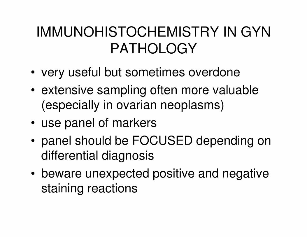
IMMUNOHISTOCHEMISTRY IN GYN
PATHOLOGY
• very useful but sometimes overdone
• extensive sampling often more valuable (especially in ovarian neoplasms)
• use panel of markers• use panel of markers
• panel should be FOCUSED depending on differential diagnosis
• beware unexpected positive and negative staining reactions

TOPICS TO DISCUSS
• typing of ovarian carcinoma
• uses of WT1
• primary versus secondary ovarian adenocarcinoma (adenocarcinoma of unknown origin, cytology specimens)origin, cytology specimens)
• markers of sex cord-stromal tumours
• typing of uterine carcinoma
• endometrial versus cervical adenocarcinoma
• cervical neuroendocrine carcinomas
• TTF1 in gynaecological neoplasms
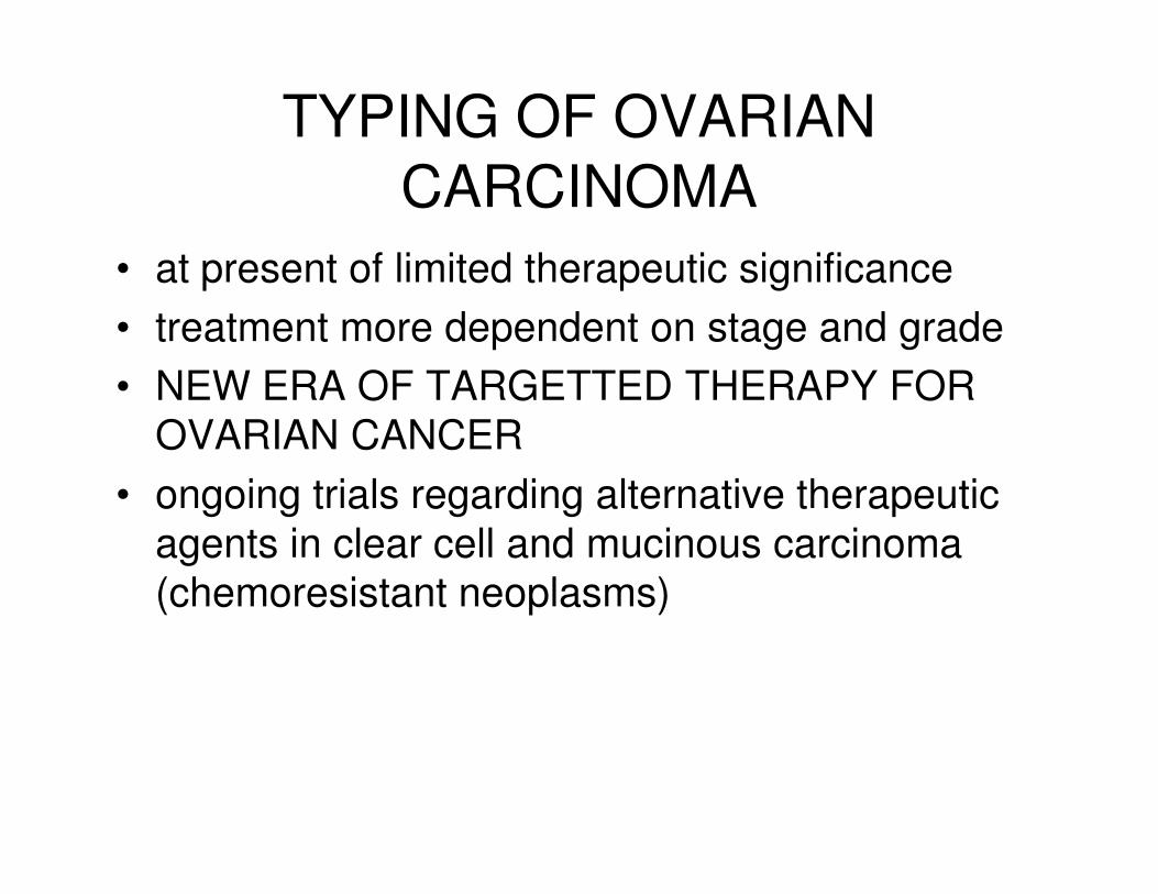
TYPING OF OVARIAN
CARCINOMA
• at present of limited therapeutic significance
• treatment more dependent on stage and grade
• NEW ERA OF TARGETTED THERAPY FOR
OVARIAN CANCEROVARIAN CANCER
• ongoing trials regarding alternative therapeutic
agents in clear cell and mucinous carcinoma
(chemoresistant neoplasms)
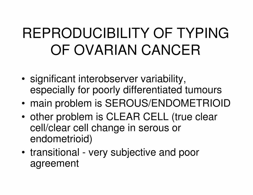
REPRODUCIBILITY OF TYPING
OF OVARIAN CANCER
• significant interobserver variability, especially for poorly differentiated tumours
• main problem is SEROUS/ENDOMETRIOID• main problem is SEROUS/ENDOMETRIOID
• other problem is CLEAR CELL (true clear cell/clear cell change in serous or endometrioid)
• transitional - very subjective and poor agreement
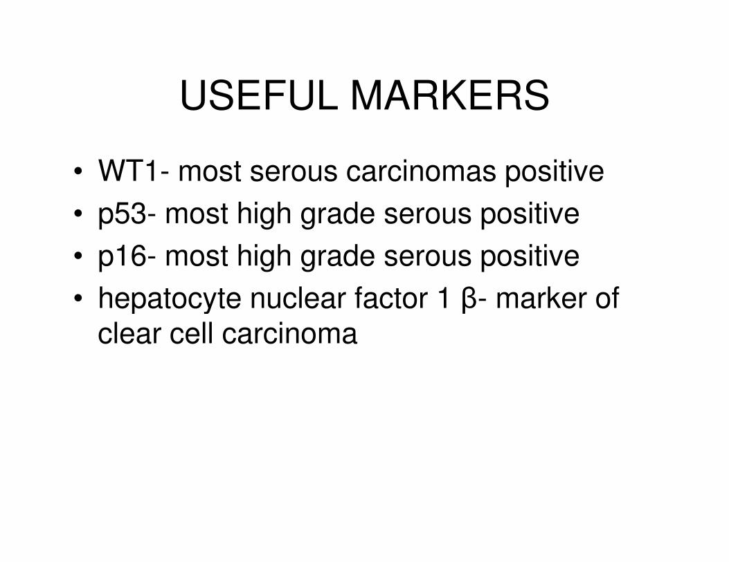
USEFUL MARKERS
• WT1- most serous carcinomas positive
• p53- most high grade serous positive
• p16- most high grade serous positive
• hepatocyte nuclear factor 1 β- marker of • hepatocyte nuclear factor 1 β- marker of clear cell carcinoma
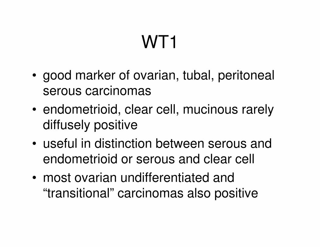
WT1
• good marker of ovarian, tubal, peritoneal serous carcinomas
• endometrioid, clear cell, mucinous rarely diffusely positivediffusely positive
• useful in distinction between serous and endometrioid or serous and clear cell
• most ovarian undifferentiated and “transitional” carcinomas also positive
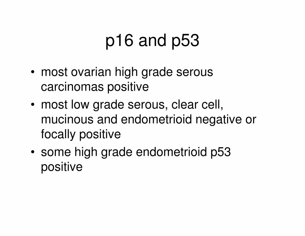
p16 and p53
• most ovarian high grade serous carcinomas positive
• most low grade serous, clear cell, mucinous and endometrioid negative or mucinous and endometrioid negative or focally positive
• some high grade endometrioid p53 positive
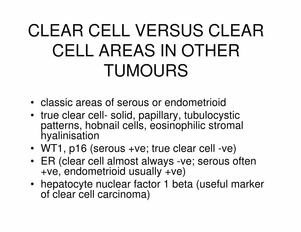
CLEAR CELL VERSUS CLEAR
CELL AREAS IN OTHER
TUMOURS
• classic areas of serous or endometrioid
• true clear cell- solid, papillary, tubulocystic • true clear cell- solid, papillary, tubulocystic patterns, hobnail cells, eosinophilic stromal hyalinisation
• WT1, p16 (serous +ve; true clear cell -ve)
• ER (clear cell almost always -ve; serous often +ve, endometrioid usually +ve)
• hepatocyte nuclear factor 1 beta (useful marker of clear cell carcinoma)

POST -CHEMOTHERAPY
• increasing tendency to neoadjuvant chemotherapy followed by surgery
• ongoing trials (CHORUS in UK)
• post-chemotherapy serous carcinomas • post-chemotherapy serous carcinomas may have clear cytoplasm and mimic clear cell carcinoma
• maintain characteristic immunophenotype

HEPATOCYTE NUCLEAR
FACTOR 1 BETA
• good marker of ovarian (and uterine) clear cell carcinoma (discovered from gene expression studies)
• endometriosis (associated and not • endometriosis (associated and not associated with clear cell carcinoma) may be +ve
• occasionally other neoplasms +ve
• need more studies (and new monoclonal antibody)
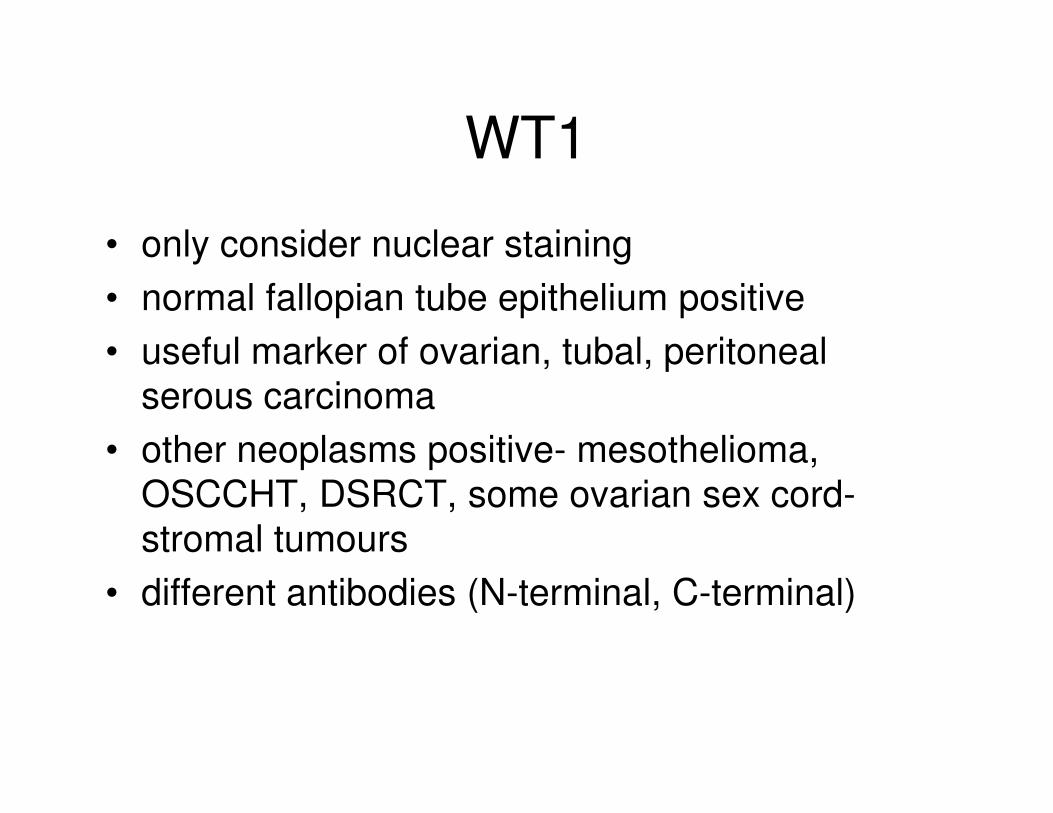
WT1
• only consider nuclear staining
• normal fallopian tube epithelium positive
• useful marker of ovarian, tubal, peritoneal
serous carcinomaserous carcinoma
• other neoplasms positive- mesothelioma,
OSCCHT, DSRCT, some ovarian sex cord-
stromal tumours
• different antibodies (N-terminal, C-terminal)
USES
• confirmation of serous carcinoma (adenocarcinoma of unknown origin; typing of ovarian carcinoma; distinction from metastatic breast carcinoma)from metastatic breast carcinoma)
• uterine versus extrauterine serous carcinoma
• OSCCHT versus mimics
COEXISTENT SEROUS
CARCINOMAS
• USC may disseminate widely and involve ovaries, even within polyp (also EIC)
• OSC may involve uterus
• ? which is primary
• ? independent synchronous or metastatic disease
• management may differ
• WT1 may be of value
• much more likely to be positive in OSC than USC (however, some overlap with occasional USC diffusely positive)occasional USC diffusely positive)
• if WT1 negative, doubt whether OSC
OVARIAN SMALL CELL CARCINOMA OF HYPERCALCAEMIC TYPE
• usually young females (peak in 2nd and 3rd
decades)decades)
• may occur in older females
• hypercalcaemia in two-thirds
• WT1 almost always +ve
WT1
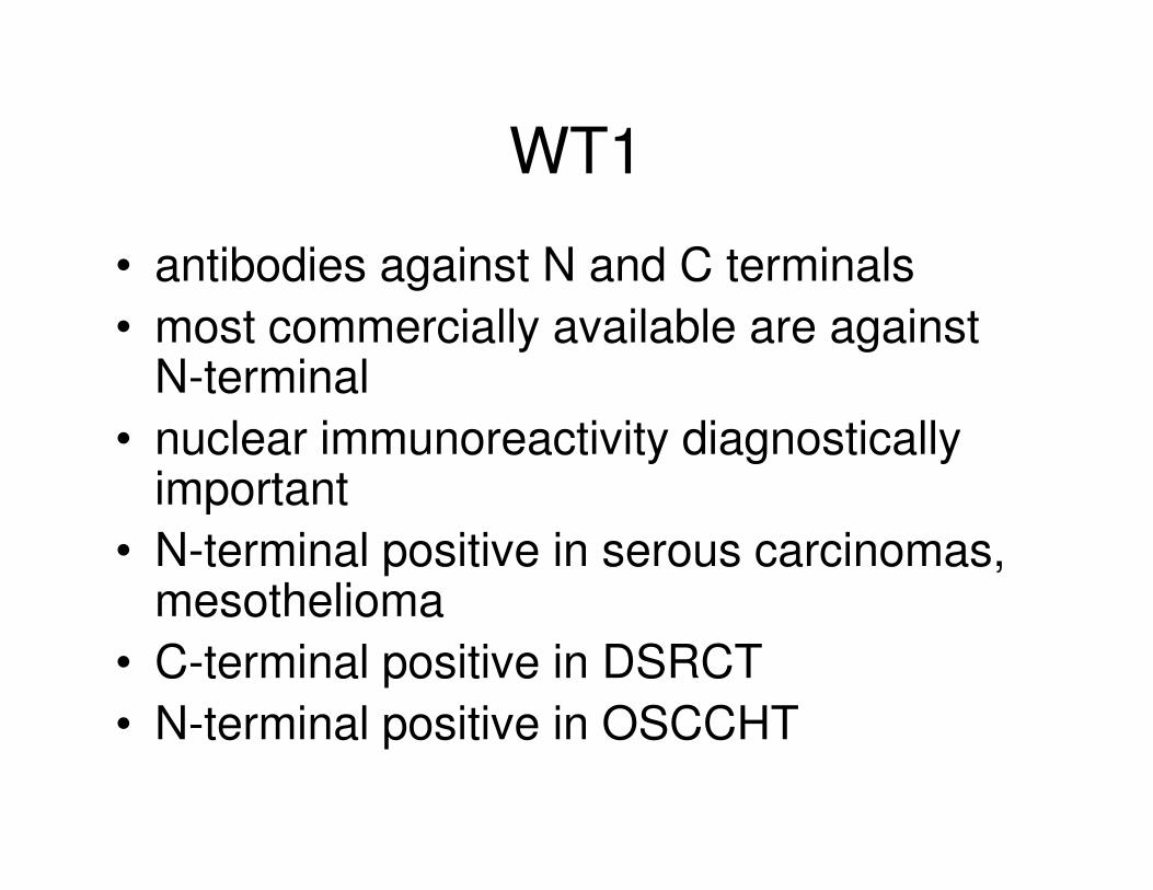
• antibodies against N and C terminals
• most commercially available are against N-terminal
• nuclear immunoreactivity diagnostically • nuclear immunoreactivity diagnostically important
• N-terminal positive in serous carcinomas, mesothelioma
• C-terminal positive in DSRCT
• N-terminal positive in OSCCHT

PRIMARY VERSUS SECONDARY OVARIAN ADENOCARCINOMA
• also disseminated peritoneal adenocarcinoma
and cytology specimens
• CK7/CK20
• CEA• CEA
• CDX2
• CA125
• CA19.9
• ER, PR
• WT1,TTF1
CK7 and CK20
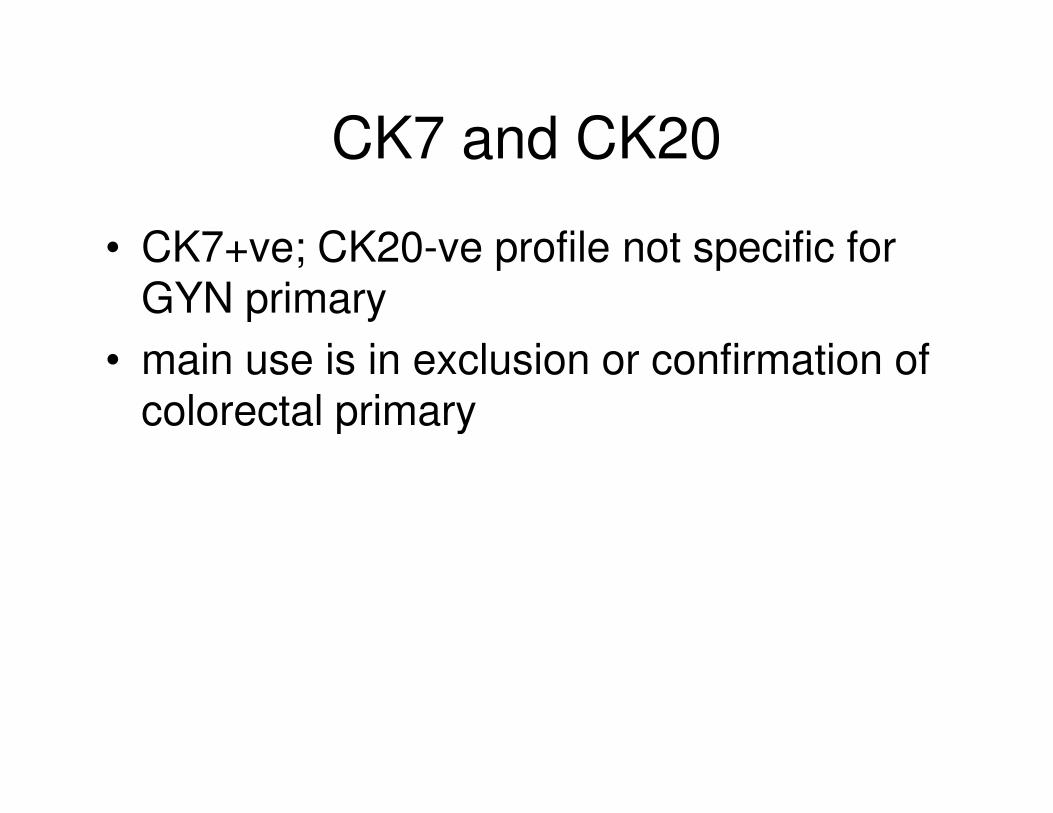
• CK7+ve; CK20-ve profile not specific for GYN primary
• main use is in exclusion or confirmation of colorectal primarycolorectal primary

METASTATIC COLORECTAL CARCINOMA
• histologically may mimic endometrioid
(pseudoendometrioid), mucinous or rarely clear cell
ovarian carcinoma
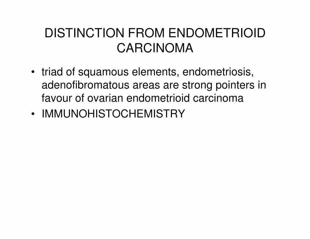
DISTINCTION FROM ENDOMETRIOID CARCINOMA
• triad of squamous elements, endometriosis,
adenofibromatous areas are strong pointers in
favour of ovarian endometrioid carcinoma
• IMMUNOHISTOCHEMISTRY• IMMUNOHISTOCHEMISTRY
PANEL
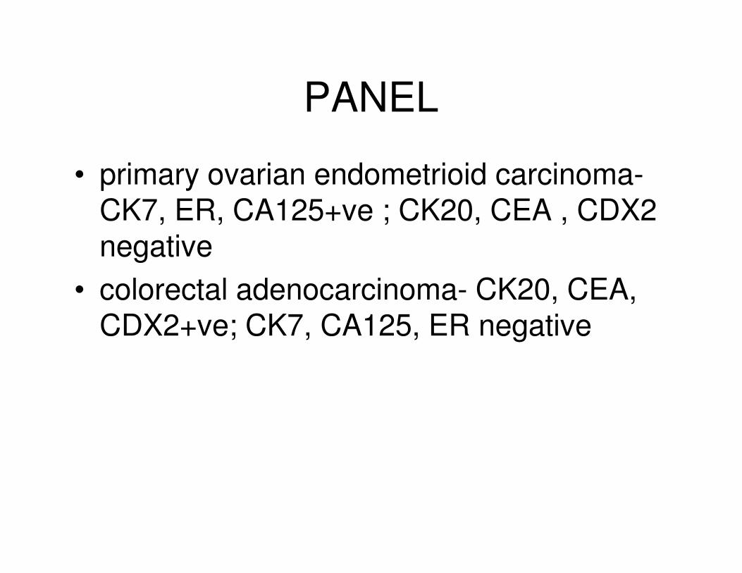
• primary ovarian endometrioid carcinoma-CK7, ER, CA125+ve ; CK20, CEA , CDX2 negative
• colorectal adenocarcinoma- CK20, CEA, • colorectal adenocarcinoma- CK20, CEA, CDX2+ve; CK7, CA125, ER negative
• in distinction between primary ovarian mucinous
adenocarcinoma and metastatic colorectal
carcinoma, immunohistochemistry is of less
value
• many primary ovarian mucinous carcinomas
exhibit positivity with intestinal markers (CK20,
CEA, CDX2, CA19.9), either focal or diffuse
• colorectal carcinomas with a mucinous
appearance often exhibit focal CK7 positivity
In general:-
• ovarian mucinous carcinomas are diffusely positive with CK7 and focally positive with CK20; may also be CEA, CA19.9, CDX2 positivepositive
• mucinous colorectal carcinomas usually diffusely positive with CK20 and focally with CK7
EXCEPTIONS OCCUR
UPPER GI ADENOCARCINOMA
• stomach, pancreas, biliary tree
• very similar immunophenotype to primary ovarian mucinous neoplasms- NO RELIABLE MARKER TO DISTINGUISH RELIABLE MARKER TO DISTINGUISH (DPC4 may be of value but not in widespread use)
• pancreas often CA125 positive
ER/PR• useful in adenocarcinoma in suggesting gynaecological
or breast primary
• occasionally other carcinomas positive (pancreas may
be PR +ve)
• pathologists often forget to use as part of panel
METASTATIC CERVICAL
ADENOCARCINOMA
• in most cases diagnosis is obvious
• rarely ovarian mass is first manifestation
• small cervical adenocarcinomas may • small cervical adenocarcinomas may
metastasise to the ovary
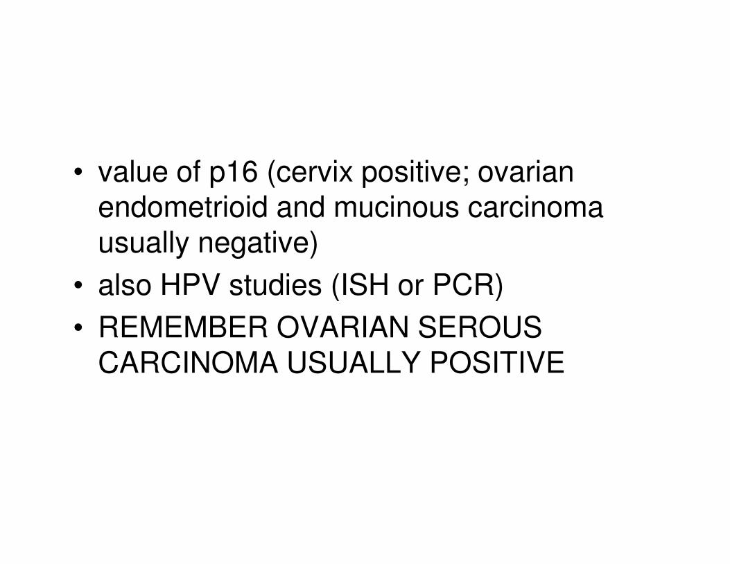
• value of p16 (cervix positive; ovarian endometrioid and mucinous carcinoma usually negative)
• also HPV studies (ISH or PCR)• also HPV studies (ISH or PCR)
• REMEMBER OVARIAN SEROUS CARCINOMA USUALLY POSITIVE
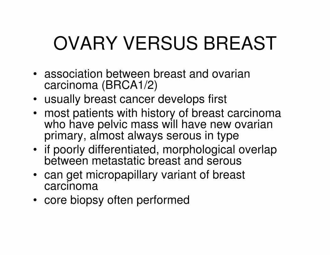
OVARY VERSUS BREAST
• association between breast and ovarian carcinoma (BRCA1/2)
• usually breast cancer develops first
• most patients with history of breast carcinoma who have pelvic mass will have new ovarian who have pelvic mass will have new ovarian primary, almost always serous in type
• if poorly differentiated, morphological overlap between metastatic breast and serous
• can get micropapillary variant of breast carcinoma
• core biopsy often performed
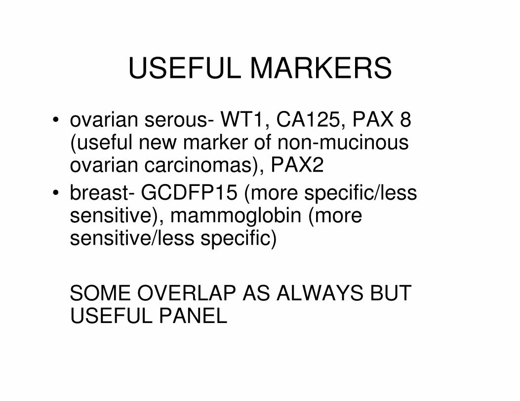
USEFUL MARKERS
• ovarian serous- WT1, CA125, PAX 8 (useful new marker of non-mucinous ovarian carcinomas), PAX2
• breast- GCDFP15 (more specific/less • breast- GCDFP15 (more specific/less sensitive), mammoglobin (more sensitive/less specific)
SOME OVERLAP AS ALWAYS BUT USEFUL PANEL
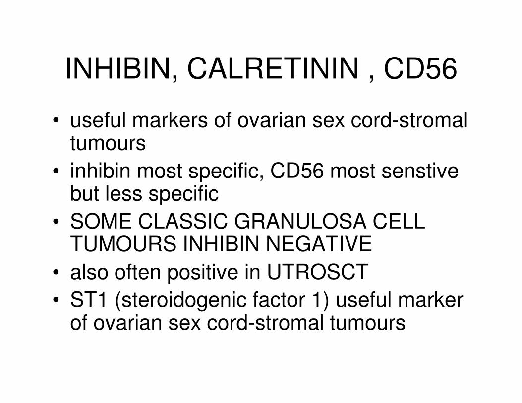
INHIBIN, CALRETININ , CD56
• useful markers of ovarian sex cord-stromal tumours
• inhibin most specific, CD56 most senstive but less specificbut less specific
• SOME CLASSIC GRANULOSA CELL TUMOURS INHIBIN NEGATIVE
• also often positive in UTROSCT
• ST1 (steroidogenic factor 1) useful marker of ovarian sex cord-stromal tumours

CD56
• extremely sensitive marker of ovarian sex cord-
stromal tumour (84 of 85 cases)
• more sensitive than inhibin or calretinin
• lacks specificity (neuroendocrine tumours • lacks specificity (neuroendocrine tumours
positive)
• membranous and weaker cytoplasmic staining
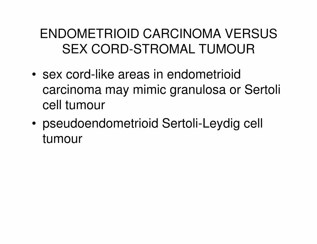
ENDOMETRIOID CARCINOMA VERSUS SEX CORD-STROMAL TUMOUR
• sex cord-like areas in endometrioid carcinoma may mimic granulosa or Sertoli cell tumour
• pseudoendometrioid Sertoli-Leydig cell • pseudoendometrioid Sertoli-Leydig cell tumour
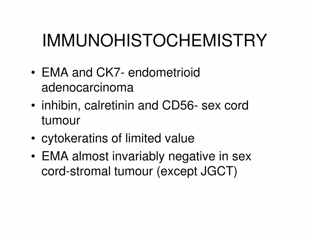
IMMUNOHISTOCHEMISTRY
• EMA and CK7- endometrioid adenocarcinoma
• inhibin, calretinin and CD56- sex cord tumourtumour
• cytokeratins of limited value
• EMA almost invariably negative in sex cord-stromal tumour (except JGCT)
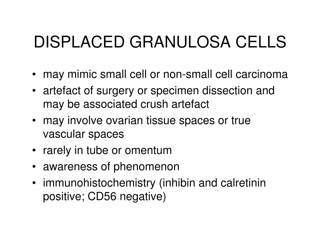
DISPLACED GRANULOSA CELLS
• may mimic small cell or non-small cell carcinoma
• artefact of surgery or specimen dissection and
may be associated crush artefact
• may involve ovarian tissue spaces or true • may involve ovarian tissue spaces or true
vascular spaces
• rarely in tube or omentum
• awareness of phenomenon
• immunohistochemistry (inhibin and calretinin
positive; CD56 negative)

Distinction Between Endometrial and
Endocervical Adenocarcinoma
• morphological differences but can be similarsimilar
• looks for clues (squamous elements, foam cells)
• primary surgery may differ
• adjuvant therapy may differ

Panel
• ER
• vimentin
• monoclonal CEA
• p16• p16
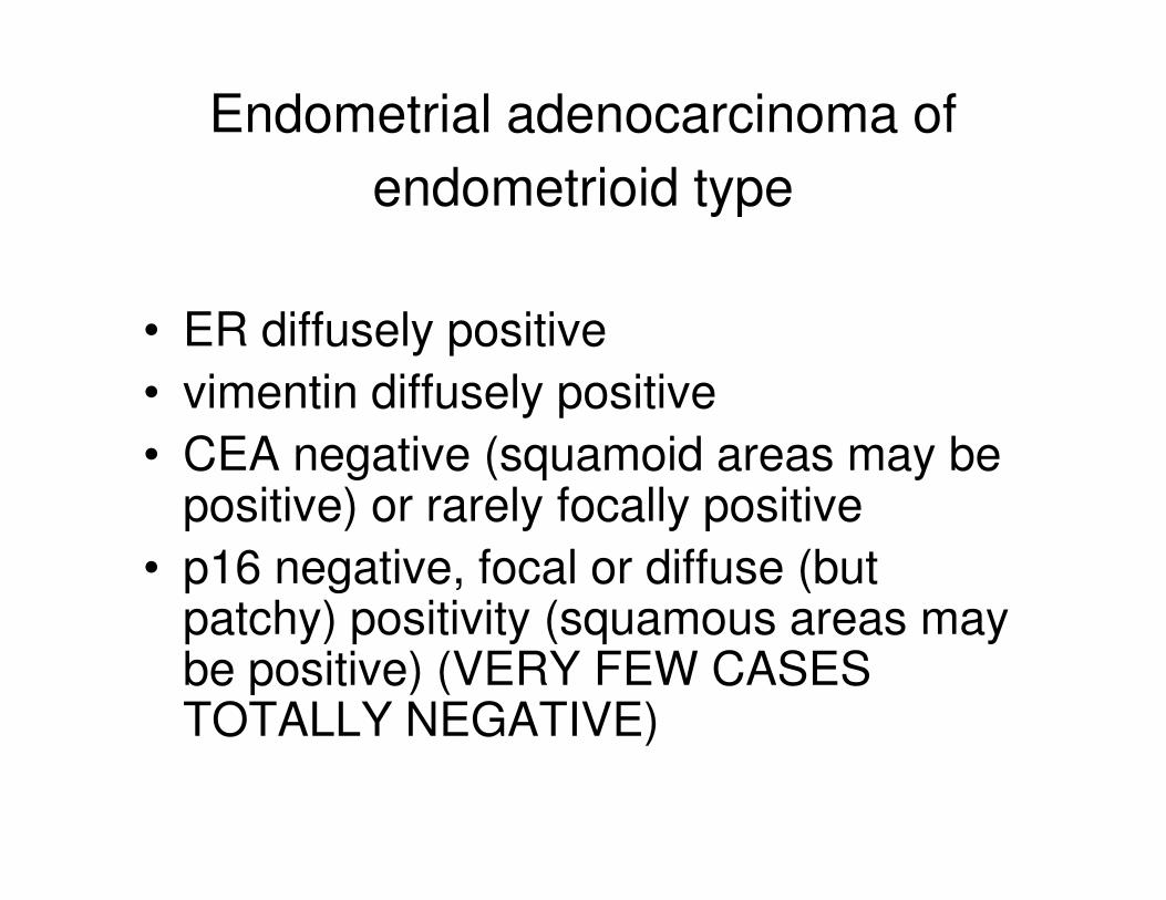
Endometrial adenocarcinoma of
endometrioid type
• ER diffusely positive
• vimentin diffusely positive
• CEA negative (squamoid areas may be • CEA negative (squamoid areas may be positive) or rarely focally positive
• p16 negative, focal or diffuse (but patchy) positivity (squamous areas may be positive) (VERY FEW CASES TOTALLY NEGATIVE)

Endocervical Adenocarcinoma
• ER negative or weakly positive
• vimentin negative
• CEA usually positive
• p16 diffusely positive• p16 diffusely positive
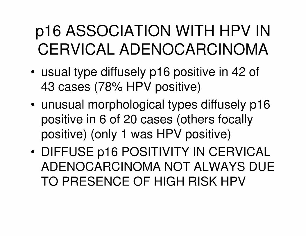
p16 ASSOCIATION WITH HPV IN
CERVICAL ADENOCARCINOMA
• usual type diffusely p16 positive in 42 of 43 cases (78% HPV positive)
• unusual morphological types diffusely p16 positive in 6 of 20 cases (others focally positive in 6 of 20 cases (others focally positive) (only 1 was HPV positive)
• DIFFUSE p16 POSITIVITY IN CERVICAL ADENOCARCINOMA NOT ALWAYS DUE TO PRESENCE OF HIGH RISK HPV
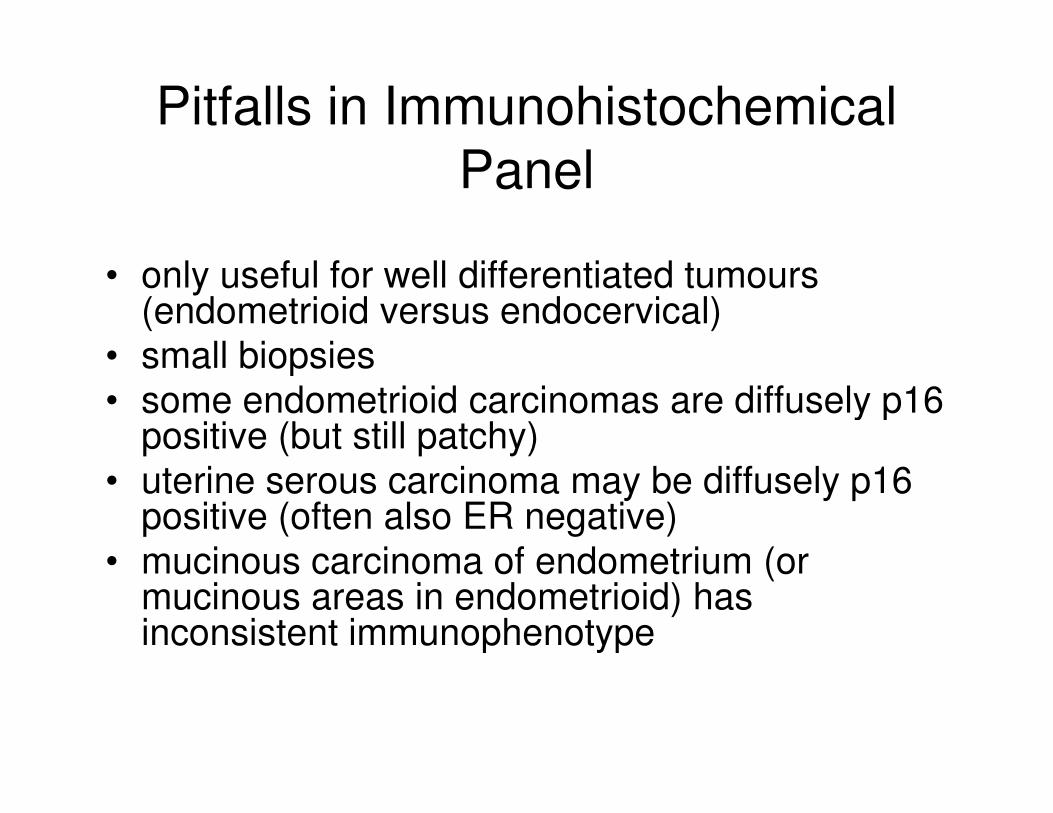
Pitfalls in Immunohistochemical
Panel
• only useful for well differentiated tumours (endometrioid versus endocervical)
• small biopsies
• some endometrioid carcinomas are diffusely p16 • some endometrioid carcinomas are diffusely p16 positive (but still patchy)
• uterine serous carcinoma may be diffusely p16 positive (often also ER negative)
• mucinous carcinoma of endometrium (or mucinous areas in endometrioid) has inconsistent immunophenotype
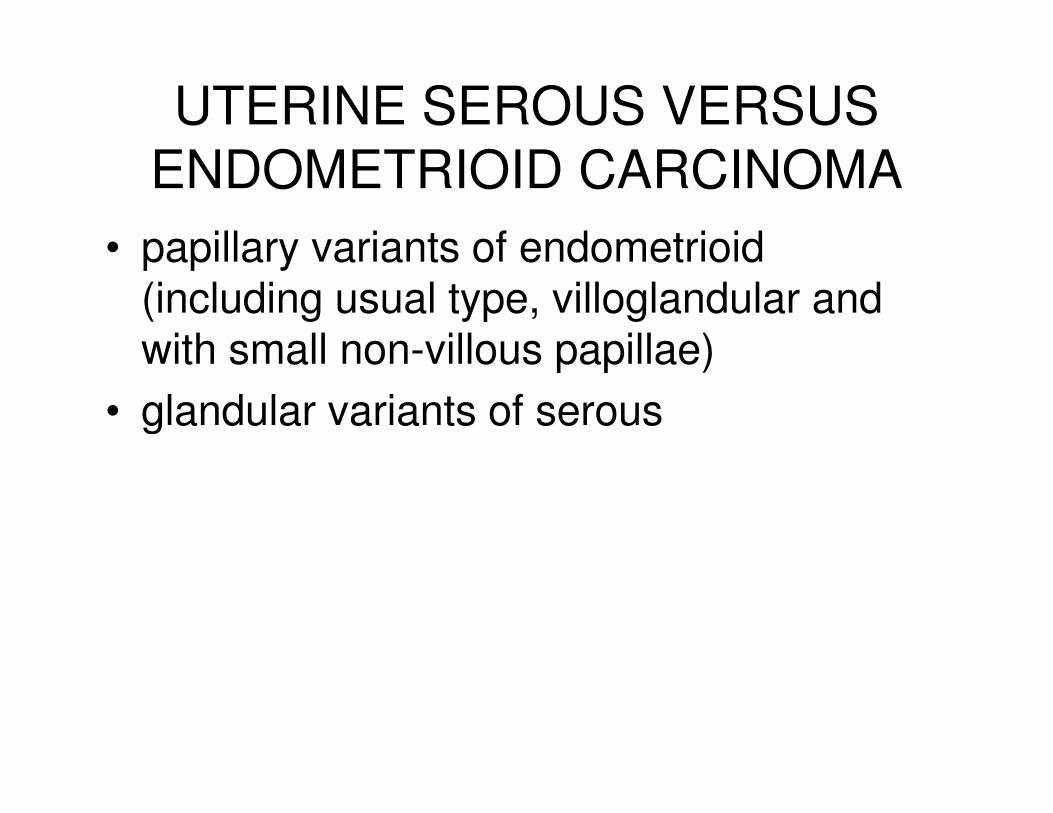
UTERINE SEROUS VERSUS
ENDOMETRIOID CARCINOMA
• papillary variants of endometrioid (including usual type, villoglandular and with small non-villous papillae)
• glandular variants of serous• glandular variants of serous

SEROUS ADENOCARCINOMA
• NOT papillary serous adenocarcinoma
• many cases have predominant or exclusive glandular architecture

DISTINCTION BETWEEN
SEROUS AND ENDOMETRIOID
• important
• surgical operation may differ
• adjuvant therapy may differ• adjuvant therapy may differ
• ultimate prognosis will differ
• DISTINCTION BASED ON MORPHOLOGY +/-IMMUNOHISTOCHEMISTRY
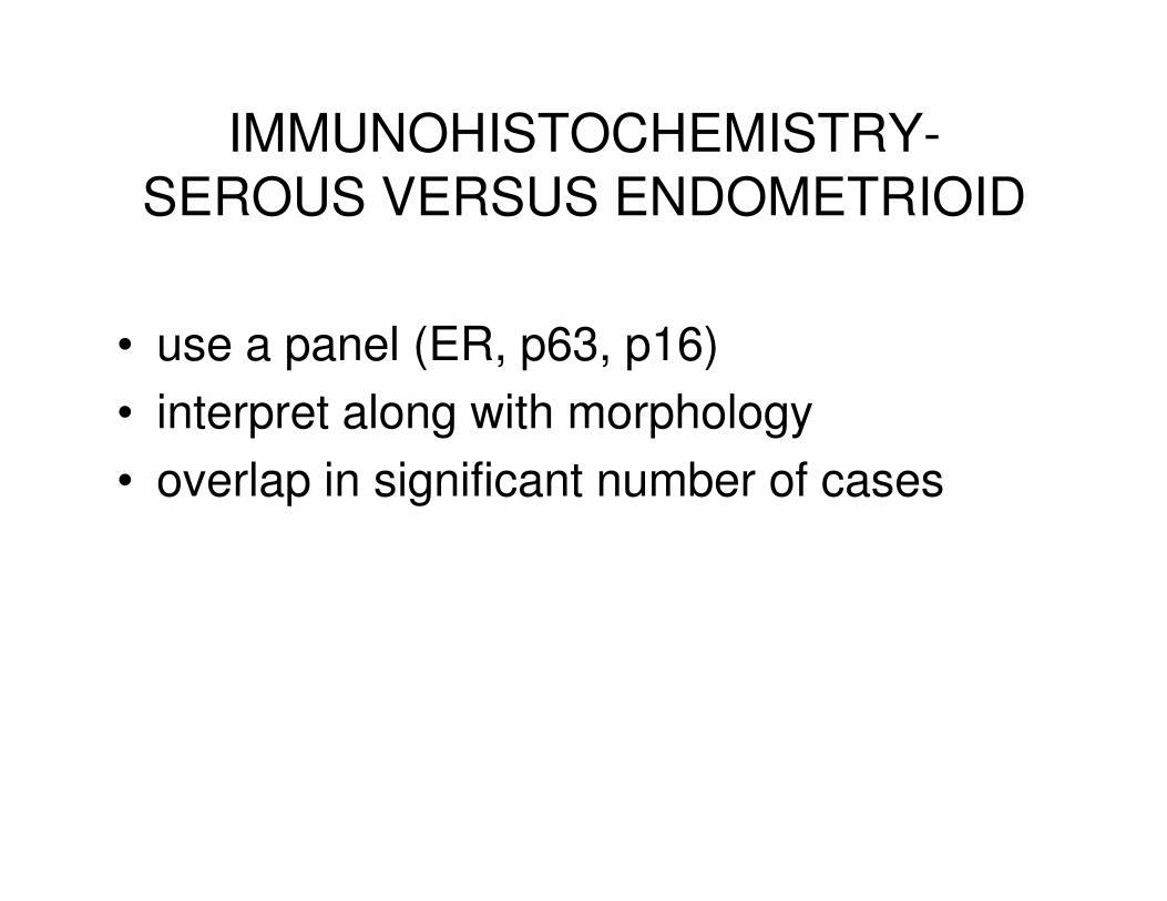
IMMUNOHISTOCHEMISTRY-
SEROUS VERSUS ENDOMETRIOID
• use a panel (ER, p63, p16)
• interpret along with morphology
• overlap in significant number of cases
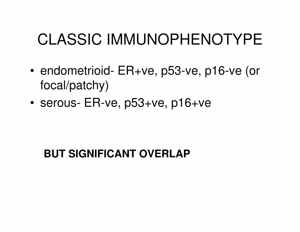
CLASSIC IMMUNOPHENOTYPE
• endometrioid- ER+ve, p53-ve, p16-ve (or focal/patchy)
• serous- ER-ve, p53+ve, p16+ve
BUT SIGNIFICANT OVERLAP
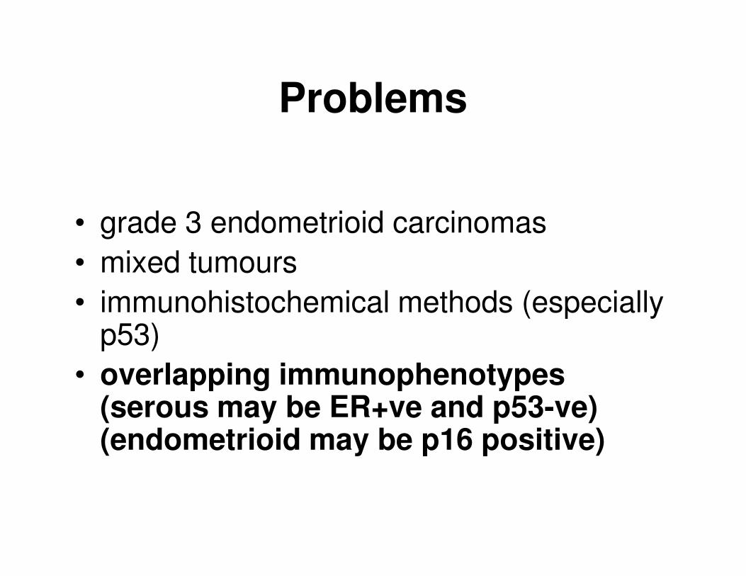
Problems
• grade 3 endometrioid carcinomas
• mixed tumours
• immunohistochemical methods (especially • immunohistochemical methods (especially p53)
• overlapping immunophenotypes (serous may be ER+ve and p53-ve) (endometrioid may be p16 positive)
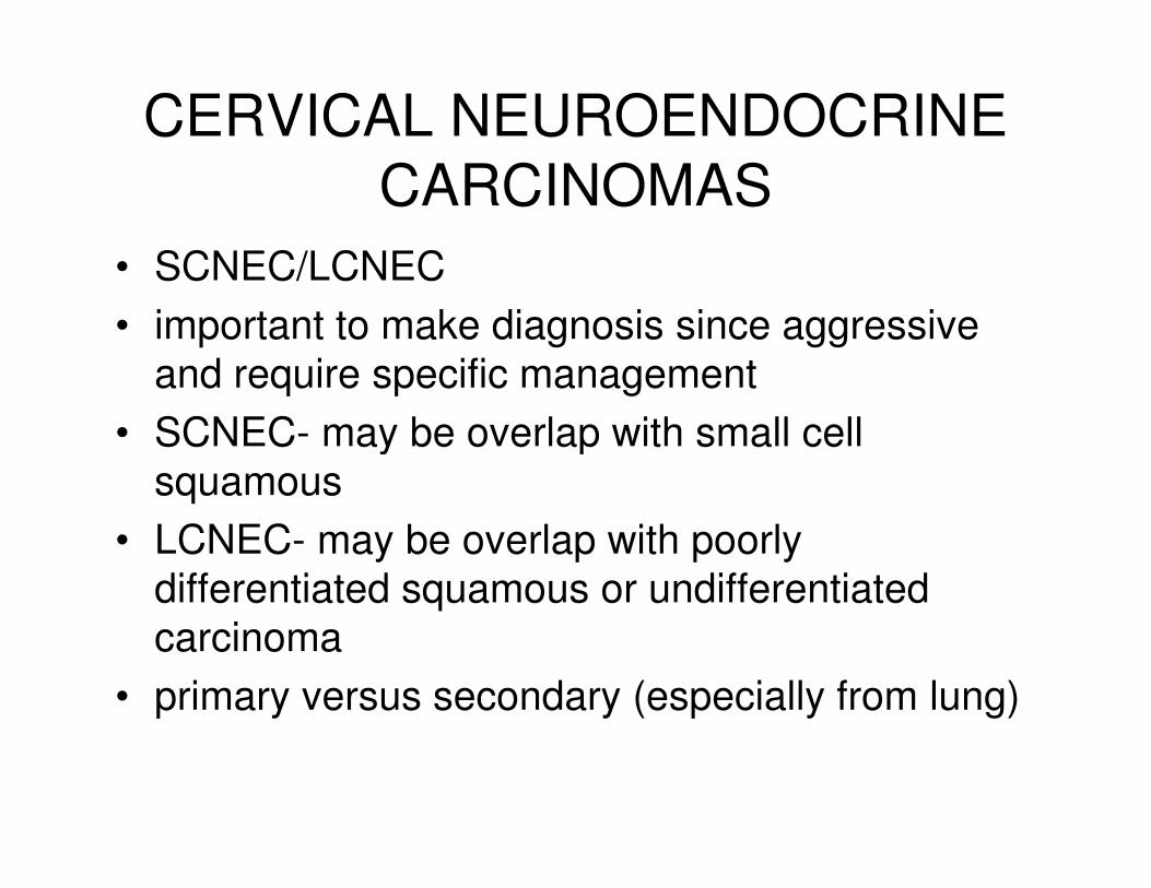
CERVICAL NEUROENDOCRINE
CARCINOMAS
• SCNEC/LCNEC
• important to make diagnosis since aggressive
and require specific management
• SCNEC- may be overlap with small cell • SCNEC- may be overlap with small cell
squamous
• LCNEC- may be overlap with poorly
differentiated squamous or undifferentiated
carcinoma
• primary versus secondary (especially from lung)

STUDY-SCNEC (n=13), LCNEC (n=8)
MARKERS
• AE1/3
• chromogranin, CD56, synaptophysin, PGP9.5
• TTF1• TTF1
• p16
• p63,
• CK7/20
• neurofilament
• CD99

AE1/3
• 85% SCNEC and 75% LCNEC positive

NEUROENDOCRINE MARKERS
• 52% chromogranin positive
• 90% CD56 positive
• 90% synaptophysin positive
• 43% PGP9.5 positive• 43% PGP9.5 positive

SCNEC
• 3 positive with all 4 neuroendocrine markers
• 6 positive with 3 markers
• 2 positive with 2 markers• 2 positive with 2 markers
• 2 positive with 1 marker
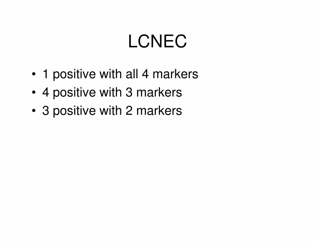
LCNEC
• 1 positive with all 4 markers
• 4 positive with 3 markers
• 3 positive with 2 markers
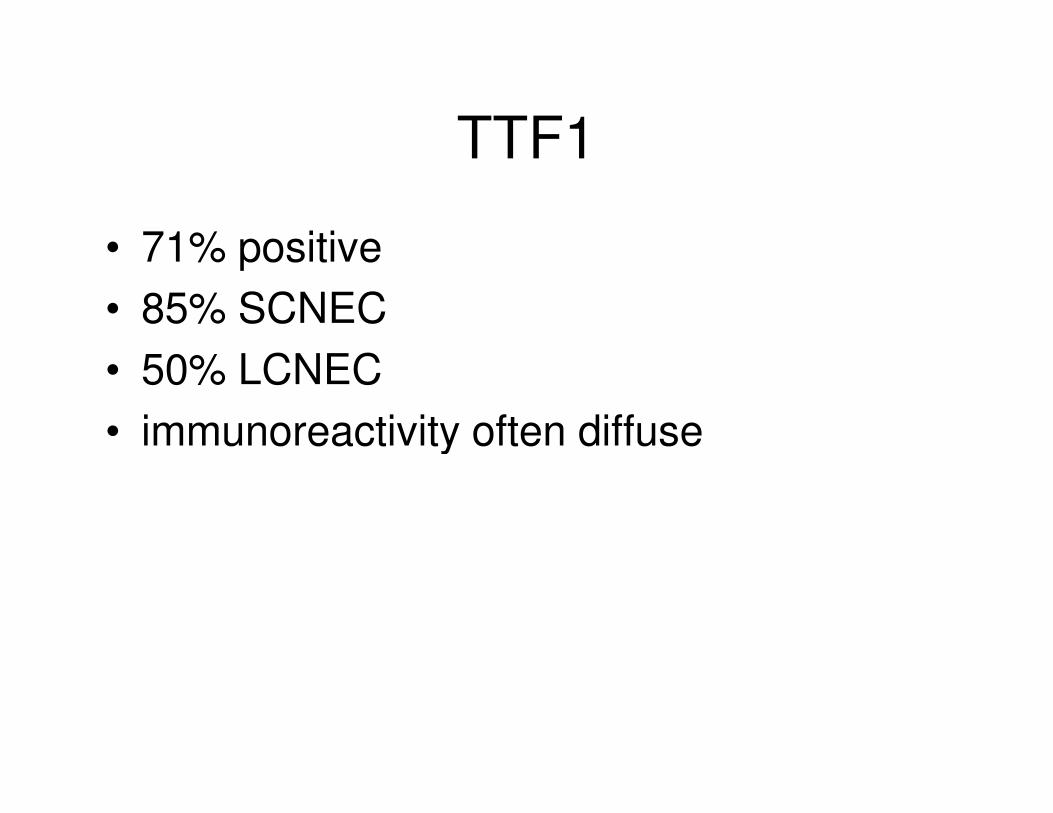
TTF1
• 71% positive
• 85% SCNEC
• 50% LCNEC
• immunoreactivity often diffuse• immunoreactivity often diffuse

TTF1
• high percentage of cervical neuroendocrine carcinomas positive
• of no value in distinction from a pulmonary metastasismetastasis
• ? useful marker of neuroendocrine carcinoma
• different clones (SPT24 versus 8G7G3/1)
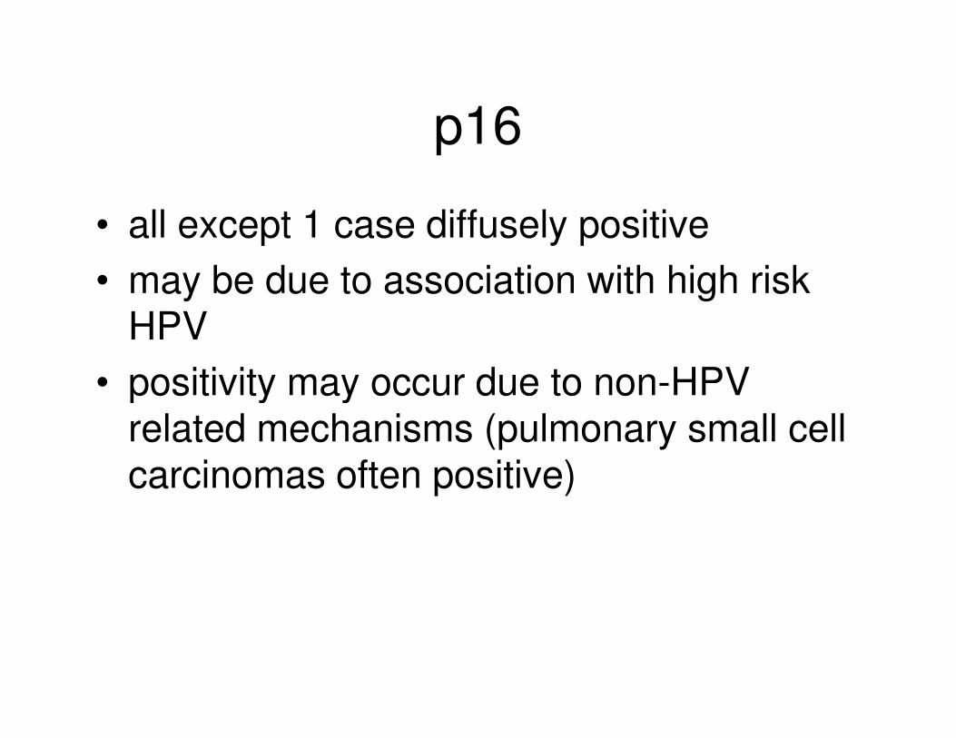
p16
• all except 1 case diffusely positive
• may be due to association with high risk HPV
• positivity may occur due to non-HPV • positivity may occur due to non-HPV related mechanisms (pulmonary small cell carcinomas often positive)
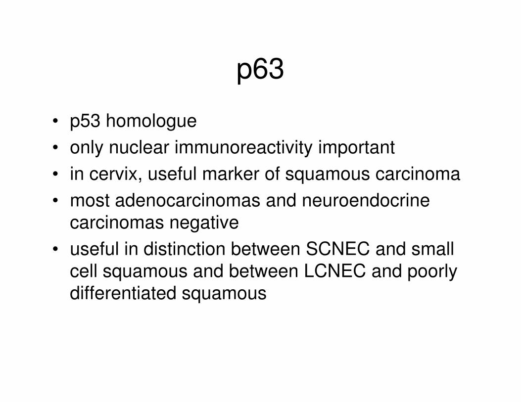
p63
• p53 homologue
• only nuclear immunoreactivity important
• in cervix, useful marker of squamous carcinoma
• most adenocarcinomas and neuroendocrine • most adenocarcinomas and neuroendocrine
carcinomas negative
• useful in distinction between SCNEC and small
cell squamous and between LCNEC and poorly
differentiated squamous
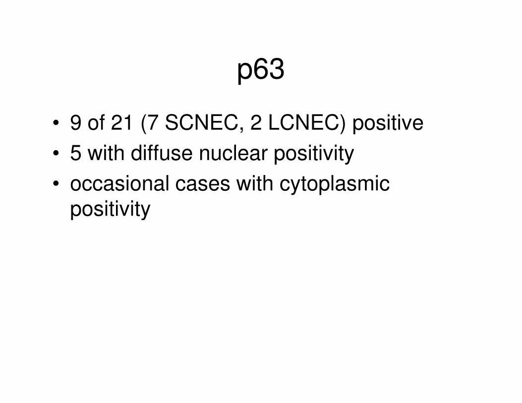
p63
• 9 of 21 (7 SCNEC, 2 LCNEC) positive
• 5 with diffuse nuclear positivity
• occasional cases with cytoplasmic positivitypositivity
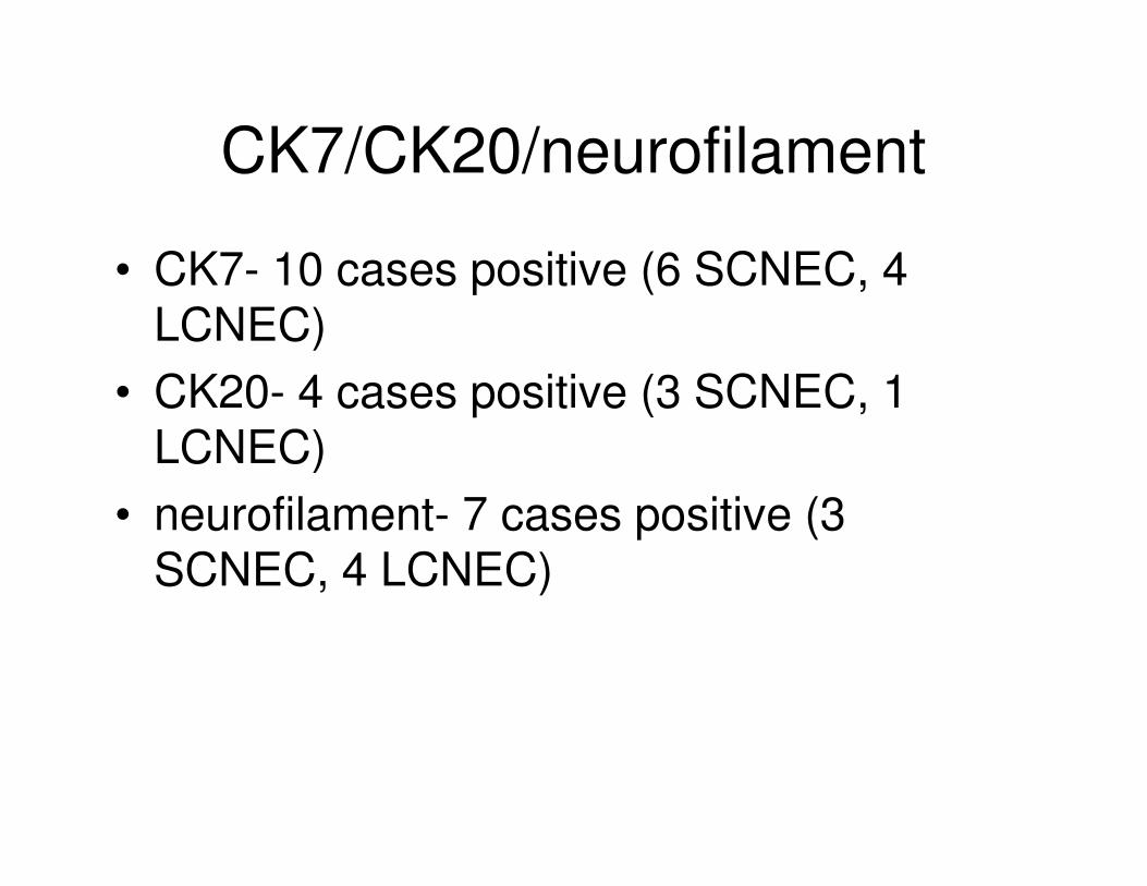
CK7/CK20/neurofilament
• CK7- 10 cases positive (6 SCNEC, 4 LCNEC)
• CK20- 4 cases positive (3 SCNEC, 1 LCNEC)LCNEC)
• neurofilament- 7 cases positive (3 SCNEC, 4 LCNEC)
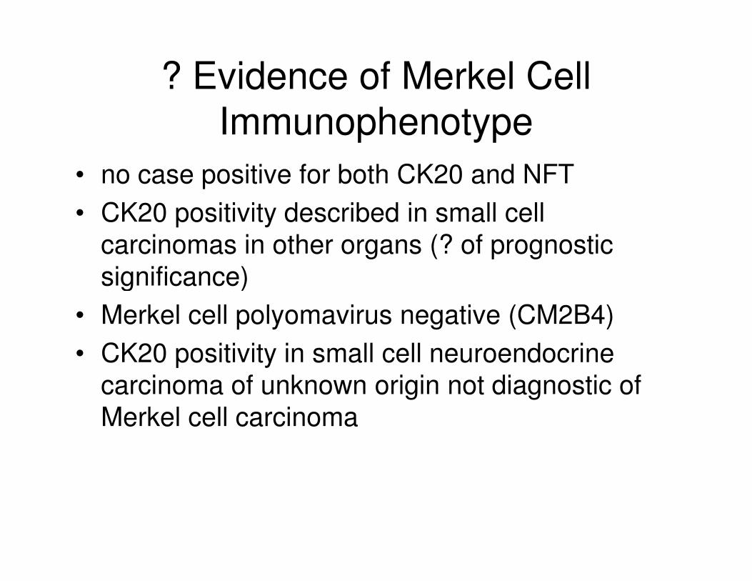
? Evidence of Merkel Cell
Immunophenotype
• no case positive for both CK20 and NFT
• CK20 positivity described in small cell
carcinomas in other organs (? of prognostic
significance)significance)
• Merkel cell polyomavirus negative (CM2B4)
• CK20 positivity in small cell neuroendocrine
carcinoma of unknown origin not diagnostic of
Merkel cell carcinoma

CD99
• marker of Ewing family of tumours
• 6 cases with membranous immunoreactivity (4 SCNEC, 2 LCNEC)
• 2 cases diffusely positive• 2 cases diffusely positive
• illustrates overlap with Ewing family of tumours
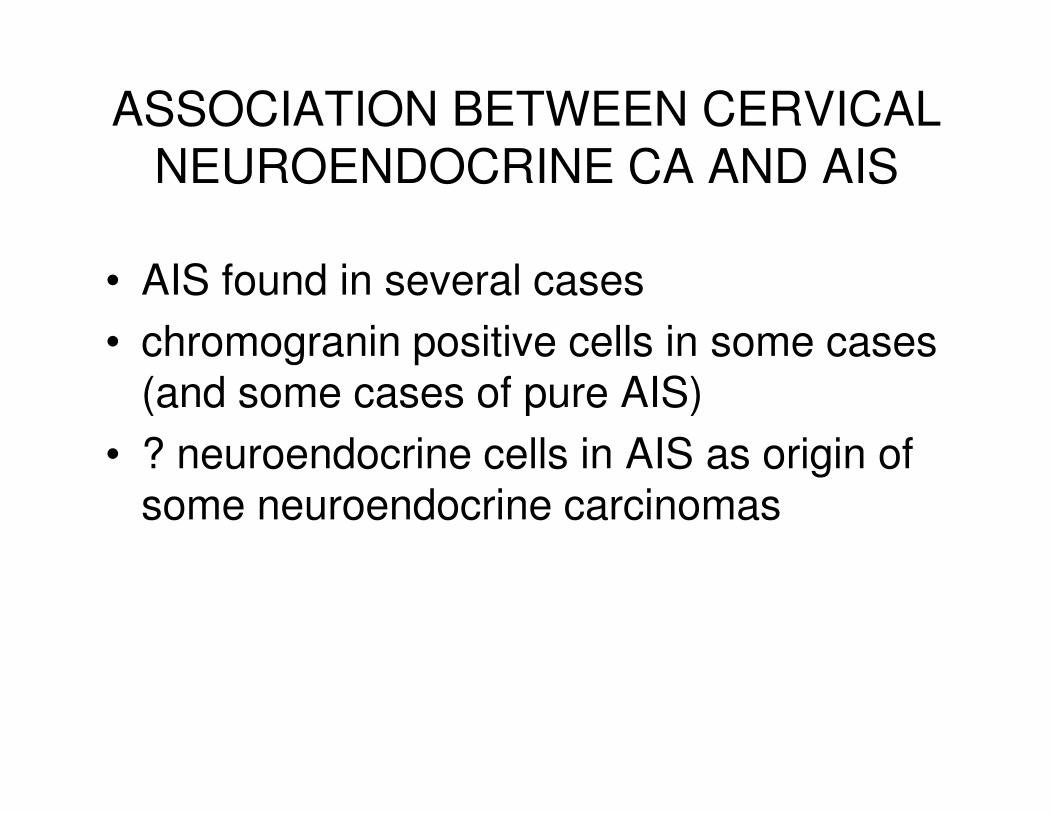
ASSOCIATION BETWEEN CERVICAL
NEUROENDOCRINE CA AND AIS
• AIS found in several cases
• chromogranin positive cells in some cases (and some cases of pure AIS)(and some cases of pure AIS)
• ? neuroendocrine cells in AIS as origin of some neuroendocrine carcinomas

TTF1 IN GYNAECOLOGICAL
NEOPLASMS
• unusual cases of struma ovarii (use with thyroglobulin)
• neuroendocrine neoplasms
• metastasis from lung/thyroid• metastasis from lung/thyroid
• some gynaecological adenocarcinomas positive (some diffusely so)

UNUSUAL STRUMA OVARII
• clear cells
• oxyphil cells
• cystic struma ovarii
• unusual thyroid type adenocarcinomas• unusual thyroid type adenocarcinomas

MALIGNANT STRUMA OVARII
• sometimes not typical papillary or follicular carcinoma

TTF1 IN GYN
ADENOCARCINOMAS
• 7 of 19 ovarian serous carcinomas
• 1 of 28 cervical adenocarcinomas
• 6 of 31 uterine endometrioid adenocarcinomasadenocarcinomas
• 3 of 13 uterine serous carcinomas

CLINICAL HISTORY
• female 67
• found to have a right lung mass on routine radiological examination
• underwent right middle lobectomy

PAST MEDICAL HISTORY
• TAH and BSO four years previously for endometrial carcinomaendometrial carcinoma
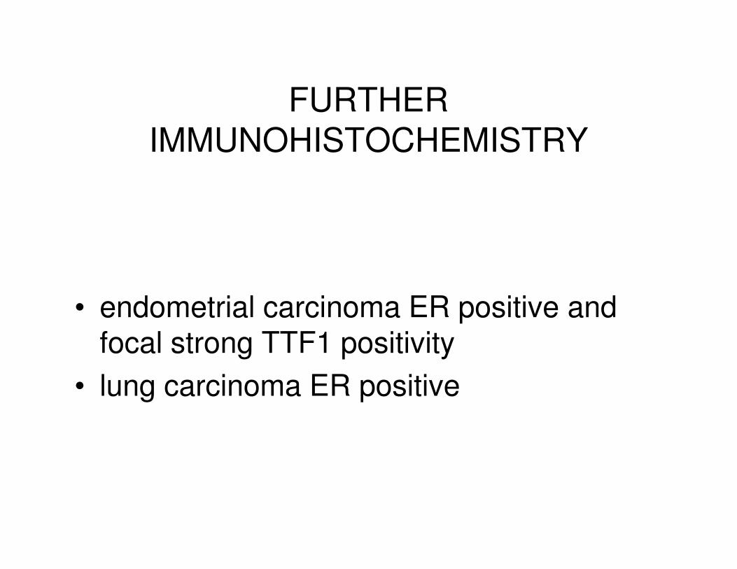
FURTHER
IMMUNOHISTOCHEMISTRY
• endometrial carcinoma ER positive and focal strong TTF1 positivity
• lung carcinoma ER positive
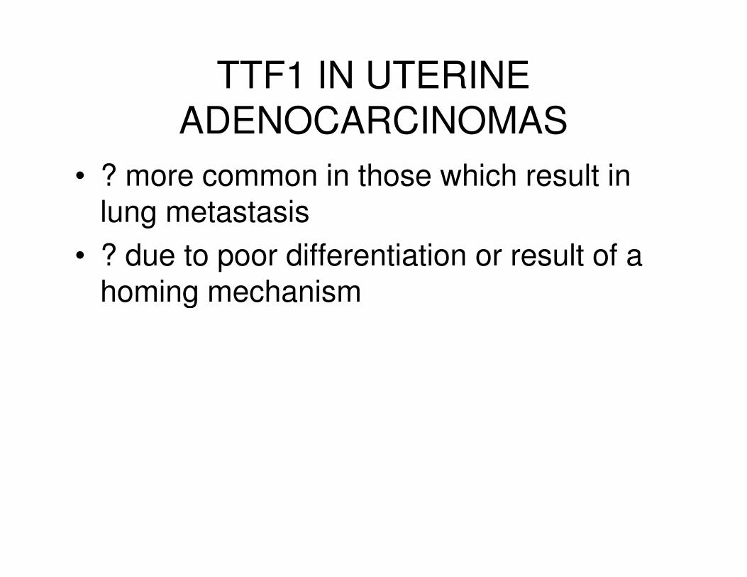
TTF1 IN UTERINE
ADENOCARCINOMAS
• ? more common in those which result in lung metastasis
• ? due to poor differentiation or result of a homing mechanismhoming mechanism

1
IMMUNOHISTOCHEMISTRY IN GYNAECOLOGICAL PATHOLOGY
W Glenn McCluggage
Royal Group of Hospitals Trust
Belfast
Northern Ireland
Immunohistochemistry plays an important role in various diagnostic scenarios in
gynaecological pathology. However, immunohistochemistry is sometimes overdone and
it is always to be remembered that immunohistochemistry is an adjunct technique and an
aid to careful morphological examination. Since no antibody is totally specific for any
given tumour and since unexpected positive and negative staining reactions may occur,
panels of markers should always be used and these panels should be carefully focused
depending on the differential diagnosis under consideration. In many diagnostic
scenarios, especially in the examination of an unusual ovarian neoplasm, judicious
sampling is sometimes more useful than immunostains. However, when carefully used,
immunohistochemistry can be extremely useful and is paramount in diagnosis in many
cases. Several topics are discussed in this talk which aims to concentrate on new
developments regarding immunohistochemistry in the female genital tract.
TYPING OF OVARIAN CARCINOMAS
Ovarian carcinomas comprise a heterogeneous group of neoplasms. Each tumour type
has a different underlying pathogenesis and natural behaviour. Although currently,
management of ovarian carcinoma is largely dependent on factors such as tumour grade
and stage, and not usually on cell type, it is important to accurately type ovarian
carcinomas to ascertain whether the various neoplasms have a different behaviour
independent of stage and other clinicopathological parameters. Typing is also important

2
since some tumours, such as mucinous and clear cell carcinoma, seem to exhibit a poor
response to platinum-based chemotherapy. In fact, tumour type may be a more reliable
predictor of response to chemotherapy than tumour grade and there are ongoing trials
regarding different therapeutic regimes in ovarian clear cell and mucinous carcinomas.
Morphology remains the mainstay in typing of ovarian carcinomas but
immunohistochemistry may be of value as a supplement in problematic cases. From
personal experience, typing is reproducible for well differentiated tumours of the four
most common types, namely serous, endometrioid, clear cell and mucinous (there may be
problems in mucinous carcinomas in distinguishing between a primary and secondary
neoplasm and there is significant variability amongst pathologists as to what constitutes
invasion in ovarian mucinous neoplasms but typing a tumour as mucinous is generally
not problematic). However, there are significant problems with regard to typing poorly
differentiated carcinomas. Many of the difficulties relate to the categories of high grade
serous, high grade endometrioid and undifferentiated carcinoma in which there is
morphological overlap. Another source of disagreement is the categorization of clear cell
areas within ovarian carcinomas, specifically whether these represent a clear cell
carcinoma or component of clear cell carcinoma or clear cell areas within a serous,
endometrioid or undifferentiated carcinoma.
Some pathologists tend to diagnose poorly differentiated ovarian carcinomas as serous in
type while others classify them as endometrioid or mixed serous and endometrioid. My
personal opinion is that the majority of these are serous carcinomas. In this distinction,
WT1 immunohistochemical staining may be of value. Most primary ovarian (as well as
primary peritoneal and tubal) serous carcinomas exhibit diffuse nuclear positivity with
WT1 while most endometrioid adenocarcinomas are negative or focally positive. p53
may also be of value in that most serous carcinomas are diffusely positive while most
endometrioid adenocarcinomas are negative, although some high grade endometrioid
neoplasms are positive. p16 (p16INK4A), a cyclin-dependent kinase IV inhibitor which
is integral to pRb mediated control of the G1-S phase transition of the cell cycle, is more
likely to be diffusely and strongly positive in serous than in endometrioid
adenocarcinomas (the majority of high grade serous carcinomas are diffusely positive
3
with p16 while most low grade serous, endometrioid, mucinous and clear cell carcinomas
are negative or focally positive). Vimentin and nuclear ß-catenin positivity favours an
endometrioid adenocarcinoma, although not all cases are positive. In problematic cases, I
would recommend WT1 immunohistochemical staining as an adjunct to help distinguish
between a high grade serous and a high grade endometrioid adenocarcinoma.
Characteristically in ovarian clear cell carcinoma, an admixture of growth patterns is
present, including solid, glandular, tubulocystic and papillary. Hobnail cells and
eosinophilic hyalinised stroma are common features. Most clear cell carcinomas are
diagnosed without difficulty but there is a tendency to overdiagnose clear cell carcinoma
or a clear cellcarcinoma component within a mixed neoplasm due to the presence of clear
cell areas within other types of ovarian carcinoma, especially serous and to a lesser extent
endometrioid. The presence of more typical areas of serous or endometrioid
adenocarcinoma are useful pointers in diagnosis (sometimes a combination of clear cell
and endometrioid adenocarcinoma occurs) and it is stressed that the mere presence of
clear cells does not constitute a clear cell carcinoma. WT1 is usually negative in ovarian
clear cell carcinoma, as is p53 and p16. ER may also be of value in that moat clear cell
carcinomas are negative while many serous and endometrioid adenocarcinomas are
positive. Recently, hepatocyte nuclear factor 1 beta has emerged as a useful
immunohistochemical marker of ovarian (and uterine) clear cell carcinoma. Most of the
other morphological subtypes are negative, although there has been only limited
investigation of the expression of this marker in morphological types of ovarian
carcinoma other than clear cell.
Transitional carcinoma is a rare variant of ovarian epithelial malignancy which is subject
to considerable interobserver variability in diagnosis. Some, including myself, make the
diagnosis rarely and feel that most cases which are so-diagnosed are serous or
endometrioid carcinomas with a transitional-like growth pattern while others diagnose
transitional carcinoma not uncommonly. Ovarian transitional carcinomas are negative
with markers which are commonly expressed in urothelial transitional carcinoma, such as
4
CK20, uroplakin III, thrombomodulin and p63. Rather, they exhibit a CK7 positive/CK20
negative immunophenotype, similar to other ovarian carcinomas, and exhibit Mullerian
rather than urothelial differentiation. They often exhibit nuclear positivity with WT1,
suggesting that many represent variants of high grade serous carcinoma. In contrast,
Brenner tumours exhibit true urothelial differentiation and express markers which are
commonly positive in normal urothelium and urothelial neoplasms, such as uroplakin III,
thrombomodulin and p63. Many undifferentiated ovarian carcinomas are WT1 positive,
suggesting that these merely represent the extreme end of the spectrum of poor
differentiation in serous carcinomas.
Rare hepatoid carcinomas arise within the ovary; these may or may not be associated
with a component of more usual epithelial tumour. In the absence of the latter, these
neoplasms are morphologically indistinguishable from a metastatic hepatocellular
carcinoma or a metastatic hepatoid carcinoma from other organs, such as the stomach.
All these tumours, in addition to hepatoid yolk-sac tumour, exhibit a similar
immunophenotype with positive staining with α fetoprotein (αFP) and hepPAR 1.
There is an increasing tendency to administer up-front chemotherapy to patients with
advanced ovarian carcinoma. The morphological features of post-chemotherapy ovarian
carcinoma often differ markedly from that of native untreated tumour. In some cases, it
may be difficult to identify residual tumour cells while in other cases a misdiagnosis of
clear cell carcinoma may be made because of the presence of abundant clear cytoplasm, a
direct consequence of chemotherapy treatment. It has recently been demonstrated that
ovarian carcinomas treated by pre-operative chemotherapy retain their chemonaieve
immunophenotype even if the morphological response is marked. Most advanced stage
ovarian carcinomas are serous in type and following chemotherapy treatment are
immunoreactive with antibodies such as CK7, CA125, WT1, ER, p16 and p53, markers
which are characteristically positive in serous carcinomas. These markers may be useful
in identifying residual tumour cells and in typing the neoplasm when a pre-chemotherapy
biopsy has not been obtained.
5
DISTINCTION BETWEEN PRIMARY AND SECONDARY OVARIAN
ADENOCARCINOMA
Most ovarian adenocarcinomas are readily classified as primary or metastatic. However,
significant problems still arise in distinguishing between a primary ovarian
adenocarcinoma of endometrioid or mucinous type and a metastatic adenocarcinoma.
Although these difficulties have been highlighted in recent years, problems still arise.
With a history of a carcinoma elsewhere and with bilateral ovarian adenocarcinomas, the
possibility of a secondary is likely to be immediately considered by the pathologist.
However, patients with no known history of a primary extraovarian neoplasm may
present with a secondary adenocarcinoma in the ovary, which may be unilateral or
bilateral, and this is an area where immunohistochemistry may be of value, although
there are significant diagnostic pitfalls. At this point, I will note that it is my experience
that immunohistochemistry is often resorted to with undue haste and without careful
consideration of the gross and microscopic pathological features. Almost always there
are features which should result in a definitive diagnosis of or strong consideration of a
metastasis; of course, in these cases immunohistochemistry may be extremely useful to
support the gross and microscopic suspicion of a secondary. I will discuss in the
following sections the immunophenotype of adenocarcinomas of diverse organs,
especially concentrating on those primary sites which are likely to metastasize to the
ovary or present with disseminated peritoneal malignancy as ovarian cancer commonly
does. The value of immunohistochemistry in defining the relationship between the two
neoplasms in the not uncommon scenario of simultaneously occurring adenocarcinomas
of the uterine corpus and ovary is discussed in a separate section below.
Metastatic Colorectal Adenocarcinoma
Differential cytokeratin (CK7 and CK20) staining has received much attention in the
literature. The main value of these markers is in the distinction between a primary
ovarian endometrioid or mucinous adenocarcinoma and a metastatic colorectal
6
carcinoma, especially between an endometrioid adenocarcinoma and a colorectal
carcinoma with a pseudo-endometrioid appearance. The former are usually diffusely
CK7 positive and CK20 negative while the latter exhibit the converse immunophenotype,
although there may be focal CK7 immunoreactivity in a metastatic colorectal
adenocarcinoma and focal CK20 staining of an ovarian endometrioid adenocarcinoma.
In this distinction, I usually combine CK7 and CK20 with CA125 and oestrogen receptor
(ER) (typically positive in an ovarian endometrioid adenocarcinoma) and
carcinoembryonic antigen (CEA) and CDX2 (typically positive in a colorectal
adenocarcinoma) . CDX2 is an intestinal transcription factor which is diffusely positive
with nuclear immunoreactivity in most colorectal carcinomas. However, focal positivity
may be seen in ovarian endometrioid adenocarcinomas and occasional cases are diffusely
positive (personal observations). Squamous morules in endometrioid carcinomas are also
typically diffusely CDX2 positive. The histological features useful in the distinction
between an ovarian endometrioid adenocarcinoma and a metastatic colorectal
adenocarcinoma have been extensively described and will not be repeated here, except to
mention that one or more of the triad of endometriosis, adenofibromatous areas and
squamous differentiation may be useful pointers to an endometrioid adenocarcinoma.
Immunohistochemistry is less useful in distinguishing between a primary ovarian
mucinous carcinoma or borderline mucinous tumour and a metastatic colorectal
adenocarcinoma with a mucinous appearance, although it may still be helpful. Problems
arise because colorectal carcinomas with a mucinous appearance may exhibit CK7
positivity. Moreover, most primary ovarian mucinous carcinomas and borderline
tumours are of intestinal type and, as a consequence, often express enteric markers such
as CK20, CA19.9, CEA and CDX2 and are negative with Mullerian markers such as ER
and CA125. CK20 staining is usually focal while immunoreactivity with the other
enteric markers may be focal or widespread. Helpfully, primary ovarian mucinous
tumours usually retain their diffuse CK7 positivity, although this is not always the case.
There is also a subset of primary ovarian mucinous tumours which arise in a teratoma
which may be totally overgrown by the mucinous neoplasm. These mucinous tumours
often exhibit overt intestinal differentiation and closely mimic a colorectal
7
adenocarcinoma. Not unexpectedly, they may exhibit an identical immunophenotype to a
primary colorectal adenocarcinoma and are often diffusely positive with enteric markers
such as CK20, CEA and CDX2 and CK7 negative. Other markers which have been
reported to be of value in distinguishing between a primary ovarian endometrioid or
mucinous adenocarcinoma and a metastatic colorectal adenocarcinoma include villin, β
catenin, MUC2 and P504S (racemase), these typically being positive in colorectal
carcinomas. However, in general these may be of limited value in an individual case
since many primary ovarian mucinous carcinomas exhibit intestinal differentiation and
may be positive, at least focally, with one or more of these markers. It is stressed that β
catenin staining should be nuclear in order to be of diagnostic value since this pattern of
immunoreactivity is associated with β catenin or adenomatous polyposis coli (APC) gene
mutation which is characteristic of most colorectal adenocarcinomas. Some ovarian
endometrioid adenocarcinomas also exhibit nuclear positivity, often confined to the
squamous elements, since they are associated with β catenin mutation.
Occasional metastatic colorectal adenocarcinomas in the ovary have a clear cell
appearance and potentially might be misdiagnosed as a primary ovarian clear cell
carcinoma, although the morphological features of the two tumours are almost always
significantly different. An identical panel of markers used to distinguish a metastatic
colorectal adenocarcinoma from a primary ovarian endometrioid adenocarcinoma is
useful.
Metastatic Pancreatic and Biliary Adenocarcinoma
When these neoplasms metastasise to the ovary they may closely resemble a primary
ovarian mucinous tumour, either malignant, borderline or rarely even benign. Although
these are often bilateral ovarian neoplasms with associated omental and peritoneal
disease, this is not always the case and occasionally metastatic pancreatic or biliary
adenocarcinoma presents as a large unilateral solid or cystic mass, apparently confined to
the ovary. Immunohistochemistry plays a limited role in the distinction between
metastatic pancreatic or biliary adenocarcinoma and a primary ovarian mucinous tumour
8
since they exhibit a similar immunophenotype with regard to differential cytokeratin
staining, ie they are most commonly diffusely positive with CK7 and focally with CK20.
However, any pattern of CK7 and CK20 staining potentially occurs in mucinous
carcinomas of the ovary, pancreas and biliary tree. These neoplasms also exhibit a
similar immunophenotype with regard to CEA, CA19.9 and CDX2, potentially being
negative, focally or diffusely positive with each of these markers, such that in an
individual case immunohistochemistry is of little value. These tumours are usually ER
negative. DPC4 staining may be of value since in approximately 50% of pancreatic
adenocarcinomas this tumour suppressor gene is inactivated by allelic loss with resultant
absence of immunoreactivity. Negative staining with DPC4 is therefore suggestive of a
pancreatic primary while positive staining is of no value. Pancreatic adenocarcinomas
are often positive with CA125 and uncommonly exhibit nuclear immunoreactivity with
PR. Rare acinar cell carcinomas of the pancreas metastasise to the ovary and cause
problems in diagnosis. These neoplasms exhibit positivity with trypsin and, or,
chymotrypsin.
Metastatic Gastric Adenocarcinoma
Immunohistochemistry is also of limited value in distinguishing between a metatastic
gastric adenocarcinoma and a primary ovarian mucinous tumour. With regard to the
immunophenotype of gastric adenocarcinoma, similar comments pertain to those already
discussed for pancreatic and biliary adenocarcinoma. However, most metastatic gastric
carcinomas in the ovary have the typical appearance of a Krukenberg tumour with signet
ring cells which are very rare in a primary ovarian mucinous adenocarcinoma.
Occasional metastatic gastric adenocarcinomas in the ovary have an intestinal pattern and
mimic a primary ovarian endometrioid or mucinous carcinoma. Again,
immunohistochemistry is of little value.
Metastatic Appendiceal Adenocarcinoma

9
Appendiceal adenocarcinoma may spread to involve one or both ovaries, more commonly
the right in the case of unilateral involvement. The ovarian involvement may occur in the
setting of pseudomyxoma peritonei (PMP) or outside of this setting. It is beyond the
scope of this review to detail the relationship between the coexistent appendiceal, ovarian
and peritoneal disease in PMP, except to say that there is abundant evidence that, in the
majority of cases, the appendix is the primary source of disease. Some of this evidence
comes from immunohistochemical studies demonstrating that the epithelial elements at
all sites express enteric markers, such as CK20, CEA, MUC2 and CDX2 and are CK7
negative, in keeping with intestinal differentiation. However, in rare cases of PMP no
appendiceal lesion is found in spite of examining the appendix in its entirety, the ovary
being the source of the disease. In these cases, the ovarian mucinous tumour typically
exhibits overt intestinal differentiation with diffuse CK20, CEA and CDX2
immunoreactivity and is thought to represent an intestinal mucinous neoplasm arising
within a teratoma; other teratomatous elements may or may not be identified. It seems
that only overt intestinal type mucinous epithelium has the capacity to give rise to PMP.
Metastatic appendiceal carcinomas in the ovary outside the setting of PMP may comprise
a mucin secreting adenocarcinoma resembling a colorectal primary and, on other
occasions, a signet ring adenocarcinoma, morphologically resembling a gastric primary.
In my experience, the appendix is often not considered as a possible site of primary with
a metastatic ovarian mucinous or signet ring carcinoma. In some cases, the appendiceal
neoplasm has a goblet cell carcinoid-like appearance with focal immunoreactivity for
neuroendocrine markers. Immunohistochemistry may be of value in distinguishing
between an appendiceal and a gastric primary for a signet ring carcinoma. Most
metastatic gastric signet ring carcinomas are positive with CK7 while CK20 is variable.
Conversely, most appendiceal signet ring carcinomas exhibit an overt enteric
immunophenotype and are diffusely CK20, CEA and CDX2 positive while CK7 is
negative or focally positive.
Metastatic Breast Carcinoma
10
Breast carcinoma metastatic to the ovary may mimic a primary ovarian endometrioid,
serous or undifferentiated carcinoma. It is a not uncommon scenario that a patient with a
history of breast carcinoma presents with a pelvic mass and the differential diagnosis lies
between metastatic spread and a separate ovarian primary. Some of these patients have a
germline BRCA1 or BRCA2 mutation, although this is a not uncommon scenario in
patients without these mutations. In most cases, the pelvic tumour represents a new
ovarian primary, usually of serous type. Immunohistochemically, metastatic breast
carcinoma is usually positive with CK7 and negative with CK20. ER and PR are often
positive, as is HER2. Hormone receptor positivity may be useful both in diagnosis and in
determining the likely response to adjuvant therapy. Hormone receptor positivity is, of
course, not specific for a metastatic breast cancer since many primary ovarian and other
gynaecological malignancies are positive. Gross cystic disease fluid protein 15
(GCDFP15) and mammaglobin are commonly positive in breast carcinomas. The former
is a relatively specific marker of breast cancer but its sensitivity is low. Mammaglobin is
a more sensitive marker of breast carcinoma than GCDFP15 but is less specific since
some gynaecological adenocarcinomas are positive, although expression of this marker
has not been extensively studied in primary ovarian adenocarcinomas. In the distinction
between a metastatic breast carcinoma and an ovarian high grade serous carcinoma, WT1
and CA125 may be of value since these are commonly positive in serous carcinomas but
usually negative in metastatic breast carcinoma, although a small percentage of the latter
may be positive with both markers. It has been shown recently that PAX8 is of value in
the distinction between a metastatic breast carcinoma (PAX8 negative) and an ovarian
serous or endometrioid adenocarcinoma (usually PAX8 positive). In this distinction,
PAX8 has been found to be more sensitive and more specific than WT1. Another recent
study suggested PAX2 to be of value in the distinction between an ovarian serous
carcinoma (PAX2 positive) and a breast carcinoma (PAX2 negative).
Metastatic Cervical Adenocarcinoma
11
Cervical adenocarcinoma rarely metastasises to the ovary, usually in a patient with a
known history of a cervical neoplasm or the metastasis is discovered at radical
hysterectomy for the cervical tumour. However, occasionally the metastatic lesion is
discovered in a patient who is not known to have a cervical neoplasm . The metastatic
tumour may closely mimic a primary ovarian endometrioid or mucinous carcinoma and a
high index of suspicion by the pathologist is needed to make the diagnosis. In the
distinction between a primary ovarian endometrioid carcinoma and a metastatic cervical
adenocarcinoma with an endometrioid appearance, ER and p16 staining may be of value.
ER is usually diffusely positive in an ovarian endometrioid carcinoma and negative or
focally positive in a cervical adenocarcinoma. Conversely, diffuse p16 staining (this is a
surrogate of the presence of high risk human papilloma virus (HPV) in the cervix) is the
rule in most cervical adenocarcinomas while the majority of ovarian endometrioid
carcinomas are negative. These markers may be combined with molecular studies to
demonstrate the presence of HPV. Similarly, p16 staining and HPV studies may be
useful in distinguishing a primary ovarian mucinous carcinoma or borderline tumour
from a metastatic cervical adenocarcinoma. ER is of little value in this regard as both
tumour types are typically negative. As stated earlier, many high grade ovarian serous
carcinomas are diffusely positive with p16 due to non-HPV related mechanisms;
morphologically there is usually little resemblance between these and a metastatic
cervical adenocarcinoma.
Metastatic Pulmonary Adenocarcinoma
Pulmonary adenocarcinomas rarely metastasise to the ovary usually, but not always, in a
patient with a known primary lung tumour. These may mimic an ovarian serous,
endometrioid or mucinous adenocarcinoma. Primary pulmonary and ovarian
adenocarcinomas are usually diffusely positive with CK7 and negative with CK20.
Thyroid transcription factor 1 (TTF1) is a sensitive marker of a primary pulmonary
adenocarcinoma. However, we have recently shown that some gynaecological
adenocarcinomas of ovarian, endometrial and cervical origin are positive, usually with
focal staining but occasionally with diffuse immunoreactivity. Positive ovarian tumours
12
have included both those of serous and endometrioid type. ER may be of value in that
many ovarian serous and endometrioid carcinomas are positive while pulmonary
adenocarcinomas are negative. Pulmonary small cell carcinomas may also metastasise to
the ovary and are TTF1 positive but small cell neuroendocrine carcinomas arising in
other organs may also be immunoreactive.
Metastatic Renal Carcinoma
Rarely a renal clear cell carcinoma may metastasise to the ovary and potentially be
mistaken for a primary ovarian clear cell carcinoma. Usually the morphological
appearances differ with a characteristic admixture of architectural patterns in the primary
ovarian neoplasm which commonly arises in endometriosis. A panel of markers may be
of use. Renal clear cell carcinoma is usually negative with both CK7 and CK20 and
positive with CD10 and renal cell carcinoma (RCC) marker. Conversely, primary
ovarian clear cell carcinomas are almost invariably diffusely CK7 positive and negative
with RCC marker; some may be CD10 positive. ER immunoreactivity is uncommon in
primary ovarian clear cell carcinomas but occasional tumours are positive. Therefore,
positive ER staining is suggestive of a primary ovarian clear cell carcinoma but negative
staining is of no value. As stated earlier, ovarian transitional carcinomas express
Mullerian and not urothelial markers. Positive staining with urothelial markers, such as
uroplakin III, thrombomodulin and p63, may assist in confirming a metastatic urothelial
transitional carcinoma in the ovary.
ADENOCARCINOMA OF UNKNOWN PRIMARY IN PERITONEAL OR
OMENTAL BIOPSY
I have already detailed those markers of value in distinguishing between a primary
ovarian adenocarcinoma and a metastatic adenocarcinoma from various sites. These may
also be useful in the not uncommon scenario of a metastatic adenocarcinoma of unknown
13
origin in a peritoneal or omental biopsy or in a peritoneal fluid specimen. In a peritoneal
fluid specimen, I would also carry out Ber EP4 immunocytochemical staining in order to
exclude a mesothelioma, diffuse positivity with this marker being supportive of an
adenocarcinoma. There are a few other general comments with regard to the pathological
interpretation of an adenocarcinoma in an omental or peritoneal biopsy which are worth
emphasizing. The first is that the likely primary site should be formulated on the
histological appearances before immunohistochemistry is undertaken. A large majority
(80-90%) of advanced stage (stage 3 and 4) ovarian carcinomas are serous in type and, as
discussed, diffuse nuclear WT1 immunoreactivity is extremely useful in confirming a
serous carcinoma which could have arisen either in the ovary, fallopian tube or
peritoneum. Primary ovarian, tubal and peritoneal serous carcinomas are all usually
diffusely WT1 positive, in contrast to uterine serous carcinoma. With a mucinous
adenocarcinoma in a peritoneal or omental biopsy in a female the ovary is commonly
suggested as a likely primary site. However, primary ovarian mucinous carcinomas are
relatively uncommon and advanced stage ovarian mucinous carcinomas are rare;
therefore with a mucinous adenocarcinoma in a peritoneal or omental biopsy, an ovarian
primary is unlikely, the colorectum, pancreas, biliary tree, appendix or stomach being
more likely sites of primary. In my experience, if the ovary is mentioned as a possible
primary site in the pathology report the patient is often referred to a gynaecological
oncologist because surgical debulking is typically indicated for an advanced ovarian
cancer in contrast to the situation pertaining to other primary sites. Some pathologists
erroneously equate a CK7 positive/CK20 negative immunophenotype in an
adenocarcinoma of unknown origin with an ovarian primary; it is stressed that
adenocarcinomas of many organs exhibit this pattern of differential cytokeratin staining
and that the main value of differential cytokeratin staining is in helping to confirm or
exclude a primary colorectal neoplasm. CA125 positivity is also often erroneously
equated with an ovarian adenocarcinoma but this is a relatively unspecific marker, with
adenocarcinomas of many sites potentially being positive.

14
ENDOMETRIAL CARCINOMAS INVOLVING OVARY AND SYNCHRONOUS
ENDOMETRIAL AND OVARIAN CARCINOMAS
A not uncommon scenario is simultaneous involvement of the uterine corpus and one or
both ovaries by an adenocarcinoma. Most commonly, these adenocarcinomas are
endometrioid in type but sometimes they are serous. With different morphological
tumour types, for example an endometrioid adenocarcinoma in the uterus and a serous
adenocarcinoma in the ovary, it is clear that these represent independent primary
neoplasms, although mixed endometrioid and serous carcinomas not uncommonly occur
within the uterus and the serous element may preferentially metastasise even when this
represents a minor component of the primary neoplasm. The most common scenario is
the presence of an endometrioid adenocarcinoma within the uterus and one or both
ovaries. This association is found in approximately 5% of uterine endometrioid
adenocarcinomas and 10-15% of ovarian endometrioid adenocarcinomas. In such cases,
the neoplasms may represent synchronous independent primaries or metastasis from the
uterus to the ovary or vice versa. Careful pathological examination (discussed below) is
the basis for deciding the relationship between the tumours. The question of whether
these represent synchronous independent or metastatic carcinomas is of importance since
adjuvant therapies may differ, as will the prognosis. For example, with an early stage,
low grade endometrioid adenocarcinoma of the uterine corpus and a separate independent
stage IA or IB well differentiated endometrioid adenocarcinoma of the ovary, it is likely
that no adjuvant therapy will be given. Conversely, adjuvant therapy is indicated with an
ovarian tumour which has spread to the uterus (stage II) or with an endometrial
carcinoma metastatic to the ovary (stage IIIA).
It is currently considered that early stage, low grade endometrioid adenocarcinomas
involving the uterus and one or both ovaries most likely represent synchronous
independent primary neoplasms. The prognosis in such cases is usually good. Careful
pathological examination using “common sense” criteria is the basis for deciding the
relationship between the uterine and ovarian neoplasms. Adjacent endometrial
hyperplasia in the case of the uterine tumour and endometriosis (which may be subtle) or
15
a component of benign or borderline adenofibroma in the case of the ovarian neoplasm
are pointers towards an origin in these organs. With a deeply myoinvasive endometrial
tumour exhibiting prominent lymphovascular invasion and tumour deposits in and on the
surface of both ovaries, a uterine primary with ovarian metastasis is likely. The pattern of
ovarian involvement may be a clue to a secondary neoplasm; for example, a nodular
pattern of tumour growth, surface implants and prominent lymphovascular invasion
within and surrounding an ovary which shows significant preservation of its parenchyma
are clues to metastatic involvement. With endometrioid adenocarcinomas involving the
uterus and one or both ovaries, immunohistochemistry is of no value in ascertaining the
relationship between the tumours as the immunophenotype of a primary ovarian and
uterine endometrioid adenocarcinoma is essentially identical.
With a serous carcinoma involving the uterus and one or both ovaries, the situation is
different. Uterine serous carcinoma (USC) has a marked propensity for extrauterine
spread which may occur even with a small primary tumour apparently confined to the
endometrium. Similarly, the presumed precursor lesion of USC, serous endometrial
intraepithelial carcinoma (serous EIC), may be associated with extrauterine involvement
without demonstrable endometrial stromal or myometrial invasion. USC and serous EIC
may arise within endometrial polyps and in such cases may be associated with
extrauterine involvement. It is theoretically possible that, analogous to the situation with
endometrioid carcinomas, the uterine and extrauterine (usually ovarian, peritoneal or
omental) disease could represent independent primary neoplasms, indicative of a “field-
change” effect. However, currently with serous neoplasia it is considered much more
likely that multifocal disease represents spread from one organ to another. The pattern of
ovarian involvement described above may be a clue that one is dealing with a metastatic
neoplasm in that organ. In problematic cases, WT1 staining may assist in distinguishing
between a primary USC with metastasis to the ovary and independent synchronous
neoplasms or metastasis from the ovary to the endometrium. Most ovarian serous
carcinomas (and primary tubal and peritoneal serous carcinomas) exhibit diffuse nuclear
positivity with WT1. In contrast, USC is usually negative. However, the percentage of
USCs exhibiting WT1 immunoreactivity has varied between studies and some cases are
16
positive, occasionally with diffuse immunoreactivity. It can be summarized that, although
the published studies are somewhat contradictory and there is overlap, diffuse WT1
positivity in a serous neoplasm favours an ovarian, tubal or peritoneal origin. In contrast,
negative staining is a pointer towards a primary uterine neoplasm.
MARKERS OF OVARIAN SEX CORD-STROMAL TUMOURS
Ovarian sex cord-stromal tumours comprise a heterogeneous group of neoplasms which
may mimic a range of other lesions and as such immunohistochemistry is often resorted
to as an aid to diagnosis. In recent years, several antibodies have come to the fore as
useful markers of this group of neoplasms. Probably the best known are inhibin and
calretinin. Although inhibin is positive in a high percentage of sex cord-stromal tumours,
some cases are negative, including occasional examples of typical adult granulosa cell
tumour. This may shake the confidence of the pathologist and it is stressed that a
diagnosis of adult granulosa cell tumour and other sex cord-stromal tumours can be
confidently made in the absence of inhibin immunoreactivity. Inhibin immunoreactivity
is usually maintained in recurrent and metastatic granulosa cell tumours. Many purely
fibromatous neoplasms are inhibin negative as often are poorly differentiated Sertoli-
Leydig cell tumours; most of the other morphological types of sex cord-stromal tumour
are positive in the majority of cases. In most, but not all, studies which directly compare
the two markers, calretinin has been shown to be a slightly more sensitive, but less
specific, marker than inhibin of the sex cord-stromal group of tumours as a whole. Other
markers which may be positive in this group of neoplasms are CD99, melan A, CD10,
ER, PR, relaxin-like factor, Mullerian inhibiting substance, S100 and WT1, although all
of these have limitations with regard to specificity and,or,sensitivity and are of limited
value in diagnosis. In fact, they may result in diagnostic confusion; for example, CD10
positivity in a sex cord-stromal tumour may lead to consideration of an endometrial
stromal sarcoma if it is not realised that the former group of neoplasms may be positive.
It has recently been demonstrated that WT1 is positive in most ovarian Sertoli cell
17
tumours and is useful in distinguishing these from endometrioid neoplasms and carcinoid
tumours. WT1 may be positive in other sex cord-stromal tumours but the range of
immunoreactivity in these neoplasms has not been extensively investigated. CD10 is a
well known marker of endometrial stromal neoplasms which when occurring in the
ovary, either as a primary neoplasm or metastatic from the uterus, may mimic a sex cord-
stromal tumour. CD10 is positive in many sex cord-stromal tumours, albeit usually with
low intensity, in contrast to the diffuse strong immunoreactivity which is the rule in most
endometrial stromal neoplasms. Recently, it has been shown that CD56 is an extremely
sensitive marker of sex cord-stromal tumours with positivity, usually both membranous
and cytoplasmic, in almost all cases. However, although an extremely sensitive marker,
the value of CD56 is somewhat limited by its poor specificity as many other neoplasms
which may be in the differential diagnosis of a sex cord-stromal tumour, such as
carcinoid tumour, are positive. In the sometimes problematic distinction between a sex
cord-stromal tumour and an endometrioid adenocarcinoma with a sex cord-like pattern or
between an endometrioid adenocarcinoma and a pseudoendometrioid Sertoli-Leydig cell
tumour a panel of markers is recommended, including inhibin, calretinin, CD56,
epithelial membrane antigen (EMA) and CK7. EMA and CK7 are positive in
endometrioid carcinomas but are usually negative in ovarian sex cord-stromal tumours,
although some juvenile granulosa cell tumours have been shown to be positive with
EMA. Many sex cord-stromal tumours are positive with cytokeratin markers, often with
dot-like cytoplasmic immunoreactivity, and these are no value in the distinction from an
epithelial neoplasm. Inhibin and other sex cord markers stain non-neoplastic luteinised
ovarian stromal cells which may be numerous in many ovarian neoplasms. This may
result in problems with interpretation and close attention should be paid to which cells are
positive.
OVARIAN NEOPLASMS COMPOSED OF SMALL ROUND CELLS
The differential diagnosis of an ovarian neoplasm composed of small round cells is wide.
The prototypical ovarian neoplasm composed of small round cells is ovarian small cell
18
carcinoma of hypercalcaemic type (OSCCHT). Although termed OSCCHT, only about
two-thirds of these neoplasms are associated with paraneoplastic hypercalcaemia and so
the presence of a normal serum calcium does not preclude this diagnosis; moreover,
occasionally other ovarian neoplasms, such as dysgerminoma and clear cell carcinoma,
may be associated with hypercalcaemia. A variety of other tumours may enter into the
differential diagnosis of an ovarian neoplasm composed of small round cells, including
ovarian small cell carcinoma of pulmonary (neuroendocrine) type, metastatic small cell
neuroendocrine carcinoma, the various small round cell tumours of childhood, malignant
lymphomas and leukaemias, sex cord-stromal tumours (including adult and juvenile
granulosa cell tumour and poorly differentiated Sertoli-Leydig cell tumour),
undifferentiated carcinoma, malignant melanoma, desmoplastic small round cell tumour
(DSRCT) and peripheral and central primitive neuroectodermal tumour (PNET). The
immunophenotype of many of these neoplasms is well known and identical to when they
occur at more usual sites. Only a few points are emphasised here. It is stressed that a
panel of markers should always be used and careful morphological examination is
paramount in diagnosis because of immunophenotypic overlap between the various
neoplasms. Extensive sampling may also assist; for example some primary ovarian small
cell carcinomas of pulmonary type are associated with a component of more usual
surface epithelial neoplasm which may be focal.
WT1 (antibody against N-terminal) is a useful marker of OSCCHT; a large majority of
cases exhibiting diffuse nuclear positivity. OSCCHT is variably positive with
cytokeratins, EMA, Ber EP4, calretinin, CD10 and p53 but WT1 is currently the most
useful immunohistochemical marker. Germ cell markers, such as OCT4 and c-kit, are
negative, arguing against a germ cell origin for this enigmatic neoplasm. The
immunophenotype of the large cell variant of OSCCHT does not differ significantly from
the more usual type. Some other neoplasms in the differential diagnosis such as
undifferentiated carcinoma, endometrial stromal sarcoma and sex cord tumours may also
be WT1 positive. WT1 is negative in primary ovarian small cell carcinoma of pulmonary
type. DSRCT, which in females may mimic an ovarian malignancy because of bilateral
ovarian involvement , exhibits a polyphenotypic immunophenotype with coexpression of
19
epithelial, mesenchymal and neural markers, including anti-cytokeratins, EMA, vimentin,
neurone specific enolase, CD57, CD15 and Ber EP4. Cytoplasmic dot-like desmin
immunoreactivity is especially common. DSRCT also exhibits nuclear positivity with
WT1 but with an antibody against the C-terminal; with antibodies against the N-terminal
there is characteristically cytoplasmic staining. TTF1 is positive in metastatic pulmonary
small cell carcinoma but it is stressed that neuroendocrine carcinomas primary in other
organs may also be positive. Both central and peripheral PNETs may occur in the ovary,
the former usually as a component of a teratoma. The latter is positive with CD99 and
FLI-1, although neither of these are specific markers; for example CD99 may be positive
in sex cord-stromal tumours, rhabdomyosarcoma and some malignant lymphomas, all of
which may enter into the differential diagnosis of an ovarian small round cell neoplasm.
MARKERS OF VALUE IN TYPING ENDOMETRIAL CARCINOMA
A dual model of endometrial carcinogenesis is well established. Type 1 carcinoma, the
prototype of which is endometrioid adenocarcinoma, is usually a low grade, oestrogen-
dependent neoplasm which arises on a background of endometrial hyperplasia in the
perimenopausal or early postmenopausal age group. In contrast, type 2 carcinoma, the
prototype of which is uterine serous carcinoma (USC) is a high grade neoplasm which is
not oestrogen-dependent and which usually arises in elderly postmenopausal women
from an atrophic endometrium. It is important to distinguish between type 1 and a type 2
neoplasm since treatments may differ, as does the prognosis. Usually the morphological
distinction between an endometrioid adenocarcinoma and USC is straightforward but, on
occasions, it may be difficult for a number of reasons and it should be remembered that
combined endometrioid and serous neoplasms are not uncommon. Endometrioid
adenocarcinoma may have a focal or diffuse papillary growth pattern, including the
villoglandular variant and the variant referred to as endometrioid carcinoma with small
non-villous papillae, and this may result in overdiagnosis of USC. Conversely a glandular
variant of USC exists with little or no papillary formation which may result in diagnosis
of an endometrioid adenocarcinoma. This is an argument for the nomenclature USC
rather than uterine papillary serous carcinoma. The nuclear features are different
20
between endometrioid adenocarcinoma and USC but, in problematic cases,
immunostaining with p53, p16 and ER may be of value. Endometrioid adenocarcinoma,
especially of low grade, is usually positive with ER and is largely negative with p53.
However, some endometrioid adenocarcinomas, particularly but not exclusively those of
high grade, may be positive with p53 and negative with ER. Some of these p53 positive
cases are not associated with p53 mutation and this may represent accumulation of wild-
type p53. Conversely, USC characteristically exhibits diffuse nuclear positivity with p53
and is negative with ER. The combination of p53 and ER may be useful in diagnosing
USC, especially the glandular variant. Diffuse positivity with ER and negative staining
with p53 in a papillary endometrial adenocarcinoma is against a USC. As already stated,
the glandular variant of USC may be overlooked and misdiagnosed as an endometrioid
adenocarcinoma. Generally, in endometrioid adenocarcinomas with good glandular
formation, the nuclear features are low grade and, although exceptions occur, the nuclear
and architectural grades are usually broadly similar. In a tumour with good glandular
differentiation throughout, but with marked cytologic atypia, a diagnosis of the glandular
variant of USC should be considered. p53 and ER may be useful in this scenario, as
already described. However, some cases exhibit unexpected staining patterns; for
example, some serous carcinomas are ER positive and/or p53 negative. p16 may also be
useful in the distinction between a uterine endometrioid and a serous carcinoma. The
latter are typically diffusely p16 positive while the former may be negative, focally
positive or occasionally diffusely immunoreactive (see section below on distinction
between endometrial and cervical adenocarcinoma). Even in those cases of endometrioid
adenocarcinoma which exhibit diffuse p16 immunoreactivity, positive staining is usually
patchy with areas of negative staining.
Previous reports have drawn attention to the propensity for USC and its presumed
precursor lesion endometrial intraepithelial carcinoma (EIC) to involve and be largely
confined to otherwise benign endometrial. EIC and USC may focally involve polypoid or
non-polypoid endometrium and be easily overlooked. With EIC and small foci of USC
arising in benign endometrial polyps or non-polypoid endometrium, there is usually an
abrupt transition from glands lined by atrophic or weakly proliferative endometrial
21
epithelium to glands lined by cells with nuclear features characteristic of EIC or USC.
Pre-existing glands in EIC are lined by cells with markedly pleomorphic nuclei, a high
nuclear to cytoplasmic ratio, nuclear hyperchromatism, prominent nucleoli and a high
mitotic rate, often with abnormal mitoses. p53 and MIB1 staining may be useful in
highlighting the areas of EIC which, if they are focal, may cause problems in diagnosis.
The cells of EIC (and USC) are characteristically strongly positive with p53, exhibit a
high proliferation index with MIB1 and are ER negative. This contrasts with the
surrounding endometrial epithelial cells which are ER positive, p53 negative and exhibit
a low proliferation index with MIB1. An occasional problem in endometrial polyps and
non-polypoid endometrium is that metaplastic changes may occur which can be
associated with a degree of nuclear atypia. In such instances, staining with the
aforementioned antibodies may be useful in distinguishing metaplastic epithelia (p53
negative or focally positive, low proliferation index with MIB1, ER positive) from EIC.
Many epithelial metaplasias within the endometrium are not entirely negative but exhibit
a pattern of staining which has been referred to as weak and heterogenous.
MARKERS USEFUL IN THE DISTINCTION BETWEEN ENDOMETRIAL AND
ENDOCERVICAL ADENOCARCINOMA
On occasions, adenocarcinoma is present in preoperative endometrial and cervical
biopsies and ascertaining the site of origin may be difficult. This is of importance in that
a simple hysterectomy is usually performed for an endometrial cancer whereas radical
hysterectomy is generally undertaken for a cervical carcinoma. Preoperative imaging may
or may not assist in determining the tumour origin. Problems may also occur in a
hysterectomy specimen where tumour involves both the uterine corpus and the cervix and
the choice of adjuvant therapy may depend on the site of origin. Typically the
morphology differs between a usual cervical adenocarcinoma and an endometrial
adenocarcinoma of endometrioid type, with foam cells and squamous elements being
more common in the latter neoplasms, but there may be considerable histological overlap.
22
A panel of markers comprising ER, vimentin, p16 and monoclonal CEA may be of value.
Endometrial adenocarcinomas of endometrioid type typically exhibit diffuse nuclear ER
and cytoplasmic vimentin positivity. CEA is usually negative or focally positive,
although the squamous elements which are common in these neoplasms may be
immunoreactive. In contrast, cervical adenocarcinomas are usually, but not always, CEA
positive. Vimentin is usually negative and ER is typically negative or there is focal weak
positivity. p16 may also be of use in distinguishing between an endometrial
adenocarcinoma of endometrioid type and a cervical adenocarcinoma. Cervical
adenocarcinomas usually exhibit diffuse p16 positivity due to the presence of high risk
HPV while endometrial adenocarcinomas of endometrioid type are typically negative or
focally positive. Some case are diffusely positive but even then positive areas are usually
admixed with areas of negative staining. The squamous elements of endometrioid
carcinomas may be positive. Uterine serous carcinoma (similar to ovarian serous
carcinoma) is often diffusely positive with p16, due to non-HPV related mechanisms, and
it is stressed the panel of markers discussed is only of value in distinguishing a usual
cervical adenocarcinoma from an endometrial adenocarcinoma of endometrioid type.
Molecular studies for HPV may also be of value since cervical adenocarcinomas are
typically positive whereas endometrial adenocarcinomas are almost always negative. The
panel of markers discussed also assists in cases of subtle cervical stromal invasion by
endometrioid adenocarcinoma of the uterine corpus. In such cases, the tumour within the
cervix may be morphologically bland without a stromal reaction and may mimic
cervicaladenocarcinoma in situ, a primary cervical adenocarcinoma or even cervical
mesonephric remnants. Rarely, an endometrioid adenocarcinoma of the uterine corpus
and a premalignant or malignant endocervical glandular lesion coexist and the
aforementioned panel of markers helps to clarify the relationship between the two
neoplasms.
The question also arises as to the immunophenotype of a mucinous adenocarcinoma of
the endometrium and an endometrioid adenocarcinoma of the cervix; is the
immunophenotype more dependent on the site of origin or the pattern of differentiation?
One study which addressed this issue found that if a tumour exhibited diffuse positivity

23
with ER and vimentin then it was almost certainly of endometrial origin. I have observed
that some mucinous adenocarcinomas of the endometrium and endometrioid carcinomas
with mucinous differentiation may be vimentin negative and CEA positive, this
immunophenotype overlapping with that of a cervical adenocarcinoma. However, these
endometrial neoplasms typically retain their nuclear ER positivity and are p16 negative or
focally positive. I also stress that in an individual tumour, unexpected staining reactions
may occur with one or more of the markers discussed. This can result in potential
diagnostic problems and illustrates that the immunohistochemistry is always to be
interpreted in light of the clinical, radiological, gross pathological and microscopic
findings.
IMMUNOHISTOCHEMISTRY OF CERVICAL NEUROENDOCRINE
CARCINOMAS
In attempting to confirm neuroendocrine differentiation, the four most commonly used
markers are chromogranin, CD56, synaptophysin and PGP9.5. In our series of 21 cases
of cervical neuroendocrine carcinoma (13 small cell neuroendocrine carcinoma
(SCNECs), 8 large cell neuroendocrine carcinoma (LCNECs)), chromogranin, CD56,
synaptophysin and PGP9.5 were positive in 57, 90, 90 and 43% of cases respectively.
All neoplasms were positive with at least one of the four markers. Chromogranin is
widely regarded as the most specific neuroendocrine marker available but, as illustrated
by our study, the sensitivity is lower than that of other markers with the exception of
PGP9.5. Although some neoplasms were diffusely positive, there were several cases
where chromogranin appeared negative on initial inspection but high power examination
revealed small foci of cytoplasmic positivity with a punctate pattern. Thus, when
evaluating chromogranin immunoreactivity, careful inspection of the entire slide at high
power is necessary. Of the four most commonly used neuroendocrine markers, PGP9.5
seems the least sensitive while CD56 and synaptophysin are the most sensitive. This is
broadly in agreement with the results of a previous study where 22 of 25 cervical
SCNECs stained with CD56 compared to 16 of 25 with synaptophysin and 8 of 25 with
chromogranin. However, CD56 lacks specificity and cervical carcinomas of non-

24
neuroendocrine type can be positive, including some squamous carcinomas and
adenocarcinomas (personal observations). In attempting to confirm neuroendocrine
differentiation, it is best to utilise a panel of markers, including chromogranin,
synaptophysin and CD56, since sometimes only one of these is positive. Only 4 of our
cases were positive with all four neuroendocrine markers. Although all SCNECs were
positive with at least one neuroendocrine marker, I make the point that this diagnosis can
be made on the basis of the typical morphological features, even if all the neuroendocrine
markers are negative. p63 (discussed later) may be of value in this regard in the
distinction from a small cell squamous carcinoma. While at present, it is considered that a
diagnosis of cervical LCNEC requires neuroendocrine marker positivity in the context of
an appropriate morphology, it is possible that as more experience with these neoplasms is
gathered, a diagnosis will be possible on morphology alone in the absence of
neuroendocrine marker positivity, as has been suggested for the corresponding
pulmonary neoplasms.
Our study illustrates that TTF1 nuclear immunoreactivity is very common in cervical
neuroendocrine carcinomas with positive staining in 11 (85%) and 4 (50%) cases of
SCNEC and LCNEC respectively. Altogether 71% of cases were positive. Staining may
be focal but is commonly diffuse. TTF1 is a widely used and well known useful
immunohistochemical marker of thyroid and pulmonary neoplasms, including primary
pulmonary adenocarcinomas and small cell carcinomas. However, TTF1 may be positive
in primary neuroendocrine carcinomas of other organs. For example, TTF1 nuclear
immunoreactivity was found in 39% of small cell carcinomas of the urinary bladder in
one study and in another 80% of extrapulmonary small cell carcinomas were positive,
although in most studies the percentage of extrapulmonary small cell carcinomas positive
with TTF1 is much less than this. There has been only limited investigation of TTF1
immunoreactivity in cervical neuroendocrine carcinomas. We previously reported TTF1
nuclear immunoreactivity in three cervical LCNECs and other studies have found
positivity in a small number of cervical SCNECs. As stated, our study illustrates that
cervical neuroendocrine carcinomas of small cell and large cell type are commonly
positive with TTF1 and this should not be misconstrued as evidence of a metastasis from
25
a primary pulmonary neoplasm. The proportion of cervical SCNECs exhibiting TTF1
positivity is greater in our series than in other studies. The reason for this is not clear but
may be due to the use of different antibodies and/or methods of staining. One study
which examined TTF1 immunoreactivity in colonic adenocarcinomas found that with the
SPT24 anti-TTF1 clone, there was staining of a small number of neoplasms which were
all were negative with the 8G7G3/1 clone. The authors concluded that the diagnostic
value of TTF1 depends on the antibody clone used. The SPT24 clone seems to have a
stronger affinity for TTF1 protein but may result in a few positive colorectal
adenocarcinomas. We used an antibody against the SPT24 clone while most other studies
used antibodies against the 8G7G3/1 clone. Given the high percentage of cervical
neuroendocrine carcinomas that are TTF1 positive, we feel that immunoreactivity with
this marker may be a useful pointer that a poorly differentiated neoplasm represents a
neuroendocrine carcinoma. However, it should be borne in mind that some
gynaecological adenocarcinomas, including primary cervical adenocarcinomas, exhibit
TTF1 nuclear immunoreactivity with occasional cases being diffusely positive. In one
study, more gynaecological adenocarcinomas were TTF1 positive using antibodies
against the SPT24 clone than against other clones. Whether those cases which stain with
antibodies against the SPT24 clone but not with antibodies against the 8G7G3/1 clone
represent false positive staining or is simply a result of increased affinity for TTF1
protein (and by implication false negative staining of some cases with antibodies against
the 8G7G3/1 clone) is not clear and should be determined by further study.
All the neuroendocrine carcinomas in our study, except one LCNEC, were diffusely
positive with p16 which, in the cervix, is regarded as a surrogate marker of the presence
of high risk HPV. While in most cases, diffuse p16 immunoreactivity may be secondary
to the presence of oncogenic HPV, diffuse p16 expression may also occur in various
neoplasms, such as ovarian and uterine serous carcinomas and leiomyosarcomas, due to
non-HPV related mechanisms and pulmonary small cell carcinomas are commonly
positive with this marker.

26
p63 is regarded as a useful immunohistochemical marker of squamous differentiation
within a cervical carcinoma, as most squamous carcinomas exhibit diffuse nuclear
positivity while most adenocarcinomas and neuroendocrine carcinomas are negative or
focally immunoreactive. In one study, 75% of cervical squamous carcinomas were
diffusely positive compared to no adenocarcinomas or neuroendocrine carcinomas.
Neuroendocrine carcinomas of small cell and large cell type may be mistaken for a
squamous carcinoma and, in conjunction with neuroendocrine markers and TTF1, p63
staining is useful in this distinction. However, we found a significant number of cervical
neuroendocrine carcinomas to exhibit nuclear immunoreactivity with p63, including 5
with diffuse positivity. These cases were also positive with three or four neuroendocrine
markers. Thus, p63 nuclear immunoreactivity in a cervical neoplasm cannot be taken as
unequivocal evidence of a squamous carcinoma. There were three cases which exhibited
cytoplasmic p63 immunoreactivity. This is considered to represent non-specific staining.
Several of our cases exhibited positive staining with CK20, including one with diffuse
immunoreactivity and one with cytoplasmic dot-like staining. Merkel cell carcinomas
(primary cutaneous neuroendocrine carcinomas) are commonly CK20 positive with
cytoplasmic dot-like immunoreactivity and positivity of some of our cases suggests a
Merkel cell immunophenotype. This has been described in primary small cell carcinomas
at extracutaneous sites, for example the salivary gland, the oesophagus and the vagina. In
fact, small cell carcinomas of the major salivary glands have been divided into pulmonary
type and Merkel cell type on the basis of CK20 immunoreactivity and this has been
shown to be of prognostic significance with CK20 positive neoplasms having a better
prognosis. It would be interesting to investigate whether CK20 immunoreactivity in a
cervical neuroendocrine carcinoma is of prognostic significance, as it is in the salivary
gland. In order to further investigate a possible Merkel cell immunophenotype, we
stained our cases with neurofilament, a sensitive marker of Merkel cell carcinoma. In 7
cases, there was positive staining of scattered individual or small groups of cells. No case
was positive with both CK20 and neurofilament. While CK20 and neurofilament
immunoreactivity can be taken as evidence of a Merkel cell immunophenotype, it is also
27
possible that positivity with these antibodies is merely indicative of the fact that highly
aggressive neoplasms may abberantly express various markers. CK20 immunoreactivity
in primary cervical neuroendocrine carcinomas also illustrates that positivity with this
marker cannot be taken as unequivocal evidence of a Merkel cell carcinoma when faced
with a neuroendocrine carcinoma of unknown origin. CK20 immunoreactivity has been
reported previously in a small number of cervical SCNECs but only a limited number of
cases have been stained with this marker. In one study, 1 of 11 cervical SCNECs
expressed CK20. Recently, a polyomavirus (referred to as Merkel cell polyomavirus) has
been discovered which is present in most cutaneous Merkel cell carcinomas but not in
SCNECs in other organs. We stained our CK20 positive cases with the CM2B4
monoclonal antibody generated against a predicted antigenic epitope on the Merkel cell
virus T antigen. In a recent tissue microarray study, 75% of Merkel cell carcinomas were
immunoreactive with CM2B4 but all skin tumours with a combined squamous and
neuroendocrine phenotype were negative, as were all pulmonary neuroendocrine
carcinomas. In our study, the 4 CK20 positive cases were negative with CM2B4, arguing
against a true Merkel cell carcinoma. Several of our neoplasms expressed CK7 which is
typically negative in neuroendocrine carcinomas arising in various organs. Similar to the
expression of CK20, this may indicate aberrant staining in an aggressive neoplasm.
CD99 staining was undertaken to investigate whether there is immunohistochemical
overlap with neoplasms in the Ewing family of tumours (EFT) or primitive
neuroectodermal tumour, occasional examples of which have been described in the cervix
and elsewhere in the lower female genital tract. Several of our cases were positive,
including two with diffuse membranous immunoreactivity. Neoplasms in the EFT and
cervical neuroendocrine carcinomas both typically occur in young females and these
tumours may be difficult to distinguish, especially if cytokeratins and some or all
neuroendocrine markers are negative in the latter. To complicate matters, some
neoplasms in the EFT exhibit immunoreactivity with cytokeratins and neuroendocrine
markers. Given these observations, we undertook molecular studies for HPV in 4 cases
which exhibited CD99 immunoreactivity. One case each contained HPV16 and 18,
arguing against a neoplasm in the EFT since these are not known to be HPV associated
28
and in the others there was insufficient tumour DNA for HPV analysis. Most
neuroendocrine carcinomas of the cervix of small cell and large cell type are associated
with oncogenic HPVs, with HPV 18 being the most common infecting viral type.
If a diagnosis of a neuroendocrine carcinoma of small cell or large cell type is made on a
punch biopsy of an obvious cervical neoplasm, then chemotherapy is generally given and
surgical resection not undertaken, even with a tumour confined to the cervix. This is
because of the high risk of systemic spread. Given this fact, adjacent tissues are usually
not available for histological examination. However, there is thought to be an association
between cervical neuroendocrine carcinomas and premalignant or malignant endocervical
glandular lesions. In two of our neoplasms, adjacent endocervical glands exhibited AIS
and interestingly in both of these cases, scattered individual chromogranin positive
neuroendocrine cells were present in the AIS. This has been noted previously in a single
cervical SCNEC and raises the possibility that neuroendocrine cells in AIS are the origin
of at least some cervical neuroendocrine carcinomas. We stained 13 cases of cervical AIS
with chromogranin and individual positive cells were present in 4 of these. Argyrophilic
cells were noted previously in 7 of 25 cases of cervical AIS.
TTF1 IMMUNOREACTIVITY IN GYNAECOLOGICAL ADENOCARCINOMAS
TTF1 is widely regarded as a useful immunohistochemical marker of primary thyroid and
pulmonary neoplasms. Most primary pulmonary adenocarcinomas and neuroendocrine
carcinomas are positive. As discussed, TTF1 may also be positive in neuroendocrine
carcinomas arising primary in other organs. In the ovary, TTF1 may be of value in
diagnosing unusual morphological variants of struma ovarii, including those with clear
cells or oxyphilic cells and cystic variants, and thyroid type adenocarcinomas. In two
recent studies, we have shown TTF1 nuclear immunoreactivity in a significant percentage
of primary adenocarcinomas of the cervix, uterine corpus and ovary. In most cases,
positive staining was focal but occasionally it was diffuse. TTF1 was positive in 7 of 19
ovarian serous carcinomas, 1 of 28 cervical adenocarcinomas, 6 of 31 uterine
endometrioid adenocarcinomas and 3 of 13 uterine serous carcinomas. This illustrates

29
that TTF1 nuclear immunoreactivity in an adenocarcinoma is not diagnostic of a
pulmonary primary.