immunoperoxidase quantitation of 4-aminobiphenyl- and...
TRANSCRIPT

Vol. 6, 193-199, March 1997 Cancer Epidemiology, Biomarkers & Prevention 193
Immunoperoxidase Quantitation of 4-Aminobiphenyl- and Polycyclic
Aromatic Hydrocarbon-DNA Adducts in Exfoliated Oral and
Urothelial Cells of Smokers and Nonsmokers’
Ta-Ming Hsu, Yu-Jing Zhang, and Regina M. Santella2
Division of Environmental Health Sciences, Columbia School of Public Health
and Columbia Presbyterian Cancer Center, New York, New York 10032
Abstract
Immunoperoxidase methods using two antibodies weredeveloped for detection and quantitation of DNA damagein single cells. A monoclonal antibody that recognizes 4-
aminobiphenyl (4-ABP)-DNA adducts was initially testedon liver tissues of BALBIC mice treated with 4-ABP, thenapplied to the detection of adducts in oral mucosa andexfoliated urothelial cells of smokers and nonsmokers.Levels of 4-ABP-DNA in exfoliated urothelial cells wereelevated in each of 20 smokers (mean relative stainingintensity, 517 ± 137) compared with age-, race-, and sex-matched nonsmokers (313 ± 79; P < 0.0005).Significantly higher damage levels were also observed inoral mucosa cells of smokers compared with nonsmokers(552 ± 157 versus 326 ± 101; P < 0.0005). A polyclonalantiserum that recognizes benzo(a)pyrene andstructurally related polycyclic aromatic hydrocarbon(PAH) diol epoxide-DNA adducts was also applied to thesame study samples after validation by staining of 10T1J2cells treated with (±)-trans-anti-benzo(a)pyrene diol
epoxide. Smokers had higher levels of PAH-DNA damagein oral mucosa and exfoliated urothelial cells thannonsmokers (oral mucosa cells, 684 ± 107 versus 370 ± 83;
P < 0.0005; urothelial cells, 689 ± 72 versus 495 ± 57; P <
0.0005). A similar 2-3-fold range in relative staining wasfound in smokers and nonsmokers for both 4-ABP- andPAIl-DNA, suggesting the importance of individual
differences in capacity to metabolize the carcinogens and/or
repair damaged DNA. Significant correlations were foundamong the biomarkers in both cell types. This noninvasivemethod, requiring small numbers of cells and with arelatively low cost, will be useful for monitoring DNAdamage in large-scale molecular epidemiology studies.
Introduction
Cigarette smoke contains several classes of compounds with dem-onstrated carcinogenic or cocarcinogenic activity, including nitro-
Received 7/12/96; revised 9/27/96; accepted 9/30/96.
The costs of publication of this article were defrayed in part by the payment ofpage charges. This article must therefore be hereby marked advertisement in
accordance with 18 U.S.C. Section 1734 solely to indicate this fact.I This work was supported by an award from the Lucille P. Markey Charitable
Trust and National Institute of Environmental Health Sciences Grant 05116.
2 To whom requests for reprints should be addressed, at Columbia University, 701
West 168th Street, New York, NY 10032.
samines, PAHs,3 aromatic amines, unsaturated aldehydes, and
phenolic compounds (1). Ofthese, benzo(a)pyrene, an indicator ofexposure to PAH, has been linked to increased risk of lung cancerin both active smokers and in nonsmokers passively exposed to
environmental tobacco smoke (2-4). 4-ABP, an aromatic amine,is a common link between cigarette smoking and urinary bladdercancer (5, 6). Smokers have a 2-10-fold increased risk for devel-
oping bladder cancer compared with nonsmokers (7).A number of methods have been developed for quantitation
of DNA damage resulting from environmental carcinogens such ascigarette smoke, and these include immunoassays, GC/MS, syn-
chronous fluorescence spectrophotometry, and 32P-postlabeling(8-16). Most methods require the isolation of bulk DNA fromtissue or blood samples, and thus only average adduct concentra-
tions can be determined. Other disadvantages of these methodsinclude the requirement for relatively large amounts of DNA orhigh levels of radioactivity. Invasiveness of sample collection also
sometimes limits application of these assays.To overcome these limitations, we have been adapting our
antibodies recognizing carcinogen-damaged DNA to quantitativeimmunohistochemical analysis of adducts. Our initial studies usedimmunofluorescence methods to detect aflatoxin � -DNA in livertissue of hepatocellular cancer patients (17, 18), 8-methoxypsor-
alen-DNA in skin biopsies of psoralen-treated psoriasis patients(19), and PAM-DNA in skin biopsies of coal tar-treated psoriasispatients (20). We have also used quantitative immunofluorescence
methods to measure PAH-DNA in human lymphocytes resultingfrom occupational or environmental exposure (21) and 4-ABP-DNA in liver and bladder tissues of treated animals (22). Because
background autofluorescence of oral mucosa cells interfered withthe immunofluorescence assay, a quantitative immunoperoxidase
method using biotinylated secondary antisera and streptavidin-conjugated peroxidase was developed for detection of PAH-DNAdamage in human oral mucosa cells and demonstrated higherlevels of staining in cells from smokers compared with nonsmok-ers (23). This method was also used to determine that 4-ABP-DNA in tumor tissues of bladder cancer patients increased with
smoking (24).In the current study, the immunoperoxidase method was
further expanded to measurement of 4-ABP-DNA in exfoliatedoral and urothelial cells, and PAH-DNA in urothelial cells from
smokers and nonsmokers. The major advantages of this methodinclude the localization of adducts in specific cells and therequirement for small numbers of cells making the method
applicable to biopsy samples. Oral mucosa and exfoliated
urothelial cells, target tissues for smoking-induced cancers, can
3 The abbreviations used are: PAH, polycycic aromatic hydrocarbon; 4-ABP,
4-aminobiphenyl; GCIMS, gas chromatography/mass spectroscopy; BPDE-I,
7R,8S-dihydroxy-9S, lOR-epoxy-7,8,9, l0-tetrahydrobenzo(a)pyrene; DAB, dia-minobenzidine; ABC, avidin-biotin complex.
on July 11, 2018. © 1997 American Association for Cancer Research. cebp.aacrjournals.org Downloaded from

194 4-ABP- and PAH-DNA in Exfoliated Cells
4 T-M. Hsu, Y-J. Zhang, and R. M. Santella, unpublished results.
be readily and repeatedly collected by noninvasive methods andare ideal samples for biological monitoring of humans for thecarcinogenic effects of cigarette smoking.
Materials and Methods
Human Study. Volunteers were recruited after approval by
the Institutional Review Board by advertisement around Co-lumbia Presbyterian Medical Center. Smokers were currentlysmoking one or more packs of cigareues per day and had
smoked for more than 5 years. Race-, sex-, and age (within 5years)-matched nonsmokers were selected as a low-exposure
group for a total of 20 pairs.After informed consent was obtained, oral mucosa cells
were collected by gently rinsing the mouth with 1 X PBS, andslides were prepared by cytocentrifugation. Exfoliated urothe-
hal cells obtained from urine contain both transitional and
squamous cells (25). Occasionally, a small number of granularand white blood cells were also observed. As in a previousreport (26), more cells were obtained from the same volume of
urine collected from females than from males. As a result, itwas necessary to collect 30-50 ml of urine for females, and90-250 ml for males, to ensure sufficient cells for analysis.
After three washes with sucrose buffer to dissolve contaminat-ing crystals (27), slides were prepared by cytocentrifugation.
A self-administered questionnaire was used to collect in-
formation on socioeconomic status, active and passive exposureto cigarette smoke, and dietary, occupational, medical, andresidential history. A series of oral cells was also collected atdifferent time points from a smoker who decreased smokingfrom 5 packs/day to 1 pack/week (days 0, 7, 17, and 26 since
smoking level decreased).Batches of 8-10 slides containing paired samples of smokers
and nonsmokers were coded before staining and assayed togetherwith controls. Controls for the 4-ABP-DNA assay consisted of
frozen liver tissue sections from mice treated with 0 or 80 mg/kg4-ABP as described (22). For the PAH-DNA assay, lOTl/2 cells
cultured in eight-chambered slides were treated with 0, 5, 10, 20,
and 40 �LM BPDE-I as described (24).
Immunoperoxidase Staining of 4-ABP-DNA Adducts.Mouse liver tissues, human oral mucosa, and exfoliated urothe-hal cells were stained by the same protocol essentially as
described previously (23), except that NiCl2 was added to theDAB reagent as indicated by the manufacturer (Vector Labo-ratories, Burlingame, CA). Slides were first washed with PBSfor 10 mm and treated with RNase (100 jig/mi; Sigma Chem-ical Co., St. Louis, MO) at 37#{176}Cfor 1 h to degrade any RNApresent. After another 10-mm wash with PBS, slides weretreated with 10 �tg/ml proteinase K (Sigma) at room tempera-
ture for 10 mm to remove histone and nonhistone proteins fromthe DNA and increase antibody accessibility, and were thenwashed. To denature the DNA, slides were incubated with 4N
HC1 for 7 mm at room temperature to further increase antibody
accessibility, then with 50 mist Tris base at room temperaturefor 5 mm. A 15-mm wash was conducted after this step.Nonspecific binding was blocked with 1.5% normal goat Se-
rum, and slides were incubated overnight at 4#{176}Cwith anti-4-ABP-DNA monoclonal antibody 3C8 developed from a mouseimmunized with 4-ABP-modified calf thymus DNA (22). Toobtain optimal staining (clear nuclear and low backgroundstaining), antibody was diluted 1:10 in 1.5% normal goat serumfor oral mucosa cells, 1 :80 for bladder cells, and 1 : 100 formouse liver sections. After washing, slides were incubated withthe anti-mouse secondary antiserum at 37#{176}Cfor 30 ruin fol-
lowed by another 15-mn wash. Treatment with 0.3% H2O2 in
methyl alcohol at room temperature for 30 mm was used toquench endogenous peroxidase activity. Elite mouse ABC andDAB kits (Vector) were used for visualization of bound antisera
as directed by the manufacturer. Slides were dehydrated andcleaned in serial ethyl alcohol and xylene, and mounted withPermount (Fisher Scientific, Pittsburgh, PA).
Immunoperoxidase Staining of PAH-DNA Adducts.
Staining for PAH-DNA was similar to the method described above
for 4-ABP-DNA, except that a rabbit polyclonal antiserum 1 (21)was used (1:800 diluted in 1.5% normal horse serum for lOTl/2,
oral, and urothelial cells). This anti-BPDE-I-DNA antiserumcross-reacts with DNA modified by several other PAH-diol ep-
oxides.4 Thus, the antiserum recognizes a class of adducts ratherthan just those of BPDE-I. Because the antiserum was derivedfrom rabbit, an Elite anti-rabbit ABC kit was used.
Specificity of Staining. To demonstrate staining specificity,cells from smokers and nonsmokers were pretreated withDNase (100 jtg/mi for 1 h at 37#{176}C)before staining, stained with
a nonspecific antibody (8G1; 1: 10 dilution) recognizing DNAdamage produced by the photoactivated drug 8-methoxypsor-
alen (28), or with antiserum preabsorbed with specific antigen(4-ABP-DNA for monoclonal antibody 3C8 and BPDE-I-DNA
for polyclonal antiserum 1; 1 �tg/.tl for 20 mm at room tern-perature) before use. An additional set of control experirnentswas carried out by preabsorbing antibody 3C8 with BPDE-I-
DNA and antiserum 1 with 4-ABP-DNA. Oral cells from asmoker were also stained with the combination of both anti-
bodies at the same final concentration as used individually andwith a combination of anti-mouse and rabbit ABC kit reagents.
Quantitation of DNA Adducts. A Cell Analysis System 200microscope (Becton Dickinson, San Jose, CA) was used to
measure the relative intensity of nuclear staining in 30 ran-domly selected cells with good morphology using the CellMeasurement Program software package. Data presented are
the object average absorbance multiplied by 1000. Stainingvariability was determined by repeat analysis of 15% of smoker
and nonsmoker samples.The difference in mean value of relative staining intensity
between smokers and nonsmokers was analyzed by paired
test. Spearman rank correlation, a nonparametric test, was usedto examine the association between each biomarker because therelative staining intensity of 4-ABP- or PAH-DNA adductsamong all subjects was not always normally distributed. Two-sided P values were calculated, and values <0.05 were con-sidered significant.
Results
Immunoperoxidase Quantitation of 4-ABP-DNA Adducts.Immunoperoxidase staining of liver tissues from 4-ABP-treatedmice indicated specific nuclear staining in treated liver tissuesand weak background staining in control tissue (not shown). A
dose-related increase in staining was observed with relativestaining intensities of 189 ± 42, 247 ± 30, 329 ± 53, 368 ±69, 459 ± 38, and 656 ± 76 in animals treated with 0, 4, 10,20, 40, and 80 mg/kg, respectively. These tissues were analyzedpreviously by a quantitative immunofluorescence method (22).By that method, the difference in relative staining between liver
tissue from animals treated with the highest (80 mg/kg) andlowest (4 mg/kg) dose of 4-ABP was much larger than thatobserved with the immunoperoxidase staining (1 3-fold versus
on July 11, 2018. © 1997 American Association for Cancer Research. cebp.aacrjournals.org Downloaded from

Cancer Epidemiology. Biomarkers & Prevention /95
A
C
1�
SS
O
�.
toB
D
.
S
4
Fig. 1. Immunoperoxidase staining of exfoliated urothelial cells from smoker 3 (A( and nonsmoker 50 (B( with monoclonal antibody 3C8 recognizing 4-ABP-DNA
damage. Preabsorption ofprimary antibody with 4-ABP-DNA before use on cells from smoker 21 (C( and pretreatment ofcells from smoker 10 with DNase before staining
with primary antibody (D). X400.
2.7-fold). Data on GC/MS analysis of 4-ABP-DNA adduct
levels in DNA isolated from the same samples were also
available (22). There were significant correlations betweenimmunoperoxidase staining and either immunofluorescence
staining (r = 0.99, P < 0.001 ) or GC/MS analysis of DNAadducts (r = 0.94, P < 0.01). To ensure the consistency ofstaining intensity in human samples on different days, tissues
from the 0 mg/kg- and 80 mg/kg-treated mice were stained sideby side with each batch of human samples. There was nosignificant difference in repeat staining of these tissues. The
coefficient of variation ranged from 5 to 18% (ii = 5).
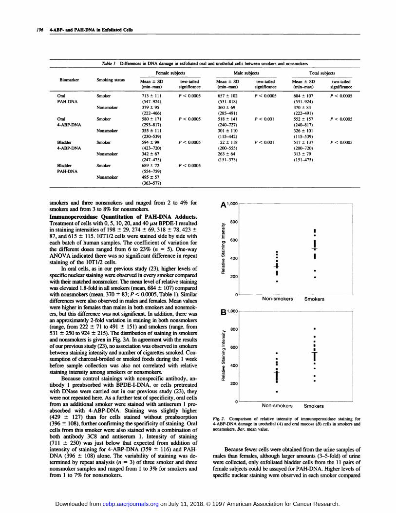
When the immunoperoxidase method was applied to thehuman samples, higher levels of nuclear staining for 4-ABP-DNAwere observed in the exfoliated urothelial cells of every smokercompared with their matched nonsmoker, except for one pair.Representative staining in smoker 3 and nonsmoker 50 is illus-
trated in Fig. 1 , A and B, respectively. Mean level of relativestaining in all smokers (517 ± 137) was I .7-fold higher than in
nonsmokers (313 ± 79: P < 0.0005 by paired t test: Table I).
Similar differences between smokers and nonsmokers were ob-served in males and females. Mean values were significantlyhigher in females than males in urothelial cells of both smokers
and nonsmokers (P < 0.01 ). The distribution of staining in smok-ers and nonsmokers is given in Fig. 14. An approximately 3-foldvariation in staining was observed both in nonsmokers (range,
151 ± 47 to 475 ± 103) and smokers (range, 200 ± 64 to 720 ±185). Among smokers, no association was found between relativestaining and number of cigarettes smoked per day.
Preabsorption of primary antibody with 4-ABP-DNA before
use decreased staining in urothelial cells of smoker 2 1 from 720 ±
185 to 84 ± 19 (Fig. 1C) and of nonsmoker 39 from 327 ± 84 to
70 ± 32 (not shown). Pretreatment of slides with DNase also
decreased staining in smoker 10 from 659 ± 15 1 to 129 ± 30 (Fig.
1D) and to 97 ± 31 in nonsmoker 39 (not shown). As an additionaltest of staining specificity. at a later time point oral cells from oneadditional smoker were stained with antibody 3C8 with or withoutpreabsorption with BPDE-I-DNA. There was little change in stain-ing intensity with (314 ± 101) or without (359 ± I 16) preabsorp-tion with an alternate DNA adduct. Variability of staining wasdetermined by repeat analysis (n = 3) of three smoker and threenonsmoker samples and ranged from 2 to 3% for smokers and
from 5 to 9% for nonsmokers.In oral cells, specific 4-ABP-DNA nuclear staining was also
observed, with higher levels of staining detected in every smokercompared with their matched nonsmoker. The mean level ofrelative staining was elevated 1.7-fold in smokers (552 ± I 57)
compared with nonsmokers (326 ± 101 ; P < 0.0005: Table 1).Similar differences were observed when male and female subjectswere analyzed separately. Mean levels were higher in females thanmales in both smokers and nonsmokers, but this difference was notsignificant. In addition, there was a 3-4-fold variation in staining
in nonsmokers (range, I 15 ± 57 to 539 ± 157) and smokers(range, 240 ± 80 to 815 ± 202, Fig. 2B). Again, no associationwas found in smokers between staining intensity and number of
cigarettes smoked.
Staining specificity was confirmed as for the urothelial cells.Preabsorption of primary antibody with 4-ABP-DNA before usealso decreased staining in oral cells of smoker 22 from 8 15 ± 202
to 125 ± 46 and of nonsmoker 48 from 305 ± 153 to 1 10 ± 32.Pretreatment of slides with DNase gave a value of 85 ± 26 forsmoker 22 and 63 ± 19 for nonsmoker 48. Variability of stainingwas determined by repeat analysis (ii = 3) of samples from three
on July 11, 2018. © 1997 American Association for Cancer Research. cebp.aacrjournals.org Downloaded from
I
I: I
-I-I
1 ,00C
�. 800
Cl)Ca,C
.; 600C
Ci 400
200
C
B �o0C
I 800
.; 600C
C
:i200
Non-smokers Smokers
i I-I
- Non-smokers Smokers
Fig. 2. Comparison of relative intensity of immunoperoxidase staining for4-ABP-DNA damage in urothelial (A) and oral mucosa (B) cells in smokers and
nonsmokers. Bar, mean value.
196 4-ABP- and PAll-DNA in Exfoliated Cells
Table 1 Differe nces in DNA damage in exfoliated oral and urothelial cells between smokers and nonsmokers
Biomarker Smoking status
Female subjects Male subjects Total subjects
M� ± SD �-�iled Mean ± SD two-tailed Mean ± SD two-tailed(nun-max) significance (mm-max) significance (mm-max) significance
Oral Smoker 713 ± Ill P < 0.0005 657 ± 102 P < 0.0005 684 ± 107 P < 0.0005
PAH-DNA
Nonsmoker
(547-924)
379 ± 95
(222-466)
(531-818)
360 ± 69
(285-491)
(531-924)
370 ± 83
(222-491)
Oral Smoker 580 ± 171 P < 0.0005 518 ± 141 P < 0.001 552 ± 157 P < 0.0005
4-ABP-DNA
Nonsmoker
(293-817)
355 ± Ill
(230-539)
(240-727)
301 ± 110
(115-442)
(240-817)
326 ± 101
(115-539)
Bladder Smoker 594 ± 99 P < 0.0005 22 ± I 1 8 P < 0.001 5 17 ± I 37 P < 0.0005
4-ABP-DNA
Nonsmoker
(423-720)
342 ± 67
(247-475)
(200-555)
263 ± 64
(151-373)
(200-720)
3 13 ± 79
(151-475)
Bladder Smoker 689 ± 72 P < 0.0005
PAR-DNA
Nonsmoker
(554-759)
495 ± 57
(363-577)
smokers and three nonsmokers and ranged from 2 to 4% forsmokers and from 3 to 8% for nonsmokers.
Immunoperoxidase Quantitation of PAH-DNA Adducts.Treatment ofcells with 0, 5, 10, 20, and 40 ,LM BPDE-I resultedin staining intensities of 198 ± 29, 274 ± 69, 318 ± 78, 423 ±
87, and 615 ± 1 15. 1OT1/2 cells were stained side by side witheach batch of human samples. The coefficient of variation for
the different doses ranged from 6 to 23% (n = 5). One-wayANOVA indicated there was no significant difference in repeat
staining of the 1OT1/2 cells.In oral cells, as in our previous study (23), higher levels of
specific nuclear staining were observed in every smoker comparedwith their matched nonsmoker. The mean level ofrelative stainingwas elevated 1.8-fold in all smokers (mean, 684 ± 107) comparedwith nonsmokers (mean, 370 ± 83; P < 0.0005, Table 1). Similar
differences were also observed in males and females. Mean valueswere higher in females than males in both smokers and nonsmok-
era, but this difference was not significant. In addition, there wasan approximately 2-fold variation in staining in both nonsmokers(range, from 222 ± 71 to 491 ± 151) and smokers (range, from531 ± 250 to 924 ± 215). The distribution of staining in smokersand nonsmokers is given in Fig. 3A. In agreement with the resultsof our previous study (23), no association was observed in smokers
between staining intensity and number ofcigareues smoked. Con-sumption of charcoal-broiled or smoked foods during the 1 week
before sample collection was also not correlated with relativestaining intensity among smokers or nonsmokers.
Because control stainings with nonspecific antibody, an-tibody 1 preabsorbed with BPDE-I-DNA, or cells pretreatedwith DNase were carried out in our previous study (23), theywere not repeated here. As a further test of specificity, oral cells
from an additional smoker were stained with antiserum 1 pre-absorbed with 4-ABP-DNA. Staining was slightly higher(429 ± 127) than for cells stained without preabsorption
(396 ± 108), further confirming the specificity of staining. Oralcells from this smoker were also stained with a combination of
both antibody 3C8 and antiserum 1. Intensity of staining(711 ± 250) was just below that expected from addition ofintensity of staining for 4-ABP-DNA (359 ± 1 16) and PAH-DNA (396 ± 108) alone. The variability of staining was de-
terinined by repeat analysis (n = 3) of three smoker and threenonsmoker samples and ranged from 1 to 3% for smokers andfrom 1 to 7% for nonsmokers.
Because fewer cells were obtained from the urine samples ofmales than females, although larger amounts (3-5-fold) of urinewere collected, only exfoliated bladder cells from the 1 1 pairs of
female subjects could be assayed for PAR-DNA. Higher levels ofspecific nuclear staining were observed in each smoker compared
on July 11, 2018. © 1997 American Association for Cancer Research. cebp.aacrjournals.org Downloaded from

II
II
III
I
A � ,ooc
�. 800
Ca,C
.; 600C
C
I 400
200
C
1,000
I,800
.; 600C
C:1400
200
Non-smokers Smokers
-I-
-I-
Non-smokers Smokers
(1 Mean staining intensity in 30 randomly selected cells ± SD.
Cancer Epidemiology, Biomarkers & Prevention 197
Table 2 Correlations between 4-ABP and PAH-DNA in exfoliated oral and
bladder cells in females only (n = 22) and in all subjects (n = 40)
4-ABP-DNA PAH-DNA 4-ABP-DNA
Oral cells Oral cells Bladder cells
4-ABP-DNA 1.000
Oral cells
PAH-DNA 0738�.b 1.000
Oral cells 0.723”’
4-ABP-DNA 0592�.b 0.7 14” 1.000
Bladder cells 0.58l�.c 0.672”’
PAH-DNA 0536a.b #{216}6�ja.b 0687�.b
Bladder cells
“P <0.001.b Correlation for female subjects only.
‘ Correlation for all subjects.
Table 3 Immunoperoxidase detection of 4-ABP-DNA and PAH-DNA in oral
cells of a smoker who reported decreasing smoking intensity from 5 packs per
day to 1 pack per week
Relative staining intensity”Days since reduced
smoking level 4-ABP-DNA
0 262±77 573±43
7 202±78 369±55
16 134±36 330±35
26 89±33 310±42
Fig. 3. Comparison of relative intensity of immunoperoxidase staining for
PAH-DNA damage in oral mucosa (A) and urothelial (B) cells in smokers and
nonsmokers. Bar, mean value.
with her matched nonsmoker control (not shown). The mean levelof relative staining was elevated 1 .4-fold in female smokers (mean,689 ± 72) compared with nonsmokers (mean, 495 ± 57; P <
0.0005; Table 1). The staining variation in nonsmokers wasslightly higher than in smokers. It ranged from 363 ± 105 to577 ± 143 in nonsmokers and from 554 ± 184 to 759 ± 189 in
smokers (Fig. 3B). Again, no association was found between
relative staining intensity and number of cigarettes smoked insmokers, and consumption of charcoal-broiled or smoked foods insmokers and nonsmokers.
Preabsorption of primary antiserum with BPDE-I-DNAbefore use on urothelial cells decreased staining of smoker 20from759 ± l89to249 ± 87andofnonsmoker3l from363 ±
105 to 221 ± 35. Staining with a nonspecific antiserum rec-ognizing DNA damage produced by 8-methoxypsoralen alsodecreased relative staining in smoker 21 from 759 ± 189 to246 ± 93 and in nonsmoker 31 from 363 ± 105 to 191 ± 59.
Pretreatment of slides with DNase gave a value of 190 ± 60 insmoker 20 and 197 ± 39 in nonsmoker 31. Variability ofstaining was determined by repeat analysis (n 3) of twosmoker and two nonsmoker samples and ranged from 2 to 3%for smokers and from 4 to 6% for nonsmokers.
Correlations between 4-ABP- and PAH-DNA in ExfoliatedOral and Bladder Cells. Correlations between the two types ofDNA damage in the different cells in all subjects were significant
(P < 0.001 ; Spearman rank correlation; Table 2). Among the threecombinations, the highest association was seen between PM!- and4-ABP-DNA in oral mucosa cells, followed by the correlation
between PAH-DNA in oral cells and 4-ABP-DNA in exfoliatedurothelial cells and that between 4-ABP-DNA in oral and exfoli-
ated urothelial cells. For female subjects in which data on PAH-DNA in urothelial cells was also available, correlations for all six
combinations of biomarkers were determined. Similar to the re-sults in total subjects, significant correlations were observed for allcombinations (P < 0.001).
Detection of 4-ABP- and PAH-DNA Damage in Oral Mu-
cosa Cells of a Smoker Who Decreased Smoking. Oral cellswere obtained at different time points from a smoker who dramat-ically decreased, but did not stop, smoking (5 packs/thy at baseline
to 1 pack/week). Decreased relative staining was observed in cells
stained for either PAH-DNA or 4-ABP-DNA (Table 3).
Discussion
Iminunoperoxidase Quantitation of 4-ABP-DNA Damage.Previously, we had developed an immunofluorescence method
for detection of 4-ABP-DNA, demonstrated a dose-response rela-tionship between administered dose and relative fluorescencestaining intensity in mouse liver tissues, and found an excellentcorrelation with GCIMS detection ofDNA damage (22). A similar
dose-response was found in the present study for immunoperoxi-dase staining. The specificity of staining was further confirmed by
preabsorbing antibody with an alternate modified DNA, BPDE-I-DNA, and by demonstrating no change in staining intensity. Sig-nificant correlations were observed between quantitative immu-noperoxidase intensity and either immunofluorescence stainingintensity (r = 0.99) orlevelofDNA adduct determined by (iC/MS
(r 0.94). However, there was a much smaller increase in im-munoperoxiclase (2.7-fold) than immunofluorescence staining (13-
fold) for animals treated with 80 compared with 4 mg/kg 4-ABP(20-fold difference in dose). This may be due to the detectionlimits and/or dynamic range of each method. High background
on July 11, 2018. © 1997 American Association for Cancer Research. cebp.aacrjournals.org Downloaded from

198 4-ABP- and PAH-DNA in Exfoliated Cells
staining related to nonspecific binding of reagents in the immu-noperoxidase method may be responsible for the shallow slope of
the dose-response curve. An 8-fold difference in immunoperoxi-
dase staining between liver tissue of mice treated with 80 or 4mg/kg 4-ABP was found after subtracting the value for intensity of
staining of control tissue (mean = 189) from that for the treatedanimal tissues. Although background staining in the immunoflu-
orescence method is much lower, subtraction of the staining in-
tensity of control tissue also increased the difference in stainingbetween the highest and lowest dose-treated animals (from 13-foldto 21-fold). Inefficient digestion with proteinase K or incomplete
denaturation of DNA during the staining procedure, leading tolower antibody binding to adduct, and/or nonlinear enzyme am-
plification of signal may also reduce the ability of the immunoper-
oxidase method to efficiently quantitate adduct levels.In this study, higher levels of specific nuclear staining were
observed in exfoliated urothelial cells in every smoker compared
with their matched nonsmoker, except for one pair. A 1.7-foldhigher level of relative staining intensity was observed in smokers
than in nonsmokers (P < 0.0005). We previously observed a
2.4-fold higher staining intensity in bladder biopsies of smokingthan nonsmoking bladder cancer patients (24). Comparable resultswere found by 32P-postlabeling of DNA isolated from human
exfoliated urothelial cells (29). A smoking-related adduct was
found to be 1.7-fold higher in smokers compared with nonsmok-
ers, and correlated significantly with the amount of tobacco the
individual smoked and the level of 4-ABP-hemoglobin adducts in
the red blood cells. Fivefold higher adduct levels in smokerscompared with nonsmokers were observed in another study of
human bladder biopsy samples using butanol enrichment before
posfiabeling (5). Among these studies, measured 4-ABP-DNA
adduct levels ranged from 1 adductll07 nucleotides to 1 adduct/i0� nucleotides (5, 16, 29, 30). After dosing for 6 weeks, 4-ABP-
DNA adducts in exfoliated urothelial cells of dogs were identical
and equal in level to adducts found in intact urinary bladder (27).This further strengthens the applicability of monitoring carcino-
gen-DNA adduct levels in the target organ using exfoliated urothe-
lial cells.Specific nuclear staining of 4-ABP-DNA in two cell types
(oral mucosa and exfoliated urothelial cells) was observed in
nonsmokers as well as smokers. It is not surprising that theimmunohistochemical assay can also detect DNA damage in
nonsmokers. Although, in this study, only 1 of 20 nonsmoking
subjects reported environmental tobacco smoke exposure, this
is a potential source of exposure. Structurally related corn-pounds (e.g., 4-nitrobiphenyl in diesel exhaust; Ref. 31), are
other possible sources of 4-ABP-DNA adducts in nonsmokers.
In addition, the background staining of 4-ABP-DNA in non-smokers may also be partially due to the low cross-reactivity of
antibody 3C8 with unadducted DNA (22).
Immunoperoxidase Quantitation of PAH-DNA Adducts.We reported previously specific detection of adducts in BPDE-
I-treated lOTl/2 cells (23). The addition of nickel chloride to
the DAB solution resulted in darker staining and a larger
difference in staining intensity of cells treated with the highestdose compared with control cells than in our previous report(3.1-fold versus 1.7-fold). Specific nuclear staining was ob-
served in oral mucosa cells of both smokers and nonsmokers.The 1.7-fold increase in damage levels observed in this study is
similar to that of our previous study (2-fold; Ref. 23). It is also
comparable to the 3-fold difference in PAH-DNA in mononu-clear cells of smokers and nonsmokers using an ELISA with asimilar antiserum (32). In addition, oral mucosa cells have been
used in several studies to monitor exposure to PAH by 32P-
postlabeling with adduct levels ranging from 1 .6/106 to l/10�
(8, 10, 33-35). Significant 2-3-fold differences were found
between smokers, and either nonsmokers or ex-smokers insome studies (33-35), although no association between smok-
ing and adduct level was observed in others (8, 10). We alsoobserved an almost additive effect when simultaneously stain-ing oral cells for both 4-ABP- and PAH-DNA, suggesting that
multiple antigens can be detected in the same sample.
Specific nuclear staining for PAH-DNA in both cells typeswas observed in nonsmokers. Because PAHs are ubiquitous,
these adducts may come from exposure to environmental airpollution, including tobacco smoke. Exposure to PAHs from
other sources such as water, soil, or green leafy vegetables may
also contribute to adduct formation (36). Twofold to 3-foldvariation in staining in the two cell types was also observed in
both smokers and nonsmokers, which may be due to individual
genetic variation in metabolism of carcinogens, as well as
differences in exposure levels. However, larger interindividual
differences were found in previous studies on white blood cell
DNA from individuals exposed to similar levels (37-40).
Correlations between Biomarkers and Other Factors. No
association between relative staining and consumption of char-coal-broiled and smoked foods was observed; however, we still
cannot rule out this means of exposure. Accurate dietary dataare difficult to obtain, and study subjects reported low fre-
quency of consumption of these foods. The small sample sizealso reduced our ability to detect differences between charcoal-
broiled/smoked food consumption and adduct levels. The lackof association between the number of cigarettes smoked per day
and relative staining intensity of each biomarker in the smokerswas not unexpected, because all smokers in this study smoked
20-40 cigarettes/day. A linear, dose-related increase in stain-
ing intensity for 4-ABP-DNA was observed in our previousstudy of bladder biopsies of bladder cancer patients classified
into four groups: nonsmokers, and smokers of 1-19, 20-40,
and >40 cigarettes/day (24). In an endeavor to validate markersin a highly exposed group, the heavy exposure of the subjects
in this study may have reduced our ability to see dose-responserelationships. Differences in smoking habits, types of cigarettes
(air- or flue-cured), and genetic factors that influence carcino-
gen metabolism and DNA repair may also affect adduct for-
mation in individuals. Other environmental exposures or dif-
ferences in diet or lifestyle of study subjects are other possible
explanations for the lack of association with number of ciga-rettes smoked. Significant correlations among the biomarkers
demonstrated that they are good indicators of exposure to
cigarette smoke and a good measure of biologically effectivedose in the body. Analysis of samples collected at four differenttime points from a smoker who reported decreased smoking
demonstrated lower relative staining for both 4-ABP- and PAH-DNA between days 0 and 7. This is comparable to the estimated
turnover rate of oral mucosa cells. A study of the disappearance
of micronuclei in radiation therapy patients estimated as 5-7
days the time frame of cell migration from the basal layer toexfoliation (41). For urothelial cells, the estimated cell turnoverrate is much longer (50-200 days) than oral cells (5 days) (42).
However, with chronic exposure, measurement of DNA dam-
age in these cell types may reflect current, as well as previous,
exposure to carcinogens.The immunohistochemical method developed here allows
investigation of adduct formation at the individual cell level,
with high specificity and relatively low cost. The major limi-
tation is that it is semiquantitative and provides information
only on relative levels of carcinogen-DNA adducts. The re-
on July 11, 2018. © 1997 American Association for Cancer Research. cebp.aacrjournals.org Downloaded from

Cancer Epidemiology, Biomarkers & Prevention 199
quirement for small numbers of cells allows application to
biopsy samples and exfoliated cells that are easily collected,and represents a simple way to obtain human tissue samples bynoninvasive methods. As target tissues for smoking-induced
cancers, DNA damage observed in these cells may also reflectthe potential risk of an individual to develop cancer. In additionto the study of the mechanisms of oral and bladder carcinogen-esis, these methods may also be valuable in more generalstudies of exposure to environmental carcinogens. We are cur-rently using these biomarkers as intermediate end points in anantioxidant vitamin intervention study in heavy smokers.
References
1. Janoff, A., Pryor, W. A., and Bengali, Z. H. Effects of tobacco smoke
components on cellular and biochemical processes in the lung. Am. Rev. Respir.
Dis., 136: 1058-1064, 1987.
2. IARC. Monographs on the Evaluation of Chemicals to Humans: TobaccoSmoking, pp. 244-270. [ARC, 1986.
3. Surgeon General. The Health Consequences of Smoking. Cancer. A Report of the
Surgeon General. Washington, DC: United States Department of Health, 1982.
4. National Research Council. Environmental Tobacco Smoke. Measuring cx-
posures and assessing health effects. National Academy of Sciences. Washington,
DC: National Academy Press, 1986.
5. Talaska, G., Al-Juburi, A. Z. S. S., and Kadlubar, F. F. Smoking related
carcinogen-DNA adducts in biopsy samples of human urinary bladder: identifi-cation of N-(deoxyguanosin-8-yl)-4-aminobiphcnyl as a major adduct. Proc. Natl.
Acad. Sci. USA, 88: 5350-5354, 1991.
6. Talaska, G., Dooley, K. L., and Kadlubar, F. F. Detection and characterization
of carcinogen-DNA adducts in exfoliated urothelial cells of cigarette smokers:
association with smoking, hemoglobin adducts and urinary mutagenicity. Cancer
Epidemiol., Biomarkers & Prey., 1: 61-66, 1991.
7. Mommsen, S., and Aagaard, J. Tobacco as a risk factor in bladder cancer.Cancer (Phila.), 4: 335-338, 1983.
8. Dunn, B. P., and Stich, H. F. 32P-Postlabeling analysis of aromatic DNA adducts
in human oral mucosal cells. Carcinogenesis (Lond.), 7: 1 1 15-1 120, 1986.
9. Bryant, M. S., Vineis, P., Skipper, P. L., and Tannenbaum, S. R. Hemoglobin
adducts of aromatic amines: associations with smoking status and type of tobacco.
Proc. Natl. Acad. Sci. USA, 85: 9788-9791, 1988.
10. Chacko, M., and Gupta, R. C. Evaluation of DNA damage in the oral mucosa
of tobacco users and non-users by 32P-postlabeling assay. Carcinogenesis
(Lond.), 9: 2309-2313, 1988.
1 1 . Santella, R. M. DNA adducts in humans as biomarkers of exposure to environ-
mental and occupational carcinogens. Environ. Care. Rev., C9: 57-81, 1991.
12. Beach, A. C., and Gupta, R. C. Human biomonitoring and the 32P-postla-
beling assay. Carcinogenesis (Lond.), 13: 1053-1074, 1992.
13. Talaska, G., and Hoon Roh, J. 32P-postlabelling and mass spectrometric
methods for analysis of bulky, polyaromatic carcinogen-DNA adducts in humans.
J. Chromatogr., 580: 293-323, 1992.
14. Poirier, M. C. Antisera specific for carcinogen-DNA adducts and carcinogen-
modified DNA: applications for detection of xenobiotics in biological samples.
Mutat. Res., 288: 31-38, 1993.
15. Weston, A. Physical methods for the detection of carcinogen-DNA adducts
in humans. Mutat. Res., 288: 19-29, 1993.
16. Talaska, G., Schamer, M., Skipper, P., Tannenbaum, S., Caporaso, N.,
Kadlubar, F., Bartsch, H., and Vineis, P. Techniques for noninvasive human
monitoring. Environ. Health Perspect., 99: 289-291, 1993.
17. Zhang, Y-J., Chen, C. J., Haghighi, B., Yang, G. Y., Hsieh, L. L., Wang, L.
W., and Santella, R. M. Quantitation of aflatoxin B 1-DNA adducts in woodchuckhepatocytes and rat liver tissues by indirect immunofluorescence analysis. Cancer
Res., 51: 1720-1725, 1991.
18. Zhang, Y-J., Chen, C. J., Lee, C. S., Haghighi, B., Yang, G. Y., Wang, L. W.,
Feitelson, M., and Santella, R. Aflatoxin B1-DNA adducts and hepatitis B virusantigens in hepatocellular carcinoma and non-tumorous liver tissue. Carcinogen-
esis (Lond.), 12: 2247-2252, 1991.
19. Santella, R. M., Yang, X. Y., DeLco, V. A., and Gasparro, F. P. Detection and
quantification of 8-methoxypsoralen-DNA adducts. In: H. Bartsch, K. Hemminki,and 1. K. O’Neil (eds.), Methods for Detecting DNA Damaging Agents in Human:
Applications in Cancer Epidemiology and Prevention, pp. 333-340. Lyon,
France: IARC, 1988.
20. Zhang, Y-J., Li, Y., DeLco, V. A., and Santella, R. M. Detection of DNA
adducts in skin biopsies ofcoal tar-treated psoriasis patients: immunofluorescence
and 32P postlabeling. Skin Pharmacol., 3: 171-179, 1990.
21 . Motykiewicz, G., Malusecka, E., Grzybowska, E., Chorazy, M., Zhang. Y-J.,
Perera. F. P., and Santella, R. M. Immunohistochemical quantitation of polycyclicaromatic hydrocarbon-DNA adducts in human lymphocytes. Cancer Res., 55:
1417-1422, 1995.
22. Al-Atrash, J., Zhang, Y-J., Lin, D., Kadlubar, F. F., and Santella, R. M.
Quantitative immunohistochemical analysis of 4-aminobiphenyl-DNA in cultured
cells and mice: comparison to gas chromatography/mass spectroscopy analysis.
Chem. Res. Toxicol., 8: 747-752, 1995.
23. Zhang, Y-J., Hsu, T-M., and Santella, R. M. Immunoperoxidase detection of
polycyclic aromatic hydrocarbon-DNA adducts in oral mucosa cells of smokers
and nonsmokers. Cancer Epidemiol., Biomarkers & Prey., 4: 133-138, 1995.
24. Curigliano, G., Zhang, Y-J., Wang, L. Y., Flamini, G., Alcini, A., Ratio, C.,
Giustacchini, M., Alcini, E., Cittadini, A., and Santella, R. M. Immunohisto-chemical quantitation of 4-arninobiphenyl-DNA adducts and p53 nuclear over-
expression in TI bladder cancer of smokers and nonsmokers. Carcinogenesis
(Lond.), 17: 911-916, 1996.
25. Schulte, J., King, C. D., King, E. B., Macdonald, D. A., and Jassie, M. P. Asingle technique for recognizing abnormal epithelial cells in urinary sediment.
J. Urol., 89: 615-625, 1963.
26. Warner, M. L., Moore, L. E., Smith, M. T., Kalman, D. A., Fanning, E., and
Smith, A. H. Increased micronuclei in exfoliated bladder cells of individuals who
chronically ingest arsenic-contaminated water in Nevada. Cancer Epidemiol.,Biomarkers & Prey., 3: 583-590, 1994.
27. Talaska, G., Dooley, K. L., and Kadlubar, F. F. Detection and characteriza-
tion of carcinogen-DNA adduct in exfoliated urothelial cells from 4-aminobiphe-
nyl-treated dogs by 32P-postlabeling and subsequent thin-layer and high pressureliquid chromatography. Carcinogenesis (Lond.), 11: 639-646, 1990.
28. Santella, R. M., Dharmaraja, N., Gasparro, F. P., and Edelson, R. L. Mono-
clonal antibodies to DNA modified by 8-methoxypsoralen and ultraviolet A light.
Nucleic Acids Res., 13: 2533-2544, 1985.
29. Jones, N. J., McGregor, A. D., and Waters, R. Detection of DNA adducts in
human oral tissue: correlation of adduct levels with tobacco smoking and differ-
ential enhancement of adducts using the butanol extraction and nuclease P1 of 32P
postlabeling. Cancer Res., 53: 1522-1528, 1993.
30. Talaska, G., Schamer, M., Skipper, P., Tannenbaum, S. R., Caporaso, N.,
Unruh, L., Kadlubar, F., Bartsch, H., Malaveille, C., and Vineis, P. Detection of
carcinogen DNA adducts in exfoliated bladder cells of cigarette smokers: asso-
ciation with smoking, hemoglobin adducts and urinary mutagenicity. Cancer
Epidemiol., Biomarkers & Prey., 1: 61-66, 1991.
31. Phillips, D. H., and Hewer, A. DNA adducts in human urinary bladder and
other tissues. Environ. Health Perspect., 99: 45-49, 1993.
32. Bryant, M. S., Skipper, P. L., and Tannenbaum, S. R. Hemoglobin adducts of
4-aminobiphenyl in smokers and nonsmokers. Cancer Res., 47: 602-608, 1987.
33. Santella, R. M., Grinberg-Funes, R. A., Young, T. L., Dickey, C., Singh, V.
N., Wang, L. W., and Perera, F. P. Cigarette smoking related polycyclic aromatic
hydrocarbon-DNA adducts in peripheral mononuclear cells. Carcinogenesis
(Lond.), 13: 2041-2045, 1992.
34. Foiles, P. G., Miglietta, L. M., Quart, A. M., Quart, E., Kabat, G. C., andHecht, S. S. Evaluation of 32P-postlabeling analysis of DNA from exfoliated oral
mucosa cells as a means of monitoring exposure of the oral cavity to genotoxic
agents. Carcinogenesis (Lond.), 10: 1429-1434, 1989.
35. Stone, J. G., Jones, N. J., McGregor, A. D., and Waters, R. Development of a
human biomonitoring assay using buccal mucosa: comparison of smoking-related
DNA adducts in mucosa versus biopsies. Cancer Res.. 55: 1267-1270, 1995.
36. Toxicological Profile for Benzo(a)pyrene. Washington. DC: United States
Department of Health and Human Services, 1990.
37. Perera, F. P., Hemminki. K., Young, T. L., Santella, R. M., Brenner, D., and
Kelly, G. Detection of polycyclic aromatic hydrocarbon-DNA adducts in whiteblood cells of foundry workers. Cancer Res., 48: 2288-2291, 1988.
38. van Schooten, F. J., van Leeuwen, F. E., Hillebrand, M. J. X.. deRijke, M. E.,Hart, A. A. M., van Veen, H. G., Oosterink, S., and Kriek, E. Determination of
benzo(a)pyrene diol epoxide-DNA adducts in white blood cell DNA from coke-
oven workers: the impact of smoking. J. Natl. Cancer Inst., 82: 927-933, 1990.
39. Schoket, B., Phillips, D. H., Hewer, A., and Vincze, I. 32P-postlabeling
detection of aromatic DNA adducts in peripheral blood lymphocytes from alu-minum production plant workers. Mutat. Res., 260: 89-98, 1991.
40. Ovrebo, S., Haugen, A., Phillips, D. H., and Hewer, A. Detection of poly-
cyclic aromatic hydrocarbon-DNA adducts in white blood cells from coke oven
workers: correlation with job categories. Cancer Res., 52: 1510-1514, 1992.
41 . Stich, H. F., San, R. H. C., and Rosin, M. P. Adaptation of the DNA-repair
and micronucleus test to human cell suspensions and exfoliated cells. Ann. N.Y.
Acad. Sci., 407: 93-105, 1983.
42. Clayson, D. B., and Lawson, T. A. Mechanisms of bladder carcinogenesis.
In: J. G. Connolly (ed), Carcinoma ofthe Bladder, pp. 91-100. New York: Raven
Press, 1981.
on July 11, 2018. © 1997 American Association for Cancer Research. cebp.aacrjournals.org Downloaded from

1997;6:193-199. Cancer Epidemiol Biomarkers Prev T M Hsu, Y J Zhang and R M Santella oral and urothelial cells of smokers and nonsmokers.polycyclic aromatic hydrocarbon-DNA adducts in exfoliated Immunoperoxidase quantitation of 4-aminobiphenyl- and
Updated version
http://cebp.aacrjournals.org/content/6/3/193
Access the most recent version of this article at:
E-mail alerts related to this article or journal.Sign up to receive free email-alerts
Subscriptions
Reprints and
To order reprints of this article or to subscribe to the journal, contact the AACR Publications
Permissions
Rightslink site. Click on "Request Permissions" which will take you to the Copyright Clearance Center's (CCC)
.http://cebp.aacrjournals.org/content/6/3/193To request permission to re-use all or part of this article, use this link
on July 11, 2018. © 1997 American Association for Cancer Research. cebp.aacrjournals.org Downloaded from