impact of different stages of intrauterine inflammation on
TRANSCRIPT

RESEARCH ARTICLE
Impact of different stages of intrauterine
inflammation on outcome of preterm
neonates: Gestational age-dependent and
-independent effect
Carlo Pietrasanta1,2, Lorenza PugniID1*, Daniela Merlo3, Barbara Acaia4, Dario Consonni5,
Andrea Ronchi1, Manuela Wally Ossola4, Beatrice Ghirardi1,2, Ilaria Bottino1,2, Fulvia
Milena Cribiu3, Silvano Bosari3,6, Fabio Mosca1,2
1 NICU Fondazione IRCCS Ca’ Granda Ospedale Maggiore Policlinico, Milan, Italy, 2 University of Milan,
Department of Clinical Sciences and Community Health, Milan, Italy, 3 Pathology Unit, Fondazione IRCCS
Ca’ Granda Ospedale Maggiore Policlinico, Milan, Italy, 4 Gynecology and Obstetrics Unit, Fondazione
IRCCS Ca’ Granda Ospedale Maggiore Policlinico, Milan, Italy, 5 Epidemiology Unit, Fondazione IRCCS Ca’
Granda Ospedale Maggiore Policlinico, Milan, Italy, 6 University of Milan, Department of Pathophysiology
and Transplantation, Milan, Italy
Abstract
Objective
To investigate the impact of different stages of intrauterine inflammation (IUI) on neonatal
outcomes, before and after adjusting for gestational age (GA) and other perinatal
confounders.
Methods
This was an observational, prospective, single-center cohort study including all eligible neo-
nates with GA < 35 weeks and/or birth weight� 1500 g born at a 3rd level Neonatal Intensive
Care Unit between 2011 and 2014. Pathological patterns of placenta, membranes and cord
were classified according to Redline’s criteria. Multivariable linear and logistic regression
models were applied, either including or not GA among the covariates.
Results
Of the 807 enrolled neonates, 134 (16.6%) had signs of IUI: among these, 54.5% showed
just histological chorioamnionitis (HCA), 25.4% had HCA + funisitis (FUN) stage 1, and
20.1% had HCA + FUN stage 2–3. At univariate analysis, HCA increased the risk for retinop-
athy of prematurity (ROP) and bronchopulmonary dysplasia, while FUN (any stage) had a
deleterious impact on all outcomes investigated. After adjustment for covariates not includ-
ing GA, HCA was a risk factor only for ROP (OR = 2.8, CI: 1–7.8), while FUN (any stage)
was still associated with increased ORs for all outcomes (p <0.01). Upon inclusion of GA in
the regression model, the results differed remarkably. HCA was associated with lower risk
for mechanical ventilation (OR = 0.3, CI: 0.1–0.7) and need for surfactant (OR = 0.5, CI:
PLOS ONE | https://doi.org/10.1371/journal.pone.0211484 February 8, 2019 1 / 15
a1111111111
a1111111111
a1111111111
a1111111111
a1111111111
OPEN ACCESS
Citation: Pietrasanta C, Pugni L, Merlo D, Acaia B,
Consonni D, Ronchi A, et al. (2019) Impact of
different stages of intrauterine inflammation on
outcome of preterm neonates: Gestational age-
dependent and -independent effect. PLoS ONE 14
(2): e0211484. https://doi.org/10.1371/journal.
pone.0211484
Editor: Cheryl S. Rosenfeld, University of Missouri
Columbia, UNITED STATES
Received: August 23, 2018
Accepted: January 15, 2019
Published: February 8, 2019
Copyright: © 2019 Pietrasanta et al. This is an open
access article distributed under the terms of the
Creative Commons Attribution License, which
permits unrestricted use, distribution, and
reproduction in any medium, provided the original
author and source are credited.
Data Availability Statement: All relevant data are
within the manuscript and its Supporting
Information files.
Funding: The authors received no specific funding
for this work.
Competing interests: The authors have declared
that no competing interests exist.
brought to you by COREView metadata, citation and similar papers at core.ac.uk
provided by AIR Universita degli studi di Milano

0.2–0.9), while FUN (any stage) worsened clinical conditions at birth (p <0.05), increased
the risk for early-onset sepsis (p <0.01), and increased the length of mechanical ventilation
(FUN stage 2–3 only, RC = 6.5 days, CI: 2–11). No other outcome was affected.
Conclusions
IUI, especially FUN, negatively impact most neonatal morbidities, but its effect is partially
reverted adjusting for GA. Considered that GA is an intermediate variable interposed
between prenatal causes of prematurity and outcomes, the appropriateness of adjusting for
GA may be questionable.
Introduction
Acute perinatal intrauterine inflammation (IUI) is one of the leading causes of preterm birth
worldwide and its prevalence increases with decreasing gestational age (GA): up to 70% of pre-
term births at 23–25 weeks of GA are associated with IUI [1–3]. IUI may involve both mater-
nal and fetal compartment, with signs of inflammation limited to the membranes chorion and
amnion (maternal inflammation, referred to as histological chorioamnionitis, HCA) or also
within the walls of umbilical cord vessels (fetal inflammation or funisitis, FUN) [4]. These his-
topathological patterns still represent the gold standard for the diagnosis of IUI and few differ-
ent classification systems have been proposed over the past 30 years [5–8].
To diagnose IUI before or immediately after birth, earlier than histopathological examina-
tion, the use of several clinical and biochemical signs has been proposed [9–14]. Nonetheless,
none of them nor any specific combination have been proved to provide acceptable sensitivity
and specificity for the diagnosis of IUI up to now, while the new descriptive term of “intrauter-
ine inflammation or infection or both”, abbreviated as “Triple I”, has been recently proposed
to harmonize different clinical definitions and replace the term chorioamnionitis in the clinical
context [15,16].
Although IUI is a well-known cause of preterm birth, the correlation between its different
stages or grades and neonatal adverse outcomes is far from being clarified [17]. In the last
decades, numerous studies have attempted to establish whether, and to what extent, IUI might
negatively affect the short- and long-term outcome of preterm neonates. Several conditions
peculiar of prematurity, such as respiratory distress syndrome (RDS), bronchopulmonary dys-
plasia (BPD) [18,19], neurological short- and long-term adverse outcomes [20–23], sepsis
[24,25], retinopathy of prematurity (ROP) [26], and the patency of ductus arteriosus (PDA)
[27] have been variably correlated to either a clinical or histological diagnosis of IUI, with dis-
cordant results. The inconsistency observed across studies can be attributed largely to the
enrollment of different study populations, the use of different diagnostic criteria and methods,
and to whether or not potential confounding factors were taken into account. Particularly,
although most studies considered GA at birth among other confounding factors, few authors
clearly explored the role of GA independently of other covariates.
Considering controversial results among existing studies, we decided to perform a study to
investigate the importance of different stages of perinatal IUI, histologically diagnosed, on neo-
natal outcomes of preterm birth, before and after adjusting for antenatal and perinatal vari-
ables and with a specific focus on the adjustment for GA.
Intrauterine inflammation and neonatal outcomes
PLOS ONE | https://doi.org/10.1371/journal.pone.0211484 February 8, 2019 2 / 15

Materials and methods
Study population and design
This was an observational, prospective, single-center study conducted at the Neonatal Inten-
sive Care Unit (NICU) of Fondazione IRCCS Ca’ Granda Ospedale Maggiore Policlinico of
Milan, Italy, between November 2011 and December 2014. The study protocol was approved
by the Ethics Committee of the Hospital (Comitato Etico Milano Area B—Fondazione IRCCS
Ca’ Granda Ospedale Maggiore Policlinico Milano). Written informed consent was obtained
from parents for the inclusion in the study, and all procedures were in accordance with the
Helsinki Declaration of 1975, as revised in 2008.
All inborn neonates with a GA <35 weeks and/or a birth weight (BW)�1500 g admitted to
the NICU were consecutively enrolled in the observational study. Exclusion criteria were
being outborn, the presence of major congenital anomalies, lack of parental consent or a miss-
ing pathological examination of fetal adnexa.
Histopathological examination of placenta, chorioamniotic membranes and umbilical cord
was performed by a single pathologist expert in perinatal pathology, aware of GA and BW but
blinded to other clinical data. At least 7 samples were collected, included in paraffin, sectioned
and stained with hematoxylin and eosin: 1 samples from the membranes, 3 from the umbilical
cord, and 3 from the placental disc, plus additional samples from macroscopically abnormal
areas. Pathological patterns of placenta, membranes and cord were described and classified
according to Redline’s criteria [7].
Neonates were classified into four groups, based on the result of pathological examination
of fetal adnexa: Group 0 (controls, no inflammation detected), Group 1 (HCA, presence of
HCA of any stage or grade, without involvement of umbilical cord or chorionic plate vessels),
Group 2 (HCA + FUN 1, presence of HCA plus FUN stage 1), and Group 3 (HCA + FUN 2–3,
presence of HCA plus FUN stage 2–3).
Data collection and definitions
Data from mothers and neonates were prospectively collected using an electronic database.
The following maternal variables were recorded: age at delivery, ethnicity, premature (at least
1 hour before the onset of contractions)–rupture of the membranes (PROM), PROM >24
hours, clinical chorioamnionitis, antibiotic therapy in labor, prenatal steroids, pre-eclampsia.
A preterm birth was defined as “indicated” in case of induced vaginal delivery or cesarean sec-
tion in the absence of preterm labor. Clinical chorioamnionitis was defined as the presence of
at least two of the following criteria during labor, in absence of other known cause: maternal
fever>38˚C, maternal leukocytosis >15,000 leukocytes/mm3, maternal C-reactive protein
(CRP) >10 mg/dL, foul-smelling or purulent amniotic fluid, persistent maternal heart rate
>100 bpm or fetal heart rate>160 bpm.
Moreover, we recorded the following neonatal data: GA (dated by first trimester ultrasound
crown-rump length measurement, when available, or calculated from the first day of last men-
strual period), BW, sex, mode of delivery, twins, small for gestational age (SGA) defined as
BW<10th percentile according to Fenton [28], need for resuscitation (at least ventilation with
mask), supplemental oxygen, intubation and surfactant at birth, Apgar score at 1 and 5 min-
utes of life, presence of RDS, total doses of surfactant, duration of invasive and non-invasive
ventilation (including heated humidified high flow nasal cannulae), occurrence of culture-
proven early-onset (within the first 72 hours of life) sepsis (EOS), late-onset (after the first 72
hours of life) sepsis (LOS), PDA, intraventricular hemorrhage (IVH), ROP, BPD, and death
during hospitalization.
Intrauterine inflammation and neonatal outcomes
PLOS ONE | https://doi.org/10.1371/journal.pone.0211484 February 8, 2019 3 / 15

RDS was defined as PaO2 <50 mmHg in room air, central cyanosis in room air, a require-
ment for supplemental oxygen to maintain PaO2 >50 mmHg, or a requirement for supple-
mental oxygen to maintain a pulse oximeter saturation over 85% within the first 24 hours of
life, and a chest radiograph consistent with RDS (reticulogranular appearance to lung fields
with or without low lung volumes and air bronchograms) within the first 24 hours of life [29].
BPD was defined according to Jobe’s criteria [30]. IVH was defined according to Volpe’s classi-
fication by head ultrasound scan [31], and ROP was defined according to the international
classification for retinopathy of prematurity [32].
Statistical analysis
We calculated regression coefficients (RCs), odds ratios (ORs), 95% confidence intervals (CIs),
and two-sided p-values for each of the three groups with IUI in comparison with the control
group using univariate and multivariable linear (for quantitative variables) or logistic (for
dichotomous variables) regression models. Variables included in regression models were
selected a priori. To investigate the role of GA independently of the other covariates (maternal
age, ethnicity, antenatal steroids, sex, SGA neonate, and indicated preterm birth), models
including GA at delivery were compared with those without GA at delivery. Standard errors of
RCs and log(ORs) were adjusted to take into account the outcome correlations within twin
births. Statistical analysis was performed using Stata 15 (StataCorp. 2017, College Station, TX).
Results
During the study period a total of 1024 neonates with GA <35 weeks and/or BW�1500 g
were born at the study site. Of these, 217 (21.2%) were excluded because of unavailable placen-
tal examination or major congenital malformation. A total of 807 babies were enrolled, with
mean GA 31.9 ± 2.7 weeks and mean BW 1656 ± 535 g. Females were 47.1%. The full dataset is
available in supporting information (S1 File).
Of the 807 enrolled neonates, 134 (16.6%) had signs of fetal adnexa inflammation: among
these, 73 (54.5%) showed just HCA, 34 (25.4%) had signs of HCA + FUN 1, and 27 (20.1%)
had signs of HCA + FUN 2–3. The incidence of both HCA alone and FUN decreased progres-
sively with increasing GA, dropping from 12.5% at 22–24 weeks of gestation to 2.4% at 33–34
weeks (HCA) and from 45.8% at 23–24 weeks to 8% at 33–34 weeks (FUN, any stage),
respectively.
The neonates born to mothers with any IUI had lower GA and lower BW compared to the
control group, and a stepwise reduction in both GA and BW was correlated with increasing
stages of IUI, from chorioamnionitis to the highest stages of fetal response (all p<0.05 com-
pared to the control group, Table 1). A progressive decrease in the incidence of caesarean sec-
tion and pre-eclampsia was recorded from neonates born without HCA (90.1% and 14.1%,
respectively) to neonates with FUN stage 2–3 (59.3% and 0, respectively). A similar, stepwise
decrement was found for indicated preterm birth (from 57.8% in the absence of IUI to 3.7%
among neonates with FUN stage 2–3, p<0.01), while the incidence of PROM, clinical chor-
ioamnionitis and administration of peripartum antibiotics increased together with the severity
of IUI. Particularly, clinical signs of maternal chorioamnionitis were recorded in 81.5% of neo-
nates with HCA + FUN stage 2–3, 47.1% of neonates with FUN stage 1, and 15.1% of neonates
with HCA alone (all p<0.01 compared to the control group) (Table 1).
FUN, both at stage 1 and stage 2–3, was associated with significantly lower Apgar scores at
1 and 5 minutes of life, and an increased need for resuscitation and oxygen in the delivery
room compared to the control group (all p<0.01, Table 2).
Intrauterine inflammation and neonatal outcomes
PLOS ONE | https://doi.org/10.1371/journal.pone.0211484 February 8, 2019 4 / 15

Both early and advanced stages of FUN had a deleterious impact on neonatal outcomes,
increasing the ORs for invasive and non-invasive ventilation, the need for surfactant, and the
incidence of RDS, EOS, LOS, PDA, IVH, ROP, BPD, and death (all p<0.05, Table 2). Maternal
HCA alone had a much more limited effect on neonatal outcomes, with an increased OR for
ROP (OR = 3.4, 95% CI = 1.2–9.1, p = 0.02) and BPD (OR = 2.3, 95% CI = 1.1–4.7, p = 0.03).
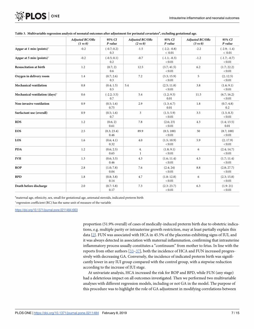
Upon multivariable analysis without GA as a covariate (Table 3), HCA alone was not signif-
icantly associated with any neonatal adverse outcome, except for an increased risk for ROP
(OR = 2.8, 95% CI = 1–7.8, p = 0.04). Conversely, neonates with histological signs of FUN,
independently of the stage, showed worse adaptation at birth, with lower Apgar scores,
increased need for resuscitation/oxygen in the delivery room, and increased ORs for mechani-
cal ventilation, surfactant use, RDS, EOS, LOS, PDA, IVH, ROP, BPD, and death before dis-
charge (all p<0.01). Only the need for non-invasive ventilation was less affected, with not
significant ORs in neonates with FUN stage 2–3.
After the inclusion of GA in the regression model, the results differed remarkably (Table 4).
HCA alone was protective against mechanical ventilation (OR = 0.3, 95% CI = 0.1–0.7,
p<0.01) and against the need for exogenous surfactant (OR = 0.5, 95% CI = 0.2–0.9, p = 0.05);
it was still associated with an increased risk for ROP (OR = 3.3, 95% CI = 1–10.2, p = 0.04).
The effects of FUN were also greatly tempered, being limited to significantly reduced Apgar
score at 1 minute of life (RC = -0.6, 95% CI = -1.2 to -0.1), increased need for resuscitation
(OR = 6.1, 95% CI = 1.4 to 25.3) and oxygen in the delivery room (OR = 3.2, 95% CI = 1 to
9.3), and increased incidence of EOS (OR = 85, 95% CI = 9.4 to 100) for FUN stage 1; reduced
Apgar scores (RC = -1, 95% CI = -1.7 to -0.4 for Apgar at 1 min; RC = -0.5, 95% CI = -0.9 to
-0.1 for Apgar at 5 min), increased duration of mechanical ventilation (RC = 6.5, 95% CI = 2
to 11) and increased incidence of EOS (OR = 37.6, 95% CI = 6.7 to 100) for FUN stage 2–3.
Table 1. Maternal and perinatal characteristics of enrolled neonates.
No IUIGroup 0n = 673
HCAGroup 1n = 73
HCA+Fun 1 Group 2n = 34
HCA+Fun 2–3 Group 3n = 27
P value(1 vs 0)
P value(2 vs 0)
P value(3 vs 0)
Maternal age
(mean ± SD)
35.1 ± 6.1 36.3 ± 5.8 34 ± 5.7 33.4 ± 7.3 0.93 0.59 0.34
Caucasian ethnicity (%) 578/673 (86) 63/73 (86.3) 27/24 (79.4) 14/27 (51.9) 0.93 0.31 <0.01
PROM (%) 208/673 (30.9) 38/73 (52) 21/34 (61.7) 26/27 (96.3) <0.01 <0.01 <0.01
PROM >24 h (%) 75/208 (36.1) 20/38 (52.6) 10/21 (47.6) 18/26 (69.2) 0.07 0.32 <0.01
Indicated preterm birth (%) 389/673 (57.8) 27/73 (37) 2/34 (5.9) 1/27 (3.7) <0.01 <0.01 <0.01
Clinical choriamnionitis (%) 25/671 (3.7) 11/73 (15.1) 16/34 (47.1) 22/27 (81.5) <0.01 <0.01 <0.01
Maternal antibiotics (%) 192/673 (28.5) 39/73 (53.4) 20/34 (58.8) 24/27 (88.9) <0.01 <0.01 <0.01
Prenatal steroids (%) 524/671 (78.1) 63/73 (86.3) 25/34 (73.5) 26/27 (96.3) 0.14 0.57 0.05
Pre-eclampsia (%) 95/673 (14.1) 5/73 (6.8) 1/34 (2.9) 0/27 0.09 0.1 n.a.
Caesarean section (%) 605/673 (90.1) 63/73 (86.3) 24/34 (70.6) 16/27 (59.3) 0.35 <0.01 <0.01
Male (%) 351/673 (52.1) 46/73 (63) 17/34 (50) 13/27 (48.1) 0.09 0.82 0.68
SGA (%) 115/673 (17.1) 17/73 (23.3) 0/34 0/27 0.18 n.a. n.a.
Twin (%) 361/673 (53.6) 30/73 (41.1) 14/34 (41.2) 4/27 (14.8) 0.08 0.21 <0.01
Gestational age
(mean ± SD)
32.3 ± 2.5 31.4 ± 3.1 28.9 ± 3.4 28.3 ± 3.8 <0.01 <0.01 <0.01
Birth weight
(mean ± SD)
1704 ± 518 1492 ± 501 1385 ± 640 1235 ± 562 <0.01 <0.02 <0.01
n.a.: not applicable
https://doi.org/10.1371/journal.pone.0211484.t001
Intrauterine inflammation and neonatal outcomes
PLOS ONE | https://doi.org/10.1371/journal.pone.0211484 February 8, 2019 5 / 15

The correlation between any stage of FUN and all other clinically relevant neonatal outcomes
was completely abolished after inclusion of GA in the regression model.
Discussion
In the present study, we evaluated the impact of different stages of perinatal IUI on short-term
outcomes of neonates less than 35 weeks’ gestation exposed to no IUI, HCA alone, and HCA
plus FUN, before and after adjusting for antenatal and perinatal confounders and with a spe-
cific focus on the adjustment for GA.
We found an overall prevalence of histological IUI of 16.6%, slightly lower than that
reported by other authors [33–36]. Some characteristics of the population studied, such as the
wide GA range, the prevalent Caucasian ethnicity of the mothers, and the considerable
Table 2. Incidence of neonatal outcomes and unadjusted linear (quantitative outcomes) or logistic (dichotomous outcomes) regression analysis.
No IUIGroup 0n = 673
HCAGroup 1n = 73
HCA+Fun 1Group 2n = 34
HCA+Fun 2–3Group 3n = 27
RC/ORs and CI(1 vs 0)
RC/ORs and CI(2 vs 0)
RC/ORs and CI(3 vs 0)
Apgar at 1 min
(median, range)
8, 0–10 8, 0–9 5,5, 1–9 5, 1–9 -0.3
-0.8; 0.1
-1.5
-2.1; -0.8
-2.5
-3.2; -1.7
Apgar at 5 min
(median, range)
9, 2–10 9, 3–10 8, 5–10 8, 3–10 -0.2
-0.6; 0.1
-0.8
-1.2; -0.3
-1.4
-1.8; -0.9
Resuscitation at birth (%) 320/673
(47.5)
40/73
(54.8)
31/34
(91.2)
24/27
(88.9)
1.3
0.8; 2.2
11.4
3.4; 38.3
8.8
2.6; 29.6
Oxygen in delivery room (%) 175/672
(26)
25/73
(34.2)
24/34
(70.5)
19/27
(70.4)
1.5
0.8; 2.6
6.8
3.1; 14.5
6.7
2.9; 15.7
Mechanical ventilation (%) 128/673
(19)
13/73
(17.8)
19/34
(55.8)
14/27
(51.8)
0.9
0.4; 1.8
5.4
2.6; 11.1
4.6
2.1; 10.1
Mechanical ventilation (days) 2.1 3.9 8.5 15.6 1
-1.8; 3.8
5.4
1.4; 9.5
12.2
7.6; 16.7
Non-invasive ventilation (%) 364/673
(54.1)
39/73
(53.4)
26/34
(76.5)
20/27
(74.1)
0.9
0.6; 1.6
2.8
1.2; 6.1
2.4
1; 5.8
Non-invasive ventilation (days) 10.8 16.3 30 26.7 2.6
-3.9; 9
11.9
2.6; 21.3
13.6
2.9; 24.2
Surfactant use (overall) 204/673
(30.3)
21/73
(28.8)
19/34
(55.9)
17/27
(62.9)
0.9
0.5; 1.7
2.9
1.5; 5.7
3.9
1.7; 8.7
Surfactant use (>1 dose) 67/673
(9.9)
9/73
(12.3)
10/34
(29.4)
11/27
(40.7)
1.2
0.5; 2.9
4.6
2; 10.7
7.7
3.1; 18.9
RDS
(%)
341/673
(50.7)
41/73
(56.2)
30/34
(88.2)
23/27
(85.2)
1.2
0.7; 2.1
7.3
2.5; 21.3
5.6
1.9; 16.4
EOS
(%)
3/673
(0.45)
1/73
(1.37)
4/34
(11.8)
2/27
(7.41)
3.1
0.3; 30.3
29.8
6.3; 100
17.8
2.8; 100
LOS
(%)�43/672
(6.4)
8/72
(11.1)
6/33
(18.2)
8/27
(29.6)
1.8
0.8; 4.4
3.3
1.3; 8.2
6.2
2.5; 15
PDA
(%)
72/673
(10.7)
10/73
(13.7)
11/34
(32.4)
12/27
(44.4)
1.3
0.6; 2.7
4
1.9; 8.4
6.7
2.9; 15
IVH
(%)
40/673
(5.9)
7/73
(9.59)
9/34
(26.5)
9/27
(33.3)
1.7
0.6; 4.2
5.7
2.3; 13.7
7.9
3.3; 18.9
ROP
(%)�21/661
(3.2)
7/70
(10)
6/30
(20)
7/25
(28)
3.4
1.2; 9.1
7.6
2.5; 23.2
11.8
4.4; 31.8
BPD
(%)�60/656
(9.2)
13/70
(18.6)
9/28
(32.1)
10/23
(43.5)
2.3
1.1; 4.7
4.7
1.9; 11.8
7.6
3.2; 18.4
Death before discharge (%) 19/673
(2.8)
5/73
(6.9)
6/34
(17.6)
5/27
(18.5)
2.5
0.9; 6.9
7.4
2.7; 19.9
7.8
2.6; 23.1
�percentage is calculated on survived neonates
https://doi.org/10.1371/journal.pone.0211484.t002
Intrauterine inflammation and neonatal outcomes
PLOS ONE | https://doi.org/10.1371/journal.pone.0211484 February 8, 2019 6 / 15
proportion (51.9% overall) of cases of medically-induced preterm birth due to obstetric indica-
tions, e.g. multiple parity or intrauterine growth restriction, may at least partially explain this
data [2]. FUN was associated with HCA in 45.5% of the placentas exhibiting signs of IUI, and
it was always detected in association with maternal inflammation, confirming that intrauterine
inflammatory process usually constitutes a “continuum” from mother to fetus. In line with the
reports from other authors [33–37], both the incidence of HCA and FUN increased progres-
sively with decreasing GA. Conversely, the incidence of indicated preterm birth was signifi-
cantly lower in any IUI group compared with the control group, with a stepwise reduction
according to the increase of IUI stage.
At univariate analysis, HCA increased the risk for ROP and BPD, while FUN (any stage)
had a deleterious impact on all outcomes investigated. Then we performed two multivariable
analyses with different regression models, including or not GA in the model. The purpose of
this procedure was to highlight the role of GA adjustment in modifying correlations between
Table 3. Multivariable regression analysis of neonatal outcomes after adjustment for perinatal covariatesa, excluding gestational age.
Adjusted RC/ORs(1 vs 0)
95% CIP value
Adjusted RC/ORs(2 vs 0)
95% CIP value
Adjusted RC/ORs(3 vs 0)
95% CIP value
Apgar at 1 min (points)� -0.2 (-0.7; 0.2)
0.3
-1.5 (-2.2; -0.8)
< 0.01
-2.2 (-2.9; -1.4)
< 0.01
Apgar at 5 min (points)� -0.2 (-0.5; 0.1)
0.2
-0.7 (-1.1; -0.3)
<0.01
-1.2 (-1.7; -0.7)
<0.01
Resuscitation at birth 1.2 (0.7; 2)
0.6
12.5 (3.7; 41.5)
<0.01
6.2 (1.7; 22.2)
<0.01
Oxygen in delivery room 1.4 (0.7; 2.6)
0.3
7.2 (3.3; 15.9)
<0.01
5 (2; 12.5)
<0.01
Mechanical ventilation 0.8 (0.4; 1.5)
0.5
5.4 (2.5; 11.8)
<0.01
3.8 (1.6; 9.1)
<0.01
Mechanical ventilation (days)� 0.6 (-2.2; 3.5)
0.7
5.4 (1.2; 9.5)
0.01
11.5 (6.7; 16.2)
<0.01
Non-invasive ventilation 0.9 (0.5; 1.6)
0.75
2.9 (1.3; 6.7)
0.01
1.8 (0.7; 4.8)
0.2
Surfactant use (overall) 0.9 (0.5; 1.6)
0.7
3 (1.5; 5.9)
<0.01
3.5 (1.5; 8.3)
<0.01
RDS 1.2 (0.6; 2)
0.61
7.8 (2.6; 23)
<0.01
4.3 (1.4; 13.5)
0.01
EOS 2.5 (0.3; 23.4)
0.46
89.9 (8.5; 100)
<0.01
50 (8.7; 100)
<0.01
LOS 1.6 (0.6; 4.1)
0.32
4.0 (1.5; 10.9)
<0.01
5.9 (2; 17.9)
<0.01
PDA 1.2 (0.6; 2.5)
0.65
4.
1
(1.8; 9.1)
<0.01
6 (2.4; 14.7)
<0.01
IVH 1.3 (0.6; 3.5)
0.46
4.3 (1.6; 11.4)
<0.01
4.3 (1.7; 11.4)
<0.01
ROP 2.8 (1.0; 7.8)
0.04
7.6 (2.4; 24)
<0.01
8.8 (2.8; 27.7)
<0.01
BPD 1.8 (0.8; 3.8)
0.14
4.7 (1.8: 12.8)
<0.01
6 (2.3; 15.8)
<0.01
Death before discharge 2.0 (0.7: 5.8)
0.17
7.3 (2.3: 23.7)
<0.01
6.3 (1.9: 21)
<0.01
amaternal age, ethnicity, sex, small for gestational age, antenatal steroids, indicated preterm birth
�regression coefficient (RC) has the same unit of measure of the variable
https://doi.org/10.1371/journal.pone.0211484.t003
Intrauterine inflammation and neonatal outcomes
PLOS ONE | https://doi.org/10.1371/journal.pone.0211484 February 8, 2019 7 / 15

risk factors, namely perinatal IUI, and neonatal outcomes, and to speculate on the appropriate-
ness of adjusting for GA itself. The other covariates (maternal age, ethnicity, antenatal steroids,
sex, SGA neonate, and indicated preterm birth), were selected a priori as they may affect, either
positively or negatively, neonatal outcome. Without GA as a covariate, but still adjusting for
the other confounders, results were not greatly affected compared to unadjusted analysis, espe-
cially for what concerns the effect of FUN. In fact, HCA alone was confirmed as a risk factor
for ROP, but not anymore for BPD, while neonates born with both FUN stage 1 and stage 2–3
still had increased ORs for almost all the evaluated outcomes.
After inclusion of GA in the regression model, results differed dramatically. HCA alone was
protective against the need for mechanical ventilation and surfactant, but it was confirmed as a
risk factor for ROP. The reduced need for mechanical ventilation and surfactant, in the
absence of a reduced OR for RDS, may support the hypothesis that a mild, just maternal peri-
natal inflammation would be protective against postnatal inflammatory stimuli inducing lung
Table 4. Multivariable regression analysis of neonatal outcomes after adjustment for perinatal covariatesa, including gestational age.
Adjusted RC/ORs(1 vs 0)
95% CIP value
Adjusted RC/ORs(2 vs 0)
95% CIP value
Adjusted RC/ORs(3 vs 0)
95% CIP value
Apgar at 1 min (points)� 0 (-0.4; 0.4)
0.99
-0.6 (-1.2; -0.1)
0.05
-1 (-1.7; -0.4)
<0.01
Apgar at 5 min (points)� -0.1 (-0.3; 0.2)
0.6
-0.1 (-0.5; 0.2)
0.5
-0.5 (-0.9; -0.1)
0.03
Resuscitation at birth 0.8 (0.4; 1.6)
0.6
6.1 (1.4; 25.3)
0.01
2.5 (0.7; 8.5)
0.1
Oxygen in
delivery room
1 (0.5; 2.1)
0.88
3.2 (1; 9.3)
0.03
2.2 (0.8; 5.6)
0.11
Mechanical ventilation 0.3 (0.1; 0.7)
<0.01
1.9 (0.6; 6.2)
0.3
0.9 (0.2; 4.5)
0.94
Mechanical ventilation (days)� -0.4 (-3.1; 2.4)
0.8
0.9 (-2.3; 4.5)
0.6
6.5 (2; 11)
<0.01
Non-invasive ventilation 0.7 (0.4; 1.2)
0.2
1.1 (0.3; 3.9)
0.93
0.6 (0.2; 2.1)
0.46
Surfactant use (overall) 0.5 (0.2; 0.9)
0.05
0.6 (0.2;2.3)
0.59
1.2 (0.4; 3.5)
0.71
RDS 0.7 (0.4; 1.4)
0.4
2.8 (0.6; 12.5)
0.17
1.4 (0.4; 4.9)
0.59
EOS 2.4 (0.3; 18.7)
0.4
85 (9.4; 100)
<0.01
37.6 (6.7; 100)
<0.01
LOS 1.2 (0.3; 3.8)
0.8
0.9 (0.3; 2.7)
0.86
1.2 (0.2; 6.1)
0.83
PDA 0.6 (0.2; 1.6)
0.3
0.9 (0.3; 2.4)
0.83
1.8 (0.4; 8.3)
0.44
IVH 0.8 (0.3; 2.1)
0.65
1 (0.3; 3.6)
0.9
0.9 (0.2; 3.5)
0.9
ROP 3.3 (1; 10.2)
0.04
2 (0.4; 12.6)
0.4
1.7 (0.3; 11)
0.59
BPD 1.5 (0.6; 3.6)
0.42
1.2 (0.3; 4.5)
0.79
2.2 (0.2; 27.1)
0.57
Death before discharge 1.3 (0.4: 4.8)
0.65
2 (0.6: 7.1)
0.3
1.3 (0.4: 4.7)
0.4
aGA, maternal age, ethnicity, sex, small for gestational age, antenatal steroids, indicated preterm birth
�regression coefficient (RC) has the same unit of measure of the variable
https://doi.org/10.1371/journal.pone.0211484.t004
Intrauterine inflammation and neonatal outcomes
PLOS ONE | https://doi.org/10.1371/journal.pone.0211484 February 8, 2019 8 / 15

damage and subsequent need for ventilation, as highlighted by some authors [38,39]. Alterna-
tively, considering that the risk of non-invasive ventilation was not affected by HCA, it could
be hypothesized that IUI, accelerating lung development as demonstrated in several studies on
animal models [40,41], causes less severe RDS requiring only non-invasive ventilation. As for
the increased incidence of ROP, unexpectedly not confirmed in neonates with FUN, it has
been suggested that perinatal inflammation may be involved in the pathogenesis of ROP [42].
However, a recent meta-analysis by Mitra and coll. [26] concluded that chorioamnionitis was
significantly associated with ROP (any stage) as well as with severe ROP (stage� 3) upon
unadjusted analyses, but such association disappeared on subanalysis of the studies adjusting
for GA. Certainly, the association between IUI and ROP is difficult to demonstrate due to
many possible confounding factors, such as extreme prematurity and oxygen therapy [43].
Considering GA as a covariate in the logistic regression model, the relationship between
FUN and neonatal outcome also changed considerably. FUN remained an independent risk
factor for lower Apgar scores, an increased need for resuscitation at birth and oxygen in the
delivery room (FUN stage 1), an increased number of days of mechanical ventilation (FUN
stage 2–3), and for EOS (FUN any stage). In 2005, Lau and coll. [35], in a study conducted in a
large cohort of neonates with a mean GA of 32–33 weeks admitted to the NICU, reported after
adjusting for confounding factors an increased mortality and morbidity, including RDS, BPD,
EOS, LOS, necrotizing enterocolitis (NEC), PDA, and IVH when chorioamnionitis was associ-
ated with a fetal inflammatory response. Conversely, most of the studies conducted in more
recent years [36,37], including this one, failed to confirm or only partially confirmed these
findings. In a retrospective study by Lee and coll. [37], conducted to evaluate in a relatively
large group of neonates less than 34 weeks’ gestation if there was a stepwise increase in neona-
tal morbidities according to the stage or grade of acute HCA and FUN, the incremental trends
of each neonatal outcome were found to be not significant after adjusting for confounding var-
iables including GA at birth. In a subsequent study [36] on the relationship between all stages
and grades of HCA and FUN and neonatal mortality and morbidity, only a high grade of fetal
inflammation was significantly associated with BPD and NEC after adjusting for GA. Cer-
tainly, the increasingly widespread use of antenatal steroid may explain why some recent stud-
ies have failed to demonstrate an association between IUI and neonatal adverse outcomes. A
meta-analysis published in 2011 [44] highlighted an association between antenatal corticoste-
roids administration and reduced incidence of RDS, IVH, and PDA, especially in the absence
of maternal clinical chorioamnionitis. As a matter of fact, in the study by Lau and coll. prenatal
steroids were administered in a percentage ranging from 40 to 60%, while in the most recent
studies, including ours, they were administered in more than 80% of neonates.
As expected, we found a strong association between any stage of FUN and EOS even after
including GA in the regression model. Contrary with what is seen for RDS, IVH, PDA and,
albeit to a lesser extent, for BPD, antenatal steroids do not seem to have a protective effect on
EOS [44]. Many studies have linked HCA, FUN, and even more clinical chorioamnionitis with
EOS [21,35,45,46], although other studies have not confirmed this association [36,37], proba-
bly because maternal antibiotic therapy can affect culture results or the causative pathogen
may be a mycoplasma, not identifiable with traditional culture methods.
Furthermore, in our study the presence of FUN stage 1 was strongly associated with an
increased need for resuscitation and oxygen in the delivery room, independently of GA. This
finding, not confirmed for FUN stage 2–3 possibly because of the small sample size, is not
emphasized by many authors [35], and suggests that when the umbilical cord vessels are
involved in inflammation, the preterm baby is very ill at birth and this can seriously worsen its
outcome.
Intrauterine inflammation and neonatal outcomes
PLOS ONE | https://doi.org/10.1371/journal.pone.0211484 February 8, 2019 9 / 15

Lastly, the increased length of mechanical ventilation we found in the neonates with FUN
stage 2–3, in the absence of higher odds for RDS and BPD, is not easy to interpret. We can
hypothesize that the more severe clinical conditions at birth in neonates with FUN may cause
a more severe acute lung disease, requiring prolonged mechanical ventilation before switching
to a non-invasive ventilation. Moreover, the role of IUI in the development of neonatal pulmo-
nary disease is far from being clarified. Despite evidence from animal models suggest a
reduced incidence of RDS and an increased incidence of BPD in neonates exposed to IUI [47–
49], the results of the numerous clinical studies on this topic are often contradictory. In partic-
ular, according to some authors, the presence of FUN in association with HCA appears to
have a protective effect on the development of RDS [50–52] or BPD [1,53], findings not con-
firmed by other studies [35,36,54–56]. Regarding the development of BPD, the various insults
(mechanical ventilation, oxygen, infection) that can damage the lung after birth must be con-
sidered. Some authors agree that the fetal lung, exposed to a prolonged pro-inflammatory
stimulus in uterus, responds to postnatal inflammatory stimuli in a more pathologic way com-
pared to a naïve lung [57]. On the other hand, studies on animal models have shown that a
first exposure to an inflammatory stimulus can increase or decrease the response to a second
injurious stimulus exposure depending on the time interval between exposures themselves
[58,59].
The dichotomy between results obtained before and after adjustment for GA reflects the
debate currently ongoing on the so called “independent role” of perinatal inflammation in
shaping subsequent neonatal health [60]. As reported by Wilcox and coll. [61], GA is a conse-
quence of any underlying cause of preterm birth, like IUI, and should be more properly con-
sidered as an “intermediate” variable rather than an “independent” variable. The adjustment
for intermediate variables may in turn be misleading and favor a biased effect of other uncon-
trolled variables, rarely missing, that collide on the outcome. This bias can be so impactful that
the relationship between IUI and neonatal adverse outcome may be even reversed after adjust-
ment for GA. This issue has been also explored by other authors, like Dessardo and coll. [62],
who tested a statistical approach incorporating path analysis to evaluate direct and indirect
causality between exposure to HCA or FUN and the development of BPD. From this perspec-
tive, the inclusion of GA as a covariate, although recalling a common practice, may be strongly
misleading.
Furthermore, the purpose of adjusting for GA is ideally to compare similar neonates with
and without IUI, considering those without IUI as the control group. This assumption raises
another subtle issue, because the components of this control group, when preterm neonates
are involved, are rarely “control” subjects: rather they hide other pathological causes of pre-
term birth, first of all intrauterine growth restriction [2]. Thus, the effect of IUI on any neona-
tal outcome is not referred to healthy neonates, but to neonates born because of other causes
of prematurity. The issue has been recently explored by Torchin and coll. [56] and Gagliardi
and coll. [63], who demonstrated in large cohorts of preterm neonates that the apparent pro-
tective effect of IUI against the risk of developing BPD was due to the high rate, in the non-IUI
group, of neonates affected by fetal growth restriction, a known predisposing condition for
BPD according to the “vascular hypothesis”. Thus, the lack of a healthy control group in most
studies may alter the real impact of IUI on neonatal outcomes. Interestingly, this issue seems
to be neglected also in experimental models of IUI, where the effect of IUI itself is frequently
investigated against a healthy, untreated control group, instead of a more appropriate group of
animals with experimentally-induced fetal growth restriction.
The strengths of our study are rigorous definitions and staging of IUI, and prospective, sin-
gle-center collection of data that reduces the variability sometimes affecting multicenter
Intrauterine inflammation and neonatal outcomes
PLOS ONE | https://doi.org/10.1371/journal.pone.0211484 February 8, 2019 10 / 15

studies. We recognize some weaknesses of our work, such as the relatively small sample size,
the wide GA range, and the relatively low incidence of IUI in our population.
In conclusion, our study shows that the influence of IUI on neonatal outcomes, without
including GA among the covariates in multivariable regression analyses, is limited to an
increased incidence of ROP, while FUN has a strong negative impact on most neonatal mor-
bidities. With the inclusion of GA in regression models, results differ radically, with HCA
reducing the need for mechanical ventilation and surfactant and FUN being a clear risk factor
only for EOS and for worse clinical conditions in the delivery room. Considered that GA is an
intermediate variable interposed between prenatal conditions favoring preterm delivery and
neonatal outcomes, it is at least questionable whether the adjustment for GA, a common and
accepted practice when investigating the correlation between IUI and neonatal outcomes, is
methodologically correct. In order to better understand the independent role of IUI in shaping
neonatal outcomes, we believe that further clinical studies and experimental models should
not focus on GA adjustment or on the comparison between neonates affected or not by IUI
but, rather, on a direct comparison between neonates affected by IUI and homogeneous
groups of preterm neonates carrying other antecedents of preterm birth, such as placental vas-
cular disorders, in order to have a more realistic picture of the different causes of prematurity
and their really “independent” consequences.
Supporting information
S1 File. Spreadsheet containing all data included in the manuscript. Enlisted patients are
completely anonymized.
(XLS)
Acknowledgments
The authors would like to thank all nurses of the Neonatal Intensive Care Unit and midwives
of the Gynecology and Obstetrics Unit of Fondazione IRCCS Ca’ Granda Ospedale Maggiore
Policlinico of Milan for their kind collaboration.
Author Contributions
Conceptualization: Carlo Pietrasanta, Lorenza Pugni, Barbara Acaia, Manuela Wally Ossola,
Silvano Bosari, Fabio Mosca.
Data curation: Carlo Pietrasanta, Lorenza Pugni.
Formal analysis: Carlo Pietrasanta, Lorenza Pugni, Daniela Merlo, Dario Consonni, Fulvia
Milena Cribiu.
Investigation: Carlo Pietrasanta, Daniela Merlo, Andrea Ronchi, Beatrice Ghirardi, Ilaria Bot-
tino, Fulvia Milena Cribiu.
Methodology: Carlo Pietrasanta, Lorenza Pugni, Daniela Merlo, Dario Consonni.
Project administration: Carlo Pietrasanta, Lorenza Pugni, Silvano Bosari, Fabio Mosca.
Resources: Lorenza Pugni, Silvano Bosari, Fabio Mosca.
Supervision: Carlo Pietrasanta, Lorenza Pugni, Silvano Bosari, Fabio Mosca.
Visualization: Carlo Pietrasanta, Lorenza Pugni, Dario Consonni.
Writing – original draft: Carlo Pietrasanta, Lorenza Pugni.
Intrauterine inflammation and neonatal outcomes
PLOS ONE | https://doi.org/10.1371/journal.pone.0211484 February 8, 2019 11 / 15

Writing – review & editing: Carlo Pietrasanta, Lorenza Pugni, Daniela Merlo, Barbara Acaia,
Dario Consonni, Andrea Ronchi, Manuela Wally Ossola, Beatrice Ghirardi, Ilaria Bottino,
Fulvia Milena Cribiu, Silvano Bosari, Fabio Mosca.
References1. Lahra MM, Beeby PJ, Jeffery HE. Intrauterine inflammation, neonatal sepsis, and chronic lung disease:
A 13-year hospital cohort study. Pediatrics. 2009; 123:1314–9. https://doi.org/10.1542/peds.2008-0656
PMID: 19403497
2. Goldenberg RL, Culhane JF, Iams JD, Romero R. Epidemiology and causes of preterm birth. Lancet.
2008; 371:75–84. https://doi.org/10.1016/S0140-6736(08)60074-4 PMID: 18177778
3. Goldenberg RL, Hauth JC, Andrews WW. Intrauterine infection and preterm delivery. N Engl J Med.
2000; 342:1500–7. https://doi.org/10.1056/NEJM200005183422007 PMID: 10816189
4. Kim CJ, Romero R, Chaemsaithong P, Chaiyasit N, Yoon BH, Kim YM. Acute chorioamnionitis and funi-
sitis: definition, pathologic features, and clinical significance. Am J Obstet Gynecol. 2015; 213:S29–52.
https://doi.org/10.1016/j.ajog.2015.08.040 PMID: 26428501
5. Salafia CM, Weigl C, Silberman L. The prevalence and distribution of acute placental inflammation in
uncomplicated term pregnancies. Obstetrics and Gynecology. 1989; 73:383–9. PMID: 2915862
6. Redline RW. Inflammatory responses in the placenta and umbilical cord. Semin Fetal Neonatal Med.
2006; 11:296–301. https://doi.org/10.1016/j.siny.2006.02.011 PMID: 16621749
7. Redline RW, Faye-Petersen O, Heller D, Qureshi F, Savell V, Vogler C. Amniotic infection syndrome:
nosology and reproducibility of placental reaction patterns. Pediatr Dev Pathol. 2003; 6:435–48. https://
doi.org/10.1007/s10024-003-7070-y PMID: 14708737
8. Andrews WW, Goldenberg RL, Faye-Petersen O, Cliver S, Goepfert AR, Hauth JC. The Alabama Pre-
term Birth study: polymorphonuclear and mononuclear cell placental infiltrations, other markers of
inflammation, and outcomes in 23- to 32-week preterm newborn infants. Am J Obstet Gynecol. 2006;
195:803–8. https://doi.org/10.1016/j.ajog.2006.06.083 PMID: 16949415
9. Popowski T, Goffinet F, Maillard F, Schmitz T, Leroy S, Kayem G. Maternal markers for detecting early-
onset neonatal infection and chorioamnionitis in cases of premature rupture of membranes at or after
34 weeks of gestation: a two-center prospective study. BMC Pregnancy Childbirth. 2011; 11:929.
10. Sung JH, Choi SJ, Oh SY, Roh CR, Kim JH. Revisiting the diagnostic criteria of clinical chorioamnionitis
in preterm birth. BJOG. 2017; 124:775–783. https://doi.org/10.1111/1471-0528.14176 PMID:
27365145
11. Chaemsaithong P, Romero R, Korzeniewski SJ, Martinez-Varea A, Dong Z, Yoon BH, et al. A point of
care test for interleukin-6 in amniotic fluid in preterm prelabor rupture of membranes: a step toward the
early treatment of acute intra-amniotic inflammation/infection. J Matern Fetal Neonatal Med. 2016;
29:360–7. https://doi.org/10.3109/14767058.2015.1006621 PMID: 25758620
12. Tita ATN, Andrews WW. Diagnosis and management of clinical chorioamnionitis. Clin Perinatol. 2010;
37:339–54. https://doi.org/10.1016/j.clp.2010.02.003 PMID: 20569811
13. Romero R, Miranda J, Chaiworapongsa T, Korzeniewski SJ, Chaemsaithong P, Gotsch F, et al. Preva-
lence and clinical significance of sterile intra-amniotic inflammation in patients with preterm labor and
intact membranes. Am J Reprod Immunol. 2014; 72:458–74. https://doi.org/10.1111/aji.12296 PMID:
25078709
14. Park CW, Moon KC, Park JS, Jun JK, Romero R, Yoon BH. The involvement of human amnion in histo-
logic chorioamnionitis is an indicator that a fetal and an intra-amniotic inflammatory response is more
likely and severe: clinical implications. Placenta. 2009; 30:56–61. https://doi.org/10.1016/j.placenta.
2008.09.017 PMID: 19046766
15. Doty MS, Salafia C, Shen-Schwarz S, Guzman E, Saade G, Chauhan S. Histologic Funisitis and Likeli-
hood of Intrauterine Inflammation or Infection: A Case-Control Study. Am J Perinatol. 2018; 35:858–
864. https://doi.org/10.1055/s-0037-1620232 PMID: 29365327
16. Peng CC, Chang JH, Lin HY, Cheng PJ, Su BH. Intrauterine inflammation, infection, or both (Triple I): A
new concept for chorioamnionitis. Pediatr Neonatol. 2017; 59: 231–237. https://doi.org/10.1016/j.
pedneo.2017.09.001 PMID: 29066072
17. Pugni L, Pietrasanta C, Acaia B, Merlo D, Ronchi A, Ossola MW, et al. Chorioamnionitis and neonatal
outcome in preterm infants: a clinical overview. J Matern Fetal Neonatal. Med 2016; 29:1525–9. https://
doi.org/10.3109/14767058.2015.1053862 PMID: 26135227
18. Thomas W, Speer CP. Chorioamnionitis is essential in the evolution of bronchopulmonary dysplasia–
The case in favour. Paediatr Respir Rev. 2014; 15:49–52. https://doi.org/10.1016/j.prrv.2013.09.004
PMID: 24128984
Intrauterine inflammation and neonatal outcomes
PLOS ONE | https://doi.org/10.1371/journal.pone.0211484 February 8, 2019 12 / 15
19. Lacaze-Masmonteil T. That chorioamnionitis is a risk factor for bronchopulmonary dysplasia–the case
against. Paediatr Respir Rev 2014; 15:53–5. https://doi.org/10.1016/j.prrv.2013.09.005 PMID:
24120077
20. Ylijoki M, Ekholm E, Haataja L, Lehtonen L; Pipari study group. Is chorioamnionitis harmful for the brain
of preterm infants? A clinical overview. Act Obstet Gynecol Scand. 2012; 91:403–19.
21. Pappas A, Kendrick DE, Shankaran S, Stoll BJ, Bell EF, Laptook AR, et al. Chorioamnionitis and early
childhood outcomes among extremely low-gestational-age neonates. JAMA pediatr. 2014; 168:137–
11. https://doi.org/10.1001/jamapediatrics.2013.4248 PMID: 24378638
22. Korzeniewski SJ, Romero R, Cortez J, Pappas A, Schwartz AG, Kim CJ, et al. A “multi-hit” model of
neonatal white matter injury: cumulative contributions of chronic placental inflammation, acute fetal
inflammation and postnatal inflammatory events. J Perinat Med. 2014; 42:1–2. https://doi.org/10.1515/
jpm-2013-0200
23. Shi Z, Ma L, Luo K, Bajaj M, Chawla S, Natarajan G, et al. Chorioamnionitis in the development of cere-
bral palsy: a meta-analysis and systematic review. Pediatrics. 2017; 139:e20163781–17. https://doi.
org/10.1542/peds.2016-3781 PMID: 28814548
24. Ogunyemi D, Murillo M, Jackson U, Hunter N, Alperson B. The relationship between placental histopa-
thology findings and perinatal outcome in preterm infants. J Matern Fetal Neonatal Med. 2009; 13:102–
9.
25. Strunk T, Doherty D, Jacques A, Simmer K, Richmond P, Kohan R, et al. Histologic chorioamnionitis is
associated with reduced risk of late-onset sepsis in preterm infants. Pediatrics. 2012; 129:e134–41.
https://doi.org/10.1542/peds.2010-3493 PMID: 22157134
26. Mitra S, Aune D, Speer CP, Saugstad OD. Chorioamnionitis as a risk factor for retinopathy of prematu-
rity: a systematic review and meta-analysis. Neonatology. 2014; 105:189–99. https://doi.org/10.1159/
000357556 PMID: 24481268
27. Park HW, Choi YS, Kim KS, Kim SN. Chorioamnionitis and patent ductus arteriosus: a systematic
review and meta-analysis. PloS One. 2015; 10:e0138114. https://doi.org/10.1371/journal.pone.
0138114 PMID: 26375582
28. Fenton TR, Kim JH. A systematic review and meta-analysis to revise the Fenton growth chart for pre-
term infants. BMC pediatrics. 2013; 13:59. https://doi.org/10.1186/1471-2431-13-59 PMID: 23601190
29. Horbar JD, Rogowski J, Plsek PE, Delmore P, Edwards WH, Hocker J, et al. Collaborative quality
improvement for neonatal intensive care. NIC/Q Project Investigators of the Vermont Oxford Network.
Pediatrics. 2001; 107:14–22. PMID: 11134428
30. Jobe AH, Bancalari E. Bronchopulmonary dysplasia. Am J Respir Crit Care Med. 2001; 163:1723–9.
https://doi.org/10.1164/ajrccm.163.7.2011060 PMID: 11401896
31. Volpe JJ. Neurology of the newborn. 5th ed. Philadelphia: Saunders. 2008.
32. International Committee for the Classification of Retinopathy of Prematurity. The International Classifi-
cation of Retinopathy of Prematurity revisited. Arch Ophthalmol. 2005; 123:991–9. https://doi.org/10.
1001/archopht.123.7.991 PMID: 16009843
33. Edwards RK. Chorioamnionitis and labor. Obstet Gynecol Clin North Am. 2005; 32:287–96. https://doi.
org/10.1016/j.ogc.2004.12.002 PMID: 15899361
34. Lahra MM, Jeffery HE. A fetal response to chorioamnionitis is associated with early survival after pre-
term birth. Am J Obstet Gynecol. 2004; 190:147–51. https://doi.org/10.1016/j.ajog.2003.07.012 PMID:
14749651
35. Lau J, Magee F, Qiu Z, Hoube J, Dadelszen Von P, et al. Chorioamnionitis with a fetal inflammatory
response is associated with higher neonatal mortality, morbidity, and resource use than chorioamnioni-
tis displaying a maternal inflammatory response only. Am J Obstet Gynecol. 2005; 193:708–13. https://
doi.org/10.1016/j.ajog.2005.01.017 PMID: 16150264
36. Yamada N, Sato Y, Moriguchi-Goto S, Yamashita A, Kodama Y, Sameshima H, et al. Histological
severity of fetal inflammation is useful in predicting neonatal outcome. Placenta. 2015; 36:1490–3.
https://doi.org/10.1016/j.placenta.2015.10.021 PMID: 26565600
37. Lee Y, Kim HJ, Choi SJ, Oh SY, Kim JS, Roh CR, et al. Is there a stepwise increase in neonatal morbidi-
ties according to histological stage (or grade) of acute chorioamnionitis and funisitis?: effect of gesta-
tional age at delivery. J Perinat Med. 2015; 43:259–67. https://doi.org/10.1515/jpm-2014-0035 PMID:
25153209
38. Backstrom E, Lappalainen U, Bry K. Maternal IL-1beta production prevents lung injury in a mouse
model of bronchopulmonary dysplasia. Am J Respir Cell Mol Biol. 2010; 42:149–60. https://doi.org/10.
1165/rcmb.2008-0287OC PMID: 19411613
Intrauterine inflammation and neonatal outcomes
PLOS ONE | https://doi.org/10.1371/journal.pone.0211484 February 8, 2019 13 / 15

39. Choi CW, Lee J, Oh JY, Lee SH, Lee HJ, Kim BI. Protective effect of chorioamnionitis on the develop-
ment of bronchopulmonary dysplasia triggered by postnatal systemic inflammation in neonatal rats.
Pediatr Res. 2016; 79:287–94. https://doi.org/10.1038/pr.2015.224 PMID: 26551413
40. Kramer BW. Antenatal inflammation and lung injury: prenatal origin of neonatal disease. J Perinatol.
2008; 28:S21–7. https://doi.org/10.1038/jp.2008.46 PMID: 18446173
41. Jobe AH. Antenatal associations with lung maturation and infection. J Perinatol. 2005; 25:S31–5.
https://doi.org/10.1038/sj.jp.7211317 PMID: 15861169
42. Sood BG, Madan A, Saha S, Schendel D, Thorsen P, Skogstrand K, et al. Perinatal systemic inflamma-
tory response syndrome and retinopathy of prematurity. Pediatr Res. 2010; 67:394–400. https://doi.org/
10.1203/PDR.0b013e3181d01a36 PMID: 20032809
43. Hartnett ME. Pathophysiology and mechanisms of severe retinopathy of prematurity. Ophthalmology.
2015; 122:200–10. https://doi.org/10.1016/j.ophtha.2014.07.050 PMID: 25444347
44. Been J, Degraeuwe P, Kramer B, Zimmermann L. Antenatal steroids and neonatal outcome after chor-
ioamnionitis: a meta-analysis. BJOG. 2011; 118:113–122. https://doi.org/10.1111/j.1471-0528.2010.
02751.x PMID: 21054759
45. Soraisham AS, Singhal N, McMillan DD, Sauve RS, Lee SK, Network CN. A multicenter study on the
clinical outcome of chorioamnionitis in preterm infants. Am J Obstet Gynecol. 2009; 200:372.e1–6.
46. Robert Lee SY, Leung CW. Histological chorioamnionitis–implication for bacterial colonization, labora-
tory markers of infection, and early onset sepsis in very-low-birth-weight neonates. J Matern Fetal Neo-
natal Med. 2011; 25:364–8. https://doi.org/10.3109/14767058.2011.579208 PMID: 21609204
47. Bry K, Lappalainen U, Hallman M. Intraamniotic interleukin-1 accelerates surfactant protein synthesis in
fetal rabbits and improves lung stability after premature birth. J Clin Invest. 1997; 99:2992–9. https://doi.
org/10.1172/JCI119494 PMID: 9185523
48. Kramer BW, Kallapur S, Newnham J, Jobe AH. Prenatal inflammation and lung development. Semin
Fetal Neonatal Med. 2009; 14:2–7. https://doi.org/10.1016/j.siny.2008.08.011 PMID: 18845493
49. Moss TJM, Westover AJ. Inflammation-induced preterm lung maturation: lessons from animal experi-
mentation. Paediatr Respir Rev. 2017; 23:72–7. https://doi.org/10.1016/j.prrv.2016.10.004 PMID:
27856214
50. Been JV, Rours IG, Kornelisse RF, Passos VL, Kramer BW, Schneider TA, et al. Histologic chorioam-
nionitis, fetal involvement, and antenatal steroids: effects on neonatal outcome in preterm infants. Am J
Obstet Gynecol. 2009; 201:587.e1–8.
51. Lahra MM, Beeby PJ, Jeffery HE. Maternal versus fetal inflammation and respiratory distress syn-
drome: a 10-year hospital cohort study. Arch Dis Child Fetal Neonatal Ed. 2009; 94:F13–6. https://doi.
org/10.1136/adc.2007.135889 PMID: 18463119
52. Lee J, Oh KJ, Park CW, Park JS, Jun JK, Yoon BH. The presence of funisitis is associated with a
decreased risk for the development of neonatal respiratory distress syndrome. Placenta. 2011; 32:235–
40. https://doi.org/10.1016/j.placenta.2010.11.006 PMID: 21216461
53. Plakkal N, Soraisham AS, Trevenen C, Freiheit EA, Sauve R. Histological chorioamnionitis and bronch-
opulmonary dysplasia: a retrospective cohort study. J Perinatol. 2013; 33:441–5. https://doi.org/10.
1038/jp.2012.154 PMID: 23238570
54. Hartling L, Liang Y, Lacaze-Masmonteil T. Chorioamnionitis as a risk factor for bronchopulmonary dys-
plasia: a systematic review and meta-analysis. Arch Dis Child Fetal Neonatal Ed. 2012; 97:F8–F17.
https://doi.org/10.1136/adc.2010.210187 PMID: 21697236
55. Ballard AR, Mallett LH, Pruszynski JE, Cantey JB. Chorioamnionitis and subsequent bronchopulmon-
ary dysplasia in very-low-birth weight infants: a 25-year cohort. J Perinatol. 2016; 36:1045–8. https://
doi.org/10.1038/jp.2016.138 PMID: 27583395
56. Torchin H, Lorthe E, Goffinet F, Kayem G, Subtil D, Truffert P, et al. Histologic Chorioamnionitis and
Bronchopulmonary Dysplasia in Preterm Infants: The Epidemiologic Study on Low Gestational Ages 2
Cohort. J Pediatr. 2017; 187:98–104.e3. https://doi.org/10.1016/j.jpeds.2017.05.019 PMID: 28583707
57. Jobe AH. Mechanisms of Lung Injury and Bronchopulmonary Dysplasia. Am J Perinatol. 2016;
33:1076–8. https://doi.org/10.1055/s-0036-1586107 PMID: 27603539
58. Eklind S, Mallard C, Leverin AL, Gilland E, Blomgren K, Mattsby-Baltzer I, et al. Bacterial endotoxin sen-
sitizes the immature brain to hypoxic—ischaemic injury. Eur J Neurosci. 2001; 13:1101–6. PMID:
11285007
59. Eklind S, Mallard C, Arvidsson P, Hagberg H. Lipopolysaccharide induces both a primary and a second-
ary phase of sensitization in the developing rat brain. Pediatr Res. 2005; 58:112–6. https://doi.org/10.
1203/01.PDR.0000163513.03619.8D PMID: 15879289
60. Thomas W, Speer CP. Chorioamnionitis: important risk factor or innocent bystander for neonatal out-
come? Neonatology. 2011; 99:177–87. https://doi.org/10.1159/000320170 PMID: 20881433
Intrauterine inflammation and neonatal outcomes
PLOS ONE | https://doi.org/10.1371/journal.pone.0211484 February 8, 2019 14 / 15

61. Wilcox AJ, Weinberg CR, Basso O. On the pitfalls of adjusting for gestational age at birth. Am J Epide-
miol. 2011; 174:1062–8. https://doi.org/10.1093/aje/kwr230 PMID: 21946386
62. Dessardo NS, Mustać E, Dessardo S, Banac S, Peter B, Finderle A, et al. Chorioamnionitis and chronic
lung disease of prematurity: a path analysis of causality. Am J Perinatol. 2012; 29:133–40. https://doi.
org/10.1055/s-0031-1295654 PMID: 22147641
63. Gagliardi L, Rusconi F, Bellu R, Zanini R; Italian Neonatal Network. Association of maternal hyperten-
sion and chorioamnionitis with preterm outcomes. Pediatrics. 2014; 134:e154–61. https://doi.org/10.
1542/peds.2013-3898 PMID: 24913788
Intrauterine inflammation and neonatal outcomes
PLOS ONE | https://doi.org/10.1371/journal.pone.0211484 February 8, 2019 15 / 15