impact of endografting on the thoracic aortic anatomy
TRANSCRIPT

HAL Id: hal-00990700https://hal.archives-ouvertes.fr/hal-00990700
Submitted on 30 Oct 2018
HAL is a multi-disciplinary open accessarchive for the deposit and dissemination of sci-entific research documents, whether they are pub-lished or not. The documents may come fromteaching and research institutions in France orabroad, or from public or private research centers.
L’archive ouverte pluridisciplinaire HAL, estdestinée au dépôt et à la diffusion de documentsscientifiques de niveau recherche, publiés ou non,émanant des établissements d’enseignement et derecherche français ou étrangers, des laboratoirespublics ou privés.
Impact of Endografting on the Thoracic AorticAnatomy: Comparative Analysis of the Aortic
Geometry before and after the Endograft ImplantationMarco Midulla, Ramiro Moreno, Anne Nègre-Salvayre, Franck Nicoud,
Jean-Pierre Pruvo, Stéphan Haulon, Hervé Rousseau
To cite this version:Marco Midulla, Ramiro Moreno, Anne Nègre-Salvayre, Franck Nicoud, Jean-Pierre Pruvo, et al.. Im-pact of Endografting on the Thoracic Aortic Anatomy: Comparative Analysis of the Aortic Geometrybefore and after the Endograft Implantation. CardioVascular and Interventional Radiology, SpringerVerlag, 2014, 37 (1), pp.69-76. �10.1007/s00270-013-0601-7�. �hal-00990700�
CLINICAL INVESTIGATION ARTERIAL INTERVENTIONS
Impact of Endografting on the Thoracic Aortic Anatomy:Comparative Analysis of the Aortic Geometrybefore and after the Endograft Implantation
Marco Midulla • Ramiro Moreno • Anne Negre-Salvayre •
Franc Nicoud • Jean Pierre Pruvo • Stephan Haulon •
Herve Rousseau
Received: 20 December 2012 / Accepted: 30 January 2013 / Published online: 13 March 2013
� Springer Science+Business Media New York and the Cardiovascular and Interventional Radiological Society of Europe (CIRSE) 2013
Abstract
Purpose Although the widespread acceptance of thoracic
endovascular aortic repair (TEVAR) as a first-line treat-
ment option for a multitude of thoracic aortic diseases,
little is known about the consequences of the device
implantation on the native aortic anatomy. We propose a
comparative analysis of the pre- and postoperative geom-
etry on a clinical series of patients and discuss the potential
clinical implications
Methods CT pre- and postoperative acquisitions of 30
consecutive patients treated by TEVAR for different
pathologies (20 thoracic aortic aneurysms, 6 false aneu-
rysms, 3 penetrating ulcers, 1 traumatic rupture) were used
to model the vascular geometry. Pre- and postoperative
geometries were compared for each patient by pairing and
matching the 3D models. An implantation site was iden-
tified, and focal differences were detected and described.
Results Segmentation of the data sets was successfully
performed for all 30 subjects. Geometry differences
between the pre- and postoperative meshes were depicted
in 23 patients (76 %). Modifications at the upper implan-
tation site were detected in 14 patients (47 %), and among
them, the implantation site involved the arch (Z0–3) in 11
(78 %).
Conclusion Modeling the vascular geometry on the basis
of imaging data offers an effective tool to perform patient-
specific analysis of the vascular geometry before and after
the treatment. Future studies will evaluate the conse-
quences of these changes on the aortic function.
Keywords TEVAR � Stent graft � Thoracic aorta �Anatomy � Segmentation � Geometry
Introduction
Thoracic endovascular aortic repair (TEVAR) was pre-
sented to the medical community in the mid-1990s [1];
since then, technical evolutions of the devices and
improvements in the treatment strategies have placed
the endovascular repair as a first-line treatment option for a
multitude of thoracic aortic diseases [2]. In spite of a
M. Midulla (&) � J. P. Pruvo
Cardiovascular and Interventional Radiology, University
Hospital of Lille, Lille, France
e-mail: [email protected]
J. P. Pruvo
e-mail: [email protected]
R. Moreno � H. Rousseau
Department of Radiology, Rangueil University Hospital,
Toulouse, France
e-mail: [email protected]
H. Rousseau
e-mail: [email protected]
R. Moreno � A. Negre-Salvayre � H. Rousseau
INSERM, UMR 1048, I2MC, Toulouse, France
e-mail: [email protected]
F. Nicoud
CNRS, UMR 5149 I3M, CC 051, University Montpellier II,
Montpellier, France
e-mail: [email protected]
S. Haulon
Department of Vascular Surgery, University Hospital of Lille,
Lille, France
e-mail: [email protected]
123
Cardiovasc Intervent Radiol (2014) 37:69–76
DOI 10.1007/s00270-013-0601-7

multitude of clinical trials, institutional registers and case
reports, little is known about the consequences of the
device implantations on the native aortic status, in term of
anatomy and function.
Actually, vascular anatomy has a crucial role for treat-
ment planning and long-term outcomes of TEVAR. Auto-
mated methods for quantitative mapping of the aortic arch
have been developed for TEVAR issues [3–5] on the basis
of CT imaging. According to these experiences, the new
approaches to the analysis of the vascular geometry by
postprocessing techniques would help to improve preop-
erative planning and lead toward more patient-specific
treatments. The significance of aortic anatomy (angulation,
diameter) in achieving adequate stent graft fixation and seal
[4, 6], and subsequently endoleak formation [7] has been
extensively highlighted.
Besides this evidence, other investigators have focused
on the role of the vascular geometry in the aortic hemo-
dynamics by proposing applications of computational fluid
dynamics (CFD) in order to provide realistic simulations of
blood flow [8–11]. All these experiences have highlighted
the importance of geometry variations of the aortic anat-
omy explaining subsequent hemodynamic properties of the
blood flow.
We analyzed in a clinical series the modifications of the
geometry induced by TEVAR on the aortic anatomy by
comparing the 3D vascular volumes obtained by CT
imaging-based modeling, and we discuss their potential
clinical implications.
Materials and Methods
Population
Institutional database searches for TEVAR cases per-
formed between January 2008 and January 2010 in two
academic vascular centers were retrospectively reviewed.
Thirty consecutive patients with good-quality imaging (24
men; median age 71 years, range 28–87 years) treated by
TEVAR for different thoracic aortic pathologies (20 tho-
racic aortic aneurysms, 6 false aneurysms, 3 penetrating
ulcers, 1 acute traumatic aortic rupture) were enrolled
(Table 1). Patients with aortic dissections were excluded
from this study because of the complexity of the vascular
geometry in this aortic disease requiring separate manual
segmentation of the true and false lumen.
Image Analysis and Geometry Reconstruction
A commercially available software for analysis of 3D data
sets and mesh generation (Amira 5.0, TGS, Mercury
Computer Systems, USA), was used to analyze the CT
images and segment the aortic volumes. Two senior radi-
ologists with more than 5 years’ experience in cardiovas-
cular imaging (M.M. and H.R.) retrospectively reviewed
the quality of acquisition.
Inclusion criterion was a good quality pre- and postop-
erative imaging available for the analysis. A minimum
slice thickness of 3 mm with minimum interval recon-
struction of 1 mm were considered mandatory for a good
quality imaging. For each enrolled patient, a couple of
DICOM data sets (pre- and posttreatment examinations)
were archived. Aortic volume from each data set was
segmented by the level-set technique [12] to generate the
corresponding inner wall surface. The process included the
whole aortic volume from the ascending to the descending
aorta; the roots of the supra-aortic trunks were segmented
along 3–4 cm above the origins. At the endograft implan-
tation site, the in-stent surface was extracted. Segmentation
of the landing zones was extended at least 3 cm proximal
and 3 cm distal, respectively, to the proximal and distal
sealing zones, as previously described for the analysis of
aortic morphometry [4].
The 3D results of the modeling process were expressed and
saved in stereolithography (.stl) format, which was suitable for
both geometry analysis and further CFD applications.
Geometry Analysis
A patient-specific analysis of the geometry was performed
by comparing the 3D meshes obtained for each patient
from the native data sets (pre- and postoperative acquisi-
tions). Both volumes were loaded on the software interface:
Table 1 Patient characteristics
Characteristic Value
Age (year), median (range) 71 (28–87)
Gender
Male 24 (80 %)
Female 6 (30 %)
Pathology
Thoracic aortic aneurysm 20 (67 %)
False aneurysm 6 (20 %)
Penetrating ulcer 3 (10 %)
Acute traumatic aortic rupture 1 (3 %)
Proximal landing zone
Z0 1 (3 %)
Z1 2 (6.5 %)
Z2 6 (20 %)
Z3 14 (47 %)
Z4 7 (23.5 %)
70 M. Midulla et al.: Anatomical Impact of TEVAR
123

the transparency mode was used for plotting the preoper-
ative volume, while for the postoperative view, either a
shaded surface or a tetrahedral grid was adopted. This
allowed overlapping of the two meshes and permitted
recognition of the shape and contours of the two aortic
volumes; the preoperative volume was shifted over the
postoperative and 2D sections from the original postoper-
ative data set could be overlaid in order to well depict the
implantation site. Classical classification in zones 0–5 [13]
was used to identify the landing zones. Analysis of the
geometries was focused on the implantation zone: a good
correspondence between the two meshes was defined as a
same morphology of the vascular profiles when matching
the two geometries. Focal differences were detected and
described as focal narrowing of the mesh profiles, modified
curvatures, skewness of the contours (Table 2).
Results
Image Analysis and Geometry Reconstruction
TEVAR was performed in 23 patients (76 %) with 1 stent
graft and in 7 patients (24 %) with 2 stent grafts. Different
devices were implanted (3 Talent, Medtronic, Minneapolis,
MN, USA; 3 Relay, Bolton Medical, Sunrise, FL, USA; 11
Valiant, Medtronic; 20 Zenith TX 2, Cook, Bloomington, IN,
USA). The proximal landing zone was at zone 0 (Z0) in 1
patient (3 %), at Z1 in 2 patients (6.5 %), at Z2 in 6 patients
(20 %), at Z3 in 14 patients (47 %), and at Z4 in 7 patients
(23.5 %). The distal landing zone was localized in the
descending aorta (zones 4 or 5) in all patients but one (zone 3).
Segmentation of the data sets was successfully performed for
all 30 subjects. Thus, a total of 60 three-dimensional meshes
were available for the comparative analysis. Each geometry
reconstruction process required approximately 20 min.
Geometry Analysis
Comparative analysis of the pre- and postoperative geom-
etries was possible for every patient. Good correspondence
between the pre- and postoperative vascular geometry was
observed in 7 cases (24 %). Geometric differences between
the pre- and postoperative meshes were depicted in 23
patients (76 %). In particular, modifications at the upper
implantation site were detected in 14 patients (47 %),
corresponding to 7 focal narrowing, 5 modifications of the
arch curvature, and 3 cases of skewness of the vascular
walls. Among them, the implantation site involved the arch
(Z0–3) in 11 patients (78 %) (Figs. 1, 2, 3). Modifications
of middle implantation site was observed in 6 cases: 4
changes of the aortic curvature (Fig. 4) and 2 focal nar-
rowing of the aortic profile. A skewness of the contours
characterized the postoperative geometry in 4 cases
(Fig. 5). Modifications at the distal implantation site were
observed in 4 patients (10 %) for slight irregularities of the
contours. No stenosis or sharp tortuosity was observed.
Discussion
An institutional project dedicated to evaluate the impact of
the endografting on the native thoracic aorta has been
developed over the last 3 years at our institution. The
present report, integrated into this project, is focused on the
aortic anatomy. To our knowledge, it is the first patient-
specific analysis on this topic on clinical series. Different
authors have focused attention on the evaluation of the
geometry of the thoracic aorta in relation to TEVAR issues,
but none of these studies proposed a systematic compara-
tive evaluation to initial anatomy for each patient [2, 4, 5,
14]. Some of these reports even proposed automated tools
for quantitative characterization of the aortic morphology
for subsequent improvements in device design, preopera-
tive planning, and long-term outcomes [3]. The role of
aortic tortuosity in relation with endoleak development has
been assessed [4, 15], and some authors have recently
suggested the significance of particular radiological con-
figurations of the stent graft (‘‘bird beak’’) with the risk of
type I and III endoleak [16]. These investigations evaluated
CT or MR data from postimplantation acquisitions.
Here we present a patient-specific comparative analysis
of the pre- and postoperative geometries focused on the
implantation site. In nearly half of the patients (47 %), the
geometry changed (stenosis and curvature changes) at the
upper implantation zone. Interestingly, in 70 % of these,
the proximal sealing was in the arch (Z1, Z2, and Z3).
These findings confirm that the arch is a critical zone for
the device delivery; according to previous reports, aortic
arch tortuosity may affect late endoleak formation more
than 6 months after TEVAR [15]. In Fig. 3, the aortic
geometry has a coarctation aspect as a result of the stent
graft morphology. This feature has already been described
in patient-specific models of aortic dissections generated
Table 2 Comparative analysis
Characteristic Proximal landing In-stent zone Distal
landing
Geometry modification 14 (46 %)a 13 (43 %) 3 (10 %)
Modified curvature 5 (17 %) 4 (13 %) 0
Focal narrowing 7 (23 %) 3 (10 %) 0
Contour skewness 3 (10 %) 4 (13 %) 3 (10 %)
a In patient 4, the postoperative geometry at the upper implantation
site was characterized by a focal narrowing plus a sharp curvature of
the arch (see text and Fig. 1)
M. Midulla et al.: Anatomical Impact of TEVAR 71
123
for investigation of hemodynamics using CFD before and
after thoracic aortic treatment [11].
The combination of MR and CT imaging with CFD for
obtaining realistic simulations of the aortic flow has been
largely described with both abdominal and thoracic EVAR
[8–10, 17, 18]. In this methodological approach to func-
tional imaging, geometry extraction plays a primary role in
defining the fluid structure interactions to perform the
calculations required to simulate the flow behavior. Virtual
stent graft implantations or modifications of the pre- and
postoperative geometries have been performed on the
native meshes derived from the imaging data sets by dif-
ferent authors [9, 10, 19]. In our study, the whole set of 3D
meshes (60 geometries), both before and after surgery, was
obtained by segmentation of the native data sets. This
allowed for performing realistic assessments of the vas-
cular geometry, particularly regarding the morphological
changes induced by the endovascular treatment. Virtual
stent graft implantation in the aortic arch has been per-
formed by alignment of real grafts with projected 2D plane
from CTA images [1, 5]. As shown in Figs. 1 and 3, our
findings underline that the morphologic configuration of
the device in the arch—as a result of its mechanical
behavior during deployment as well as the intrinsic char-
acteristics of the structure—is hardly predictable, and tre-
mendous modifications of the aortic anatomy can occur
after the implantation. In the next step of our project, the
use of patient-specific geometries will allow to perform
more realistic simulations of the aortic hemodynamics in
combination with CFD. A comparative analysis of the
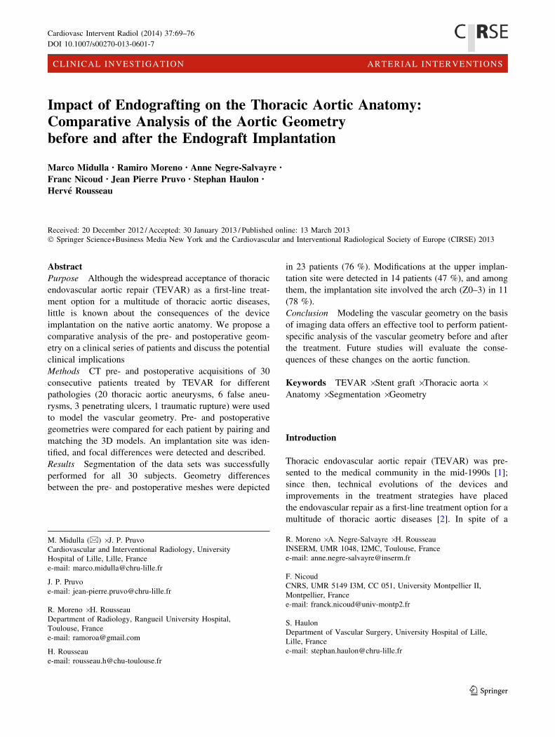
Fig. 1 Impact of TEVAR on the vascular geometry in a patient
treated for a large thoracic aortic aneurysm of the arch and the
proximal descending aorta, requiring endograft landing at Z2.
A Multiplanar reconstructions (MPR) before (left) and after (right)
the treatment. Asterisks show the lesion. B Reconstruction of the pre-
(left, transparency mode) and postoperative (right, outlined mode)
aortic geometries obtained by segmentation of the CT data sets.
C Overlapping of the 2 volumes confirmed left subclavian artery
origin exclusion (asterisk) and revealed marked changes of the native
anatomy after the device deployment, particularly related to the
modification of aortic arch curvature. A sharp angulation is induced
by the stent graft configuration at Z3 (arrow) and characterizes the
postoperative mesh
72 M. Midulla et al.: Anatomical Impact of TEVAR
123
assessments on the pre- and postoperative status could be
performed, using the entire data set, to investigate the
functional implications of both major and even slight
changes in the geometry (Fig. 5).
This pilot study has several limitations. First, it is just a
first step toward assessing the impact of endografting on
the native thoracic aorta. It is an observational report about
an early experience with the comparative analysis of pre-
and postoperative 3D geometries. No quantitative assess-
ment has been obtained because no automated tool for the
quantification of the vascular geometry was available at the
time we began the study. Different authors have proposed
various approaches for the quantification of the aortic
morphology, and in particular the arch. Most of them are
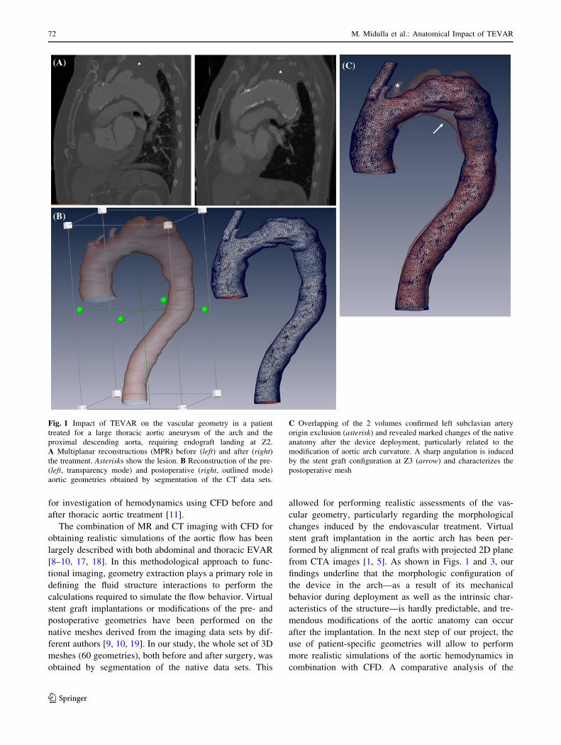
Fig. 2 Geometry analysis of a TEVAR for a descending thoracic
aortic aneurysm with proximal landing zone at Z3. A Segmentations
of the vascular volumes were performed systematically for the pre-
(left) and postoperative (right) acquisitions. B Interface of the
software used for comparing the 2 geometries. The device implan-
tation induced a sharp angulation of the postoperative anatomy (right)
at the isthmic region
M. Midulla et al.: Anatomical Impact of TEVAR 73
123

based on commercially available or in-house software that
calculates the luminal centerline to extract diameters and
different parameters related to vessel tortuosity, such as the
tortuosity index [4], the curvature index, and the radius of
curvature [2, 14]. Visual assessment of tortuosity is sub-
jective, and manual techniques for calculation of the radius
and the angulation at the fixation zone do not reflect fully
the complex and varied morphology of the diseased aorta
Fig. 3 Impact of TEVAR at the upper implantation site. A Axial
images showing a false aneurysm of the isthmic region in a patient
with past history of trauma. Asterisk indicates, on postoperative
examination, the lesion excluded by the endograft. B The 2
geometries were modeled and C compared by matching the volumes.
D The use of the transparency mode for plotting the preoperative
geometry allows to recognize the vascular contours of both meshes
(the postoperative geometry is plotted as a shaded surface, asterisk).
Modifications of the curvature with a focal narrowing were detected
at the upper implantation site after the treatment
74 M. Midulla et al.: Anatomical Impact of TEVAR
123
[4, 20, 21]. The application of dedicated software could be
proposed in next studies for quantitatively characterize the
geometric changes and better assess the impact of endo-
grafting on the aortic anatomy.
The second limitation is the absence of a standard pro-
tocol for the CT acquisitions, especially for preoperative
examinations, as patients were referred to our center by
different institutions from a wide region. Patients were
enrolled consecutively when pre- and postoperative
DICOM data with slice thickness of 3 mm and minimum
interval reconstruction of 1 mm were available. These are
limiting acquisition parameters already proposed for
geometry reconstruction of the aortic anatomy in retro-
spective studies [17, 18]. A prospective approach with
standard acquisition protocols defined for pre- and post-
operative imaging would homogenize the geometry
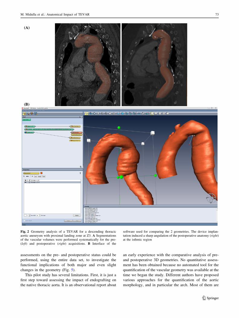
Fig. 4 Impact of thoracic endografting at the middle implantation
site in a patient treated for a huge aneurysm of the left subclavian
artery (LSA). A The 2 volumes (preoperative acquisition shown as
transparent, postoperative acquisition plotted as a tetrahedral grid) are
overlapped on a 2D sagittal section from the native preoperative
acquisition. The LSA is patent before the treatment with the
thrombosed aneurysm around (asterisks). B A focal narrowing with
sharp angulation (arrowheads) characterizes the postoperative vas-
cular anatomy at the middle implantation site. 2D section from the
postoperative acquisition shows the implanted stent graft
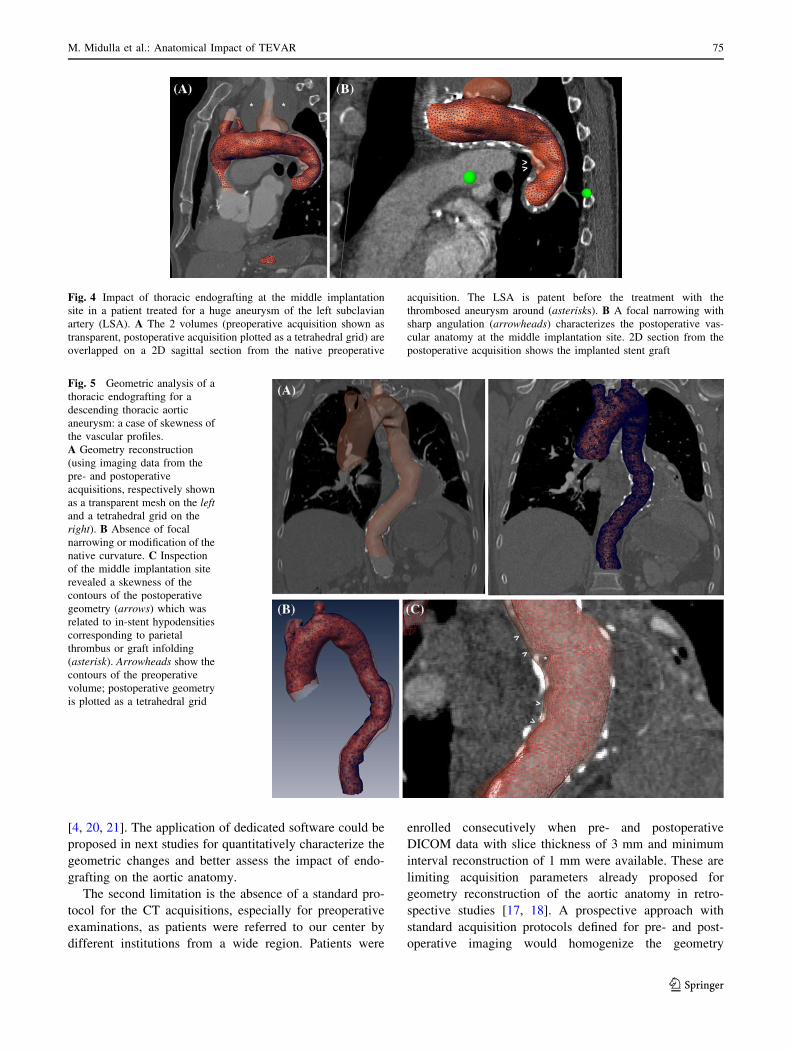
Fig. 5 Geometric analysis of a
thoracic endografting for a
descending thoracic aortic
aneurysm: a case of skewness of
the vascular profiles.
A Geometry reconstruction
(using imaging data from the
pre- and postoperative
acquisitions, respectively shown
as a transparent mesh on the left
and a tetrahedral grid on the
right). B Absence of focal
narrowing or modification of the
native curvature. C Inspection
of the middle implantation site
revealed a skewness of the
contours of the postoperative
geometry (arrows) which was
related to in-stent hypodensities
corresponding to parietal
thrombus or graft infolding
(asterisk). Arrowheads show the
contours of the preoperative
volume; postoperative geometry
is plotted as a tetrahedral grid
M. Midulla et al.: Anatomical Impact of TEVAR 75
123

extraction for future trials and eventual development of
dedicated software for quantitative analysis of the
geometry.
In conclusion, modeling the vascular geometry by means
of imaging data offers an effective tool to perform patient-
specific analysis of the vascular geometry before and after
treatment. This preliminary comparative study demonstrates
that the vascular anatomy can undergo radical changes after
aortic endografting. The next step of our project will use the
acquired set of reconstructed geometries to investigate the
functional consequences of these modifications by a com-
bined CT-CFD approach. A better understanding of the
impact of endografting on the native thoracic aorta is crucial
for future technical developments and clinical improve-
ments in the management of pathology.
Acknowledgments Marco Midulla was supported during the
development of his PhD project, of which this article is a part, by a
grant from the French Society of Radiology (SFR). The entire project
OCFIA is supported by the French National Agency for Research
(ANR 07-CIS7-006-01).
Conflict of interest The authors declare that they have no conflict
of interest.
References
1. Dake MD, Miller DC, Semba CP et al (1994) Transluminal
placement of endovascular stent-grafts for the treatment of
descending thoracic aortic aneurysms. N Engl J Med 331:
1729–1734
2. Nakatamari H, Ueda T, Ishioka F et al (2011) Discriminant
analysis of native thoracic aortic curvature: risk prediction for
endoleak formation after thoracic endovascular aortic repair.
J Vasc Interv Radiol 22(974):979.e2
3. Rengier F, Worz S, Godinez WJ et al (2011) Development of
in vivo quantitative geometric mapping of the aortic arch for
advanced endovascular aortic repair: feasibility and preliminary
results. J Vasc Interv Radiol 22:980–986
4. Ueda T, Takaoka H, Raman B et al (2011) Impact of quantita-
tively determined native thoracic aortic tortuosity on endoleak
development after thoracic endovascular aortic repair. AJR Am J
Roentgenol 197:W1140–W1146
5. Worz S, von Tengg-Kobligk H, Henninger V et al (2010) 3-D
quantification of the aortic arch morphology in 3-D CTA data for
endovascular aortic repair. IEEE Trans Biomed Eng 57:
2359–2368
6. Bortone AS, De Cillis E, D’Agostino D, de Luca Tupputi
Schinosa L (2004) Endovascular treatment of thoracic aortic
disease: four years of experience. Circulation 110(11 suppl
1):II262–II267
7. Serag AR, Bergeron P, Mathieu X et al (2007) Identification of
proximal landing zone limit for proper deployment of aortic arch
stentgraft after supra-aortic great vessels transposition. J Cardio-
vasc Surg (Torino) 48:805–807
8. Midulla M, Moreno R, Baali A et al (2012) Haemodynamic
imaging of thoracic stent-grafts by computational fluid dynamics
(CFD): presentation of a patient-specific method combining
magnetic resonance imaging and numerical simulations. Eur
Radiol 22:2094–2102
9. Prasad A, To LK, Gorrepati ML et al (2011) Computational
analysis of stresses acting on intermodular junctions in thoracic
aortic endografts. J Endovasc Ther 18:559–568
10. Figueroa CA, Taylor CA, Chiou AJ et al (2009) Magnitude and
direction of pulsatile displacement forces acting on thoracic
aortic endografts. J Endovasc Ther 16:350–358
11. Tse KM, Chiu P, Lee HP, Ho P (2011) Investigation of hemo-
dynamics in the development of dissecting aneurysm within
patient-specific dissecting aneurismal aortas using computational
fluid dynamics (CFD) simulations. J Biomech 44:827–836
12. Sethian JA (1999) Level set methods and fast marching methods:
evolving interfaces in computational geometry, fluid mechanics,
computer vision, and materials science. Cambridge University
Press, New York
13. Fillinger MF, Greenberg RK, McKinsey JF, Chaikof EL (2010)
Reporting standards for thoracic endovascular aortic repair (TE-
VAR). J Vasc Surg 52:1022–1033
14. Sze DY, van den Bosch MA, Dake MD et al (2009) Factors
portending endoleak formation after thoracic aortic stent-graft
repair of complicated aortic dissection. Circ Cardiovasc Interv
2:105–112
15. Czerny M, Grimm M, Zimpfer D et al (2007) Results after
endovascular stent graft placement in atherosclerotic aneurysms
involving the descending aorta. Ann Thorac Surg 83:450–455
16. Ueda T, Fleischmann D, Dake MD et al (2010) Incomplete
endograft apposition to the aortic arch: bird-beak configuration
increases risk of endoleak formation after thoracic endovascular
aortic repair. Radiology 255:645–652
17. Molony DS, Kavanagh EG, Madhavan P et al (2010) A compu-
tational study of the magnitude and direction of migration forces
in patient-specific abdominal aortic aneurysm stent-grafts. Eur J
Vasc Endovasc Surg 40:332–339
18. Molony DS, Callanan A, Morris LG et al (2008) Geometrical
enhancements for abdominal aortic stent-grafts. J Endovasc Ther
15:518–529
19. Filipovic N, Milasinovic D, Zdravkovic N et al (2011) Impact of
aortic repair based on flow field computer simulation within the
thoracic aorta. Comput Methods Programs Biomed 101:243–252
20. Sternbergh WC 3rd, Money SR, Greenberg RK, Chuter TA
(2004) Influence of endograft oversizing on device migration,
endoleak, aneurysm shrinkage, and aortic neck dilation: results
from the Zenith Multicenter Trial. J Vasc Surg 39:20–26
21. Bowman JN, Silverberg D, Ellozy S et al (2009) The role of
anatomic factors in predicting success of endovascular repair of
thoracic aortic aneurysms. Vasc Endovascular Surg 44:101–104
76 M. Midulla et al.: Anatomical Impact of TEVAR
123