impaired urinary concentration vasopressin and its...
TRANSCRIPT

Impaired Urinary Concentration after Vasopressin
and Its Gradual Correction in
Hypothalamic Diabetes Insipidus
Avway R. HABINGTONand HEIzZ VALINFrom the Department of Physiology, Dartmouth Medical School,Hanover, NewHampshire
A B S T RA C T This study utilized rates with here-ditary hypothalamic diabetes insipidus (D.I.) inorder to explore possible mechanisms which pre-vent full urinary concentration after acute admin-istration of vasopressin in hypothalamic D.I. andwhich correct this concentrating defect with pro-longed therapy.
It was found: (a) that the concentrating defectpersisted even when the urinary osmolal excretionof D. I. rats was reduced to that of normal ani-mals; (b) that the defect was not corrected morerapidly if larger doses of vasopressin were given;(c) that it persisted even when the D.I. rats weredeprived of drinking water after vasopressin wasgiven; (d) that there was osmotic equilibrationbetween urine and renal papilla at a time when theconcentrating defect was still evident; and (e)that the correction of the defect was associatedwith progressive and significant rise of the papil-lary osmolality.
These studies appear to rule out osmotic diure-sis, accumulation of exogenous vasopressin, per-sistent primary polydipsia, or delay in the induc-tion of membrane permeability as causes for theconcentrating defect. Rather, subnormal osmolalityof the renal papilla, which can be corrected onlygradually, accounts for the initial concentrating
Dr. Harrington's present address is Department ofMedicine, University of Wisconsin, Madison, Wis. 53706.Address requests for reprints to Dr. Heinz Valtin, De-partment of Physiology, Dartmouth Medical School,Hanover, N. H. 03755
Received for publication 31 August 1967 and in re-vised form 6 November 1967.
defect and the long time required for its correction.Reduction of water content and increase of urea
content are primarily responsible for restorationof papillary osmolality to normal.
INTRODUCTIONIt is well known that urinary concentration inresponse to supramaximal doses of vasopressin isimpaired in both experimental (1, 2) and clinical(3) states of water diuresis. The gradual correc-
tion of this defect with prolonged administrationof vasopressin has received less attention, althoughit has been pointed out by several authors (1, 4,5), and convincing data demonstrating this cor-
rection were presented by Burka (6). A satis-factory explanation for these findings, however,has remained wanting (7).
Both the initial impairment and its gradual cor-
rection can be clearly demonstrated in rats withhereditary hypothalamic diabetes insipidus (D.I.)(8). In the experiments to be described, severalpossible mechanisms for this concentrating defectand its gradual response to treatment in D.I. rats
have been explored. The studies suggest a gradualrise in the osmolality of the renal papillary inter-stitial tissue as the most plausible explanation.
METHODSEach experiment will be described in relation to the hy-pothesis which it was designed to test.
All animals,-both normal and D.I. of the Brattleborostrain (9), were obtained from our own breeding colony.Adult rats of both sexes were used, and in any givenexperiment, normal animals and rats with D.I. were
502 The Journal of Clinical Investigation Volume 47 1968

matched for age and sex. Except when noted otherwise,rats had free access to Purina Labena rat pellets (Ralston,Purina Co., St. Louis, Mo.) and to drinking water.
Vasopressin tannate in oil' was used throughout andgiven subcutaneously. During collection periods, rats werekept in individual metabolism cages. Urine was collectedunder mineral oil at room temperature. Urine osmolalitywas determined in a Fiske osmometer. Osmolalities ofpapillary tissues were calculated as the sum of urea plus2 (Na+ + K+ + NH4+). The concentrations of these soluteswere determined by methods previously described (10).Significance of the results was evaluated by Student's ttest (11).
RESULTS
The concentrating defect and its gradual correc-tion. Fig. 1 illustrates the progressive rise inurine osmolality as D.I. rats are given dailyinjections of vasopressin tannate in oil for 5 wk.In contrast, the urine osmolality of normal ratsincreased at the very onset of treatment and,except for one unexplained fluctuation, rose nofurther. After 5 wk of treatment, the mean urineosmolality of D.I. rats was about the same as thatof normal rats given peanut oil (8), but not asgreat as that of normal rats treated with vaso-pressin (Fig. 1). In order to determine whetherfurther treatment would completely normalize theurinary concentrating mechanism in D.I. rats, wecontinued injections of vasopressin in five D.I.rats for a total of 57 days. By this time their meanurine osmolality was 2657 mOsm/kg, which is notsignificantly different from the mean of the treatednormal rats.
Urine flow decreased progressively during thecourse of treatment in the D.I. rats, but not inthe normal animals.
Osmotic diuresis. The bottom of Fig. 1 indi-cates that urinary osmolal excretion was verymuch higher in untreated D.I. rats than in nor-mals; this finding may be related to the greaterfood consumption of D.I. animals (12). WhenD.I. rats were treated with vasopressin, theirosmolal excretion declined toward the levels seenin normal rats. This observation suggested thatthe initial subnormal response of D.I. rats tovasopressin might be due to osmotic diuresis,which is known to reduce maximum concentratingability (13). The pattern of osmolal excretionduring treatment of D.I. rats (Fig. 1) renders
1 Pitressin tannate in oil, Parke, Davis & Co., Detroit,Mich.
this explanation most unlikely. Even by the 2ndday of treatment, there had been a sharp decline,and although the total excretion remained higherthan in the normals, this small difference in osmo-tic load is unlikely to have caused the very largedifferences in urinary concentration that were stillapparent on days 2 and 9. Furthermore, by day 22,the urinary osmolal excretion of D.I. rats was lessthan that of normals, and yet the urinary concen-tration remained significantly lower than in nor-mals by at least 500 mOsm/kg through the 28thday of treatment.
The role of solute excretion was explored fur-ther by depriving D.I. rats of food, thereby lower-ing their rate of osmolal excretion to that ofnormal rats (Fig. 2). Five normal rats (threefemales, two males) and six D.I. rats (three ofeach sex) which had previously been on food andwater ad lib. were tested during the second 24 hrof treatment with vasopressin. During this testperiod the normal rats continued to have freeaccess to food and water, but the D.I. rats hadaccess only to water. Through this maneuver, theosmolal excretion in D.I. rats was reduced tothe level seen in normal animals. Nevertheless, themean urinary concentration remained some 1000mOsm/kg lower in the former group.
The urine osmolality in fasted D.I. rats wasslightly higher than we would have expected onthe 2nd day of vasopressin treatment in fed D.I.rats. This phenomenon may have been due notonly to decreased excretion of nonurea solutes, butalso to dehydration. For unknown reasons, fastedD.I. rats drank less than normals, even thoughthey had free access to water, and their urineoutput exceeded their fluid intake. This situation,plus insensible loss of water, must have resultedin a considerable negative fluid balance, and ac-counted for about 30% of the weight loss of fastedD.I. rats in this experiment.
One might suspect that the subnormal urineosmolality of the fasted D.I. rats was related tothe known tendency of dietary nitrogen restrictionto reduce urinary concentrating ability (14-16);but several findings suggest that this effect playedno important role in this experiment. The urinaryexcretion of urea was almost halved in the fastedD.I. rats (Fig. 2). This phenomenon may haveoccurred mainly because of increased reabsorptionof urea from the distal nephron under the influ-
Impaired Urinary Concentration after Vasopressin in Diabetes Insipidus 503

0 5 10 15 20 25 30 35
URINE FLOWml per 100g
per Day
NORMAL 3.3 2.9
D. I.
12,000
11,000 -
10,000 FOSMOLAL
9000 FEXCRETION
8000,.Osm per 100 9
per Doy7000 1
6000
72.8 11.43.0
7.3
0 5 10DAYS OF TREATMENT (1.0 UNIT
2.7 2.1 2.6
4.1 3.1 3.32.5
2.5
15 20 25 30 35VASOPRESSIN TANNATE IN OIL
FIGURE 1 Mean values on eight normal and nine diabetes insipidus (D.I.) rats, before treatment (datato left of day 0) and during course of 37 daily, subcutaneous injections of vasopressin tannate in oil.All animals ate and drank ad lib. throughout the study. Except for the values at 35 and 37 days of treat-ment, all urine osmolalities in D.I. rats were significantly different from those in normal animals (P <0.05). Urine flows in D.I. rats were also significantly different from those of normal rats (P < 0.05) ex-
cept during the final two collection periods. During daily control injections of peanut oil vehicle, valuesdid not deviate from those before treatment (8).
504 A. R. Harrington and H. Valtin
3500
3000
URINE
2500
2000
NORMAL
00,~~~~~~
DAS
* DIABETES INSIPIDUS
0. .
OSMOLALITY
1500 1mOsm/ kg
1000I
500 1
0
NDIABETES INSIPIDUS
7/0'
NORMAL
1.DAILY)
I I I I I I I m

FED
2500 f
2000tI
4
en0
U)0
wzFE
1500 I.m-
E0E 10001
500 I
0
P-values Uosm
UOsm*V
- T4
0z
0-
VASOPRESSIN1.0 UNIT S.C.
DAILY X 2
FED
+4
0
OSMOLAL z
EXCRETION
0~~~~~c]
< 0.001
<0.001
<0.001
>0.50
WATER INTAKE
(ml per 100 g per Day)URINE FLOW
8.3 69.8
2.0 59.0
6.5 2.5
2.1 3.6
BODY WEIGHT(g)
60
x 50
.4wa: 40
30
347 274 343 247
0 x
0 0
FIGURE 2 Data on five normal and six D.I. rats. The columns represent the mean ± SE of urine os-molalities. During the 24 hr immediately after the second injection of vasopressin tannate in oil, theD.I. rats were fasted in order to reduce their solute excretion to that of the normal rats. For unknownreasons, fasting also caused the D.I. rats to drink significantly less than the normals, even thoughthey had free access to drinking water. Urinary excretion of urea was about the same in fasted D.I.rats as in normals, and the proportion of the total urine osmolality which was contributed by ureawas only slightly and not significantly less in fasted D.I. rats than in fed normal and D.I. animals.For interpretation, see text. The differences in water intake, urine flow, and body weight betweenD.I. rats and normals were significant both before and after vasopressin. In the case of the urea val-ues, only the difference in urea excretion between D.I. and normal rats before vasopressin was sig-nificant statistically (P < 0.05).
ence of vasopressin, rather than because of dietarynitrogen restriction, for the urea excretion infasted D.I. rats did not drop significantly belowthat of the normals. Furthermore, the proportionof the total urine osmolality which was contributedby urea was 52-53% in normal and fed D.I.
animals, and diminished to only 48% in fastedD.I. rats. The last value, which is not significantlylower than the others, compares with 49%o inD.I. rats which had not been deprived of dietaryprotein and had been treated with vasopressin for3 consecutive days (10). Weconclude, therefore,
Impaired Urinary Concentration after Vasopressin in Diabetes Insipidus 505
FASTED
04
I10,000
z 211
z00
0 a8000 w °o CPx 0
6000 W--J 0I< Cfi40.
4000 0 a0n°00
2000
E
a>:
0 co
D
0w0
OD
z0 a
6000 t awa:.@Cc 0>x
4000 i CP
4 0w oa:
2000 Df
z 00 Fc: @
-.
vy By

that the concentrating defect in question is ac-counted for very little, if at all, by osmoticdiuresis.
Accumulation of exogenous vasopressin. Thepossibility was considered that gradual accumula-tion of exogenous vasopressin in D.I. rats givendaily injections might have caused their increasingconcentrating ability. It would follow from thisargument that the dose of vasopressin we used(1.0 U/day) was not supramaximal and thathigher urine concentrations could be achievedsooner if more vasopressin were used. The experi-ment illustrated in Fig. 3 was done in order totest this possibility.
Three groups of D.I. rats, each consisting oftwo females and two males, were given daily in-jections of 0.5 U, 1.0 U, and 2.5 U of vasopressin,respectively, for 3 days, and urine was collectedfor 24 hr on each day of treatment. There wasno significant difference between the groups inthe degree of urine concentration achieved on anyone day, although the expected progressive risein urine osmolality was seen in each group. Thefact that a fivefold range in drug dosage causedno significant difference in urine concentrationstrongly suggests that a daily injection of even0.5 U would have been supramaximal.
Habitual polydipsia. It seemed possible thatD.I. rats, which consume an average of 80% oftheir body weight as drinking water each day (9),continue to drink excessively after replacementtherapy with vasopressin is begun. In that case,primary polydipsia might be responsible for theinitial concentrating defect.
This hypothesis was explored by testing the
1600-
1200URINE
OSMOLALITY800-
mosm/ kg
400-
0*
O 0.5 unit0 1.0 unitX 2.5 units
DAY I
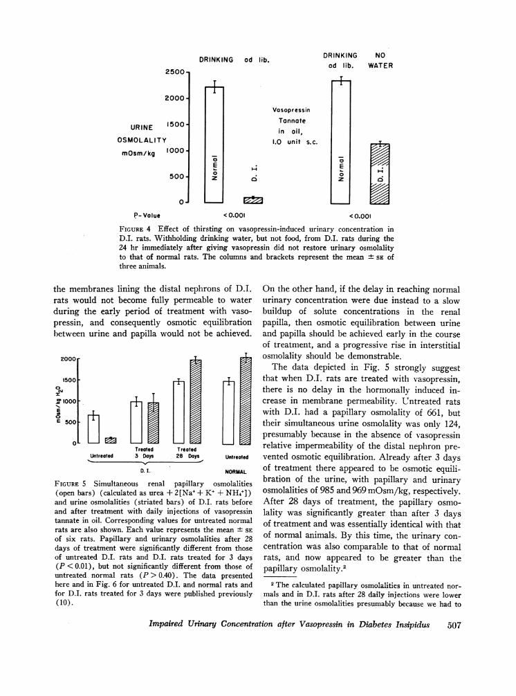
response to vasopressin of D.I. rats which weredenied access to drinking water (Fig. 4). Threenormal male rats and three D.I. males wereallowed to eat and drink ad lib. during a 24 hrcontrol period without treatment. The mean urineosmolality of the normal rats during this periodwas 2227 mOsm/kg, that of the D.I. rats was 116.Each animal, both normal and D.I., was thengiven 1.0 U of vasopressin tannate in oil. Duringthe subsequent 24 hr normal rats were allowedfood and water ad lib., while D.I. rats wereallowed food but no water. During this final periodurine was collected every 6 hr in order to avoiddilution of urine from the D.I. rats by urineformed immediately after the vasopressin wasgiven.
The right-hand columns in Fig. 4 represent themean urine osmolalities during the final 6 hr.Even though the D.I. rats were dehydrated andhad lost an average of 10% of their body weight,their urine osmolality remained nearly 1100mOsm/kg lower than that of the normal animals.Thus, habitual polydipsia cannot be responsiblefor the concentrating defect.
Progressive increase in membrane permeabilityvs. progressive rise in papillary osmolality. Vaso-pressin probably increases the permeability of themammalian distal nephron to water (17-19). Thebiochemical reactions which bring about thischange are not yet known, but it seems likely thatthese reactions involve one or more enzyme sys-tems (20). If that is so, then the time requiredfor adaptation of these systems (21) might ac-
count for the long delay before D.I. rats respondnormally to vasopressin. According to this view,,
±h
DAY 2
FIGURE 3 Effect of vasopressin dosageon urinary concentration in D.I. rats.The columns and brackets represent themean + SE of four rats. Numbered daysrefer to days of treatment with vasopres-sin tannate in oil, subcutaneously.
DAY 3
506 A. R. Harrington and H. Valtin
DRINKING od lib.
2500-
2000-
URINE 1500OOSMOLALITY
mOsm/kg 1000.
500-
0.1
P-Value
To
E0zI
DRINKING NOad lib. WATER
VasopressinTonnatein oil,
1.0 unit s.c.
-4
0;
0E0z
< 0.001 < 0.001
i
l
FIGURE 4 Effect of thirsting on vasopressin-induced urinary concentration inD.I. rats. Withholding drinking water, but not food, from D.I. rats during the24 hr immediately after giving vasopressin did not restore urinary osmolalityto that of normal rats. The columns -and brackets represent the mean ± SE ofthree animals.
the membranes lining the distal nephrons of D.I.rats would not become fully permeable to waterduring the early period of treatment with vaso-pressin, and consequently osmotic equilibrationbetween urine and papilla would not be achieved.
2000
1500 T
0
.Cl 1000TE
E 500
Treated TreatedUntreated 3 Days 28 Days Untreated
D. I. NORMAL
FIGURE 5 Simultaneous renal papillary osmolalities(open bars) (calculated as urea + 2[Na+ + K+ + NH4+] )and urine osmolalities (striated bars) of D.I. rats beforeand after treatment with daily injections of vasopressintannate in oil. Corresponding values for untreated normalrats are also shown. Each value represents the mean ± SEof six rats. Papillary and urinary osmolalities after 28days of treatment were significantly different from thoseof untreated D.I. rats and D.I. rats treated for 3 days(P <0.01), but not significantly different from those ofuntreated normal rats (P > 0.40). The data presentedhere and in Fig. 6 for untreated D.I. and normal rats andfor D.I. rats treated for 3 days were published previously(10).
On the other hand, if the delay in reaching normalurinary concentration were due instead to a slowbuildup of solute concentrations in the renalpapilla, then osmotic equilibration between urineand papilla should be achieved early in the courseof treatment, and a progressive rise in interstitialosmolality should be demonstrable.
The data depicted in Fig. 5 strongly suggestthat when D.I. rats are treated with vasopressin,there is no delay in the hormonally induced in-crease in membrane permeability. Untreated ratswith D.I. had a papillary osmolality of 661, buttheir simultaneous urine osmolality was only 124,presumably because in the absence of vasopressinrelative impermeability of the distal nephron pre-vented osmotic equilibration. Already after 3 daysof treatment there appeared to be osmotic equili-bration of the urine, with papillary and urinaryosmolalities of 985 and 969 mOsm/kg, respectively.After 28 days of treatment, the papillary osmo-lality was significantly greater than after 3 daysof treatment and was essentially identical with thatof normal animals. By this time, the urinary con-centration was also comparable to that of normalrats, and now appeared to be greater than thepapillary osmolality.2
2 The calculated papillary osmolalities in untreated nor-mals and in D.I. rats after 28 daily injections were lowerthan the urine osmolalities presumably because we had to
Impaired Urinary Concentration after Vasopressin in Diabetes Insipidus 507

400 r PAPILLARY SODIUM CONTENT
OL-g H20 per
100 g Wet Tissue
40or
0>3000
0
--. 200
(Aa,0
rE 100E
87 85 81
PAPILLARY UREA CONTEN
Treated Treated~Untreated 3 Days 28 Days
D. I.
81 FIGURE 6 Renal papillary content of water, so-
dium (top), and urea (bottom) 'in D.I. rats be-fore and after treatment with daily injections ofvasopressin tannate in oil. Figures for untreatednormal rats are also shown. The columns andbrackets represent the mean - SE of the same
six animals for which values are given in Fig. 5.No significant difference of sodium content was
found among these groups. The contents of bothurea and water after 3 days of treatment were
significantly different from those of untreatedD.I. rats, from those of D.I. rats treated for28 days, and from normal rats (P < 0.05).There were no significant differences betweenany values in D.I. rats treated for 28 days andthose of untreated normal rats (P > 0.50). D.S.
=dry solids.
Untreated
NORMAL
Fig. 6 shows the main causes for the progressiverise in D.I. papillary osmolality which has beendepicted in Fig. 5. Although previous work by us
(10) had shown that the sequestration of papillarysodium did not increase in D.I. rats after threedaily injections of vasopressin, there remainedthe possibility that more prolonged treatmentmight raise the papillary sodium content(mmoles/100 g of dry solids). As Fig. 6 shows, however,even 28 days of treatment failed to raise this
go down an osmotic gradient from the papillary tip to-ward the medulla in order to obtain enough tissue foranalysis. This methodological error must have been mini-
mized when papillary osmolality was determined after 3
days of treatment, for the interstitial osmotic gradientis much less steep when less concentrated urine is formed
(22). Nevertheless, to the extent that the calculatedpapillary osmolality may have been an underestimate ofthe value at the very tip of the papilla, complete osmoticequilibration may not have occurred after 3 days oftreatment.
variable significantly. It might be more meaning-ful to express contents as mmoles/100 g of urea-
free dry solids. Although the slight rise whichwould occur in the sodium content if it were cal-culated in these units is still not significant, itmust be admitted that the rise might reflectslightly increased sodium sequestration duringprolonged treatment with vasopressin. However,this possible effect does not appear to contributenearly so importantly to the rise in total papillaryosmolality as do the significant and progressiverise in the content of urea and the progressive andsignificant decline in the content of water. Thelatter effect would of course tend to raise the con-
centrations (mmoles/kg of tissue water) of allsolutes in the papilla. Thus, although tissue analy-ses admittedly do not provide direct measurementsof the interstitial fluid volume and its contents,the striking differences between the behavior of
508 A. R. Harrington and H. Valtin
unO 300C,00z 200an
0E l00E
OL

sodium on the one hand, and urea and water onthe other, strongly suggest that the progressiverise in papillary osmolality as D.I. rats are treatedwith vosopressin appears to be due principally, butnot necessarily solely, to two factors: the decreas-ing content of water and the increasing contentof urea.
DISCUSSION
There is now a fairly long list of conditions thatcan cause failure of the kidneys to concentrateurine, even in the presence of large amounts ofendogenous or exogenous vasopressin (7). Mostof these conditions can be ruled out as causes ofthe concentrating defect in hypothalamic D.I. inhumans and in rats, because in these the kidneysare normal grossly and histologically (23), thereare no known abnormalities of electrolytes, suchas calcium or potassium (6, 12), and appreciable,albeit subnormal, concentration of the urine isusually achieved with the very first injection ofvasopressin. The phenomenon being considered inthis report is unique in that it represents only apartial unresponsiveness and one which can becorrected with the single measure of prolongedtreatment with vasopressin.
Several possible explanations for the concen-trating defect, such as solute diuresis, accumula-tion of exogenous vasopressin, habitual polydipsia,and enzyme adaptation appear to have been ruledout by the present experiments. The last, adapta-tion of enzymes involved in the action of vaso-pression, is further rendered most unlikely by thefindings that the concentrating defect in humanscannot be prevented by giving large doses ofexogenous vasopressin during the period of ex-perimental overhydration (1), and that when D.I.rats are used as bioassay preparations they aremore, rather than less, sensitive to intravenousvasopressin (24-26). Furthermore, contrary tothe expectation if enzyme adaptation were in-volved, the sensitivity of the bioassay cannot beenhanced by prior prolonged treatment withexogenous vasopressin (25).
In an earlier report we suggested that the initialdefect and its gradual correction in D.I. ratsmight be due to competitive inhibition by anabnormal polypeptide which these rats might pro-duce instead of vasopressin (8). This hypothesishas since been disproved (27). It is conceivable
that oxytocin might act as a competitive inhibitorof exogenous vasopressin in hypothalamic D.I.Such a mechanism seems unlikely in water-loadedrats and humans, however, since excessive releaseof oxytocin would not be expected in overhydrated,normal subjects.
A number of possible mechanisms have beenpostulated in the past to explain the concentratingdefect during experimental and clinical water di-uresis. Most of these have invoked an increasein the volume or decrease in the osmolality of theextracellular fluid, the ultimate effect possibly be-ing cellular overhydration and consequent inabilityof vasopressin to act or to reach its site of action(1, 2, 28, 29). If rats with D.I. represent a com-parable experimental model for the concentratingdefect under discussion, there is considerable evi-dence against such explanations. The concentratingdefect persisted in dehydrated D.I. rats (Fig. 4),which must have had contracted fluid volumes.Even in D.I. rats drinking ad lib., the serumosmolality and sodium coicentrations are signifi-cantly higher than in normal rats ( 23). Thisfinding suggests a state of mild dehydrationand contraction of the extracellular fluid volume.3
Several authors have alluded to the possibilitythat the cause of the concentrating defect may liein decreased concentration of the papillary inter-stitium. The data presented in Fig. 5 seem toleave little doubt that this explanation is correctin the case of D.I. rats. Although Fig. 6 shows thata slow and progressive rise in the papillary con-tent of urea and a concomitant gradual and pro-gressive decline in the content of water are pri-marily responsible for the slow buildup of papillaryosmolality, these studies do not elucidate themechanism (s) by which these changes are broughtabout.
3 The Friedmans (12) found very slight changes in theopposite direction, but this may have been because theycorrected extracellular fluid volumes to 100 g of bodyweight. Since D.I. rats are very much leaner than nor-mals of the same age, one would expect the proportion oftheir body weight which is water to be greater than nor-mal. This possible explanation is strengthened by theFriedmans' finding of no significant difference in extra-cellular fluid volumes between normal rats and those withsurgically induced D.I. of similar body weights. Therealso appear to be no significant differences between ratswith hereditary D.I. and normals in total and intracellu-lar water content of gastrocnemius muscle and aorta.
Impaired Urinary Concentration after Vasopressin in Diabetes Insipidus 509

ACKNOWLEDGMENTSWe thank Mrs. Robert A. Garrity for valuable technicalhelp.
This work was supported by U. S. Public Health Serv-ice research grant AM-08469-GM from the National In-stitute of Arthritis and Metabolic Diseases, and an in-stitutional grant from the American Cancer Society.Dr. Harrington was a Postdoctoral Trainee in Physi-ology, supported by National Heart Institute Train-ing Grant HE-5322. Dr. Valtin received U. S. PublicHealth Service Research Career Program Award 6-K3-GM-21,786 from the National Institute of General Medi-cal Sciences.
REFERENCES
1. Epstein, F. H., C. R. Kleeman, and A. Hendrikx.1957. The influence of bodily hydration on the renalconcentrating process. J. Clin. Invest. 36: 629.
2. deWardener, H. E., and A. Herxheimer. 1957. Theeffect of a high water intake on the kidney's abilityto concentrate the urine in man. J. Physiol. (Lon-don). 139: 42.
3. Alexander, C. S., D. M. Filbin, and S. A. Fruchtman.1959. Failure of vasopressin to produce normal urineconcentration in patients with diabetes insipidus. J.Lab. Clin. Med. 54: 566.
4. Kleeman, C. R., M. H. Maxwell, and S. Witlin. 1958.Functional isosthenuria. Arch. Internal Med. 101:1023.
5. Dies, F., S. Rangel, and A. Rivera. 1961. Differentialdiagnosis between diabetes insipidus and compulsivepolydipsia. Ann. Internal Med. 54: 710.
6. Burka, E. R. 1962. Renal function in diabetes insipidus.Arch. Internal Med. 109: 717.
7. Epstein, F. H. 1966. Disorders of renal concentratingability. Yale J. Biol. Med. 39: 186.
8. Harrington, A. R., and H. Valtin. 1965. Vasopressineffect on urinary concentration in rats with hereditaryhypothalamic diabetes insipidus (Brattleboro strain).Proc. Soc. Exptl. Biol. Med. 118: 448.
9. Valtin, H., W. H. Sawyer, and H. W. Sokol. 1965.Neurohypophysial principles in rats homozygous andheterozygous for hypothalamic diabetes insipidus(Brattleboro strain). Endocrinology. 77: 701.
10. Valtin, H. 1966. Sequestration of urea and nonureasolutes in renal tissues of rats with hereditary hypo-thalamic diabetes insipidus: effect of vasopressin anddehydration on the countercurrent mechanism. J. Clin.Invest. 45: 337.
11. Hill, A. B. 1961. Principles of Medical Statistics. Ox-ford University Press, Inc., NewYork. 7th edition. 146.
12. Friedman, S. M., and C. L. Friedman. 1965. Salt andwater distribution in hereditary and in induced hypo-thalamic diabetes insipidus in the rat. Can. J. Physiol.Pharmacol. 43: 699.
13. Rapoport, S., W. A. Brodsky, C. D. West, and B.Mackler. 1949. Urinary flow and excretion of solutesduring osmotic diuresis in hydropenic man. Am. J.Physiol. 156: 433.
14. Hendrikx, A., and F. H. Epstein. 1958. Effect offeeding protein and urea on renal concentrating abilityin the rat. Am. J. Physiol. 195: 539.
15. Crawford, J. D., A. P. Doyle, and J. H. Probst. 1959.Service of urea in renal water conservation. Am. J.Physiol. 196: 545.
16. Radford, E. P., Jr. 1959. Factors modifying watermetabolism in rats fed dry diets. Am. J. Physiol. 196:1098.
17. Ullrich, K. J., G. Rumrich, and G. Fuchs. 1964.Wasserpermeabilitat und transtubalirer Wasserflusscorticaler Nephronabschnitte bei verschiedenen Di-uresezustanden. Arch. Ges. Physiol. 280: 99.
18. Morel, F., M. Mylle, and C. W. Gottschalk. 1965.Tracer microinjection studies of effect of ADH onrenal tubular diffusion of water. Am. J. Physiol. 209:179.
19. Grantham, J. J., and M. B. Burg. 1966. Effect ofvasopressin and cyclic AMPon permeability of iso-lated collecting tubules. Am. J. Physiol. 211: 255.
20. Handler, J. S., R. W. Butcher, E. W. Sutherland, andJ. Orloff. 1965. The effect of vasopressin and oftheophylline on the concentration of adenosine 3', 5'-phosphate in the urinary bladder of the toad. J. Biol.Chem. 240: 4524.
21. Knox, W. E., V. H. Auerbach, and E. C. C. Lin.1956. Enzymatic and metabolic adaptations in animals.Physiol. Rev. 36: 164.
22. Ullrich. K. J., F. 0. Drenckhahn, and K. H.Jarausch. Untersuchungen zum Problem der Harkom-zentrierung und-verdilnnung. 1955. tVber das osmotischeVerhalten von Nierenzellen und die begleitende Elek-trolytanhiufung im Nierengewebe bei verschiedenenDiuresezustinden. Arch. Ges. Physiol. 261: 62.
23. Valtin, H., and H. A. Schroeder. 1964. Familial hypo-thalamic diabetes insipidus in rats (Brattleborostrain). Am. J. Physiol. 206: 425.
24. Sawyer, W. H., and H. Valtin. 1967. Antidiureticresponses of rats with hereditary hypothalamic dia-betes insipidus to vasopressin, oxytocin, and nicotine.Endocrinology. 80: 207.
25. Vierling, A. F., J. B. Little, and E. P. Radford, Jr.1967. Antidiuretic hormone bio-assay in rats withhereditary hypothalamic diabetes insipidus (Brattle-boro strain). Endocrinology. 80: 211.
26. Jones, J. J., and J. Lee. 1967. The value of rats withhereditary hypothalamic diabetes insipidus for thebioassay of vasopressin. J. Endocrinol. 37: 335.
27. Sawyer, W. H., and H. Valtin. 1965. Inhibition ofvasopressin antidiuresis by extracts of pituitaries fromrats with hereditary hypothalamic diabetes insipidusand by oxytocin. Endocrinology. 76: 999.
28. Levinsky, N. G., D. G. Davidson, and R. W. Berliner.1959. Changes in urine concentration during pro-longed administration of vasopressin and water. Am.J. Physiol. 196: 451.
29. Hays, R. M., and A. Leaf. 1961. The problem ofclinical vasopressin resistance: in vitro studies. Ann.Internal Med. 54: 700.
510 A. R. Harrington and H. Valtin