vasopressin is secreted from the neurohypophysis following
TRANSCRIPT

J. Phyaiol. (1985), 367, pp. 253-265 253With 1 plate and 5 text-figureMPrinted in Great Britain
INHIBITING THE RABBIT CAUDAL VENTROLATERAL MEDULLAPREVENTS BARORECEPTOR-INITIATED SECRETION OF VASOPRESSIN
BY W. W. BLESSING AND J. 0. WILLOUGHBYFrom the Centre for Neuroscience, Department of Medicine, Flinders University of
South Australia, Bedford Park, S.A. 5042, Australia
(Received 22 January 1985)
SUMMARY
1. The A 1 noradrenergic neurones are known to project from the caudal ventro-lateral medulla to the vasopressin-secreting neuroendocrine cells in the hypo-thalamus. They therefore represent a possible central pathway from the medullato the hypothalamus for baroreceptor-initiated secretion of vasopressin.
2. We tested this hypothesis in the anaesthetized rabbit. Muscimol, a y-aminobutyric-acid-receptor agonist, was injected into the caudal ventrolateralmedulla to inhibit the A 1 noradrenergic neurones.
3. Secretion of vasopressin, measured by radioimmunoassay, was initiated eitherby arterial haemorrhage or by constriction of the inferior vena cava.
4. After injection ofvehicle into the caudal ventrolateral medulla, or after injectionof muscimol into nearby control areas, both haemorrhage and constriction of theinferior vena cava produced the expected elevation in plasma vasopressin.
5. After injection of muscimol into the caudal ventrolateral medulla, secretion ofvasopressin in response to haemorrhage and to constriction of the inferior vena cava,was completely abolished.
6. The A 1 noradrenergic neurones may be the sole pathway transmitting thereflex for baroreceptor-initiated secretion of vasopressin from the medulla to thehypothalamus.
INTRODUCTION
Vasopressin is secreted from the neurohypophysis following haemorrhage or severehypotension, a reflex originating from baroreceptors within the atria and great vessels(Share, 1974; Wang, Sundet, Hakumaki & Goetz, 1983). Afferent fibres in the IXthand Xth cranial nerves convey baroreceptor information to the nucleus tractussolitarius, whence it reaches vasopressin-secreting neuroendocrine cells in the hypo-thalamus by an as yet undefined pathway (Clark & Rocha E Silva, 1967; Yamashita& Koizumi, 1979; Kalia & Mesulam, 1980; Spyer, 1982). The caudal ventrolateralmedulla contains the A1 noradrenergic neurones (P1. 1) which project directly tovasopressin-secreting cells in the supraoptic and paraventricular nuclei (McKellar &Loewy, 1981; Sawchenko & Swanson, 1982; Blessing, Jaeger, Ruggiero & Reis, 1982).The nucleus tractus solitarius is known to project to the caudal ventrolateral medulla(Loewy & Burton, 1978; Ricardo & Koh, 1978; Sawchenko & Swanson, 1982), so that
W. W. BLESSING AND J. 0. WILLOUGHB Y
the pathway mediating baroreceptor-initiated secretion of vasopressin may includeneurones in this region.
Functional studies support this suggestion. Neurones in the ventrolateral medullawith projections to the hypothalamus are affected by baroreceptor-derived input(Ciriello & Caverson, 1984a, b). Excitatory agents applied to the caudal area elevateplasma vasopressin (Bisset, Feldberg, Guertzenstein & Rocha E Silva, 1975; Feldberg& Rocha E Silva, 1978; Sved, Blessing & Reis, 1985). Inhibitory agents, applied tothe same area, prevent the release of vasopressin normally seen after carotid occlusion(Feldberg & Rocha E Silva, 1981). Although lesioning the AI cell bodies in the rabbitincreases plasma vasopressin (Blessing, Sved & Reis, 1982), subsequent studies haveshown that this is due to an initial excitatory effect of the lesions (Blessing &Willoughby, 1985a, b). Although some electrophysiological studies show that micro-ionophoretically applied noradrenaline inhibits magnocellular neurones (Barker,Crayton & Nicoll, 1971; Moss, Urban & Cross, 1972; Arnauld, Cirino, Layton &Renaud, 1983) more recent studies have demonstrated a convincing excitatory effect(Day & Renaud, 1984; Day, Ferguson & Renaud, 1984; Tanaka, Kaba, Saito & Seto,1984; Kannan, Yamashita & Osaka, 1984).
In the present study we have tested the hypothesis that one of the functions of A 1noradrenergic neurones is to act as an excitatory link in the central pathwaymediating the secretion of vasopressin in response to haemorrhage and hypotension.We have done this by measuring the baroreceptor-initiated secretion of vasopressinafter inhibiting neuronal function in the caudal ventrolateral medulla using localapplication of muscimol, a long acting y-aminobutyric-acid-receptor agonist(Johnston, Curtis, de Groat & Duggan, 1968; DeFeudis, 1980).
METHODS
Animal and surgical proceduresMale New Zealand White rabbits (2-3 kg) were used. They were housed with free access to food
and water, and were transferred to the laboratory in small cages in which they remained whileexperiments without general anaesthesia were performed. Preparatory surgical procedures werecarried out under halothane anaesthesia, one to two weeks before experiments. Experimentalstudies in anaesthetized animals were carried out under urethane (1-4 g/kg), infused over 30 mininto a marginal ear vein. After establishment of anaesthesia, the rabbit was intubated andmechanically ventilated with oxygen-enriched air after muscle relaxation with curare (0 75 mg/kg).Rectal temperature was maintained at 38 'C.
In the preliminary operation, in some animals, an inflatable-cuff constrictor was placed aroundthe inferior vena cava, just above the diaphragm (Korner, Shaw, West & Oliver, 1972). Tubing,connected to the cuff, was left subcutaneously in a dorsal position, from which it could subsequentlybe retrieved. By graded inflation ofthe cuff it was possible to reduce venous return, thereby loweringatrial filling and systemic arterial pressure.Experiments on the medulla oblongata were performed under anaesthesia with the head fixed
in a Kopf stereotaxic holder. The medulla was exposed by incision and retraction of theatlanto-occipital membrane, without affecting the occipital bone or the cerebellum. The degreeofneck flexion was adjusted so that the dorsal surface of the medulla was horizontal. The medullarysurface was covered in warm Ringer solution.
Measurement of arterial pre&sure, heart rate and blood ga8 ten8ion8Arterial pressure and heart rate were recorded on a Grass Model 7 Polygraph via a Statham P23
ID pressure transducer connected to a catheter in the central ear artery (unanaesthetized animals)
254

Al NORADRENERGIC NEURONES AND VASOPRESSIN
or in the femoral artery (anaesthetized animals). Mean arterial pressure was obtained byelectronically damping the phasic signal and heart rate (heart rate) obtained from the pulse wavewith a Grass Model 7P4F tachograph. Arterial blood gases were measured on 1 ml samples usingan IL 513 pH/Blood Gas Analyser (Lexington, MA).
Measurement of plasma vasopressinBlood (1-3 ml) for assay of vasopressin was obtained from the arterial catheter and replaced
immediately by warm Ringer solution. Samples were heparinized, stored on ice and centrifugedwithin 3 hours. The resulting plasma was stored at -20 C until assay. Concentrations ofvasopressin were determined by radioimmunoassay, using an antibody supplied by Dr J. D.Fernstrom, Pittsburg, U.S.A., following extraction from plasma by cation exchange chromato-graphy (Van Itallie & Fernstrom, 1982). Recovery after extraction was 75-100%. Vasopressin foriodination was purchased from Bachem (California). Bound and free vasopressin were separatedusing polyethylene glycol. The sensitivity of the assay was 1-128 pg/tube, and the intra- andinterassay variabilities were 13% and 14% respectively. The assay failed to detect oxytocin at aconcentration of 32 pg/tube and crossreacted 2-5% with 64 pg/tube oxytocin.
Intramedullary injectionsStereotaxic injections were made through long, fine, glass micropipettes with bevelled tips (o.d.
50 jsm). The experimental site, where A 1 neurones are located (Blessing, Chalmers & Howe, 1978),was 1 mm caudal to the rostral border of the area postrema, 3 mm lateral to the midline and 3 mmbelow the dorsal surface of the medulla. Control sites (P1. 1) were either 2-5 mm dorsal or 1-5 mmmedial to the experimental site. Muscimol hydrochloride (Sigma), 1 nmol per 0-2 #l of Ringersolution, was injected bilaterally in approximately 2 s. The Ringer solution contained 0.1 %horseradish peroxidase (Sigma) to mark the centre of the injection site. Micropipettes were removed1 min after injection.
Experimental protocolUnanaesthetized rabbits - inferior vena cava constriction. A central ear artery cannula was inserted
and the subcutaneous tubing for cuff inflation located. After a 15 min rest period, control valuesfor mean arterial pressure, heart rate and plasma vasopressin were obtained. The inferior vena cavacuff was then inflated for 60 or 90 s, using sufficient inferior vena cava constriction to reduce meanarterial pressure by 20-70 mmHg. Plasma vasopressin was measured 1 and 5 min after deflationof the cuff. After results from these pilot experiments were obtained, we performed furtherexperiments in which the inferior vena cava cuff was inflated to a degree sufficient to reduce meanarterial pressure by at least 30 mmHg for 3 min. Arterial blood gases were measured during thelast 15 s of the constriction period and mean arterial pressure, heart rate and plasma vasopressinwere measured 1 and 5 min after cuff deflation.
Anaesthetized rabbits- inferior vena cava constriction. Control values for mean arterial pressure,heart rate, plasma vasopressin and blood gases were obtained after surgical preparation. EitherRinger solution (0-2,l) or muscimol (1 nmol in 0-2,1 of Ringer solution) was then injectedbilaterally into the caudal ventrolateral medulla. Cardiovascular variables and plasma vasopressinwere reassessed after 5 min. The cuff constrictor was then inflated for 3 min. A sample for bloodgas assessment was obtained during the final 15 s. Plasma vasopressin was measured 1 and 5 minafter cuff deflation.
Anaesthetized rabbits - haemorrhage. Rabbits without inferior vena cava cuffs were used. Controlvalues were obtained for all variables and then either vehicle or muscimol was injected into thecaudal ventrolateral medulla. In additional control experiments, muscimol (1 nmol) was injectedinto either the dorsal or the medial site (P1. 1). Variables were reassessed after 5 min. Blood wasthen withdrawn from the femoral artery (15-20 ml/kg) to reduce mean arterial pressure toapproximately 40 mmHg. This procedure occupied approximately 3 min. Blood gas analysis wasperformed on the last blood removed. Plasma vasopressin was measured after a further 2-5 min.The blood was then returned to the rabbit to ensure survival for histological analysis of the injectionsite.
255

W. W. BLESSING AND J. 0. WILLOUGHBY
Hi8tological analy8i8Accurate localization of microinjections was confirmed at the conclusion of each experiment by
perfusing the animal with fixative and processing the brain for catecholamine fluorescencehistochemistry (Blessing et al. 1978), horseradish peroxidase activity and for Nissl substance. Somesections, from other rabbits, not injected with peroxidase, were processed for tyrosine hydroxylaseimmunoreactivity (PI. 1 C), using an antibody provided by Dr T. H. Joh, New York, U.S.A. Theantiserum was used at a dilution of 1 in 5,000 and the sections processed using the avidin-biotin-peroxidase procedure (Vectastain, CA).
RESULTS
Control levels of plasma vasopressinResting plasma vasopressin values were invariably less than 10 ng/l in unanaes-
thetized rabbits (mean 4+ 2 ng/l, n = 23). Approximately 10% of urethane-anaesthetized rabbits had control levels greater than 20 ng/l. We excluded all datafrom these animals from further analysis. Elevated resting vasopressin levels werelikely to occur if the anaesthetic was infused too rapidly or if transient hypotensionoccurred during surgical procedures.
Plasma vasopressin after constriction of the inferior vena cava and blood gas analysisIn pilot experiments we varied the duration and degree of constriction of the
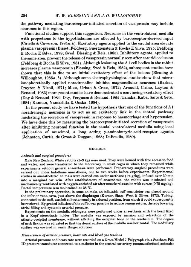
inferior vena cava, recording the fall in mean arterial pressure and the rise in heartrate during constriction, and measuring plasma vasopressin levels 1 and 5 min aftercessation of constriction. The unanaesthetized rabbits rested quietly in their cagesduring these procedures. Constriction periods of 60, 90 and 180 s all increased plasmavasopressin. The 5 min level was approximately 40% of the 1 min level, in bothunanaesthetized rabbits and in anaesthetized rabbits subjected to 180 s of inferiorvena cava constriction (Fig. 1). This is consistent with published values of approxi-mately 4 min for the plasma half-life of vasopressin (Lauson, 1974), indicating thatvasopressin secretion ceased promptly after restoration of venous return through theinferior vena cava, in both unanaesthetized and anaesthetized rabbits.For the 90 s constriction period, in unanaesthetized rabbits, we systematically
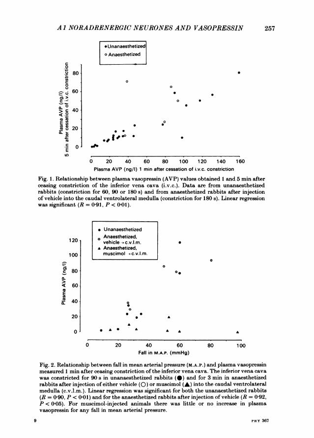
adjusted the degree of balloon inflation to produce graded falls in mean arterialpressure. This resulted in a significant relationship between the fall in mean arterialpressure and the subsequent post-constriction value for plasma vasopressin (Fig. 2),a relationship also present in anaesthetized rabbits after injection of vehicle into thecaudal ventrolateral medulla.Both inferior vena cava constriction and arterial haemorrhage caused a metabolic
acidosis, without a major change in pH because a degree of respiratory alkalosisdeveloped. Hypoxaemia was never observed.These results suggest that the constriction stimulus used resulted in secretion of
vasopressin by a baroreceptor-mediated mechanism.
Inferior vena cava constriction in anaesthetized rabbits after intra-medullary injectionsInjection of Ringer solution into the caudal ventrolateral medulla did not affect
mean arterial pressure, heart rate or plasma vasopressin (Fig. 3). Subsequentconstriction of the inferior vena cava for 3 min reduced mean arterial pressure and
256
AI NORADRENERGICNEURONES AND VASOPRESSIN
* Unanaesthetizedo Anaesthetized
c0
.S2 80
0
_ < 60
C
C 4000
CI
EmIL
0
0
0
0 50
.
0
* 0
0 0
logo *
0 20 40 60 80 100 120 140 160Plasma AVP (ng/l) 1 min after cessation of i.v.c. constriction
Fig. 1. Relationship between plasma vasopressin (AVP) values obtained 1 and 5 min afterceasing constriction of the inferior vena cava (i.v.c.). Data are from unanaesthetizedrabbits (constriction for 60, 90 or 180 s) and from anaesthetized rabbits after injectionof vehicle into the caudal ventrolateral medulla (constriction for 180 s). Linear regressionwas significant (R = 0-91, P < 0-01).
* Unanaesthetized
0 Anaesthetized,vehicle -+c.v.l.m.
* Anaesthetized,muscimol -*c.v.l.m.
0
0
0.
* A S
00
0 0
AA
20 40 60Fall in M.A.P. (mmHg)
80 100
Fig. 2. Relationship between fall in mean arterial pressure (M.A.P.) and plasma vasopressinmeasured 1 min after ceasing constriction of the inferior vena cava. The inferior vena cavawas constricted for 90 s in unanaesthetized rabbits (@) and for 3 min in anaesthetizedrabbits after injection of either vehicle (0) or muscimol (A) into the caudal ventrolateralmedulla (c.v.l.m.). Linear regression was significant for both the unanaesthetized rabbits(R = 0 90, P < 0 01) and for the anaesthetized rabbits after injection of vehicle (R = 0-92,P < 005). For muscimol-injected animals there was little or no increase in plasmavasopressin for any fall in mean arterial pressure.
PHY 367
257
S120
100
0-c 80
< 60EU)m 40
20
0
A
0
A A A
9
258 W. W. BLESSING AND J. 0. WILLOUGHB Y
100 Vehicle MuscimolC.v.l.m. C. v. l.m.
:, 80 (5) (7)0)CD
> 60
EX 40
20
A o0
120 f
I r ~~~~~~~~~. ... ..F 80 .~~~~~ ~~~...... ....... a
.. .-...i..;R,,333
® 80
£400
en 200
-on, E|li in3-
C C:~~~[ Pre-injection
LII 5 min after injection
m~l 1 mmn after cessation of i v c. constriction
* 5 mmn after cessation of i v c. constriction
m ValIue during i.v c. constriction
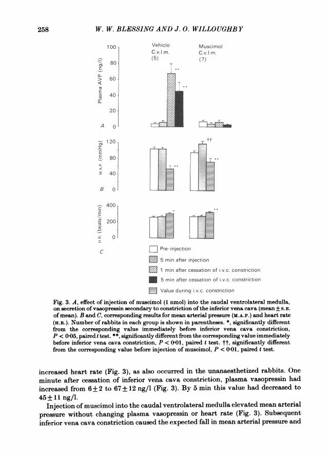
Fig. 3. A, effect of injection of muscimol (1 nmol) into the caudal ventrolateral medulla,on secretion of vasopressin secondary to constriction of the inferior vena cava (mean +5S.E.Of mean). B and C, corresponding results for mean arterial pressure (M.A.P.) and heart rate(H.a.). Number of rabbits in each group is shown in parentheses. *, significantly differentfrom the corresponding value immediately before inferior vena cava constriction,P c 005, paired t test. **", significantly different from the corresponding value immediatelybefore inferior vena cava constriction, P < 0G01, paired t test. tt, significantly differentfrom the corresponding value before injection of muscimol, P < 0 0t, paired t test.
increased heart rate (Fig. 3), as also occurred in the unanaesthetized rabbits. Oneminute after cessation of inferior vena cava constriction, plasma vasopressin hadincreased from 6+2 to 67+12 ng/l (Fig. 3). By 5 mmn this value had decreased to45+ 11 ng/l.
Injection of muscimol into the caudal ventrolateral medulla elevated mean arterialpressure without changing plasma vasopressin or heart rate (Fig. 3). Subsequentinferior vena cava constriction caused the expected fall in mean arterial pressure and

AI NORADRENERGIC NEURONES AND VASOPRESSIN 259
Vehicle Muscimol Muscimol MuscimolC.v.l.m. C.v.I.m. Dorsal site Medial site(6) (7) (5) (5)
120 .
100
-s80CL
< 60EU)E 40a-
20
A 0
120 __t
80E.....
TJ g Pre-ILjec after injection *....... ......
Fig. 400 A,
ofv~opes seonPre-ijetionar atetijetiniaatehaemorrhagemas. omen.BndC
corresponding data for mean arterial pressure (M.A.P.) and heart rate (H.n.). Numberof rabbits in each group shown in parentheses. Significantly different from previouscorresponding value, *P < O O5, **P < 001, paired t test.
increase in heart rate, but plasma vasopressin remained entirely unchanged frompre-constriction values (Fig. 3). Constriction of the inferior vena cava in vehicle andmuscimol injected groups caused similar falls in mean arterial pressure (50+10 and49+10 mmHg respectively) and rises in heart rate (60+16 and 42+13 beats/mmn)but the final mean arterial pressure was slightly higher in the muscimol-injected group(72 ± 9 compared to 54 ± 9 mmHg), because mean arterial pressure was higher in thisgroup immediately before inferior vena cava constriction. Maximal inferior vena cavaconstriction readily reduced arterial pressure to levels as low as 30 mmHg invehicle-injected rabbits. In muscimol-injected animals it was usually impossible toreduce pressure below 60 mmHg even with maximal inferior vena cava constriction.
9-2
W. W. BLESSING AND J. 0. WILLOUGHBY
However, analysis of results for individual rabbits in the muscimol-injected group(Fig. 2) revealed no relationship between fall in mean arterial pressure and subsequentplasma vasopressin. Indeed the muscimol-injected animal with the most effectivehypotensive stimulus (mean arterial pressure falling from 138 to 40 mmHg) had nodetectable rise in plasma vasopressin (Fig. 2).
Haemorrhage in anaesthetized rabbits after intra-medullary injectionsInjection of Ringer solution into the caudal ventrolateral medulla or injection of
muscimol into dorsal and medial control sites did not alter mean arterial pressure,heart rate or plasma vasopressin (Fig. 4). After arterial haemorrhage in these rabbits,plasma vasopressin increased to over 100 ng/l (Fig. 4). Injection of muscimol intothe caudal ventrolateral medulla increased mean arterial pressure without change inplasma vasopressin (Fig. 4). Before haemorrhage, plasma vasopressin was 4+1 ng/l.After haemorrhage it was 9+ 3 ng/l, not significantly changed from the pre-haemorrhage value (P > 0 05, paired t test). The level of mean arterial pressure afterhaemorrhage was 44+ 6 mmHg, not significantly different from the correspondinglevel in the control group (39+ 7 mmHg, P > 0 05, t test for independent means). Toreduce mean arterial pressure to basal levels, it was necessary to remove more blood(20 ml/kg) from the muscimol-injected animals than from the vehicle-injectedanimals (15 ml/kg).
DISCUSSION
The results presented show that injection of muscimol into the caudal ventrolateralmedulla prevents the release of vasopressin normally seen after haemorrhage orhypotension. The medullary region where muscimol had this effect was shown to berestricted. Injections dorsal or medial to the caudal ventrolateral medulla did notaffect the release of vasopressin. The effective site includes A 1 noradrenergic neuronesand, as can be seen in P1. 1, these cells and their dendritic processes correspond withthe injected region.
Inhibition of vasopressin secretion by application of y-aminobutyric-acid-receptoragonists to the caudal ventrolateral medulla is in accord with increases in the levelof this hormone observed after blockade of the same receptors with bicuculline(Feldberg & Rocha E Silva, 1978; Sved, Blessing & Reis, 1985). These two results,taken together, suggest that A 1 neurones stimulate vasopressin-secreting cells and,in turn, are themselves tonically inhibited by a y-aminobutyric-acid-containinginput. The latter may well derive from cell bodies in the nucleus tractus solitarius(Blessing, Oertel & Willoughby, 1984).We used reasonably physiological methods to produce baroreceptor-initiated
secretion ofvasopressin. We were careful to maintain arterial oxygen tension, therebyminimizing the chemoreceptor stimulation which would be induced by carotidocclusion, as used by Feldberg & Rocha E Silva (1981). Changes in cerebral bloodflow, secondary to haemorrhage and reduced venous return, presumably would besimilar in both our control and our experimental groups.We again observed an increase in arterial pressure following activation of y-
aminobutyric acid receptors in the caudal ventrolateral medulla, in agreement with
260
Al NORADRENERGIC NEURONES AND VASOPRESSIN
previous studies in the rabbit (Blessing & Reis, 1982, 1983) and the rat (Willette,Kreiger, Barcas & Sapru, 1983). This finding differs from that of Feldberg and hiscollaborators. Although Feldberg & Guertzenstein (1976) described a caudal nicotine-sensitive vasodepressor area on the ventral surface of the medulla of the cat, noincrease in arterial pressure was detected when y-aminobutyric acid was applied tothis region (Feldberg & Rocha E Silva, 1981), nor did bicuculline cause dramatichypotension (Feldberg & Rocha E Silva, 1978), as occurs in the rabbit and the rat(Blessing & Reis, 1983; Willette, Barcas, Kreiger & Sapru, 1984). Recent work in thecat, using glutamate-induced excitation, has re-emphasized the importance of thecaudal vasodepressor area (McAllen & Woollard, 1983). Guertzenstein & Lopes (1984)have shown that inhibition of the caudal region with pentobarbitone elevates arterialpressure and it is likely that neuroactive amino acids will also prove to have reciprocaleffects on vasomotor tone and plasma vasopressin in this species.Our findings indicate that inhibition of a discrete region in the caudal ventrolateral
medulla prevents excitation of vasopressin-secreting neurones in the hypothalamusand produces excitation of vasomotor neurones in the spinal cord. Excitation of thesame medullary region has the converse effect. The identity of the neuronesresponsible for the vasomotor effects remains an open question. Projection areas ofA l cells do not include the spinal cord (Blessing, Goodchild, Dampney & Chalmers,1981; Westlund, Bowker, Zeigler & Coulter, 1983), so that these cells could onlyinfluence spinal vasomotor centres by an indirect, presently unknown, route. Incontrast, the well-documented hypothalamic projection of A 1 neurones means thatthese are the cells likely to be responsible for the regulation of plasma vasopressin.Over 80 %, and possibly all, of the neurones in the caudal ventrolateral medulla withdirect projections to the hypothalamus belong to the A 1 group (Sawchenko &Swanson, 1982; Blessing, Jaeger et al. 1982). The studies of Day & Renaud (1984)and Day et al. (1984) show that 6-hydroxydopamine-induced destruction of nor-adrenergic nerve terminals in the supraoptic and paraventricular nuclei abolishesneuroexcitation of the neurosecretory neurones produced by stimulating the caudalventrolateral medulla. Moreover, interruption of noradrenergic axons in the ponsinterferes with secretion of vasopressin in response to haemorrhage (Lightman, Todd& Everitt, 1984). Finally, application of noradrenaline to the supraoptic nucleusreleases vasopressin into the circulation, apparently by activation of an alpha 1adrenoceptor (Milton & Paterson, 1974; Urano & Kobayashi, 1978; Willoughby,Jervois, Menadue & Blessing, 1985).
Our results do not exclude the possibility ofalternative inputs to the A 1 cells. Bisset& Chowdrey (1984) emphasize the importance, for vasopressin secretion, of a nicotinicreceptor located on neurones in the caudal ventrolateral medulla. These authorspropose that this receptor is located on cholinergic cells which project from the caudalventrolateral medulla to the hypothalamus. However, available anatomical studiesshow that the only cholinergic neurones in the region belong to the vagal preganglionicgroup in the nucleus ambiguus (Kimura, McGeer, Peng & McGeer, 1981; Armstrong,Saper, Levey, Wainer & Terry, 1983). Furthermore, the hypothesis is difficult toreconcile with the results of the 6-hydroxydopamine experiments described above.Because A 1 noradrenergic neurones appear to excite the neurosecretory cells, the
261

W. W. BLESSING AND J. 0. WILLOUGHBY
results of Bisset and Chowdrey could equally well be explained by postulating thatthe nicotinic receptors are on A 1 cells.
Alternative inputs to the vasopressin-secreting cells are known to come from themedian preoptic nucleus, with connexions from the circumventricular organs(Miselis, Shapiro & Hand, 1979). Circulating angiotensin is believed to activateneurones in the circumventricular organs but evidence suggests that this system playslittle part in the release of vasopressin in response to haemorrhage (Wang et al. 1983;Feuerstein, Johnson, Zerbe, Davis-Kramer & Faden, 1984). The A2 noradrenergic
BaroreceptorsIxx ~~~~Noradrenaline
/ - C~~~~~A AVP
Fig. 5. Schematic outline of the hypothesized central neural pathways and transmitteragents mediating the baroreceptor-initiated secretion of vasopressin. The scheme is notintended to exclude alternative projections, either inhibitory or excitatory, from thenucleus tractus solitarius to the A 1 cells. Abbreviations: n.t.s., nucleus tractus solitarius;s.o.n.-p.v.h., supraoptic and paraventricular nuclei.
neurones, within the nucleus tractus solitarius, project directly to the hypothalamus,but not to the vasopressin-secreting neuroendocrine cells (Sawchenko & Swanson,1982).Thus the central pathway for baroreceptor-mediated secretion of vasopressin may
be that shown in Fig. 5, as first suggested, in more general terms, by Feldberg & RochaE Silva (1981). The complete prevention ofvasopressin release observed in the presentstudy implies that neurones in the caudal ventrolateral medulla are essential,excitatory elements in the pathway transmitting the baroreceptor-initiated vaso-
pressin--secretion reflex from the medulla to the hypothalamus. The A 1 noradrenergiccells are likely to be the neurones involved.
We thank Ms Margaret Menadue, Ms Peta Jervois and Mr Paul Cranwell for technical assistance.Ms Maria Kay, in the laboratory of Professor J. P. Chalmers, implanted the inferior vena cava cuffoccluders. Dr T. H. Joh, New York, U.S.A., provided the antiserum to tyrosine hydroxylase andDr J. Fernstrom, Pittsburgh, U.S.A., provided the antiserum to vasopressin. The work was
supported by the National Health and Medical Research Council and the Neurosurgical ResearchFoundation of South Australia.
262
A 1 NORADRENERGIC NEURONES AND VASOPRESSIN
REFERENCES
ARMSTRONG, D. M., SAPER, C. B., LEVEY, A. I., WAINER, B. H. & TERRY, R. D. (1983). Distributionof cholinergic neurons in rat brain: demonstrated by the immunocytochemical localization ofcholine acetyltransferase. The Journal of Comparative Neurology 216, 53-68.
ARNAULD, E., CIRINO, M., LAYTON, B. S. & RENAUD, L. P. (1983). Contrasting actions of aminoacids, acetylcholine, noradrenaline and leucine enkephalin on the excitability of supraopticvasopressin-secreting neurons. Neuroendocrinology 36, 187-196.
BARKER, J. L., CRAYTON, J. W. & NICOLL, R. A. (1971). Supraoptic neurosecretory cells: adrenergicand cholinergic sensitivity. Science 171, 208-210
BISSET, G. W. & CHOWDREY, H. S. (1984). A cholinergic link in the reflex release of vasopressinby hypotension in the rat. Journal of Physiology 354, 523-545.
BISSET, G. W., FELDBERG, W., GUERTZENSTEIN, P. G. & ROCHA E SILVA JR., M. (1975). Vasopressinrelease by nicotine: the site of action. British Journal of Pharmacology 54, 463-474.
BLESSING, W. W., CHALMERS, J. P. & HOWE, P. R. C. (1978). Distribution of catecholamine-containing cell bodies in the rabbit central nervous system. Journal of Comparative Neurology179, 407-424.
BLESSING, W. W., GOODCHILD, A. K., DAMPNEY, R. A. L. & CHALMERS, J. P. (1981). Cell groupsin the lower brain stem of the rabbit projecting to the spinal cord, with special reference tocatecholamine-containing neurons. Brain Research 221, 35-55.
BLESSING, W. W., JAEGER, C., RUGGIERO, D. & REIS, D. J. (1982). Hypothalamic projection ofmedullary catecholamine neurons in the rabbit: demonstration by formaldehyde-gluteraldehydeinduced catecholamine fluorescence and HRP retrograde transport. Brain Research Bulletin 9,279-326.
BLESSING, W. W., OERTEL, W. H. & WILLOUGHBY, J. 0. (1984). Glutamic acid decarboxylaseimmunoreactivity is present in perikarya of neurons in nucleus tractus solitarius of rat. BrainResearch 322, 346-350.
BLESSING, W. W. & REIS, D. J. (1982). Inhibitory cardiovascular function of neurons in the caudalventrolateral medulla of the rabbit: relationship to the area containing A 1 noradrenergic cells.Brain Research 253, 161-171.
BLESSING, W. W. & REIS, D. J. (1983). Evidence that GABA and glycine-like inputs inhibitvasodepressor neurons in the caudal ventrolateral medulla of the rabbit. Neuroscience Letters 37,57-62.
BLESSING, W. W., SVED, A. F. & REIS, D. J. (1982). Destruction of noradrenergic neurons in rabbitbrainstem elevates plasma vasopressin, causing hypertension. Science 217, 611-613.
BLESSING, W. W. & WILLOUGHBY, J. 0. (1985a). Tetrodotoxin elevates arterial pressure but notplasma vasopressin when injected into the caudal ventrolateral medulla of the rabbit. NeuroscienceLetters 53, 259-262.
BLESSING, W. W. & WILLOUGHBY, J. 0. (1985b). Excitation of neuronal function in rabbit caudalventrolateral medulla elevates plasma vasopressin. Neuroscience Letters (in the Press).
CIRIELLO, J. & CAVERSON, M. M. (1984 a). Direct pathway from neurons in the ventrolateral medullarelaying cardiovascular afferent information to the supraoptic nuclei in the cat. Brain Research292, 221-228.
CIRIELLO, J. & CAVERSON, M. M. (1984b). Ventrolateral medullary neurons relay cardiovascularinputs to the paraventricular nucleus. American Journal of Physiology 246, R968-978.
CLARKE, B. J. & ROCHA E SILVA JR., M. (1967). An afferent pathway for the selective release ofvasopressin in response to carotid occlusion and haemorrhage in the cat. Journal of Physiology191, 529-542.
DAY, T. A., FERGUSON, A. V. & RENAUD, L. P. (1984). Facilitatory influence of noradrenergicafferents on the excitability of rat paraventricular nucleus neurosecretory cells. Journal ofPhysiology 355, 237-249.
DAY, T. A. & RENAUD, L. P. (1984). Electrophysiological evidence that noradrenergic afferentsselectively facilitate the activity of supraoptic vasopressin neurons. Brain Research 303, 233-240.
DEFEUDIS, F. V. (1980). Binding studies with muscimol; relation to synaptic y-aminobutyric acidreceptors. Neuroscience 5, 675-688.
263
W. W. BLESSING AND J. 0. WILLOUGHBY
FELDBERG, W. & GUERTZENSTEIN, P. G. (1976). Vasodepressor effects obtained by drugs acting onthe ventral surface of the brain stem. Journal of Physiology 258, 337-355.
FELDBERG, W. & ROCHA E SILVA JR., M. (1978). Vasopressin release produced in anaesthetizedcats by antagonists of y-aminobutyric acid and glycine. British Journal of Pharmacology 62,99-106.
FELDBERG, W. & ROCHA E SILVA JR., M. (1981). Inhibition of vasopressin release to carotidocclusion by y-aminobutyric acid and glycine. British Journal of Pharmacology 72, 17-24.
FEUERSTEIN, G., JOHNSON, A. K., ZERBE, R. L., DAVIS-KRAMER, R. & FADEN, A. I. (1984).Anteroventral hypothalamus and hemorrhagic shock: cardiovascular and neuroendocrine res-ponses. American Journal of Physiology 246, R551-557.
GUERTZENSTEIN, P. G. & LOPES, 0. U. (1984). Cardiovascular responses evoked from the nicotine-sensitive area on the ventral surface of the medulla oblongata in the cat. Journal of Physiology347, 345-360.
JOHNSTON, G. A. R., CURTIS, D. R., DE GROAT, W. C. & DUGGAN, A. W. (1968). Central actions ofibotenic acid and muscimol. Biochemical Pharmacology 17, 2488-2489.
KALIA, M. & MESULAM, M. (1980). Brain stem projections of sensory and motor components of thevagus complex in the cat: the cervical vagus and nodose ganglion. Journal of ComparativeNeurology 193, 435-465.
KANNAN, H., YAMASHITA, H. & OSAKA, T. (1984). Paraventricular neurosecretory neurons:
synaptic inputs from the ventrolateral medulla in rats. Neuroscience Letters 51, 183-188.KIMURA, H., MCGEER, P. L., PENG, J. H. & McGEER, E. G. (1981). The central cholinergic system
studied by choline acetyltransferase immunohistochemistry in the cat. The Journal ofComparativeNeurology 200, 151-201.
KORNER, P. I., SHAW, J., WEST, M. J. & OLIVER, J. R. (1972). Central nervous control ofbaroreceptor reflexes in the rabbit. Circulation Research 31, 637-652.
LAUSON, H. D. (1974). Metabolism of the neurohypophysial hormones. In Handbook of Physiology,vol. 7, Endocrinology, vol. IV, ed. GREEP, R. O., ASTWOOD, E. B., KNOBIL, E., SAWYER, W. H. &GEIGER, S. R., pp. 287-394. Bethesda, MD: American Physiological Society.
LIGHTMAN, S. L., TODD, K. & EVERITT, B. (1984). Ascending noradrenergic projections from thebrainstem: evidence for a major role in the regulation of blood pressure and vasopressin secretion.Experimental Brain Research 55, 145-151.
LOEWY, A. D. & BURTON, H. (1978). Nuclei of the solitary tract: efferent projections to the lowerbrain stem and spinal cord of the cat. Journal of Comparative Neurology 181, 421-450.
MCALLEN, R. M. & WOOLLARD, S. (1983). Exploration of the cat's ventral medullary surface bymicroinjections of excitant amino acid. Journal of Physiology 346, 35P.
MCKELLAR, S. & LOEWY, A. D. (1981). Organization of some brain stem afferents to theparaventricular nucleus of the hypothalamus in the rat. Brain Research 217, 351-357.
MILTON, A. S. & PATERSON, A. T. (1974). a microinjection study of the control of antidiuretichormone release by the supraoptic nucleus of the hypothalamus in the cat. Journal of Physiology241, 607-628.
MISELIS, R. R., SHAPIRO, R. E. & HAND, P. J. (1979). Subfornical organ efferents to neural systemsfor control of body water. Science 205, 1022-1025.
Moss, R. L., URBAN, I. & CROSS, B. A. (1972). Microelectrophoresis of cholinergic and aminergicdrugs on paraventricular neurons. American Journal of Physiology 232, 310-318.
RICARDO, J. A. & KOH, E. T. (1978). Anatomical evidence of direct projections from the nucleusof the solitary tract to the hypothalamus, amygdala, and other forebrain structures in the rat.Brain Research 153, 1-26.
SAWCHENKO, P. E. & SWANSON, L. W. (1982). The organisation of noradrenergic pathways fromthe brainstem to the paraventricular and supraoptic nuclei in the rat. Brain Research Review 4,275-325.
SHARE, L. (1974). Blood pressure, blood volume, and the release of vasopressin. In Handbook ofPhysiology, vol. 7, Endocrinology, vol IV, ed. GREEP, R. O., ASTWOOD, E. B., KNOBIL, E., SAWYER,W. H. & GEIGER, S. R., pp. 243-256. Bethesda, MD: American Physiological Society.
SPYER, K. M. (1982). Central nervous integration of cardiovascular control. Journal of ExperimentalBiology 100, 109-128.
SVED, A. F., BLESSING, W. W. & REIS, D. J. (1985). Caudal ventrolateral medulla can altervasopressin and arterial pressure. Brain Research Bulletin 14, 227-232.
264
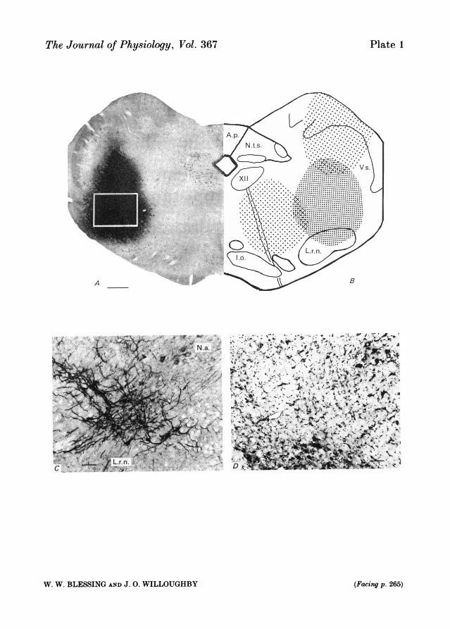
Plate 1The Journal of Physiology, Vol. 367
Kn
A
4* tj
"t r-V....ti 4'
.1r , A
a'*.
'%r .' .L .JtA.. WV
-- ' 'Is'-,s .
r. 1?.
)435. -' . 4<
W. W. BLESSING AND J. 0. WILLOUGHBY (Facing p. 265)

Al NORADRENERGIC NEURONES AND VASOPRESSIN
TANAKA, J., KABA, H., SAITO, H. & SETO, K. (1984). The action of the Al noradrenergic regionon physically firing neurons in the rat paraventricular nucleus. Brain Research 310, 138-141.
URANO, A. & KOBAYASHI, H. (1978). Effects of noradrenaline and dopamine injected into thesupraoptic nucleus on urine flow rate in hydrated rats. Experimental Neurology 60, 140-150.
VAN ITALLIE, C. M. & FERNSTROM, J. D. (1982). Osmolal effects on vasopressin secretion in thestreptozotocin-diabetic rat. American Journal of Physiology 242, E411-417.
WANG, B. C., SUNDET, W. D., HAKUMAKI, M. 0. K. & GOETZ, K. L. (1983). Vasopressin and reninresponses to haemorrhage in conscious, cardiac-denervated dogs. American Journal of Physiology245, H 399-405.
WESTLUND, K. N., BOWKER, R. M., ZIEGLER, M. G. & COULTER, J. D. (1983). Noradrenergicprojections to the spinal cord of the rat. Brain Research 263, 15-31.
WILLETTE, R. N., BARCAS, P. P., KREIGER, A. J. & SAPRU, H. N. (1984). Endogenous GABAergicmechanisms in the medulla and the regulation of blood pressure. Journal of Pharmacology andExperimental Therapeutics 230, 34-39.
WILLETTE, R. N., KRIEGER, A. J., BARCAS, P. P. & SAPRU, H. N. (1983). Medullaryy-aminobutyricacid (GABA) receptors and the regulation of blood pressure in the rat. Journal of Pharmacologyand Experimental Therapeutics 226, 893-899.
WILLOUGHBY, J. O., JERVOIS, P. M., MENADUE, M. F. & BLESSING, W. W. (1985). Stimulation ofalpha 1 adrenoceptors in the supraoptic nucleus elevates plasma vasopressin in the unanesthetisedrat. Neuroscience Letters supply. 19, S 109.
YAMASHITA, H. & KoIZUMI, K. (1979). Influence ofcarotid and aortic baroreceptorson neurosecretoryneurons in supraoptic nuclei. Brain Research 170, 259-277.
EXPLANATION OF PLATE
A, photomicrograph of Nissl stain of rabbit caudal ventrolateral medulla. The rostro-caudal level,1 mm caudal to the anterior border of the area postrema, is at the level of the exiting hypoglossalnerve rootlets and thus corresponds with the caudal, nicotine-sensitive, depressor area describedin the cat. The dark area is horseradish peroxidase reaction product, indicating the centre of theinjection site and the direction of spread. The size of the area depended on the concentration ofhorseradish peroxidase included in the injection. Bar = 0-6 mm. B, diagrammatic representationof the experimental site in the caudal ventrolateral medulla and the two control injection sites,dorsal and medial to the experimental site. C, photomicrograph of tyrosine hydroxylase immuno-reactive cell bodies and dendritic processes located in the region shown by the rectangle in A,demonstrating that A 1 neurones are located in the centre of the injection site. Bar = 100 um. D,photomicrograph of the same region from a different animal, stained for Nissl substance. Bycomparing C and D one can verify that the A 1 neurones occur in the region between the caudalcells of the nucleus ambiguus and the lateral reticular nucleus. Bar = 100 lm. Abbreviations: A.p.,area postrema; Lo., inferior olive; L.r.n., lateral reticular nucleus; N.a., nucleus ambiguus; N.t.s.,nucleus tractus solitarius; Vs., spinal nucleus of the trigeminal nerve; XII, hypoglossal nucleus.
265