importance and potential impact of liver fluke in cattle ...aciar.gov.au/files/node/9010/mn133 part...
TRANSCRIPT

Importance and potential impact of liver fluke in cattle and buffalo
D.B. Copeman and R.S. Copland

2Importance and potential impact of liver fluke in cattle and b2Importance and potential impact of liver fluke in cattle and b OVERCOMING LIVER FLUKE IN SOUTH-EAST ASIA
Introduction In this study the potential importance of liver fluke was estimated by considering the impacts of infection at the level of individual animals, farms or smallholdings, regions, countries and globally. Economic costs have been estimated for different reasons including the justification and creation of research or control programs, to design control programs to ensure that they are targeted correctly, and to assess the impact of control programs. The total global economic loss attributed to fasciolosis has been estimated earlier to be more than US$3 billion per year (FAO 1994).
This chapter reviews studies that may be used to estimate the losses caused by fasciolosis, with a focus on large ruminants in Asia. It is estimated that 300 million bovines are exposed to fasciolosis worldwide. The review builds on an earlier comprehensive review (Spithill et al. 1999) in which impacts on weight gain, draught performance, fertility and lactation were summarised and used to develop dollar estimates of lost productivity. These authors highlight the difficulty of such estimation, citing:
• incompleteinformationonalltheelementsofofftake (including milk, draught power, meat and asset accumulation) from cattle and buffalo production systems
• therisksofextrapolationfromexperimentsonFasciola hepatica (in cattle and sheep)
• thehighvariabilitywithinandbetween tropical production systems in breed, sex and cropping systems
• presenceofotherparasiteandinfectiousdiseases.
In the face of such incomplete and diverse information it is worthwhile to question the value of making broad estimates of loss. However, there is no doubt that control programs need to be justified and supported by accurate economic data as there is increasing competition for public and private investment. Also, even broad estimates will help to direct research towards new approaches to control and projects most likely to be beneficial.
The practical questions that stimulate these studies include the following.
• HowdoesliverflukeinfectioncausedbyFasciola gigantica reduce the productive outputs of cattle and buffalo in tropical Asia?
• Whatarethebestwaystomeasureproductionlosses at the local level?
• Whatarethebenefitsandcostsofsuppressivedrenching trials as a way of measuring local impact and demonstrating production losses to extension agents and farmers?
• CanGISmethodsbeusedtopredictareaswhereprevalence is sufficiently high to cause significant production loss?
• Canrecommendationsbemadeonthebestwaysto assess impact to justify, plan, implement and monitor liver fluke control programs?
The following sections review studies that estimate the existing and potential losses from F. gigantica in tropical cattle and buffalo systems. The main lessons to be drawn from these studies and the gaps that need to be filled by further research are discussed.
Importance and potential impact of liver fluke in cattle and buffalo
D.B. Copeman and R.S. Copland

2Importance and potential impact of liver fluke in cattle and b3IMPORTANCE AND POTENTIAL IMPACT OF LIVER FLUKE IN CATTLE AND BUFFALO
national and global impactsLiver fluke infection (fasciolosis) of cattle and buffalo is one of the most important parasitic diseases in Indonesia (Partoutomo et al. 1985) and its impact has been estimated in several studies. Thorbecke and van der Pluijm (1993) estimated that, of the overall losses from livestock diseases in Indonesia, losses from fasciolosis are second only to Newcastle disease. Prevalence of liver fluke in parts of Indonesia has been estimated at 25–90% in cattle and buffalo (Edney and Muchlis 1962; Soesetya 1975; Beriajaya 1979; Edney and Muchlis 1962; Soesetya 1975; Beriajaya and Soetedjo 1979), and in sheep and goats (Beriajaya and Copeman 1997). Estimates of the annual economic losses have been made by several authors (Table 2.1). The figure of A$96 million (US$65 million) (Winrock 1986) accounted only for the cost of lost meat production. The total annual cost of lost meat production, lost draught power and reduced fertility in infected cattle/buffalo has subsequently been assessed at A$176 million (US$107 million).
The losses in some other countries in Asia, including Cambodia, Vietnam and the Philippines, can be estimated where the farming practices in countries are similar to Indonesia and prevalence rates for fasciolosis are comparable. In 1997 the size of the cattle and buffalo herd in Asia was estimated at 589 million (FAO 1997) with the Indonesian herd (15 million) representing 2.5% of the Asian total. Using a conservative scenario of 10% prevalence and annual loss per infected animal of US$42, economic losses in cattle and buffalo alone exceeded US$2.4 billion in Asia. Similar calculations for the African cattle herd of 201 million (FAO 1997), where prevalence rates are similar to Asia, predict losses at US$0.84 billion, bringing total world losses to at least US$3.2 billion. The worldwide annual loss from fasciolosis is a substantial figure by any valuation and possibly greater than the earlier estimate of US$2 billion by Boray (1985).
Recent studies in Cambodia (Sothoeun 20071) have confirmed the impact of fasciolosis in areas of high prevalence. Where prevalence exceeds 30%, for example, annual weight gains are reduced by
1 Reproduced as an appendix to this monograph
20–40 kg, pregnancy rate is lowered by 10% and there is 2.5 kg less liver available for consumption. Taking into account these losses and the costs of implementing a practical control program the net benefit per head of cattle and buffalo in these high risk zones of Cambodia is 274–326 thousand riel (A$76–91).
Sources of production losses
weight gain
Monitoring weight gain of young animals is a useful way to assess the impacts of infection and interventions to prevent or control infection, improved nutrition or treatment with an anthelmintic. Younger growing animals are more susceptible to infection, less costly to maintain under experimental conditions and any results can be applied to production systems that are producing young growing animals for sale. A linear relationship between burden of adult F. gigantica and weight gain has been described in yearling zebu cattle. It was found that each fluke
South-East Asian countries rely heavily on the draught power of livestock but liver fluke infection can markedly weaken cattle and buffaloes.

2Importance and potential impact of liver fluke in cattle and b4Options for the control of liver fluke. OVERCOMING LIVER FLUKE IN SOUTH-EAST ASIA
table 2Importance and potential impact of liver fluke in cattle and b.1Overview. Prevalence of fasciolosis caused by Fasciola gigantica in several Asian countries.
Country range(%)
mean(%)
number of animals sampled
reference
Cambodia7–50 38 273 Sothoeun (unpublished)
245 575 Sothoeun et al. (2006) 0–57 12 1,406 Tum et al. (2004)
Overall estimate 18 (409 animals infected out of 2,254 sampled)Indonesia
25–48 36 Suweta (1982) Cattle 61
Buffaloes 3125 Soetedjo (unpublished)
Overall estimate 38 (Average of prevalence data)Philippines
35–100 Tongson (1978) Cattle Female 49 133 Molina et al. (2005)
Male 27 119Buffaloes Female 50 24
Male 37 8Cattle 33 250 E. Abonado (unpublished)
Buffaloes 47 3269 252 M.F. Guinsatao and N.B. Salcedo (unpublished)
Overall estimate 45 (318 animals infected out of 711 sampled)Vietnam
23–90 Luong (unpublished), 21–30% in goatsCattle 76% 140 Luong et al. (1999), ACIAR Meeting Balitvet
Indonesia (106 infected)Buffaloes 76 63 (47 infected)
Coastal 5.3–27.9 11,252 Luong (unpublished) (2,650 infected)Delta 13–59.2
Overall Vietnam 27.2% (3,119 infected out of 11,455 sampled)thailand
Beef 212 Pholpark (unpublished)Dairy 7
Buffaloes 70–85 12 Srihakim and Pholpark (1991)
laosCattle 9 76 Vongthilath (unpublished)
Buffaloes 21 274 Cattle 0–81 23 107 Douangngeun (unpublished)
Buffaloes 38 243Overall estimate 26 (182 animals infected out of 700 sampled)India
Cattle 24.4 516 Sanyal and Singh (1995)39 977 Gupta et al. (1986)
China 27 120,000 Jiangang (unpublished)

2Importance and potential impact of liver fluke in cattle and b5IMPORTANCE AND POTENTIAL IMPACT OF LIVER FLUKE IN CATTLE AND BUFFALO
reduced the potential annual gain by about 200 g, with infected animals achieving only about half the annual weight gain of that shown by control animals (Sewell 1966). Weight change in infected animals is also a useful parameter to monitor because most cases of fasciolosis are subclinical.
One deficiency of these types of studies is that many animals can compensate for reduced growth when the cause (e.g. liver fluke) is removed and so the total impact has not been measured accurately. Further, comparisons across studies are difficult because of differences in levels of infection, the age and sex of animals, the interaction between level of nutrition and pathogenic effect, and the difference between breeds and within breeds in resistance to infection. It is sufficient to conclude at this point that reports of weight-gain reduction need to be accompanied by descriptions of all the factors known to contribute to the effect of fasciolosis, including sex, age, breed and previous exposure.
draught performance
Anaemia resulting from fasciolosis has been shown to reduce work output by 7–15% (Roberts et al. 1991). Combined with a further indirect reduction of 20% in potential work capacity in animals whose growth has been restricted by fluke infection, it can be concluded that liver fluke can seriously lower the work potential of both cattle and buffalo. The economic significance of this may, however, be changing rapidly in production systems where hand tractors are replacing animals as sources of draught power. In many systems, however, even if there is a gradual reduction in their use, draught power from large ruminants remains important and should not be ignored. Indeed, as the prices for fossil fuel increase, draught animal power may remain viable in more isolated areas, particularly with poorer farmers.
The cost of the reduced draught capacity caused by F. gigantica may be measured as the opportunity cost to a farmer caused by the longer time taken by infected animals to perform a specific task; this amounts to about 27–35% more time with buffaloes according to the conclusions of J.A. Roberts and D.B. Copeman (unpublished). As the average draught animal in Indonesia is used in land preparation for growing rice only about 23 days per year, the
opportunity cost for a farmer with infected buffalo in this situation is thus the value of his labour for about seven days per year.
Further evidence that infection with F. gigantica adversely affects draught capacity was collected by Suhardono (2001) in Indonesia. Farmers were surveyed during the second year of a trial to measure the effects in Ongole cattle of a single treatment with triclabendazole administered in July, about six weeks after harvest of the second seasonal rice crop in the area. The survey revealed that animals treated for fasciolosis (with triclabendazole) were used twice as many days as untreated animals for preparing land for planting rice. This result suggests that farmers recognised that the treated cattle performed better than those that were untreated. Furthermore, those with untreated animals avoided the opportunity cost associated with increased time to prepare their land by hiring animals that had received treatment. Thus, where this hiring option is available, the economic cost associated with reduced work capacity in animals infected with F. gigantica may be the cost of hiring replacement animals for land preparation rather than the opportunity cost of a farmer’s labour.
Fertility
A link has also been observed between infection with F. gigantica, anaemia and fertility (Suhardono 2001). There were significantly longer intercalving intervals and a lower packed cell volume in Ongole cows in Indonesia infected with F. gigantica than in those treated with triclabendazole each July for two years. In his study, Suhardono (2001) found that treated cows had a mean intercalving interval of 18.5 months whereas in untreated cows the interval was 31.5 months. It is thus reasonable to conclude that infection with F. gigantica is likely to adversely affect reproduction. Furthermore, the extent may be proportional to the degree of anaemia induced, an outcome that varies according to the level of nutrition, level of infection and breed.
lactation
Needham (1977) found no measurable difference in weaning weight of calves (an indicator of milk output of their dams) from cows infected with

2Importance and potential impact of liver fluke in cattle and b6 OVERCOMING LIVER FLUKE IN SOUTH-EAST ASIA
F. gigantica and those treated each 8–12 weeks with an adulticide. Such contradictory reports are to be expected due to differences between studies in factors likely to affect milk output. These include level of nutrition, size of infection and differences in resilience to infection between individuals and breeds. However, at present, too few studies have been undertaken to enable meaningful prediction of the extent to which any of these determinants affects lactation in animals infected with F. gigantica.
Sources of prevalence dataThe first step in establishing the importance and cost of fasciolosis is to determine the prevalence of infestation. A simple approach to estimating the national impact of fasciolosis is to estimate prevalence in different systems, determine the loss caused by infestation per animal per year in each of these systems, and then multiply these to provide an overall estimate of impact.
No comprehensive, countrywide surveys of prevalence have been conducted in South or South-East Asia. Prevalence is difficult to establish because of the dependence on local physical and climatic conditions for the survival of the intermediate host snail (see Chapter 3). Prevalence may vary from 0% to 100% over a comparatively short distance (Srihakim and Pholpark 1991; Tum et al. 2004). Therefore, figures given for the national prevalence may conceal certain areas of high risk and high prevalence and hence loss. Nevertheless, national figures are presented in Table 2.1 to provide an overview.To establish prevalence, faeces need to be collected for faecal egg counts or livers examined for parasites in abattoirs. Both of these techniques are subject to inaccuracies. The faecal egg count method has a low sensitivity with many false negatives. Sothoeun et al. (2006) reported that 27% of animals with F. gigantica in their livers yielded negative faecal egg counts. Therefore, prevalence estimates based on faecal egg counts may underestimate true prevalence. On the other hand, prevalence data based on abattoir surveys may be inaccurate because older and sometimes sick animals are generally slaughtered (Sothoeun et al. 2006; Luong, unpublished).
The epidemiology of fasciolosis suggests that the prevalence would be higher in wetter, more fertile areas (see Chapter 3). These high-risk intensive agricultural areas are often associated with high numbers of cattle and buffaloes.
A further source of bias is that districts selected for study have often been chosen because preliminary data indicated these were areas of high risk. This is illustrated by the data from Cambodia. Sothoeun et al. (2006) found prevalence levels of 24.7%. However, a preliminary whole country study by Tum et al. (2004) found a national average prevalence of 11.6% (163 infected animals out of 1406 tested). Multiple methods were used to estimate prevalence and the level of infection at which each animal was categorised as ‘infected’ varied widely. Nonetheless, assuming that each infected animal lost production by an average amount determined by experimentation some national figures for the economic impact of the fasciolosis can be generated (Table 2.2).
regional observations of production lossesGiangxi province, China
Fasciolosis is found in cattle, buffaloes and goats in all provinces of China with Fasciola gigantica found in 10 southern provinces. Most research has been on Fasciola hepatica and sometimes F. hepatica and F. gigantica were not distinguished. From 1949 to 1989 several national surveys were conducted in which major mortalities in goats, cattle and buffalo were identified. Fasciola gigantica is found only in south China, south of the Changjiang River. Fasciola hepatica and F. gigantica are found in varying proportions, often together in the same animals. In Guangxi F. gigantica was more common than F. hepatica and in some counties the infection rates are above 90%.
There are about 10 million cattle and buffaloes in Guangxi. They are owned by smallholder farmers who represent about 80% of the 40 million people in the province. Most of the area is mountainous and cattle and buffaloes are allowed to graze freely in hilly areas. Fasciolosis is more serious in rice-producing areas.

2Importance and potential impact of liver fluke in cattle and b7IMPORTANCE AND POTENTIAL IMPACT OF LIVER FLUKE IN CATTLE AND BUFFALO
table 2Importance and potential impact of liver fluke in cattle and b.2Importance and potential impact of liver fluke in cattle and b Estimated number of cattle and buffaloes infected with Fasciola gigantica in various countries of Asia in areas of high or low prevalence. As an approximate indication of the total costs in each area the number of infected animals has been multiplied by the cost per infected animal derived from Sothoeun (2007, see also appendix) of between A$82 and A$98. Livestock numbers are derived from FAO (2004) and the prevalence data from Table 2.1.
Country total number of large ruminants
(thousands)
Prevalence scenario
total number of infected animals
(thousands)
range of total losses (millions of
Australian dollars)
Cambodia 3,625 High (18.1%) 656 54–64Low (11.0%) 399 33–39
Indonesia 14,000 High (38.2%) 5,355 439–525Low (20.0%) 2,800 230–274
Philippines 5,799 High (44.8%) 2,598 213–255Low (34.0%) 1,972 162–193
Vietnam 7,050 High (27.0%) 1,903 156–186Low (15.0%)) 1,057 87–104
Thailand 7,000 High (11.8%) 826 68–81Low (11.8%) 826 68–81
Laos 2,350 High (26.0%) 611 50–60Low (15.0%) 352 29–34
China 129,348 High (27.2%) 35,182 2,885–3,448Low (12.0%) 15,521 1,273–1,521
India 283,200 High (25.0%) 70,800 5,806–6,938Low (10.0%) 28,320 2,322–2,775
Total 452,372 High (26%) 117,932 9,670–11,557Low (11%) 51,248 4,202–5,022

2Importance and potential impact of liver fluke in cattle and b8Immunology and assessment of resistance to fasciolosis in smal OVERCOMING LIVER FLUKE IN SOUTH-EAST ASIA
India
In India fasciolosis is widespread and is primarily caused by F. gigantica although F. hepatica is reported in the temperate Himalayan region. Building of dams and the establishment of new irrigation systems have further widened the distribution of Fasciola by creating more water-covered areas suitable for propagation of its intermediate hosts, the lymnaeid snails. Thus, fasciolosis has started to appear in the semi-arid and arid regions of western India where it was hitherto non-existing.
The onset and advancement of monsoon rains have a profound effect on the incidence and seasonality of fasciolosis in India. Most of the available information on the prevalence of F. gigantica comes from abattoir surveys and coprological studies on animals visiting clinics, and is thus biased. It is, however, apparent that the prevalence of fasciolosis in a tropical country like India is largely determined by rainfall and production systems. A review of some of the recently conducted surveys indicate a high level of incidence in the endemic areas throughout India but in the endemic areas of the northern plains, a high 10–39% infection was recorded in cattle and buffalo. A nationwide survey in dairy animals organised by the National Dairy Development Board (NDDB) indicated two critical periods in the year: July–September and February–March (Sanyal and Singh 1995).
lao Pdr
A survey of 76 cattle and 274 buffaloes killed at the slaughterhouse of the Vientiane municipality revealed infection with F. gigantica in the livers of 9% of cattle and 21% of buffaloes. At the slaughterhouse of Luang Prabang only 1 of 2 cattle and 17 buffaloes examined were infected.
Fasciola gigantica, Toxocara vitulorum and strongyles are regarded as the most important parasites of ruminants in the Lao PDR. Fasciolosis has a substantial negative effect on production but is not a significant cause of mortality. It is recommended that valuable animals are treated but preventive programs that rely on chemotherapy are unlikely to be sustainable (Vongthilath, unpublished; Douangngeun, unpublished.)
Philippines
Fasciolosis is still the leading cause of morbidity and mortality in ruminants in the Philippines. Fasciolosis research has always played second fiddle to research on the more dramatic and explosive diseases with international implications, which would account for the meagre research output. The low research priority given by the national government to fasciolosis and other economically important parasitic diseases and the lack of trained manpower for research have all contributed to a lack of qualitative and quantitative information.
Of the 252 cattle examined in Cotabato (Mindanao) in 1997, 173 (69%) were found infected with F. gigantica based on the presence of Fasciola eggs in the faeces. Cattle older than four years had the highest prevalence (82%), for those two to four years old the prevalence was 78% and for those five months to two years, prevalence was 53%. The prevalence was 72% in females and 62% in males. Barangay Bannawag had the highest prevalence proportion (92%), followed by Katidtuan (89%). Proportions of animals infected among eight villages ranged from 36% to 73%.
A study in two slaughterhouses (Mlang and Kabacan) in 1999 determined the prevalence of fasciolosis in cattle and carabaos. All animals from Mlang were negative for fasciolosis. Of 282 animals examined from Kabacan, 83 (33%) cattle and 15
Growth rates and market value of livestock are reduced by liver fluke infection.
Greg Hood

2Importance and potential impact of liver fluke in cattle and b9IMPORTANCE AND POTENTIAL IMPACT OF LIVER FLUKE IN CATTLE AND BUFFALO
(47%) carabaos were positive. There was a higher prevalence of fasciolosis in cattle and carabaos older than six years (69% and 59%, respectively). Carabaos three to six years old had a prevalence of 25% and those younger than one to three years 17% (Molina et al. 2005).
thailand
The economic loss from fasciolosis in cattle and buffalo throughout Thailand has been assessed at not less than 100 million Baht (about US$3 million). Recent investigations have shown that the average prevalence of F. gigantica in cattle and buffalo in Thailand was 12%. However, the prevalence varies considerably between villages, ranging from 0% to 85%. Prevalence is high in areas surrounding dams or large ponds in which Lymnaea auricularia rubiginosa, the intermediate host of F. gigantica is found. The disease has a seasonal pattern on which control of the disease is based. All cattle and buffalo older than eight months should be treated for liver flukes each September. In addition, animals in poor condition should be treated in April to prevent severe losses, especially in high-prevalence areas or where strategic treatment was missed.
Diagnostic results of the Northeast Regional Veterinary Research and Diagnostic Center in 1998 revealed that the average prevalence proportions of infection with F. gigantica in beef cattle, dairy cattle and buffaloes in north-east Thailand were 22%, 7% and 7% respectively. However, the prevalence varied considerably between villages.
Humans are also infected with F. gigantica. From 1967 to 1990, 25 cases of human fasciolosis were reported in Thailand, of which 19 occurred in the north-east. Since then at least 10–20 new cases of human fasciolosis have been confirmed in the Khon Kaen University Hospital each year. Fasciolosis should therefore be regarded as a newly emerged zoonosis and an important public health problem (Pholpark, unpublished).
Cambodia
A national survey was conducted in Cambodia to identify zones of high prevalence for further studies into the epidemiology of this infection. Faecal samples were collected from 273 cattle and buffaloes in October and November 1998 and
examined for F. gigantica eggs. The 14 villages in this study were in Saang district of Kandal province and in Cheung Prey district of Kompong Cham province. The proportion of animals with Fasciola eggs in their faeces varied greatly from village to village. All villages in Cheung Prey district had < 20% prevalence and the mean of samples from these villages was 7%. In Saang district, one of the six villages had a low prevalence, two had a medium level (20–50%), and two had a high prevalence (> 50%). The average prevalence in Saang district was 38% (Suon et al. 2006).
Vietnam
Between 1996 and 1998 faeces of 11,252 cattle or buffaloes from different regions of Vietnam were examined for eggs of F. gigantica. Prevalence varied from 5–28% in the coastal areas to 13–59% in the delta areas. In earlier studies age was shown to be an important determinant of infection. Prevalence increased from 16% in animals younger than three years to 37% in animals older than five years. An abattoir survey of 495 cattle and buffaloes also revealed increasing prevalence with increasing age. At three months of age, 6.8% of animals examined were infected. Prevalence in animals from four to six months old was 11%, from 7–12 months 36.4% and from 12–24 months 45.5% (Luong, unpublished).
nepal
Among the diseases of ruminants, fasciolosis is probably the most common and perhaps one of the most important causes of livestock deterioration in Nepal. It is widespread throughout the country, affecting all species of ruminant livestock, including yaks and yakows of the Himalayas. The different local names of this disease, such as namle, mate and lew, in different regions are proof of its continued existence for many years in Nepalese animals.
Singh et al. (1973) reported an infection rate of 50–90% in animals in areas below 1800 m and estimated it caused an annual economic loss of NRs 200 million (US$20 million). Recent studies have indicated a similar prevalence of the disease but a higher estimate of economic loss (US$37 million) calculated only on decreased buffalo milk and buffalo meat production (Mahato 1993; Mahato et al. 1997).

30 OVERCOMING LIVER FLUKE IN SOUTH-EAST ASIA
Although infections with F. hepatica have been reported, infection with F. gigantica is the most common and widespread cause of fasciolosis throughout the country. Among the species of lymnaeid snails found in Nepal, Lymnaea auricularia race rufescens, L. auricularia sensu stricto and L. viridis have been identified as the important intermediate hosts.
The pre-monsoon rains together with rice cultivation practices promote habitat creation for Lymnaea spp. over a wide area. After the monsoon when rice is harvested, animals acquire infection during grazing from heavily contaminated rice fields. Rice straw, the principal food of large ruminants during the dry season (December–April), is another important source of infection with Fasciola, especially in stall-fed animals. Thus, it appears that there is a good case for introducing appropriate management practices to prevent animals becoming infected.
In Nepal, attempting to control the intermediate hosts using molluscicides, even during the dry season, is of limited value because of the numerous permanent habitats and the great biotic potential and aestivating ability of the lymnaeid snails. Epidemiological studies have revealed that most snails are infected by fluke eggs deposited on pasture during March–May and again in October–November. This pattern suggests that administration of anthelmintic in February and again in late August to control pasture contamination is an appropriate strategy for the control of fasciolosis in Nepal.
Suppression trials with anthelminticsApplying anthelmintics to kill off existing infections and prevent new ones has made it possible in a few cases to measure production loss in situ. That is, infected and uninfected animals have been compared in their normal production environment. The value of such studies is that an upper limit of acceptable benefits for deworming or other control measures can be estimated without having to rely on extrapolation from experimental results. The disadvantage remains, however, that to estimate the total effect, treatment needs to be continued for at least a complete season. Even then the impacts on
reproductive rate are not included. The results of the three studies described below have been important in increasing the accuracy of production loss estimates and raising awareness among scientists, policymakers and farmers, of the magnitude and extent of the liver fluke problem. These, in themselves, are important outcomes.
Philippines
A two-year suppressive treatment study begun in February 2000 compared irrigated and rainfed cropping systems. Selection of the study areas was based on the recommendation of livestock technicians. The number of animals, willingness of the animal owners and barangay officials to cooperate, accessibility of the area and whether they were from a Christian or Muslim area were all taken into account.
Takepan was chosen for the irrigated cropping system, and Colambog for the rainfed area. Both areas are in Pikit, Cotabato province and inhabited by both Muslim and Christians. Buffaloes and cattle were weighed, drenched with triclabendazole (Fasinex-Novartis) at a dose rate of 24 mg/kg body weight and 12 mg/kg body weight respectively. Two other areas, Dagupan in Kabacan and Datu Paglas in Maguindanao, serve as controls for the rainfed and irrigated areas, respectively. Data such as age, sex, body weight, reproductive performance, feeding system, draught power and function of the animals were gathered from both the treated and the control groups every four months for two years. All animals used in the study were marked with ear tags. The buffaloes are used for draught power for land preparation in lowland and in upland areas and for hauling farm products such as coconuts, rice, corn and other farm products from the field to the market. Cattle, however, were used for meat and breeding and not for draught power. Cattle and carabaos (buffaloes) graze communal areas near the dikes or river banks and canals and, from the rice planting to harvesting period, are fed with grasses by a cut and carry system or allowed to graze.
Information programs and consultations with barangay (district) officials and animal owners were conducted before the study began. Information about the project and its activities, about fasciolosis and about possible benefits from the study were

31Overview.IMPORTANCE AND POTENTIAL IMPACT OF LIVER FLUKE IN CATTLE AND BUFFALO
explained. Faecal egg counts were done to determine the prevalence of fasciolosis in the respective areas.
The average age at first calving was 4.0 years old in carabaos and 2.6 in cattle. The age group of one day old to three years old was more common than the older age groups in all study areas. There were more cattle (643) than carabaos (501) and more female animals (772) than male (340) and castrated animals (32). The mean live-weight gain per day was higher in treated animals in the irrigated area (0.32 kg) than in the control group (0.24 kg). However, in the rainfed area it was higher in the control group (0.35 kg) than in the treated group (0.29 kg). Generally, cattle had higher weight gain than in buffaloes in all study areas except in the control irrigated area with few cattle. The intercalving interval in buffaloes in control treated and rainfed areas was the same (19 months) but was higher in the treated irrigated areas (13 months) than in the control irrigated area.
In cattle, the intercalving interval was 16 months and 14 months in treated rainfed and irrigated areas and 15 months in control rainfed. However, data on the intercalving interval for controls in irrigated areas are not yet available. The prevalence rate for fasciolosis infection during the preliminary sampling in the study areas ranged from 33% to 95%.
Indonesia
A study by Suhardono (2001) demonstrated that the treatment of cattle reared in West Java in association with intensive production of irrigated rice with triclabendazole in July significantly reduces their level of exposure to infection with F. gigantica over the following 12 months. In comparison with untreated animals treated animals had improved reproductive and draught performance and higher packed cell volume (PCV) values, and yearlings had a higher weight gain. It was concluded that the timing of such
Liver fluke reduces the output of buffalo and cattle milk for consumption by calves and human consumers.
ILRI/Steve Mann

32Importance and potential impact of liver fluke in cattle and b OVERCOMING LIVER FLUKE IN SOUTH-EAST ASIA
annual treatment should be about six weeks after harvest of the second seasonal rice crop in an area. Furthermore, to achieve the best effect, all animals sharing common grazing should receive treatment.
Treatment in July may be regarded as strategic as it is about six weeks after the end of the period when rice is harvested, which is when most infection with F. gigantica occurs in this area.
Cambodia
The impact of fasciolosis on weight gain, reproductive performance, draught capacity and PCV of cattle was studied in the upper delta of the Mekong River in Cambodia where the risk of infection with F. gigantica is high (Tum et al. 2004). Farmers in two villages in Saang province, Prek Samrong and Preak Trang, participated in the study. Cattle in Preak Samrong were maintained free from infection with F. gigantica by treating them every three months for nine months with triclabendasole at 12 mg/kg body weight. No control of F. gigantica was practised in Preak Trang but cattle received the placebo Zanisef every three months. At the start of the study, there were 224 cattle in Preak Samrong village made up of 60 castrated males, 113 females, 49 male calves and 2 bulls. In Preak Trang village, there were 202 cattle comprising 13 castrated males, 144 females, 41 male calves and 4 bulls. Every three months all animals were weighed, faeces were collected for examination for Fasciola eggs, blood was collected for estimation of PCV, and condition score, skin coat, and draught strength were recorded. Weights of pregnant females were adjusted to remove the effects of pregnancy.
In Preak Samrong 52 animals were sold: 13, 26 and 13 during first, second and third periods of three months respectively. In Preak Trang, 69 cattle were sold during the same periods comprising 32, 14 and 23 animals respectively.
Mean weight gains of males and females were similar for the age group 0–0.5 years and for 0.6–1.5 years so were combined in analyses of differences between control and treated groups. Nine months after observations began treated males plus females in both the 0.5–1.5 years and 1.6–2.5 years age groups had gained significantly more weight than comparable animals in the control village (Table 2.3). During the same period weight gains of treated and control castrated males three years of age and over were similar, as were weight gains of females in this age group (Sothoeun et al. 2006).
Predicting impacts using geographical information systemsA geographic information systems (GIS) model for mapping the risk of fasciolosis in cattle and buffaloes was developed for the Kingdom of Cambodia using determinants of inundation, proximity to rivers, land use, slope, elevation, and the density of cattle and buffaloes. Determinants were subjectively weighted according to their perceived relative importance before combining them to produce a risk map of fasciolosis. The model estimates that 28% of Cambodia is potentially at risk of fasciolosis with
table 2Importance and potential impact of liver fluke in cattle and b.3 Comparison of mean weight gains ± SE of treated and control groups over the nine months of observations in Cambodia.
weight gains (kg, mean ± SE)
Groups compared treated Control P value
Male + female 0.5–1.5 years 99.0 ± 10.3 74.2 ± 4.9 0.02
Male + female 1.6–2.5 years 86.4 ± 4.4 73.9 ± 4.3 0.05
Castrated male 3 years and over 45.1 ± 4.9 43.1 ± 4.7 0.77
Females 3 years and over 39.3 ± 6.7 28.3 ± 4.8 0.19

33IMPORTANCE AND POTENTIAL IMPACT OF LIVER FLUKE IN CATTLE AND BUFFALO
Figure 2Importance and potential impact of liver fluke in cattle and b.1Overview. The risk of transmission of fasciolosis due to Fasciola gigantica (adjusted by animal density) as predicted by the GIS model developed by Tum et al. (2004) for Kampong Cham province and the four districts that were surveyed for prevalence of fasciolosis in cattle to test the model.
areas of high and moderate risk concentrated in southern and central Cambodia. The estimates of risk reflect the actual prevalence of fasciolosis in most districts surveyed, suggesting that the epidemiological determinants and weightings used to produce the model were appropriate. These results will be progressively refined as more detailed field surveys are completed to fully validate the model. A comparison between levels of risk predicted by the maps and field measurements of prevalence in 11 provinces (n = 1406) showed general agreement, which suggested that the epidemiological determinants and weightings used to produce the maps are appropriate (Tum et al. 2004). One constraint on the validity of this conclusion was that prevalence was measured at the provincial level, a very large unit, and animals were not sampled at random. An additional study sought to correct this deficiency by measuring prevalence at a more
detailed scale, at district level. Faecal samples were collected from a randomly selected set of animals in four districts in one province for areas at high, moderate and no risk. This result supported the earlier conclusion that there is a good relationship between prevalence and risk predicted by the GIS model (Figure 2.1).
In Thailand (Pholpark, unpublished observations) fasciolosis is one of the most important parasitic diseases in adult cattle and buffaloes in north-east Thailand. Prevalence varies considerably between villages. It is higher in areas surrounding dams or large ponds in which Lymnaea (Radix) auricularia rubiginosa, the snail intermediate host of F. gigantica, is found. Strategic treatment with an effective drug is considered to be the most effective control measure. In this study, instead of performing an epidemiology survey, a GIS is being applied to define the fasciolosis risk areas and the

34Options for the control of liver fluke. OVERCOMING LIVER FLUKE IN SOUTH-EAST ASIA
appropriate time for strategic treatment of animals. In a pilot GIS study in Khonkaen province during 1999 the parameters used for analysis of the data were surface water, rainfall, temperature, and the boundary of the province, district or subdistrict. From results of the pilot study the high Fasciola risk areas and the time for strategic treatment were defined quite accurately when compared with the results of the epidemiological study that had been conducted from 1982 to 1984 (Srikitjakarn et al. 1988). However, the parameters used were considered too imprecise. To improve their predictive precision, seven parameters are now being used for analysis of data in the current GIS field trial in Kalasin province. They are:
• temperature
• rainfall
• surfacewater
• boundaryofriver,brookorirrigationcanal
• slopeclass
• boundarybetweenwetlandsandgrazingareas
• theboundaryoftheprovince,district, or sub-district.
ConclusionsIt is evident from these studies that comprehensive data on the impact of fasciolosis over several years are difficult and expensive to obtain. This is due mostly to the long-term and chronic nature of the disease, its multiple effects on productivity and the difficulty of making an accurate diagnosis. There is also a natural bias in many studies, including those reported here, to focus on areas where the problems are known and not undertake a systematic survey with a wider statistical base. Nonetheless there is no doubt that the disease is widespread throughout most of tropical Asia with hotspots in areas susceptible to seasonal flooding and irrigated rice production. The exact figures will always be open to discussion but a consensus view would be that the impacts are relatively high and often unrecognised.
The challenge is how to use the information. Two pathways are suggested by the data reported here. One is to make use of increasingly accessible technologies based on GIS mapping to make predictions about places and systems that can be targeted for control programs. As more data are
Animals infected by liver fluke produce fewer offspring and the interval between litters is longer.
Doug
Gra
y

35IMPORTANCE AND POTENTIAL IMPACT OF LIVER FLUKE IN CATTLE AND BUFFALO
collected the predictive models for such programs will become more accurate and useful to planners and extension services. The second pathway is to develop relatively simple ways to demonstrate to farmers and extension workers the impacts of fasciolosis and the benefits of control. Computer models and reports are not sufficient to convince them that their time and resources should be used for fluke control when there are so many other competing demands. The use of suppressive drenching with monitoring of the impacts by farmers and extension workers themselves may provide the required stimulus.
references Beriajaya and Copeman D.B. 1997. An estimate
of seasonality and intensity of infection with gastrointestinal nematodes in sheep and goats in West Java. Jurnal Ilmu Ternak dan Veteriner 2, 270–276.
Beriajaya and Soetedjo R. 1979. Annual report, Lembaga Penelitian Penyakit Hewan. Research Institute for Veterinary Science: Bogor, Indonesia.
Boray J.C. 1985. Flukes of domestic animals. Pp. 179–218 in ‘Parasites, pests and predators’, ed. by S.M. Gaafar, W.E. Howard and R.E. Marsh. Elsevier: New York.
Dalton J.P. 1999. Fasciolosis. CAB International: Wallingford, UK.
Edney J.M. and Muchlis A. 1962. Fascioliasis in Indonesian livestock. Communications Veterinariae 6, 49–62.
FAO 1994. Diseases of domestic animals caused by liver flukes: epidemiology, diagnosis and control of Fasciola, paramphistome, Dicroceoelium, Eurytrema and schistosome infections of ruminants in developing countries. FAO: Rome.
FAO 1997. FAOSTAT online database, at: <http://faostat.fao.org>. Accessed 3 April 2008.
Gupta R.P., Yadav C.L. and Ruprah N.S. 1986. The epidemiology of bovine fasciolosis (Fasciola gigantica) in Haryana state. Indian Veterinary Journal 63, 187–190.
Kumar P. and Pachauri S.P. 1989. Efficacy of albendazole against Fasciola gigantica infection in buffaloes with particular reference to milk production. Journal of Veterinary Parasitology 3, 35–39.
Luong To Thu and Phan Dich Lan 1999. Fasciolosis in cattle and buffalo in Vietnam. In ‘Control of fasciolosis in Indonesia, Cambodia and the Philippines. Proceedings of the Second Annual Fasciolosis Control Coordination Meeting (ACIAR Project AS1/96/160)’, Davao, Southern Mindanao.
Mahato S.N. 1993. Epidemiology and pathogenesis of fasciolosis in eastern Nepal. Ph.D. Thesis, University of Edinburgh.
Mahato S.N., Harrison L.J.S. and Hammond J.A. 1997. Epidemiological basis of the control of fasciolosis in Nepal. Bulletin of Veterinary Science and Animal Husbandry Nepal 25, 22–26.
Many smallholders rely on large ruminants to transport farm produce. Liver fluke infection can reduce their pulling power.
Alice Kenney

36 OVERCOMING LIVER FLUKE IN SOUTH-EAST ASIA
Molina E.C., Gonzaga E.A. and Lumbayo L.A. 2005. Prevalence of infection with Fasciola gigantica and its relationship to carcase and liver weights, fluke and egg counts in slaughter cattle and buffaloes in South Mindanao, Philippines. Tropical Animal Health and Production 37, 215–221.
Needham A.J.E. 1977. Observations on the economics of treatment of Fasciola gigantica infestation in cattle in Rhodesia. Rhodesian Veterinary Journal 8, 14–20.
Partoutomo S., Ronohardjo P., Wilson A.J. and Stevenson P. 1985. Review of diseases in Indonesia affecting draught power in domestic animals. Pp. 140–146 in ‘Draught animal power for production’, ed. by J.W. Copland. ACIAR: Canberra.
Roberts J.A., Bakrie B., Copeman D.B. and Teleni E. 1991. An assessment of the work output of buffalo infected with Fasciola gigantica. ACIAR: Canberra.
Sanyal P.K. and Singh D.K. 1995. Administration of fenbendazole in urea molasses block to dairy buffaloes in India. Tropical Animal Health and Production 27, 186–190.
Sewell M.M.H. 1966. The pathogenesis of fascioliasis. Veterinary Record 78, 98–105.
Simpson J.R., Kunkle W., Courtney C.H. and Shearer J.K. 1985. Economic analysis of controlling liver fluke. Agri-Practice 6, 20–24.
Singh N.B., Basnyat B.M., Eichenberger G. and Bommeli W. 1973. Report on preparatory phase of parasite control project. HMG/SATA: Kathmandu, Nepal.
Soesetya R.H.B. 1975. The prevalence of Fasciola gigantica infection in cattle in East Jawa, Indonesia. Malaysian Veterinary Journal 6, 5–8.
Sothoeun S. 2007. Fasciolosis of cattle and buffaloes and its control measures. Technical implementation procedure, Ministry of Agriculture, Forestry and Fisheries, Kingdom of Cambodia. (Reproduced as an appendix to this monograph.)
Sothoeun S., Davun H. and Copeman B. 2006. Abattoir study on Fasciola gigantica in Cambodian cattle. Tropical Animal Health and Production 38, 113–115.
Soun S., Hol D., Siek S., McLean M.and Copeman B. 2006. Seasonal differences in the incidence of infection with Fasciola gigantica in cambodian cattle. Tropical Animal Health and Production 38, 23–28.Spithill T.W., Smooker P.M. and Copeman B. 1999. Fasciola gigantica: epidemiology, control, immunology and molecular biology. Pp. 465–520 in ‘Fasciolosis’, ed. by J.P. Dalton. CAB International: Wallingford, UK.
Srihakim S. and Pholpark M. 1991. Problem of fascioliasis in animal husbandry in Thailand. Southeast Asian Journal of Tropical Medicine and Public Health 22 Supplement, 352–355.
Srikitjakarn L., Pholpark M., Leidl K., Loehr K.F. and Hoerchner F. 1988. The epidemiology and control of bovine fascioliasis in Northeast Thailand. Thai Journal of Veterinary Medicine 18, 9–22.
Suhardono D. 2001. Epidemiology and control of fasciolosis by Fasciola gigantica in ongole cattle in West Java. Ph.D. Thesis, Tropical Veterinary Science, James Cook University of North Queensland: Townsville Australia.
Suweta I.G.P. 1982. Economic loss due to liver fluke infestation in cattle implicative to interactions in the environmental conditions of the agricultural ecosystem in Bali. [Gangguan ekonomi cacing hati pada sapi implikasi interaksi dalam lingkungan hidup pada edosistem pertanian di pulau Bali]. Universitas Padjadjaran, Ph.D. Thesis: Bandung, Indonesia:
Thorbecke E., van der Pluijm T. 1993. Rural Indonesia: Socioeconomic development in a changing environment. NYU Press: New York.
Tongson M.S. 1978. A national fascioliasis control program for the Philippines (a professorial lecture). Pp 106–117 in ‘Proceedings of the Annual Convention of the Veterinary Practitioners Association of the Philippines’.
Tum S., Puotinen M.L. and Copeman D.B. 2004. A geographic information systems model for mapping risk of fasciolosis in cattle and buffaloes in Cambodia. Veterinary Parasitology 12, 141–149.

Epidemiology of Fasciola gigantica in cattle and buffalo
Suhardono and D.B. Copeman
38Immunology and assessment of resistance to fasciolosis in smal OVERCOMING LIVER FLUKE IN SOUTH-EAST ASIA
Epidemiology of Fasciola gigantica in cattle and buffalo
Suhardono and D.B. Copeman
IntroductionThe patterns of infection of liver fluke are determined by the interaction of environmental factors, the biology of the parasite and the production system in which cattle and buffalo are expected to perform. Given the essential relationship between the parasite Fasciola and the water-dwelling snails of the genus Lymnaea that act as intermediate hosts, it is no surprise that liver fluke disease is most common in rice production systems (or areas providing a suitable habitat for the intermediate host), which may be contaminated with parasite-containing cattle faeces. These systems are not static, with gradual transition from single to double, to sometimes triple rice crops per year, and increasing reliance on machinery to replace large ruminants as a source of draught power. Nonetheless is it is still common for cattle and buffalo to be allowed to graze on recently harvested rice paddies, and straw to be cut and carried to stall-fed animals. Cattle faeces, possibly containing parasite eggs, are also used as fertiliser in many rice production systems.
A series of experiments and surveys from ACIAR projects are described which, when taken with other published work from other parts of Asia, identify opportunities for intervention and control. The results underscore the paradigm that has dominated modern integrated control of parasites: that control can only occur when the life cycle, environmental interactions and production systems are well understood.
The practical questions that stimulated these studies include the following.
• Whatistheseasonalpatternofliverflukeinfection and how best can it be assessed in smallholder farming systems?
• Whatistheroleofsnailsinthetransmissionof Fasciola gigantica in each of the production systems studied?
• Whatarethesourcesofinfectionforsnails,andfor large ruminants by metacercariae, and can these sources be eliminated?
• Whatopportunitiesareavailableforbiologicalcontrol through competition with the intermediate stages of F. gigantica?
• Howdosourcesandpatternsofinfectiondifferbetween rice-based systems and more extensive grazing systems?
the parasiteFasciola gigantica is one of the most important parasites of cattle and buffalo in the humid tropical areas of the world. In some regions it may also be important in sheep, goats and other domestic animals. Wild herbivores are susceptible, but laboratory animals are not readily infected. Human infection may be more common in endemic areas than the occasional case reports suggest. Hammond and Sewell (1975) proposed that F. gigantica is better adapted to cattle than sheep in that it is more infective and lives longer in cattle.
The intermediate hosts of F. gigantica are tropical aquatic snails which thrive in clear, stagnant or slow-moving water with high oxygen content and abundant aquatic vegetation (Kendall 1954). Such ecological situations are typically found at the fringes of rivers or lakes where the water levels are stable, and in irrigated rice fields throughout the humid tropics. Fasciola gigantica is transmitted worldwide by snails not readily distinguishable on morphological grounds or on grounds of their ecological requirements from the single superspecies Lymnaea auricularia sensu lat. (Kendall 1965). The life cycle
3

39EPIDEMIOLOGY OF FAsCiOLA gigAnTiCA IN CATTLE AND BUFFALO
3 Once the water level drops too fast during the dry season for the fringing aquatic vegetation to persist or the oxygen level drops too low, the habitat will be rendered unsuitable for these snails (Kendall 1954). In irrigated rice fields the population of snails is influenced by the availability of water for irrigation and the stage of growth of the crop. Snails and their eggs surviving from the previous crop may colonise recently planted rice fields or they may enter with water introduced to flood the field after planting. Their numbers then increase over the next few months before declining again a few weeks before harvest (Widjajanti 1989). Despite being aquatic, the snail hosts of F. gigantica are able to survive desiccation in the shade on the surface of the soil for some weeks. After one month, Widjajanti (1989) observed that mean survival time was reduced by about one-third, and egg masses by about one-half relative to hydrated controls. However, she found no adverse affect of desiccation for one month on subsequent hatchability of eggs. The eggs did not hatch until rehydrated, prompting her to propose this as a possible mechanism for survival of the population in habitats subject to periodic desiccation.
Figure 3.1Overview. The life cycle of Fasciola gigantica (w.p.i. = weeks post infection).
of F. gigantica (Figure 3.1) may also be completed in some other species of snail including Lymnaea stagnalis, Lymnaea pergera, Lymnaea tomentosa, Lymnaea truncatula, and Lymnaea palustris. However, as F. gigantica does not normally occur outside the range of L. auricularia sensu lat., it may be concluded that the contribution of other snails to the endemicity of F. gigantica is minor. In lowland tropical areas L. auricularia breeds throughout the year in favourable habitats (Chartier et al. 1990). The population of these snails in rivers may be negatively related to rainfall, reflecting the disruption of the habitat of the snails by flooding and also their dispersion by floodwater, as they spend up to 70% of their time floating at the surface (Widjajanti 1989). Once water levels stabilise, the population level of snails increases and is most numerous while these conditions persist. The duration and timing of this favourable period for snails may be only a few months at the end of the wet season in closed water bodies, or persist throughout the dry season in slow-moving rivers (Mzembe and Chaudhry 1979; Chartier et al. 1990; Tembely et al. 1995).
ADULTS in the bile ducts (from about 9 w.p.i.)
25 to > 100 days
LARVAE in hepatic parenchyma
Ingested by host
METACERCARIAE encyst on immersed herbage near the
surface or in water
EGGS in faeces(12 to 20+ w.p.i.)
EGGS in faeces(12 to 20+ w.p.i.)
MIRACIDIA
Penetrate aquatic lymnaeid snail, e.g. L. a. rubiginosa
CERCARIAE REDIAE SPOROCYSTS
4Options for the control of liver fluke.0 OVERCOMING LIVER FLUKE IN SOUTH-EAST ASIA
The time taken for miracidia to develop in eggs of F. gigantica and for sporocysts, rediae, and cercariae to develop in snails varies with temperature. Grigoryan (1958) considered 24°C to 26°C optimal for development of miracidia, found that eggs did not survive temperatures more than 43°C and desiccation was rapidly fatal. At 27°C cercariae may develop as early as 26 days after snails are infected (al Kubaisee and Altaif 1989). Development of larvae in the snail becomes slower as the temperature drops and it eventually stops. Below 16°C only a succession of daughter redial generations are produced, but they switch to production of cercariae when the mean temperature is raised to 20°C (Dinnick and Dinnick 1964). Cercariae are shed in up to 15 waves (usually three or fewer) one to eight days apart over a period of up to 50 days (Grigoryan 1958; Da Costa et al. 1994; Dreyfuss and Rondelaud 1994). Maximum shedding at the optimum temperature range of 25°C to 27°C occurs 46–50 days after infection (Dinnick and Dinnick 1963; Asanji 1988). The number of cercariae produced per snail is usually a few hundred, but varies from fewer than one hundred to over a thousand.
After release from the snail, cercariae exist as metacercariae. About two-thirds attach to various objects near the surface to the water (Ueno and Yoshihara 1974). The remainder become floating cysts (Dreyfuss and Rondelaud 1994). Floating cysts may move with the flow of water to become a source of infection where the habitat is unsuitable for snails and therefore presumed safe from infection. The duration of survival of metacercariae is inversely related to temperature and directly to the degree of hydration. In water at 26°C, a high proportion of metacercariae remained viable for six weeks, but few were viable after 10 weeks. At 30°C and 35°C, however, few metacercariae remained viable after five and two weeks respectively. When metacercariae are stored out of water, the duration of their viability is directly related to relative humidity, and inversely to temperature and exposure to sunlight. All metacercariae stored at 21–31°C and 30–50% relative humidity were dead by day 35 (Grigoryan 1959). Desiccated metacercariae exposed to direct sunlight were all dead within eight hours. In lowland equatorial regions, therefore, aquatic habitats should be safe to graze about two months
after death of snails but this period will be extended in cooler habitats for up to six months. Similarly, metacercariae which become dry on aquatic vegetation as a result of receding water levels or on hay are likely to be no longer infectious after about six weeks in lowland tropical areas but may survive up to about four months in cooler climates.
Exposure of animals to infection with F. gigantica from rivers and lakes may be restricted to only a few weeks each year. Infection usually occurs about two months after the habitat becomes stable (and thus favourable for snails) towards the end of the wet season and persists during the dry season for a few weeks after the habitat is no longer suitable for snails. Exposure of animals to infection from irrigated rice fields occurs when stock graze stubble, eat rice stalks after harvest, or drink water flowing from the fields. Exposure will occur throughout the year in regions where rice cropping is continuous, but only for a few weeks after harvest when rice cropping is seasonal. Infection is highest from fields fertilised with animal manure.
Newly encysted metacercariae require at least 24 hours to become infective (Boray 1969). Larval flukes develop in the hepatic parenchyma and, in cattle, enter the bile ducts about 89 days after infection (Guralp et al. 1964). The prepatent period is about 14 weeks (Grigoryan 1958; Guralp et al. 1964; Sewell 1966). The output of eggs rises for the first 12–14 weeks after eggs appear in the faeces then falls to low levels (Sewell 1966; Prasitirat et al. 1996). With the same infecting dose, faecal egg counts are up to 80% lower in buffaloes than in cattle (Prasitirat et al. 1996). Hammond and Sewell (1975) found the number of F. gigantica in cattle begins to fall about 28 weeks after infection. Most survived less than a year but some survive up to four years (Alicata and Swanson 1941; Hammond and Sewell 1975).
A sound understanding of relationships between the life cycle of F. gigantica and the system of seasonal flooding of rice paddies in South-East Asian countries (Figure 3.2) is essential for developing control strategies for fasciolosis.

4Options for the control of liver fluke.1Overview.EPIDEMIOLOGY OF FAsCiOLA gigAnTiCA IN CATTLE AND BUFFALO
Figure 3.2Importance and potential impact of liver fluke in cattle and b (a) The annual cycle of rice production on irrigated or rainfed rice paddy showing the development of the snail population and infective liver fluke metaceraciae.
Figure 3.2Importance and potential impact of liver fluke in cattle and b (b) The annual cycle of rice production and grazing on the banks of seasonal lakes and rivers showing the development of the snail population and Infective liver fluke metaceraciae.
Irrigated or rainfed rice paddy
End of dry season
Bunds for flood irrigation
Former water line
Former water line
Irrigation water
Rice stubble from last season
Rice stubble from last season
End of dry season
End of growing season
End of growing season
Irrigated or rainfed rice paddy
Bank of dry lake
Bank of lake
Irrigated or rainfed rice paddy
Rice planted
Rice planted as waste recedes
After harvesting
Rice harvested
Irrigated or rainfed rice paddy
Bank of lake
Bank of lake exposed when water recedes
Snail eggs from last wet period
Snail eggs from last wet period
3. Metacercariae attached to rice plants
below water
3. Metacercariae attached to rice plants
below water
2. Intermediate stages of F. gigantica infect
snails
2. Intermediate stages of F. gigantica infect
snails
1. Faeces containing F. gigantica eggs
1. Faeces containing F. gigantica eggs
Metacercariae on rice straw if cut below water line
Metacercariae on rice straw if cut below water line
Metacercariae exposed when paddy
drained
Metacercariae exposed when water recedes
Snail eggs hatch. Snails colonise paddy
Snail eggs hatch. Snails colonise paddy

4Options for the control of liver fluke.2Importance and potential impact of liver fluke in cattle and b OVERCOMING LIVER FLUKE IN SOUTH-EAST ASIA
Eggs in faecesThe transmission cycle of F. gigantica is dependent on the expulsion and survival of eggs in faeces until the eggs hatch and the resulting miracidia are released into water. Management of dung has the potential to reduce transmission, especially when cattle and buffalo dung are used to fertilise rice paddies; a common practice throughout Asia.
The projects studying eggs per se have focused on:
• reducingthenumbersofeggsthatsurviveandhatch through an examination of environmental factors that enhance and reduce survival
• improvingmethodstodetecteggsinfaeces—which had the dual purpose of improving field diagnosis of infection and evaluation of control strategies under field conditions.
Survival of eggs in dung heaps
An experiment was conducted to evaluate the impact of exposure to sun on the eggs of F. gigantica. Eggs were placed in dung heaps located in the shade or exposed to the sun, and examined at intervals for up to 14 weeks. Fewer viable eggs were recovered from Petri dishes than dung heaps and the rate and extent of decline in viability of eggs was greater in dung exposed to the sun than in shaded dung. This difference was attributed to the higher temperature in dung in the sun than the shade, due to the effect of direct sunlight on exposed dung and a higher rate of fermentation in exposed than shaded dung.
It was concluded that strategies for storing dung which would reduce the risk it poses for infecting L. rubiginosa with F. gigantica when used as fertiliser in rice fields include:
• storingdunginthesunratherthantheshade,preferably in a thin layer to heat and desiccate it
• mixingacarbohydratewiththestoreddungtoincrease heat through fermentation (Suhardono et al. 2006a).
To further test the hypothesis that increasing the heat generation during a composting process would reduce egg development, a trial was conducted (Suhardono and G. Adiwinata, unpublished) using a microbial additive that has been adopted by some farmers in Indonesia as a means of improving the quality of manure as organic fertiliser. It is expected that the technology will also be useful for controlling fasciolosis through generation of heat, interfering with the development of eggs of F. gigantica.
Three types of marketed micro-organism (Stardec®, Gama 96®, and Em-4®) were used in this study. There were five heaps of faeces, each 150 cm long, 60 cm wide and 30–40 cm high, four of which were treated with microbes and one used as a non-treated control, all located in the shade. Group I was mixed with Stardec®, Group II with Gama 96®, Group III with Em-4® (remixed if the temperature was more than 50°C), and Group IV was faeces mixed with Em-4® (remixed every 24 hours). Five samples of faeces from groups I and II were collected every week for three weeks. They were then incubated in the dark at room temperature for three weeks, with a change of water every week, to promote development of Fasciola eggs. A similar procedure was applied to faeces in groups III and IV but samples were taken every day for four days. The temperature in the pile of faeces in Group I was 41–43°C from days 1 to 7 then decreased rapidly. In Group II, the temperature was 37–40°C. In Groups III and IV from 12 hours post incubation the temperature was 42–60°C. The hatchability of eggs in the non-treated control group was 71%. Almost no eggs developed in Groups I, III and IV from faeces collected any time after deposition. From Group II, however, a few eggs developed from the sample collected in week 1 but none in weeks two and three. From this study it can be concluded that addition of micro-organism in the process of
Detecting eggs in faeces remains the commonest way to diagnose liver fluke infection.
Dick
Cop
land
4Options for the control of liver fluke.3EPIDEMIOLOGY OF FAsCiOLA gigAnTiCA IN CATTLE AND BUFFALO
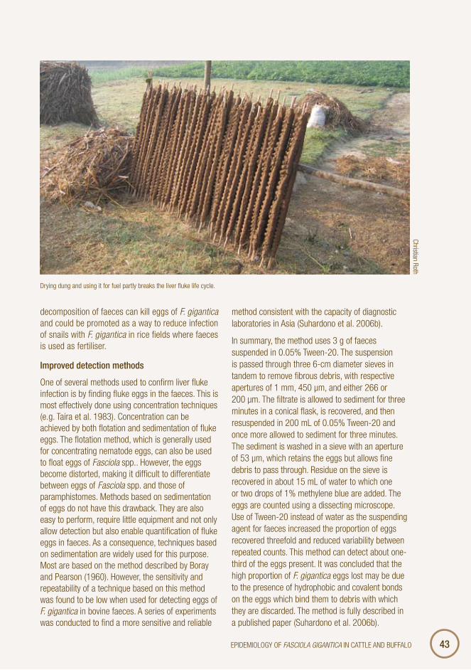
decomposition of faeces can kill eggs of F. gigantica and could be promoted as a way to reduce infection of snails with F. gigantica in rice fields where faeces is used as fertiliser.
Improved detection methods
One of several methods used to confirm liver fluke infection is by finding fluke eggs in the faeces. This is most effectively done using concentration techniques (e.g. Taira et al. 1983). Concentration can be achieved by both flotation and sedimentation of fluke eggs. The flotation method, which is generally used for concentrating nematode eggs, can also be used to float eggs of Fasciola spp.. However, the eggs become distorted, making it difficult to differentiate between eggs of Fasciola spp. and those of paramphistomes. Methods based on sedimentation of eggs do not have this drawback. They are also easy to perform, require little equipment and not only allow detection but also enable quantification of fluke eggs in faeces. As a consequence, techniques based on sedimentation are widely used for this purpose. Most are based on the method described by Boray and Pearson (1960). However, the sensitivity and repeatability of a technique based on this method was found to be low when used for detecting eggs of F. gigantica in bovine faeces. A series of experiments was conducted to find a more sensitive and reliable
method consistent with the capacity of diagnostic laboratories in Asia (Suhardono et al. 2006b).
In summary, the method uses 3 g of faeces suspended in 0.05% Tween-20. The suspension is passed through three 6-cm diameter sieves in tandem to remove fibrous debris, with respective apertures of 1 mm, 450 μm, and either 266 or 200 μm. The filtrate is allowed to sediment for three minutes in a conical flask, is recovered, and then resuspended in 200 mL of 0.05% Tween-20 and once more allowed to sediment for three minutes. The sediment is washed in a sieve with an aperture of 53 μm, which retains the eggs but allows fine debris to pass through. Residue on the sieve is recovered in about 15 mL of water to which one or two drops of 1% methylene blue are added. The eggs are counted using a dissecting microscope. Use of Tween-20 instead of water as the suspending agent for faeces increased the proportion of eggs recovered threefold and reduced variability between repeated counts. This method can detect about one-third of the eggs present. It was concluded that the high proportion of F. gigantica eggs lost may be due to the presence of hydrophobic and covalent bonds on the eggs which bind them to debris with which they are discarded. The method is fully described in a published paper (Suhardono et al. 2006b).
Drying dung and using it for fuel partly breaks the liver fluke life cycle.
Christian Roth

4Options for the control of liver fluke.4Options for the control of liver fluke. OVERCOMING LIVER FLUKE IN SOUTH-EAST ASIA
Snail ecology
west Java
Fasciolidae need one intermediate host, a freshwater gastropod from the superfamily of pulmonate snail, family Lymnaeidae. In tropical regions L. auricularia sensu lat. serves as intermediate host of F. gigantica (Hubendick 1951; Kendall 1954). Besides Fasciola the snail also serves as an intermediate host of other trematodes (Basch and Lie 1965; Boray 1985; Estuningsih 1991). Lymnaea rubiginosa is a fully freshwater snail (Van Benthem Jutting 1954) and serves as intermediate host of F. gigantica in Indonesia (Boray 1980; Muchlis 1985). Other species of Lymnaea have been found in Indonesia (Boray 1980) but have proven to be totally resistant to F. gigantica infection. The distribution of L. rubiginosa in Indonesia is widely spread and the biology of the snail is well described by Widjajanti (1990). Little is known of the population dynamic of this snail in the area where agricultural activities are conducted intensively. Since it is known that the life cycle of the liver fluke is heavily dependent on L. rubiginosa then it is clear that the distribution of the disease will be
Manure used as fertiliser contains liver fluke eggs, and flooding of rice paddies before planting allows snail populations to build up.
determined by that of the snail. The aim of this study is to determine the population dynamics of the snail L. rubiginosa in the rice paddy environment and its infection with fluke trematodes.
A field study was conducted in five villages in the subdistrict of Surade in West Java. In summary the findings were that the population dynamics of L. rubiginosa in the paddy fields vary with the cropping practices of wet paddy and that the population was very high during the wet season. More snails were found close to human habitation and more of these were infected with trematode larvae. Passive migration in streams formed by heavy rain is the most important way that snails are disseminated into new habitats such as paddy fields. In the dry season most snails die from lack of water, with the surviving snails being mainly in streams, rivers or water springs. There is no sign of snail aestivation during droughts. Infection with F. gigantica in snails occurred throughout the year with the peak in May, October and February. Infection with non-Fasciola, mainly echinostome, tended to occur more in the dry season than in the wet.
A more detailed study was conducted in West Java to catalogue all the trematodes that use L. rubiginosa as a first intermediate host and to identify their definitive hosts (Estuningsih and Copeman 1996). Trematode larvae in 3,253 L. rubiginosa were collected from irrigated rice fields at five sites around Bogor, and another 2,875 from Surade. Four types of cercariae were found in snails from the Bogor area: echinostome, strigeid, Trichobilharzia sp. and xiphidiocercariae; whereas in snails from Surade there were xiphidiocercariae and cercariae of F. gigantica, schistosoma sp. and echinostomes.
The larval echinostomes found in L. rubiginosa from the Bogor area, and adult echinostomes in domestic ducks and chickens which grazed harvested rice fields in this area were both identified as Echinostoma revolutum. Since no echinostome was found in 24 rats, 11 lizards or 35 frogs caught in the vicinity of the Bogor rice fields, it was concluded that domestic ducks and chickens were the main definitive hosts for E. revolutum in the area. The implication here is that the prevalence of cercaria of F. gigantica in the snail population is a very poor indicator of the prevalence in the definitive hosts of interest.
4Options for the control of liver fluke.5EPIDEMIOLOGY OF FAsCiOLA gigAnTiCA IN CATTLE AND BUFFALO
Cambodia
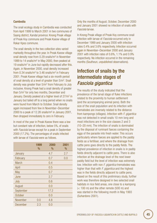
The snail ecology study in Cambodia was conducted from April 1999 to March 2001 in two communes of Saang district, Kandal province: Knong Preak village of Preak Koy commune and Preak Kseow village of Rokar Kpos commune.
The snail density in the two collection sites varied markedly throughout the year. In Preak Kseow village snail density rose from 0.29 snails/m² in November 1999 to 14 snails/m² in May 2000, then peaked at 19 snails/m2 in June but rapidly decreased after this. Again, in November 2000, snail density increased from 0.34 snails/m² to 3.46 snails/m² in February 2001. Preak Kseow village had a six-month period of snail density at a level of greater than 5/m2. Snail density was greater than 5/m2 from February to July inclusive. Knong Preak had a snail density of greater than 5/m2 for only two months, December and January. Density peaked at a higher level of 27/m2 in January but tailed off to a long period when no snails were found from March to October. Snail density again increased from few in November–December 2000 to a peak of 12.26 snails/m² in January 2001, then dropped immediately to zero in February.
In most of the year in Preak Kseow there was a low but constant rate of infection, below 5%, of snails with Fasciola larvae except for a peak in September 2000 (17.2%). The percentages of snails infected with larvae of Fasciola were as follows.
1Overview.999 %
2Importance and potential impact of liver fluke in cattle and b000 %
2Importance and potential impact of liver fluke in cattle and b001Overview.%
January 4.7 2.7
February 0.7 0.0
March 0.4
April 0.5
May 0.7
June 2.7
July 0.6
August 0.0
September 17.2
October 0.0
November 0.0 4.8
December 2.3 0.0
Only the months of August, October, December 2000 and January 2001 showed no infection of snails with Fasciola larvae.
In Knong Preak village of Preak Koy commune snail infection with larvae of Fasciola occurred only in December 1999 and January 2000 with infection rates 0.4% and 3.6% respectively. Infection occurred again in November–December 2000 and January 2001 with infection rates of 0.6%, 1.1% and 0.9% respectively. No infection occurred in the remaining months (Southeun, unpublished observations).
Infection of snails by the intermediate stages of Fasciola giganticaThe results of the study indicated that the prevalence of the larval stage of fluke infections related to the proximity to human settlement (and the accompanying animal pens). Both the size of the snail population and its infection with trematodes are inversely related to the distance from kampongs (villages). Infection with F. gigantica was not detected in small snails 10 mm long and most infections are in the size classes 2 and 3 (10–20 mm). The infection of snails is dominated by the disposal of ruminant faeces containing the eggs of the parasite into fresh water. This occurs particularly where faeces are applied to the paddy fields as a fertiliser, and where the drainage from cattle pens goes directly to the paddy fields. The highest prevalence of infection in snails is in paddy fields directly adjacent to cattle pens. There is also infection at the drainage level of the next lower paddy field but the level of infection was extremely low. Infection with non F. gigantica trematodes was higher than that with F. gigantica and the highest was in the fields directly adjacent to cattle pens. Based on the result of this preliminary study, further work was therefore designed in two selected snail habitats in rice field areas, one close to a kampong (< 100 m) and the other remote (500 m) and was started in the following month in May 1993 (Suhardono 2001).

4Options for the control of liver fluke.6 OVERCOMING LIVER FLUKE IN SOUTH-EAST ASIA
Competition with Echinostoma
Echinostoma in other hosts
In a series of experiments to investigate further the host range of Echinostoma, 50 mallard ducks as well as native chickens, rats and edible frogs were examined for the presence of E. revolutum (Gonzaga and colleagues, unpublished observations). The study aimed to show the percentage of ducks, chickens, field rats and edible frogs with echinostomosis and which organs were commonly inhabited by the trematodes, and compared the carcass weight of infected and non-infected ducks.
Echinostoma revolutum were found in the caecum and large intestine of 24 (48%) mallard ducks, 27 (54%) native chickens and 23 (46%) rats examined. There were no E. revolutum found in the alimentary tract of edible frogs.
It was concluded that infection of E. revolutum is sufficiently common in mallard ducks, native chickens, and rats in the Kabacan region that they
would be a suitable resource for biological control of infection with F. gigantica
location and infectivity of metacercariae
distribution on rice straw
The distribution of metacercariae of F. gigantica on fresh rice stalks was determined by cutting the lower 40 cm of stalks into lengths of 10 cm and feeding 10 kg from each level to groups of two previously uninfected merino sheep (Table 3.1). Two additional sheep were each infected with 250 metacercariae as positive controls and one uninfected sheep acted as a negative control. Seven to ten weeks later all sheep were killed and immature flukes harvested. It was found that sheep fed with the lower 10 cm of the stalks harboured 98% of the flukes recovered from animals fed rice stalks. Thirty-three and 41% respectively of the dose of metacercariae given to the two positive control sheep were recovered as
table 3.1Overview. Number and size of Fasciola gigantica recovered from Fasciola-naïve merino sheep infected with metacercariae of F. gigantica or fed portions of rice stems cut 0–10, 10–20, 20–30 or 30–40 cm from the ground.
Sheepno.
Source of infection/distance of cut stalk
from the ground
week sheep killed
number of F. gigantica recovered
length ± Sd(mm)
width ± Sd(mm)
n
603 Nil 6.5 0 0 0 0
611 250 mc 8 83 8.8 ± 1.5 1.9 ± 0.4 37
612 250 mc 7.5 103 6.5 ± 1.0 1.3 ± 0.3 78
628 0–10* 10.5 137 10.1 ± 2.1 2.2 ± 0.4 49
629 0–10 10.5 113 12.2 ± 2.0 2.4 ± 0.5 53
623 10–20 10.5 4 13.2 ± 1.8 2.7 ± 0.3 2
A60 10–20 10 0 0 0 0
500 20–30 10.5 1 12.5 2.5 1
98 20–30 0 0 0 0 0
102 30–40 0 0 0 0 0
708 30–40 0 0 0 0 0
* = distance in centimetres of the pieces of rice stem from ground levelmc = metacercariae of F. gigantican = number of flukes measured

4Options for the control of liver fluke.7EPIDEMIOLOGY OF FAsCiOLA gigAnTiCA IN CATTLE AND BUFFALO
larval flukes. It was concluded that rejection of the lower portion of rice stalks previously immersed in water would help control infection with F. gigantica in animals fed on fresh rice straw (Suhardono et al. 2006d).
Effect of temperature and humidity
The viability of the metacercariae of F. gigantica when stored in water at 13°C for up to 23 weeks, exposed to the sun for up to eight hours or stored at a range of temperatures and humidities for up to 10 weeks was studied (Table 3.2). Excysted metacercariae were categorised microscopically as viable (motile and undamaged), dubious (not motile and undamaged) or dead (visible necrosis). Infectivity of viable and dubious metacercariae and unselected reference metacercariae held in water at 7°C for 20 or more days was assessed by comparing numbers of flukes recovered from infected merino sheep. Mean recovery rates were respectively 54.6%, 7.2% and 37.2% for viable, dubious and unselected metacercariae. Metacercariae immersed in water remained viable longer than those allowed to desiccate. Viability was promoted by decreasing temperature and increasing humidity. Exposure to direct sun killed metacercariae within eight hours. Results indicated that in lowland Indonesian irrigated rice paddies, metacercariae immersed in water are
likely to survive for less than five weeks while those that become desiccated will survive less than two weeks. This information, together with the option of exposing fresh rice stalks to direct sunlight before feeding them to livestock, can help farmers reduce infection with F. gigantica (Suhardono et al. 2006c).
Sources of metacercariae for infection
Rice straw is believed to be the main source of infection of cattle and buffaloes with F. gigantica in areas where irrigated rice is grown extensively (Ueno et al. 1975; Harrison et al. 1996). However, this conclusion has been based on largely circumstantial evidence on the ecology of the snail intermediate host and observations that prevalence of fasciolosis in draught animals is high in lowland rice irrigation areas (Edney and Muchlis 1962; Soesetya 1978; Morel and Mahato 1987; Suhardono and Estuningsih 1989). Furthermore, the possibility that water from rice fields may also be a source of metacercariae for animals and that there may be other sources has not been investigated.
The objective of this study was to identify the sources of metacercariae of F. gigantica infecting cattle in a lowland area of West Java where two tandem irrigated crops of rice are grown most years followed by either a period of fallow or a dryland crop.
table 3.2Importance and potential impact of liver fluke in cattle and b Percentage viability of metacercariae of Fasciola gigantica exposed to the sun or shaded while being maintained damp or dry at ambient temperature of about 30°C.
Hoursexposed
Percentage viable
under sunshine under shade
damp dry damp dry
live dubious live dubious live dubious live dubious
0 88 6 88 6 88 6 88 6
2 94 5 29 10 96 4 84 10
4 33 37 6 18 83 10 86 11
6 0 19 0 0 81 12 81 8
8 3 11 0 19 69 14 0 55

4Options for the control of liver fluke.8Immunology and assessment of resistance to fasciolosis in smal OVERCOMING LIVER FLUKE IN SOUTH-EAST ASIA
Water and rice stubble from recently harvested rice fields containing L. rubiginosa infected with F. gigantica, and water and forage from other habitats of L. rubiginosa where free-grazing cattle were observed to eat or drink were fed to merino sheep. They were then monitored, either by counting fluke numbers in the liver at necropsy or by observing Fasciola eggs in faeces, to determine sources of viable metacercariae. Infection with F. gigantica was acquired from the clear effluent water and water containing sediment from rice fields, and from rice stalks, but not from irrigation channels, the only other habitat of L. rubiginosa in the study area. It was concluded that the most important risk factor for cattle to become infected with F. gigantica was to allow them to graze in recently harvested rice fields. Fields most heavily contaminated were those flooded again after harvest and risk of infection was greatly increased by feeding fresh herbage to cattle from such fields or by allowing animals to drink from rice fields (Suhardono 2001).
Influence of production systems
Seasonal variation in Cambodia
A study was conducted in Cambodia to generate baseline data on the incidence and level of infection of Fasciola in cattle and buffalo. The study was conducted in six villages in four communes of Saang district of Kandal province and eight villages in four communes in Cherng Prey district of Kampong Cham province. The 373 families directly involved in the project either brought their animals for study or were interviewed. The project examined 1,790 cattle, 5,126 faecal samples, 596 cattle livers and 8,008 snails.
To understand the epidemiology of fasciolosis, the research included an abattoir study, a tracer study, an agricultural cycle study and a snail ecology study. In addition, impacts of fasciolosis on production were monitored in a suppressive treatment trial.
For the abattoir study, a total of 645 cattle were examined at the district abattoirs (Cherng Prey district of Kompong Cham province and Saang district of Kandal province) and of these 502 head (77.82%) were identified as originating from within Saang district.
Nearly all the cattle examined were described as the crossbred type, which is a cross between the imported large tall-bodied white-coated Haryana breed and the smaller local breed. A total of 644 cattle had their sex recorded. Of these, 82.9% were castrated males, 15.2% females and 1.9% bulls. The average age of cattle brought for slaughter was seven years old, with a range from two to 12 years old. Females were on average younger (5.6 years) than castrated males (7.4 years).
A total of 575 faeces samples were examined for Fasciola eggs, of which 142 (24.7%) were positive. Of the 596 cattle examined at the slaughterhouse, 176 (30%) had liver damage. Female cattle with both liver damage and eggs in their faeces had lower body condition than other females. There were no differences in bodyweight or condition between male cattle that had eggs in their faeces, liver damage, both, or neither of these.
There is some evidence of a seasonal infection with Fasciola. Positive faecal samples rose from a low of 11% in December 1998 to 34% in April 1999, 28% in May and 55% in June, then declined progressively for the remainder of 1999. These results indicate that infection was highest over the months from about November to April. Liver damage by Fasciola infection was high in December, January, February and March (39%, 36%, 43% and 37% respectively) and then decreased, thus providing additional evidence that infection with Fasciola is seasonally highest at the end and start of the year.
The average cattle liver weight was 6.2 kg (range 5.3–10.4 kg). In the 32 damaged livers examined there were means of 30.5 adult and 40.3 immature flukes per liver. The average weight of damaged liver tissue was 2.3 kg (range 0.3–10.4 kg).
A tracer study was conducted in four villages in three communes of Saang district, Kandal province: Preak Kseow village of Rokar Kpos commune, Svay Tany and Preak Run villages of Preak Koy commune and Ang village of Krang Yov commune. The study began in April 1999 and was completed at the end of March 2001. A total of 446 cattle in the target area were used (Preak Kseow village 130 head, Svay Tany 99 head, Preak Run 129 head and Ang 88 head). All were treated with triclabendazole (Fasinex) at 12 mg/kg body weight (0.1 ml/kg) to free them from any pre-existing Fasciola infection.

4Options for the control of liver fluke.9EPIDEMIOLOGY OF FAsCiOLA gigAnTiCA IN CATTLE AND BUFFALO
Using a prepatent period for Fasciola of four months, it was calculated that cattle were infected in all villages from September 1999 to March or April 2000. In Svay Tany village infection began in September (6%) and reached a peak in April (50%). Between May and July no cattle were infected, then some were infected from August of the following year. In Preak Kseow village incidence rose from 9% in September to the peak of 87% in April. No infection occurred from May to July, however, it began again in August. In Preak Run village incidence rose from 3% in September to 37% in December, dropped to 12% in February, rose again in March and reached a peak in April (57.2%). Once again the infection began from the following August. In Ang village, only small percentages of cattle were infected with Fasciola (1.6–4.0%). Infection occurred from September to March intermittently.
Tracer studies thus confirmed the pattern of infection with Fasciola suggested by the abattoir study, that incidence of infection is highest from October to April. It also indicated that April was a peak month for infection and that little infection occurred from May to August (Southeun et al., unpublished).
Seasonal variation in Indonesia
There are reports on prevalence proportions from most countries where infection with F. gigantica is endemic. Few studies, however, have attempted to define monthly or seasonal incidence rates even though this information provides a logical basis for rational decisions on control. Fasciolosis in Indonesia mainly occurs in cattle and buffaloes and is widespread in the main islands (Adiwinata 1955; Kraneveld 1924; Soesetya 1975; Nurdin and Salsabila 1979; Rivai 1979; Soemardi and Rubino 1979; Tarmudji and Ginting 1983; Tarmudji et al. 1983). In Indonesia old reports recognised the presence of more than one species of Fasciola (Krijgsman 1933). However, Mukhlis (1985) concluded only one species of liver fluke occurred in indigenous cattle and it was F. gigantica. The recent work of Kurniasih et al. (1996) supported this conclusion, but from molecular studies introduced the possibility of their being more than one subspecies of F. gigantica.
To complete their life cycle liver flukes use lymnaeid snails as their intermediate host. On the island of Java, there is only one species of snail from the
Seasonal water sources are focal points for grazing and the continual cycling of liver fluke infection through snails.
ILRI/Steve Mann

50 OVERCOMING LIVER FLUKE IN SOUTH-EAST ASIA
family Lymnaeidae, L. javanica, which is synonymous with L. rubiginosa (van Benthem Jutting 1956). Lymnaea rubiginosa is an aquatic snail which spends most of its time floating at the surface of water (Widjajanti 1989). It thrives in the clear, slowly moving shallow water of irrigated rice fields.
In most countries of South-East Asia the main reason for raising cattle in rice-cropping communities is to provide a source of power during the processing of land before it’s cultivated (Jarrige 1980). They also provide manure which is used as fertiliser in rice fields and an alternative source of income if the crop fails. The management of these animals is closely integrated with cropping practices. Farmers mostly feed their cattle with crop residues as most do not have enough land to grow special fodder for their animals.
two seasonal studies—Monthly fluctuations of faecal Fasciola egg counts were monitored in 60 Ongole cattle each four weeks for 13 months in five villages in the subdistrict of Surade, West Java. Faecal egg counts of F. gigantica were also estimated for additional groups of 30 cattle selected each month from the same villages. It was concluded that estimates of prevalence of infection with F. gigantica are likely to be conservative when based on faecal egg counts. It was proposed that the sudden drop in apparent prevalence during December followed by gradual
recovery during the following few months was a consequence of immune stimulation (induced by an influx of metacercariae acquired by animals from rice stubble at the beginning of the harvest season in January) which either causes loss of adult worms or suppression of their egg output (Figures 3.3 and 3.4).
The incidence and time of infection with F. gigantica in Ongole cattle in a rice-cropping environment in West Java was undertaken using 30 Ongole tracer cattle each four weeks for 16 months in 1993–94. It was found that over 80% of infection was acquired from January to June which coincided with the period of harvest of irrigated rice. It is also concluded that the main source of infection with F. gigantica in cattle is fresh rice straw, especially that from fields within about 200 m of a cattle pen.
In parallel with the green revolution of Indonesia, rice stubble has become abundant. This has been due mainly to high-yield varieties of rice being introduced, a relatively short time from planting to harvest and an increase in irrigation (Heytens 1991). Because more rice stubble is available, it has become a major dietary component for cattle. Irrigated rice is increasingly producing two crops per year and this also serves to maintain a high population of L. rubiginosa. The close relationship between rice cultivation and cattle management and the suitability of irrigated rice fields as a habitat for L. rubiginosa causes the pattern of liver
Figure 3.3. Prevalence of Fasciola gigantica eggs and mean egg counts per gram of faeces (EPG) ± SD of each batch of 30 Ongole cattle chosen each four weeks to become tracers, before they were treated with triclabendazole.
Figure 3.4Options for the control of liver fluke.. Incidence of Fasciola gigantica infection as detected by eggs in faeces in relation to monthly rainfall and the seasonal activities for rice production.
80
60
40
20
0M J J A S O N D D J F M A M
1993 1994
EPG Prevalence (%)
Prevalence of infection
EPG values
100
50
0
75
50
25
0M J J A S O N D D J F M A M J J A
1993 1994
Incidence (%) Rainfall (mm)Monthly
rainfall (mm)
Harvesting rice
5002500
Harvestingrice
Transplantingrice
Incidence (%)

51Overview.EPIDEMIOLOGY OF FAsCiOLA gigAnTiCA IN CATTLE AND BUFFALO
table 3.3 Key research finding and their implications for control.
key finding Implication for control programs
Fluke eggs are susceptible to heat and exposure to the sun.
Dung could be composted or exposed to sun before being spread as fertiliser. This can supplement the containment of dung to prevent contamination of waterways and rice paddies.
Laboratory methods for counting fluke eggs can be improved.
Diagnosis of suspected cases and in field survey can be improved. Evaluation of interventions in the field can be made more accurate.
Composting and heat induced by additives can reduce infectivity of eggs.
Different methods of composting need to be evaluated for their impact on fluke egg survival.
The role of Lymnea rubiginosa is confirmed in areas of high fluke prevalence in Indonesia.
Knowing which species and their distribution is an important aspect of identifying areas of potential problems.
Many parasite species found in Lymnea, including Echinostoma in Java.
Many species use Lymnea as an intermediate host, therefore accurate identification of cercariae is essential.
Echinostoma found in a range of warm- blooded vertebrates in the Philippines.
Echinostoma may be very widespread and has the potential to interfere with transmission of Fasciola.
Distribution of metacercaria is limited to the bottom 10 cm of rice straw.
Cutting rice straw higher than 10 cm would prevent infection being carried with fodder to stalled animals.
Exposure of metacercariae on rice straw to sun reduces their viability.
Leaving contaminate rice straw in the sun before feeding could reduce exposure to infection.
Prepatent periods in Ongole cattle confirmed to be 18 weeks (4 months).
This conforms to expectations but needs to be confirmed if local experimental studies are to be conducted.
Pattern of infection in Cambodia consistent with 4-month prepatent period for infection.
Once established that Lymnea is the intermediate host and Fasciola the cause of disease similar control principles can be applied across the region.
In Cambodia the highest level of infection occurs between October and April, with no new infections between May and August.
Timing of chemical treatments and interventions to prevent infection can be focused on periods of non-transmission and transmission, as appropriate.
Similar patterns in Indonesia with tracer studies provide a clearer picture.
Infection is intimately associated with the cycle of rice planting and harvesting in Indonesia.
Changes in rice production system need to take account of implications for liver fluke infection and control programs.

52Importance and potential impact of liver fluke in cattle and b OVERCOMING LIVER FLUKE IN SOUTH-EAST ASIA
fluke disease in cattle to vary directly with the intensity of cropping of irrigated rice (Edney and Muchlis 1962).
No information is documented on the time of acquisition of and source of infection with F. gigantica by cattle in Indonesia. As prevalence is highest in rice-growing areas, this study was undertaken to determine the time of acquisition of F. gigantica in Ongole cattle raised in a farming area dominated by the cultivation of irrigated rice.
Implications for control programsThe main findings present in this chapter have been tabulated (Table 3.3) and their implications for potential control programs listed.
references Adiwinata R.T. 1955. Helminth parasites in mammals
and poultry in Indonesia. (Cacing-cacing yang berparasit pada hewan menyusui dan unggas di Indonesia). Hemerazoa 62, 229–247.
Alicata J.E. and Swanson L.E. 1941. Observations on the longevity of the liver fluke, F. gigantica in cattle. American Journal of Veterinary Research 3, 417–419.
al Kubaisee R.Y. and Altaif K.I. 1989. Comparative study of sheep and buffalo isolates of F. gigantica in the intermediate host Lymnaea auricularia. Research in Veterinary Science 47, 273–274.
Asanji M.F. 1988. The snail intermediate host of F. gigantica and the behaviour of miracidia in host selection. Bulletin of Animal Health and Production in Africa 36, 245–250.
Basch P.F. and Lie K.J. 1965. Multiple infection of larval trematodes in one snail. Medical Journal of Malaya 20, 59.
Boray J.C. 1969. Experimental fascioliasis in Australia. Advances in Parasitology 60, 114–124.
Boray J.C. 1982. Molluscan hosts and trematodes in the Pasific Basin. Pp. 81–106 in ‘Biology and control of endoparasites’ ed. by L.E.A. Symons, A.D. Donald and J.K. Dineen. Academic Press: New York.
Chartier C., Ngota A., Lonu L. and Cabaret J. 1990. Dynamics of Lymnaea natalensis populations in the Bunia area (Ituri, Haut-Zaire). Annales de Parasitologie Humaine et Comparee 65, 177–182.
Da Costa C., Dreyfuss G., Rakotondravao, Rondelaud D. and Da Costa C. 1994. Several observations concerning cercarial sheddings of Fasciola gigantica from Lymnaea natalensis. Parasite 1, 39–44.
Dinnick J.A. and Dinnick N.N. 1963. Effect of the seasonal variations of temperature on the development of F. gigantica in the snail host in the Kenya highlands. Bulletin of Epizootic Diseases of Africa 11, 197–207.
Dinnick J.A. and Dinnick N.N. 1964. The influence of temperature on the succession of redial and cercarial generations of Fasciola gigantica. Parasitology 54, 59–65.
Dreyfuss G. and Rondelaud D. 1994. Comparative study of cercarial shedding by Lymnaea tomentosa Pfeiffer infected with either F. gigantica Cobbold or F. hepatica Linne. Bulletin de la Societe Francaise de Parasitologie 12, 43–54.
Edney J.M. and Muchlis A. 1962. Fascioliasis in Indonesian livestock. Communications Veterinariae 6, 49–62.
Estuningsih S.E. 1991. Studies on trematodes infecting Lymnaea rubiginosa in West Java. M.Sc Thesis. Tropical Veterinary Sciences and Agriculture, James Cook University of North Queensland: Townsville, Australia.
Estuningsih S.E. and Copeman D.B. 1996.Trematode larvae in Lymnaea rubiginosa and their definitive host in irrigated rice fields in West Java. Jurnal Ilmu Ternak dan Veteriner 1, 200–205.
Grigoryan G.A. 1958. Experimental study of F. gigantica infestation in sheep. Trudy Armyansk Inst Zhivotnovod. i Vet 3, 155–168.

53EPIDEMIOLOGY OF FAsCiOLA gigAnTiCA IN CATTLE AND BUFFALO
Grigoryan G.A. 1959. Exsperimental’nye dannye po prodolzhitol’nosti sokhranoniya adolescariev Fasciola hepatica (Data from experiments to find the survival span of metacercariae of Fasciola gigantica). Veterinariya 36, 35–37.
Guralp N., Ozcan C. and Simms B.T. 1964. Fasciola gigantica and fascioliasis in Turkey. American Journal of Veterinary Research 25, 196–210.
Hammond J.A. and Sewell M.M.H. 1975. Experimental infections of cattle with F. gigantica: numbers of parasites recovered after varying periods of infection. Tropical Animal Health and Production 7, 105–113.
Harrison L.J.S., Hammond, J.A. and Sewell, M.M.H. 1996. Studies on helminthosis at the Centre for Tropical Veterinary Medicine. Tropical Animal Health and Production 28, 23–39.
Heytens, P. 1991. Rice production system. In ‘Rice policy in Indonesia’, ed. by S. Pearson, W. Falcon, P. Heytens, E. Monke and R. Naylor, Cornell University Press: Ithaca, London.
Hubendick, B. 1951. Recent lymnaeidae. Almquist & Wicksells: Stockholm.Jarrige R. 1980. Herbivores in agricultural ecosystem. Pp 763–823 in ‘Digestive physiology and metabolism in ruminants’. ed. by Y. Ruckebusch and P. Thivend, AVI Publishing Company: Westport, CT, USA.
Kendall S.B. 1954. Fascioliasis in Pakistan. Annals of Tropical Medicine and Parasitology 48, 307–313.
Kendall S.B. 1965. Relationships between the species of Fasciola and their molluscan hosts. Advances in Parasitology 3, 59–98.
Kraneveld F.C. 1924. Bijdrage tot de therapie der distomatosis in Nederlandsch-Indie. Nederlandsch Indie Bladen von Diergeneskunde 36, 3–63.
Krijgsman B.J. 1933. The parasite worms of mammals and birds in the Nederlandsch-Indie. (De in zoogdieren en vogels parasiteerende wormen van Nederlandsch-Indie). Nederlandsch-Indi Bladen von Diergeneskunde 22, 366–379.
Kurniasih, Barker S.C. and Cribb T.H. 1996. Phylogeny of Fasciola species (Digenea) in internal transcribed spacer ribosomal RNA from Indonesian domestic animals. (Abstract). In ‘Pasean 1996. Parasites and Southeast Asia—now’. Bali, September 1996. (Program and Abstract. p. 30).
Morel A.M. and Mahato S.N. 1987. Epidemiology of fascioliasis in the Koshi hills of Nepal. Tropical Animal Health and Production 19, 33–38.
Mukhlis A. 1985.The identity of liver fluke (Fasciola sp.) and its life cycle in Indonesia. (Identitas cacing hati (Fasciola sp.) dan daur hidupnya di Indonesia). Ph.D. Thesis. Institut Pertanian: Bogor, Indonesia.
Mzembe S.A.T. and Chaudhry M.A. 1979. The epidemiology of fascioliasis in Malawi: I. The epidemiology of the intermediate host. Tropical Animal Health and Production 11, 246–250.
Nurdin M.S. and Salsabila A. 1979. Trematodes in slaughtered animals in West Sumatra. Veterinary Parasitology 5, 82 (Abstract).
Prasitirat P., Thammasart S., Chompoochan T., Nithiuthai S. and Taira S. 1996. The dynamics of antibody titres and faecal egg output in cattle and buffalo following infection with 500 and 1,000 Fasciola gigantica metacercariae. Thai Journal of Veterinary Medicine 26, 85–89.
Rivai M. 1979. Losses caused by Fasciola hepatica in infected cattle and buffaloes slaughtered at Padang Panjang abattoir. Veterinary Parasitology 5, 81 (Abstract).
Sewell M.M.H. 1966. The pathogenesis of fascioliasis. Veterinary Record 78, 98–105.
Soemardi A.W. and Rubino 1979. Distribution of Fasciola hepatica in cattle and buffalo in Timor. Veterinary Parasitology 5, 81 (Abstract).
Soesetya R.H.B. 1975. The prevalence of Fasciola gigantica infection in cattle in East Java, Indonesia. Malaysian Veterinary Journal 6, 5–8.
Suhardono D. 2001. Epidemiology and control of fasciolosis by Fasciola gigantica in ongole cattle in West Java. Ph.D. Thesis, Tropical Veterinary Science, James Cook University of North Queensland: Townsville Australia.

54Options for the control of liver fluke. OVERCOMING LIVER FLUKE IN SOUTH-EAST ASIA
Suhardono and Estuningsih, S.E. 1989. Contamination rate of metacercariae in rice straws in the endemic area of fasciolosis. (Tingkat kontaminasi jerami padi oleh metacercariae di daerah endemik fascioliasis). In Proceedings Pertemuan Ilmiah Ruminansia. Ruminansia Besar. Pusat Penelitian dan Pengembangan Peternakan, Badan Penelitian dan Pengembangan Pertanian: Bogor, Indonesia.
Suhardono, Roberts J.A and Copeman D.B. 2006a. The longevity of eggs of F. gigantica in bovine faeces. Tropical Animal Health and Production 38, 379–382.
Suhardono, Roberts J.A and Copeman D.B. 2006b. Optimisation of the procedure for counting the eggs of Fascioloa gigantica in bovine faeces. Tropical Animal Health and Production 38, 383–387.
Suhardono, Roberts J.A and Copeman D.B. 2006c. The effect of temperature and humidity on longevity of metacercariae of F. gigantica. Tropical Animal Health and Production 38, 371–387.
Suhardono, Roberts J.A and Copeman D.B. 2006d. Distribution of metacercariae of F. gigantica on rice straw. Tropical Animal Health and Production 38, 117–119.Taira N., Suzuki K. and Boray J.C. 1983. Detection and quantitation of Fasciola eggs in cattle faeces using the beads technique. Japan Journal of Parasitology 32, 279–286.
Tarmudji and Ginting Ng. 1983. The rate of liver destruction due to fasciolosis in female Friesian Holstein in the district of Malang, East Java: A histopathological examination. (Derajat kerusakan hati akibat fascioliasis pada sapi-sapi Friesian Holstein betina di kabupaten Malang, Jawa Timur: Suatu tinjauan histopatologik). Penyakit Hewan 15, 83–85.
Tarmudji, Iskandar T. and Hamid H. 1983. Fasciolosis in cattle in East Java between 1979 and 1982, based on histopathological examination. (Kasus fascioliasis pada sapi-sapi di Jawa Timur, berdasarkan hasil pemeriksaan patologik antara 1979–1982. Penyakit Hewan 15, 91–94.
Tembely S., Coulibaly E., Dembele K., Kayentao O. and Kouyate B. 1995. Intermediate host populations and seasonal transmission of Fasciola gigantica to calves in central Mali, with observations on nematode populations. Preventive Veterinary Medicine 22, 127–136.
Ueno H. and Yoshihara S. 1974. Vertical distribution of Fasciola gigantica metacercariae on stem of rice plant grown in a pot. National Institute of Animal Health Quarterly 14, 54–60.
van Benthem Jutting W.S.S. 1950. Systematic studies on the non-marine mollusca of the Indo-Australian archipelago. II. Critical revision of the Javanese pulmonate land-snails of the families Helicarionidae, Pleurodontidae, Fruticicolidae & Streptaxidae. Treubia 20, 381–505.
Widjajanti S. 1989. Studies on the biology of Lymnaea rubiginosa. M.Sc. Thesis, Tropical Veterinary Science and Agriculture. James Cook University of North Queensland: Townsville, Australia.