improved vascular organization enhances … vascular organization enhances functional integration of...
TRANSCRIPT
Corrections
APPLIED BIOLOGICAL SCIENCESCorrection for “Improved vascular organization enhances func-tional integration of engineered skeletal muscle grafts,” byJacob Koffler, Keren Kaufman-Francis, Shandalov Yulia, EgoziDana, Amiad Pavlov Daria, Amir Landesberg, and ShulamitLevenberg, which appeared in issue 36, September 6, 2011, ofProc Natl Acad Sci USA (108:14789–14794; first publishedAugust 30, 2011; 10.1073/pnas.1017825108).The authors note that, due to a printer’s error, the author
name Shandalov Yulia should instead appear as Yulia Shandalov,the author name Egozi Dana should instead appear as DanaEgozi, and the author name Amiad Pavlov Daria should insteadappear as Daria Amiad Pavlov. The corrected author line ap-pears below. The online version has been corrected.
Jacob Koffler, Keren Kaufman-Francis, Yulia Shandalov,Dana Egozi, Daria Amiad Pavlov, Amir Landesberg,and Shulamit Levenberg
www.pnas.org/cgi/doi/10.1073/pnas.1120303109
EVOLUTIONCorrection for “Duplication and partitioning in evolution andfunction of homoeologous Q loci governing domestication char-acters in polyploid wheat,” by Zengcui Zhang, Harry Belcram,Piotr Gornicki, Mathieu Charles, Jérémy Just, Cécile Huneau,Ghislaine Magdelenat, Arnaud Couloux, Sylvie Samain, BikramS. Gill, Jack B. Rasmussen, Valérie Barbe, Justin D. Faris, andBoulos Chalhoub, which appeared in issue 46, November 15,2011, of Proc Natl Acad Sci USA (108:18737–18742; first pub-lished October 31, 2011; 10.1073/pnas.1110552108).The authors note that, due to a printer’s error, on page 18737,
right column, first paragraph, line 13, “T. turgidum” should in-stead appear as “T. aestivum.” Additionally, on page 18738, leftcolumn, second full paragraph, line 7, “T. turgidum” should in-stead appear as “T. aestivum.” Lastly, on page 18739, left column,third full paragraph, line 4, “T. turgidum” should instead appearas “T. aestivum.”
www.pnas.org/cgi/doi/10.1073/pnas.1121066109
GENETICSCorrection for “Meiotic double-strand breaks occur once per pairof (sister) chromatids and, viaMec1/ATRand Tel1/ATM, once perquartet of chromatids,” by Liangran Zhang, Nancy E. Kleckner,Aurora Storlazzi, and Keun P. Kim, which appeared in issue 50,December 13, 2011, of Proc Natl Acad Sci USA (108:20036–20041;first published November 28, 2011; 10.1073/pnas.1117937108).The author line should appear as “Liangran Zhang, Keun P.
Kim, Nancy E. Kleckner, and Aurora Storlazzi.”Additionally, the authors note that Aurora Storlazzi should be
listed as an additional corresponding author. The corrected authorline and correspondence footnote appear below. The online ver-sion has been corrected.
Liangran Zhang, Keun P. Kim, Nancy E. Kleckner1,and Aurora Storlazzi1
1To whom correspondence may addressed. E-mail: [email protected] or [email protected].
www.pnas.org/cgi/doi/10.1073/pnas.1120345109
www.pnas.org PNAS | January 24, 2012 | vol. 109 | no. 4 | 1353
CORR
ECTIONS
Improved vascular organization enhances functionalintegration of engineered skeletal muscle graftsJacob Kofflera,b, Keren Kaufman-Francisa,b, Yulia Shandalova, Dana Egozic, Daria Amiad Pavlova,Amir Landesberga, and Shulamit Levenberga,1
aBiomedical Engineering Faculty; bInter-departmental Program in Biotechnology, Technion-Israel Institute of Technology, Haifa 32000, Israel; andcDepartment of Plastic and Reconstructive Surgery, Rambam Health Care Campus, Haifa 31096, Israel
Edited* by Robert Langer, Massachusetts Institute of Technology, Cambridge, MA, and approved July 25, 2011 (received for review December 9, 2010)
Severe traumatic events such as burns, and cancer therapy, ofteninvolve a significant loss of tissue, requiring surgical reconstructionby means of autologous muscle flaps. The scant availability ofquality vascularized flaps and donor site morbidity often limit theiruse. Engineered vascularized grafts provide an alternative for thisneed. This work describes a first-time analysis, of the degree of invitro vascularization and tissue organization, required to enhancethe pace and efficacy of vascularized muscle graft integration invivo. While one-day in vitro was sufficient for graft integration,a three-week culturing period, yielding semiorganized vessel struc-tures and muscle fibers, significantly improved grafting efficacy. Im-planted vessel networks were gradually replaced by host vessels,coupled with enhanced perfusion and capillary density. Upregula-tion of key graft angiogenic factors suggest its active role in promot-ing the angiogenic response. Transition from satellite cells to maturefibers was indicated by increased gene expression, increased capil-lary to fiber ratio, and similar morphology to normal muscle. Wesuggest a “relay” approach in which extended in vitro incubation,enabling the formation of a more structured vascular bed, allowsfor graft-host angiogenic collaboration that promotes anastomosisand vascular integration. The enhanced angiogenic response sup-ports enhanced muscle regeneration, maturation, and integration.
angiogenesis ∣ tissue engineering ∣ vascular biology ∣ regenerativemedicine
Flaps serve as important elements in reconstructive surgery,however, the availability of quality autologous muscle flaps,
donor site morbidity, length of procedures, and cosmetic con-cerns present significant limiting factors (1–6). Tissue survivalis dependent on oxygen supply, which is limited to a diffusion dis-tance of up to 300 μm from a supplying blood vessel (7, 8). There-fore, long-term survival and function is depending on rapidvascularization, which provides for the metabolic needs of the tis-sue. Suitable flaps lacking a sufficient blood supply may undergonecrosis (9). Engineered vascularized muscle graft may offer aclinically relevant alternative to the autologous muscle flap. How-ever, neither the in vitro and in vivo interactions between thevasculature network and muscle fibers, nor the influence of theseinteractions on graft survival and postimplantation viability, havebeen fully elucidated (10). This work describes the dynamics ofvascularized muscle graft integration in vivo. More specifically,we investigated the degrees of prevascularization and myogenesisrequired for improved implantation prospects. Extended incuba-tion periods in vitro, yielded a semiorganized vascularized graftthat actively contributed to angiogenesis and enhanced perfusionin vivo. In turn, accelerated host vasculature ingrowth and repla-cement of the preorganized vascular-like network, were observed.The improved angiogenic response influenced muscle fiber inter-actions with host blood vessels, along with their spatial organiza-tion and maturation. This improved integration had similarmorphology to the normal muscle tissue, thus presenting new in-sights into the approaches of vascular and muscle integration.
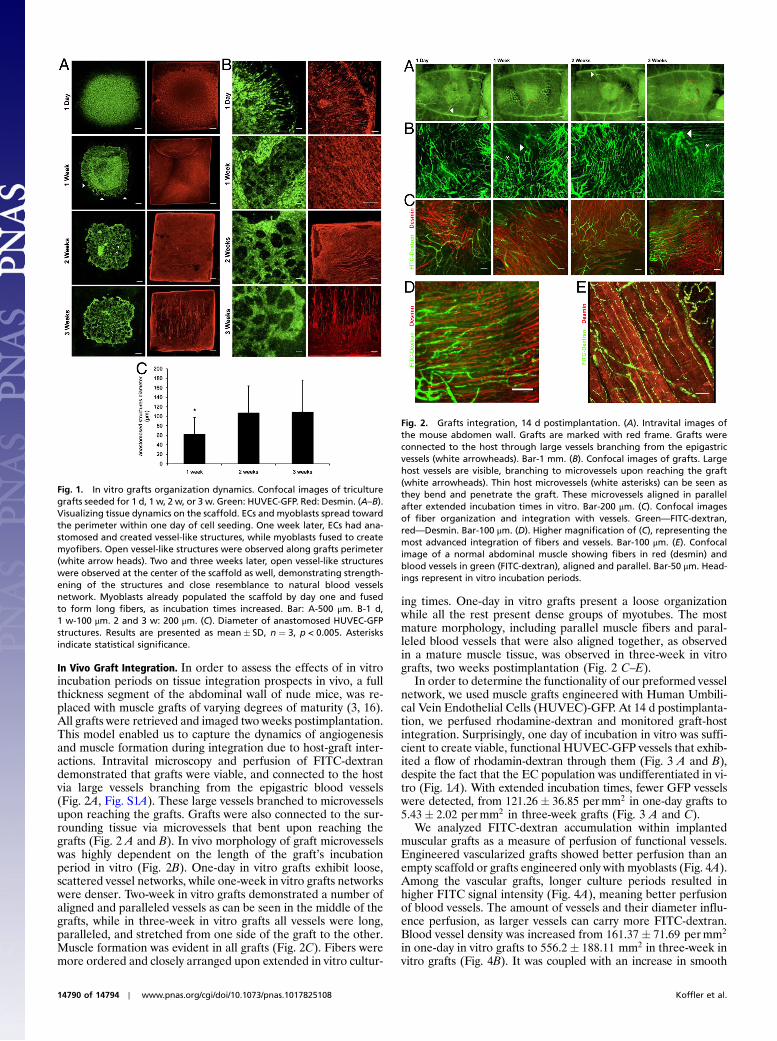
ResultsIn Vitro Tissue Dynamics.Grafts were constructed using a triculturesystem of endothelial cells (ECs), myoblasts, and foreskin fibro-blasts on biodegradable Surgisis scaffolds SIS is a resorbable,acellular bioscaffold, composed of extracellular matrix proteinsderived from the jejunum of pigs, and has been shown to be com-pletely replaced by the host within 90 d (11, 12). SIS contains avariety of factors including VEGF and FGF2 that are knownangiogenic factors (12, 13), thus it has the potential to promoteremodeling instead of a scar tissue (14). The triculture tissueproliferated and differentiated on the scaffold in vitro (Fig. 1).Within the first day of seeding, ECs had already spread from thecenter, where they had been seeded, and covered most of thescaffold area (Fig. 1 A and B). Following one week of incubation,ECs organized in two, spatially-distinct populations, where thelower layer included anastomosed ECs arranged in network-likestructures, while the upper layer population was composed ofsingle ECs. As the latter population gradually disappeared overthe ensuing two and three weeks of incubations, most of theanastomosed structures expanded in diameter (Fig. 1C). At earlystages of culturing, a number of anastomosed structures locatedalong the tissue perimeter were open, yet with time, those situ-ated at the tissue center opened as well. By three weeks postseed-ing, most structures were open and demonstrated vessel-like,branched networks (Fig. 1A). A dense and uniform myoblastpopulation was observed, where fusion to multinucleated myofi-bers was already noted within one week of seeding. After threeweeks in culture, straight, long fibers were observed extendingacross the scaffold (Fig. 1B). Grafts did not change in shape orsize, as can be seen in Fig. 1A, which shows the entire surface ofthe scaffolds, up to the perimeters. We have shown before thatmyobalsts grown on poly-lactic co glycolic acid (PLGA) scaffoldcan exert forces that change its shape (15). However, this effectwas not observed in the three-culture system, on SIS scaffolds,where most of the cells were ECs. One can affect cells and tissueorganization using mechanical loading, oxygen gradient, or elec-trical stimulus. For example, we have shown before that differentscaffold stiffness influence myoblasts viability, organization, andmyotube formation (15). We did not use any of these methods,therefore the observed pattern of aligned fibers is probably theintrinsic organizing pattern of the myoblasts. ECs and myoblastsproliferated on the scaffold and began their respective differen-tiation processes but did not integrate to form classic, parallel andaligned fibers and vessels as in the mature muscle.
Author contributions: J.K. and S.L. designed research; J.K., K.K.-F., Y.S., D.E., D.A.P., and A.L.performed research; J.K., D.A.P., S.L., and A.L. analyzed data; and J.K. and S.L. wrotethe paper.
The authors declare no conflict of interest.
*This Direct Submission article had a prearranged editor.1To whom correspondence should be addressed. E-mail: [email protected].
This article contains supporting information online at www.pnas.org/lookup/suppl/doi:10.1073/pnas.1017825108/-/DCSupplemental.
www.pnas.org/cgi/doi/10.1073/pnas.1017825108 PNAS Early Edition ∣ 14789 of 14794
APP
LIED
BIOLO
GICAL
SCIENCE
S
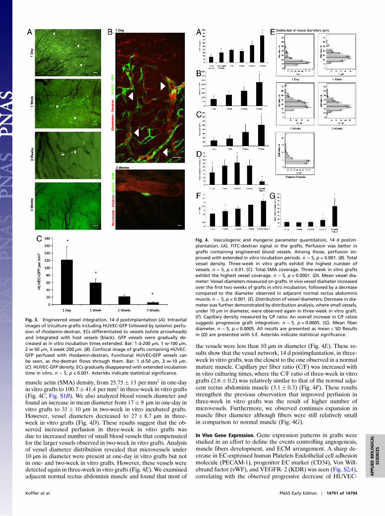
In Vivo Graft Integration. In order to assess the effects of in vitroincubation periods on tissue integration prospects in vivo, a fullthickness segment of the abdominal wall of nude mice, was re-placed with muscle grafts of varying degrees of maturity (3, 16).All grafts were retrieved and imaged two weeks postimplantation.This model enabled us to capture the dynamics of angiogenesisand muscle formation during integration due to host-graft inter-actions. Intravital microscopy and perfusion of FITC-dextrandemonstrated that grafts were viable, and connected to the hostvia large vessels branching from the epigastric blood vessels(Fig. 2A, Fig. S1A). These large vessels branched to microvesselsupon reaching the grafts. Grafts were also connected to the sur-rounding tissue via microvessels that bent upon reaching thegrafts (Fig. 2 A and B). In vivo morphology of graft microvesselswas highly dependent on the length of the graft’s incubationperiod in vitro (Fig. 2B). One-day in vitro grafts exhibit loose,scattered vessel networks, while one-week in vitro grafts networkswere denser. Two-week in vitro grafts demonstrated a number ofaligned and paralleled vessels as can be seen in the middle of thegrafts, while in three-week in vitro grafts all vessels were long,paralleled, and stretched from one side of the graft to the other.Muscle formation was evident in all grafts (Fig. 2C). Fibers weremore ordered and closely arranged upon extended in vitro cultur-
ing times. One-day in vitro grafts present a loose organizationwhile all the rest present dense groups of myotubes. The mostmature morphology, including parallel muscle fibers and paral-leled blood vessels that were also aligned together, as observedin a mature muscle tissue, was observed in three-week in vitrografts, two weeks postimplantation (Fig. 2 C–E).
In order to determine the functionality of our preformed vesselnetwork, we used muscle grafts engineered with Human Umbili-cal Vein Endothelial Cells (HUVEC)-GFP. At 14 d postimplanta-tion, we perfused rhodamine-dextran and monitored graft-hostintegration. Surprisingly, one day of incubation in vitro was suffi-cient to create viable, functional HUVEC-GFP vessels that exhib-ited a flow of rhodamin-dextran through them (Fig. 3 A and B),despite the fact that the EC population was undifferentiated in vi-tro (Fig. 1A). With extended incubation times, fewer GFP vesselswere detected, from 121.26� 36.85 permm2 in one-day grafts to5.43� 2.02 permm2 in three-week grafts (Fig. 3 A and C).
We analyzed FITC-dextran accumulation within implantedmuscular grafts as a measure of perfusion of functional vessels.Engineered vascularized grafts showed better perfusion than anempty scaffold or grafts engineered only with myoblasts (Fig. 4A).Among the vascular grafts, longer culture periods resulted inhigher FITC signal intensity (Fig. 4A), meaning better perfusionof blood vessels. The amount of vessels and their diameter influ-ence perfusion, as larger vessels can carry more FITC-dextran.Blood vessel density was increased from 161.37� 71.69 permm2
in one-day in vitro grafts to 556.2� 188.11 mm2 in three-week invitro grafts (Fig. 4B). It was coupled with an increase in smooth
Fig. 1. In vitro grafts organization dynamics. Confocal images of triculturegrafts seeded for 1 d, 1 w, 2 w, or 3 w. Green: HUVEC-GFP. Red: Desmin. (A–B).Visualizing tissue dynamics on the scaffold. ECs and myoblasts spread towardthe perimeter within one day of cell seeding. One week later, ECs had ana-stomosed and created vessel-like structures, while myoblasts fused to createmyofibers. Open vessel-like structures were observed along grafts perimeter(white arrow heads). Two and three weeks later, open vessel-like structureswere observed at the center of the scaffold as well, demonstrating strength-ening of the structures and close resemblance to natural blood vesselsnetwork. Myoblasts already populated the scaffold by day one and fusedto form long fibers, as incubation times increased. Bar: A-500 μm. B-1 d,1 w-100 μm. 2 and 3 w: 200 μm. (C). Diameter of anastomosed HUVEC-GFPstructures. Results are presented as mean� SD, n ¼ 3, p < 0.005. Asterisksindicate statistical significance.
Fig. 2. Grafts integration, 14 d postimplantation. (A). Intravital images ofthe mouse abdomen wall. Grafts are marked with red frame. Grafts wereconnected to the host through large vessels branching from the epigastricvessels (white arrowheads). Bar-1 mm. (B). Confocal images of grafts. Largehost vessels are visible, branching to microvessels upon reaching the graft(white arrowheads). Thin host microvessels (white asterisks) can be seen asthey bend and penetrate the graft. These microvessels aligned in parallelafter extended incubation times in vitro. Bar-200 μm. (C). Confocal imagesof fiber organization and integration with vessels. Green—FITC-dextran,red—Desmin. Bar-100 μm. (D). Higher magnification of (C), representing themost advanced integration of fibers and vessels. Bar-100 μm. (E). Confocalimage of a normal abdominal muscle showing fibers in red (desmin) andblood vessels in green (FITC-dextran), aligned and parallel. Bar-50 μm. Head-ings represent in vitro incubation periods.
14790 of 14794 ∣ www.pnas.org/cgi/doi/10.1073/pnas.1017825108 Koffler et al.
muscle actin (SMA) density, from 25.75� 13 permm2 in one-dayin vitro grafts to 100.7� 41.4 permm2 in three-week in vitro grafts(Fig. 4C, Fig. S1B). We also analyzed blood vessels diameter andfound an increase in mean diameter from 17� 9 μm in one-day invitro grafts to 31� 10 μm in two-week in vitro incubated grafts.However, vessel diameters decreased to 27� 8.7 μm in three-week in vitro grafts (Fig. 4D). These results suggest that the ob-served increased perfusion in three-week in vitro grafts wasdue to increased number of small blood vessels that compensatedfor the larger vessels observed in two-week in vitro grafts. Analysisof vessel diameter distribution revealed that microvessels under10 μm in diameter were present at one-day in vitro grafts but notin one- and two-week in vitro grafts. However, these vessels weredetected again in three-week in vitro grafts (Fig. 4E).We examinedadjacent normal rectus abdominis muscle and found that most of
the vessels were less than 10 μm in diameter (Fig. 4E). These re-sults show that the vessel network, 14 d postimplantation, in three-week in vitro grafts, was the closest to the one observed in a normalmature muscle. Capillary per fiber ratio (C/F) was increased within vitro culturing times, where the C/F ratio of three-week in vitrografts (2.6� 0.2) was relatively similar to that of the normal adja-cent rectus abdominis muscle (3.1� 0.3) (Fig. 4F). These resultsstrengthen the previous observation that improved perfusion inthree-week in vitro grafts was the result of higher number ofmicrovessels. Furthermore, we observed continues expansion inmuscle fiber diameter although fibers were still relatively smallin comparison to normal muscle (Fig. 4G).
In Vivo Gene Expression. Gene expression patterns in grafts werestudied in an effort to define the events controlling angiogenesis,muscle fibers development, and ECM arrangement. A sharp de-crease in EC-expressed human Platelets Endothelial cell adhesionmolecule (PECAM-1), progenitor EC marker (CD34), Von Will-ebrand factor (vWF), and VEGFR- 2 (KDR) was seen (Fig. S2A),correlating with the observed progressive decrease of HUVEC-
Fig. 3. Engineered vessel integration, 14 d postimplantation (A). Intravitalimages of triculture grafts including HUVEC-GFP followed by systemic perfu-sion of rhodamin-dextran. ECs differentiated to vessels (white arrowheads)and integrated with host vessels (black). GFP vessels were gradually de-creased as in vitro incubation times extended. Bar: 1 d-200 μm, 1 w-100 μm,2 w-50 μm, 3 week-200 μm. (B). Confocal image of grafts containing HUVEC-GFP perfused with rhodamin-dextran, Functional HUVEC-GFP vessels canbe seen, as rho-dextran flows through them. Bar: 1 d-50 μm, 3 w-10 μm.(C). HUVEC-GFP density. ECs gradually disappeared with extended incubationtime in vitro. n ¼ 5, p < 0.001. Asterisks indicate statistical significance.
Fig. 4. Vasculogenic and myogenic parameter quantitation, 14 d postim-plantation. (A). FITC-dextran signal in the grafts. Perfusion was better ingrafts containing engineered blood vessels. Among those, perfusion im-proved with extended in vitro incubation periods. n ¼ 5, p < 0.001. (B). Totalvessel density. Three-week in vitro grafts exhibit the highest number ofvessels. n ¼ 5, p < 0.01. (C). Total SMA coverage. Three-week in vitro graftsexhibit the highest vessel coverage. n ¼ 5, p < 0.0001. (D). Mean vessel dia-meter: Vessel diameters measured on grafts. In vivo vessel diameter increasedover the first two weeks of grafts in vitro incubation, followed by a decreasecompared to the diameter observed in adjacent normal rectus abdominismuscle. n ¼ 5, p < 0.001. (E). Distribution of vessel diameters: Decrease in dia-meter was further demonstrated by distribution analysis, where small vessels,under 10 μm in diameter, were observed again in three-week in vitro graft.(F). Capillary density measured by C/F ratio: An overall increase in C/F ratiossuggests progressive graft integration. n ¼ 5, p < 0.0005. (G). Mean fiberdiameter. n ¼ 5, p < 0.0005. All results are presented as mean� SD Resultsin (D) are presented with S.E. Asterisks indicate statistical significance.
Koffler et al. PNAS Early Edition ∣ 14791 of 14794
APP
LIED
BIOLO
GICAL
SCIENCE
S
GFP in vivo as incubation times extended (Fig. 3A). In contrast,the human morphogenes, capillary morphogenesis genes (CMG-1, CMG-2, and CD39), angiogenic factors (VEGF-A, VEGF-B,and FGF2) and the hypoxia marker HIF1α, all critical factorsin angiogenesis, vessel survival, and stabilization (17–25), were stillhighly expressed in three-week in vitro grafts (Fig. S2A). We havepreviously reported (16) that in the presence of ECs, foreskinfibroblasts differentiate to smooth muscle cells, colocalize withECs, and secrete VEGF in vitro. To address this issue we labeledHFF cells with DiI (1,1'-dioctadecyl-3,3,3',3'-tetramethylindocar-bocyanine perchlorate) prior to implantation and visualize themat the end point. Here we show that HFF cells survived and differ-entiated to smooth muscles exhibiting close interaction with bloodvessels (Fig. S1C). This observation means that they were thesource of the additional human angiogenic factors expression. In-creased expression of the mouse ECs genes PECAM-1, Flk1, andthrombomodulin (THBD), supported formation and stabilizationof host blood vessels (21) (Fig. S2B), while human ECs decreasedin number (Fig. S2A). We observed a phenomena where lowexpression levels of mouse HIF1α,VEGF-AVEGF-B, FGF-2, An-goipoietin-2, CMG-1, and CMG-2 genes were observed while theirhuman counterparts were highly expressed. (Fig. S2B). On the otherhand it was also interesting to note that PDGF-B, which is importantfor vessel stabilization, was highly expressed by the host, and by thegraft as well. Mousemuscle markers Pax7, c-Met, N-cam,M-cadher-in, Myf5, and myogenin were upregulated (Fig. S2C), suggestingsubstantial muscle regeneration involving satellite cells activation,proliferation, differentiation, fusion to myofibers, and maturation(26–30). This expression profile was accompanied by increasedexpression of ECM components (Fig. S2D), including collagen 1,collagen 3, collagen 4, collagen 6, laminin chains α5, β1, and fibro-nectin. Expression of MuSK, DOK7, and laminin chains α4, α2, β2,γ1, all associated with neuro-muscular junctions (NMJ) (31–35),were not upregulated in the grafts, relatively to one-day in vitrograft. The neural marker β3-Tubulin, demonstrates that the NMJin one-day in vitro graft was only partially developed (Fig. S1F), sug-gesting that it is not yet fully formed and organized.
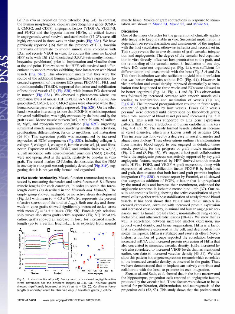
In Vivo Muscle Functionality.Muscle function (contraction) was as-sessed by measuring the passive and active forces at 6–8 differentmuscle lengths for each construct, in order to obtain the force-length curves (as described in the Materials and Methods). Theempty group showed negligible or no active stress development(Fig. 5A) with mean Fa ¼ 6.3� 7.6%, (Fa represents the percentof active stress out of the total at Lmax). Both one-day and three-week in vitro grafts showed significantly increased active stresswith mean Fa ¼ 34.5� 20.4% (Fig. 5B). Force-length relation-ship curves also stress grafts active response (Fig. 5C). Most tri-culture grafts showed an increase in force for increased musclelength (up to a certain length-Lmax), as expected from normal
muscle tissue. Movies of graft contractions in response to stimu-lation are shown in Movie S1, Movie S2, and Movie S3.
DiscussionOne of the major obstacles for the generation of clinically applic-able tissue is to keep it viable in vivo. Successful implantation isdependent on revascularization and anastomosis of the implantwith the host vasculature, otherwise ischemia and necrosis set in.This study reveals the in vivo dynamics of graft vascular integra-tion and angiogenesis. The degree of the vascular bed organiza-tion in vitro directly influences host penetration to the graft, andthe remodeling of the vascular network. Incubation of one day,where ECs were not organized yet (Fig. 1A), was sufficient fordifferentiation and anastomosis with the host (Fig. 3 A and B).This short incubation was also sufficient to yield blood perfusionthat was better than grafts without ECs (Fig. 4A). However, invivo perfusion and vessel density improved dramatically as incu-bation time lengthened to three weeks and ECs were allowed tobe better organized (Fig. 1A, Fig. 4 A and B). This observationwas followed by increased coverage with smooth muscle cellsleading to stabilization and maturation of vessels (Fig 4C,Fig S1B). The improved preorganization resulted in faster repla-cement of graft vessels by host vessels. Fewer GFP vesselspermm2 were detected until barely seen on three-week grafts,while total number of blood vessel permm2 increased (Fig. 3 Aand C). This result was supported by ECs gene expressionpatterns that showed a reduction in human endothelial markers(Fig. 4 A and B). The newly formed vessels exhibit an increasein vessel diameter, which is a known result of ischemia (36).This increase was followed by a decrease in vessel diameter andaligned morphology. These observations outline a transformationfrom massive blood supply to one engaged in detailed tissueneeds, providing for the progress of graft muscle maturation(Fig. 2 C and D, Fig. 4E). We suggest a host-graft collaborationwhere the angiogenic process was actively supported by key graftangiogenic factors, expressed by HFF derived smooth musclecells. HIF1α, FGF2, and VEGF-A graft expression, along withexpression of vessel stabilization factor PDGF-B by both hostand graft, demonstrate that both host and graft promote implantintegration (Fig. S2B). A recent report by Frontini, et al. showedthat exogenous addition of FGF9, a factor that is upregulatedby the mural cells and increase their recruitment, enhanced theangiogenic response in ischemic mouse hind limb (37). Our re-sults enhance this finding, showing the active contribution of graftmural cells together with host stabilization of newly formed bloodvessels. It has been shown that VEGF and PDGF mRNA in-creased expression, correlate with increased protein expressionand increased vessel density, in animal and human angiogenic sce-narios, such as human breast cancer, non-small-cell lung cancer,melanoma, and atherosclerotic lesions (38–42). We show that aswell; a correlation between increased mRNA expression, im-proved vessel density, and tissue organization. Hif1α is a factorthat is constitutively expressed in the cell, and degraded in nor-moxia. In hypoxia, Hif1α is stabilized and exerts its effect. Never-theless, a number of groups reported the correlation betweenincreased mRNA and increased protein expression of Hif1α thatalso correlated to increased vascular density. Hif1α increased le-vels also correlated to increased VEGF levels that, as mentionedearlier, correlate to increased vascular density (43–51). We alsoshow this pattern in our gene expression research which correlatesto the increased vascular density, as observed in the grafts. Thus,we have demonstrated that an implant can actively contribute andcollaborate with the host, to promote its own integration.
Shen, et al. and Suda, et al. showed that in the bone marrow andthe hippocampus, progenitor cells respond to angiogenic factors,produced by the vascular bed. These factors were shown to be es-sential for proliferation, differentiation, and neurogenesis of theprogenitor cells (52, 53). This study shows that the preimplanted
Fig. 5. In vivo functionality. (A). Empty constructs showed negligible activestress developed for the different lengths (n ¼ 4), (B). Triculture graftsshowed significantly increased active stress (n ¼ 12). (C). Curvilinear force-length relationship could be observed only in triculture grafts. p < 0.05.
14792 of 14794 ∣ www.pnas.org/cgi/doi/10.1073/pnas.1017825108 Koffler et al.
vascular bed is essential for graft integration and organogenesis invivo. In a regenerated muscle, once the myotubes are formed, theygo through a maturation process during which they become inner-vated and vascularized, resulting in myofibers (10). Myofibers arethen packed together by connective tissues to provide strength tothe muscle. The parallel alignment of the myoblasts during fusionis the key to what gives the myofibers their ability to produce theforce necessary for movement and strength (10, 54). We havebrought together the vascular bed and myoblasts to improve mus-cle regeneration. Our grafts exhibited an increase in fiber diameterand capillary per fiber ratio in vivo (Fig. 4 E and F) as incubationtime is prolonged. Further tissue maturation, similar to the normalrectus abdominis muscle, was evident by coalignment of blood ves-sels with muscle fibers in three-week in vitro graft (Fig. 2 C–E).This coalignment was not demonstrated in grafts engineered withonly myoblasts, (Fig. S1D). Another control of empty SIS scaffoldin vivo, showed scattered blood vessels and single, unfused, myo-blasts population that resembled the single cell population ob-served in one-day in vitro grafts (Fig. S1E, Fig. 1B). All stagesin the muscle regeneration process, from satellite cells to maturemuscle, were demonstrated by real-time PCR, showing increasedexpression of relevant markers (Fig. S2C). This maturation wasaccompanied by an increase in the expression of ECM components(Fig. S2D). These components have structural roles in interstitialconnective tissue and basement membrane development duringangiogenesis and myogenesis, and are important to vessel functionand stabilization, as well as muscle structure and force transduction(26, 32, 33, 54–56).
The functional study showed that our triculture graft contractsin vivo in response to stimulation, while the empty scaffold didnot exhibit any activity. Although we could not show significantdifferences in activity between one-day and three-week in vitrografts it was clear that three-week in vitro grafts were highlyintegrated with the host muscles, which made it very difficult todissect the grafts based on boundary visibility (Fig. S3 B–D). Theactive force was about 35% of an intact muscle. This result wasexpected because our additional data support the fact that thegrafts are not fully matured in terms of muscle fibers maturation(Fig. 4 F–G, Fig. S2D, Fig. S1F) and NMJ formation after only14 d in vivo. Nevertheless, contraction after such a short integra-tion period is very impressive.
The results of this study substantiate the need for vasculariza-tion and emphasize its importance for implant integration. Wesuggest a unique “relay” approach to engineered vascularizedgraft integration in vivo. The relay is the competition betweenthe various tissue organizations states examined in vitro, to resultin a more advanced maturation and integration in vivo. The moreadvanced organization (three-week in vitro grafts) resulted infaster anastomosis, maturation, and replacement by the host. Intissue engineering and cell therapy where endothelial cells arebeing used, investigators seeking to find their implanted cells orvessels in vivo, so they can show a prolonged and sustained effectover time (57). We suggest the opposite. We show that increasedincubation allows for a more advanced organization that resultsin better perfusion increased vessel density and maturity, butgradual decrease in implanted HUVEC-GFP. Meaning once theimplanted vessels anastomosed and perfused, the host is able toquickly remodel the blood vessel network to its needs. Anotherresult is a more advanced fiber maturation and morphologywhere vessels and fibers are parallel and aligned, as in a maturemuscle. We show the effect of ECs addition to the graft, and theresulted formation of tissue structures—the vascular tree, fibersorganization, and their interaction. In addition, we suggest a col-laboration in the angiogenic process, where some angiogenicfactors were expressed by the graft (VEGF-A, VEGF-B, FGF2,,and HIF1α) and not by the host, while one factor (PDGF-B) washighly expressed by both. This suggested collaboration supportedthe observed improved integration, which results in partial func-
tionality 14 d postimplantation, and morphology and organiza-tion that resemble the normal mature muscle organization.
Materials and MethodsCell Culture. C2C12 mouse myoblast cells [American Type Culture Collection(ATCC)] were cultured in DMEM supplemented with 20% FBS, and 2.5%Hepes buffer. Human Umbilical Vein Endothelial Cells (HUVEC, Clonetics)and HUVEC-GFP (Angio-Proteomie) were cultured in endothelial cell medium(EGM-2, EGM-MV, and their respective bullet kits, Cambrex Bio ScienceWalkersville, Inc.). Human foreskin fibroblasts were cultured in DMEMsupplemented with 10% FBS and 1% nonessential amino acids.
Graft Preparation. Four-ply Surgisis (Cook Biotech Inc.) were rehydrated ac-cording to the manufacturer’s instructions. For seeding, the desired numberof cells were pooled and resuspended in 3□l culturemedium. The suspensionwas placed on the scaffold and was allowed to be absorbed (1.5 h, 37 °C, 5%CO2), after which 3 mL of 1∶1 HUVEC-GFP:C2C12 media were added. Mediumwas replaced every other day. We have previously refined these conditions ofmedia and cell numbers (16).
Fibroblast Labeling. Cells were labeled [45 min, room temperature (RT)],directly on culture plates, with 3 μL∕mL Vybrant DiI (In Vitrogen) dilutedin medium, and then washed three times with PBS.
Implantation of Muscle Grafts. All surgical procedures were conducted accord-ing to protocols approved by the Institutional Animal Care and Use Commit-tee. Male, 8 w old, nudemice (Harlan Laboratories) were divided randomly tofour groups, five mice per group. Mice were anesthetized using a ketamine:xylazine cocktail at a dose of 35 μL∕20 g delivered with a 25-gauge needle. Asmall incision was made allowing access to the linea-alba and surroundingtissue, where a 3 × 2 mm full thickness defect segment was removed. Musclegrafts were sutured in place using four 8-0 silk sutures. All mice were mon-itored closely for 1–2 h to ensure full recovery from the anesthesia. Mice wereanesthetized two weeks later and 10 mg∕mL FITC-dextran or rhodamine-dextran (Sigma) were perfused through the tail vein. Grafts were either im-aged or immediately placed into RNAlater buffer (Qiagen) until RNA extrac-tion was performed as described below.
Tissue Processing and Immunohistochemical Staining. Whole mount: Graftswere fixed in 4% paraformaldehyde (30min, RT) followed by extensive wash-ing in PBS and overnight blocking (10% FBS, 0.1 g glycine, 0.1% Triton X-100,in PBS). Skeletal muscles were labeled using a goat polyclonal desmin anti-body (Santa Cruz Biotechnology), smooth muscles were labeled using mouseSMA (DAKO), mouse blood vessels were labeled using FITC-ILB4 (Vector Labs),and NMJ was labeled using mouse anti βIII-tubulin (Promega). All antibodieswere diluted in fresh blocking serum and incubated with samples overnightat 4 °C. Following extensive washings, donkey anti-goat, or goat anti-mouseCy3 antibodies (Jackson ImmunoResearch Laboratories Inc.) and DAPI (Sigma)were diluted in PBS and incubated with the grafts (3 h, RT). Serial sections:Grafts were fixed for 30 min in 10% neutral buffered formalin, routinelyprocessed, and embedded in paraffin. Serial sections (5 μm) were placedon silanized slides for immunohistochemistry as described above.
Imaging. Intravital images were captured using an Olympus fluorescent stereomicroscope (SZX12). In vitro and ex-vivo images were taken with a Zeiss LSM510 Meta Confocal microscope and Leica TCS-LSI confocal microscope. Slideswere imaged using a Zeiss fluorescent microscope (Axiovert-200M).
Image Analysis. FITC intensity was measured using NIH ImageJ software, aswere vessels and muscle fiber numbers and diameters. For HUVEC-GFP andvessel area calculations, images were segmented followed by quantificationof signal area.
Statistical Analysis. Results were analyzed using one-tail ANOVA followedby Tukey's test using JMP 8.0 (SAS); α < 0.05 was considered statisticallysignificant.
RT-PCR Analysis. Total RNA was isolated using RNEasy Midi Kit (Qiagen)according to the manufacturer’s muscle tissue isolation protocol. RT-PCRwas performed using High Capacity cDNA Reverse Transcription Kit (AppliedBiosystems). TaqMan assays were performed using custom gene expressionplates and probes (Applied Biosystems), as detailed in Table S1. All humanand mouse sequences were confirmed unique and noncrossreactive by
Koffler et al. PNAS Early Edition ∣ 14793 of 14794
APP
LIED
BIOLO
GICAL
SCIENCE
S
BLAST. Results were processed using DataAssist Software (Applied Biosys-tems) and visualized using ArrayStar software.
Force Measurements. The constructs were dissected in a bath perfused withmodified Krebs-Henseleit solution containing (in mM) 143.7 Na+, 5.0 K+,130.4 Cl−, 1.2 Mg2+, 2.26 PO4−, 19.0 HCO3−, and 10.0 glucose; pH wasset at 7.40 by adjusting the flow of the gas mixture (95% O2, 5% CO2). Cal-cium concentration was 1.5 mM. The constructs were dissected based on theborders of the scaffold with small ends of surrounding tissue on each side forforce transducer and motor arm insertion (Fig. S3A). The dimensions of allthe constructs were fairly similar of about 4 × 3 × 1 mm. The experimentalbath was placed on a stage of an inverted microscope (TE300, Nikon).The bath was perfused at a constant rate, and the temperature was keptat 25 °C. The constructs were stimulated at 0.2 Hz. The stimulation amplitudewas 1.5 times the stimulus threshold for maximal force production. Force
was measured by a silicone strain gauge (SensorOne AE801). Muscle lengthwas controlled by a fast servomotor (Aurora Scientific, model 308B) andmonitored by a precise capacitance sensor attached to the motor axis.Tetanic stimulation (50 Hz) was performed following every regular twitch.Passive stress was calculated from baseline force at each length and activestress was calculated from the developed steady force at tetanus. Musclelength was measured from the calibrated signal of servomotor location.Muscle length and force were sampled at 5,000 Hz. Each measurementwas repeated five times, and the results were averaged in order to reducethe random noise.
ACKNOWLEDGMENTS. The authors would like to thank the Technion’s LorryLokey Infrastructure Unit and the Russell Berrie Nanotechnology Institute.This research was supported by the Israeli Ministry of Science, Levi EshkolScholarship, and Marie Curie International Reintegration Grants.
1. Atisha D, Alderman AK (2009) A systematic review of abdominal wall functionfollowing abdominal flaps for postmastectomy breast reconstruction. Ann Plast Surg63:222–230.
2. Baechler MF, Groth AT, Nesti LJ, Martin BD (2010) Soft tissue management of warwounds to the foot and ankle. Foot Ankle Clin 15:113–138, http://www.ncbi.nlm.nih.gov/pubmed/19067424.
3. de Vries Reilingh TS, et al. (2007) Autologous tissue repair of large abdominal walldefects. Brit J Surg 94:791–803.
4. Falco EE, Roth JS, Fisher JP (2008) Skeletal muscle tissue engineering approaches toabdominal wall hernia repair. Birth Defects Res C Embryo Today 84:315–321, http://www.ncbi.nlm.nih.gov/pubmed?term=20189120%5Buid%5D.
5. Mertsching H, et al. (2009) Generation and transplantation of an autologous vascu-larized bioartificial human tissue. Transplantation 88:203–210.
6. Netscher DT, Baumholtz MA (2009) Chest reconstruction: I. Anterior and anterolateralchestwall andwounds affecting respiratory function. Plast Reconstr Surg 124:240e–252e.
7. Laschke MW, et al. (2006) Angiogenesis in tissue engineering: breathing life intoconstructed tissue substitutes. Tissue Eng 12:2093–2104.
8. Polykandriotis E, Arkudas A, Horch RE, Sturzl M, Kneser U (2007) Autonomouslyvascularized cellular constructs in tissue engineering: opening a new perspectivefor biomedical science. J Cell Mol Med 11:6–20.
9. Guo L, Pribaz JJ (2009) Clinical flap prefabrication. Plast Reconstr Surg 124:e340–350.10. Gayraud-Morel B, Chretien F, Tajbakhsh S (2009) Skeletal muscle as a paradigm for
regenerative biology and medicine. Regen Med 4:293–319.11. Gilbert TW, Stewart-Akers AM, Badylak SF (2007) A quantitative method for evaluat-
ing the degradation of biologic scaffold materials. Biomaterials 28:147–150.12. Woods AM, Rodenberg EJ, Hiles MC, Pavalko FM (2004) Improved biocompatibility of
small intestinal submucosa (SIS) following conditioning by human endothelial cells.Biomaterials 25:515–525.
13. Badylak S, Liang A, Record R, Tullius R, Hodde J (1999) Endothelial cell adherence tosmall intestinal submucosa: an acellular bioscaffold. Biomaterials 20:2257–2263.
14. Hodde J (2006) Extracellular matrix as a bioactive material for soft tissue reconstruc-tion. ANZ J Surg 76:1096–1100.
15. Levy-Mishali M, Zoldan J, Levenberg S (2009) Effect of scaffold stiffness on myoblastdifferentiation. Tissue Eng Pt A 15:935–944.
16. Levenberg S, et al. (2005) Engineering vascularized skeletal muscle tissue. Nat Biotech-nol 23:879–884.
17. Goepfert C, et al. (2001) Disordered cellular migration and angiogenesis in cd39-nullmice. Circulation 104:3109–3115.
18. Jackson SW, et al. (2007) Disordered purinergic signaling inhibits pathological angio-genesis in cd39/Entpd1-null mice. Am J Pathol 171:1395–1404.
19. Wagatsuma A (2007) Endogenous expression of angiogenesis-related factors in re-sponse to muscle injury. Mol Cell Biochem 298:151–159.
20. Lokmic Z, Mitchell GM (2008) Engineering the microcirculation. Tissue Eng Part B-Rev14:87–103.
21. Carmeliet P, Conway EM (2001) Growing better blood vessels. Nat Biotechnol19:1019–1020.
22. Norrby K (2006) In vivo models of angiogenesis. J Cell Mol Med 10:588–612.23. Dowling O, et al. (2003) Mutations in capillary morphogenesis gene-2 result in
the allelic disorders juvenile hyaline fibromatosis and infantile systemic hyalinosis.Am J Hum Genet 73:957–966.
24. Fedak PW (2008) Paracrine effects of cell transplantation: modifying ventricularremodeling in the failing heart. Semin Thorac Cardiovasc Surg 20:87–93, http://www.ncbi.nlm.nih.gov/pubmed/18707639.
25. Zhang F, et al. (2009) VEGF-B is dispensable for blood vessel growth but critical for theirsurvival, and VEGF-B targeting inhibits pathological angiogenesis. Proc Natl Acad SciUSA 106:6152–6157.
26. Charge SB, Rudnicki MA (2004) Cellular and molecular regulation of muscle regenera-tion. Physiol Rev 84:209–238.
27. Le Grand F, Rudnicki MA (2007) Skeletal muscle satellite cells and adult myogenesis.Curr Opin Cell Biol 19:628–633.
28. Tajbakhsh S (2009) Skeletal muscle stem cells in developmental versus regenerativemyogenesis. J Intern Med 266:372–389.
29. Tedesco FS, Dellavalle A, Diaz-Manera J, Messina G, Cossu G (2010) Repairing skeletalmuscle: regenerative potential of skeletal muscle stem cells. J Clin Invest 120:11–19.
30. Wallace GQ, McNally EM (2009) Mechanisms of muscle degeneration, regeneration,and repair in the muscular dystrophies. Annu Rev Physiol 71:37–57.
31. d’Houtaud S, et al. (2009) Synapse formation and regeneration (Translated from fre).Neurochirurgie 55:S49–62 (in fre).
32. Durbeej M (2010) Laminins. Cell Tissue Res 339:259–268.33. Grounds M (2008) Complexity of Extracellular Matrix and Skeletal Muscle Regen-
eration. Skeletal Muscle Repair and Regeneration, ed SSaT Partridge (Springer,Dordrecht, The Netherlands), pp 269–301.
34. Milholland RB, Gordon H (2007) A role for acetylcholine receptors in their own aggre-gation on muscle cells. Dev Neurobiol 67:999–1008.
35. Tzu J, Marinkovich MP (2008) Bridging structure with function: structural, regulatory,and developmental role of laminins. Int J Biochem Cell Biol 40:199–214.
36. Anonymous (2007) Grabb and Smith’s Plastic Surgery. (LippincottWilliams andWilkins,Philadelphia), 6th ed, p 43.
37. Frontini MJ, et al. (2011) Fibroblast growth factor 9 delivery during angiogenesisproduces durable, vasoresponsive microvessels wrapped by smooth muscle cells.Nat Biotechnol 29:421–427.
38. Battegay EJ, Rupp J, Iruela-Arispe L, Sage EH, Pech M (1994) PDGF-BB modulatesendothelial proliferation and angiogenesis in vitro via PDGF beta-receptors. J Cell Biol125:917–928.
39. Forsberg K, Valyi-Nagy I, Heldin CH, Herlyn M, Westermark B (1993) Platelet-derivedgrowth factor (PDGF) in oncogenesis: development of a vascular connective tissuestroma in xenotransplanted human melanoma producing PDGF-BB. Proc Natl AcadSci USA 90:393–397.
40. Li J, et al. (1995) Induction of vascular endothelial growth factor gene expression byinterleukin-1 beta in rat aortic smooth muscle cells. J Biol Chem 270:308–312.
41. Scott PA, et al. (1998) Differential expression of vascular endothelial growth factormRNA vs protein isoform expression in human breast cancer and relationship toeIF-4E. Br J Cancer 77:2120–2128.
42. Yuan A, et al. (2000) Correlation of total VEGF mRNA and protein expression withhistologic type, tumor angiogenesis, patient survival and timing of relapse innon-small-cell lung cancer. Int J Cancer 89:475–483.
43. Fu D, Dai A, Hu R, Chen Y, Zhu L (2008) Expression and role of factor inhibiting hypoxia-inducible factor-1 in pulmonary arteries of rat with hypoxia-induced hypertension.Acta Biochim Biophys Sin 40:883–892.
44. Furlan D, et al. (2007) Up-regulation and stabilization of HIF-1alpha in colorectalcarcinomas. Surg Oncol 16:S25–27.
45. Jiang H, Feng Y (2006) Hypoxia-inducible factor 1alpha (HIF-1alpha) correlated withtumor growth and apoptosis in ovarian cancer. Int J Gynecol Cancer 16:405–412.
46. Lee S, Garner EI, Welch WR, Berkowitz RS, Mok SC (2007) Over-expression of hypoxia-inducible factor 1 alpha in ovarian clear cell carcinoma. Gynecol Oncol 106:311–317.
47. Liang B, et al. (2010) Correlation of hypoxia-inducible factor 1alpha with angiogenesisin liver tumors after transcatheter arterial embolization in an animal model. Cardio-vasc Inter Rad 33:806–812.
48. Mori R, et al. (2010) The relationship between proangiogenic gene expression levels inprostate cancer and their prognostic value for clinical outcomes. Prostate 70:1692–1700.
49. Qiu MZ, et al. (2011) Expressions of hypoxia-inducible factor-1alpha and hexokinase-IIin gastric adenocarcinoma: the impact on prognosis and correlation to clinicopatho-logic features. Tumour Biol 32:159–166.
50. Wang W, et al. (2009) Expression and correlation of hypoxia-inducible factor-1alpha,vascular endothelial growth factor and microvessel density in experimental rat hepa-tocarcinogenesis. J Int Med Res 37:417–425.
51. Yohena T, et al. (2009) Upregulation of hypoxia-inducible factor-1alpha mRNA and itsclinical significance in non-small cell lung cancer. J Thorac Oncol 4:284–290.
52. Shen Q, et al. (2004) Endothelial cells stimulate self-renewal and expand neurogenesisof neural stem cells. Science 304:1338–1340.
53. Suda T, Arai F, Hirao A (2005) Hematopoietic stem cells and their niche. Trends Immu-nol 26:426–433.
54. Grounds MD, Sorokin L, White J (2005) Strength at the extracellular matrix-muscleinterface. Scand J Med Sci Spor 15:381–391.
55. Francis ME, Uriel S, Brey EM (2008) Endothelial cell-matrix interactions in neovascular-ization. Tissue Eng Part B-Rev 14:19–32.
56. Jain RK (2003) Molecular regulation of vessel maturation. Nat Med 9:685–693.57. Koike N, et al. (2004) Tissue engineering: creation of long-lasting blood vessels.Nature
428:138–139.
14794 of 14794 ∣ www.pnas.org/cgi/doi/10.1073/pnas.1017825108 Koffler et al.