in eukaryotes, large primary transcripts are processed to smaller, mature mrnas. recap (1) what was...
TRANSCRIPT

In eukaryotes, large primary transcripts are processed to smaller, mature mRNAs.
RECAP (1)
What was first evidence for this precursor-product relationship?

RNA is unstable – it can cleave itself.
RECAP (2)
Self-splicing introns utilize this suicidal tendency and contortionist ability to direct self-cleavage at precisely defined sites
RNA can fold into complex structures (hairpins and interactions between hairpins).

Chemistry underlying the cleavage is the basis of other RNA-based cleavage
events
And also the reason for the RNA world hypothesis

Splicing in eukaryotes probably relies on the same chemistry as self-splicing group II introns.
RECAP (3)
A complex RNA+protein machine is used to precisely define splice sites.
Splicing substrates in eukaryotes much more varied, and can’t rely on 2o and 3o structure alone to define splice sites.
intron or intervening sequence
exon

What are the proteins doing to facilitate splicing?
Catalysis? – Prp8Quality control? – Prp16

U2 snRNA
U6 snRNA
U5 snRNA intron
In the catalytically active spliceosome, the U2, U5 and U6 snRNAs make very specific contacts with the splice sites.
What are the proteins doing in catalysis?
exon n
exon n+1

Prp8 is at the heart of the catalytically active spliceosome
Prp8 mutants are splicing defective.Prp8 cross links to crucial U5, U6, 5’-SS, 3’-SS and
branch point residues.Prp8 interacts with Brr2 and Snu114, which unwind
U4/U6 and allow U2 to pair with U6.Many Prp8 mutations suppress splicing defects caused
by 5’-SS, 3’-SS and branch point mutations.

Crystal structure of Prp8 reveals a cavity of appropriatedimensions to position spliceosomal RNAs for catalysis.
Structural domains of Prp8 (endonuclease, reverse transcriptase) suggest ancient evolutionary origins as a homing
endonuclease.
Prp8Group II intron

Mobile genetic elements provide an example of RNPcomplexes in which proteins and RNAs cooperate for specificity
group II self-splicing intron encodes an endonuclease (E)
maturase (M) and reverse transcriptase (RT) that are used
for integration of the mobile element back into the genome. The intron,
E, M, and RT form an RNP and the 2’OH of the intron directs cleavage of the first strand of the target DNA.
Group II self-splicing intron forms the core of an RNP thatcan direct cleavage of other nucleic acid polymers.

Splicing is dynamic, with sequential regulated alterationsin RNA:RNA and RNA:protein interactions

DEAD-box ATPases found at every step

Splicing error rates range from 1 in 1000 to 1 in 100,000
DEAD-box ATPasesimplicated in quality control

Monomeric (vs. “AAA” ATPases)RNA-dependent ATPases~300 aa domain with 7 signature motifs (e.g. eponymous tetrapeptide)2 RecA-like folds bind ATP, RNA (“closed form”)Conformation opens upon ATP hydrolysis (i.e. switch-like)
8 essential spliceosomal DEAD-box ATPases in yeast (more in mammals)
In vitro: Most catalyze RNA-dependent ATP hydrolysis (ATPase)Some catalyze ATP-dependent RNA unwinding (“helicase”)
In vivo???? Likely most are “RNPases”, destabilizing RNA:protein complexes
Transitions regulated by DEAD-box ATPases

PRP16
Prp16-1 mutant was identified in a screen for reduced-fidelity mutants:
Mutate branchpoint A to C in a splicing reporterMutagenize cells ->Select for improved splicing of reporter
Repeat selection by mutagenesis of cloned PRP16 gene ->
- New suppressors all map to the conserved DEAD-box domain- In vitro, mutant Prp16 proteins have reduced ATPase activity
Conclude:
Prp16 modulates the fidelity of splicing by an ATP-dependent mechanism

PRP16 exhibits helicase activity in vitro

Hypothesis: Prp16 promotes fidelity by acting as a timer for an essential conformation rearrangement
1) branchpoint mutations -> slow conformational rearrangement -> rejection
2) suppressor mutations in Prp16 -> more time

Prp16 joins the party after the formation of the full spliceosome, when the U1 and the U4 snRNPs are detached and the remaining U2, U5 and U6 snRNAs are rearranged.
Prp16

before transesterification if the reaction is slow, ie branch point mutant (br). DEAH mutant (D473A) of Prp16 can stabilize the association of Cwc25 with the spliceosome to promote the transesterification reaction even with a branchpoint mutant.
Prp2-mediated dissociation of SF3a/b
Yju2 and Cwc25 bind to the spliceosome to promote the first tranesterification reaction
Prp16 binds to the spliceosome after the binding of Yju2 and Cwc25
Prp16 mediates the dissociation of Cwc25 and Yju2
after the transesterification reaction if the reaction is fast,

Conclusion:ATPases promote specificity by discriminating against slow substrates

How are the splice sites identified?
In budding yeast, sequences at 5’ ss and 3’ ss are highly conserved, most genes contain only a single intron, and introns are short
Intron definition model posits that introns are first thing identified for yeast splicing

2.4 Mb
260 kb intronHuman Dystrophin gene
Genes in higher eukaryotes have many exons and introns can be very large
How are the splice sites identified?

The same primary transcript can be spliced many different ways (estimated 90% of genes experience alternative splicing)
How are the splice sites identified?

Because of the intron length and lack of specificity of splice sites, most introns contain numerous cryptic splice sites in addition to bona fide alternative splice sites.
How are the splice sites identified?

How are the splice sites identified?
x
outcomes of 5’splice site (ss) mutants (x)
activates cryptic 5‘ss ( ), but only if there is one within 100-300 bp of original 5’ ss
5’ ss 3’ ss

How are the splice sites identified?
outcomes of 5‘ss mutants
x
If there isn’t a nearby cryptic ss, then skip the exon altogether and ignore perfectly good 3’ ss of the upstream intron
5’ ss 3’ ss

How are the splice sites identified?
beta-globin mutants that create a new 3’ ss within an intron:
x
also create a new exon???
5’ ss 3’ ss

Splicing is co-transcriptional and all introns assayed are spliced within 5-10 minutes of transcription of the downstream exon and 3’ splice site, regardless of intron size (1 kb or 240 kb)

Average human exon is ~ 150 bp

The SR Proteins are a family of proteins with a common domain structure of 1 or 2 RNP RNA binding domains (also called RRMs) and a C-terminal domain rich in SR dipeptides.
These proteins are involved in many aspects of splicing, but most significantly they bind to Exonic Splicing Enhancers (ESEs) and stimulate spliceosome assembly at the adjacent sights.
It is thought that most exons carry ESE’s and require SR proteins for exon recognition.
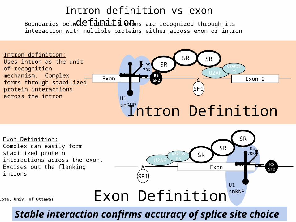
Intron definition vs exon definition
AU2AF
Exon 1
U1snRNP
RS70K
RSSF2
U2AF35RS
SF1
Exon 2
SRIntron definition:Uses intron as the unit of recognition mechanism. Complex forms through stabilized protein interactions across the intron
SRSR
Intron Definition
Exon
U1snRNP
RS70K
RSSF2A
U2AFU2AF35
RS
SF1
SR
SRSR
Exon Definition: Complex can easily form stabilized protein interactions across the exon. Excises out the flanking introns
Exon DefinitionStable interaction confirms accuracy of splice site choice
(Cote, Univ. of Ottawa)
Boundaries between introns & exons are recognized through its interaction with multiple proteins either across exon or intron

Differential size distributions of exons (~50 to 300 nt) vs. introns (<100-100,000 nt)
• SR protein - preferentially binds to exon sequences - mark the 5’ & 3’ splicing sites in conjunction w/ U1 & U2 during transcription
• hnRNP - heterogenous nuclear ribonucleoproteins (twice the diameter of nucleosome) - consists at least eight different proteins - compacts introns, thereby masking cryptic splicing sites - preferentially binds to introns, but also bind to exons, although less frequently
Why are exons preferentially recognized?

Big Question:What triggers the switch from Exon- to Intron-Defined interactions?

Internal exons first and last exons
What about the terminal exons?

Vertebrate external exons

Bioreg proposal fodder: higher eukaryotes have two splicing systems
Minor spliceosome, consists of U11, U12, U4atac, U6atac, and U5
About 100-fold less abundant than major spliceosome
Splices ~ 0.2% of introns in vertebrates

Bioreg proposal fodder: secondary structures within introns can promote exon recognition
RNA bridge brings splicing factor binding site close to exon, so to promote exon recognition

Recap 1
In higher eukaryotes, there isn’t much sequence information encoded in the 3’ss, 5’ss, or branch point

Recap “exon definition” step involves interactions between the splice sites across the exon and special sequences in the exon called Exonic Splicing Enhancers (ESE).
Recap 2

Defining an exon involves the specific stabilization or
destabilization of splice site recognition
Stabilization: exon inclusionDestabilization: exon skipping

Regulation of alternative splicing involves the specific stabilization or destabilization
of splice site recognition
Stabilization: exon inclusionDestabilization: exon skipping

How would you identify cis-regulatory sequences responsible for alternative splicing ?
Examine RNA Splicing of Transfected Splicing Reporters to identify cis-regulatory regions
ReporterPlasmid
Transfection
Mutational analysis finds an element necessary for exoninclusion
Alternatively spliced Not alternatively spliced

Four classes of splicing regulatory elements: Exonic Splicing Enhancers, Exonic Splicing Silencers (ESS), Intronic Splicing
Enhancers (ISE), and Intronic Splicing Silencers (ISS).
1 2 3+
1 2 3-
1 2 3-
1 2 3+
ESE
ESS
ISE
ISS

How would an Intronic Splicing Silencer work

SR proteins generally bind ESE, ESS, ISE, and ISSs

SR Proteins bind to specific RNA elements using their RNA binding domains similar to those in the Sex-Lethal protein.

Characterization of an ESE and SR protein in flies
Sex differentiation in flies controlled by AS CascadeDsx: weak 3’SS next to female-specific exonTra/Tra2 (females) promotes recruitment of U2AF
Sequence-specific RRM -> binds 13-nte. RepeatsRS domain interacts w U2AF RS domain
Proof of concept: Convert ESE to MS2 binding site -> activated by MS2:RS

hnRNP contain RRMs but not SR domain
Can block sterically, tighter binding affinity than U2AF
hnRNP function at ISSs
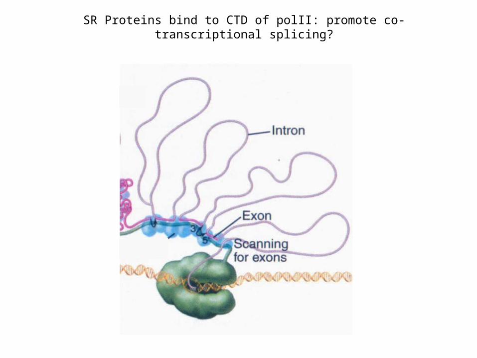
SR Proteins bind to CTD of polII: promote co-transcriptional splicing?

CTD of RNA pol II plays important role in pre-mRNA splicing
(Kornblihtt et al, 2004)

Does splice site strength affect alternative splicing?

A connection between chromatin and splicing
include exonIIIc by repress exonIIIb
include exonIIIb, repress exon IIIc,via Epithelial splicing regulatory
protein

mRNA export - formation of an export competent mRNP
Sees formation of mRNP as transcription commences
Balbiani Rings (Chironomus tentans)
• Why export as a protein/DNA complex? RNAs are too big and lack the signals to interact w/ nuclear export receptors
• Specific “adaptor” proteins must first bind to the RNA and chaperone this molecule to the export receptor, which, in turn, guides the RNA across the NPC
Follow mRNP through NPC

(Stutz & Izaurralde,2003)
Factors involved in mRNA export are co-transcriptionally recruited
• THO complex: major role in transcriptional elongation and recruitment of mRNA export factors
Model from yeast:
• Mex67 - promotes translocation across NPC
• Yra1 - mRNA export factor, interacts with Mex67

(Cullen, 2003)
(Sub2p)
(Yra1p) (Mtr2p)
(Mex67p)
(yeast homolog is indicated in parentheses)
Proteins involved in the nuclear export of mRNAs

(Linder & Stutz, 2001)
• Sub2, Yra1p and hnRNP proteins such as Npl3p associate co-transcriptionally with the mRNA in yeast.
• In mammalian cells, Aly/REF(Yra1) and UAP56(Sub2) are part of the exon-junction complex (EJC) on the spliced mRNA (not shown). UAP56 is replaced by the TAP-p15 (Mex67-Mtr2 in yeast) heterodimers
• The Mex67-Mtr2 heterodimers mediate the interaction of the mRNP with components of the nuclear pore complex (NPC).
• The DEAD box protein Dbp5p is required for release of mRNP on the cytoplasmic side of the NPC.
• DEAD box-mediated ATPase activities important for mRNA export are indicated by stars.
Path of transporting mRNA to the nuclear pore complex

Genetic approach to identify genes involved in mRNA export process
(Lei et al, 2003)
Mutagenized cells or collection of non-essential gene KOs
Non-essential genes
essential genes
Growth at permissive temperature
Shift to non-permissive temperature
RNA FISH w/ oligo dT
RNA FISH w/ oligo dT

(Stutz & Izaurralde, 2003)
Nuclear mRNA accumulation is observed after shifting mex67 TS mutant to the restrictive temperature (37°C)
Visualization of poly(A) mRNA is accomplished by in situ using fluorescently-labeled oligo-dT probe
Mex67(yeast) and NXF1(Drosophila) are essential genes involved in mRNA export

• Yra1p and Nab2p are essential for mRNP docking to the Mlp export gate at the nuclear periphery.
• mRNP complexes produced in the GFP-yra1-8 mutant strain are retained by the Mlp selective filter.
• mRNP stalling negatively feeds back on mRNA synthesis.
• Loss of Mlp1p or Mlp2p alleviates the negative effect on mRNA synthesis and allows a fraction of transcripts to reach the cytoplasm.
(Vinciguerra et al., 2005)
Linking mRNA biogenesis with mRNA export: Mlp proteins
Mlp proteins: filamentous proteins on the nuclear side of NPC

(Vinciguerra & Stutz, 2004)
• The perinuclear Mlp1p protein contributes to mRNP surveillance by retaining unspliced transcripts within the nucleus
• This is achieved possibly via recognition of a component associated with the 5´ splice site.
Mlp proteins act as selective filters at NPC entrance

• Nab2p, a shuttling mRNA binding protein involved in polyA tail length regulation, directly interacts with Mlp proteins.
• Possible mechanism: by signaling proper 3´ end formation.
Nab2 is responsible for the docking of mRNPs to Mlp
(Vinciguerra & Stutz, 2004)