in-fusion® hd cloning kit user manual - …® hd cloning kits are designed for fast, directional...
TRANSCRIPT
In-Fusion® HD Cloning KitUser Manual
PT5162-1 (PR133833)April 2011
United States/Canada800.662.2566
Asia Pacific+1.650.919.7300
Europe+33.(0)1.3904.6880
Japan+81.(0)77.543.6116
Clontech Laboratories, Inc.A Takara Bio Company1290 Terra Bella Ave.Mountain View, CA 94043Technical Support (US)E-mail: [email protected]
Use
r M
anu
al

In-Fusion® HD Cloning Kit User Manual
Protocol No. PT5162-1 www.clontech.com Clontech Laboratories, Inc. Version No. PR133833 A Takara Bio Company
2
Table of Contents
I. Introduction .............................................................................................................................3
II. List of Components ................................................................................................................5
III. Additional Materials Required ...............................................................................................5
IV. PCR and Experimental Preparation .......................................................................................6
V. Which Protocol Should You Follow? ......................................................................................9
VI. Protocol I: In-Fusion Cloning Procedure w/Spin-Column Purification ...............................9
VII. Protocol II: In-Fusion Cloning Procedure w/Cloning Enhancer Treatment ....................... 10
VIII.Transformation Procedure .................................................................................................... 11
IX. Expected Results ................................................................................................................... 11
X. Troubleshooting Guide .........................................................................................................12
XI. Appendix A: Quick In-Fusion Cloning Protocol ..................................................................14
XII. Appendix B: pUC19 Linearized Vector Information ...........................................................15
Contact Us For Assistance
Customer Service/Ordering: Technical Support:
Telephone: 800.662.2566 (toll-free) Telephone: 800.662.2566 (toll-free)
Fax: 800.424.1350 (toll-free) Fax: 650.424.1064
Web: www.clontech.com Web: www.clontech.com
E-mail: [email protected] E-mail: [email protected]

Clontech Laboratories, Inc. www.clontech.com Protocol No. PT5162-1A Takara Bio Company Version No. PR133833
3
In-Fusion® HD Cloning Kit User Manual
I. Introduction
In-Fusion® HD Cloning Kits are designed for fast, directional cloning of one or more fragments of DNA into any vector. The cornerstone of In-Fusion Cloning technology is Clontech’s proprietary In-Fusion Enzyme, which fuses DNA fragments e.g. PCR-generated sequences and linearized vectors, efficiently and precisely by recognizing a 15 bp overlap at their ends. This 15 bp overlap can be engineered by designing primers for amplification of the desired sequences. In-Fusion HD Kits offer increased cloning efficiency over previous generations of In-Fusion Kits, especially for longer fragments, short oligonucleotides, and multiple fragments.
Clone any insert, into any location, within any vector you choose•
Efficiently clone a broad range of fragment sizes •
Clone multiple DNA fragments simultaneously into any vector in a single reaction! •
No restriction digestion, phosphatase treatment, or ligation required •
Final constructs are seamless with no extra or unwanted base pairs •
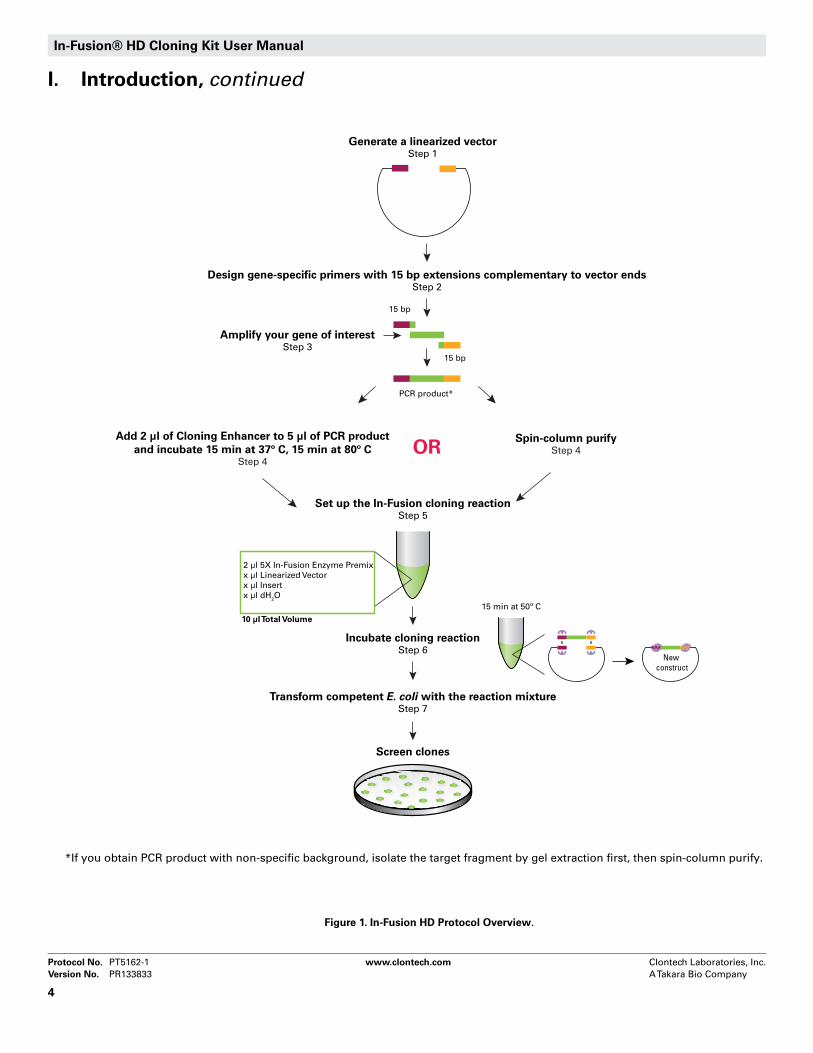
The table below is a general outline of the protocol used for the In-Fusion HD Cloning Kits. This outline is further illustrated in Figure 1. Please refer to the specified pages for details on performing each step.
Table I. In-Fusion HD Protocol OutlineStep Action Pages
1Select a base vector and identify the insertion site. Linearize the vector by restriction enzyme digestion or inverse PCR and purify.
6
2Design PCR primers for your gene of interest with 15 bp extensions (5’) that are complementary to the ends of the linearized vector.
7-8
3Amplify your gene of interest with a high-fidelity DNA polymerase. Verify on an agarose gel that your target DNA has been amplified and determine the integrity of the PCR product.
9
4Spin-column purify your PCR product OR treat it with Cloning Enhancer.
Spin-Column Protocol I (p.9) OR
Cloning Enhancer Protocol II (p.10)
5
Set up your In-Fusion cloning reaction:2 μl of 5X In-Fusion HD Enzyme Premix X μl of Linearized Vector X μl of Insert X μl of dH20 to a Total Reaction Volume of 10 μl. Mix well.
Spin-Column Protocol I (p.9) OR
Cloning Enhancer Protocol II (p.10)
6 Incubate the reaction for 15 min at 50°C, then place on ice. Spin-Column Protocol I (p.9)
ORCloning Enhancer Protocol II (p.10)
7Transform competent cells with 2.5 μl of the reaction mixture from Step 6.
11
In-Fusion® HD Cloning Kit User Manual
Protocol No. PT5162-1 www.clontech.com Clontech Laboratories, Inc. Version No. PR133833 A Takara Bio Company
4
Figure 1. In-Fusion HD Protocol Overview.
I. Introduction, continued
2 μl 5X In-Fusion Enzyme Premixx μl Linearized Vectorx μl Insertx μl dH2O
10 μl Total Volume
Set up the In-Fusion cloning reactionStep 5
Amplify your gene of interestStep 3
Add 2 µl of Cloning Enhancer to 5 µl of PCR productand incubate 15 min at 37º C, 15 min at 80º C
Step 4
Incubate cloning reactionStep 6
15 min at 50º C
15 bp
15 bp
PCR product*
Transform competent E. coli with the reaction mixtureStep 7
Generate a linearized vectorStep 1
Design gene-specific primers with 15 bp extensions complementary to vector endsStep 2
ORSpin-column purify
Step 4
Screen clones
x x
New construct
*If you obtain PCR product with non-specific background, isolate the target fragment by gel extraction first, then spin-column purify.

Clontech Laboratories, Inc. www.clontech.com Protocol No. PT5162-1A Takara Bio Company Version No. PR133833
5
In-Fusion® HD Cloning Kit User Manual
II. List of Components
In-Fusion HD Cloning Kits
Components
Cat. Nos. 639648 639649 639650
Rxns. 10 rxns 50 rxns 100 rxns
5X In-Fusion HD Enzyme Premix 20 μl 100 μl 200 μl
pUC19 Control Vector, linearized (50 ng/μl)
5 μl 15 μl 30 μl
2 kb Control Insert (40 ng/μl) 10 μl 30 μl 60 μl
In-Fusion HD Cloning Kits are available in 10, 50 and 100 reaction sizes. Kits can also be purchased with Stellar™ Competent Cells, NucleoSpin Extract II, and/or Cloning Enhancer.
Store all components at –20°C.
III. Additional Materials Required
The following materials are required but not supplied:
Ampicillin• (100 mg/ml stock) or other antibiotic required for plating the In-Fusion reaction
LB (Luria-Bertani) medium• (pH 7.0)
LB/antibiotic plates •
SOC medium•
Competent Cells•We recommend the use of Stellar Competent Cells. If you decide to use other commercially-available competent cells (e.g., DH10B, DH5α), make sure that they have a transformation efficiency ≥ 1.0 x 10⁸ cfu/µg. Many In-Fusion HD Cloning Kits come with Stellar Competent Cells, but you can also purchase the cells separately in various formats.
Cloning Enhancer • (Cat. Nos. 639613, 639614 & 639615) [Optional] Cloning Enhancer is provided with some of the In-Fusion HD Cloning Kits and can also be purchased sepa-rately. Cloning Enhancer removes background template DNA and PCR residue, eliminating the need for PCR insert purification prior to cloning when a single PCR product (i.e., no background) is obtained.
Spin Columns—NucleoSpin Extract II• (Cat. Nos. 740609.10, 740609.50 & 740609.250) [Optional]NucleoSpin Extract II is provided with some of the In-Fusion HD Cloning Kits and can also be purchased separately. Spin columns can be used to purify PCR products, if non-specific background or multiple bands are visible on an agarose gel. When spin columns are needed, we recommend NucleoSpin Ex-tract II. NucleoSpin Extract II is provided with some of the In-Fusion HD Cloning Kits and can also be purchased separately.

In-Fusion® HD Cloning Kit User Manual
Protocol No. PT5162-1 www.clontech.com Clontech Laboratories, Inc. Version No. PR133833 A Takara Bio Company
6
IV. PCR and Experimental PreparationA. Preparation of a Linearized Vector by Restriction DigestionTo achieve a successful In-Fusion reaction, you must first generate a linearized vector. The linearized vector can be generated using restriction enzymes (single or double digests) or by PCR.
Due to differences in cutting efficiencies, different restriction enzymes will generate different amounts of background. Generally speaking, two enzymes cut better than any single enzyme. Efficiency of digestion will always be better if the restriction sites are as far apart as possible. In addition, increasing the enzyme digestion time and the digestion reaction volume will reduce the background.
Recommendations for preparation of a linearized vector by restriction enzyme digestion:
Incubate your restriction digest as directed by the restriction enzyme supplier. For many enzymes, 1. incubation from 3 hours to overnight can increase linearization and reduce background.
After digestion, purify the linearized vector using any available PCR purification kit. We recommend gel 2. purification using the NucleoSpin Extract II Kit.
[Control]3. Check the background of your vector by transforming 5–10 ng of the linearized and purified vector into competent cells.
If the background is high, continue digesting the vector for a longer time after the addition of more restric-tion enzyme(s). Incubate 2 hours to overnight. Gel purify the remainder of the vector and transform again.
B. PCR Amplification of Target Fragment
For most DNA polymerases, 100 pg–1 ng of plasmid DNA is typically enough to use as a PCR template. However, if you are amplifying from a pool of cDNA, the amount of template DNA required depends on the relative abundance of the target message in your mRNA population.
The In-Fusion method is not affected by the presence or absence of A-overhangs, so you can use any thermostable DNA polymerase for amplification, including proofreading enzymes. For the best results, we recommend using our Advantage® HD Polymerase Mix (Cat. No. 639241), which offers high-fidelity, efficient amplification of long gene segments (>1 kb), and automatic hot start for increased specificity and reduced background. For high yields and error-free amplification of inserts up to 5 kb, we recommend using the Advantage HF 2 enzyme supplied in our Advantage HF 2 PCR Kits (Cat. Nos. 639123 & 639124).
If you will be performing PCR with Advantage HD Polymerase, we recommend using the following amounts of template (for a 50 μl reaction):
Human Genomic DNA 5 ng–200 ng•
E. coli • Genomic DNA 100 pg–100 ng
cDNA 1 ng–200 ng•
Plasmid DNA 10 pg–1 ng•
If you choose not to use Advantage HD, we recommend that you use a robust, high fidelity, thermostable DNA polymerase that is capable of hot start PCR.
When PCR cycling is complete, analyze your PCR product by agarose gel electrophoresis to confirm that you have obtained a single DNA fragment and to estimate the concentration of your PCR product. Quantify the amount of DNA by measuring against a known standard or DNA mass ladder run on the same gel.

Clontech Laboratories, Inc. www.clontech.com Protocol No. PT5162-1A Takara Bio Company Version No. PR133833
7
In-Fusion® HD Cloning Kit User Manual
IV. PCR and Experimental Preparation, continuedC. PCR Primer Design Primer design and quality are critical for the success of the In-Fusion reaction. In-Fusion allows you to join two or more fragments, e.g. vector and insert (or multiple fragments), as long as they share 15 bases of homology at each end. Therefore, In-Fusion PCR primers must be designed in such a way that they generate PCR products containing ends that are homologous to those of the vector. Figure 2 outlines the guidelines for primer design and Figure 3 gives specific examples of In-Fusion PCR primers.
When designing In-Fusion PCR primers, consider the following:
Every In-Fusion primer must have two characteristics: The 5’ end of the primer must contain 15 bases 1. that are homologous to 15 bases at one end of the DNA fragment to which it will be joined (i.e., the vector or another insert). The 3’ end of the primer must contain sequence that is specific to the target gene.
The 3’ portion of each primer should:2.
be gene-specific. •
be between 18–25 bases in length, and have a GC-content between 40–60%.•
have a melting temperature (T• m) between 58–65°C. The Tm difference between the forward and reverse primers should be ≤ 4°C, or you will not get good amplification. Note: The Tm should be calculated based upon the 3’ (gene-specific) end of the primer, and NOT the entire primer. If the calculated Tm is too low, increase the length of the gene-specific portion of the primer until you reach a Tm of between 58–65°C.
not contain identical runs of nucleotides. The last five nucleotides at the 3’ end of each primer should •contain no more than two guanines (G) or cytosines (C).
Avoid complementarity within each primer to prevent hairpin structures, and between primer pairs to 3. avoid primer dimers.
You can perform a BLAST search to determine if the 3’ portion of each primer is unique and specific (at 4. www.ncbi.nlm.nih.gov/BLAST/).
Clontech provides an online tool (at 5. http://bioinfo.clontech.com/infusion/) that simplifies In-Fusion PCR primer design for standard cloning reactions. Simply provide your vector sequence, the restriction enzyme(s) used to linearize the vector (if that is the chosen method for linearization), and the primer sequence required to amplify your region of interest.
We generally use desalted oligonucleotide primers in PCR reactions. However, primer quality can de-6. pend on the vendor and varies from lot to lot. If your primer quality is particularly poor (i.e., has many premature termination products), or your primers are longer than 45 nucleotides, they may need to be PAGE purified; however, we usually find this is unnecessary.
D. Control ReactionsWhen using the In-Fusion kit for the first time, we strongly recommend that you perform the positive and negative control reactions in parallel with your In-Fusion cloning reaction. The positive control should consist of a circular vector of known concentration (competent cells should give >2 x 10⁸ cfu/μg), and the negative control should consist of a known amount of your linearized vector (see Section IX for Expected Results). Performing the control reactions will verify that the system is working properly. The 2 kb Control Insert included in the In-Fusion HD Cloning Kits has already been purified, so there is no need for further treatment prior to the cloning reaction.

In-Fusion® HD Cloning Kit User Manual
Protocol No. PT5162-1 www.clontech.com Clontech Laboratories, Inc. Version No. PR133833 A Takara Bio Company
8
IV. PCR and Experimental Preparation, continued
Figure 3. Examples of primers designed for In-Fusion cloning. The above figure shows examples of primers designed with recognition sites for restriction enzymes that generate: 5’ overhangs (Panel A), blunt ends (Panel B), and 3’ overhangs (Panel C). The primer sequences are shown in bold. The Xs represent bases corresponding to the gene or sequence of interest. Additional nucleotides (indicated with a black box) have been added to each primer in order to reconstruct the restriction sites. They are not part of the 15 bases of sequence homology.
HindIII
5'-...ATA CAT TAT ACG AAG TTA TCA G AGC TTT CTA GAC CAT TCG TTT GGC G...-3'
SalI
3'-...TAT GTA ATA TGC TTC AAT AGT CAG CT AA GAT CTG GTA AGC AAA CCG C...-5'
5'-G AAG TTA TCA GTC GAC XXX XX...-3'
3'-...X XXX XXT TCG AAA GAT CTG GTA-5'
5' Forward Primer
3' Reverse Primer
pDNR-CMV sequence
SmaI
5'-...TCA GTC GAC GGT ACC GGA CAT ATG CCC GGG AAT TCC TGC AGG ATC CGC T...-3'
SmaI
3'-...AGT CAG CTG CCA TGG CCT GTA TAC GGG CCC TTA AGG ACG TCC TAG GCG A...-5'
5'-ACC GGA CAT ATG CCC GGG XXX...-3'
3'-...XXX GGG CCC TTA AGG ACG TCC-5'
5'-AG TTA TCA GTC GAC GGT ACC XXX...-3'
KpnI
KpnI
3'-...GTA ATA TGC TTC AAT AGT CAG CTG C CA TGG CCT GTA TAC GGG CCC TTA A...-5'
3'-...XXX CCA TGG CCT GTA TAC GGG CC-5'
5'-...CAT TAT ACG AAG TTA TCA GTC GAC GGT AC C GGA CAT ATG CCC GGG AAT T...-3'
A
B
C
Brackets indicate bases to be included in the
15 b region of homology
Linearized Vectors with5' Overhangs
Linearized Vectors with Blunt ends
LinearizedVectors with3' Overhangs
Forward Primer
Reverse Primer
append with your specific sequence
Reverse Primer
Forward Primer
Forward Primer
Reverse Primer
Guidelines for universal primer designTo determine the 15 b homology sequence to be incorporated into each primer, start at the 5’ end of each DNA strand in the linearized vector (*).The region of homology for a particular primer consists of bases that are complementary to the first 15 bases at the 5’ end of a particular DNA strand.
This means that the bases complementary to 5' overhangs are included in the primer sequence, but the bases in 3’ overhangs are not.
15 14 13 12 11 10 9 8 7 6 5 4 3 2 1
1 2 3 4 5 6 7 8 9 10 11 12 13 14 15
15 14 13 12 11 10 9 8 7 6 5 4 3 2 1
15 14 13 12 11 10 9 8 7 6 5 4 3 2 1
1 2 3 4 5 6 7 8 9 10 11 12 13 14 15
1 2 3 4 5 6 7 8 9 10 11 12 13 14 15
*
*
*
*
*NNNNNNNNNNNNNNNNNN
NNNNNNNNNNNNNNNNNNNNNN
NNNNNNNNNNNNNNN
NNNNNNNNNNNNNNN
NNNNNNNNNNNNNNN
NNNNNNNNNNNNNNNNNNNNNNNNNNNN
NNNNNNNNNNNNNNN
NNNNNNNNNNNNNNNNNN
NNNNNNNNNNNNNNN
NNNNNNNNNNNNNNNNNNNNNN
NNNNNNNNNNNNNNN
*
Figure 2. Universal primer design for the In-Fusion System. Successful insertion of a PCR fragment requires that the PCR insert share 15 bases of homology with the ends of the linearized vector. This sequence homology is added to the insert through the PCR primers. For vectors with sticky ends, bases complementary to 5’ overhangs are included in the primer sequence; bases in the 3’ overhangs are not. See Figure 3 for specific examples. An online tool is also provided to assist in primer design and can be found at http://bioinfo.clontech.com/infusion/.

Clontech Laboratories, Inc. www.clontech.com Protocol No. PT5162-1A Takara Bio Company Version No. PR133833
9
In-Fusion® HD Cloning Kit User Manual
V. Which Protocol Should You Follow?Following PCR, verify by agarose gel electrophoresis that your target fragment has been amplified. If a single band of the desired size is obtained, you can EITHER spin-column purify (follow Protocol I), OR treat your PCR product with Cloning Enhancer (follow Protocol II). However, if non-specific background or multiple bands are visible on your gel, isolate your target fragment by gel extraction, then spin-column purify (follow Protocol I). If you use PCR to amplify your vector and insert and you obtain both a PCR-amplified vector AND PCR-amplified fragment(s) without non-specific background, you can use the Quick In-Fusion Cloning Protocol provided in Appendix A.
A. Procedure for Spin-Column Purification of PCR FragmentsIf non-specific background bands are observed on an agarose gel, isolate your target fragment by gel 1. extraction, then spin-column purify. If a single band of the desired size is obtained, then spin-column purify.
Spin-column purify your PCR product (e.g., insert) by using a silica-based purification system, such as 2. NucleoSpin Extract II. During purification, avoid nuclease contamination and exposure of the DNA to UV light for long periods of time.
After purification, proceed with the In-Fusion Cloning Procedure for Spin Column-Purified PCR Frag-3. ments (Section VI.B).
B. In-Fusion Cloning Procedure for Spin-Column Purified PCR FragmentsIn general, good cloning efficiency is achieved when using 50-200 ng of vector and inserts respectively, regardless of their length. More is not better. If the size of the PCR fragment is shorter than 0.5 kb, maximum cloning efficiency may be achieved by using less than 50 ng of fragment.
Set up the In-Fusion cloning reaction1. :
5X In-Fusion HD Enzyme Premix 2 µl
Linearized Vector x µl*
Purified PCR Fragment x µl*
dH2O (as needed) x µl
Total Volume 10 µl
*For reactions with larger volumes of vector and PCR insert (> 7 µl of vector + insert), double the amount of enzyme premix, and add dH20 for a total volume of 20 µl.
Adjust the total reaction volume to 10 µl using deionized H2. 2O and mix the reaction.
Incubate the reaction for 3. 15 min at 50°C, then place on ice.
Continue to the Transformation Procedure (Section VIII). You can store the cloning reactions at –20°C 4. until you are ready.
TABLE II. RECOMMENDED IN-FUSION REACTIONS FOR PURIFIED FRAGMENTS
Rxn Component Cloning Rxn Negative Control Rxn
Positive Control Rxn
Purified PCR fragment 10–200 ng* – 2 µl of 2 kb control insert
Linearized vector 50–200 ng** 1 µl 1 µl of pUC19 control vector
5X In-Fusion HD Enzyme Premix 2 µl 2 µl 2 µl
Deionized water to 10 µl to 10 µl to 10 µl
*<0.5 kb: 10-50 ng, 0.5 to 10 kb: 50-100 ng, >10 kb: 50-200 ng**<10 kb: 50-100 ng, >10 kb: 50-200 ng
VI. Protocol I: In-Fusion Cloning Procedure w/Spin-Column Purification
BREAK
PROTOCOL
PROTOCOL

In-Fusion® HD Cloning Kit User Manual
Protocol No. PT5162-1 www.clontech.com Clontech Laboratories, Inc. Version No. PR133833 A Takara Bio Company
10
B. In-Fusion Cloning Procedure for Cloning Enhancer-Treated PCR Fragments
Set up the In-Fusion cloning reaction:1.
5X In-Fusion HD Enzyme Premix 2 µl
Linearized Vector x µl*
Cloning Enhancer-Treated PCR Fragment x µl**
dH2O (as needed) x µl
Total Volume 10 µl
*Use 50–200 ng of linearized vector.
**Use 1–2 µl of Cloning Enhancer-Treated fragments, regardless of their length. The total volume of Cloning Enhancer-
Treated PCR fragments should be up to 4 µl per 10 µl reaction. If you obtain a low product yield from your PCR reac-
tion, we recommend purification of PCR fragments instead of Cloning Enhancer-Treatment.
Adjust the total reaction volume to 10 µl using deionized H2. 2O and mix the reaction.
Incubate the reaction for 3. 15 min at 50°C, then place on ice.
Continue to the Transformation Procedure (Section VIII). If you cannot transform cells immediately, store 4. the cloning reactions at –20°C until you are ready.
NOTE:If you use PCR to amplify your vector and insert and you obtain both a PCR-amplified vector and PCR-amplified frag-ments without non-specific background you may use the Quick In-Fusion Cloning Protocol provided in Appendix A.
PROTOCOL
Attention
VII. Protocol II: In-Fusion Cloning Procedure w/Cloning Enhancer Treatment
A. Procedure for Treating Unpurified PCR Fragments with Cloning Enhancer
Before setting up the In-Fusion cloning reaction, treat unpurified PCR products (e.g. fragments) as follows:
Add 2 µl of Cloning Enhancer to 5 µl of the PCR reaction. 1.
Incubate at 37°C for 15 minutes, then at 80°C for 15 minutes in a PCR thermal cycler. If you used more 2. than 100 ng of DNA as a template in the PCR reaction, extend the 37°C incubation step to 20 minutes. If you are using a water bath or heat block rather than a thermal cycler, extend each of the incubation steps to 20–25 minutes.
Proceed with the In-Fusion Cloning Procedure for Cloning Enhancer-Treated PCR Fragments (Section 3. VII.B). If you cannot proceed immediately, store treated PCR reactions at –20°C until you are ready.
Attention
IMPORTANT:DO NOT treat purified PCR products with the Cloning Enhancer.
BREAK
BREAK
PROTOCOL

Clontech Laboratories, Inc. www.clontech.com Protocol No. PT5162-1A Takara Bio Company Version No. PR133833
11
In-Fusion® HD Cloning Kit User Manual
VIII. Transformation Procedure
A. Procedure for Transformation Using Stellar™ Competent CellsThe following protocol has been optimized for transformation using Stellar Competent Cells. If your In-Fusion Kit does not include Stellar Competent Cells, Clontech sells Stellar Competent Cells separately in several formats. If you are not using Stellar Competent Cells, you may need to dilute the In-Fusion reaction mixture prior to transformation to increase cloning efficiency (See Table III, Troubleshooting Guide). We strongly recommend the use of competent cells with a transformation efficiency >1 x 10⁸ cfu/ug.
Follow the protocol provided with your Stellar Competent Cells to transform the cells with 2.5 µl of the 1. In-Fusion reaction mixture. If you are using other competent cells, please follow the transformation protocol provided with your cells.
Place 1/1002. th–1/5th of each transformation reaction into separate tubes and bring the volume to 100 µl with SOC medium. Spread each diluted transformation reaction on a separate LB plate containing an antibiotic appropriate for the cloning vector (i.e., the control vector included with the Kit requires 100 µg/ml of ampicillin).
Centrifuge the remainder of each transformation reaction at 6000 rpm for 5 min. Discard the supernatant 3. and resuspend each pellet in 100 µl fresh SOC medium. Spread each sample on a separate LB plate containing the appropriate antibiotic. Incubate all of the plates overnight at 37°C.
The next day, pick individual isolated colonies from each experimental plate. Isolate plasmid DNA using 4. a standard method of your choice (e.g. miniprep). To determine the presence of insert, analyze the DNA by restriction digestion or PCR screening.
IMPORTANT:
DO NOT add more than 5 µl of the reaction to 50 µl of competent cells. More is not better. Using too much of the reaction mixture inhibits the transformation.
IX. Expected Results
The positive control plates typically develop several hundred white colonies when using cells with a minimum transformation efficiency of 1 x 10⁸ cfu/μg. The negative control plates should have few colonies.
The number of colonies on your experimental plates will depend on the amount and purity of the PCR product and linearized vector used for the In-Fusion cloning reaction.
• Thepresenceofalownumberofcoloniesonbothplates—typically,afewdozencolonies— is indicative of either transformation with too much of the reaction, or poor DNA/primer quality.
• Thepresenceofmany (hundreds)ofcolonieson thenegativecontrol is indicativeof incomplete vector linearization.
Attention
PROTOCOL

In-Fusion® HD Cloning Kit User Manual
Protocol No. PT5162-1 www.clontech.com Clontech Laboratories, Inc. Version No. PR133833 A Takara Bio Company
12
X. Troubleshooting Guide
TABLE III. TROUBLESHOOTING GUIDE FOR IN-FUSION EXPERIMENTS
A. No or Few Colonies Obtained from Transformation
Description of Problem Explanation Solution
Low transformationefficiency
Transformed with too much In-Fusion reaction
Do not add more than 5 µl of the In-Fusion reaction to 50 µl of competent cells (see Section VIII for details).
Competent cells are sensitive to the In-Fusion enzyme
If your cloning efficiency is low, you may obtain better re-sults if you dilute the reaction. For some cell strains, it may be better to dilute the In-Fusion reaction with TE buffer 5-10 times prior to transformation (add 40-90 µl to 10 µl In-Fusion reaction).
Bacteria were not competent
Check transformation efficiency. You should obtain >1 x 10⁸ cfu/µg; otherwise use fresh competent cells.
Low quality DNA fragments
Low DNA concentration in reaction
It is imperative to obtain the highest DNA concentration possible in your In-Fusion reaction. Either the amount of vector or the amount of PCR fragment was too low. We recommend using between 50 ng and 200 ng of vector, depending on its size (see Table II).
Gel purification introduced contaminants
If your fragment was gel purified, it is imperative to obtain the highest DNA concentration possible in your In-Fusion reaction. The total volume of purified vector and insert should not exceed 5 µl.
When possible, optimize your PCR amplification reactions such that you generate pure PCR products and use the Cloning Enhancer (see Section VII.A for details).
Suboptimal PCR product
Repeat PCR amplification and purify product using a different method of purification. Alternatively, perform phenol:chloroform extraction on your original PCR product, followed by ethanol precipitation.
Primer sequences are incorrect
Check primer sequences to ensure that they provide 15 bases of homology with the region flanking the insertion site (see Section IV.C).
If you do not obtain the expected results, use the following guide to troubleshoot your experiment. To confirm that your kit is working properly, perform the control reactions.

Clontech Laboratories, Inc. www.clontech.com Protocol No. PT5162-1A Takara Bio Company Version No. PR133833
13
In-Fusion® HD Cloning Kit User Manual
X. Troubleshooting Guide, continued
TABLE III. TROUBLESHOOTING GUIDE FOR IN-FUSION EXPERIMENTS
B. Large Numbers of Colonies Contained No Insert
Description of Problem Explanation Solution
Large numbers of colonies obtained with no insert
Incomplete linearization of your vector
It is important to remove any uncut vector prior to use in the In-Fusion reaction. If necessary, recut your vector and gel purify.
Contamination of In-Fusion reaction by plasmid with same antibiotic resistance
If your insert was amplified from a plasmid, closed circular DNA may have carried through purification and contaminated the cloning reaction:
a) To ensure the removal of any plasmid contamination, we recommend linearizing the template DNA before performing PCR.
b) If you spin-column purify your insert, treat-ing the PCR product with DpnI before purifica-tion will help to remove contaminating tem-plate DNA.
Plates too old or contained incorrect antibiotic
Be sure that your antibiotic plates are fresh (<1 month old). Check the antibiotic resistance of your fragment.
C. Clones Contained Incorrect Insert
Large number of colo-nies contain incorrect insert
Your PCR product contained non-specific sequences
If your PCR product is not a single distinct band, then it may be necessary to gel purify the PCR product to ensure cloning of the correct insert. See Section VI for more information.

In-Fusion® HD Cloning Kit User Manual
Protocol No. PT5162-1 www.clontech.com Clontech Laboratories, Inc. Version No. PR133833 A Takara Bio Company
14
XI. Appendix A: Quick In-Fusion Cloning Protocol
A. In-Fusion Cloning Procedure for a PCR-Amplified Vector & Fragment
Set up the In-Fusion cloning reaction:1.
5X In-Fusion HD Enzyme Premix 2 µl
PCR Linearized Vector 1–2 µl*
PCR Fragment 1–2 µl*
Cloning Enhancer 1 µl
dH2O (as needed) x µl
Total Volume 10 µl
*The total volume of PCR vector & fragment(s) should be up to 4 µl per 10 µl reaction.
Adjust the total reaction volume to 10 µl using deionized H2. 2O and mix the reaction.
Incubate the reaction for 3. 15 min at 37°C, followed by 15 min at 50°C, then place on ice.
Continue to the Transformation Procedure (Section VIII). If you cannot transform cells immediately, store 4. the cloning reactions at –20°C until you are ready.
NOTE:If you obtain both a PCR vector and PCR fragment(s) without non-specific background, you may use the following Quick In-Fusion Cloning Protocol to perform Cloning Enhancer-Treatment and In-Fusion reaction in the same tube. PROTOCOL

Clontech Laboratories, Inc. www.clontech.com Protocol No. PT5162-1A Takara Bio Company Version No. PR133833
15
In-Fusion® HD Cloning Kit User Manual
Notice to Purchaser
Clontech products are to be used for research purposes only. They may not be used for any other purpose, including, but not limited to, use in drugs, in vitro diagnostic purposes, therapeutics, or in humans. Clontech products may not be transferred to third parties, resold, modified for resale, or used to manufacture commercial products or to provide a service to third parties without written approval of Clontech Laboratories, Inc.
Your use of these products and technologies is subject to compliance with any applicable licensing requirements described on the product’s web page at http://www.clontech.com. It is your responsibility to review, understand and adhere to any restrictions imposed by such statements.
Clontech, the Clontech logo, Advantage, In-Fusion, and Stellar are trademarks of Clontech Laboratories, Inc. All other marks are the property of their respective owners. Certain trademarks may not be registered in all jurisdictions. Clontech is a Takara Bio Company. ©2011 Clontech Laboratories, Inc.
This document has been reviewed and approved by the Clontech Quality Assurance Department.
XII. Appendix B: pUC19 Linearized Vector Information
pUC19
2690 bp
lacZα(part B)
pUC ori
Ampr
In-Fusion Cloning Site
lacZα(part A)
MCS
GAATTCGAGC TCGGTACCCG GGGATCCTTAAGCTCG AGCCATGGGC CCCTAG
SmaI
XbaIXmaI
AccIHincII
SacI SalI SphI
Partial BamHI Site
EcoRI HindIIIPstIKpnI
CTAGGAGA TCTCAGCTGG ACGTCCGTAC GTTCGAACCGATCCTCT AGAGTCGACC TGCAGGCATG CAAGCTTGG In-Fusion
Cloning Site Partial BamHI Site
Figure 4. pUC19 Linearized Vector Map and Multiple Cloning Sites (MCS). pUC19 is a commonly used, high copy number cloning vector. This linearized version was generated by PCR, and contains the blunt ends shown in the MCS sequence above. The vector encodes the N-terminal fragment of β-galactosidase (lacZα), which allows for blue/white colony screening (i.e., α-complementation), as well as a pUC origin of replication and an ampicillin resistance gene that allow propagation and selection in E. coli.