in middle-age and elderly individuals the fractured
TRANSCRIPT

Page 1/9
Minimally Invasive Pedicle Screw Fixation, Includingthe Fractured Vertebra, Combined withPercutaneous Vertebroplasty for Treatment of AcuteThoracolumbar Osteoporotic Compression Fracturein Middle-Age and Elderly Individualshong liu
Orthopedic Institutejinwei Xu
Orthopedic Instituteguanrong Sun
Orthopedic Instituteweifeng Shi
Orthopedic Instituteliming Xiang
Orthopaedic Hospitalshan Chen ( [email protected] )
HangZhou Hospital of Traditional Chinese Medical
Research article
Keywords: Thoracolumbar vertebroplasty, Pedicle screw �xation, osteoporotic
Posted Date: August 9th, 2021
DOI: https://doi.org/10.21203/rs.3.rs-750840/v1
License: This work is licensed under a Creative Commons Attribution 4.0 International License. Read Full License

Page 2/9
AbstractBackground: To evaluate the feasibility, e�cacy, and safety of minimally invasive pedicle screw (MIPS)�xation, including the fractured vertebra, combined with percutaneous vertebroplasty (PVP) for thetreatment of acute thoracolumbar osteoporotic compression fracture in middle-age and elderlyindividuals.
Methods: Between January 2016 and August 2019, a total of 30 patients, with a mean age of 69.4 years(range, 58–75 years), who experienced thoracic or lumbar fracture without neurological de�cits,underwent the MIPS procedure combined with PVP. Preoperative and postoperative pain were assessedusing a visual analog scale (VAS) and Oswestry Disability Index (ODI). Cobb angles and anterior columnheight were measured on lateral radiographs before surgery and at 3 days, 1, 3, and 6 months, and 1 and2 years at �nal follow-up after surgery.
Results: All patients underwent surgery successfully, with a mean follow-up of 18.2 ± 5.7 months (range,12–45 months). Mean preoperative VAS score decreased from 7.3±2.2 to 1.4±0.3 at the �nal follow-up(p<0.05). Mean preoperative ODI decreased from 84.2±10.3 to 18.8±7.5 (p<0.05) at the �nal follow-up.The Kyphosis angle of operative segment was improved from preoperative (21.38±1.68)°to(4.01 ± 1.38)°3 days postoperatively and(5.02±1.09)°at �nal follow up (p<0.05).The anterior vertebralheight was improved from preoperative(49.86±6.50)% to(94.01±1.79)% 3 days postoperatively and(91.80±1.88)% at �nal follow up (p<0.05). No signi�cant changes in vertebral body height restorationwere observed during 2 years of follow-up after surgery. In addition, there were no instrumentationfailures or complications in any of the patients.
Conclusions: MIPS, including the fractured vertebra, combined with PVP, was a reliable and safeprocedure, with satisfactory clinical and radiological results for the treatment of thoracolumbarosteoporotic compression fracture in patients without neurological de�cits.
IntroductionThe incidence of osteoporotic vertebral fractures (OVCF) has increased rapidly in recent years due to theaging population. Surgical intervention for OVCF has been widely accepted as an effective treatmentmethod because it has several advantages, including immediate spinal stability and more reliablerestoration of sagittal alignment, vertebral height, and canal dimension[1]. Some studies have described atechnique involving 4-screw �xation immediately adjacent to the fractured vertebra combined withcement augmentation. Clinical and biomechanical studies have con�rmed that addition of pedicle screw�xation in fractured vertebrae signi�cantly improved stability compared with conventional 4-screw�xation for thoracolumbar fractures[2, 3]. Minimally invasive surgery has been successfully used in thetreatment of vertebral fractures, and more studies have reported that the technique is associated with lesssoft tissue destruction, reduced intraoperative blood loss, and shorter hospitalization[4]. In the presentstudy, we designed a minimally invasive pedicle screw (MIPS) �xation technique, including the fractured

Page 3/9
vertebra, combined with percutaneous vertebroplasty (PVP) to treat patients with thoracolumbarosteoporosis vertebral compressive fractures, which could prevent secondary fractures after PVP [5]. Thepurpose of this study was to evaluate the feasibility, e�cacy, and safety of MIPS �xation, including thefractured vertebra, combined with PVP.
Methods
Surgical Procedure After general tracheal anesthesia, the fractured and adjacent vertebral pedicles are identi�ed under C-arm�uoroscopy of 6 pedicle projections, and an intact pedicle of the injured vertebra is �tted with screws(preoperative computed tomography scan), and the entry site to the pedicle is located at the junctionbetween the lateral border of the superior articular facet and the bisecting midline of the transverseprocess. Once the pedicle is identi�ed, a pedicle probe is used to enter the pedicle, and preoperativeanteroposterior and lateral roentgenograms and computed tomography (CT) scans through the pediclesof the vertebral body to be instrumented are assessed to determine the optimal angle of entry in both thecoronal and sagittal planes. Pedicle integrity is veri�ed in all �ve quadrants to ensure the presence of asolid tube of bone and that violation of the spinal canal or inferiorly into the neuroforamen has notoccurred. The tip of the needle is positioned at the 10 and 2 points of the left and right pedicle. Thepuncture angle (anteroposterior, introversion angle 10°–15°) is determined and �ve hollow pedicle screwsare torqued in. C-arm �uoroscopy is activated again and the tip of the screw is positioned in the front one-quarter to one-third of the vertebral body to con�rm that the position is satisfactory. One side of the threescrews is �xed �rst, and then the screws are torqued in and the nuts are tightened. The order of �xation isinjured vertebra, lower vertebrae, and upper vertebrae. According to the degree of compression of theinjured vertebra before the operation, the reduction should be performed between the injured vertebra andthe upper vertebra. Fluoroscopy is used to con�rm that the working channel enters the front one-third ofthe injured vertebra. A bone cement injection tube with an inner core is inserted. After mixing the bonecement to the dough stage, it is injected under �uoroscopic guidance. When the bone cement �owsbackward and approaches the posterior edge of the vertebral body, the injection is immediately stopped.The side connecting rod is �xed and C-arm �uoroscopy is activated to con�rm satisfactory positioning ofthe implant, and the skin is sutured.
Postoperative management Antibiotics were routinely administered to prevent infection, with standing or walking under the protectionof a thoracolumbar brace for 2 weeks after the operation, with the lumbar brace removed 1.5 monthsafter the operation.
Clinical assessment

Page 4/9
The following clinical indexes were observed and recorded: operation duration; surgical blood loss;amount of cement instillation; and number of cement leakages. A visual analog scale (VAS) was used toevaluate back pain. VAS pain assessments were performed immediately, preoperatively, at 3,14 days, at 1,3, 6,12 months and the �nal follow-up after surgery. The Oswestry disability index (ODI) was used toevaluate functional outcome, the plain radiographs were captured preoperatively, immediatelypostoperatively, and at the �nal follow-up. The following parameters were observed and analyzed: localkyphotic angle, de�ned as the angle measured between the superior endplate of the upper vertebra andthe inferior endplate of the lower vertebra; and percentage of anterior height of the fractured vertebra. Theanterior height of the fractured vertebra was de�ned as the anterior height of the fractured vertebradivided by the normal anterior height of the vertebra. Cobb angles and anterior vertebral body height weremeasured on lateral radiographs. The fractured and restored heights were calculated as a percentage ofthe estimated intact vertebral body height by averaging the anterior height from the adjacent levels.
Statistical analysis Data are expressed as mean ± standard deviation (SD) and were analyzed using SPSS version 22.0 (IBMCorporation, Armonk, NY, USA). The paired Student’s t-test and Dunnett’s t test were used to evaluatechanges in data at different times, and the VAS score, Cobb angle in the sagittal plane, and anteriorheight ratio of injured vertebrae were assessed using X-ray. The anterior vertebral height ratio and sagittalCobb angle at different time were compared using repeated measures analysis of variance. Pairwisecomparisons at different times after the operation were performed using Dunnett’s t test; differences withp < 0.05 were considered to be statistically signi�cant.
ResultsThirty patients (10 male, 20 female) who experienced thoracolumbar fracture(s) without neurologicalsymptoms were treated using the surgical method described above. Lesions were identi�ed at thefollowing anatomical locations: T11 (n = 2); T12 (n = 8); L1 (n = 12); L2 (n = 5); and L3 (n = 3). Thefractures were classi�ed as type A1.1 (n = 6 [20%]), A1.2 (n = 5 [16.7%]), and A1.3 (n = 19 [63.3%]). Allpatients underwent surgery successfully as planned, with a mean follow-up of 18.2 ± 5.7 months (range,12–45 months). The mean duration of surgery was 83.2 ± 23.5 min (range, 72–108 min), with a meanblood loss of 98.5 ± 18.6 ml (range, 65–120 ml), and a mean instillation of cement of 2.1 ± 0.7 ml (range,1.5–3 ml). Cement leakage was observed in 5 (16.3%) cases, 3 at the adjacent discal space and 2 at theanterior of the fractured vertebra, none of which had clinical consequences. There was no infection andno neurological complications. No screw pullouts were found at the �nal follow-up. The meanpreoperative back pain score according to VAS decreased from 7.3 ± 2.2 to 2.2 ± 1.1 at discharge (p < 0.05), and was 1.4 ± 0.3 at the �nal follow-up (p < 0.05). The mean ODI preoperative decreased from 84.2 ± 10.3 to 18.8 ± 7.5 at the �nal follow-up (p < 0.05). The Kyphosis angle and anterior vertebral heightpreoperatively, postoperatively, and at the �nal follow-up period are reported in Table 1. The Kyphosisangle of operative segment was improved from preoperative (21.38 ± 1.68)° to(4.01 ± 1.38)°3 days

Page 5/9
postoperatively and(5.02 ± 1.09)°at �nal follow up (p < 0.05).The anterior vertebral height was improvedfrom preoperative(49.86 ± 6.50)% to(94.01 ± 1.79)% 3 days postoperatively and (91.80 ± 1.88)% at �nalfollow up (p < 0.05).
Table 1
Variations of each measured parameter preoperatively, postoperatively and at latest follow-up (measuredas mean ± SD).
Time Kyphosis angle(°)
Anterior height(%)
VAS ODI
Preop 21.38 ± 1.68 49.86 ± 6.50 7.3± 2.2 84.2± 10.3 84.2 ± 10.3 84.2 ± 10.3
Postop 3days
3days
4.01 ± 1.38 94.01 ± 1.79 2.7± 1.7 27.7±11.5
Postop(1months)
4.67 ± 1.50 93.16 ± 1.83 2.3± 1.5 25.2±9.5
Postop(3months)
4.93 ± 1.43 92.94 ± 1.84 2.2± 0.5 23.1±8.9
Postop(6months)
4.97 ± 1.42 92.83 ± 1.82 1.5± 0.6 21.5±7.8
Postop(12months)
4.99 ± 1.31 92.53 ± 1.78 1.4± 0.5 19.2±8.2
�nal follow-up 5.02 ± 1.09 91.80 ± 1.88 1.4 ± 0.3
18.8 ± 7.5
Note Comparison of the anterior vertebral body height among preoperation 3 days after operationand last follow-up,F=1169.135 P < 0.05;Comparison of the sagittal Cobb angle among preoperation 3days after operation and last follow-up,F= 403.697 P < 0.05
DiscussionSurgical treatment can restore vertebral body height, correct angular deformity, decompress neural tissue,and facilitate rapid mobilization and rehabilitation.Percutaneous pedicle screw internal �xation has beensuccessfully used in the treatment of OVCF and compared with standard open approaches is associatedwith less soft tissue destruction (muscle atrophy and denervation), reduced intraoperative blood loss, andshorter hospitalization. The integrity of the posterior spinal complex preserved[4]. In the past, surgicalprocedures included the midline and multi�dus approach, which resulted in pain and complicationsassociated with spinal degeneration. Short-segment pedicle screw internal �xation for thoracolumbarfractures has been widely used in clinical practice. Posterior �xation of the injured vertebra in the shortsegment of the 4 screws should be used most extensively [6]; however, some studies have con�rmed

Page 6/9
many problems with this method[7]. The reasons are as follows: the injured vertebra forms a so-called“eggshell.” The short segment �xation combined with injured vertebral screw placement can be used toeffectively treat OVCF in aging patients[8], following the principle of three-point �xation which can bettercorrect the kyphosis of fractures and maintain reduction[9]. The safety and advantages of injuredvertebral screws in reduction and maintenance of reduction have been recognized. However, a currentlyencountered problem is whether unilateral vertebral screw placement combined with a short segmentpedicle screw can achieve satisfactory reduction and maintain good reduction. Some authors havecompared the biomechanical stability effects of 4, 5, and 6 pedicle screw �xation in the treatment ofthoracolumbar fractures using fresh spine cadaver specimens[10]. The results demonstrated that 5 or 6screws could improve the biomechanical stability of the vertebrae, and the stability effect was better thanthat of the traditional 4 screws. There was no signi�cant difference in stability between 5 and 6 screws.
In the present study, percutaneous short-segment pedicle screw �xation, including the fractured vertebra,combined with PVP was performed in middle-age and elderly individuals, and the results demonstratedthat a good curative effect could be obtained[11].The improvement was signi�cant 3 days after surgeryand ODI compared with that before surgery, and there was no statistically signi�cant difference betweenCobb angles at the last follow-up and 2 weeks after surgery. Moreover, the Cobb and wedge angles couldeffectively maintain postoperative VAS scores and ODI, which were signi�cantly decreased comparedwith preoperative values. No serious complications occurred, indicating that this technique can effectivelyrestore and maintain spinal sequence, assist in relieving pain and improve clinical function. Inpercutaneous posterior short segment pedicle �xation combined with PVP, short-segment pedicle �xationwas used to alter spinal force conduction and obtain a strong �xed and stable central column, whichenhances the stability of the anterior column to prevent mechanical access. Meanwhile, local release ofhigh temperature through bone cement prevents rapid pain relief from chemical access, improving thelocal biomechanical stability of the fracture area and the anterior central column. Unilateral PVP of theinjured vertebra was used to avoid the loss of the anterior column again, and the pain were blocked bystrengthening stability and local heat release[12].
There are some limitations concerning our study. It is a retrospective study, the number of patients islimited, the follow-up time is short, and there is a lack of control group comparing other possible surgicalapproaches, Randomized controlled trials should be conducted to verify its safety and e�cacy in futurestudies.
Declarations
Ethics approval and consent to participateThis study was conducted in accordance with the Declaration of Helsinki. This study was conducted withapproval from the Ethics Committee of Xiaoshan District Hospital of TCM and Hangzhou Hospital ofTCM. Written informed consent was obtained from all participants.

Page 7/9
Consent for PublicationWritten informed consent was obtained from all participants.
Availability of data and materialThis data will not be shared, because in recent years, although many scholars have explored this invarious aspects, its pathological mechanism remains unclear and there are no standard diagnosticcriteria. In order to determine the effective method for preventing and treating this disease, it is necessaryto proceed with more large-scale and clinical studies.
Competing interestsThe authors declare that they have no competing interests. There is no funding source.
Funding There is no funding source.
Authors' contributionsLH was accountable for the integrity and analysis of the data, and the writing of the manuscript. XJWand SGR were accountable for the integrity and analysis of the data. SWF and XLM were accountable forthe analysis of the data. CS was accountable for the conception of the research. All authors read andapproved the �nal manuscript.
AcknowledgementsThe authors would like to thank all participating patients, as well as the study nurses, co-investigators,and colleagues who made this trial possible.
References1. Scheer JK, Bakhsheshian J, Fakurnejad S, Oh T, Dahdaleh NS. Z.A. Smith.Evidence-Based Medicine
of Traumatic Thoracolumbar Burst Fractures: A Systematic Review of Operative Management across20 Years Global Spine J.5 (1) (2015), pp. 73–82.
2. Sun C, Guan G, Liu X, Zhang H, Wang B. Comparison of short-segment pedicle �xation with versuswithout inclusion of the fracture level in the treatment of mild thoracolumbar burst fractures. Int JSurg. 2016;36:352–7.
Page 8/9
3. Norton RP, Milne EL, Kaimrajh DN, Eismont FJ, Latta LL, Williams SK. Biomechanical analysis of four-versus six-screw constructs for short-segment pedicle screw and rod instrumentation of unstablethoracolumbar fractures. Spine J. 2014;14(8):1734–9.
4. Schnake KJ, Schroeder GD, Vaccaro AR, Oner C. AOSpine classi�cation systems (subaxial,thoracolumbar). J Orthop Trauma. 2017;31(Suppl 4):14–23.
5. Wang B, Zhao CP, Song LX, Zhu L. Balloon kyphoplasty versus percutaneous vertebroplasty forosteoporotic vertebral compression fracture: a meta-analysis and systematic review. J Orthop SurgRes. 2018;22(1):264.
�. Kanna RM, Shetty AP, Rajasekaran S. Posterior �xation including the fractured vertebra for severeunstable thoracolumbar fractures. Spine J. 2015;15(2):256 – 64ã .
7. Krüger A, Zettl R, Ziring E, Mann D, Schnabel M, Ruchholtz S. Kyphoplasty for the treatment ofincomplete osteoporotic burst fractures. Eur Spine J. 2010;19(6):893–900.
�. Han L Quan, RenFu, et al. E�cacy comparison of percutaneous pedicle instrumentation combinedwith vertebral fracture �xation or vertebral augmentation for treatment of osteoporoticthoracolumbar fractures in elderly patients. Chin J TraumaMarch. 2017;33(3):213–8.
9. Zeng ZL,Cheng LM,Qian L,et a1.Unilateral pedicle screw �xation through the pedicle of fracturedvertebra in combination with the short segment of pedicle screw in the treatment of thoracolumbarfracture of mild to moderate instability[J].Chinese journal of surgery.2012,50(3).–237.
10. Norton RP, Milne EL, Kaimrajh DN, Eismont FJ, Latta LL, Williams SK. Biomechanical analysis of four-versus six-screw constructs for short-segment pedicle screw and rod instrumentation of unstablethoracolumbar fractures. Spine J. 2014;14(8):1734–9.
11. Huang YS, Ge CY, Feng H,et al.Bone cement-augmented short-sement pedicle for kummell diseasewith Spinal Canal stenosis[J].Med SciMonit2018,14(24):928–935.
12. Long 12ChengX, Xu HQ, Huang JH, Li YL. FB. Comparison of unilateral versus bilateral percutaneouskyphoplasty for the treatment of patients with osteoporosis vertebral compression fracture (OVCF): asystematic review and meta-analysis. Eur Spine J. 2016;25(11):3439–49.
Figures
Page 9/9
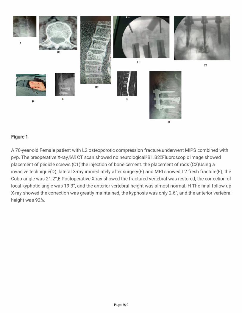
Figure 1
A 70-year-old Female patient with L2 osteoporotic compression fracture underwent MIPS combined withpvp. The preoperative X-ray, A CT scan showed no neurological B1.B2 Fluoroscopic image showedplacement of pedicle screws (C1),the injection of bone cement. the placement of rods (C2)Using ainvasive technique(D), lateral X-ray immediately after surgery(E) and MRI showed L2 fresh fracture(F), theCobb angle was 21.2°,E Postoperative X-ray showed the fractured vertebral was restored, the correction oflocal kyphotic angle was 19.3°, and the anterior vertebral height was almost normal. H The �nal follow-upX-ray showed the correction was greatly maintained, the kyphosis was only 2.6°, and the anterior vertebralheight was 92%.