in vitro rna synthesis from exogenous dengue viral rna ...dengue hemorrhagic/dengue shock syndrome...
TRANSCRIPT

1
In vitro RNA Synthesis from Exogenous Dengue Viral RNA Templates Requires Long
Range Interactions Between 5’- and 3’-Terminal Regions that Influence RNA structure
Shihyun You1¶, Barry Falgout2, Lewis Markoff2, and R. Padmanabhan1*
1Department of Biochemistry and Molecular Biology, University of Kansas Medical Center,
Kansas City, KS 66160-7421 and 2 DVP, CBER, FDA, Rockville, MD 20852-1448
Corresponding Author:
Dr. R. PadmanabhanDepartment of Biochemistry & Molecular BiologyUniversity of Kansas Medical Center3901 Rainbow Blvd.Kansas City, KS 66160-7421Tel. 913-588-7018Fax 913-588-7440E-mail: [email protected]
Running Title: Structural Requirements for Dengue Viral RNA Synthesis in vitro
Copyright 2001 by The American Society for Biochemistry and Molecular Biology, Inc.
JBC Papers in Press. Published on February 5, 2001 as Manuscript M010923200 by guest on M
arch 15, 2020http://w
ww
.jbc.org/D
ownloaded from

2
SUMMARY
Viral replicases of many positive-strand RNA viruses are membrane-bound complexes of
cellular and viral proteins that include viral RNA-dependent RNA polymerase (RdRP). The in
vitro RdRP assay systems that utilize cytoplasmic extracts from viral-infected cells or purified
components and exogenous RNA templates are useful to understand the mechanism of viral
replication in vivo. Such a system was developed for dengue virus utilizing exogenous dengue
viral RNA templates. Our results indicated that in vitro RNA synthesis at the 3’untranslated
region (UTR) required the presence of the 5’-terminal region (TR) and the two cyclization
(CYC) motifs suggesting a functional interaction between the TRs. In this study, using a
psoralen-UV crosslinking method and an in vitro RdRP assay, we analyzed structural
determinants for physical and functional interactions. Exogenous RNA templates that were used
in the assays contained deletion mutations in the 5’-TR and substitution mutations in the 3’-stem-
loop (SL) structure that included those that would disrupt the predicted pseudoknot structure.
Our results indicate that there is physical interaction between the 5’-TR and 3’-UTR that
requires only the CYC motifs. RNA synthesis at the 3’-UTR, however, requires long range
interactions involving the 5’-UTR, CYC motifs, and the 3’-SL region which includes the tertiary
pseudoknot structure.
by guest on March 15, 2020
http://ww
w.jbc.org/
Dow
nloaded from

3
INTRODUCTION
Dengue viruses type 1-4 are members of the flavivirus family of positive strand RNA
viruses. The diseases caused by dengue viruses range from a simple form of dengue fever to a
more complex form of dengue hemorrhagic fever/shock syndrome, which exhibits considerable
morbidity and mortality, especially among children in the tropical and subtropical regions of the
world (for reviews, see (1-3)). It is currently estimated that about 40% of the world’s population
living in these areas is at risk for dengue viral diseases and about 5% of about one million
dengue hemorrhagic/dengue shock syndrome cases are fatal (1). Dengue virus type 2 (DEN2) is
the most prevalent serotype identified over a 10-year period in epidemiological studies. DEN2
has a single stranded RNA genome consisting of 10,723nt (in New Guinea-C strain) (4) which
encodes a single polyprotein arranged in the order, NH2-C-prM-E-NS1-NS2A-NS2B-NS3-
NS4A-NS4B-NS5-COOH. The RNA genome contains a type I cap at the 5’end but lacks a poly
A tail at the 3’end (5). The polyprotein precursor is processed in the endoplasmic reticulum,
where the action of the cellular signal peptidase results in three structural proteins, the
components of the virion, the capsid (C), precursor membrane protein (prM), and the envelope
(E) protein (6-8). The remainder of the polyprotein codes for at least seven nonstructural proteins
which are generated by the combination of both the cellular signal peptidase and the virally-
encoded, two-component serine protease, NS2B/NS3.
The NS3 protein of flaviviruses has a serine protease catalytic triad within the N-
terminal region of 180 amino acid residues (9,10) that requires NS2B for protease activity (11-
20). The conserved motifs found in the NTP-binding proteins and the DEXH family of RNA
helicases were also identified within a region from acid residue 160 to the carboxy terminus of
the NS3 protein ((10,21,22); for a review, see (23)). The RNA helicase activity of NS3 is thought
by guest on March 15, 2020
http://ww
w.jbc.org/
Dow
nloaded from

4
to be required in flaviviral replication and is involved in unwinding of a putative double-
stranded RNA replicative intermediate (5,24). In addition, the West Nile viral and dengue viral
NS3 proteins exhibit 5’-RNA triphosphatase activity ((25); Bartelma, Winter and Padmanabhan,
unpublished results).
The largest of the flaviviral proteins, NS5, contains conserved motifs found in many viral
RNA-dependent RNA polymerases (RdRP) (26,27), suggesting a role for NS5 in viral RNA
replication. In addition to the RdRP motifs, NS5 has conserved motifs found in RNA
methyltransferases (28,29) at the N-terminal region although this activity has not been
experimentally demonstrated for any flaviviral NS5. 5’ RNA triphosphatase, guanylyl
transferase, and RNA methyltransferase are the three enzyme activities required for 5’ capping
(30,31). Since NS3 and NS5 exist as a complex in dengue virus-infected cells (32), they are
likely to be the components of not only the viral replicase but also the enzyme complex involved
in 5’ capping.
Positive-strand viral RNA genomes contain recognition motifs for specific initiation of
minus- (-) and plus (+)-strand RNA synthesis. These promoter elements are usually contained
within the 5’- and 3’-terminal regions (TR), especially in the conserved stem-loop (SL)
structures that are shown to be important for viral RNA replication in many (+)-strand RNA
viruses (33-45). In flavivirus RNAs, two conserved sequences (CS1 and CS2) within the 3’-
untranslated region (UTR) and conserved SL structures and cyclization (5’-CYC and 3’-CYC)
motifs within the 3’ and 5’ TRs have been identified by sequence and secondary structure
prediction analyses (46-49). Recent studies suggested a possible role for the 3’-SL structure in
flavivirus viral RNA replication (45). The 3’-UTR of West Nile virus is known to interact with
the translation elongation factor, eF-1α (50) although the biological significance of this
by guest on March 15, 2020
http://ww
w.jbc.org/
Dow
nloaded from

5
interaction is not clear. In addition, the 3’-UTR of the Japanese encephalitis viral RNA was
found to interact with NS3 and NS5, the putative components of the viral replicase (51) that exist
as a heteromeric complex in infected cells (32,51).
To study the mechanism of viral replication in molecular detail, we reported the
development of an in vitro assay system which utilized exogenous subgenomic RNA templates
containing these conserved elements at the 5’ and 3’-TRs. A subgenomic RNA containing the 5’-
and 3’-UTRs and the two CYC motifs was active as a template for RNA synthesis in vitro. RNA
synthesis at the 3’end of the 3’-UTR in the subgenomic RNA gave rise to two labeled products.
The first was a double-stranded RNA which was resistant to RNase A under high ionic strength
and had a mobility identical in size to the template RNA. The second product was twice the size
of the input RNA template. This product was converted to a template-size RNA species upon
digestion with RNase A under high ionic strength. These results suggested that the 2X product
had a hairpin-like structure with an RNase A-sensitive single-stranded region and that this
product was formed by a mechanism involving a snapback of the 3’-end of the template and
elongation by the viral RdRP. Moreover, when the 3’-UTR RNA alone was used as a template, it
did not form a 2X hairpin product in the in vitro RdRP assay with infected C6/36 cell extracts.
However, addition of 5’-TR230nt RNA in trans activated RNA synthesis from the 3’-UTR
resulting in the formation of this 2X product of the 3’-UTR in the RdRP assay. Furthermore, it
was shown that the CYC motifs are important for RNA synthesis in vitro because if the motif in
the template 3’-UTR RNA or the activator 5’-TR230nt RNA was mutated, RNA synthesis was
significantly reduced but was restored when the mutant motifs were complementary to each
other (52).
by guest on March 15, 2020
http://ww
w.jbc.org/
Dow
nloaded from

6
The results of our previous study supported the conclusion that a functional interaction
between the 5’-TR and 3’-UTR is required for RNA synthesis at the 3’-end of the positive-
stranded template. In this study, we sought to determine whether there is any physical interaction
between the 5’-TR and the 3’-UTR. Moreover, the 5’-TR RNA which we had used in our
previous study contained the 5’-UTR as well as the 5’-CYC motif. Therefore, it remained to be
established whether the 5’-UTR was required or only the 5’-CYC motif alone was sufficient for
physical interaction with the 3’-UTR as well as for RNA synthesis. It also remained to be
established whether the conserved 3’-SL structure of the 3’-UTR played any role in RNA
synthesis in vitro. Using a psoralen-UV cross-linking method, we now show that there is
physical interaction between the two ends of the dengue viral RNA that is dependent on the two
cyclization motifs. However, our results also indicate that the interaction between the two ends
alone is not sufficient for RNA synthesis in vitro at the 3’-UTR and that the 5’-UTR is also
required for this process. For analyzing the importance of 3’-SL structure in RNA synthesis, we
used the mutants that had previously been characterized for their replication efficiencies in vivo
in the context of full length infectious dengue viral RNAs (45). Some of these 3’- SL mutants
were constructed by nucleotide substitutions that either maintained or disrupted base pairings
within the dengue viral 3’-SL; others were constructed by substituting different regions of the
dengue viral 3’-SL with corresponding regions of West Nile virus 3’-SL structures. We
engineered these 3’-SL mutations into the subgenomic RNAs. Analysis of these mutant
subgenomic RNA templates in the in vitro RdRP assays revealed that the secondary structure as
well as tertiary pseudoknot structure (53) of the 3’-SL region were required for efficient RNA
synthesis. In general, the 3’-SL mutations that interfered with the infectivity of the genomic
RNAs in vivo also had similar effects on the template efficiencies in the in vitro RdRP assays. A
by guest on March 15, 2020
http://ww
w.jbc.org/
Dow
nloaded from

7
few 3’-SL mutations that were defective in viability in vivo or exhibited “host range” phenotype
(45) did not affect template efficiency in vitro suggesting that the defect in vivo could be at a step
other than the initial negative strand RNA synthesis.
by guest on March 15, 2020
http://ww
w.jbc.org/
Dow
nloaded from

8
EXPERIMENTAL PROCEDURES
1) Preparation of DEN2 infected cell lysates
Aedes albopictus cells (C6/36 cells) were cultured as described (54,55). LLC-MK2 cells
were cultured at 37oC in DMEM supplemented with 10% fetal bovine serum. Cells were
infected by DEN2 (New Guinea C-strain) for 3 days using approximately 5 plaque forming
units/cell. Cell lysates from infected C6/36 or LLC-MK2 cells were prepared as described (52).
The viral proteins, NS3 and NS5, were detected in the cell lysates by Western blotting using
rabbit polyclonal antibodies, followed by a chemiluminescence detection system (ECL system
from Amersham Pharmacia Biotech) (16,56).
2) Plasmid Constructs
A. Plasmids containing cDNAs encoding subgenomic RNAs with mutant CYC motifs--- The
preparation of pSY-2 plasmid containing the wild type 5’-CYC and 3’-CYC motifs,
TCAATATGC and GCATATTGA, respectively, and the PCR fragment containing the 5’-
TR230nt-mutCYC or the 3’-UTR373nt mutCYC motif was as described (52). The PCR fragments
were phosphorylated by polynucleotide kinase, and cloned into pSP64 vector (Promega,
Madison, WI) that was digested with SmaI and PvuII and dephosphorylated at the blunt ends to
prevent religation. The pSY-1 plasmid (52) was digested with two sets of double digestions: for
cloning the 5’-CYC motif, the plasmid was digested with EcoRI +SalI, and for cloning the 3’-
CYC motif, with NcoI+PflM1. For construction of subgenomic RNA containing the 5’-CYCmut
motif, the pSP64 intermediate plasmid was digested with EcoRI and SalI and the 200nt fragment
containing the 5’-CYC was isolated; from the EcoRI + SalI digest of pSY1 plasmid, the Sal
fragment (950nt) containing the wild type 3’-CYC motif and the EcoRI-SalI vector fragment (2.9
kb) were also isolated and ligated with the 200nt fragment to yield the pSY-1-5’-CYCmut
by guest on March 15, 2020
http://ww
w.jbc.org/
Dow
nloaded from

9
plasmid. Similarly, the NcoI-PflM1 fragment (225 nt) containing the 3’-CYCmut motif was
isolated from the pSP64 intermediate plasmid clone and was ligated to the two fragments derived
from the NcoI + PflM1 digest of the pSY-1 plasmid: NcoI fragment (430nt) and the vector
fragment (3.3 kb). In both ligation reactions, the vector fragments of 2.9 kb and 3.3 kb were
dephosphorylated prior to ligation. The pSY-2 plasmid derivatives (containing the cDNA coding
for the 770 nt long subgenomic RNA) were obtained from the pSY-1 plasmid constructs by
digesting the respective plasmids with XmnI and Bsu36I, followed by treatment with the E. coli
Klenow fragment and ligation of the blunt ends. The subgenomic RNA construct containing both
5’- and 3’-CYCmut motifs was constructed by digesting the respective pSY-2 construct
containing a single mutant motif with AlwN1 and NcoI. The 1.2 kb fragment containing the 5’-
CYCmut motif was ligated to the vector fragment generated from the pSY-2 plasmid containing
the 3’-CYCmut motif to yield the pSY-2- 5’3’-CYCmut plasmid.
B. Plasmid Constructs containing cDNAs encoding 3’-SL mutant subgenomic RNAs
In a previous study, several substitution mutations in the 3’-SL structure of DEN2 were
generated by repositioning of the DEN2 sequences within the 3’-stem either without disrupting
base pairing (D2-SL(a) and –(b) mutants) or with disruption of base pairing within the top half
of the long stem (D2-SL(c) mutant) as well as by replacing different segments of 3’-SL with
corresponding regions from West Nile virus (WN) 3’-SL. The effects of these mutations in the
context of DEN2 infectious clones were analyzed by their replication efficiencies and
infectivities in C6/36 and LLC-MK2 cells (45). To analyze the effects of these 3’-SL mutations
on the exogenous RNA template efficiencies in the in vitro RdRP assays, plasmids encoding the
3’-SL mutant 3’-UTR RNAs and subgenomic RNAs containing the 3’-SL mutations were
constructed by PCR. The 3’-SL mutants of the 3’-UTR were first constructed by using the 5’-
by guest on March 15, 2020
http://ww
w.jbc.org/
Dow
nloaded from

10
primer consisting of the T7 promoter sequence (underlined) upstream of the 3’ UTR start site, 5’-
TAATACGAC TCACTATAGAAGGCAAAACTAACATGAAAC-3’ and a specific 3’-primer
depending on the mutant. Four different 3’-end primers used for PCR were as follows: D2 3’:
5’-AGAACCTGTTGATTCAA-3’, WN 3’: 5’-AGATCCTGT GTTCTC-3’, mutE 3’: 5’-
AGAACCCTGTGTTCTC-3’, mutF 3’: 5’-AGATCCGTTGATTCAA-3’. To amplify the wild
type D2-SL, D2/WN-SL(mutA), -SL(mutD), D2-SL(a), -SL(b), -SL(c), and D2-SL(C71G)
mutants, the 5’ primer and D2 3’ primer were used. For the D2/WN-SL, D2/WN-SL(mutB) and
-SL(mutC) mutants, the WN 3’ primer was used. To amplify the D2/WN-SL(mutE) and -
SL(mutF) mutants, the mutE 3’ and mutF 3’ primers were used, respectively. PCR was for 30
cycles in the order: 94oC for 1.5’, 40oC for 1.5’, and 72oC for 2’. To increase the efficiency of in
vitro transcription, a second PCR was carried out with a different 5’- primer containing the T7
promoter (underlined), 5’-TTACGAATTCGAGCTCGCCCTAAT ACGACTCACTATAG-3’, and
the same 3’primer which was used for the first PCR of each mutant 3’-SL. The sequences
upstream of the T7 promoter were found to increase the efficiency of in vitro transcription
probably by increasing the stability of T7 RNA polymerase interaction with the promoter.
The amplified PCR products were purified from agarose gel by Qiagen gel extraction kit.
Plasmids encoding the subgenomic RNAs containing 3’-SL mutations were constructed by
cloning the PCR products as follows. The 5’ end of PCR products was phosphorylated by T4
polynucleotide kinase and subcloned into the SmaI-digested and dephosphorylated pSP64 vector.
Clones which contained the 3’-UTR inserts upstream of the SP6 promoter were selected (series
pUPT7-‘X’ clones where ‘X’ is a specific 3’-SL mutation). Each intermediate clone was cleaved
with ApaI and XbaI and the ~ 200nt fragment was cloned into the ApaI/XbaI digested pSY-2
by guest on March 15, 2020
http://ww
w.jbc.org/
Dow
nloaded from

11
plasmid (52). The regions of the 3’-SL structure replacements in the subgenomic RNA
constructs were confirmed by DNA sequencing.
C. Plasmids construct containing the pseudoknot mutation in the 3’-SL---In order to disrupt the
potential pseudoknot structure in WN-3’SL structure (WN-3’SL(C73G)), a site-directed
mutagenesis was performed by PCR. Two PCR products were generated with overlapping
sequences using two sets of primers. PCR1 was obtained using the 5’ primer (T7-3’UTR), 5’-
TAATACGACTCACTATAGAAGGCAAAACTAACATGAAAC-3’ that corresponds to the
T7 promoter (underlined) followed by the first 21nt of 3’-UTR, and 3’ primer (C73G(A)), 5’-
TTGTGCAGAGCACAAGATCTCCTAG-3’. PCR2 was produced using the 5’ primer
(C73G(B)), 5’-CTAGGAGATCTTGTGCTCTGCACAA-3’ and 3’WN primer, 5’-
AGATCCTGTGTTCTC-3’. The underlined complementary nucleotides in bold letters in
C73G(A/B) primers represent the mutated pseudoknot nucleotide C to G. The two products,
PCR1 and PCR2 were mixed and used for a third PCR in the presence of the primer set T7-
3’UTR and 3’WN primers. The final PCR product was purified and used as template for second
PCR as described above. The PCR product was treated with T4 polynucleotide kinase. This
PCR fragment was first subcloned into pSP64-Sma I/PvuII cut plasmid. The intermediate
plasmid was digested by ApaI/ Xba I and subcloned into the Apa I/Xba I digested pSY-2 plasmid
to yield the plasmid encoding the subgenomic RNA containing the pseudoknot mutation. The
region of the mutation was confirmed by DNA sequencing.
3) Preparation of DNA fragments for use as templates in the in vitro RNA transcription by
T7 RNA polymerase
A) PCR products encoding the subgenomic RNA770nt (WT and CYC mutants): To generate the
subgenomic RNA770nt WT as well as the CYC motif mutants without any additional 3’ terminal
by guest on March 15, 2020
http://ww
w.jbc.org/
Dow
nloaded from

12
nucleotides, PCR was performed using a plasmid construct encoding a specific subgenomic
RNA as a template and two primers, the 5’-primer containing the EcoR I site, 5’-
AGCTATGACCATGATTACGAATTC-3’ that corresponds to the sequences upstream region of
the T7 promoter and the pSP64 vector sequence in each plasmid and the 3’-primer, 5’-
AGAACCTGTTGATTCAACAGCACC-3’, which anneals with the 3’-end region of the 3’-
UTR. PCR was performed for 35 cycles of each step: 94°C for 1.5 min, 66°C for 1.5 min, and
72°C for 2 min.
B) PCR product encoding the subgenomic RNA674nt (without 5’-UTR): The DNA template for
preparation of the subgenomic RNA674nt that lacks the 5’-UTR (96nt) but is otherwise identical
to the subgenomic RNA770nt was prepared by PCR using pSY-2 as the template and the two
primers: the 5’-primer, 5’-TAATACGACTCACTATAGATGAATAA CCAACGAAAA
AAGGCG-3’, containing the T7 promoter (underlined) and the region beginning with the
downstream capsid coding sequence. The 3’-primer contains the complementary sequence of the
3’-end of the 3’-UTR. A second PCR was performed using the 5’-primer described in Section
2A and the same 3’ primer as mentioned above.
C) PCR product encoding the 5’-TR230nt RNA: PCR was carried out with the pSY-2 as a template
and two primers: the 5’-primer same as in Section 3A, and the 3’-primer, 5’-
TGAGGTCCTCGTCCTG-3’ (the underlined sequence is in the vicinity of Bsu36I site).
D) PCR fragment for the 5’TR180nt RNA transcript: PCR was performed with the same 5’primer
and the 3’ primer, 5’-CTGTTGTACAGTCGACACGCG-3’.
E) PCR fragment for the 5’TR130nt RNA transcript: To generate 5’-TR 130nt RNA without the 5’-
UTR, PCR was performed as described for the production of the 5’-TR230nt PCR fragment except
that the 5’-primer used for making the subgenomic RNA674nt (Section B) was employed.
by guest on March 15, 2020
http://ww
w.jbc.org/
Dow
nloaded from

13
F) Production of PCR products containing mutations in the 3’-SL region: To generate the
subgenomic RNA templates with various mutations in the 3’-SL region, the corresponding
plasmid construct described above was used as a template for PCR using the same 5’-primer as
used for the subgenomic RNA770nt described above along with an appropriate 3’-primer. Again
four different 3’-end primers (see Section 2B) were used for PCR. All PCR products were
purified from an agarose gel using the Qiagen gel extraction kit.
4) Preparation of in vitro RNA transcripts
In vitro transcription was performed at 37oC for three hours in a 100 µl reaction volume
as described by the manufacturer (Promega, Madison, WI) (see also ref. 52 except 40 units of T7
RNA polymerase was used). The in vitro transcripts were quantitated spectrophotometrically
and their integrity was verified by electrophoresis on 3.5% polyacrylamide/7M urea gel followed
by staining with acrydine orange.
5) In vitro RNA dependent RNA polymerase (RdRP) assay with exogenous RNA
transcripts
The RdRP assays using the DEN2-infected Aedes albopictus (C6/36) or the LLC-MK2
cell extracts and the various exogenous RNA templates were carried out as described (52). In
the RdRP assays, where the various 5’-TR RNAs were added in trans to the reaction mixtures
containing the 3’-UTR454nt RNA template (3 µg), the RNAs were added in equimolar amounts.
The template efficiencies of the various 3’-UTR454nt RNAs containing different 3’-SL mutations
were assayed by incubating 3’-UTR454nt RNAs (3 µg each) with 1.2 µg of the 5’ TR180nt RNA.
The reactions were terminated by phenol extraction (phenol:chloroform, 5:1, pH 4.5), followed
by chloroform extraction (chloroform : isoamyl alcohol, 24:1), and the unincorporated
radiolabeled CTP was removed by passage through a G30 Spin column (Bio-Rad, USA). After
by guest on March 15, 2020
http://ww
w.jbc.org/
Dow
nloaded from

14
ethanol precipitation, the RdRP assay products were analyzed on a 1.5% agarose-formaldehyde
gel followed by autoradiography (52). In the RdRP assays where the subgenomic RNA770nt
containing the various 3’-SL mutations was used as templates, 3 µg of each of the RNAs were
used per assay.
6) Analysis of the polarity of the RdRP product by RNase A digestion followed by RNase H
mapping
The RdRP reaction was first carried out under standard conditions at 30oC for 1.5 h using the
infected C6/36 extracts and the RNA products were purified by phenol:CHCl3 extraction and
ethanol precipitation. RNA pellet was then resuspended in 20 µl H2O and purified by using the
G-30 Spin column as described above. An aliquot of RNA was digested with RNase A under
high ionic strength conditions as described (52) except in a 50 µl reaction mixture containing 300
ng RNase A. The RNAse A-digested sample was phenol:CHCl3 extracted and precipitated with
ethanol. This step expected to remove the single-stranded loop region of the hairpin was found to
be necessary before annealing with the oligodeoxynucleotides followed by RNase H digestion.
Aliquots of RNA sample was resuspended in RNase H reaction mixture (29 µl) containing 50
mM Tris-HCl pH 8/0.1 M NaCl/2mM MgCl2/1mM dithiothreitol, 6.6 µM oligodeoxynucleotide
of either positive- or negative-strand polarity, and formamide (30% final concentration). The
reaction mixture was heated at 95oC for 2 min and slowly cooled down to 65oC by turning off
the heating source. It was then chilled in ice and 3 units of RNase H was added and incubated at
37oC for 30 min. The reaction was terminated by phenol:CHCl3 extraction followed by ethanol
precipitation. RNA products were analyzed by denaturing formaldehyde/agarose gel
electrophoresis (52). The sequences of oligodeoxynucleotides were selected from a 19 nt region
by guest on March 15, 2020
http://ww
w.jbc.org/
Dow
nloaded from

15
(between 231 and 250nt from the 3’-end of 3’-UTR) for annealing. RNase H digestion would
generate two fragments, 530nt and 230nt in length.
RNA : RNA interaction analysis
(A) Preparation of a radiolabeled RNA template: The various 5’-TR RNAs were radiolabeled
during in vitro transcription. DNA template (1µg) was mixed with 20 µl of 1X reaction buffer
for in vitro trancription as mentioned above, except containing 12.5 µM CTP, 30 µCi of [α-32P]
CTP (800 Ci/mmole) and 10 units of T7 RNA polymerase. The reaction mixture was incubated
at 37°C for one hour. Then, DNase I (1 unit) was added and the reaction was incubated at 37°C
for 15 min to digest the DNA template. Labeled RNA was purified by phenol:chloroform
extraction and by using a Micro-spin P-30 column (Bio-Rad) followed by ethanol precipitation.
The radiolabeled RNA transcript was run on a polyacrylamide(4%)/7M urea gel and eluted from
the gel as described (52). The radioactivity of the eluted RNA was measured by a scintillation
counter.
(B) Psoralen/UV cross linking: To study the physical interaction between the 5’-TR RNA and
3’-UTR RNA, the psoralen/UV cross-linking method was used (57). Briefly, the radiolabeled
5’-TR RNA (0.3 x 10⁴ cpm) and 5 µg of the unlabeled 3’-UTR RNA were denatured at 90°C for
2 min and chilled quickly on ice. Then, 25 µl of annealing buffer (50 mM sodium
cacodylate/300 mM KCl) was added and the mixture was incubated at 70°C for 20 min. The
samples were supplemented with 5 mM MgCl2, and were incubated at 37°C for an additional 15
min. The samples were kept on ice until 2 µl of AMT-psoralen (4’-Aminomethyl-4’,5’,8’-
trimethylpsoralen; 1 mg/ml in RNase-free water; Sigma) was added, followed by further
incubation on ice for 15 min in the dark. The samples were transferred to a 96-well flat bottom
plate on ice. The plate was exposed to UV irradiation (365nm) at a distance of 2-3 cm for 30
by guest on March 15, 2020
http://ww
w.jbc.org/
Dow
nloaded from

16
min. The samples were transferred back to microfuge tubes, followed by phenol extraction and
ethanol precipitation. The precipitated and cross-linked RNA samples were analyzed on a
formaldehyde (2.2M)- agarose (1.5%) gel. The gel was dried under vacuum and was subjected
to autoradiography.
Secondary structure modeling of RNA
The predicted secondary structures of the mutant subgenomic RNAs were analyzed by
using the RNA structure algorithm developed by Zuker et al. (58). Among the predicted
structures for each RNA, the most stable forms based on ∆G values were chosen for
comparisons.
by guest on March 15, 2020
http://ww
w.jbc.org/
Dow
nloaded from

17
RESULTS
Analysis of the polarity of the RdRP product by RNase H digestion
In a previous study (52), we reported the first in vitro RdRP assay that utilizes exogenous
subgenomic DEN2 RNA templates and membrane-bound cytoplasmic extracts from the DEN2-
infected mosquito (C6/36) cells. Using a subgenomic RNA template that contained wild type 5’-
and 3’-UTRs and CYC motifs in this assay system, we found that two RNA products were
formed; one had the same mobility and size as the input RNA template and the second product
had a faster mobility on a partially denaturing PAGE/7M urea gel but a mobility that was
consistent with a size twice that of the template RNA on a completely denaturing formaldehyde
agarose gel. The RNA product that was twice the size of the input RNA (2X) was converted to a
product with the same mobility as the template RNA (1X) upon digestion with RNase A. These
results suggested that 2X product was a double-stranded hairpin RNA formed via a snap-back
mechanism by the 3’-end elongation of the input RNA template. The 1X RNA species was also
resistant to RNase A suggesting that this double-stranded RNA product was formed either by de
novo initiation of RNA synthesis by RdRP at the 3’-end of the template RNA or it was formed
by a cleavage of the 2X hairpin product by a nuclease present in the cytoplasmic extract at the
single-stranded loop region. Since the predominant RNA product that was formed in the in vitro
RdRP assay was the 2X hairpin product, the structural requirements of the template for the
formation of the 2X product was analyzed in this study.
First, the polarity of the radiolabeled RNA formed in the RdRP reaction carried out using
the subgenomic RNA770nt template of (+) polarity was determined by RNase H mapping. The
oligodeoxynucleotides were designed to anneal with the region 231-250nt from the 3’-end of the
genomic RNA so that RNase H digestion would be expected to yield fragments of 520nt and
by guest on March 15, 2020
http://ww
w.jbc.org/
Dow
nloaded from

18
230nt in length. However, denaturation of the covalently linked double-stranded RNA hairpin
followed by annealing with an oligodeoxynucleotide of either polarity followed by RNase H
digestion did not produce any discrete RNA products suggesting that complete denaturation of
the highly structured RNA was not attained under these conditions. Therefore, the 2X RNA
product formed in the standard RdRP reaction was first digested with RNase A under high salt
conditions in order to remove the single-stranded loop region which connects the unlabeled input
RNA with the newly synthesized complementary RNA strand. RNase A treatment would
produce a double-strand RNA not covalently linked which would facilitate denaturation and
annealing with the oligodeoxynucleotides (Figure 1: Panel A). Inclusion of 30% formamide in
the annealing reaction was also found to be necessary for optimum annealing of RNA-DNA
hybrids and that under these conditions, RNase H activity was not affected.
The RdRP product (Figure 1: Panel B: lane 1) was digested by RNase A under high salt
conditions. Equal aliquots were digested with RNase H after annealing with
oligodeoxynucleotide of either (+) or (-) polarity, or in the absence of any oligodeoxynucleotide
as a negative control. The RNA product without the addition of any oligodeoxynucleotide was
not susceptible to RNase H (Figure 1: Panel B: lane 2). However, the RNA product upon
digestion with RNase H gave rise to essentially two distinct fragments of 520 and 230 nt after
annealing with oligodeoxynucleotide of (+) polarity (Panel B: lane 3) but not with (-)
oligodeoxynucleotide (Panel B: lane 4). These results indicated that the labeled strand from the
RdRP reaction was of (-) polarity which is complementary to the input (+) strand RNA template.
The radiolabeled (+) RNA annealed with the (-) oligodeoxynucleotide served as a positive
control for RNase H digestion (data not shown).
5’-TR RNA and 3’-UTR RNA physically interact via the cyclization motif
by guest on March 15, 2020
http://ww
w.jbc.org/
Dow
nloaded from

19
In a previous study, we showed that in vitro RNA synthesis by the cytoplasmic extract
from dengue virus-infected cells that required specifically the 5’- and 3’-TRs of the dengue virus
RNA be present in the same template RNA molecule (in cis) or that the 5’-TR230nt RNA be added
in “trans” to the 3’-UTR373nt RNA. However, the 3’-UTR373nt RNA alone was not active to form
the 2X RNA product. The results of that study suggested that there is a functional interaction
between the 5’-and 3’-TRs that conferred to the inactive 3’-UTR373nt RNA the ability to serve as
an efficient template for RNA synthesis resulting in the formation of the 2X product in vitro.
Similar results were also obtained when the full length 3’-UTR454nt and the 5’-TR230nt RNA were
both present in the RdRP assay (data not shown).
In this study, we investigated whether there is any physical interaction between the 5’-TR
and 3’-UTR RNA by using the psoralen-UV cross-linking method. Psoralen intercalates
between base pairs and upon UV treatment it crosslinks opposite strands especially at pyrimidine
bases. The psoralen-UV method has been used extensively to study RNA-RNA and RNA-
protein interactions (57,59-64). To determine whether there is a physical interaction between the
two ends of DEN2 RNA, the 5’-TR230nt RNA was radiolabeled with [32P] in the in vitro
transcription reaction and purified from the PAGE/7M urea gel. It was then mixed with
unlabeled 3’-UTR454nt RNA and treated with psoralen, followed by UV irradiation. The
crosslinked products were analyzed by formaldehyde-agarose gel electrophoresis and
autoradiography. The results shown in Figure 2A indicate that the formation of crosslinked
products was observed only when the 5’-TR230nt and the 3’-UTR454nt RNAs containing the wild
type or mutant CYC motifs, which were complementary to each other, were present in the
psoralen/UV crosslinking reaction (lanes 3 and 10). The crosslinked products were significantly
reduced under conditions in which one of the CYC motifs was mutated which would disrupt
by guest on March 15, 2020
http://ww
w.jbc.org/
Dow
nloaded from
20
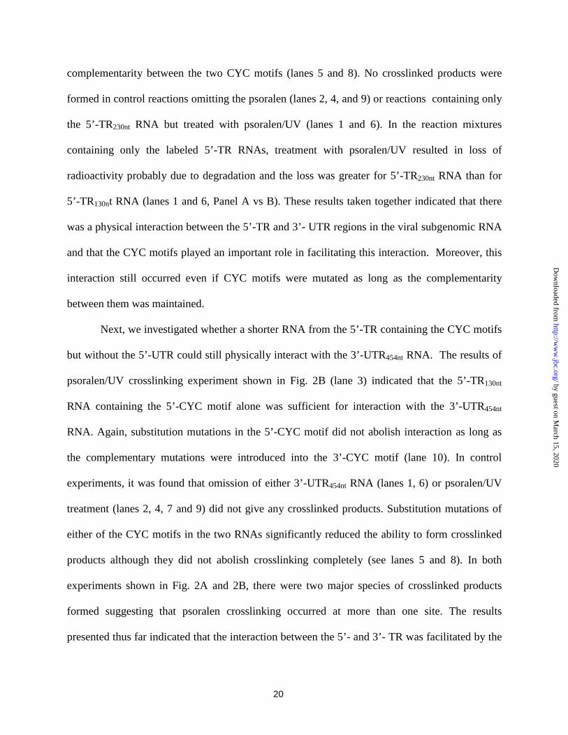
complementarity between the two CYC motifs (lanes 5 and 8). No crosslinked products were
formed in control reactions omitting the psoralen (lanes 2, 4, and 9) or reactions containing only
the 5’-TR230nt RNA but treated with psoralen/UV (lanes 1 and 6). In the reaction mixtures
containing only the labeled 5’-TR RNAs, treatment with psoralen/UV resulted in loss of
radioactivity probably due to degradation and the loss was greater for 5’-TR230nt RNA than for
5’-TR130nt RNA (lanes 1 and 6, Panel A vs B). These results taken together indicated that there
was a physical interaction between the 5’-TR and 3’- UTR regions in the viral subgenomic RNA
and that the CYC motifs played an important role in facilitating this interaction. Moreover, this
interaction still occurred even if CYC motifs were mutated as long as the complementarity
between them was maintained.
Next, we investigated whether a shorter RNA from the 5’-TR containing the CYC motifs
but without the 5’-UTR could still physically interact with the 3’-UTR454nt RNA. The results of
psoralen/UV crosslinking experiment shown in Fig. 2B (lane 3) indicated that the 5’-TR130nt
RNA containing the 5’-CYC motif alone was sufficient for interaction with the 3’-UTR454nt
RNA. Again, substitution mutations in the 5’-CYC motif did not abolish interaction as long as
the complementary mutations were introduced into the 3’-CYC motif (lane 10). In control
experiments, it was found that omission of either 3’-UTR454nt RNA (lanes 1, 6) or psoralen/UV
treatment (lanes 2, 4, 7 and 9) did not give any crosslinked products. Substitution mutations of
either of the CYC motifs in the two RNAs significantly reduced the ability to form crosslinked
products although they did not abolish crosslinking completely (see lanes 5 and 8). In both
experiments shown in Fig. 2A and 2B, there were two major species of crosslinked products
formed suggesting that psoralen crosslinking occurred at more than one site. The results
presented thus far indicated that the interaction between the 5’- and 3’- TR was facilitated by the
by guest on March 15, 2020
http://ww
w.jbc.org/
Dow
nloaded from

21
CYC motifs and that the 130nt region containing the CYC motif alone in the absence of the 5’-
UTR was sufficient for physical interaction with the 3’-UTR454nt RNA. The 5’-UTR96nt RNA
alone, lacking the 5’-CYC motif, did not interact with the 3’-UTR454nt RNA to form the
psoralen-crosslinked product (data not shown).
5’-UTR is crucial for RdRP to initiate RNA synthesis
Next, we sought to determine whether this 5’-TR130nt RNA can transactivate the 3’-
UTR454nt RNA in the in vitro RdRP assay. To address this question, in addition to the 5’-TR230nt
RNA, we used three different RNA templates in the in vitro assays: the 5’-TR180nt RNA
containing 5’-UTR and the CYC motif but shorter at the 3’-end, the 5’-TR130nt RNA lacking the
5’-UTR region, and the 5’-UTR96nt lacking the CYC motif. The 3’-UTR454nt RNA alone was
inactive in the formation of the 2X product in the RdRP assay as reported earlier (Fig. 3, lane 1).
However, a labeled RNA species of the same size as the input template RNA (1X) was formed
(lane 1). The 3’-UTR454nt RNA alone in the absence of 5’-TR was radiolabeled with varied
efficiency in the RdRP assays with different infected C6/36 extracts or with uninfected cell
lysates (data not shown; see also ref. 52). The formation of this labeled (1X) RNA species is
likely due to addition of a few nucleotides to the 3’-end of the template strand by the host
terminal nucleotidyl transferase (a membrane-bound enzyme) and this 1X product was sensitive
to RNase A digestion under high ionic strength conditions; however, the 1X RNA product
formed from the 3’-UTR RNA template in the presence of 5’-TR230nt RNA or the subgenomic
RNA template was resistant to RNase A (ref. 52). Furthermore, the results shown in Fig. 3
indicate that the 5’-TR230nt and the 5’-TR180nt RNA could both transactivate the 3’-UTR454nt RNA
(lanes 7 and 9) but the 5’-TR130nt and the 5’-UTR96nt RNAs were essentially inactive in
conferring RNA synthesis at the 3’-end of the 3’-UTR454nt RNA to form the 2X product (lanes 3
by guest on March 15, 2020
http://ww
w.jbc.org/
Dow
nloaded from

22
and 5). In control RdRP assays containing only 5’-TR RNA species as templates, the labeled 1X
and 2X products of the 5’-TR RNAs (lanes 2, 4, 6, and 8) were also formed, the former was
more predominant than the latter species. This observation was consistent with our previous
study (52) which also reported that blocking the 3’-end of the 5’-TR230nt RNA abolished its
template activity but not its ability to transactivate the 3’-UTR454nt RNA template for 3’-end
elongation. However, the presence of 3’-UTR454nt and 5’-TR RNAs together in the RdRP assay
resulted in significant suppression of RNA synthesis at the 5’-TR RNA templates for reasons
unknown (lanes 2 and 4 vs 3 and 5). Our results shown in Fig. 3, taken together, indicated that
although the 5’-TR RNA130nt containing the CYC motif alone was sufficient for physical
interaction with the 3’-UTR454nt (Fig. 2B), it was not sufficient for activation of RNA synthesis at
the 3’-end of 3’-UTR454nt RNA template and both the 5’-UTR and the 5’CYC motif are required.
In the previous study, we demonstrated a functional interaction between the 5’-TR and
the 3’-UTR for RNA synthesis at the 3’-end when both regions are in the same RNA template
such as the subgenomic RNA770nt that includes the CYC motifs. To further confirm the role of
5’-UTR and CYC motifs, four mutant subgenomic RNA770nt templates were used in the RdRP
assays; these were: subgenomic RNA674nt with a deletion of the 5’-UTR, two subgenomic
RNAs770nt containing the same substitution mutations as described before within either the 5’-
CYC or the 3’-CYC motif which would disrupt base-pairings and the subgenomic RNA770nt in
which both CYC motifs were mutated such that the complementarity was restored. (Figure 4).
The template efficiencies of these mutant subgenomic RNAs were analyzed using the in
vitro RdRP assay. The results indicated that the subgenomic RNA674nt (without the 5’-UTR) had
significantly reduced template activity for RNA synthesis (Fig. 4A). These results support the
by guest on March 15, 2020
http://ww
w.jbc.org/
Dow
nloaded from

23
conclusion reached from the transactivation assays that the 5’-UTR is necessary for the viral
RdRP to initiate RNA synthesis at the 3’-end of the RNA template (Fig. 3).
Next, the contribution of the two CYC motifs for RNA synthesis at the 3’-end of
subgenomic RNA templates was examined. The subgenomic RNA770nt, containing either of the
CYC motifs mutated or both mutated, were used as templates in RdRP assays. The results
indicate that the double mutant subgenomic RNA containing complementary mutant CYC motifs
was an equally efficient template as the wild type RNA for RNA synthesis (Figure 4B: lanes 1
and 4). Furthermore, the subgenomic RNA with the mutated 5’-CYC motif showed a dramatic
reduction in template efficiency compared to the wild type (lane 2). These results are consistent
with the conclusion reached in the previous study based on the transactivation assays (52).
However, the subgenomic RNA with the 3’-CYC mutation showed a reduced template activity
(about 60% of the wild type) (lane 3 vs lane 1). But this result was contrary to the results
obtained in the transactivation assays in that the 5’-TR with wild type CYC motif was essentially
inactive in promoting RNA synthesis from the 3’-UTR RNA containing mutant 3’-CYC motif
(52). This experiment was repeated 4 times and similar results were obtained. One possible
explanation for this difference is that the interaction between the 5’-TR containing the wild type
5’-CYC motif with the 3’-UTR RNA containing the mutant 3’-CYC motif is intramolecular and
may be more favorable as the two ends are in the same molecule compared to the scenario in
which the interaction between the two terminal regions of RNAs is intermolecular as is the case
with 5’-TR230nt/wtCYC and the 3’-UTR373nt/mutCYC used in the transactivation assays.
Importance of 3’-stem loop structure in the 3’-UTR for RNA synthesis in vitro by RdRP
The 3’-most 90-100 nucleotides of the flavivirus RNA genomes have been predicted to
form a stable stem loop (SL) structure (Figure 5: Panels A and B) (40,45,46,48,53,65-68).
by guest on March 15, 2020
http://ww
w.jbc.org/
Dow
nloaded from

24
Although the primary sequences of the region are less homologous, the 3’-SL structures are
highly conserved throughout the flavivirus family. This implies that the conserved 3’-SL
structure might have functional importance for viral replication.
Recently, Zeng et al. (45) reported the functional role of the 3’-SL structure in DEN2
viral replication and infectivity. They constructed several substitution mutations within the
DEN2 3’-SL either by rearrangement of base pairings that would disrupt or maintain secondary
structure or by replacing all or part of the 3’ SL of the DEN2 genome with the corresponding
regions of the West Nile 3’-SL, without disrupting the overall secondary structure. These
substitution mutations were cloned into the full length DEN2 infectious clone (diagrammatically
shown in Fig. 5, Panel C). In that study, monkey kidney (LLC-MK2) cells were electroporated
with the genome length RNA transcript from each mutant clone, and virus production and the
spread of infection was followed over time by immunofluorescence assay (45). The ability of the
viable mutant viruses to replicate in the mosquito (C6/36) cells and monkey kidney cells (LLC-
MK2) was also examined. From the analysis of infectivity of these RNA transcripts, it was
concluded that the stem structure of the top half and the nucleotide sequence of the 11 base
paired region in the uppermost portion of the bottom half of the 3’-SL are important for efficient
viral replication (45). Furthermore, a point mutation which would disrupt the predicted
pseudoknot structure within the 3’-SL (53) in the context of infectious clone was lethal for viral
replication and infectivity in vivo (Zeng et al., personal communication) (C71G; see Fig. 5A).
Four of the first 6 nt of the WN small loop region also had the potential to form a pseudoknot
structure with nucleotide 71 to 74 in the long stem (53), suggesting an interesting possibility that
disruption of this tertiary structure interaction could account for the loss of replication potential
of the chimeric mutants in this part of the viral genome.
by guest on March 15, 2020
http://ww
w.jbc.org/
Dow
nloaded from

25
To study the effects of these substitutions within the DEN2 3’-SL structures on their
template efficiencies in the in vitro RdRP assay system, we subcloned these 3’-SL mutations into
the plasmid coding for the subgenomic RNA. RNAs from these constructs were produced by in
vitro transcription and were used as templates in the in vitro RdRP assays using DEN2-infected,
mosquito (C6/36) as well as monkey kidney (LLC-MK2) cell lysates. The rationale for using
these two cell lysates was to examine whether there are any differences in host restriction for
RNA synthesis in vitro as was found previously with one mutant (mutF) in the in vivo infectivity
assays (45).
As shown in Figure 6, the 3’-SL mutant subgenomic RNAs showed template efficiency at
different levels. Subgenomic RNAs with the D2/WN-SL(mutA), D2-SL(a), D2-SL(b), D2/WN-
SL, D2/WN-SL(mutB), and D2/WN-SL(mutF) mutations exhibited template efficiency either
close to or only slightly reduced compared to the wild type template in the in vitro RdRP assays
that utilized either C6/36 or LLC-MK2 cell extracts for the source of viral replicase (Figure 6A
and B: lanes 1,3,4, 7, 8, and 11 vs 12). Of these mutants, mutagenesis of the top half of the
DEN2-3’-SL, either completely or partially as in D2/WN-SL(mutA), D2-SL(a), and D2-SL(b)
(see Fig. 5C) had only modest effects on virus growth compared to the wild type DEN2 genome
(45). The in vitro template efficiencies also support this finding (Fig. 6A and 6B: lanes 1, 3 and
4). In contrast, D2/WN-SL and D2/WN-SL (mutB) behaved like the wild type in the in vitro
assays whereas they were severely affected for growth in vivo (45). Moreover, the mutants
D2/WN-SL(mutD) and D2/WN-SL(mutF) exhibited similar template activities with the C6/36
extract which were closer to that of the wild type template (Fig. 6A: lanes 2 and 11 vs lane 12).
On the otherhand, with the LLC-MK2 cell extract, the –(mutD) was less active than –(mutF) but
both mutants were less active than the wild type template in the in vitro assay (Fig. 6B, lane 2 vs
by guest on March 15, 2020
http://ww
w.jbc.org/
Dow
nloaded from

26
11; compare lanes 2 and 11 with lane 12). The –(mutF) exhibited a “host-range” phenotype in
vivo in that it was severely restricted for replication in C6/36 cells but grew like the wild type in
LLC-MK2 cells (45). In contrast, the results of the in vitro RdRP assays showed little difference
in template efficiencies with the extracts from LLC-MK2- or C6/36-infected cells.
Template efficiency of the subgenomic RNAs containing D2/WN-SL(mutC) and -(mutE)
mutations exhibited a reduced template efficiency for RNA synthesis using either the LLC-MK2-
or the C6/36-infected cell extract (Figure 6A and 6B: lanes 9 and 10). Consistent with these
results, these mutants exhibited either no DEN-positive antigens (not viable) or only 10% of the
cells were positive for viral antigen (barely viable) as visualized by immunofluorescence of
DEN2-transfected LLC-MK2 cells (45). In -(mutC) and –(mutE) mutants, the mutations were
introduced at the bottom half of the 3’-stem (Figure 5C). In addition, the mutant subgenomic
RNAs containing the D2-SL(c) and D2-SL(C71G) mutations lost their template activities
dramatically when either of the two infected cell extracts was used in the in vitro assays (Figure
6A and 6B: lanes 5 and 6) which are consistent with their lethal phenotypes in vivo. In the D2-
SL(c) mutant, the lethal phenotype was attributed to the disruption of the top half of the 3’-SL
(45). The loss of template activity of the D2-SL(C71G) mutant is particularly interesting
because this subgenomic RNA contains mutation that would disrupt the predicted pseudoknot
structure based on the comparison to the homologous structure in the West Nile viral RNA 3’-SL
(53) (see Figure 5A and 5B). When the mutant genomic RNA was electroporated into LLC-MK2
cells, the mutation reverted to wild type and only the latter grew out of the transfected cells
suggesting that this pseudoknot mutant was lethal in vivo (Zeng, Falgout, and Markoff;
unpublished results). In order to prove that the tertiary interaction involving a pseudoknot
structure in the 3’-SL is important for template efficiency for RNA synthesis in vitro, we
by guest on March 15, 2020
http://ww
w.jbc.org/
Dow
nloaded from

27
engineered the C73G mutation to disrupt the pseudoknot base pairing in the D2/WN-SL
subgenomic RNA mutant in which the entire D2-SL was replaced with WN-SL. These
subgenomic RNA templates containing the wild type or mutant pseudoknot base pairs within 3’
D2-SL or 3’ WN-SL were analyzed for RNA synthesis in vitro. The results from Fig. 7 indicate
that the templates harboring a single base pair change that would disrupt the pseudoknot
structure within either of the two 3’-SLs significantly affected the template efficiency for RNA
synthesis in vitro. Based on these results, the strong template activity of D2/WN-SL(mutB) with
either of the two extracts could be explained by the possibility that the pseudoknot structure of
the West Nile 3’-SL is not affected in this mutant. These results presented in this study , taken
together, support the conclusion that RNA synthesis in vitro at the 3’-end of 3’-UTR is not only
dependent on the conserved CYC motifs and the 5’-UTR, but also more importantly, on the
determinants of conserved 3’-SL structure of the subgenomic RNA template which are
influenced by the conserved elements at the 5’ TR of viral genome.
DISCUSSION
Functional interaction between the 5’-TR and 3’-UTR required for RNA synthesis in vitro
In this study, we have further established that the newly synthesized RNA from the
subgenomic RNA template is of (-) polarity. Furthermore, the results of analyses of the structural
requirements for RNA synthesis indicate that both 5’-UTR and 3’-SL structures contribute to
template efficiency in addition to the conserved CYC motifs. The results of psolaren-UV cross-
linking experiments further establish that there is physical interaction between the 5’- and 3’-TRs
which require complementarity between the CYC motifs.
by guest on March 15, 2020
http://ww
w.jbc.org/
Dow
nloaded from

28
A number of studies have employed computer alogorithms that are capable of analyzing
predictive secondary structures as a guide or as a prelude for chemical and/or enzymatic probing
to understand RNA-RNA interactions. Therefore, we sought to compare the predictive
secondary structures of the subgenomic RNAs having wild type and mutant CYC motifs using
the software described by Zuker (58) (version 3.0). Among several structures that were
produced from each RNA molecule, the most stable structures were selected for comparison and
correlation with its in vitro template efficiency (Figure 8). As shown in Fig. 8, the subgenomic
RNAs containing the CYCwt motifs and the complementary, double-mutant CYC
(5’3’CYCmut) motifs seem to maintain a similar predicted secondary structure (Panels A and D).
Interestingly, the subgenomic RNA/3’CYCmut also seems to maintain the same overall
predicted secondary structures of the above two subgenomic RNAs (Figure 8, Panel C) although
there is no predicted base pairing between the wild type 5’-CYC and the mutated 3’-CYC motifs.
On the other hand, the predicted structures of the subgenomic RNA/5’CYCmut and the
subgenomic RNA674nt (without 5’-UTR) are quite different from that of the wild type (Panels E
and B vs Panel A). Thus the predictive analyses of RNA secondary structure suggest that
mutations in the conserved 3’-CYC motif are tolerated and the template efficiency in RNA
synthesis is not affected because the overall RNA structure is maintained. Moreover, the overall
conformation of RNA influenced by the interaction of the 5’- and 3’-terminal SL structures
seems more important for RNA synthesis than the CYC motifs which seem only to facilitate this
interaction. Further work using chemical and/or enzymatic RNA structure probing methods and
by site-directed mutagenesis is required to understand the RNA structure-template activities of
these subgenomic RNA molecules using our in vitro RdRP assay.
by guest on March 15, 2020
http://ww
w.jbc.org/
Dow
nloaded from

29
The important role of the 3’-SL structure of the viral RNA genome in dengue viral
replication in vivo was analyzed in an earlier study by Zeng et al. (45). The chimeric virus
D2/WN-SL, which had the 96nt sequence of the WN 3’-SL as a substitute for the 93nt DEN2
3’SL, showed a phenotype severely defective but not lethal in monkey kidney LLC-MK2 cells
(defined as “sublethal” in (45)). It was suggested that the possible mechanisms involving
RNA/RNA or protein /RNA interaction might have been affected which could have slowed
down the replication of the transfected genome compared to that of the wild type (45).
There was a good correlation between the template efficiency of these 3’-SL mutants in
the in vitro assays and the in vivo viabilities in the context of full length infectious clones (45)
for lethal mutations, D2-SL(c), D2/WN-SL(mutC) and D2-SL(C71G) as well as for the sublethal
mutant, D2/WN-SL(mutE). Moreover, this correlation could also be extended for the mutants,
D2-SL(a) and D2-SL(b) and the D2/WN-SL(mutA) in which substitutions were made within the
upper half of the 3’-SL (Fig. 5C). The good accord between the in vivo and the in vitro results
with regard to the latter three mutants support the previous conclusion that the secondary
structure rather than DEN2-specific nucleotide sequence of the top half of the 3’-SL is a major
determinant for viral replication.
Notable exceptions to this good correlation are the two mutants, D2/WN-SL(mutB) and
the D2/WN-SL (mutB and WN in Fig.6A), which exhibited significant template activity in vitro,
but were lethal or sublethal, respectively, in vivo (45). It was previously reported that the 3’SL
structures of the DEN and WN viral genome contain a pseudoknot structure between the ‘G’ in
the loop of the small SL and the ‘C’ in the bulge of the bottom half of the 3’SL although the
length of the base-paired regions involved in the 3’-SL structures of the DEN2 and WN RNA
genomes are different (see ref. (53)) (illustrated in Figure 5A and 5B). Our study clearly
by guest on March 15, 2020
http://ww
w.jbc.org/
Dow
nloaded from
30
indicates that the template efficiency of the pseudoknot mutant D2-SL(C71G) with a single
nucleotide substitution, was significantly reduced suggesting that the pseudoknot structure within
the 3’-SL could play an important role in RNA synthesis in vitro. This result also suggests that
perhaps the near-wild type template activities of the mutants, D2/WN-SL(mutB) and the
D2/WN-SL, might be due to the possibility that the potential pseudoknot structure of the West
Nile 3’-SL could be preserved in these mutants. This hypothesis was confirmed by analysis of
the C73G mutation in the D2/WN-SL RNA, which would be expected to disrupt this pseudoknot
structure, and the resultant mutant subgenomic RNA had significantly reduced template
efficiency for RNA synthesis in vitro. The same explanation could be considered for the mutant
subgenomic RNA with D2/WN-SL(mutF), the regions containing the DEN2 sequences are
presumably involved in the pseudoknot structure although the bottommost half of the 3’-SL is
from the West Nile RNA. Thus, this –(mutF) subgenomic RNA template had significant
retention of template efficiency in vitro. There is good correlation between the template
efficiency in vitro and the in vivo viability of the mutant D2/WN-SL(mutD) which replicated
well in both LLC-MK2 and C6/36 cells (45). However, the template efficiency of this -(mutD)
mutant RNA was greater in the extracts from the infected C6/36 cells compared with the LLC-
MK2 cells. Although our in vitro results are consistent with the notion that tertiary pseudoknot
interaction might play an important role for RNA synthesis, they do not explain why mutants,
D2/WN-SL(mutB), D2/WN-SL and D2/WN-SL(mutF), which are active as templates in vitro are
defective in vivo. One possible explanation for the disparity is that other steps in viral life cycle
which contribute to the overall viability such as defects in re-initiation of positive strand RNA
synthesis from the double stranded RNA intermediate or in viral assembly might be affected in
vivo.
by guest on March 15, 2020
http://ww
w.jbc.org/
Dow
nloaded from

31
All of the 3’-SL mutant RNAs were also able to physically interact with the 5’-TR180nt
RNA after psoralen/UV treatment (data not shown) suggesting that substitutions within the 3’-SL
do not affect the ability of the 5’-TR RNA to interact with the 3’-UTR. These results indicated
that although both 5’-TR and 3’-UTR RNAs could interact with each other, possibly mediated by
the wild type CYC motifs which are upstream of the 3’-SL, the mutations at the 3’-SL domains
of the 3’-UTR RNAs per se must have contributed to their differences in template efficiency for
RNA synthesis in vitro. These results also emphasize that the overall conformation of the 3’-SL
is more important for RNA synthesis than the CYC motifs alone.
Possible role of cross-talk between the 5’- and 3’-terminal regions of the viral RNA genome
in RNA synthesis
There is increasing evidence from studies of other positive single strand RNA viruses that
the 3’-end of the viral genome has to maintain a certain secondary and tertiary structure for RNA
synthesis (69-74). The 3’-UTR of brome mosaic virus (BMV) RNA-3 is predicted to have five
pseudoknots at the upstream end of the 3’-end tRNA-like structure (70). The deletion mutants of
RNA-3 within these regions replicated poorly, yielding no detectable RNA-3 or RNA-4 progeny.
This result suggested that the pseudoknot regions of the RNA-3 at the 3’-UTR contributed
significantly to the overall replication of the BMV genome. Deiman et al. (72) also reported
that the disruption of the stable pseudoknot structure at the 3’-end of the Turnip yellow mosaic
virus (TYMV) genome gave rise to a drop in transcription efficiency to about 50% indicating
that the stable pseudoknot structure is crucial for initiation of negative strand RNA synthesis.
Our results suggest that the cross-talk between the 5’- TR and 3’-UTR of the viral
genome might define a precise structure at the 3’ end involving tertiary pseudoknot interaction
such that the initiation of negative strand RNA synthesis could be carried out by the viral
by guest on March 15, 2020
http://ww
w.jbc.org/
Dow
nloaded from

32
replicase complex. In fact, possible cross-talk between specific regions of the viral genome of
RNA viruses has been suggested to participate in the viral replication of Qβ virus (75), BMV
(76), rhinovirus type 14 (77), poliovirus (78), hepatitis C virus (79,80), and tobacco etch virus
(81).
Proteins interacting with the flavivirus terminal regions of the RNA genome remain to be
studied. The results of this study indicate that using our in vitro RdRP assay and the wild type
and mutant subgenomic RNA templates, it is possible to study the higher order RNA structural
interactions and identify the viral and cellular factors that contribute to the stability and
functional long range interactions between the ends of the viral genome.
by guest on March 15, 2020
http://ww
w.jbc.org/
Dow
nloaded from

33
REFERENCES
1. Gubler, D. J., and Clark, G. G. (1995) Emerg Infect Dis 1(2), 55-7
2. Kautner, I., Robinson, M. J., and Kuhnle, U. (1997) J Pediatr 131(4), 516-24
3. Monath, T. P. (1994) Proc Natl Acad Sci U S A 91(7), 2395-400
4. Irie, K., Mohan, P. M., Sasaguri, Y., Putnak, R., and Padmanabhan, R. (1989)
Gene 75(2), 197-211
5. Chambers, T. J., Hahn, C. S., Galler, R., and Rice, C. M. (1990) Annu Rev
Microbiol 44, 649-88
6. Svitkin, Y. V., Lyapustin, V. N., Lashkevich, V. A., and Agol, V. I. (1984)
Virology 135(2), 536-41
7. Markoff, L. (1989) J Virol 63(8), 3345-52
8. Nowak, T., Farber, P. M., Wengler, G., and Wengler, G. (1989) Virology 169(2),
365-76
9. Bazan, J. F., and Fletterick, R. J. (1989) Virology 171(2), 637-9
10. Gorbalenya, A. E., Donchenko, A. P., Koonin, E. V., and Blinov, V. M. (1989)
Nucleic Acids Res 17(10), 3889-97
11. Chambers, T. J., Weir, R. C., Grakoui, A., McCourt, D. W., Bazan, J. F.,
Fletterick, R. J., and Rice, C. M. (1990) Proc Natl Acad Sci U S A 87(22), 8898-902
12. Preugschat, F., Yao, C. W., and Strauss, J. H. (1990) J Virol 64(9), 4364-74
13. Falgout, B., Pethel, M., Zhang, Y. M., and Lai, C. J. (1991) J Virol 65(5), 2467-
75
14. Chambers, T. J., Grakoui, A., and Rice, C. M. (1991) J Virol 65(11), 6042-50
15. Wengler, G., Czaya, G., Farber, P. M., and Hegemann, J. H. (1991) J Gen Virol
72(Pt 4), 851-8
16. Zhang, L., Mohan, P. M., and Padmanabhan, R. (1992) J Virol 66(12), 7549-54
17. Cahour, A., Falgout, B., and Lai, C.-J. (1992) Journal of Virology 66, 1535-1542
18. Falgout, B., Miller, R. H., and Lai, C.-J. (1993) Journal of Virology 67, 2034-
2042
by guest on March 15, 2020
http://ww
w.jbc.org/
Dow
nloaded from

34
19. Clum, S., Ebner, K. E., and Padmanabhan, R. (1997) J Biol Chem 272, 30715-
30723
20. Murthy, H. M., Clum, S., and Padmanabhan, R. (1999) J Biol Chem 274(9), 5573-
5580
21. Gorbalenya, A. E., Koonin, E. V., Donchenko, A. P., and Blinov, V. M. (1989)
Nucleic Acids Res 17(12), 4713-30
22. Koonin, E. V. (1992) Trends Biochem Sci 17(12), 495-7
23. Kadare, G., and Haenni, A. L. (1997) J Virol 71(4), 2583-90
24. Westaway, E. G. (1987) Adv Virus Res 33, 45-90
25. Wengler, G., and Wengler, G. (1993) Virology 197(1), 265-73
26. Poch, O., Sauvaget, I., Delarue, M., and Tordo, N. (1989) Embo J 8(12), 3867-74
27. O'Reilly, E. K., and Kao, C. C. (1998) Virology 252(2), 287-303
28. Koonin, E. V. (1993) J Gen Virol 74(Pt 4), 733-40
29. Rozanov, M. N., Koonin, E. V., and Gorbalenya, A. E. (1992) J Gen Virol 73(Pt
8), 2129-34
30. Bisaillon, M., and Lemay, G. (1997) Virology 236, 1-7
31. Wang, S. P., Deng, L., Ho, C. K., and Shuman, S. (1997) Proc Natl Acad Sci U. S.
A. 94, 9573-9578
32. Kapoor, M., Zhang, L., Ramachandra, M., Kusukawa, J., Ebner, K. E., and
Padmanabhan, R. (1995) J Biol Chem 270(32), 19100-6
33. Esteban, R., Fujimura, T., and Wickner, R. B. (1989) Embo J 8(3), 947-54
34. Nakhasi, H. L., Cao, X. Q., Rouault, T. A., and Liu, T. Y. (1991) J Virol 65(11),
5961-7
35. Pardigon, N., and Strauss, J. H. (1992) J Virol 66(2), 1007-15
36. Pogue, G. P., and Hall, T. C. (1992) J Virol 66(2), 674-84
37. Rohll, J. B., Percy, N., Ley, R., Evans, D. J., Almond, J. W., and Barclay, W. S.
(1994) J Virol 68(7), 4384-91
38. Rohll, J. B., Moon, D. H., Evans, D. J., and Almond, J. W. (1995) J Virol 69(12),
7835-44
39. Song, C., and Simon, A. E. (1995) J Mol Biol 254(1), 6-14
40. Blackwell, J. L., and Brinton, M. A. (1995) J Virol 69(9), 5650-8
by guest on March 15, 2020
http://ww
w.jbc.org/
Dow
nloaded from

35
41. Shi, P. Y., Li, W., and Brinton, M. A. (1996) J Virol 70(9), 6278-87
42. Hsue, B., and Masters, P. S. (1997) J Virol 71(10), 7567-78
43. Schuppli, D., Miranda, G., Qiu, S., and Weber, H. (1998) J Mol Biol 283(3), 585-
93
44. Stewart, S. R., and Semler, B. L. (1998) Nucleic Acids Res 26(23), 5318-26
45. Zeng, L., Falgout, B., and Markoff, L. (1998) J Virol 72(9), 7510-22
46. Rice, C. M., Lenches, E. M., Eddy, S. R., Shin, S. J., Sheets, R. L., and Strauss, J.
H. (1985) Science 229(4715), 726-33
47. Hahn, C. S., Hahn, Y. S., Rice, C. M., Lee, E., Dalgarno, L., Strauss, E. G., and
Strauss, J. H. (1987) J Mol Biol 198(1), 33-41
48. Brinton, M. A., Fernandez, A. V., and Dispoto, J. H. (1986) Virology 153(1), 113-
21
49. Brinton, M. A., and Dispoto, J. H. (1988) Virology 162(2), 290-9
50. Blackwell, J. L., and Brinton, M. A. (1997) J Virol 71(9), 6433-6444
51. Chen, C. J., Kuo, M. D., Chien, L. J., Hsu, S. L., Wang, Y. M., and Lin, J. H.
(1997) J Virol 71(5), 3466-73
52. You, S., and Padmanabhan, R. (1999) J Biol Chem 274(47), 33714-22
53. Shi, P. Y., Brinton, M. A., Veal, J. M., Zhong, Y. Y., and Wilson, W. D. (1996)
Biochemistry 35(13), 4222-30
54. Yaegashi, T., Vakharia, V. N., Page, K., Sasaguri, Y., Feighny, R., and
Padmanabhan, R. (1986) Gene 46(2-3), 257-67
55. Smith, T. J., Brandt, W. E., Swanson, J. L., McCown, J. M., and Buescher, E. L.
(1970) J Virol 5(4), 524-32
56. Zhang, L., and Padmanabhan, R. (1993) Gene 129(2), 197-205
57. Chatterjee, P. K., and Cantor, C. R. (1978) Nucleic Acids Res 5(10), 3619-33
58. Zuker, M., Jaeger, J. A., and Turner, D. H. (1991) Nucleic Acids Res 19(10),
2707-14
59. Calvet, J. P., and Pederson, T. (1981) Cell 26(3 Pt 1), 363-70
60. Thompson, J. F., and Hearst, J. E. (1983) Cell 32(4), 1355-65
61. Garrett-Wheeler, E., Lockard, R. E., and Kumar, A. (1984) Nucleic Acids Res
12(7), 3405-23
by guest on March 15, 2020
http://ww
w.jbc.org/
Dow
nloaded from

36
62. Datta, B., and Weiner, A. M. (1991) Nature 352(6338), 821-4
63. Wassarman, D. A., and Steitz, J. A. (1992) Science 257(5078), 1918-25
64. Watkins, K. P., Dungan, J. M., and Agabian, N. (1994) Cell T6(1), 171-82
65. Pletnev, A. G., Yamshchikov, V. F., and Blinov, V. M. (1990) Virology 174(1),
250-63
66. Mohan, P. M., and Padmanabhan, R. (1991) Gene 108(2), 185-91
67. Sumiyoshi, H., Hoke, C. H., and Trent, D. W. (1992) J Virol 66(9), 5425-31
*LHM: Dykes Library currently receives this title.
68. Proutski, V., Gould, E. A., and Holmes, E. C. (1997) Nucleic Acids Res 25(6),
1194-202
69. Jacobson, S. J., Konings, D. A., and Sarnow, P. (1993) J Virol 67(6), 2961-71
70. Lahser, F. C., Marsh, L. E., and Hall, T. C. (1993) J Virol 67(6), 3295-303
71. Chapman, M. R., Rao, A. L., and Kao, C. C. (1998) Virology 252(2), 458-67
72. Deiman, B. A., Kortlever, R. M., and Pleij, C. W. (1997) J Virol 71(8), 5990-6
73. Pleij, C. W., Rietveld, K., and Bosch, L. (1985) Nucleic Acids Res 13(5), 1717-31
74. Tsai, C. H., Cheng, C. P., Peng, C. W., Lin, B. Y., Lin, N. S., and Hsu, Y. H.
(1999) J Virol 73(4), 2703-9
75. Klovins, J., Berzins, V., and van Duin, J. (1998) Rna 4(8), 948-57
76. Diez, J., Ishikawa, M., Kaido, M., and Ahlquist, P. (2000) Proc Natl Acad Sci U S
A 97(8), 3913-8
77. McKnight, K. L., and Lemon, S. M. (1998) Rna 4(12), 1569-84
78. Goodfellow, I., Chaudhry, Y., Richardson, A., Meredith, J., Almond, J. W.,
Barclay, W., and Evans, D. J. (2000) J Virol 74(10), 4590-600
79. Blight, K. J., and Rice, C. M. (1997) J Virol 71(10), 7345-52
80. Ito, T., and Lai, M. M. (1997) J Virol 71(11), 8698-706
81. Haldeman-Cahill, R., Daros, J. A., and Carrington, J. C. (1998) J Virol 72(5),
4072-9
by guest on March 15, 2020
http://ww
w.jbc.org/
Dow
nloaded from
37
FOOTNOTES
This research was supported in part by National Institutes of Health Grants AI-32078 and AI-
45623. S.Y. was partly supported by the Biomedical Research Scholar Training Program of the
University of Kansas Medical Center. The costs of publication of this article were defrayed in
part by the payment of page charges. This article must therefore be hereby marked
“advertisement” in accordance with 18 U.S.C. Section 1734 solely to indicate this fact.
* To whom correspondence and reprint requests should be addressed. Dept. of Biochemistry and
Molecular Biology, University of Kansas Medical Center, 3901 Rainbow Blvd., Kansas City, KS
66160-7421. Tel: 913-588-7018; Fax: 913-588-7440; E-mail: [email protected]
¶ Present address: Laboratory of Virology and Infectious Diseases, Rockefeller University, 1230
York Avenue, New York, N.Y. 10021; e-mail: [email protected]
Abbreviations used are: CYC, cyclization; DEN2, dengue virus type 2; NS, nonstructural;
RdRP, RNA-dependent RNA polymerase; SL, stem-loop; TR, terminal region; WT, wild type;
UTR, untranslated region.
by guest on March 15, 2020
http://ww
w.jbc.org/
Dow
nloaded from

38
FIGURE LEGENDS
Fig. 1. Analysis of the polarity of the RdRP products by RNase H digestion.
Panel A: The overall scheme of RNase H analysis. Polarity of the oligodeoxynucleotides are
depicted as (+)/(-) within boxes. A solid line indicates the unlabeled input RNA of (+) polarity.
A dotted line indicates the radiolabeled RNA strand of (-) polarity synthesized in vitro by RdRP.
Panel B: The RdRP product from (+) polarity of RNA template was indicated in lane 1. Lane 2:
The RdRP product after RNase A digestion under high ionic strength, followed by RNase H
digestion in the absence of an oligodeoxynucleotide. Lane 3: The RdRP product after RNase A
digestion followed by annealing with (+) oligodeoxynucleotide and RNase H digestion. Lane 4:
The RdRP product after RNase A digestion followed by annealing with (-) oligodeoxynucleotide
and RNase H digestion. Prior digestion of the 2X hairpin product with RNase A to remove the
single-stranded loop region and addition of formamide in the annealing reaction (as described in
Experimental Procedures) were necessary for digestion of the RNA:DNA hybrid by RNase H.
Fig. 2. Analysis of RNA:RNA interaction.
Physical interaction between two RNAs was analyzed by the psoralen/UV crosslinking method
which is described in “Experimental Procedures”. The 3’-UTR454nt RNA (thick dotted line)
which had either wild type (W) or mutant (M) 3’-CYC motif was unlabeled. The 5’-TR230nt RNA
is indicated by a thick solid line with a hatched rectangular box denoting the 5’-UTR. Thick
rectangular vertical bars in 3’-UTR and 5’-TR RNAs denote CYC motifs. An ‘X’ indicates the
mutant CYC motif. An asterisk (*) denotes a radiolabeled RNA used for the crosslinking
experiment.
by guest on March 15, 2020
http://ww
w.jbc.org/
Dow
nloaded from

39
Panel A: Interaction between *5’TR230nt RNA and 3’UTR454nt RNA. Lanes 1 and 6: *5’-TR230nt
wt and 5’-TR230nt /cycMUT, respectively after psoralen/UV treatment. Lanes 2 and 3:
*5’TR230nt/wt + 3’UTR454nt/wt without and with psoralen/UV treatment, respectively. Lanes 4
and 5: *5’TR230nt/wt + 3’UTR454nt/cycMUT without and with psoralen/UV treatment,
respectively. Lanes 7 and 8: *5’TR230nt/cycMUT + 3’UTR454nt/wt without and with psoralen/UV
treatment, respectively. Lanes 9 and 10: *5’TR230nt/cycMUT + 3’UTR454nt/cycMUT without and
with psoralen/UV treatment, respectively. Although same amount of labeled 5’-TR RNA was
used for each experiment, treatment of labeled 5’-TR RNA alone with psoralen/UV seemed to
result in loss of labeled RNA which might be attributable to degradation (lanes 1 and 6; see also
Panel B).
Panel B: Interaction between 5’TR130nt RNA and 3’UTR454nt. The experiments were carried out
as described in Panel A except that labeled 5’TR130nt RNA (*) was used. The order of samples
loaded were same as in Panel A.
Fig. 3. Transactivation activities of various 5’-TR RNAs.
In order to map the minimum region of 5’-TR RNA which is necessary for transactivation of the
3’-UTR454nt for in vitro RNA synthesis, three truncated mutant 5’-TR RNAs were constructed.
The RdRP products were analyzed on a formaldehyde/agarose gel (1.8%), followed by
autoradiography. Lane 1: 3’-UTR454nt RNA alone. Lanes 2, 4, 6, and 8: 5’-UTR96nt, 5’-TR130nt,
5’-TR180nt, and 5’-TR230nt RNAs alone, respectively. Lanes 3, 5, 7, and 9: 3’-UTR454nt RNA in
the presence of the 5’-UTR96nt, 5’-TR130nt, 5’-TR180nt, and 5’-TR230nt RNA, respectively. The
labeled 1X and 2X products produced from the unlabeled 3’-UTR454nt RNA are indicated by
arrows. The unlabeled 5’-TR RNAs also served as templates in the in vitro RdRP assay and the
by guest on March 15, 2020
http://ww
w.jbc.org/
Dow
nloaded from

40
1X and 2X products are indicated as closed and open dots, respectively. In lane 7, 1X of 3'-
UTR454nt and the 2X of 5'-TR180nt were separated whereas in lane 9, the 3'-UTR454nt RNA band
was not well resolvede from the 2X product of 5'-TR230nt RNA. The presence of 3’-UTR RNA
significantly inhibited the template activity of the 5’-TRs (see lanes 3, 5, 7, and 9).
Fig. 4. In vitro RdRP assay with various subgenomic mutant RNA templates.
RNA templates were produced by in vitro transcription as described in “Experimental
Procedures”. The integrity of RNAs were verified by formaldehyde/1.8% agarose gel
electrophoresis and visualized by staining with acrydine orange.
Panel A: Analysis of the template efficiency of the subgenomic RNA674nt (without the 5’-UTR).
Lane 1: Subgenomic RNA770nt WT. Lane 2: Subgenomic RNA674nt containing the deletion of 5’-
UTR.
Panel B: Analysis of the template efficiency of the subgenomic RNA770nt WT, 5’cyc MUT,
3’cycMUT, and 5’3’double cycMUT (lanes 1-4, respectively).
Fig. 5. A schematic diagram of the 3’-SL mutations in the 3’UTR of DEN2.
Adapted from (45). Panel A: The secondary structure of the 93nt region from the 3’-end of the
DEN2 RNA. Panel B: The secondary structure of the 96nt region from the 3’-end of the West
Nile virus RNA. Pseudoknot G : C base pairing interactions within the DEN2 3’-SL and the
WN-3’-SL are also indicated as dotted circles. Panel C: Diagram of the mutations within the
3’-SL of the DEN2 RNA genome. The boldface regions in the first two rows indicate the
substituted regions from WN RNA. The bars from D2-SL(a) and D2-SL(b) indicate the
exchanged sequences. The boldface region from the D2-SL(c) shows the disrupted base-
by guest on March 15, 2020
http://ww
w.jbc.org/
Dow
nloaded from

41
pairings. D2-SL (C71G) is the pseudoknot mutant (indicated by a filled circle). The horizontal
dotted line denotes the border between upper and lower halves of the 3’-SL structure.
Fig. 6. Comparison of template efficiencies of the subgenomic RNAs with specific 3’-SL
mutations using the in vitro RdRP assays.
Each 3’-SL mutation was subcloned into the plasmid encoding the subgenomic RNA770nt.
In vitro transcribed 3’-SL mutant subgenomic RNA templates were analyzed on
formaldehyde/1.8% agarose gels, followed by staining with acrydine orange prior to their use in
the in vitro RdRP assays.
Panel A: In the previous study, the vivo viabilities of the 3’-SL mutations in the monkey kidney
(LLC-MK2) cells in the context of full length DEN2 RNA genome were determined by
immunofluorescence assays and quantitated as the percent of DEN antigen positive cells (45).
These semiquantitative values are presented here together with the template efficiencies of the
3’-SL mutant subgenomic RNAs in the in vitro RdRP assay carried out using infected LLC-MK2
extracts (see Panel C). Panel B: DEN2 infected C6/36 cell lysates were used for the in vitro
RdRP assay. Panel C: DEN2 infected LLC-MK2 cell lysates were used for the in vitro RdRP
assay.
Fig. 7. Importance of predicted pseudoknot interactions for in vitro RNA synthesis by
mutational analysis
C73G mutagenesis was carried out by PCR in the plasmid containing the D2/WN-SL in which
the entire 3’-SL of DEN2 (D2) was replaced the WN-3’-SL as described in Experimental
Procedures. Template efficiency of the wild type and mutant 3’-SL RNA templates was
determined using the C6/36 infected cell extract for the in vitro RdRP assays. Lane 1:D2 3’-SL
by guest on March 15, 2020
http://ww
w.jbc.org/
Dow
nloaded from

42
WT; lane 2: D2-SL(C71G) mutant; lane 3: D2/WN 3’-SL WT; lane 4: D2/WN 3’-SL(C73G)
mutant.
Fig. 8. Predicted secondary structure analysis of the subgenomic RNA templates
RNA structure software (version 3) by Zuker et al. (58) was used for secondary structure
analysis. Panel A: WT770nt, Panel B: Subgenomic 5’MUT, Panel C: Subgenomic 3’MUT, Panel
D: Subgenomic RNA 5’3’MUT. Panel E: Subgenomic RNA674nt
by guest on March 15, 2020
http://ww
w.jbc.org/
Dow
nloaded from


RdRP Assay with Different 5’-TR RNAs
1 2 3 4 5 6 7
This experiment was done with a different batch of infected C6/36 cell extracts. Thestandard conditions for RdRP assay were used.
Lane 1: 3’-UTR454nt only. Lane 2: 5’-TR130nt only. Lane 3: 5’-TR130nt RNA + 3’-UTR454nt
RNA. Lane 4: 5’-TR180nt RNA only. Lane 5: 5’-TR180nt RNA + 3’-UTR454nt RNA.Lane 6: 5’-TR230nt RNA only. Lane 7: 5’-TR230nt RNA + 3’-UTR454nt RNAThe labeled 1X RNA species generated from 5’-TR RNAs are indicated by opendiamond symbols. The 2X RNA species are by arrowheads. In lane 7, the 2X 5’-TR230nt
RNA and 1X 3’-UTR454nt RNA migrated together (mentioned in the text).
2X -3’-UTRRNA
1X 3’-UTRRNA1 X- 3’-UTR
RNA
by guest on March 15, 2020
http://ww
w.jbc.org/
Dow
nloaded from

Shihyun You, Barry Falgout, Lewis Markoff and R. Padmanabhanrange interactions between 5'- and 3'-terminal regions that influence RNA structure
In vitro RNA synthesis from exogenous dengue viral RNA templates requires long
published online February 5, 2001J. Biol. Chem.
10.1074/jbc.M010923200Access the most updated version of this article at doi:
Alerts:
When a correction for this article is posted•
When this article is cited•
to choose from all of JBC's e-mail alertsClick here
by guest on March 15, 2020
http://ww
w.jbc.org/
Dow
nloaded from