incidence of secondary root caries lesions in …
TRANSCRIPT
/ J of IMAB. 2014, vol. 20, issue 3/ http://www.journal-imab-bg.org 537
INCIDENCE OF SECONDARY ROOT CARIESLESIONS IN PATIENTS REFERRED FORTREATMENT IN THE FACULTY OF DENTALMEDICINE – SOFIA.
Mirela Marinova-TakorovaDepartment of Conservative Dentistry, Faculty of Dental medicine, MedicalUniversity – Sofia, Bulgaria.
Journal of IMAB - Annual Proceeding (Scientific Papers) 2014, vol. 20, issue 3Journal of IMABISSN: 1312-773X (Online)http://www.journal-imab-bg.org
SUMMARYPurpose: The aim of the presented study was to de-
termine the incidence of secondary root caries lesions in pa-tients referred for treatment in the Faculty of Dental Medi-cine – Sofia.
Material and Methods: The subjects who took partin the study were patients referred for treatment of carieslesions in the Faculty of Dental Medicine, Sofia. They wereinterviewed for smoking, presence of systematic diseasesand medications and debris and plaque were removed fromnatural teeth prior to examination.
Dental examination was carried out with a dentalmirror and a probe. Decayed, missed and filled teeth(DMFT) were recorded. Root caries lesions, restorationsof those lesions and secondary caries lesions were recordedseparately.
Results: A total number of 603 patients were exam-ined. The frequency of appearance of root caries in the in-vestigated population was 33.5% (202 patients). The wholenumber of root caries lesions was 857. Three hundred fortythree (41.4%) of those lesions were restored. Presence ofsecondary caries lesions was observed in 138 cases (39.1%).
Conclusions: Based on the data obtained from thepresented study it may be concluded that most of the rootcaries lesions remain untreated (58.8%).Secondary cariouswas diagnosed in 39.1% of the root caries restorations.Thepatients with secondary caries lesions presented with higherincidence of concomitant diseases and lower incidence ofsmoking.
Key words: root caries, secondary caries, epidemiol-ogy,
INTRODUCTIONRoot caries has become a socially significant oral dis-
ease for the elderly due to the increased life duration andthe higher number of teeth preserved for longer period [1].It is also an increasing problem for middle aged and evenyounger patients undergoing treatment or management ofperiodontal disease as well as those with prosthodontic re-constructions [2, 3, 4]. Root caries was developed in 90%of the subjects in a twelve-year follow up study of patientstreated for advanced periodontal disease [2, 5]. It was alsofound out that presence of four or more crowns is a risk fac-tor for the development of root caries [4].
The operative treatment of those lesions could becompromised by the difficult access, impaired visibility, dif-ficult moisture control, the proximity of pulp and the het-erogeneous morphology of the dentine, making quality ofadhesion not sufficiently predictable.[6] All these combinedwith the shrinkage stress generated during the polymeriza-tion of dental materials and the strain applied on the resto-ration/tooth contact surface during mastication due to theabfraction forces often leads to deterioration of adhesion,gap formation and secondary caries lesions. [7, 8]
The aim of the presented study was to determine theincidence of secondary root caries lesions in patients re-ferred for treatment in the Faculty of Dental Medicine –Sofia, Bulgaria.
MATERIAL AND METHODSThe subjects who took part in the study were patients
referred for treatment of caries lesions in the Faculty ofDental Medicine, Sofia, Bulgaria. Debris and plaque wereremoved from natural teeth prior to examination. All pa-tients were interviewed for smoking, presence of system-atic diseases and medications.
Dental examination was carried out with a dentalmirror and a probe by the author of the study. No radio-graphs were taken. Decayed, missed and filled teeth(DMFT) were recorded. Root caries lesions, restorationsof those lesions and secondary caries lesions were recordedseparately. Root caries was registered when a soft lesionwith discoloration or cavitation totally confined to the rootsurface or involving cement-enamel junction, but with in-dications that the lesion started from the root surface, werediagnosed. Recessions were measured on the vestibular andlingual surfaces if present.
A descriptive analysis of the results wasdone.(measurement of central tendency: arithmetic mean,median; measurements of variation: variance, standard de-viation, standard mean error), the hypotheses were checkedwith parametric (Student t-test for two independent samples,one-way analysis of variance (ANOVA)) and non paramet-ric (Mann-Whitney U tests, Chi-square criteria with Fish-er’s exact probabilities) methods. All calculations were per-formed by SPSS/PC v.13.0.
RESULTSA total number of 603 patients, referred for treatment
http://dx.doi.org/10.5272/jimab.2014203.537
538 http://www.journal-imab-bg.org / J of IMAB. 2014, vol. 20, issue 3/
of caries lesions in the Faculty of Dental Medicine, Sofiawere examined. 212 of them were males (35.2%) and 391(64.8%) – females. Their age varied from 25 to 85.
The frequency of appearance of root caries in the in-vestigated population was 33.5% (202 patients). Root car-ies lesions and restorations of such lesions are both includedas “root caries” in the conducted study. The whole numberof root caries lesions was 857. The mean number of lesionsper patient was 4.24. Three hundred forty three (41.4%)of those lesions were restored. The number of non-treatedwas 504 (58.8%). Presence of secondary caries lesions wasobserved in 138 cases (39.1%).
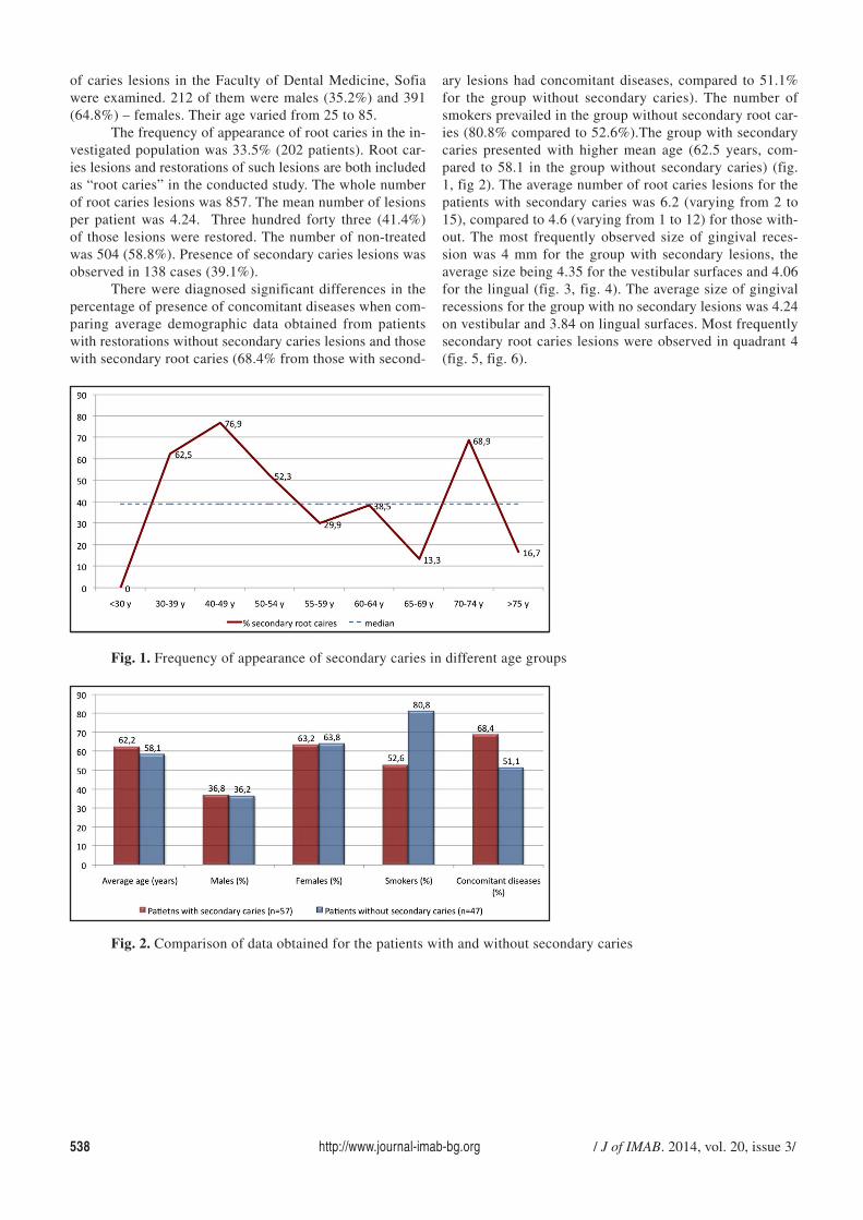
There were diagnosed significant differences in thepercentage of presence of concomitant diseases when com-paring average demographic data obtained from patientswith restorations without secondary caries lesions and thosewith secondary root caries (68.4% from those with second-
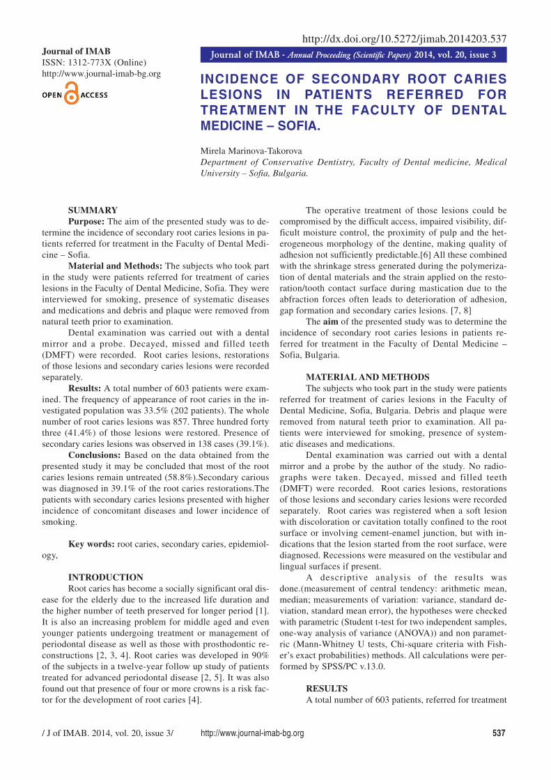
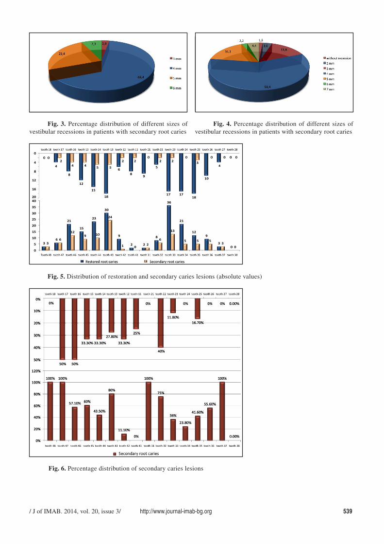
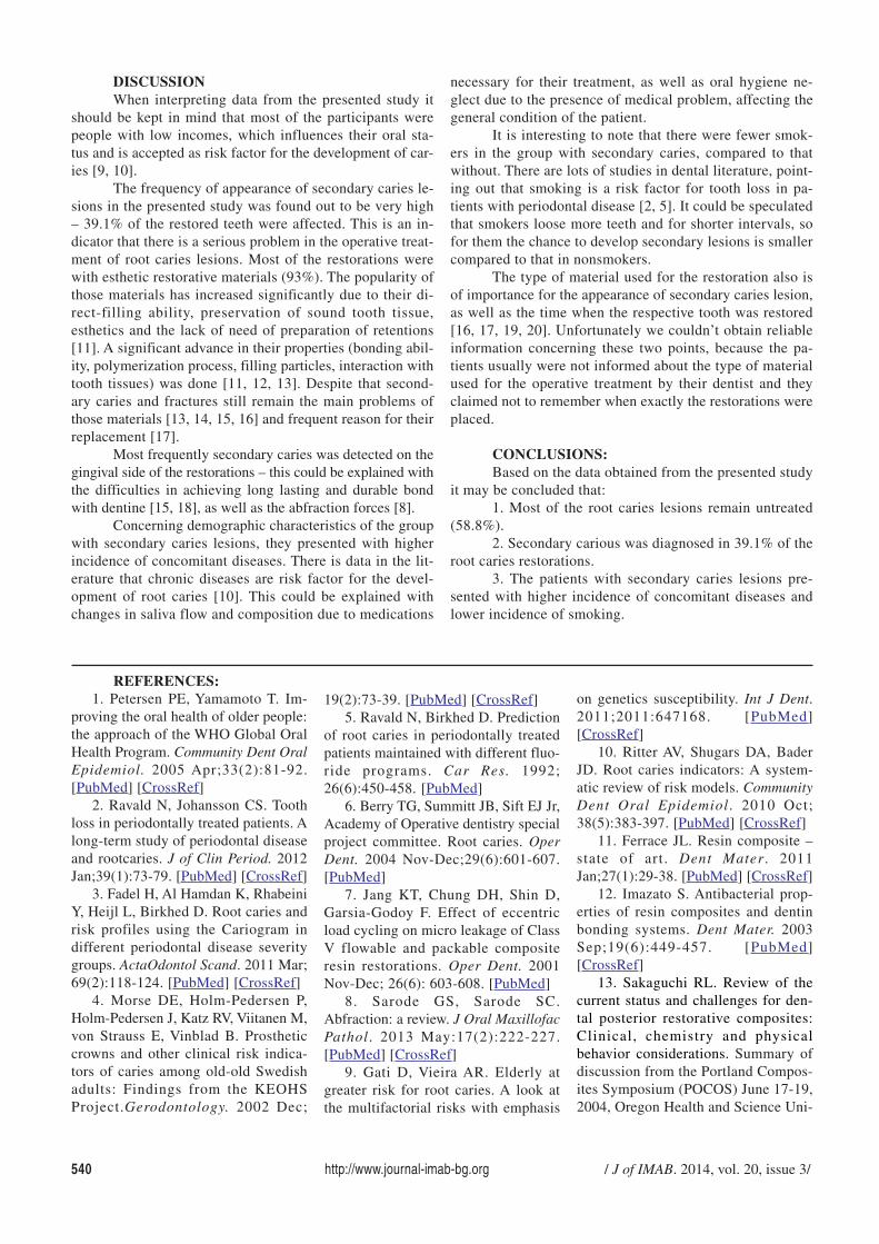
ary lesions had concomitant diseases, compared to 51.1%for the group without secondary caries). The number ofsmokers prevailed in the group without secondary root car-ies (80.8% compared to 52.6%).The group with secondarycaries presented with higher mean age (62.5 years, com-pared to 58.1 in the group without secondary caries) (fig.1, fig 2). The average number of root caries lesions for thepatients with secondary caries was 6.2 (varying from 2 to15), compared to 4.6 (varying from 1 to 12) for those with-out. The most frequently observed size of gingival reces-sion was 4 mm for the group with secondary lesions, theaverage size being 4.35 for the vestibular surfaces and 4.06for the lingual (fig. 3, fig. 4). The average size of gingivalrecessions for the group with no secondary lesions was 4.24on vestibular and 3.84 on lingual surfaces. Most frequentlysecondary root caries lesions were observed in quadrant 4(fig. 5, fig. 6).
Fig. 1. Frequency of appearance of secondary caries in different age groups
Fig. 2. Comparison of data obtained for the patients with and without secondary caries
/ J of IMAB. 2014, vol. 20, issue 3/ http://www.journal-imab-bg.org 539
Fig. 3. Percentage distribution of different sizes ofvestibular recessions in patients with secondary root caries
Fig. 4. Percentage distribution of different sizes ofvestibular recessions in patients with secondary root caries
Fig. 5. Distribution of restoration and secondary caries lesions (absolute values)
Fig. 6. Percentage distribution of secondary caries lesions
540 http://www.journal-imab-bg.org / J of IMAB. 2014, vol. 20, issue 3/
DISCUSSIONWhen interpreting data from the presented study it
should be kept in mind that most of the participants werepeople with low incomes, which influences their oral sta-tus and is accepted as risk factor for the development of car-ies [9, 10].
The frequency of appearance of secondary caries le-sions in the presented study was found out to be very high– 39.1% of the restored teeth were affected. This is an in-dicator that there is a serious problem in the operative treat-ment of root caries lesions. Most of the restorations werewith esthetic restorative materials (93%). The popularity ofthose materials has increased significantly due to their di-rect-filling ability, preservation of sound tooth tissue,esthetics and the lack of need of preparation of retentions[11]. A significant advance in their properties (bonding abil-ity, polymerization process, filling particles, interaction withtooth tissues) was done [11, 12, 13]. Despite that second-ary caries and fractures still remain the main problems ofthose materials [13, 14, 15, 16] and frequent reason for theirreplacement [17].
Most frequently secondary caries was detected on thegingival side of the restorations – this could be explained withthe difficulties in achieving long lasting and durable bondwith dentine [15, 18], as well as the abfraction forces [8].
Concerning demographic characteristics of the groupwith secondary caries lesions, they presented with higherincidence of concomitant diseases. There is data in the lit-erature that chronic diseases are risk factor for the devel-opment of root caries [10]. This could be explained withchanges in saliva flow and composition due to medications
necessary for their treatment, as well as oral hygiene ne-glect due to the presence of medical problem, affecting thegeneral condition of the patient.
It is interesting to note that there were fewer smok-ers in the group with secondary caries, compared to thatwithout. There are lots of studies in dental literature, point-ing out that smoking is a risk factor for tooth loss in pa-tients with periodontal disease [2, 5]. It could be speculatedthat smokers loose more teeth and for shorter intervals, sofor them the chance to develop secondary lesions is smallercompared to that in nonsmokers.
The type of material used for the restoration also isof importance for the appearance of secondary caries lesion,as well as the time when the respective tooth was restored[16, 17, 19, 20]. Unfortunately we couldn’t obtain reliableinformation concerning these two points, because the pa-tients usually were not informed about the type of materialused for the operative treatment by their dentist and theyclaimed not to remember when exactly the restorations wereplaced.
CONCLUSIONS:Based on the data obtained from the presented study
it may be concluded that:1. Most of the root caries lesions remain untreated
(58.8%).2. Secondary carious was diagnosed in 39.1% of the
root caries restorations.3. The patients with secondary caries lesions pre-
sented with higher incidence of concomitant diseases andlower incidence of smoking.
1. Petersen PE, Yamamoto T. Im-proving the oral health of older people:the approach of the WHO Global OralHealth Program. Community Dent OralEpidemiol. 2005 Apr;33(2):81-92.[PubMed] [CrossRef]
2. Ravald N, Johansson CS. Toothloss in periodontally treated patients. Along-term study of periodontal diseaseand rootcaries. J of Clin Period. 2012Jan;39(1):73-79. [PubMed] [CrossRef]
3. Fadel H, Al Hamdan K, RhabeiniY, Heijl L, Birkhed D. Root caries andrisk profiles using the Cariogram indifferent periodontal disease severitygroups. ActaOdontol Scand. 2011 Mar;69(2):118-124. [PubMed] [CrossRef]
4. Morse DE, Holm-Pedersen P,Holm-Pedersen J, Katz RV, Viitanen M,von Strauss E, Vinblad B. Prostheticcrowns and other clinical risk indica-tors of caries among old-old Swedishadults: Findings from the KEOHSProject.Gerodontology. 2002 Dec;
19(2):73-39. [PubMed] [CrossRef]5. Ravald N, Birkhed D. Prediction
of root caries in periodontally treatedpatients maintained with different fluo-ride programs. Car Res. 1992;26(6):450-458. [PubMed]
6. Berry TG, Summitt JB, Sift EJ Jr,Academy of Operative dentistry specialproject committee. Root caries. OperDent. 2004 Nov-Dec;29(6):601-607.[PubMed]
7. Jang KT, Chung DH, Shin D,Garsia-Godoy F. Effect of eccentricload cycling on micro leakage of ClassV flowable and packable compositeresin restorations. Oper Dent. 2001Nov-Dec; 26(6): 603-608. [PubMed]
8. Sarode GS, Sarode SC.Abfraction: a review. J Oral MaxillofacPathol. 2013 May:17(2):222-227.[PubMed] [CrossRef]
9. Gati D, Vieira AR. Elderly atgreater risk for root caries. A look atthe multifactorial risks with emphasis
on genetics susceptibility. Int J Dent.2011;2011:647168. [PubMed][CrossRef]
10. Ritter AV, Shugars DA, BaderJD. Root caries indicators: A system-atic review of risk models. CommunityDent Oral Epidemiol. 2010 Oct;38(5):383-397. [PubMed] [CrossRef]
11. Ferrace JL. Resin composite –state of art. Dent Mater. 2011Jan;27(1):29-38. [PubMed] [CrossRef]
12. Imazato S. Antibacterial prop-erties of resin composites and dentinbonding systems. Dent Mater. 2003Sep;19(6):449-457. [PubMed][CrossRef]
13. Sakaguchi RL. Review of thecurrent status and challenges for den-tal posterior restorative composites:Clinical, chemistry and physicalbehavior considerations. Summary ofdiscussion from the Portland Compos-ites Symposium (POCOS) June 17-19,2004, Oregon Health and Science Uni-
REFERENCES:
/ J of IMAB. 2014, vol. 20, issue 3/ http://www.journal-imab-bg.org 541
versity, Portland, Oregon. Dent Mater.2005 Jan;21(1):3-6. [PubMed][CrossRef]
14. Sarret DC. Clinical challengesand the relevance of materials testingfor posterior composite restorations.Dent Mater. 2005 Jan;21(1):9-20.[PubMed] [CrossRef]
15. Moreau JL, Weir MD,Giuseppetti AA, Chow LC, AntonucciJM, Xu HH. Long-term mechanicaldurability of dental nanocompositescontaining amorphous calcium phos-phate nanoparticles. J Biomed MaterRes B Appl Biomater. 2012 Jul;100(5):1264-1273. [PubMed] [CrossRef]
16. Sonbul H, Birkhed D. Risk pro-file and quality of dental restorations:A cross-sectional study. ActaOdontolScand 2010 Mar;68(2):122-128.[PubMed] [CrossRef]
17. Mjör JA, Moorland JE, Dahl JE.Reasons for replacement of restora-tions in permanent teeth in generaldental practice. Prim Dent Care. 2002Jan;9(1):31-36. [PubMed]
18. Huang C, Tay FR, Wei SH, KeiLH, Cheung GS, Pashley DH. Tensilestrength and ultrastructure of acompomer and a composite in aquousand non-aquous storage media. Am JDent. 2003 Sep;16 Spec No:82A-87A.
[PubMed]19. Hara AT, Turssi CP, Ando M,
Gonzalez-Cabezas C, Zero DT,Rodrigues AL Jr, et al. Influence offluoride releasing restorative materialon root dentine secondary caries insitu. Car Res. 2006; 40(5): 435-439.[PubMed] [CrossRef]
20. Espejo LC, Simionato MR,Barroso LP, Netto NG, Luz MA.Evaluation of three different adhesivesystems using a bacterial method todevelop secondary caries in vitro. AmJ Dent. 2010 Apr;23(2):93-97.[PubMed]
Address for correspondence:D-r. Mirela Marinova-Takorova,Department of Conservative dentistry, Faculty of Dental medicine, MedicalUniversity - Sofia1 GeorgySofiyskiblvd, 1431 Sofia, Bulgariae-mail: [email protected],
Please cite this article as: Marinova-Takorova M. Incidence of secondary root caries lesions in patients referred fortreatment in the Faculty of Dental Medicine - Sofia. J of IMAB. 2014 Jul-Sep;20(3):537-541.doi: http://dx.doi.org/10.5272/jimab.2014203.537.
Received: 29/04/2014; Published online: 18/07/2014;