sealing non-cavitated caries lesions: implications for ... · what is a non-cavitated caries...
TRANSCRIPT

Sealing NonSealing Non--Cavitated Caries Cavitated Caries Lesions: Implications For PracticeLesions: Implications For Practice
Margherita Fontana, DDS, PhDMargherita Fontana, DDS, PhDIndiana University School of DentistryIndiana University School of Dentistry
Department of Preventive and Community DentistryDepartment of Preventive and Community Dentistry
NATIONAL INSTITUTES OF HEALTHCONSENSUS DEVELOPMENT CONFERENCE
Diagnosis and Management of Dental Caries Diagnosis and Management of Dental Caries Throughout Life (March 26Throughout Life (March 26--28, 2001)28, 2001)
•“Improved caries detection and diagnostic methods would help determine the appropriate cutpoint or thresholdcutpoint or threshold separating the clinical decisionsclinical decisions to do nothing or preventively seal, or to therapeutically seal or surgically treat and restore”
(Weintraub, 2001)

Progress of Mineral Loss/DetectionProgress of Mineral Loss/Detection
(White Spot)
DiseaseDisease
DiseaseDisease
TreatmentTreatment
Treatment?Treatment?
m8

Slide 3
m8 White spots can also be in dentin....mfontan, 2/25/2005

What Level Of Assessment Do We Need For What Level Of Assessment Do We Need For Sealant Placement In Sealant Placement In Any SettingAny Setting??
Fejerskov, 2004

Variety of options change by setting, but the Variety of options change by setting, but the scientific evidence supporting management scientific evidence supporting management
strategies should be the samestrategies should be the same
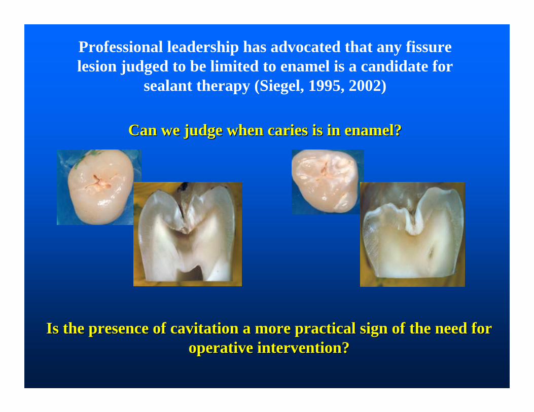
Professional leadership has advocated that any fissure lesion judged to be limited to enamel is a candidate for
sealant therapy (Siegel, 1995, 2002)
Can we judge when caries is in enamel?Can we judge when caries is in enamel?
Is the presence of cavitation a more practical sign of the need Is the presence of cavitation a more practical sign of the need for for operative intervention? operative intervention?

Indications for Occlusal SealantsIndications for Occlusal Sealants
On sound, at risk surfaces
What is a nonWhat is a non--cavitated caries lesion?cavitated caries lesion?
To arrest questionable or non-cavitated (incipient) caries lesions

White Spot /NonWhite Spot /Non--Cavitated Lesion:Cavitated Lesion:It is a subsurface lesion
External (outer) surface
Internal loss of minerals
Stages of the DiseaseStages of the Disease

Stages of the DiseaseStages of the Disease
Cavitated Lesion (Cavity):Cavitated Lesion (Cavity):A caries lesion that has lost the outer surface (leading
to a discontinuity in the surface)
MF5

Slide 9
MF5 What is referred to as a cavity in need of operative intervention, based on the previous slide by Kidd, may change with magnificationMargherita Fontana, 4/26/2006

Scientific Evidence for Caries Scientific Evidence for Caries DetectionDetection
2001 NIH Consensus Development Conference - Systematic ReviewICDAS IISelected studies

“At this time the panel senses a paradigm shift in the management of dental caries toward improved diagnosisdiagnosisof early nonearly non--cavitated lesionscavitated lesions and treatment for prevention and arrest of such lesions”
NATIONAL INSTITUTES OF HEALTHCONSENSUS DEVELOPMENT CONFERENCE
Diagnosis and Management of Dental Caries Diagnosis and Management of Dental Caries Throughout Life (March 26Throughout Life (March 26--28, 2001)28, 2001)
http://odp.od.nih.gov/consensus/cons/115/115_statement.htm
What level of assessment What level of assessment do we need for sealant do we need for sealant placement in Schoolplacement in School--
Based Programs?Based Programs?

“Clearly, since our diagnostic methods for assessing pit and fissure caries have been up to this time basically an educated guess, we must be placing sealants almost routinely over undetected incipient lesions” (Simonsen, 2002)
Occlusal surfaces:Occlusal surfaces:Typically low sensitivity, ~ 0.30,
and high specificity

2 A. VISUAL APPEARANCE
ICDASICDAS--22
Score5
DISTINCT CAVITY
Score6
EXTENSIVE CAVITY
SOUND
Score0
2. ACTIVITYDETECTION AND SEVERITY OF THE
LESION
SURFACE INTEGRITY
LOSS
Score3
OPACITYwithout
air-drying: WHITE,BROWN
Scores2W,2B
Ekstrand et al., modified by ICDAS (Ann Arbor), 2002; further modified by ICDAS (Baltimore) 2005
OPACITYwith air-drying: WHITE, BROWN
Scores1W,1B
UNDERLYING GREY
SHADOW
Score4
Lesion in Dentin Lesion in Enamel
Lesion in
Enamel/Dentin
http://www.icdas.htm
Suggested cutSuggested cut--off point: off point: Between ICDAS 2 and 3Between ICDAS 2 and 3

Probing with Sharp ExplorerProbing with Sharp Explorer……
Ekstrand et al., 1987
Traditional probing with a sharp explorer has come into question as the ultimate determinant of caries activity. The exclusive use of a “catch” by the sharp explorer to diagnose caries in pit and fissure sites should be discontinued and clinicians are being called upon to use “sharp eyes and a blunt explorer.” Also non-cavitated lesions can become cavitatedsimply through pressure from the explorer during the typical examination.
Treating caries as an Treating caries as an infectious disease. JADA infectious disease. JADA 125 (June): 2125 (June): 2--S to 15S to 15--S S (1995)(1995)

Role of Magnification in Determining Role of Magnification in Determining CavitationCavitation
•Magnification is not necessary to detect lesions using the ICDAS-2 criteria•Its use may affect the interpretation of the histological findings in relation to the criteria developed to correlate with it. For example, a category 2 tooth could be viewed as a category 3 under magnification, and this would result in more teeth being eliminated from consideration of sealants.

Radiographic ExaminationRadiographic Examination
Radiographs show that demineralization is present, but when looked at in one period of time they cannot determine ACTIVITY
Incidence of interproximal lesions in 2-3 graders is low
The ICDAS-2 criteria recognizes that some of the non-cavitated stages of the caries disease process may have already progressedinto dentin

How do we assess cavitated vs. nonHow do we assess cavitated vs. non--cavitated lesions?cavitated lesions?– Visual assessment is appropriate– Teeth can be dried with cotton rolls, gauze, or compressed
air– Explorer may be used to clean the fissures and “gently”
confirm cavitations (i.e., breaks in the continuity of the surface); do not use sharp explorer under force
– Magnification (2x-4x) can be used, but is not required – Radiographs are unnecessary, especially in programs
targeting children in grades 2 – 3– Insufficient evidence to recommend other technologies to
determine presence or absence of cavitation
SummarySummaryJ Pub Health Dent, 1995
* * *Non-Cavitated Cavitated
bfg6

Slide 17
bfg6 We suggest that you enlarge graphic "evaluate pit and fissure surfaces" and then bring each bullet in and then fade that bullet before bringing in the next bullet. In this way you will only need space for the questions and one answer at a time.Barbara Finigan Gooch, 4/17/2007

Thank You

What is the Caries Disease Process?What is the Caries Disease Process?CHO
H+
The metabolic activity of The metabolic activity of the the biofilmbiofilm on the surface on the surface
is the driving force...is the driving force...

……and/or is it the infected dentin and/or is it the infected dentin once the lesion once the lesion cavitatescavitates??

Reduction in Bacteria Counts by Reduction in Bacteria Counts by Time since Sealant Placement Time since Sealant Placement
(Griffin et al., 2007)(Griffin et al., 2007)
40.0%
50.0%
60.0%
70.0%
80.0%
90.0%
100.0%
0.03
0.15
0.23
0.35
0.50
1.00
1.00
2.00
2.00
4.00
4.00
6.00
6.00
7.00
12.00
12.00
24.00
60.00
60.00
60.00
Months since Sealant Placement
% R
educ
tion
in M
ean
Bac
teria
Cou
nts
•The percentage reduction in mean bacteria counts (4 studies) ranged from 50.8% to 99.9% and appeared to increase as time since sealant placement increased.

Dental SealantsDental Sealants
Effective Seal
Sealing infected dentin changes the oral Sealing infected dentin changes the oral environment (Kidd, 2004):environment (Kidd, 2004):
encourages arrest of demineralization, tubular sclerosis and tertiary dentin are encouraged, dentin permeability is reduced, residual microorganisms are now in a different
environment (do they change? how do they survive?)…they may become irrelevant!

Implications for PracticeImplications for Practice
Sealing non-cavitated lesions is an appropriate management alternative
for these lesions

Thank youThank you……