increased inflammatory cytokines and new collagen formation in
TRANSCRIPT

Thorax 1996;51:1253-1261
Increased inflammatory cytokines and new
collagen formation in cutaneous tuberculosisand sarcoidosis
Ben G Marshall, Arun Wangoo, H Terence Cook, Rory J Shaw
AbstractBackground- Interactions between mono-nuclear cells, vascular endothelium,fibroblasts, and cytokines during the in-flammatory reaction within a granulomahave the potential to contribute to the pro-gression to fibrosis.Methods - Biopsy specimens of six tuber-culous and eight sarcoidosis skin lesionswere examined by immunohistochemistryto seek evidence for the presence of in-flammatory and fibrotic reactions inhuman granulomatous disease. Ad-ditionally, to understand how a T cellmediated delayed type hypersensitivityreaction - a component of chronic granu-lomatous inflammation - could progressto fibrosis, the human in vivo model ofthe cutaneous tuberculin Heaf reaction topurified protein derivative (PPD) wasstudied in a group of 48 subjects.Results - Granulomas from tuberculousand sarcoidosis skin biopsy specimenswere seen to contain cells with markedstaining by antibodies to fibronectin,transforming growth factor P (pan TGF-P), and type 1 procollagen (PCP-1). Ac-centuated staining of extracellular matrixwas seen both in the granulomas and inthe peri-granulomatous regions. Lessprominent staining was observed usingantibodies against interleukin 1p (IL-10)and a-smooth muscle actin (a-SMA). Bi-opsies of Heaf reactions revealed cellsstaining for IL-1p, tumour necrosis factora (TNF-a), platelet derived growth factorB (PDGF-B), and fibronectin which weredetected as early as day 1 and persistedthroughout the 14 day study period. Cellsstaining for PCP-1 increased to greatestabundance at day 14. All these cytokineswere present in low abundance in biopsyspecimens from sites inoculated with sa-line only.Conclusions - Evidence is provided thatgranulomas in tuberculosis and sar-coidosis behave as active centres of fibro-genesis. Using the Heaf model, thetemporal relationship between the earlyappearance of cytokines and the later in-crease in the collagen precursor PCP-1linked the immune mediated chronic in-flammatory response with subsequentfibrosis and suggested that the tuberculinHeaf reaction will serve as a model forstudying the early events of granuloma
formation in patients with tuberculosisand sarcoidosis.(Thorax 1996;51:1253-1261)
Keywords: type I procollagen, delayed type hyper-sensitivity reaction, tuberculin, immunohistochemistry,granuloma.
Granulomatous diseases such as sarcoidosisand tuberculosis commonly evolve towards ir-reversible lung scarring.' In the skin tuber-culosis (particularly the lupus vulgaris form)causes fibrosis. Scarring is less common insarcoidosis of the skin, except in the papularand annular forms,2 although the immuno-histology of skin granulomas in patients withsarcoidosis is similar to that of granulomasfound in other organs such as lung and lymphnodes.3The inflammatory response in both diseases
is characterised by T cell and macrophage ac-tivation, a delayed type hypersensitivity (DTH)response, and the presence of inflammatorycytokines.4 Thus, these diseases may representmodels in which to understand the relationshipbetween immune mediated inflammation andfibrosis, as well as serving as human diseasemodels for other fibrotic lung diseases.5
Cytokines implicated in the progression fromchronic inflammation to fibrosis include inter-leukin 1 3 (IL- 1 P) 67 and tumour necrosis factora (TNF-a). The latter plays a central role inthe evolution of pulmonary fibrosis89 as well asin granuloma formation and maintenance, bothin the mouse'0 and in humans." Both IL-1B and TNF-cx are considered the primaryrecruiters of cells in granuloma growth. 12 Othercytokines such as transforming growth factorp (TGF-p) and platelet derived growth factor(PDGF) are important regulators of tissue re-pair, but may result in unresolved inflammationand fibrosis when present in excess quant-ities.3-1' There is evidence implicating bothPDGF'6 ' and TGF-P' ' in the progressionfrom chronic inflammation to fibrosis in tuber-culosis and sarcoidosis.The final common pathway in wound healing
and granulomatous fibrotic reactions is the syn-thesis of collagen by fibroblasts, particularlythe type I isoform,20 as well as other matrixproteins such as fibronectin.2' Myofibroblastsare specialised fibroblasts with morphologicaland functional features of smooth muscledifferentiation, one of a number of spindle-shaped interstitial cells capable of actively pro-ducing matrix proteins and participating inthe formation of granulation tissue. They are
Department ofRespiratory MedicineB G MarshallA WangooR J Shaw
Department ofHistopathologyH T Cook
Imperial CollegeSchool of Medicine atSt Mary's, LondonW2 1PG, UK
Correspondence to:Dr R J Shaw.Received 27 January 1996Returned to authors24 April 1996Revised version received31 May 1996Accepted for publication27 June 1996
1 253

Marshall, Wangoo, Cook, Shaw
thought to be responsible for the productionof connective tissue components which char-acterise fibrotic situations.22 Their hallmark isthe production of oc-smooth muscle actin whichmay be induced in some situations by TGF-323
In the present study we hypothesised thatthe chronic inflammatory response within cu-taneous granulomas of tuberculosis and sar-coidosis is intimately associated with theprogression to fibrosis. Biopsy specimens fromcutaneous tuberculous and sarcoidosis lesionswere examined by conventional immuno-histochemical techniques to investigate thelocal production of these cytokines, fibrogenicgrowth factors, and matrix proteins. To dissectthe local immune response and to examinethe initial kinetics of a T cell mediated in-flammatory reaction we have applied the sametechniques to the well established model of thecutaneous tuberculin Heaf test, a classical andfully defined example of a delayed type hyper-sensitivity reaction,24-26 since it may serve
as a model of the early immunopathologicalresponse in tuberculosis and sarcoidosis.
MethodsPATIENTSGranulomatous skin biopsy specimensParaffin blocks of skin biopsy specimens fromeight patients with cutaneous manifestations oftuberculosis and six patients with cutaneoussarcoidosis were drawn from the pathologyfiles from 1987 to 1993 at St Mary's Hospital.Biopsy specimens were selected because theyexhibited extensive granuloma formation andwere in a suitable condition to allow immuno-histochemical procedures. The specimens werestained with antibodies for IL-1,, fibronectin,oct-smooth muscle actin, TGF-1, and type 1procollagen. (Staining for TNF-ct and PDGF-B was not performed on these samples becausethese antibodies required frozen specimens.)
Heaf biopsy specimensThirty six healthy subjects (tuberculosis con-tacts or with a past history of tuberculosis) withstrongly positive (grade 3-4) Heaf reactionsand 12 healthy controls with Heaf grade 2-3reactions recruited from the St Mary's HospitalChest Clinic were studied. Each of the 36subjects underwent a Heaf test intradermallyinto the volar surface of the left forearm (tuber-culin PPD, 100 000 U/ml, Evans Medical Ltd,Horsham, Surrey, UK). The 12 controls re-ceived normal saline using the same method ofinjection. After a time interval of 1-14 days sixgroups of six patients each underwent twopunch skin biopsies (3 mm diameter; SteifelLaboratories (UK) Ltd, Woodburn Green,Buckinghamshire, UK) from within the areaof the site of injection on days 1, 2, 3, 6, 10,or 14 after the Heaf test. Thus, six subjectsunderwent two biopsies at each time point.Biopsy specimens were taken from the twogroups of six controls on either day 3 or 14.These procedures were performed under
sterile conditions using subcutaneous localanaesthesia (1 ml 1% lignocaine in adrenaline1 in 100 000). One biopsy specimen was fixedin 10% phosphate buffered formal saline andembedded in paraffin wax within 24 hours.Sections (5-6 [tm thick) were cut for immuno-histochemical examination with antibodies totype 1 procollagen (carboxy terminal domain),fibronectin, IL-lot, and oa-smooth muscle actin.The other biopsy specimen was snap frozen ina bath of liquid nitrogen at - 70°C in thepreservative Cryo-M-bed (Bright InstrumentsLtd, Huntingdon, Cambridgeshire, UK). Sec-tions (3-4 pim thick) were cut on a cryostat forimmunohistochemical examination with anti-bodies to TNF-ot and PDGF-B.
All subjects gave their informed consent andthe study was approved by the Parkside AreaHealth Authority ethics committee.
IMMUNOCHEMICAL EXAMINATIONSections from each paraffin wax biopsy speci-men were dewaxed, rehydrated, and treatedwith 3% hydrogen peroxide in methanol toquench the activity of endogenous peroxidase.The sections were washed in phosphatebuffered saline (PBS, pH 7.2) and trypsinised(0.5% trypsin and 0.5% chymotrypsin, SigmaChemical, Poole, Dorset, UK) for 10 minutesfor type 1 procollagen, IL- 1, and ot-smoothmuscle actin and 20 minutes for fibronectin.Non-specific protein binding was blocked with1:5 normal porcine or rabbit serum (Dako Ltd,High Wycombe, Buckinghamshire, UK) inPBS. Frozen biopsy specimens were air driedfor 10 minutes, fixed in acetone for 10 minutes,and washed in PBS for five minutes. The prim-ary antibodies used in this study were mono-clonal anti-human recombinant TNF-oc (TCSBiologicals Ltd, Buckinghamshire, UK), rabbitanti-human IL- 1 , and rabbit anti-humanPDGF-BB (Genzyme, Cambridge, Mas-sachusetts, USA), rabbit anti-human pan-TGF-1 (R&D, Abingdon, Oxon, UK), mono-clonal mouse anti-human muscle actin (DakoLtd), rabbit anti-human fibronectin (SigmaChemical), and rabbit anti-human type 1 pro-collagen. This antibody is raised against thecarboxy terminal domain of procollagen type Ipeptide which is cleaved enzymatically duringthe formation ofcollagen I and therefore detectscells actively synthesising collagen27 (gift fromM J Warburton, St George's Hospital, London,UK28).The antibodies were diluted in 1:20 porcine
or rabbit serum in PBS. The sections wereincubated with the optimal dilution ofthe prim-ary antibodies for one hour in a humidifyingchamber at room temperature (1:400 for type1 procollagen, 1:200 for ot-smooth muscle actinand fibronectin) or overnight (1:100 for IL-1 1,PDGF-B, and TNF-ot). The sections were allwashed with PBS and incubated with bio-tinylated secondary antibodies (Dako Ltd) for30 minutes, washed, and then incubated withavidin biotin peroxidase complex (Dako Ltd),washed for 15 minutes, and visualised with 3,3'-diaminobenzidine (DAB) substrate (Dako
1 254
Model for immune mediated inflammation and fibrosis
Ltd). The sections were counterstained withMayer's haematoxylin solution (Sigma Diag-nostics, St Louis, USA) for one minute.
CELL QUANTIFICATIONAfter colour development slides from the Heafbiopsy specimens were scored in a coded fash-ion by a blinded observer as follows. Slidesof paraffin wax sections (stained for type 1procollagen, IL-13, oa-smooth muscle actin,and fibronectin) were scored by counting thenumbers of positively staining nucleated cells,expressed as number/mm2 grid (Graticules Ltd,Tunbridge, Kent, UK), at three levels of dermis(superficial, middle and deep) from both peri-vascular infiltrates and interstitial dermis in-filtrates (areas of dermis selected not associatedwith vessels or sweat glands) and the meancalculated for each. Direct counting of cells inthe frozen sections was not possible as theirquality was not as good as the paraffin sectionsso each slide (stained for TNF-oa or PDGF-B)was given an intensity score on a semi-quantitative scale. Counting and grading ofslides was performed by an observer blind tothe code of the study. Specificity was as-certained when the primary antibody step wasomitted or sections were treated with purifiednon-immune rabbit or mouse serum (DakoLtd).
STATISTICAL ANALYSISResults are expressed as mean (SE). The 95%confidence intervals (CI) for the difference be-tween the two means (tuberculin Heaf testagainst control for days 3 and 14) were cal-culated using the confidence interval analysisprogramme (British Medical Journal, London,UK).
ResultsTUBERCULOSIS GRANULOMA BIOPSY SPECIMENSThere was evidence of active synthesis of col-lagen both inside and outside the granulomasof all biopsy specimens examined. Numerouscells with fibroblast morphology stainingpositively for type 1 procollagen were seenscattered throughout the granuloma core andthere was matrix staining around the marginsof the granulomas which served to delineateone granuloma from another (fig 1 a and b).In addition, there was marked staining withantibodies to TGF-,B and fibronectin both infibroblasts and in the extracellular matrix sur-rounding the granulomas, but not inside thegranulomas themselves (fig 2c, d and f). Onlyfaint staining for IL- 1p was seen, with oc-casional positively stained giant and epithelioidcells at the periphery of the granuloma. Epi-dermal staining for IL- 13B was present in allspecimens. No convincing positive staining foroa-smooth muscle actin was seen either in oraround the granulomas. In all cases dense stain-ing for oc-smooth muscle actin was present invascular smooth muscle, serving as a positivecontrol.
SARCOIDOSIS GRANULOMA BIOPSY SPECIMENSBy contrast, extensive staining for fibronectinand TGF-,B was detected throughout thegranulomas of all six sarcoidosis biopsy speci-mens with accentuation of cellular and matrixstaining around the periphery of the granul-omas (fig 2a, b and e). The cells staining withantibody for TGF-P appeared morphologicallyto be macrophages within the granulomasthemselves, but fibroblasts within the sur-rounding rim. Cells staining for type 1 pro-collagen were less prominent than those in thetuberculosis sections, both inside and aroundthe granulomas. However, the staining of thegranuloma margins appeared more prominent(fig lc and d). Occasional staining with IL-11P antibody was observed within cells at themargins of the granulomas. Again, there waslittle staining for a-smooth muscle actin in thegranulomas of these biopsy specimens. Sub-stitution of non-immune serum for each of theprimary antibodies yielded no immunostainingin specimens of tuberculosis and sarcoidosislesions, confirming the specificity of the anti-bodies used.
HEAF BIOPSY SPECIMENSCharacteristics of the delayed typehypersensitivity reactionAll subjects who underwent the tuberculin Heaftest reacted with red induration and swellingat the site of injection from day 1 onwards,reaching a peak at day 5. Sections stainedwith haematoxylin and eosin revealed a patchyperivascular interstitial dermal and periadnexalinfiltration of mononuclear cells from day 1onwards, the size and density growing to amaximum at day 14 although this was variableand it was not easy to distinguish one day'sbiopsy specimen from another. Subjects whoreceived normal saline did not show any lastingvisible reaction. Sections stained with haema-toxylin and eosin revealed very little cellularinfiltrate on either day 3 or 14.
Type 1 procollagenCellular and matrix staining was modestly butsignificantly increased from day 1 onwards (fig3d); 95% CI for the difference between themean Heaf and control perivascular infiltratecounts 3.17 to 14.60 on day 3 and 28.2 to 63.7on day 14. The number of positively stainingcells increased to a maximum at day 14 in bothperivascular and dermal infiltrates (fig le-g).These cells were morphologically fibroblasts.Control sections exhibited little cellular and nobackground staining (fig lh). Sections stainedwith rat anti-human procollagen 1 (ChemiconInternational Inc, Harrow, UK) which detectsthe cleaved amino-terminal domain of type Iprocollagen revealed similar patterns ofstaining(results not shown).
Interleukin 1/3Immunostaining for IL- 13 demonstratedstrong expression from day 1 in both peri-
1255

Marshall, Wangoo, Cook, Shaw
a b
t
c
I
4
'.. %, I F..
14O
.-4~ IF A.
d
.3S.
A,
- .0>4*
S- .4 * A
/a ',. P
*
* ft
'I
/
k, " ., 0
I
0
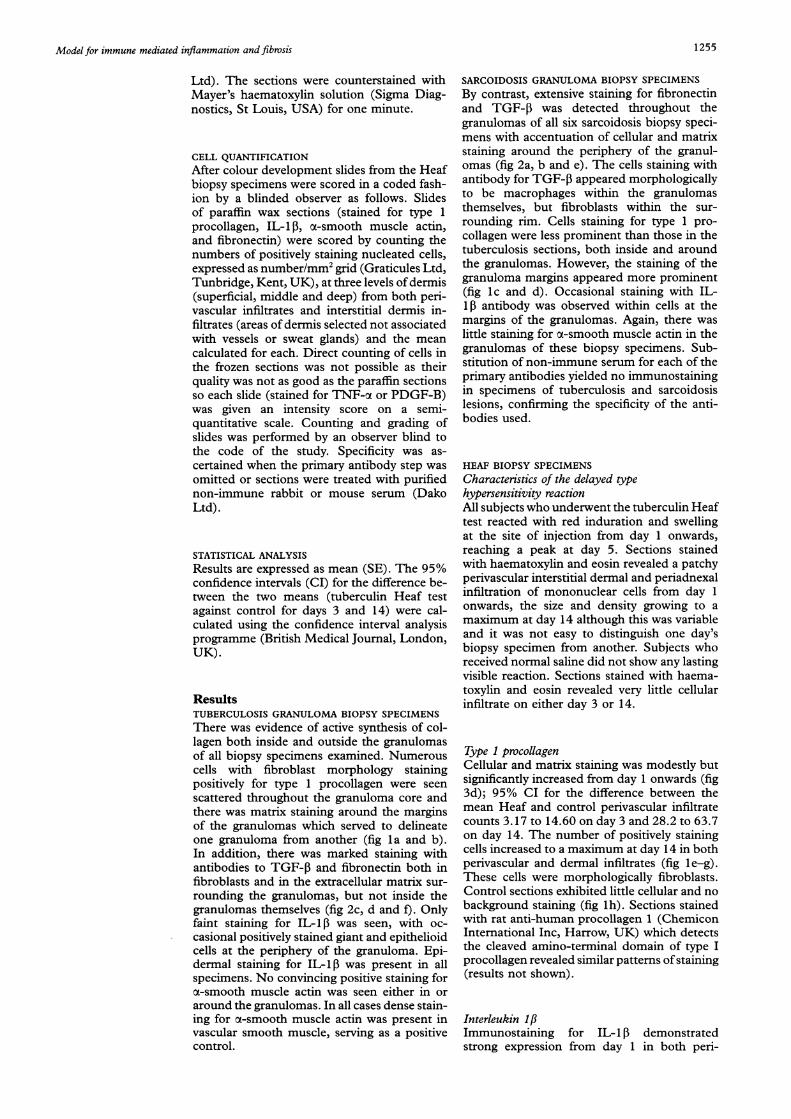
4( 19 hFigure I Photomicrographs of skin sections stained with anti-human type I procollagen antibody and counterstainedwith Mayers haematoxylin solution demonstrating (a) cutaneous tuberculosis granulomas at x 50 magnification and (b)at x 125 magnification showing numerous positively staining cells in brown with highlighting of the granulomaperiphery, (c) cutaneous sarcoidosis granulomas at x 50 magnification with less obvious cellular and matrix staining butprominent granuloma capsule staining (arrow) and (d) at x 125 magnification with multinucleate giant cell arrowed;and skin sections of tuberculin Heaf reactions stained with anti-human type I procollagen antibody and counterstainedwith Mayer's haematoxylin solution on (e) day 3 after PPD inoculation (x 125 magnification) demonstrating negligiblestaining (arrow), (j) day 10 after PPD inoculation (x 125 magnification), (g) day 14 after PPD inoculation (x 125magnification), and (h) day 3 after inoculation with saline control.
vascular and dermal infiltrates which remainedprominent in biopsy specimens collectedthroughout the 14 day study period (fig 3a).Cells staining positively were mainly foundwithin cell aggregates (fig 4a). It was not pos-sible to distinguish morphologically cell typesstaining with antibody. Absolute counts showedsignificantly increased numbers of positivelystained cells compared with controls on days
3 and 14 (95% CI for the difference betweenmean Heaf and control perivascular infiltratecounts 95.9 to 235 on day 3 and 38 to 146 onday 14) with up to one third of infiltrate cellsstaining. The pattern over the time course ofthe study was the same for the perivascular anddermal infiltrates. Only a few cells from thecontrol biopsy specimens stained for IL-1 3 ondays 3 and 14.
1256

1257Model for immune mediated inflammation and fibrosis
iIa..I ...
S .,
* -I-Aj,)
g~~ ~~IX* D~.-,\,i&._'
It.!a -F .ftI!lId;r,dX
'Af~~ ~ ~ ~ ~ U'
~~~~~~~~4~~ ~ ~ ~ -
.4-
/.-
#1 b
d
feFigure 2 Photomicrographs of skin sections of cutaneous sarcoidosis granulomas stained with (a) anti-humantransforming growth factor antibody and (b) anti-human fibronectin antibody demonstrating abundant stainingthroughout the granulomas, skin sections of cutaneous tuberculosis granulomas stained with (c) anti-human transforminggrowth factor and (d) anti-human fibronectin antibody demonstrating marked intracellular and extracellular staining inthe granuloma rim but not within the granuloma, and sections of (e) cutaneous sarcoidosis and (9 tuberculosisgranulomas with purified non-immune serum replacing primary antibody step omitted to confirm specificity of antibodystaining.
Tumour necrosis factor oxAbundant staining, most marked within theperivascular infiltrates in the skin biopsy speci-mens from tuberculin inoculated subjects, wasobserved from day 1, peaking at day 3 (fig 4d),and remaining increased until day 7. It was notpossible to differentiate the cell types stainingwith antibody to TNF-oa.
at day 3 (fig 4c), after which staining decreasedto a moderate intensity (but still increasedcompared with controls) for the duration ofthe study. In the control sections only very mildstaining or no staining was observed. It waspossible to delineate the pattern of staining andhow it related to the cellular infiltrate, butnot to determine the morphology of the cellsstaining.
Platelet derived growth factor BThere was increased cellular and matrix stain-ing from day 1 which increased to a maximum
FibronectinCell staining was increased from day 1 (95%CI for the difference between mean Heaf and
k
0.v, ".,.x

Marshall, Wangoo, Cook, Shaw
C 160E- 140-Chm 120-0o 100' 80-0.n 60
c- 40
Ji 20
0.,Ein
cD0
enIQ
-5C'.
Co
QC._11
CD0.0
2 3 6 10 14 3C 14CPPD injected site Control site
Day of biopsy after injection
C120 C
100-
80
60-
40-I
20-
0
1 2 3 6 10 14PPD injected site
PerivascularDermis
1i-- .1*3C 14C
Control site
Day of biopsy after injection
2 3 6 10 14 3C 14CPPD injected site Control site
Day of biopsy after injection
C-Ecn4
0
.0Co
I
en
Q-0
CL
1 2 3 6 10 14 3C 14CPPD injected site Control site
Day of biopsy after injection
Figure 3 Histograms demonstrating the time course of increase in abundance of (A) interleukin lfi (IL-1if), (B)fibronectin, (C) x-smooth muscle actin (o-SMA), and (D) type 1 procollagen (PCP-1) in Heaf biopsy specimens andsaline controls measured after Heaf testing or saline control inoculation. IL-i1l, fibronectin, x-SMA, and PCP-1 stainingmeasured as absolute counts per mm2 for both perivascular and interstitial dermal infiltrates.
saline control counts 10.3 to 28.8 on day 3and 10.1 to 28.2 on day 14) and remainedprominent throughout the study (fig 3b and4e). It was not possible to distinguish cell types.Of note, matrix staining was observed to bemore extensive from day 6 onwards (results notshown). In control sections there was negligiblecell or background stain noted on either daytested.
x-smooth muscle actinStaining was increased from day 1, rising to a
peak on day 10 (figs 3c and 4b) and remainingincreased compared with control countsthroughout the study (95% CI for the differ-ence between the mean Heaf and control peri-vascular infiltrate counts 2.52 to 97.80 for day3 and 3.47 to 54.1 for day 14). As before, thestaining of the dermal infiltrates reflected thatof the perivascular infiltrate and again therewas little or no staining in the control biopsyspecimens. Staining of vascular smooth muscleserved as a positive control.
DiscussionThese results indicate that granulomas frompatients with skin involvement by tuberculosisand sarcoidosis exhibit increased expression of
the growth factor TGF-13, the matrix proteinfibronectin, and evidence of new collagen for-mation on the basis of increased type 1 pro-
collagen production. The pattern of expressiondiffered between the two diseases. TGF-P andfibronectin were predominantly seen aroundtuberculous granulomas with type 1 pro-
collagen both inside and around them, whereasTGF-j3 and fibronectin was found throughoutsarcoidosis granulomas with type 1 procollagenpresent but in smaller quantities within andsurrounding the granulomatous tissue.
Excess accumulation of TGF-1 has beenreported in granulomatous lung lesions inpatients with sarcoidosis18 and tuberculosis."9The present study extended these observationsto the skin. Although the antibody used in thepresent study was a pan-TGF-3 antibody andthus did not provide information on TGF-
isoforms, the increase in TGF-,B was verymarked. The consequences of excess TGF-,Bproduction by these granulomas may be to
promote local fibrotic processes. The ob-servation that TGF-P enhances the intracellulargrowth of Mycobacterium tuberculosis inmacrophages'929 and counteracts macrophageactivation by other cytokines30 suggests thatTGF-f might be beneficial to the host onlyafter killing of mycobacteria is complete.
cl-
0cJ
0el
a)
-5.0ouIQ
5;LO
1 258

Model for immune mediated inflammation and fibrosis 1259
N I''4..
S.
'a
- .,. *.4.rv
- A *.- -.,
* '1 *.'- . - -
a
a-}
I^, *
4. 'tSV5,
a rb;W rt .4
*tMs'ma
4V'4is. 'u4 $ ''. 6
A 4
S a -tt,tAleo SVt A
V.
:i I ....13% .s
''at:...
~~~~i *- a
Ir* - ¶;, k'\ 4t '. '. ,?
;-i..*i 4 e
*a.tA
I
:' d
,f *# w
f * , ;iR 5 0 * 0 v*, fi
8 . a8 * X * t * w. f , , . . * .s * * Sw'; @ # P W; k ' \t .f h /h,,h ;J * b * J
_. ' * . _* _ w w . ox s e w /* . 4 ; . . t* S ^ . 9 %* 4 z bR s to Ft -¢ ; ) <' f 4 ,. - ,- r.t, .
* . _ . _ <, rw < z .* E t * ss 0 S _F _ t; f e
B _ . § . _ , v
* J - o * .
S _ ^ ,,s * . X < f
Figure 4 Photomicrographs of skin sections from biopsy specimens of tuberculin Heaf reactions six days after inoculationstained with antibodies against (a) interleukin-l,B at x 100 magnification (note strong positive stain of epidermis), (b)z-smooth muscle actin at x 400 magnification (note blood vessel wall staining positively, arrowed), (c) platelet derivedgrowth factor B (frozen section) at x 500 magnification, (d) tumour necrosis factor x at x 500 magnification, (e)fibronectin at x 125 magnification (note blood vessel wall staining positively) and 69 adjacent section to (e) withprimary antibody replaced by non-immune serum.
Increased amounts of type 1 procollagenin granulomas of tuberculosis and sarcoidosishave not been reported previously. Antibodiesto type 1 procollagen detect the terminal pep-tides of procollagen which are cleaved duringthe formation of mature collagen.27 The stain-ing for type 1 procollagen, which was especiallyprominent in the tuberculous granulomas,therefore indicated that active production ofnew collagen was occurring. The observationthat areas of extracellular matrix stained fortype 1 procollagen may be the result of stainingof newly cleaved carboxy terminal type 1 pro-
collagen propeptide, since it is known that thesecleavage reactions take place extracellularly,3'although these molecules are removed relativelyquickly by extracellular endopeptidases. Al-ternatively, the staining that appears to beextracellular is, in fact, delicate strands offibroblast cytoplasm. Lastly, this staining maybe type 1 procollagen peptide since a smallfraction of type 1 procollagen may escape com-plete conversion to collagen and be in-corporated into the mature crosslinkedcollagen.32 These biochemical observations arein keeping with the recognised cicatrising
.s
.Sf
.c , .. .
I
t.

Marshall, Wangoo, Cook, Shaw
nature of lupus vulgaris observed clinically.It is unclear whether the increases in type 1procollagen and TGF-P are causally linkedsince co-localisation of the staining was notdetermined in these biopsy specimens.By contrast, the increase in the pro-in-
flammatory cytokine IL-1 1B was less marked inthe mature granulomas of patients with tuber-culosis and sarcoidosis. The limited amount ofIL- 1p in the giant cells and epithelioid cells oftuberculosis and sarcoidosis granulomas is inkeeping with studies which suggest that IL-1Imay be important only in the early recruitmentstages of granuloma formation while TNF-ctmay take part in later maintenance or effectorfunctions of granulomas. ' l
Expression of fibronectin was prominent inthe skin sections of granulomas from patientswith both tuberculosis and sarcoidosis whichmay implicate fibronectin in the pathogenesis ofgranulomas. Synthesis of these proteins providesan extracellular matrix which may promote bothcell migration (important in the evolution ofthe granuloma) and maintenance of granulomaintegrity in the setting of considerable cell turn-over.34 The presence of excess amounts at theinterface of the capsule and the cellular core inall the biopsy specimens examined adds supportto the hypothesis that this is a region of activefibrogenesis. The persistence of fibronectinwithin granulomas may contribute to the balancebetween resolution and progression to fibrosis inboth tuberculosis and sarcoidosis, as is the casein granulomatous skin lesions of leprosy35 andhepatic schistosomiasis.36
In a previous study the presence of increasedamounts of TGF-1 was found to precede anincrease in expression of type 1 procollagenmRNA and protein in the tuberculin Heafbiopsymodel.37 In the present study, using the sameexperimental model, the temporal sequence ofevents involving the expression of cytokines, theappearance ofgrowth factors, and the productionof components of the extracellular matrix wasdefined in detail. The immunomodulatory cyto-kines IL- 113 and TNF-,c were expressed inabundant quantities from an early stage in thisdelayed type hypersensitivity reaction and re-mained high for the duration of the study period.This is in keeping with data from Chu et af-6who identified increases in IL- 13 and TNF-ot athigh levels throughout a seven day study periodfollowing administration of PPD in a Mantouxtest.The present study did not repeat the earlier
work in which TGF-1 was identified in theHeaf biopsy specimens37 but instead focusedon the fibrogenic agent PDGF-B. Staining offrozen sections did not allow quantifiable datato be obtained but, by using a blinded scoringsystem, production of PDGF-B was found tobe enhanced from as early as day 1 and reacheda maximum well before the maximal expressionof type 1 procollagen on day 14 (results notshown). The identification of this importantgrowth factor for fibroblasts in the Heaf biopsyspecimens may provide a mechanistic link be-tween the delayed type hypersensitivity re-sponse and the first stages of a fibrotic reaction,and is in keeping with a previous observation
that PPD stimulated lymphocytes produce in-terferon y which, in turn, increases the abund-ance of PDGF-B mRNA in macrophages.'6The expression of fibronectin by cells in the
Heaf biopsy specimens occurred as early asday 1 and was maintained for 14 days. Oneexplanation is that the cells that stained withinhours of the Heaf test insertion are, in fact, Tcells and that, following the antigenic stimulus,these T cells express T cell fibronectin, a cyto-kine recently implicated in the initiation ofdelayed type hypersensitivity reactions.38 Wewere not able to confirm this by double stainingin the present study. The accumulation offibronectin later on in the reaction might be aconsequence of fibronectin production by theexcess numbers ofmacrophages and fibroblastsprominent in the reaction at later stages. Therole of fibronectin might now be in cell ad-herence and enhancing wound healing by theprovision of a scaffold upon which collagenand other extracellular matrix proteins are de-posited. The antibody used in the present studywas raised against total fibronectin so T cellfibronectin, which shares many epitopes withother fibronectins, will cross react with epitopespresent on both plasma and cellular fibro-nectin.39 Finally, it is possible that some ofthe perivascular fibronectin staining may haveresulted from leakage from the circulation.
Unlike the situation in tuberculosis and sar-coidosis where few cells staining positively foroc-smooth muscle actin were detected, myo-fibroblasts were observed to be increased innumber following the Heaf test. This is in linewith other reports which have indicated thepersistence of excess numbers of cells stainingpositive for ot-smooth muscle actin in otherfibrotic reactions.40 It is likely that these cellsplay a part in the early stages of a lymphocytemediated inflammatory reaction, but they maynot be involved in the fibrotic process as-sociated with mature granulomas.The previous observation of an increase in
new collagen formation in association with aHeaf reaction37 was confirmed in the presentstudy. The time course of the new collagenproduction indicated that it followed the in-crease in expression of inflammatory cytokines,growth factors including PDGF-B and TGF-13,37 as well as ot-smooth muscle actin andfibronectin. There is no clinical evidence thata Heaf test results in a permanent scar, so it istherefore likely that in the longer term there isremodelling and collagen removal. However,the time course in which a cellular influx isfollowed by an increase in cytokines and growthfactors and later by evidence of new collagenproduction may serve as a useful human invivo model in which to study the initiation ofa fibrotic reaction during lymphocyte mediatedinflammation. This may shed light on similarevents that occur in tuberculosis and sar-coidosis granulomas and act as a human coun-terpart to animal studies to identify a linkbetween the delayed type sensitivity reactionand fibrosis.""A proposed scheme of the inflammatory and
profibrotic events that occur within a tuber-culosis or sarcoidosis granuloma resulting in
1 260

Model for immune mediated inflammation and fibrosis
Figure 5 Diagram of proposed scheme of inflammatory and profibrotic events within atuberculosis or sarcoidosis granuloma leading to excess deposition of extracellular matrixproteins and fibrosis.
excess deposition ofmatrix proteins and fibrosisis shown in fig 5.
The authors thank Miss Sundhiya Mandalia for her assistancewith the statistics and Professor Douglas Young for his helpwith the preparation of this manuscript. The study was fundedby St Mary's Hospital Special Trustees and the British LungFoundation.
1 Katz S. Clinical presentation and natural history of sar-coidosis. In: Fanberg BL, ed. Sarcoidosis and other granu-lomatous diseases of the lung. Switzerland: Marcel Dekker,1983:3-36.
2 Savin JA. Sarcoidosis. In: Champion RH, Burton JL, EblingFJG, eds. Textbook of dermatology. London: Blackwell Sci-entific, 1992:2383-406.
3 Quismorio Jr. FP. Immunological studies on cutaneouslesions in sarcoid. Clin Dermatol 1986;4:54-61.
4 Dannenberg Jr. AM. Roles of cytotoxic delayed-type hyper-sensitivity and macrophage-activating cell-mediated im-munity in the pathogenesis of tuberculosis. Immuno-biology 1994;191:461-73.
5 Hunninghake GW, Gadek JE, Young Jr RC, Kawanami0, Ferrans VJ, Crystal RJ. Maintenance of granulomaformation in pulmonary sarcoidosis by T-lymphocyteswithin the lung. N Engl _rMed 1980;302:594-8.
6 Dinarello CA. The biology of Interleukin-1. Interleukins.In: Kishimoto T, ed. Molecular biology and immunology.Switzerland: Karger, 1992:1-32.
7 Turino GM, Eden E. Interleukin-I secretion from humanalveolar macrophages in lung disease. _7 Clin Immunol1986;6:326-33.
8 Piguet PF, Collart MA, Grau GE, Kapanci Y, VassalliP. Tumour necrosis factor/cachectin plays a key role inbleomycin-induced pneumopathy and fibrosis. .7 Exp Med1989;170:655-63.
9 Piguet PF, Collart MA, Grau GE, Sappino A-P, Vassalli P.Requirement of tumour necrosis factor for developmentof silica-induced pulmonary fibrosis. Nature 1990;344:245-7.
10 Kindler V, Sappino A-P, Grau GE, Piguet PF, Vassali P.The inducing role of tumour necrosis factor in the de-velopment of bacterial granulomas in BCG infection. Cell1989;56:731-40.
11 Myatt N, Coghill G, Morrison K, Jones D, Cree IA. De-tection of tumour necrosis factor alpha in sarcoidosis andtuberculosis granulomas using in situ hybridisation. .7 ClinPathol 1994;47:423-6.
12 Boros DL. The role of cytokines in the formation of theschistosome egg granuloma. Immunobiology 1994;191:441-50.
13 Khalil N, O'Connor RN, Unruh HW, et al. Increased pro-duction and immunohistochemical localisation of trans-forming growth factor-f in idiopathic pulmonary fibrosis.Am Respir Cell Mol Biol 1991;5:155-62.
14 Wahl SM. Transforming growth factor f: the good, the badand the ugly. Exp Med 1994;180:1587-90.
15 Shaw RJ, Benedict SH, Clark RAF, KingJrTE. Pathogenesisof pulmonary fibrosis in interstitial lung disease. Am RevRespirDis 1991;143:167-73.
16 Wangoo A, Taylor IK, Haynes AR, Shaw RJ. Up-regulationof alveolar macrophage platelet derived growth factor-B(PDGF-B) mRNA by interferon gamma from Myco-
bacterium tuberculosis antigen (PPD)-stimulated lympho-cytes. Clin Exp Immunol 1993;94:43-50.
17 Ishioka S, Yamakido M. Role of cytokines from BAL cellsin granuloma formation. Nippon Rinsho 1994;52: 1467-72.
18 Limper AH, Colby TV, Sanders M, Asakura S, RochePC, DeRemee RA. Immunohistochemical localisation oftransforming growth factor-PI in the non-necrotizinggranulomas of pulmonary sarcoidosis. Am J Respir CritCare Med 1994;149:197-204.
19 Toossi Z, Gogate P, Shiratsuchi H, Young T, Ellner JJ.Enhanced production ofTGF-f8 by blood monocytes frompatients with active tuberculosis and presence of TGF-1in tuberculous granulomatous lung lesions. 7 Immunol1995;154:465-73.
20 Kovacs EJ. Fibrogenic cytokines: the role of immune me-diators in the development of scar tissue. Immunol Today1991;12:17-23.
21 Torikata C, Villiger B, Kuhn C, McDonald JA. Ultra-structural distribution offibronectin in normal and fibrotichuman lung. Lab Invest 1985;52:399-408.
22 Skalli 0, Gabbiani G. The biology of the myofibroblast -
relationship to wound contraction and fibroconnectivediseases. In: Clark RAF, Henson PM, eds. Molecular andcellular biology of wound repair New York: Plenum, 1988:373 -401.
23 Desmouliere A, Geinoz A, Gabbiani F, Gabbiani G. Trans-forming growth factor-: induces a-smooth muscle actinexpression in granulation tissue myofibroblasts and inquiescent and growing cultured fibroblasts. _7 Cell Biol1985;122:103-11.
24 Gibbs JH, Ferguson J, Brown RA, et al. Histometric studyof the localisation of lymphocyte subsets and accessorycells in human Mantoux reactions. I Clin Pathol 1984;37:1227-34.
25 Tsicopoulos A, Hamid Q, Vamey V, et al. Preferential mes-senger RNA expression of Thhl-type cells in classical de-layed type (tuberculin) hypersensitivity reactions in humanskin. . Immunol 1992;7:2058-61.
26 Chu CQ, Field M, Haskard D, Feldmann M, Maini RN.Detection of cytokines at the site of tuberculin-induceddelayed-type hypersensitivity in man. Clin Exp Immunol1990;90:522-9.
27 McDonald JA, Broekelmann TJ, Matheke ML, Crouch E,Koo M, Kuhn III C. A monoclonal antibody to thecarboxyterminal domain of procollagen type I visualisescollagen-synthesizing fibroblasts. _7 Clin Invest 1986;78:1237-44.
28 Warburton MJ, Ferns SA, Hughes C, Sear CH, RudlandPS. Generation of cell types with myoepithelial and me-senchymal phenotypes during the conversion of rat mam-mary tumor epithelial stem cells into elongated cells. .Natl Cancer Inst 1987;78:1191 -201.
29 Hirsch CS, Yoneda T, Averill L, Ellner JJ, Toossi Z. En-hancement of intracellular growth of Mycobacterium tuber-culosis in human monocytes by transforming growth factor-
._ Infect Dis 1994;170:1229-34.
30 Tsunawaki S, Spom M, Ding A, Nathan C. Deactivationof macrophages by transforming growth factor-P. Nature1988;334:260-4.
31 Fessler JH, Fessler LI. Biosynthesis of procollagen. Ann RevBiochem 1978;47:129-62.
32 Bomstein P, Sage H. Structurally distinct collagen types.Ann Rev Biochem 1980;49:974.
33 Remick DJ, Chensue SW, Hiserodt JC, Higashi GI, KunkelSL. Flow-cytometric evaluation of lymphocyte sub-populations in synchronously developing Schistosoma man-soni egg and Sephadex bead pulmonary granulomas. AmJPathol 1988;131:298-307.
34 Wangoo A, Cook HT, Taylor GM, Shaw RJ. Enhancedexpression of type 1 procollagen and transforming growthfactor-1 in tuberculin induced delayed type hyper-sensitivity. Y Clin Pathol 1995;48:339-45.
35 Cree IA, Nurbai S, Milne G, Swanson-Beck J. Cell deathin granulomata: the role of apoptosis. _7 Clin Pathol 1987;40:1314-9.
36 Narayanan RB, Bhutani LK, Sharma AK, Nath I. Fibro-nectin in leprosy lesions: observations using monoclonalantibodies to human fibronectin. Indian 7 Lepr 1984;56:532-9.
37 Grimaud J-A, Boros DL, Takiya C, Mathew RC, EmonardH. Collagen isotypes, laminin and fibronectin in granul-omas of the liver and intestines of Schistosoma mansoni-infected mice. Am 7 Trop Med Hyg 1987;37:335-44.
38 Godfrey HP. T cell fibronectin: an unexpected inflammatorycytokine. Lymphokine Res 1990;9:435-47.
39 Godfrey HP, Canfield LS, Kindler HL, Angadi CV, TomasekJJ, Goodman JW. Production of a fibronectin-associatedlymphokine by cloned mouse T cells. _7 Immunol 1988;141:1508.
40 McDonald JA, Kuhn C. The roles of the myofibroblast inidiopathic pulmonary fibrosis. Ultrastructural andimmunohistochemical features of sites of active extra-cellular matrix synthesis. Am3JPathol 1991;138:1257-65.
41 Miyamoto T, Kabe J, Noda M, Kobayashi N, Miura K.Physiological and pathological respiratory changes in de-layed type hypersensitivity reaction in guinea pigs. AmRev Respir Dis 1970;103:509-15.
42 Costabel U. The alveolitis of hypersensitivity pneumonitis.Eur Respir j 1988;1:5-9.
43 Andrade ZA, Reed SG, Roters SB, Sadigursky M. Immuno-pathology of experimental cutaneous leishmaniasis. Am.7Pathol 1984;114: 137-48.
44 Lemaire I. Selective differences in macrophage populationsand monokine production in resolving pulmonary gran-uloma and fibrosis. Am _7 Pathol 199 1;138:487-95.
1261