indian ckd guidelines final
DESCRIPTION
INDIAN GUIDELINESTRANSCRIPT
1
INDIAN CHRONIC KIDNEY DISEASE GUIDELINES
Indian CKD Guideline Workgroup
December 2013

2
PREFACE The first Indian guideline for chronic kidney disease was brought out in the year 2005. An update of this was long overdue. A group of experts across the country have shared their knowledge and expertise in this update of the Indian CKD guidelines. Chronic kidney disease is recognized to be a common disease, not only seen by the nephrologists but also by the specialists in other fields as well as the general practitioner. This update is targeted at nephrologists and internists. Wherever the KDIGO guidelines are available, they have been used as standard reference with modifications suited to Indian conditions. A standard format has been followed for all the guidelines. Additions in this update are guidelines on Ethical practices in dealing with CKD patients, Management of cardiovascular disease in CKD and “Prevention and treatment of contrast induced AKI”.
This is not meant to be an exhaustive textbook of nephrology, and should be read in conjunction with existing literature on various topics. We do not wish to duplicate the well known information. Indian commentary on KDIGO guidelines for CKD-MBD, which was published in 2011 in Indian Journal of Nephrology, should be read alongside and has not been added here. Since a separate workgroup is working on vaccination guidelines, that has also not been included. Finally, the KDIGO lipid guidelines are likely to appear soon, and there will be a separate commentary on those as well.
It has been a tremendous group effort of experts of different specialties, from all over the country who has interacted on many occasions, in formulating this update and has given freely their time and patience to this project. I sincerely thank Dr. Vinod Kumar K. for proof reading and help in compiling this update. This project was made feasible by unrestricted educational grant from Johnson and Johnson limited.
Dr. Gokulnath Convenor

3
NOMENCLATURE AND DESCRIPTION FOR RATING GUIDELINE RECOMMENDATIONS
We have used the terminology used by KDIGO guidelines (Table 1)
We have avoided further subdivisions into A, B, C and D due to paucity of literature available in Indian context. Uniformity has been maintained across chapters.
Table 1: KDIGO Nomenclature for guideline statements Statement Implication for patients Implication for clinicians “We recommend”
Most people in your situation Would want the recommended course of action and only a small proportion would not
Most patients should receive the recommended course of action.
“We Suggest”
The majority of people in your situation would want the recommended course of action, but many would not.
Different choices will be appropriate for different patients. Each patient needs help to arrive at a management decision consistent with her or his values and preferences.
Statement Implication for patients Implication for clinicians
4
CONTRIBUTORS
Abraham Georgi, M.D., FRCP Professor of Nephrology Pondicherry Institute of Medical Sciences and Madras Medical Mission Chennai [email protected]
Agarwal Sanjay Kumar, M.D., FRCP (edin), FASN, FAMS Professor and Head of Nephrology All India Institute of Medical Sciences New Delhi – 110 029 [email protected]
Almeida Alan, M.D., M.N.A.M.S., FISN. Former Professor of Nephrology G.S. Medical College, Mumbai – 400 013 Consultant Nephrologist P.D. Hinduja National Hospital And Research Centre, Mumbai – 400 016 [email protected]
Ballal Sudarshan, M.D., FRCP (UK) Board Certified in Internal Medicine, Nephrology and Critical Care (U.S.A) Medical Director and Chairman – Medical Advisory Board Director, Manipal Institute of Nephrology and Urology Bangalore – 560 017 [email protected]
Chakko K. Jacob, M.D., M.N.A.M.S Former Professor of Nephrology Christian Medical College and Hospital, Vellore Senior Consultant Nephrologist Baptist Hospital, Bangalore [email protected]
Dakshina Murthy KV, MD., D.M., DNB., FISN Senior Consultant Nephrologist Mahatma Sri Ramachandra Centenary Memorial Hospital and Apollo Hospital Hyderabad [email protected]
Gokulnath, M.D., D.M., DNB, FRCP(Lond) Professor and Head of Nephrology St. John’s Medical College Hospital,
Bangalore – 560 034 [email protected]
Gupta Amit, M.D., DNB, FRCP Professor of Nephrology Sanjay Gandhi Post Graduate Institute of Medical Sciences Lucknow 226 014 [email protected]
Jha Vivekanand, M.D., D.M., FAMS, FRCP Professor of Nephrology Post Graduate Institute of Medical Education And Research Chandigarh 160 012 [email protected]
Kher Vijay, M.D., D.M., FIMSA., FRACP Chairman, Division of Nephrology Medanta Kidney and Urology Institute Medanta, The Medicity Gurgaon [email protected]
Kirpalani Ashok, M.D., M.C.P.S., M.N.A.M.S (Nephro) Professor of Nephrology Bombay Hospital Institute of Medical Sciences, Mumbai 400 020 [email protected]
Kowdle Prakash, M.D., DNB. Senior Consultant Nephrologist Apollo Hospitals, Chennai 600 006 [email protected]
Nayak K. S., M.D., DNB Chief Nephrologist The Deccan Hospitals, Somajiguda Hyderabad 500 082 [email protected]
Narayan Prasad, M.D., D.M., DNB, MNAMS (Nephro) Additional Professor of Nephrology Sanjay Gandhi Post Graduate Institute of Medical Sciences Lucknow 226 014 [email protected]
Pawar Basant, M.D., D.M., DNB., FRACP Former Professor of Nephrology
5
Christian Medical College and Hospital, Ludhiana-‐141 008 [email protected]
Sharma R.K., M.D., DNB (Neph)., FAMS Professor and Head, Department of Nephrology Director, Sanjay Gandhi Post Graduate Institute Of Medical Sciences Lucknow 226 014 [email protected]
Rajapurkar Mohan, M.D. Director, Post Graduate medical studies, Mujibhai Patel Urological Hospital, Nadiad 387 001 [email protected]
Rajan Ravichandran, M.D., M.N.A.M.S. Senior Consultant and Director MIOT Institute of Nephrology MIOT hospitals Chennai – 600089 [email protected]
Ramdas Pisharody, M.D., D.M., DNB., FRCP Professor of Nephrology and Prinicipal, Trivandrum Medical College Thiruvananthapuram – 695 011 [email protected]
Rajapurkar Sujata, M.A., PhD. Medical Social Worker and Transplant Co-‐ordinator Mujibhai Patel Urological Hospital, Nadiad – 387 001 [email protected]
Ravi Raju T., M.D., D.M. Chief Consultant Nephrologist Apollo Hospital Former Director of Medical Education (Andhra Pradesh) Hyderabad.
Sakhuja Vinay, M.D., D.M., FAMS, FRCP, Professor and Head Post Graduate Institute of Medical Education and research Chandigarh 160 012 [email protected]
Sankarasubbaiyan Suresh, AB (Int. Med)., AB (Nephrology) Director, Da Vita, Nephrolife Chennai [email protected]
Shah Bharat, M.D., DNB. Director, Nephrology, Dialysis and Kidney Transplantation Global Hospital Mumbai [email protected]
Sishir Gang, M.D., D.M., DNB Chairman, Department of Nephrology Mujibhai Patel Urological Hospital Nadiad – 387 001 [email protected]
S. S. Iyengar, M.D, D.M., FRCP (UK) Senior Consultant Cardiologist Manipal Hospital Former Professor of Head Department Of Cardiology St. John’s Medical College and hospital Bangalore [email protected]
Vijay Viswanathan, M.D., Ph.D., MNAMS, FRCP (Lond), FRCP (Glas) Managing Director M.V. Hospital for Diabetes and Diabetes Research Centre Chennai – 600 013 [email protected]

6
HYPERTENSION AND ANTIHYPERTENSIVE AGENTS IN CHRONIC KIDNEY DISEASE (CKD)
Hypertension is a cause and consequence of CKD. Hypertension in CKD increases the risk of important adverse outcomes, including loss of kidney function and kidney failure, early development and accelerated progression of cardiovascular disease (CVD), and premature death.
JNC 7 defines hypertension as systolic blood pressure (SBP) > 140mm Hg or diastolic blood pressure (DBP) >90mmHg, respectively. Although common in CKD, hypertension is not a part of the definition of CKD. Approximately 50% to 75% of individuals with GFR <60mL/min/1.73 m2 (CKD stages 3-5) have hypertension.
Hypertension plays a key role in progression of CKD. In addition to controlling blood pressure, antihypertensive therapy affects other key modifiable factors related to the progression, including proteinuria, vascular stiffness and increased activity of the renin angiotensin system (RAS). Several large, controlled trials have examined the effect of antihypertensive therapy on the progression of kidney disease in patients with and without hypertension. While these trials have provided important answers about therapy, the relationships among these “progression factors” are complex, and many questions remain unanswered, especially regarding the mechanisms underlying the therapeutic benefit of the interventions.
1 GOALS OF ANTIHYPERTENSIVE THERAPY IN CKD
1.1: We suggest to individualize BP targets and agents according to age, co-existent cardiovascular disease and other co morbidities, risk of progression of CKD, presence or absence of retinopathy (in CKD patients with diabetes) and tolerance of treatment.
A J-shaped relationship between achieved BP and outcome has been observed in the elderly and in patients with vascular disease, possibly suggesting that BP cannot be reduced too far in these patients. Choice of BP-lowering agents should be tailored to the individual patient. For instance, ACEIs and ARBs are
potentially harmful in the presence of significant renovascular disease or volume depletion, or when used in combination with non-steroidal anti-inflammatory drugs (NSAIDs) or cyclooxygenase- 2 (COX-2) inhibitors.
1.2: We suggest to inquire about postural dizziness and check for postural hypotension regularly when treating CKD patients with BP-lowering drugs.
Patients with CKD, particularly the elderly and diabetic patients with autonomic neuropathy, are prone to orthostatic hypotension, which may be exacerbated by volume depletion. Many CKD patients will require combinations of drugs to control BP including vasodilators, which can cause or exacerbate postural hypotension. 2 EVALUATION OF PATIENTS
WITH CKD OR HYPERTENSION 2.1 We recommend that blood pressure
should be measured at each health encounter in all CKD patients.
2.2 We suggest that, the initial evaluation should include the following elements:
a. Description of CKD; • Type (diagnosis), level of GFR,
and level of proteinuria • Complications of decreased GFR • Risk for progression of kidney
disease b. Presence of clinical CVD and CVD
risk factors c. Comorbid conditions d. Barriers to self-management,
adherence to diet and other lifestyle modifications, adherence to pharmacological therapy and complications of pharmacological therapy
2.3 We suggest that a clinical plan should be developed for each patient, based on the stage of CKD.

7
2.4 We suggest that patients with resistant hypertension should undergo additional evaluation to ascertain the cause.
2.5 We suggest that patient with resistant hypertension should be referred to a nephrologist.
3 MEASUREMENT OF BLOOD PRESSURE IN ADULTS
3.1 We recommend that, blood pressure should be measured according to the recommendations for indirect measurement of arterial blood pressure of the American Heart Association and Seventh Report of the Joint National Committee on the Prevention, Detection, Evaluation and Treatment of High Blood Pressure (JNC 7) and patients should be taught to measure and record their blood pressure, whenever possible.
The correct method of measuring the blood pressure is described below in sequential steps.
• Relaxed, temperate setting, with the patient seated and rested
• Arm out-stretched, in line with mid-sternum and supported
• Correctly wrap a cuff containing an appropriately sized bladder around the upper arm and connect to a manometer. Cuffs should be marked to indicate the range of permissible arm circumferences; these marks should be easily seen when the cuff is being applied to an arm.
• Palpate the brachial pulse in the antecubital fossa of that arm.
• Rapidly inflate the cuff to 20 mmHg above the point where the brachial pulse disappears.
• Deflate the cuff and note the pressure at which the pulse reappears: the approximate systolic pressure.
• Re-inflate the cuff to 20 mmHg above the point at which the brachial pulse disappears
• Using one hand, place the stethoscope over the brachial artery ensuring complete skin contact with no clothing in between.
• Slowly deflate the cuff at 2–3 mmHg per second listening for the Korotkoff sounds.
Phase I: The first appearance of faint repetitive clear tapping sounds gradually increasing in intensity and lasting for at least two consecutive beats: note the systolic pressure. Phase II: A brief period may follow when the sounds soften and or 'swish'. In some patients the sounds may disappear altogether (auscultatory gap). Phase III: The return of sharper sounds becoming crisper for a short time. Phase IV: The distinct, abrupt muffling of sounds, becoming soft and blowing in quality. Phase V: The point at which all sounds disappear completely: note the diastolic pressure.
• When the sounds have disappeared, quickly deflate the cuff completely if repeating the measurement and when possible, take readings at the beginning and end of consultations.
3.2 We suggest that ambulatory blood pressure monitoring should be considered for patients with CKD for the following indications:
• Suspected white coat hypertension • Resistant hypertension • Hypotensive symptoms while taking
antihypertensive medications • Episodic hyertension • Autonomic dysfunction
4 DIETARY AND OTHER THERAPEUTIC LIFESTYLE MODIFICATIONS FOR LOWERING BP IN CKD PATIENTS:
4.1: Encourage lifestyle modification in patients with CKD to lower BP and improve long-term cardiovascular and other outcomes:
4.1.1: We recommend achieving or maintaining a healthy weight.
Though it is well documented that, weight reduction lowers BP in the general population, only observational studies are available for similar benefits in CKD

8
patients. Weight reduction strategy would have spin off beneficial effects to CKD patients in the form of reduction in proteinuria, increased insulin sensitivity and improved lipid profile. 4.1.2: We recommend lowering salt intake to
<2 g per day of sodium (corresponding to 5 g of sodium chloride), unless contraindicated.
In a systematic review of seven trials, in general population, it was evident that restricting salt intake clearly lowers BP. A low-sodium diet has been shown to further reduce BP and urine albumin or protein levels in the short term, in patients on ARBs and may be considered in those with high BP and poor response to ACE-Is or ARBs.
4.1.3: We recommend undertaking an exercise program compatible with cardiovascular health and tolerance, aiming for at least 30 minutes 5 times per week.
Two larger studies from the US Renal Data System found that CKD 5D patients who are sedentary have a higher risk of death than those who are active. A post hoc observational analysis of the Modification of Diet in Renal Disease (MDRD) study population did not identify a clear relationship between level of physical activity at baseline and the subsequent risk of death, although trends toward better outcomes for active individuals were observed.
4.1.4: We suggest limiting alcohol intake to no more than two standard drinks per day for men and no more than one standard drink per day for women.
Alcohol has been shown to produce both acute and chronic increases in BP, suggesting that restricting alcohol intake would lower BP. In a systematic review of four trials, restricting alcohol intake in the general population resulted in reduction of BP. The definition of a standard drink varies from 8 to 19.7 g of alcohol in different countries. 10 g of alcohol is equivalent to 30 ml of spirits, 100 ml of wine, 285 ml of full strength beer, and 425 ml of light beer.
5 PHARMACOLOGICAL THERAPY FOR BP MANAGEMENT IN
PATIENTS WITH DIABETES MELLITUS
5.1 We recommend to maintain a BP that is consistently ≤140mmHg systolic and ≤90mmHg diastolic in diabetic hypertensive adults with CKD and urine albumin excretion <30 mg per 24 hours (or equivalent*).
Though RCT’s have shown that reducing BP to <140/90 prevents major cardiovascular events, further lowering of the BP has not been shown to increase the benefit. In fact, many of these trials have shown serious adverse effects with only modest cardiovascular benefits in normoalbuminuric diabetic patients when BP targets are lowered.
5.2 We suggest to maintain a BP that is consistently <130mmHg systolic and <80mmHg diastolic in all hypertensive adults with diabetes with urine albumin excretion >30 mg per 24 hours (or equivalent).
Level of albuminuria predicts the adverse cardiovascular and renal outcomes, and lowering BP reduces albuminuria. In Steno study, intensive therapy to control BP <130/80 mmHg using ACEI/ARB’s in addition to other conventional measures yielded beneficial results in reducing the risk of CVD, nephropathy, retinopathy and autonomic neuropathy. Observational studies have shown that microalbuminuric patients fare worse in terms of cardiovascular and renal outcomes and, reduction in the microalbuminuria by therapeutic measures improve the outcomes.
5.3 We suggest that an ARB or ACE-I be used in adults with diabetes and CKD not on dialysis with urine albumin excretion of 30 to 300 mg per 24 hours (or equivalent).
Several trials have shown that ACEI and ARB’s are superior to placebo in controlling microalbuminuria or transition to overt proteinuria but none have studied the hard end points.
5.4 We recommend that an ARB or ACE-I be used in adults with diabetes and CKD not on dialysis with urine albumin excretion >300mg per 24 hours (or equivalent).

9
Good evidence is available in the form of RCTs with both ACEI and ARB’s in reducing the risk of renal outcomes. However there is no hard evidence for reduction of adverse cardiovascular outcomes in CKD population, but in high risk individuals in general population, there is strong evidence linking ACEI and ARB’s usage to cardiovascular protection.
6. PHARMACOLOGICAL THERAPY FOR THE BP MANAGEMENT IN NONDIABETIC KIDNEY DISEASE
6.1 We recommend to maintain a BP that is consistently ≤140mmHg systolic and ≤90mmHg diastolic in non-diabetic hypertensive adults with CKD and urine albumin excretion <30 mg per 24 hours (or equivalent).
Lower BP targets have been well documented in the general population to reduce cardiovascular risk and in CKD patients to reduce the rate of CKD progression. Several recent RCTs have not shown a benefit of lower BP targets in patients without proteinuria. In African American Study of Kidney Disease and Hypertension (AASK), which randomized participants to treatment to a MAP of either ≤92mmHg or 102 to 107mmHg, on a long-term follow-up of participants, there was a benefit associated with the lower BP target among patients with a urine protein/creatinine there was a trend toward worse outcomes in those targeted to low BP when the urine PCR was ≤220mg/g, highlighting that the target of <140/90 mmHg is sufficient for benefits and a tighter control may result in adverse outcomes in this group of patients. Similarly, in the Action to Control Cardiovascular Risk in Diabetes (ACCORD) trial, no benefit was found with regard to the primary composite outcome with a systolic BP target <120mmHg versus a target of <140mmHg.
6.2 We suggest to maintain a BP that is consistently <130mmHg systolic and <80mmHg diastolic in non-diabetic hypertensive adults with CKD and urine albumin excretion of 30 to 300 mg per 24 hours (or equivalent) and also in those with >300 mg per 24 hours (or equivalent).
Micro and macroalbuminuria are major risk factors for CVD and CKD progression. Many RCTs have shown that BP ≤130/80mmHg may reduce progression of CKD in patients with albuminuria. The evidence of BP lowering to the recommended target is stronger in patients with macro than microalbuminuria. 6.3 We suggest that an ACEI or ARB be
used in non-diabetic adults with CKD not on dialysis and urine albumin excretion of 30 to 300 mg per 24 hours (or equivalent*) in whom treatment with BP-lowering drugs is indicated.
ACE-Is and ARBs reduce albuminuria. RCTs suggest that ACE-Is or ARBs reduce progression of CKD and possibly CVD in patients with urine albumin excretion of 30 to 300mg per 24 hours.
6.4 We recommend that an ACEI or ARB be used in non-diabetic adults with CKD not on dialysis and urine albumin excretion of >300 mg/24 hours (or equivalent) in whom treatment with BP-lowering drugs is indicated.
In CKD patients with macroalbuminuria, many RCTs have shown that ARBs or ACE-Is reduce ‘hard’ outcomes such as the doubling of serum creatinine level, kidney failure, or death. Benefits have also been shown for CVD outcomes in this group in RCTs and can be extrapolated to patients with macroalbuminuria.
7 BLOOD PRESSURE MANAGEMENT IN CHILDREN WITH CKD
Because of their young age at onset of CKD and hypertension, children have a high lifetime exposure to risk factors for CVD. Thus, children with CKD are at high risk of complications from hypertension.
Measurement of blood pressure in children should be performed with age and size appropriate equipment, and blood pressure values should be interpreted according to normal values adjusted for age, gender, and height percentile.
7.1 We recommend that in children with CKD, BP lowering treatment is started
10
when BP is consistently above the 90th percentile for age, sex, and height.
In non-CKD children the goal of antihypertensive therapy is to lower the BP below 95th percentile unless concurrent conditions co-exist. Since CKD is a concurrent condition, the BP should be lowered below 90th percentile.
7.2 We suggest that in children with CKD (particularly those with proteinuria), BP be lowered to consistently achieve systolic and diastolic readings less than or equal to the 50th percentile for age, sex, and height, unless achieving these targets is limited by signs or symptoms of hypotension.
The ESCAPE trial showed significant benefit of slowing the progression of CKD when 24 hour MAP of ABPM was targeted <50th percentile for age, sex and height. In this trial fixed dose ramipril and a lower therapeutic BP target (MAP <50th percentile) delayed the progression of kidney disease. Caution has to be exercised when targeting <50th percentile, because of the adverse effects of poly-pharmacy and significant hypotension.
7.3 We suggest that an ARB or ACE-I be used in children with CKD in whom treatment with BP-lowering drugs are indicated, irrespective of the level of proteinuria.
There is a dearth of RCTs in children with CKD for hypertension in using ACEI and ARBs. Observational studies do suggest reno-protective effects of ACEI or ARB in children with CKD, with some RCT’s showing a combination of two being better than the single drug. However use of ACEI and ARB has to be individualized in children because of the risk of hyperkalemia and dietary advice.
8 BLOOD PRESSURE MANAGEMENT IN ELDERLY PERSONS WITH CKD
8.1: Tailor BP treatment regimens in elderly patients with CKD by carefully considering age, co-morbidities and other therapies, with gradual escalation of treatment and close attention to adverse events related to BP treatment, including electrolyte disorders, acute deterioration in kidney function,
orthostatic hypotension and drug side effects.
Most RCT’s have excluded patients beyond 65 years of age. Nevertheless a J shaped relationship between CKD prevalence and BP has been demonstrated, with persons having SBP of 120 to 159 mm Hg and diastolic BP of 80 to 99 mm Hg having the least prevalence. It is important to individualize the targets in elderly patients. Meta analyses of eight RCT’s in patients >80 suggested that treatment of high BP reduces risk of stroke, cardiovascular events and heart failure and no effect on total mortality. It is interesting to know that, mortality reduction was achieved in those trials with least BP reduction and lowest intensity of therapy. Most of the recent consensus document and guidelines agree that a BP <140/90 be the target in uncomplicated hypertension in elderly in the age group of 65 to 79 years. Beyond 80 years the target is difficult to set where caution is recommended when starting anti-hypertensive therapy at this age.
9. EVALUATION FOR RENAL ARTERY DISEASE (RAD)
9.1 We suggest, for patients in whom there is a clinical suspicion of RAD, the clinician should do one or more of the following:
• Estimate the probability of RAD using clinical characteristics
• Obtain a noninavasive screening test for RAD
• Refer to a nephrologist for evaluation 9.2 We suggest, for patients found to have
hemodynamically significant RAD should be referred to a nephrologist for management.
Non-invasive screening tests for RAD include duplex ultrasonography, captopril renography, captopril plasma renin activity (PRA) test, computerized tomographic angiography (CTA), and magnetic resonance angiography (MRA). Each of these methods have an inherent advantages and disadvantages, the gold standard however remains renal arteriography. Available treatment options are medical management, surgical revascularization, and percutaneous transluminal renal angioplasty with or without stenting. Optimal method of managing patients
11
is still elusive as the risk benefits of medical vs. surgical therapies have not been conclusively established.
In Indian context, in young women, the most common cause of renovascular hypertension is Takayasu’s arteritis and fibromuscular dysplasia is uncommon. However like in West, most common cause of RAS in elderly is atherosclerosis.
Angiotensin converting enzyme inhibitors (ACEI) and angiotensin receptor blockers (ARBs)
ACE inhibitors and ARBs can be used safely in most patients with CKD. ACE inhibitors and ARBs should be used at moderate to high doses, as used in clinical trials. ACE inhibitors and ARBs should be used as alternatives to each other. They may be used in combination to lower blood pressure or reduce proteinuria. When using these drugs, titrate them to the maximum tolerated therapeutic dose before adding a second-line agent.
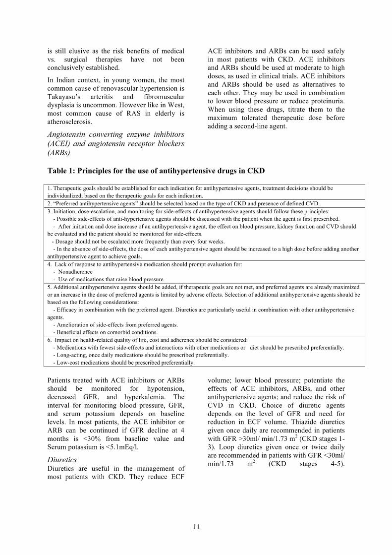
Table 1: Principles for the use of antihypertensive drugs in CKD 1. Therapeutic goals should be established for each indication for antihypertensive agents, treatment decisions should be individualized, based on the therapeutic goals for each indication. 2. “Preferred antihypertensive agents” should be selected based on the type of CKD and presence of defined CVD. 3. Initiation, dose-escalation, and monitoring for side-effects of antihypertensive agents should follow these principles: - Possible side-effects of anti-hypertensive agents should be discussed with the patient when the agent is first prescribed. - After initiation and dose increase of an antihypertensive agent, the effect on blood pressure, kidney function and CVD should be evaluated and the patient should be monitored for side-effects. - Dosage should not be escalated more frequently than every four weeks. - In the absence of side-effects, the dose of each antihypertensive agent should be increased to a high dose before adding another antihypertensive agent to achieve goals. 4. Lack of response to antihypertensive medication should prompt evaluation for: - Nonadherence - Use of medications that raise blood pressure 5. Additional antihypertensive agents should be added, if therapeutic goals are not met, and preferred agents are already maximized or an increase in the dose of preferred agents is limited by adverse effects. Selection of additional antihypertensive agents should be based on the following considerations: - Efficacy in combination with the preferred agent. Diuretics are particularly useful in combination with other antihypertensive agents. - Amelioration of side-effects from preferred agents. - Beneficial effects on comorbid conditions. 6. Impact on health-related quality of life, cost and adherence should be considered: - Medications with fewest side-effects and interactions with other medications or diet should be prescribed preferentially. - Long-acting, once daily medications should be prescribed preferentially. - Low-cost medications should be prescribed preferentially. Patients treated with ACE inhibitors or ARBs should be monitored for hypotension, decreased GFR, and hyperkalemia. The interval for monitoring blood pressure, GFR, and serum potassium depends on baseline levels. In most patients, the ACE inhibitor or ARB can be continued if GFR decline at 4 months is <30% from baseline value and Serum potassium is <5.1mEq/l.
Diuretics Diuretics are useful in the management of most patients with CKD. They reduce ECF
volume; lower blood pressure; potentiate the effects of ACE inhibitors, ARBs, and other antihypertensive agents; and reduce the risk of CVD in CKD. Choice of diuretic agents depends on the level of GFR and need for reduction in ECF volume. Thiazide diuretics given once daily are recommended in patients with GFR >30ml/ min/1.73 m2 (CKD stages 1-3). Loop diuretics given once or twice daily are recommended in patients with GFR <30ml/ min/1.73 m2 (CKD stages 4-5).
12
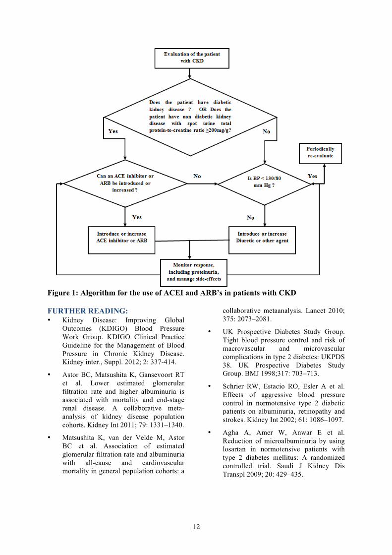
Figure 1: Algorithm for the use of ACEI and ARB’s in patients with CKD FURTHER READING: • Kidney Disease: Improving Global
Outcomes (KDIGO) Blood Pressure Work Group. KDIGO Clinical Practice Guideline for the Management of Blood Pressure in Chronic Kidney Disease. Kidney inter., Suppl. 2012; 2: 337-414.
• Astor BC, Matsushita K, Gansevoort RT et al. Lower estimated glomerular filtration rate and higher albuminuria is associated with mortality and end-stage renal disease. A collaborative meta-analysis of kidney disease population cohorts. Kidney Int 2011; 79: 1331–1340.
• Matsushita K, van der Velde M, Astor BC et al. Association of estimated glomerular filtration rate and albuminuria with all-cause and cardiovascular mortality in general population cohorts: a
collaborative metaanalysis. Lancet 2010; 375: 2073–2081.
• UK Prospective Diabetes Study Group. Tight blood pressure control and risk of macrovascular and microvascular complications in type 2 diabetes: UKPDS 38. UK Prospective Diabetes Study Group. BMJ 1998;317: 703–713.
• Schrier RW, Estacio RO, Esler A et al. Effects of aggressive blood pressure control in normotensive type 2 diabetic patients on albuminuria, retinopathy and strokes. Kidney Int 2002; 61: 1086–1097.
• Agha A, Amer W, Anwar E et al. Reduction of microalbuminuria by using losartan in normotensive patients with type 2 diabetes mellitus: A randomized controlled trial. Saudi J Kidney Dis Transpl 2009; 20: 429–435.
13
MANAGEMENT OF CARDIOVASCULAR DISEASE IN CKD Heart and kidneys are closely linked with each other through a complex array of interactions in the hemodynamic and regulatory functions of the body in maintaining homeostasis of the milieu interior. The term “cardio-renal syndrome” has been coined to emphasize the close interplay of these two organ systems in various disease states. Cardiovascular disease (CVD) includes coronary artery disease, valvular heart disease, cardiomyopathy, cardiac arrhythmias, cerebrovascular disease and peripheral vascular disease.
1 EVALUATION OF CVD IN CKD 1.1 We recommend that all CKD patients
should have assessment for cardiovascular disease.
• They should be screened for traditional and CKD related CVD risk factors • A complete clinical examination should be followed by the laboratory tests that include: • Chest X ray, ECG and Echocardiogram • Urine for albuminuria • Calculation of creatinine clearance or estimated GFR
CKD is a risk factor for CVD. This increased risk is due to the fact that these patients, apart from having traditional CVD risk factors have CKD related CVD risk factors. Some of the risk factors are shared by CKD and CVD.
2. CORONARY ARTERY DISEASE (CAD) 2.1 We recommend that all CKD patients
should be evaluated for CAD which includes;
• A detailed history and clinical examination • Low threshold for aggressive evaluation and
hospitalization for patients of CKD presenting with chest pain
• Troponin I is the preferred biomarker in acute coronary syndrome.
• ECG, Echocardiogram and stress test should be performed and results should be interpreted understanding their values and limitations.
• Diagnostic coronary angiogram should be with iso-osmolar or low osmolar contrast agents and the contrast amount should be limited to 30 ml or less, ensuring adequate hydration pre and post procedure.
There is no substitute for detailed history and clinical examination in the evaluation of CKD patient suspected to have CAD. The ischemic event (ischemia or myocardial infarction) could be silent in patients with CKD and diabetes. Patients with CKD presenting with chest pain have a 40% cardiac event rate at 30 days.
ECG in CKD patients may have longer PR interval and QT interval, partly due to concomitant medication. Increasing QRS interval and QT interval predict higher risk for heart failure and all cause mortality respectively. ST changes on ECG may be due to left ventricular hypertrophy or electrolyte abnormalities and thus are not reliable indicators of myocardial ischemia. Exercise ECG is not generally done because of poor exercise tolerance in these patients and baseline ST changes in these patients with CKD.
Stress nuclear or stress echocardiographic studies also have lower accuracy for detection of ischemia in these patients. Dobutamine stress test is reported to carry a 2 to 4% risk of transient atrial fibrillation.
Coronary calcium score by CT scan is not recommended in this population because the presence of vascular medial calcification in them interferes with the assessment of CAD.
2.2 Management
2.2.1 Pharmacotherapy • The standard therapeutic agents, that is, beta
blockers, ACE-Inhibitors, ARB’s, aldosterone antagonists and statins have a favourable risk benefit ratio
• The anticoagulant dosage has to be adjusted based on creatinine clearance
2.2.2 Coronary interventions • Avoid internal jugular vein and
radial/brachial arteries for vascular access • In UA/NSTEMI, early invasive therapy is
preferable in patients of CKD stage II and III
• In STEMI, primary PCI is the treatment of choice
• Drug eluting stents are preferable to bare metal stents

14
• CABG is preferred over PCI in patients of CKD with multivessel/left main disease, particularly in diabetic patients.
The risk benefit ratio of using drugs in patients with CAD and CKD has been evaluated. Beta blockers, ACE-inhibitors, ARB’s , aldosterone antagonists and statins have all been found to have a favourable risk benefit ratio. Many drugs, especially anticoagulants, require dose adjustment in CKD.
Special precautions should be taken to prevent contrast induced acute kidney injury, paying special attention to the optimal medical management, contrast agent and hydration. If the procedure is staged; there should be an interval of 10 days between the two procedures. However, the risk of athero-embolic renal injury increases with multiple interventions. CKD patients presenting with STEMI should receive acute reperfusion therapy as does a non- CKD patient. Primary PCI is the preferred reperfusion strategy if available within the time frame specified, because of increased bleeding risk associated with fibrinolytic therapy.
Drug eluting stents are preferred over bare metal stents since the risk of restenosis is lower with DES. However, the potential benefits of DES should be weighed against the risk of prolonged dual antiplatelet therapy, occurrence of late stent thrombosis and possibility of subsequent surgical procedures.
In patients presenting with unstable angina/non ST elevation MI, an early invasive strategy is reasonable in patients with stage 2 and stage 3 CKD. There are no adequate data for patients in stage 4/5 CKD.
There have been no randomized studies of PCI vs. CABG in this population. Clinical judgement and patient characteristics should guide the therapy. Generally, for patients with three vessel disease and/or left main disease, CABG is the treatment of choice. European guidelines recommend, in patients with mild to moderate CKD, CABG rather than PCI, when the extent of CAD justifies surgical approach and the patient’s risk profile is acceptable and the life expectancy is reasonable. Observational studies have shown better survival rates with CABG rather than PCI, and this survival advantage is probably attributable to the use of internal mammary artery grafts.
CABG patients also have reduced rates of repeat coronary revascularization.
FURTHER READING • Ronco C, Haapio M, House AA,
Anavekar N, Bellomo R. Cardiorenal Syndrome. J. Am. Coll. Cardiol.2008; 52: 1527 - 1539.
• Schmidt A, Stefenelli T. Schuster E et al. Informational contribution of noninvasive screening tests for coronary artery disease in patients on chronic renal replacement therapy. AM J Kidney Dis 2001; 37:56-63
• Karthikeyan V, Ananthasubramaniam K. Coronary risk assessment and management options in Chronic kidney disease patients prior to kidney transplantation. Curr Cardiol Rev 2009; 5:177-186.
• Taylor AJ, Cerqueira M, Hodgson JM et al. AACF/SCCT/ACR/AHA/ASE/ASNC/NAS CI/SCAI/SCMR 2010 Appropriate use Criteria for Cardiac Computed Tomography: Circulation 2010; 122:e525-e555.
• McCullough PA, Nowak RM, Foreback C etal. Performance of multiple cardiac biomarkers measured in the emergency department in patients with chronic kidney disease and chest pain. Acad Emerg Med 2002;9;1389.
• McCullough PA. Evaluation and treatment of coronary artery disease in patients with end stage renal disease. Kidney Int Suppl 2005;95;551.
• W Hamm et al. ESC guidelines for the management of acute coronary syndrome in patients presenting without persistent ST segment elevation. Eur Heart J 2011. doi:10.1093.
• Wright et al. 2011 ACCF/AHA Focused update of the guidelines for the management of patients with UA/NSTEMI JACC 2011;1920-59.
• Wijnsetal W. Guidelines on myocardial revascularisation. Eu Heart J 2010;31;2501-55

15
ASSESSMENT AND PREVENTION OF CONTRAST INDUCED AKI IN CKD
Contrast media (CM) induced nephropathy (CIN) is the third highest cause of hospital acquired acute renal failure. Permanent impairment of renal function requiring dialysis can occur in up to 10% of patients with pre existing renal failure, or in <1% of all patients who undergo per cutaneous coronary intervention using CM. CIN is defined as an absolute increase in serum creatinine level of ≥0.5mg/dl or as a relative increase of ≥25% from baseline within 3 days after CM exposure.
1. ASSESSMENT OF THE POPULATIONAT AT RISK FOR CONTRAST INDUCED – AKI (CI-AKI)
1.1 We suggest to assess the risk for CI-AKI and, in particular, screen for pre-existing impairment of kidney function in all patients who are considered for a procedure that requires intravascular (i.v. or i.a.) administration of iodinated contrast medium.
The risk of developing CI-AKI increases with worsening baseline renal function and can be as high as 50% if the baseline plasma creatinine is greater than 4 to 5 mg/dL particularly in patients with diabetic nephropathy .
1.2 We suggest to consider alternative imaging methods in patients at increased risk for CI-AKI.
Multiple studies have found that MR contrast agents when used in small doses for MR examinations have little or no nephrotoxicity. Furthermore, gadolinium-based imaging should not be performed, if at all possible, in patients with an estimated glomerular filtration rate less than 30 mL/min because of the risk of nephrogenic systemic fibrosis.
2. NONPHARMACOLOGICAL PREVENTION STRATEGIES OF CI-AKI
2.1 We suggest to use the lowest possible dose of contrast medium in patients at risk for CI-AKI and avoid repetitive, closely spaced studies (e.g., <48 hours apart).
2.2 We recommend using either iso-osmolar or low-osmolar iodinated contrast media, rather than high-osmolar iodinated contrast media in patients at increased risk of CI-AKI.
Iodinated radiocontrast agents are either ionic or non-ionic and, at the concentrations required for arteriography or computed tomography, are of variable osmolality and the same has been depicted in Table 1.
Volume depletion, nonsteroidal antiinflammatory drugs and other nephrotoxic drugs should be avoided in CKD patients undergoing contrast studies.
Table 1: Physicochemical properties of contrast media Osmolality Osmolality(mOsm/kg H2O)
High- osmolar(2100)
Low-osmolar(577) Low-osmolar (610-915)
Iso-osmolar (290)
Ionicity Ionic Ionic Non-ionic Non-ionic No. of benzene rings Monomer Dimer Monomer Dimer Viscosity at 370C (cP)
8.4 9.5 7.8-11.2 11.1
Example Diatrizoate Ioxaglate Iohexol, Iopamidol Ioversol, Iopromide
Iodixanol
3. PHARMACOLOGICAL
PREVENTION STRATEGIES OF CI-AKI
3.1 We recommend i.v. volume expansion with either isotonic sodium chloride or sodium bicarbonate solutions, in patients at increased risk for CI-AKI.

16
If there are no contraindications to volume expansion, isotonic intravenous fluids should be used prior to and continued for several hours after contrast administration. Isotonic bicarbonate is preferred over isotonic saline. A suggested regimen is a bolus of 3 mL/kg of isotonic bicarbonate for one hour prior to the procedure, and continued at a rate of 1 mL/kg per hour for six hours after the procedure. This solution can be prepared by adding 154 ml of 8.4% sodium bicarbonate (i.e., 1 mmol/ml) to 846 mL of 5% glucose solution, resulting in a
final sodium and bicarbonate concentration of 154 mmol/l each. If isotonic saline is chosen, a suggested regimen is: isotonic saline at a rate of 1 mL/kg per hour, begun at least two and preferably 6 to 12 hours prior to the procedure, and continuing for 6 to 12 hours after contrast administration. The duration of administration of fluid should be directly proportional to the degree of renal impairment (e.g., should be longer for individuals with more severe renal impairment).
Fig. 1. An algorithm for management of patients undergoing contrast investigations. 3.2 We recommend not using oral fluids
alone in patients at increased risk of CI-AKI.
3.3 We suggest using oral N-acetyl cysteine (NAC), together with i.v. isotonic crystalloids, in patients at increased risk of CI-AKI.

17
Despite conflicting data, acetylcysteine may be administered the day before and the day of the procedure, based upon its potential for benefits and low toxicity and cost. If acetylcysteine is administered, give 1200 mg orally twice daily rather than 600 mg twice daily the day before and the day of the procedure. Based upon the lack of convincing evidence of benefit and the potential risk of anaphylactoid reactions, intravenous acetylcysteine for the prevention of contrast nephropathy should be avoided.
3.4 We suggest not using theophylline or fenoldapam to prevent CI-AKI.
Many drugs have been tried to alleviate contrast induced nephropathy like dopamine, fenoldapam, theophylline etc, but none have any substantial benefit.
3.5 We suggest, in patients with CKD stage 5, using prophylactic intermittent hemodialysis (IHD) or hemofiltration (HF) may be considered.
Prevention remains the mainstay in the management of contrast induced nephropathy as no interventions are beneficial after it sets in. Shown here is a simple algorithm for prevention and risk reduction for contrast induced nephropathy.
FURTHER READING • Tepel M, Aspelin P, Lameire N. Contrast
induced nephropathy: a clinical and evidence-based approach. Circulation 2006; 113:1799–1806.
• Parfrey PS, Griffiths SM, Barrett BJ, et al. Contrast material-induced renal failure in patients with diabetes mellitus, renal insufficiency, or both. A prospective controlled study. N Engl J Med 1989; 320:143.
• Reed M, Meier P, Tamhane UU, et al. The relative renal safety of iodixanol compared with low-osmolar contrast media: a metaanalysis of randomized controlled trials. JACC Cardiovasc Interv 2009; 2:645.
• Kushner FG, Hand M, Smith SC Jr, et al. 2009 focused updates: ACC/AHA guidelines for the management of patients with ST elevation myocardial infarction (updating the 2004 guideline and 2007 focused update) and ACC/AHA/SCAI guidelines on percutaneous coronary intervention (updating the 2005 guideline and 2007 focused update) a report of the American College of Cardiology Foundation/American Heart Association Task Force on Practice Guidelines. J Am Coll Cardiol 2009; 54:2205.
• Rich MW, Crecelius CA. Incidence, risk factors, and clinical course of acute renal insufficiency after cardiac catheterization inpatients 70 years of age or older. A prospective study. Arch Intern Med 1990; 150: 1237–12
• Mehran R et al. A simple risk score for prediction of contrast induced nephropathy after percutaneous coronary intervention: development and initial validation. J Am Coll Cardiol 2004; 44:1393–1399.
• Taylor AJ, Hotchkiss D, Morse RW, et al.: Preparation for Angiography in Renal Dysfunction: a randomized trial of inpatient vs. outpatient hydration protocols for cardiac catheterization in mild o-moderate renal dysfunction. Chest 1998; 114: 1570–1574.
18
MANAGEMENT OF DIABETES IN CHRONIC KIDNEY DISEASE
Diabetic nephropathy is the leading cause of chronic kidney disease (CKD) worldwide. It is also one of the most significant long-term complications in terms of morbidity and mortality for individual patients with diabetes. Management of CKD in Indian context is challenging clinically and economically. The risk for cardiovascular disease (CVD) was 3 fold higher in South Indian nephropathic subjects when compared with their non-nephropathic counterparts. Thus, in type 2 diabetes, many patients may not reach end stage renal disease due to premature death from CVD.
1.0 SCREENING FOR DIABETIC NEPHROPATHY
1.1 We recommend that screening for diabetic nephropathy must be carried out, especially with type 2 diabetes.
1.2 We recommend that screening for microalbuminuria (MAU) is the test of choice to detect early renal injury.
This can be done by the following methods:
•Radio-immunoassay, radio-mmunodiffusion, immunoturbidimetry, laser Immuno nephlometry, enzyme-linked immunosorbant assay and dipstick test
• Of all the above methods immunoturbidimetry has the fastest turnaround time and is usually a method of choice in most laboratories
The albumin / creatinine ratio (ACR): The ACR can be determined from a random or preferably early morning urine sample. This is often the earliest test in the setting of primary care and provides a practical screening method less prone to patient’s error than timed collection.
Urinary protein/creatinine ratio It is impractical to estimate MAU in all the centres of developing countries, since its
estimation is expensive and requires sophisticated instruments. The protein excretion was assessed as the protein to creatinine ratio in random urine sample of 410 type 2 diabetic patients (M: F 264:146; mean age 55.6+9.5 years) who had regular follow-up for 6 years. During the follow-up, nephropathy (defined as persistent proteinuria of >500 mg/day with diabetic retinopathy) developed in 6.7% of those who had normal protein excretion at baseline (<100 mg/day) and in 43.4% of the mildly proteinuric subjects (100-500 mg/day) (χ2 = 41.6; P<0.001). Hence the urinary protein to creatinine ratio in a random urine sample was found to be a useful test to predict the risk of overt proteinuria.
1.3 We suggest the following timing for diabetic nephropathy screening:
• Type 1 diabetes: onset of puberty or after 5 years of disease duration
• Type 2 diabetes: begin at diagnosis
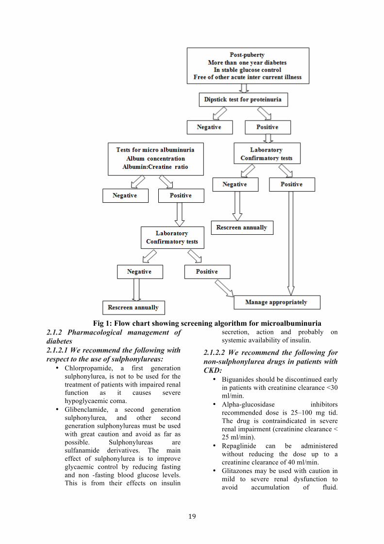
In type 1 diabetes MAU rarely occurs within 5 to 10 years of duration or before puberty. Hence screening should begin with onset of puberty or after 5 years of disease duration. In type 2 diabetes, the precise onset of disease cannot be dated. Hence screening should begin at diagnosis. In a study conducted in 205 subjects, 12.2 % of patients had persistent microalbuminuria during diagnosis of diabetes itself. Once MAU has been identified the patient should have measurements every 3 to 6 months (Fig.1).
2.0 MANAGEMENT OF DIABETES IN CKD
2.1 We recommend that the management protocol involved the following major entities:
• Management of diabetes • Management of diabetic complication
19
Fig 1: Flow chart showing screening algorithm for microalbuminuria 2.1.2 Pharmacological management of diabetes 2.1.2.1 We recommend the following with respect to the use of sulphonylureas:
• Chlorpropamide, a first generation sulphonylurea, is not to be used for the treatment of patients with impaired renal function as it causes severe hypoglycaemic coma.
• Glibenclamide, a second generation sulphonylurea, and other second generation sulphonylureas must be used with great caution and avoid as far as possible. Sulphonylureas are sulfanamide derivatives. The main effect of sulphonylurea is to improve glycaemic control by reducing fasting and non -fasting blood glucose levels. This is from their effects on insulin
secretion, action and probably on systemic availability of insulin.
2.1.2.2 We recommend the following for non-sulphonylurea drugs in patients with CKD:
• Biguanides should be discontinued early in patients with creatinine clearance <30 ml/min.
• Alpha-glucosidase inhibitors recommended dose is 25–100 mg tid. The drug is contraindicated in severe renal impairment (creatinine clearance < 25 ml/min).
• Repaglinide can be administered without reducing the dose up to a creatinine clearance of 40 ml/min.
• Glitazones may be used with caution in mild to severe renal dysfunction to avoid accumulation of fluid.
20
Concomitant administration of rosiglitazone or pioglitazone with
metformin is contraindicated.
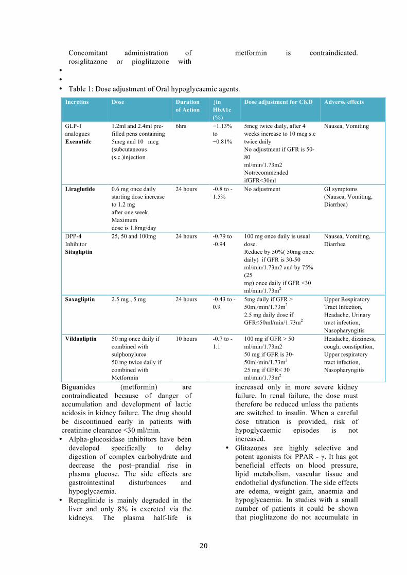
• • • Table 1: Dose adjustment of Oral hypoglycaemic agents.
Biguanides (metformin) are contraindicated because of danger of accumulation and development of lactic acidosis in kidney failure. The drug should be discontinued early in patients with creatinine clearance <30 ml/min. • Alpha-glucosidase inhibitors have been
developed specifically to delay digestion of complex carbohydrate and decrease the post–prandial rise in plasma glucose. The side effects are gastrointestinal disturbances and hypoglycaemia.
• Repaglinide is mainly degraded in the liver and only 8% is excreted via the kidneys. The plasma half-life is
increased only in more severe kidney failure. In renal failure, the dose must therefore be reduced unless the patients are switched to insulin. When a careful dose titration is provided, risk of hypoglycaemic episodes is not increased.
• Glitazones are highly selective and potent agonists for PPAR - γ. It has got beneficial effects on blood pressure, lipid metabolism, vascular tissue and endothelial dysfunction. The side effects are edema, weight gain, anaemia and hypoglycaemia. In studies with a small number of patients it could be shown that pioglitazone do not accumulate in
Incretins Dose Duration of Action
↓in HbA1c (%)
Dose adjustment for CKD Adverse effects
GLP-1 analogues Exenatide
1.2ml and 2.4ml pre-filled pens containing 5mcg and 10 mcg (subcutaneous (s.c.)injection
6hrs −1.13% to −0.81%
5mcg twice daily, after 4 weeks increase to 10 mcg s.c twice daily No adjustment if GFR is 50-80 ml/min/1.73m2 Notrecommended ifGFR<30ml
Nausea, Vomiting
Liraglutide 0.6 mg once daily starting dose increase to 1.2 mg after one week. Maximum dose is 1.8mg/day
24 hours -0.8 to -1.5%
No adjustment GI symptoms (Nausea, Vomiting, Diarrhea)
DPP-4 Inhibitor Sitagliptin
25, 50 and 100mg 24 hours -0.79 to -0.94
100 mg once daily is usual dose. Reduce by 50%( 50mg once daily) if GFR is 30-50 ml/min/1.73m2 and by 75% (25 mg) once daily if GFR <30 ml/min/1.73m2
Nausea, Vomiting, Diarrhea
Saxagliptin 2.5 mg , 5 mg 24 hours -0.43 to -0.9
5mg daily if GFR > 50ml/min/1.73m2 2.5 mg daily dose if GFR≤50ml/min/1.73m2
Upper Respiratory Tract Infection, Headache, Urinary tract infection, Nasopharyngitis
Vildagliptin 50 mg once daily if combined with sulphonylurea 50 mg twice daily if combined with Metformin
10 hours -0.7 to -1.1
100 mg if GFR > 50 ml/min/1.73m2 50 mg if GFR is 30- 50ml/min/1.73m2 25 mg if GFR< 30 ml/min/1.73m2
Headache, dizziness, cough, constipation, Upper respiratory tract infection, Nasopharyngitis
21
severe kidney disease (creatinine clearance <30 ml/min). Concomitant administration of rosiglitazone or pioglitazone with metformin is contraindicated.
• Incretins
GLP-1 analogues: The small intestine secretes glucagon-like peptide-1 (GLP-1) as well as glucose-dependent insulinotropic polypeptide (GIP) in response to food intake. These hormones stimulate insulin secretion, insulin
gene expression and pancreatic betacell growth.
Dipeptidyl peptidase-4 Inhibitors decrease the breakdown of the incretin hormone (GLP-1). Thus stimulates the secretion of insulin in a glucose dependent manner minimizing possible hypoglycemia. In general, short-acting OHAs like glipizide, repaglinide, gliclazide can be used in treating diabetes in chronic renal failure. The dose adjustment details are provided in Table 1.
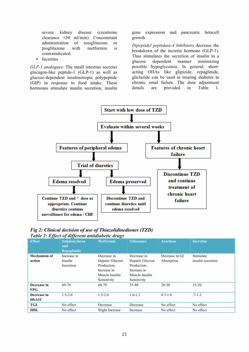
Fig 2: Clinical decision of use of Thiazolidinediones (TZD) Table 2: Effect of different antidiabetic drugs Effect Sulphonylurea
and Repaglinide
Metformin Glitazones Acarbose Incretins
Mechanism of action
Increase in Insulin Secretion
Decrease in Hepatic Glucose Production, Increase in Muscle Insulin Sensitivity
Decrease in Hepatic Glucose Production, Increase in Muscle Insulin Sensitivity
Decrease in GI Absorption
Stimulate insulin secretion
Decrease in FPG
60-70 60-70 35-40 20-30 15-30
Decrease in HbA1C
1.5-2.0 1.5-2.0 1.0-1.2 0.7-1.0 .7-1.2
TGL No effect Decrease Decrease No effect No effect HDL No effect Slight Increase Increase No effect No effect

22
2.1.2.3 We recommend the insulin be used as treatment modality of choice for diabetes when creatinine clearance < 60 ml/min. The dose of insulin must be reduced and is appropriate to use short – acting insulins.
In individuals with healthy metabolism, the liver degrades about 80% of the insulin and the kidneys about 20%. In insulin – dependent diabetic subjects, the liver and kidneys are exposed to about the same concentration of insulin owing to peripheral insulin administration and thus each degrades about half of the hormone. In kidney failure (creatinine clearance < 60 ml/min), there is
protracted action of insulin due to reduced renal degradation, which must be taken into consideration in treatment. As a rule, the dose of insulin must be reduced, owing to the better control; it is appropriate to use short – acting insulins. In general, patients with kidney failure or kidney replacement therapy should, if possible, be put on intensified insulin treatment. The American College of Physicians recommended a 25% decrease in doses of insulin, if GFR 50 - 10ml/min/1.73m2 and a 50% decrease when GFR decreased to <10ml/min/1.73m2 . The types of insulins and their onset of action and duration are listed in table [4].
Table 3: Adverse effects of different oral hypoglycemic agents Adverse effects Sulphonylureas Metformin Glitazones Acarbose DPP-4
Inhibitors Weight gain + - +/++ - - Hypoglycaemia +/++ - - - - Hypersensitivity reaction
+ - + - -
Drug interaction + - - - - Lactic Acidosis - + - - - Gastrointestinal disturbances or hepatic reaction
+ ++ ++ ++ ++
CNS disturbances (Headache, dizziness)
- - - - ++
Nasopharyngitis, Upper respiratory tract infection
- - - - +
Edema - - +/++ - -
Table 4: Types of insulin preparations Rapid-acting Vial and cartridge
Aspart (NovoRapid) Lispro(Humalog®)
Start <15 min 2-3 hrs
Short-acting (Regular) Vial and cartridge
Humulin®R Starts 30-60 min ; peak 4 hr
8 hrs
Intermediate Vial and cartridge
Neutral Protamine Hagedron (NPH) Humulin®N
Starts 1.5 hrs ; peak 7 hrs 6-12hrs
Prolonged action Humulin ®U vial only Lantus (Glargine) vial only Levemir(Detemir) Cartridge
Starts 3-4 hr ; peak less 18-24hrs
Insulin Premixes
• Regular + intermediate - Onset 30 -45 min; duration 6-12 hrs • Novolin® 10/90, 20/80, 30/70, 40/60, 50/50 • Humulin® 30/70, 20/80
23
Analogue Pre-Mix • Humalog® 25/75 (insulin lispro protamine suspension) • NovoMix 30* (protaminated insulin aspart)
2.2.2.3.1 We suggest that intraperitoneal insulin in peritoneal dialysis may be administered with caution if the clinical benefits overweigh the risk in a particular patient.
Insulin requirements are usually higher than the previous subcutaneous dose. However high rates of peritonitis have limited its use.
2.3 Management of diabetic complications
2.3.1 Diabetic Retinopathy
• We recommend that diabetic retinopathy be prevented or the progression halted by intensive glucose lowering strategies
• We recommend patients with diabetic retinopathy to be referred to an ophthalmologist for sight saving strategies
Diabetic retinopathy is the leading cause of new blindness in the general population 20 – 74 years of age. Diabetic patients are 11 times more likely to become blind than non-diabetic subjects; when retinopathy is present, this risk increases to 29 fold. In a study from south India, it was shown that 6.7% of newly diagnosed type 2 diabetic subjects had background diabetic retinopathy. About half of all diabetic patients have diabetic retinopathy at any one time. Of those patients with diabetic retinopathy, 5 – 8 % has the proliferative form. Among proteinuric patients, the prevalence of diabetic retinopathy was found to be high (60%). It was also noted that a large percentage of those who developed proteinuria during follow up developed retinopathy also
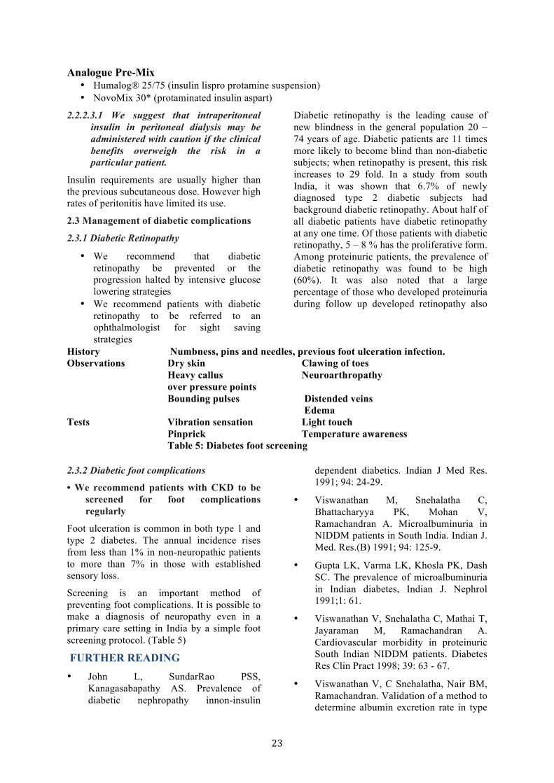
History Numbness, pins and needles, previous foot ulceration infection. Observations Dry skin Clawing of toes
Heavy callus Neuroarthropathy over pressure points Bounding pulses Distended veins
Edema Tests Vibration sensation Light touch
Pinprick Temperature awareness Table 5: Diabetes foot screening
2.3.2 Diabetic foot complications
• We recommend patients with CKD to be screened for foot complications regularly
Foot ulceration is common in both type 1 and type 2 diabetes. The annual incidence rises from less than 1% in non-neuropathic patients to more than 7% in those with established sensory loss.
Screening is an important method of preventing foot complications. It is possible to make a diagnosis of neuropathy even in a primary care setting in India by a simple foot screening protocol. (Table 5)
FURTHER READING • John L, SundarRao PSS,
Kanagasabapathy AS. Prevalence of diabetic nephropathy innon-insulin
dependent diabetics. Indian J Med Res. 1991; 94: 24-29.
• Viswanathan M, Snehalatha C, Bhattacharyya PK, Mohan V, Ramachandran A. Microalbuminuria in NIDDM patients in South India. Indian J. Med. Res.(B) 1991; 94: 125-9.
• Gupta LK, Varma LK, Khosla PK, Dash SC. The prevalence of microalbuminuria in Indian diabetes, Indian J. Nephrol 1991;1: 61.
• Viswanathan V, Snehalatha C, Mathai T, Jayaraman M, Ramachandran A. Cardiovascular morbidity in proteinuric South Indian NIDDM patients. Diabetes Res Clin Pract 1998; 39: 63 - 67.
• Viswanathan V, C Snehalatha, Nair BM, Ramachandran. Validation of a method to determine albumin excretion rate in type

24
2 diabetes mellitus. The Indian Journal of Nephrology, Vol. 13, 2003, Pg: 85-88.
• Viswanathan V, Chamukuttan S, Kuniyil S, Ramachandran A. Evaluation of a simple, random urine test for prospective analyses of proteinuria in Type 2 diabetes: a six year follow-up study. Diabetes Research and Clinical Practice. 2000; 49: 143-147.
• Vijay V, Seena R, Lalitha S, Snehalatha C, Muthu J, Ramachandran A. Significance of Microalbuminuria at diagnosis of type 2 diabetes. Diabetes Bulletin, International Journal of Diabetes in Developing countries 1998; 18: 5-6
• Vijay V, Snehalatha C, Shina K, Lalitha S, Ramachandran A. Familial aggregation
of diabetic kidney disease in Type 2 diabetes in South India. Diabetes Research and Clinical Practice, 1999; 43: 167-171.
• Ramachandran A, Snehalatha C, Vijay V, Viswanthan M. Diabetic retinopathy at the time of diagnosis of NIDDM in south Indian subjects. DRCP 32: 1996; 111 – 114.
• Vijay V, Snehalatha C, Terin M. Ramachandran A. Socio - cultural Practices that may affect the development of the diabetic foot. IDF Bulletin 1997 : 42: 10-12
• The Action to Control Cardiovascular Risk in Diabetes Study Group. Effects of intensive glucose lowering in type 2 diabetes. N Engl J Med 2008;358:2545-5
25
DYSLIPIDEMIAS IN CHRONIC KIDNEY DISEASE A large number of studies have demonstrated the benefits of lipid lowering treatment in the elderly and middle-aged men and women, smokers and non-smokers, hypertensive and non hypertensive, with higher or lower LDL levels, higher or lower cholesterol levels, higher and lower triglycerides levels, higher and lower HDL and diabetics and non-diabetics. Hence the National Kidney Foundation Disease Outcomes Quality Initiative (NKF KDOQI) clinical practice guidelines for managing dyslipidemias in CKD, last published in 2003, presumed that the same generalisation would be applicable to all stages of CKD. Subsequently, no further updates to that guideline have been made. KDIGO Lipid Guidelines are under preparation and are expected to come out soon. Important changes are likely to be made in the treatment recommendations, which will reflect the result of recent studies, especially SHARP trial.
1.0 ASSESSMENT OF DYSLIPIDEMIAS IN PATIENTS WITH CKD
We recommend that all adults and adolescents with CKD should be evaluated for dyslipidemias.
• For adults and adolescents with CKD, the assessment of dyslipidemias should include: a complete fasting lipid profile with total cholesterol, LDL, HDL, and triglycerides
• For adults and adolescents with Stage 5 CKD, dyslipidemias should be evaluated upon presentation (when the patient is stable), at 2–3 months after a change in treatment or other conditions known to cause dyslipidemias; and at least annually thereafter
• For adults and adolescents with Stage 5 CKD, a complete lipid profile should be measured after an overnight fast whenever possible
• Hemodialysis patients should have lipid profiles measured either before dialysis, or on days not receiving dialysis
2.0 SECONDARY CAUSES OF DYSLIPIDEMIAS IN PATIENTS WITH CKD
2.1 We recommend that Stage 5 CKD patients with dyslipidemias should be evaluated for remediable, secondary causes.
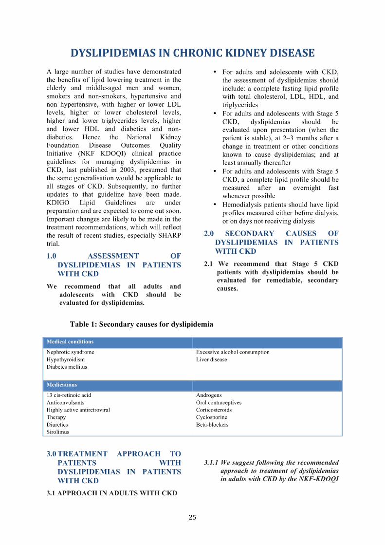
Table 1: Secondary causes for dyslipidemia
Medical conditions Nephrotic syndrome Hypothyroidism Diabetes mellitus
Excessive alcohol consumption Liver disease
Medications 13 cis-retinoic acid Anticonvulsants Highly active antiretroviral Therapy Diuretics Sirolimus
Androgens Oral contraceptives Corticosteroids Cyclosporine Beta-blockers
3.0 TREATMENT APPROACH TO
PATIENTS WITH DYSLIPIDEMIAS IN PATIENTS WITH CKD
3.1 APPROACH IN ADULTS WITH CKD
3.1.1 We suggest following the recommended
approach to treatment of dyslipidemias in adults with CKD by the NKF-KDOQI
26
guidelines and that adopted by the ATP III guidelines. (Figure 1)
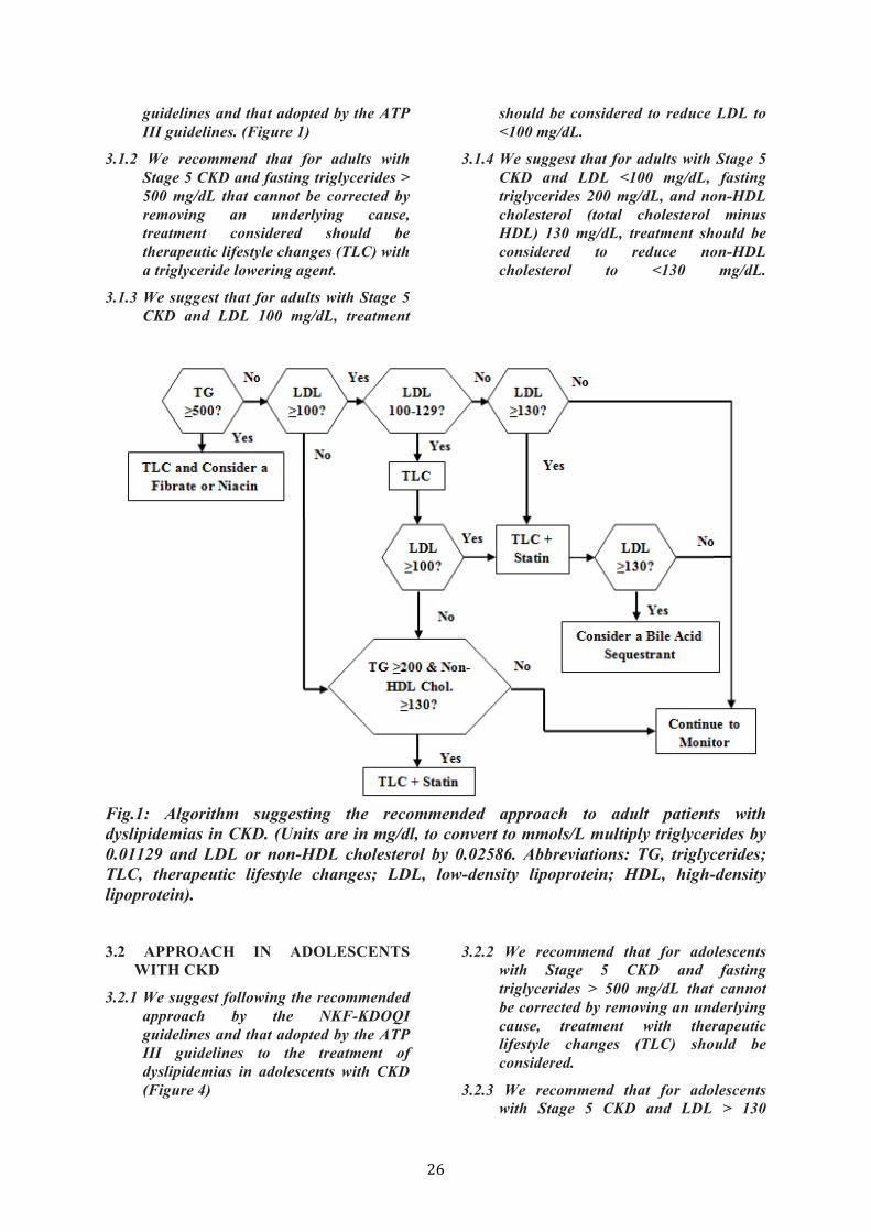
3.1.2 We recommend that for adults with Stage 5 CKD and fasting triglycerides > 500 mg/dL that cannot be corrected by removing an underlying cause, treatment considered should be therapeutic lifestyle changes (TLC) with a triglyceride lowering agent.
3.1.3 We suggest that for adults with Stage 5 CKD and LDL 100 mg/dL, treatment
should be considered to reduce LDL to <100 mg/dL.
3.1.4 We suggest that for adults with Stage 5 CKD and LDL <100 mg/dL, fasting triglycerides 200 mg/dL, and non-HDL cholesterol (total cholesterol minus HDL) 130 mg/dL, treatment should be considered to reduce non-HDL cholesterol to <130 mg/dL.
Fig.1: Algorithm suggesting the recommended approach to adult patients with dyslipidemias in CKD. (Units are in mg/dl, to convert to mmols/L multiply triglycerides by 0.01129 and LDL or non-HDL cholesterol by 0.02586. Abbreviations: TG, triglycerides; TLC, therapeutic lifestyle changes; LDL, low-density lipoprotein; HDL, high-density lipoprotein). 3.2 APPROACH IN ADOLESCENTS
WITH CKD
3.2.1 We suggest following the recommended approach by the NKF-KDOQI guidelines and that adopted by the ATP III guidelines to the treatment of dyslipidemias in adolescents with CKD (Figure 4)
3.2.2 We recommend that for adolescents with Stage 5 CKD and fasting triglycerides > 500 mg/dL that cannot be corrected by removing an underlying cause, treatment with therapeutic lifestyle changes (TLC) should be considered.
3.2.3 We recommend that for adolescents with Stage 5 CKD and LDL > 130
27
mg/dL, treatment should be considered to reduce LDL to <130 mg/dL.
3.2.4 We recommend that for adolescents with Stage 5 CKD and LDL <130 mg/dL, fasting triglycerides > 200 mg/dL, and non-HDL cholesterol (total cholesterol minus HDL) > 160 mg/dL, treatment should be considered to reduce non-HDL cholesterol to <160 mg/dL.
FURTHER READING: • Expert Panel on Detection Evaluation and
Treatment of High Blood Cholesterol in Adults. Executive Summary of the Third Report of the National Cholesterol Education Program (NCEP) Expert Panel on Detection, Evaluation, and Treatment of High Blood Cholesterol in Adults
(Adult Treatment Panel III). JAMA, 2001, 285;2486-2497.
• National Kidney Foundation. KDOQI Clinical Practice Guidelines for Managing Dyslipidemias in Chronic Kidney Disease. Am J Kidney Dis 41:S1-S92, 2003 (suppl 3)
• http://www.kdigo.org/clinical_practice_guidelines/index.php
• http://www.era-edta.org/page-8-38-0-38-erbpeuropeanrenalbestpractice.html
• http://www.renal.org/Guidelinesection/Guidelines.aspx
• http://www.cari.org.au. The CARI guidelines. Caring for Australasians with Renal Impairment
28
ANEMIA IN CHRONIC KIDNEY DISEASE DIAGNOSIS OF ANEMIA 1.1 We Suggest Diagnose anemia in adults
and children >15 years with CKD when the Hb concentration is <13.0 g/dl (<130 g/l) in males and <12.0 g/dl (<120 g/l) in females.
1.2 We suggest Diagnose anemia in children with CKD if Hb concentration is <11.0 g/dl (<110 g/l) in children 0.5–5 years, <11.5 g/dl (<115 g/l) in children 5–12 years, and <12.0 g/dl (<120 g/l) in children 12–15 years.
1.3 We suggest that no other cause other than CKD with impairment of renal function should be evident. In patients on Hemodialysis, we recommend the Hb concentration be measured from pre dialysis blood sample.
These recommended values represent the WHO definition of Anemia. Hemoglobin concentration values for anemia in children are based on US NHANES data from 1988 to 1994. Erythropoietin being costly agent should be used only after correcting iron deficiency which is common in our country. In dialysis patients Vitamin B12 and are folate deficiency to be corrected as these are water soluble vitamins & lost on dialysis.
ANEMIA INVESTIGATION 1.4.1 We suggest that following workup for
anemia be performed initially when CKD patients present with anemia
• Hb concentration • RBC indices / Peripheral smear /
Reticulocyte count • Transferrin saturation • Stool occult blood • Stool parasite test
1.4.2: We suggest that after initial work up for anemia following tests Need to be carried out based on clinical situations. A fuller work up should also include the following as indicated.
• Iron / TIBC / Ferritin • Serum B12 and red cell folate
concentrations • Differential white blood count
• Tests for haemolysis (hapatoglobin, lactate dehydrogenase)
• Serum and / or urine protein electrophoresis / immunoblotting (where available)
• Bone marrow examination in selected cases
• Assessment of occult gastrointestinal blood loss
• Intact PTH • Chronic Infections • Serum Aluminum • Patients on dialysis - adequacy of
dialysis to be assessed
Anemia of CKD patients is of varied etiology. Work up would be based on the initial clinical evaluation or lab investigation, particularly if there is clinical suspicion of haemolysis, occult blood loss and deficiency of folic acid or vitamin B12. Adequacy of dialysis too plays an important role and to be assessed in our patients. The anemia of CKD is similar to anemia of chronic inflammatory disease and erythropoietin levels are not routinely used in distinguishing Epo deficiency in a setting of CKD and the measurement of Epo level is not recommended.
FREQUENCY OF TESTING 1.5.1 We suggest, For CKD patients without
anemia, measure Hb concentration
• when clinically indicated and : at least annually in patients with CKD 3
• at least twice per year in patients with CKD 4–5ND
• at least every 3 months in patients with CKD 5HD and CKD 5PD
1.5.2 We suggest for CKD patients with anemia not being treated with an ESA, measure Hb concentration
• When clinically indicated • at least every 3 months in patients with
CKD 3–5ND and CKD 5PD • at least monthly in patients with CKD
5HD
There is minimal data about natural history of patients with CKD. The recommendation that CKD patients with anemia be evaluated periodically is based on the observation, that

29
there is a gradual decline in Hb overtime in patients with CKD when ESA is not used. The frequency of Hb monitoring however depends upon the stage of CKD, the Hb level and the rate of decline of Hb level. More frequent monitoring is required for patients on CKD 5 HD, and patients of CKD 5 PD especially those not receiving ESA. The basis of recommendation of frequency of monitoring in children is based on chronic kidney disease in children prospective cohort study of North America (CKiD), and the frequency is almost similar to adult.
ESA INITIATION 2.1.1 We recommend, address all
correctable causes of anemia (including iron deficiency and inflammatory states) prior to initiation of ESA therapy.
2.1.2 In initiating and maintaining ESA therapy, we recommend balancing the potential benefits of reducing blood transfusions and anaemia-related symptoms against the risks of harm in individual patients (e.g., stroke, vascular access loss, hypertension), Based on patient symptoms and overall clinical goals including avoidance of transfusion and improvement in anaemia-related symptoms, and after exclusion of active infection and other causes of ESA hyporesponsiveness.
2.1.3 We recommend using ESA therapy with great caution, if at all, in CKD patients with active malignancy—in particular when cure is the anticipated outcome, a history of stroke, or a history of malignancy.
2.1.4 For adult CKD ND patients with Hb concentration =>10.0 g/dl (=>100 g/l), we suggest that ESA therapy not be initiated.
2.1.5 For adult CKD ND patients with Hb concentration less than 10.0 g/dl (<100 g/l) we suggest that the decision whether to initiate ESA therapy be individualized based on the rate of fall of Hb concentration, prior response to iron therapy, the risk of needing a transfusion, the risks related to ESA therapy and the presence of symptoms attributable to anaemia.
2.1.6 For adult CKD 5D patients, we suggest that ESA therapy be used to avoid having the Hb concentration fall below 9.0 g/dl (90 g/l) by starting ESA therapy when the hemoglobin is between 9.0–10.0 g/dl (90–100 g/l).
2.1.7 We Suggest that Individualization of therapy is reasonable as some patients may have improvements in quality of life at higher Hb concentration and ESA therapy may be started above 10.0 g/dl (100 g/l).
ESA MAINTENANCE THERAPY 2.2.1 In general, we suggest that ESAs not
to be used to maintain Hb concentration above 11.5 g/dl (115 g/l) in adult patients with CKD.
2.2.2 Individualization of therapy will be necessary as some patients may have improvements in quality of life at Hb concentration above 11.5 g/dl (115 g/l) and will be prepared to accept the risks.
2.2.3 In all adult patients, we recommend that ESAs not be used to intentionally increase the Hb concentration above 13 g/dl (130g/l).
2.2.4 In all pediatric CKD patients receiving ESA therapy, we suggest that the selected Hb concentration be in the range of 11.0 to 12.0 g/dl (110 to 120 g/l).
2.2.5 For all pediatric CKD patients, we suggest that the selection of Hb concentration at which ESA therapy is initiated in the individual patient includes consideration of potential benefits (e.g., improvement in quality of life, school attendance/performance, and avoidance of transfusion) and potential harms.
Hemoglobin targets for CKD patients both dialysis and off dialysis progressively increased with the need to improve quality of life. Though a study has shown that naturally occurring Hb more than 12gm/dl (120gm/l), is not associated with increased mortality risk in CKD 5 D patients, Correction of anaemia with higher targets of Hb has found to be detrimental by normal Hematocrit study, in CKD 5D patients; and in several recent

30
randomized control trials in CKD ND patients, Viz TREAT & CHOIR, CREATE studies.
Iron deficiency anaemia along with chronic inflammatory diseases (which includes bacterial & viral infections) are the leading causes other than epo deficiency, for cause of anaemia in CKD. Since ESA’s are expensive and off late have shown to be having significant adverse effects, it is appropriate that all correctable causes of anaemia should be addressed before initiation of ESA therapy.
There is enough evidence to support treatment with ESA, if Hb concentration is below 9 gm/dl as transfusion risk is substantial and there is a significant improvement in quality of life. However there is no large RCT’s excepting the Canadian Erythropoietin study group trial of 1990 with 110 CKD HD patients, wherein correction of anaemia at these Hb levels has been studied.
In view of the out comes from the recent trials that higher Hb’s are not beneficial, and risk of transfusions are higher in those patients on dialysis, whose Hb is below 9gm/dl, it is appropriate we initiate ESA’s when the Hb between 9 & 10gm/dl.
The quality of life, age of the patients are important variables to be considered in each case. In elderly who become symptomatic with anaemia faster, early initiation of ESA therapy may be warranted.
ESA MAINTAINANCE THERAPY The upper limit of the target Hb of 11.5 gm/dl is based on the TREAT, the CHOIR and the CREATE trials, all of which evidenced harm when the Hb was raised to higher levels. However higher Hb’s may be justified in patients with high bleeding tendency when patients insist on a better quality of life. There is a strong recommendation not to raise Hb beyond 13 gm/dl because of various RCT’s showing, increased risk of cardiovascular events, renal events, stroke, hypertension and vascular access thrombosis. This is applicable to both dialysis and non dialysis patients.
ESA DOSING 2.3.1 We recommend determining the initial
ESA dose using the patient’s Hb concentration, body weight, and clinical circumstances.
2.3.2 We recommend that ESA dose adjustments be made based on the patient’s Hb concentration, rate of change in Hb concentration, current ESA dose and clinical circumstances.
2.3.3 We suggest decreasing ESA dose in preference to withholding ESA when a downward adjustment of Hb concentration is needed.
2.3.4 We suggest re-evaluate ESA dose if the patient suffers an ESA-related adverse event. Or the patient has an acute or progressive illness that may cause ESA hypo responsiveness.
In the initiation of ESA therapy, ESA dose adjustments and rates of changes have remained similar to those outlined in the 2006 KDOQI Anaemia Guideline. In general, the objective of initial ESA therapy is to achieve increase in Hb concentrations of 1.0 to 2.0 g/dl (10 to 20 g/l) per month.
This is consistent with the findings in ESA trials of CKD associated anemia where the mean initial rates of Hb concentration increases were of 0.7 to 2.5 g/dl (7 to 25 g/l) in the first 4 weeks. However, a rise in Hb of greater than 2.0 g/dl (20 g/l) over a 4-week period should be avoided.
Epoetin-alfa or epoetin-beta dosing usually starts at 20 to 50 IU/kg body weight three times a week. Darbepoetin-alfa dosing usually starts at 0.45 mcg/kg body weight once weekly by subcutaneous (SC) or IV administration, or 0.75 mcg/kg body weight once every 2 weeks by SC administration. CERA dosing starts at 0.6 mcg/kg body weight once every 2 weeks by SC or IV administration for CKD ND and CKD 5D patients, respectively, or 1.2 mg/kg body weight once every 4 weeks by SC administration for CKD ND patients. Higher baseline Hb concentrations require lower initial ESA doses, except for CERA for which there is no initial dose change. In patients with a history of CVD, thrombo-embolism or seizures, or in those with high blood pressure, the initial doses should be in the lower range. Epoetin-alfa or epoetin-beta dosage may subsequently be increased every 4 weeks by a weekly dose of 3 X 20 IU/kg if the increase of Hb is not adequate. Increases in dose should not be made more frequently than once a month. If the Hb is increasing and approaching
31
11.5 g/dl (115 g/l), the dose should be reduced by approximately 25%. If the Hb continues to increase, doses should be temporarily withheld until the Hb begins to decrease, at which point therapy should be reinitiated at a dose approximately 25% below the previous dose. Alternatively, one could simply repeat the Hb determination again in a shorter interval (e.g., weekly) and interpret any further rise, in particular in light of reticulocyte counts and their direction, before considering holding the dose. If the Hb increases by more than 1.0 g/dl (10 g/l) in any 2-week period, the dose should be decreased by approximately 25%.
Dose adjustments are generally made once in four weeks once the Hb is stable. But minimum interval dose adjustment should be 2 weeks, as the effect of change in dose will be seen only after 15 days. What is important is to avoid Hb variability which has been found and to be independent risk for the mortality though not confirmed.
ESA ADMINISTRATION 2.4.1 For CKD 5HD patients and those on
hemofiltration or hemodiafiltration therapy, we suggest either intravenous or subcutaneous administration of ESA.
2.4.2 For CKD ND and CKD 5PD patients, we suggest subcutaneous administration of ESA.
FREQUENCY OF ADMINISTRATION
2.4.3 We suggest determining the frequency of ESA administration based on CKD stage, treatment setting, efficacy considerations, patient tolerance and preference, and type of ESA.
In outpatient settings in managing CKD ND patients or CKD PD patients the choice is always subcutaneous administration. In dialysis patients in our country however short acting ESA’s preferably administered SC, as dose needed is smaller and consequent financial advantage. However in CKD 5 HD patients long acting ESA’s can be given IV as the dose doesn’t differ on the route and IV route is convenient and less painful in this situation.
When converting a patient from one ESA to another the pharmacokinetic and pharmacodynamic characteristics of the new
ESA need to be taken into consideration. The manufacturers have provided conversions from epoetin alfa or epoetin-beta to darbepoetin alfa or CERA. The conversion ratios from epoetin to darbepoetin are non-linear.
TYPE OF ESA 2.5.1 We recommend choosing an ESA
based on the balance of pharmacodynamics, safety information, clinical outcome data, costs, and availability.
Bio-similars of ESA’s which are available have to be used with great caution as there are reports of immunogenicity and variable efficacy have been reported in literature. There is no data however of these agents on long term safety of usage of these bio-similar at present, as they have not been used for many years.
FREQUENCY OF MONITORING 2.6.1 We suggest during the initiation phase
of ESA therapy, measure Hb concentration at least every 15days.
2.6.2 We suggest For CKD ND patients, during the maintenance phase of ESA therapy measure Hb concentration at least every 3 months.
2.6.3 We suggest For CKD 5D patients, during the maintenance phase of ESA therapy measure Hb concentration at least monthly.
Fortnightly monitoring of Hb had resulted in stable Hb concentrations early, after randomizations in many RCT’s. The frequency of ESA dose adjustment in maintenance therapy, is unaffected by length of action of ESA (short acting or long acting).
EVALUATING AND CORRECTING PERSISTENT FAILURE TO REACH OR MAINTAIN INTENDED HEMOGLOBIN CONCENTRATION
2.7.1 We recommend to classify patients as having ESA hypo responsiveness if they have no increase in Hb concentration after the first month of ESA treatment on appropriate weight-based dosing.
2.7.2 In patients with ESA hypo responsiveness, we suggest avoiding
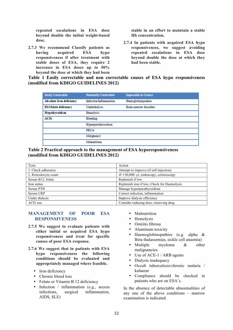
32
repeated escalations in ESA dose beyond double the initial weight-based dose.
2.7.3 We recommend Classify patients as having acquired ESA hypo responsiveness if after treatment with stable doses of ESA, they require 2 increases in ESA doses up to 50% beyond the dose at which they had been
stable in an effort to maintain a stable Hb concentration.
2.7.4 In patients with acquired ESA hypo responsiveness, we suggest avoiding repeated escalations in ESA dose beyond double the dose at which they had been stable.
Table 1 Easily correctable and non correctable causes of ESA hypo responsiveness (modified from KDIGO GUIDELINES 2012) Easily Correctable Potentially Correctable Impossible to Correct
Table 2 Practical approach to the management of ESA hyporesponsiveness (modified from KDIGO GUIDELINES 2012) Tests Action 1. Check adherence Attempt to improve (if self-injection) 2. Reticulocyte count if>130,000/ µl, endoscopy, colonoscopy Serum B12, folate Replenish if low Iron status Replenish iron if low, Check for Haemolysis Serum PTH Manage hyperparathyroidism Serum CRP Correct infection, inflammation Under dialysis Improve dialysis efficiency ACEi use Consider reducing dose, removing drug
MANAGEMENT OF POOR ESA
RESPONSIVENESS 2.7.5 We suggest to evaluate patients with
either initial or acquired ESA hypo responsiveness and treat for specific causes of poor ESA response.
2.7.6 We suggest that in patients with ESA hypo responsiveness the following conditions should be evaluated and appropriately managed where feasible.
• Iron deficiency • Chronic blood loss • Folate or Vitamin B 12 deficiency • Infection / inflammation (e.g., access
infections, surgical inflammation, AIDS, SLE)
• Malnutrition • Hemolysis • Osteitis fibrosa • Aluminum toxicity • Haemoglobinopathies (e.g. alpha &
Beta thalassemias, sickle cell anaemia) • Multiple myeloma & other
malignancies. • Use of ACE-1 / ARB agents • Dialysis inadequacy • Occult tuberculosis/chronic malaria /
kalaazar • Compliance should be checked in
patients who are on ESA’s.
In the absence of detectable abnormalities of any one of the above conditions – marrow examination is indicated.

33
2.7.7 For patients who remain hypo responsive despite correcting treatable causes, we suggest individualization of therapy, accounting for relative risks and benefits of:
• Decline in Hb concentration, • continuing ESA, if needed to
maintain Hb concentration, with due consideration of the doses required, and
• blood transfusions ADJUVANT THERAPIES 2.8.1 We suggest that androgens be limited
for use in men more than 50 years in anemia of CKD and the treatment be individualized
2.8.2 We suggest not using vitamin C, vitamin D, vitamin E, folic acid, L-carnitine, and pentoxifylline, as adjuvants to ESA Treatments.
The use of androgens as an adjuvant to ESA should be restricted to men beyond 50 because of the potential side effects. The usefulness of androgen has been highlighted in the recent meta-analysis of the various trials, which suggests the usefulness and the role for androgens in developing countries.
There is no convincing data because of lack of RCT for other adjuvants vitamin C, Vitamin D, Vitamin E, L carnitine and Pentoxifylline. Available literature is of small case series or non-randomized studies. In CKD 5 HD patients, long term use of Vitamin C is also fraught with the concern of secondary oxalosis.
EVALUATION FOR PURE RED CELL APLASIA (PRCA)
2.9.1 We suggest to investigate for possible antibody-mediated PRCA when a patient is receiving ESA therapy for more than 8 weeks develops the following: Sudden rapid decrease in Hb concentration at the rate of 0.5 to 1.0 g/dl (5 to 10 g/l) per week or requirement of transfusions at the rate of approximately 1 to 2 per week and normal platelet and white cell counts and absolute reticulocyte count less than 10,000/µl.
2.9.2 We recommend that ESA therapy be stopped in patients who develop antibody-mediated PRCA.
2.9.3 We suggest peginesatide be used to treat patients with antibody-mediated PRCA.
After exclusion of historical PRCA episodes with SC use, the present estimated incidence of PRCA episode is 0.5 cases per 10,000 patient years with all other forms of subcutaneous ESA formulations. No case has been documented in patients using IV ESA. Key factor in attributing the etiology of PRCA is to demonstrate anti EPO antibodies in patient serum. Assays available for these are enzyme linked immuno sorbent assay (ELISA), Radio immuno precipitation assay (RIPS), and bio sensor immunoassay. If a decision to treat with peginesatide is taken, it can be initiated at a dose of 0.05 to 0.075 mg/kg body weight by subcutaneous injection every 4 weeks. Subsequently, the dose needs to be adjusted to reach the desired target Hb value.
TREATMENT WITH IRON AGENTS 3.1 We suggest when prescribing iron
therapy, balance the potential benefits of avoiding or minimizing blood transfusions, ESA therapy, and anemia related symptoms against the risks of harm in individual patients (e.g., anaphylactoid and other acute reactions, unknown long-term risks).
3.2 For adult CKD patients with anemia not on iron or ESA therapy we suggest a trial of IV iron (or in CKD ND patients alternatively a 1–3 month trial of oral iron therapy) if an increase in Hb concentration without starting ESA treatment is desired and TSAT is less than 30% and ferritin is less than 500 ng/ml (≤500 µg/l)
3.3 For adult CKD patients on ESA therapy who are not receiving iron supplementation, we suggest a trial of IV iron (or in CKD ND patients alternatively a 1–3 month trial of oral iron therapy) if an increase in Hb concentration or a decrease in ESA dose is desired and TSAT is less than 30% and ferritin is less than 500 ng/ml (≤500 µg/l)
34
3.4 We suggest CKD ND patients who require iron supplementation, select the route of iron administration based on the severity of iron deficiency, availability of venous access, response to prior oral iron therapy, side effects with prior oral or IV iron therapy, patient compliance, and cost.
3.5 We suggest that guide to subsequent iron administration in CKD patients should be based on Hb responses to recent iron therapy, as well as ongoing blood losses, iron status tests (TSAT and ferritin), Hb concentration, ESA responsiveness and ESA dose in ESA treated patients, trends in each parameter, and the patient’s clinical status.
3.6 We suggest in all pediatric CKD patients with anemia not on iron or ESA therapy, oral iron (or IV iron in CKD HD patients) administration when TSAT is less than 20% and ferritin is less than 100 ng/ml (≤100 µg/l).
3.7 We suggest all pediatric CKD patients on ESA therapy who are not receiving iron supplementation, oral iron (or IV iron in CKD HD patients) administration to maintain TSAT more than 20% and ferritin more than 100 ng/ml (>100 µg/l).
The functional iron deficiency occurs in CKD patients. In these patients though TSAT and ferritin levels are not indicative of absolute iron deficiency, patients respond with enhanced erythropoiesis to IV iron. The most widely used test to asses iron status are TSAT and ferritin level. A low serum ferritin less than 30 ng/ml and TSAT less than 20% is indicative of iron deficiency. However, most CKD patients with serum ferritin more than 100 ng/ml and TSAT more than 20% do respond to supplemental iron by increase in Hb concentration and decrease in ESA dose. Hence in these guidelines we recommend, as suggested by KDIGO iron administration in anemic CKD patients with less than 30 % TSAT and less than 500 ng/ml serum ferritin, after considering the potential risk of iron administration. In CKD patients with TSAT more than 30 % and ferritin more than 500 ng/ml, the safety of providing additional iron has not been studied. We do not recommend
routine use of iron supplementation in these situations. Mode of Iron supplementation is either oral or IV. Intramuscular administration has been abandoned. The route of supplementation of iron should be on following factors.
• Severity of Anemia and iron deficiency • Response • Tolerance & adherence to prior oral administration • Cost • Ease of obtaining venous access balanced against the desire to preserve the venous access. • In CKD 5 HD patients and PD patients there is overwhelming evidence for IV iron as compared to oral Iron replacement.
IRON STATUS EVALUATION 3.8.1 We suggest to assess iron status (TSAT
and ferritin) at least every 3 months during ESA therapy, including the decision to start or continue iron therapy.
3.8.2 We suggest to Test iron status (TSAT and ferritin) more frequently when initiating or increasing ESA dose, when there is blood loss, when monitoring response after a course of IV iron, and in other circumstances where iron stores may become depleted.
These recommendations are not based on RCT’s but on consensus.
CAUTIONS REGARDING IRON THERAPY
3.8.3 When the initial dose of IV iron dextran or IV non dextran iron is administered, we suggest that patients be monitored for 60 minutes after the infusion, and that resuscitative facilities (including medications) and personnel trained to evaluate and treat serious adverse reactions be available.
Iron dextran use can cause anaphylactic reaction. Incidence is much lower with lower molecular iron dextran compared to higher molecular iron dextran. It is prudent to observe patient for 60 minutes after IV iron administration.
3.8.4 Avoid administering IV iron to patients with active systemic infections.
35
In animal models iron impairs the phagocytosis of intracellular bacterial/fungal infection. Data on CKD patients are conflicting. Hence it is advisable to avoid IV iron during active systemic infections.
USE OF RED CELL TRANSFUSION IN CHRONIC ANEMIA
4.1.1 When managing chronic anemia, we recommend avoiding, when possible, red cell transfusions to minimize the general risks related to their use.
4.1.2 In patients eligible for organ transplantation, we specifically recommend avoiding, when possible, red cell transfusions to minimize the risk of allo-sensitization.
4.1.3 When managing chronic anemia, we specifically suggest that the benefits of red cell transfusions may outweigh the risks in patients in whom: ESA therapy is ineffective (e.g., hemoglobinopathies, bone marrow failure, ESA resistance) or the risks of ESA therapy may outweigh its benefits (e.g., previous or current malignancy, previous stroke)
4.1.4 We suggest that the decision to transfuse a CKD patient with non-acute anemia should not be based on any arbitrary Hb threshold, but should be determined by the occurrence of symptoms caused by anemia.
URGENT TREATMENT OF ANEMIA 4.2.1 In certain acute clinical situations, we
suggest patients are transfused when the benefits of red cell transfusions outweigh the risks; these include:
• When rapid correction of anemia is required to stabilize the patient’s condition (e.g., acute hemorrhage, unstable coronary artery disease)
• When rapid pre-operative Hb correction is required
Allo-sensitisation due to repeated transfusions along with the risk of transfusion of blood borne viruses is the main drawback for red cell transfusions. However ESA also has inherent risks in patients with stroke and in those with malignancy. Hence a balanced approach is required when using transfusion to treat anemia.
Lueco-reduction of blood products does not decrease sensitization in a previously transplanted or in a potential transplant patient. Washed red cells are not less immunogenic than non-washed red cells. Donors specific and DR match transfusions do not reduce immunogenicity. Increased number of transfusions is associated with greater risk of sensitization.
When there is a sensitization there is a longer wait for transplantation as it is difficult to find a compatible donor with higher PRA. Waiting longer for transplantation is directly linked to lower survival. Hence when we decide on transfusion or ESA for treatment of non-acute anemia related to CKD, it is obligatory to weigh the risk and benefits in each of these modalities, in a given patient.
FURTHER READING: • Besarab A, Bolton WK, Browne JK, et al.
The effects of normal as compared with low hematocrit values in patients with cardiac disease who are receiving hemodialysis and epoetin. N Engl J Med 1998; 339: 584 – 590.
• Pfeffer MA, Burdmann EA, Chen CY, et al. A trial of darbepoetin alfa in type 2 diabetes and chronic kidney disease. N Engl J Med 2009; 361: 2019-2032.
• Singh AK, Szczech L, Tang KL, et al. Correction of anemia with epoetin alfa in chronic kidney disease. N Engl J Med 2006; 355: 2085-2098.
• Drueke TB, Locatelli F, Clyne N, et al. Normalization of Hemoglobin level in patients with chronic kidney disease and anemia. N Engl J Med 2006; 355: 2071-2084.
• National Kidney Foundation. KDOQI Clinical Practict Guidelines and Clinical Practice Recommendations for Anemia in Chronic Kidney Disease. Am J Kidney Dis 2006; 47: S11-145.
• Kalantar-Zadeh K, Hoffken B, Wunsch H, et al. Diagnosis of iron deficiency anemia in renal failure patients during the posterythropoietin era. Am J Kidney Dis 1995; 26: 292-299.
• Allegra V, Mengozzi G, Vasile A. Iron deficiency in maintenance hemodialysis

36
patients: assessment of diagnosis criteria and of three different iron treatments. Nephron 1991; 57: 175-182.
• Li H, Wang SX. Intravenous iron sucrose in Chinese hemodialysis patients with renal anemia. Blood Purif 2008; 26: 151-156.
• Srikant P., et al. Ferritin as a marker of anemia and non-anemia. J. Chem. Pharm. Res., 2010, 2(3); 400-410.
• B. Adamu, et al. Meta-Analysis of Randomized Controlled Trials on Androgens versus Erythropoietin for Anaemia of Chronic Kidney Disease: Implications for Developing Countries. International Journal of Nephrology, Vol . 2012(Article ID - 580437), 2012.
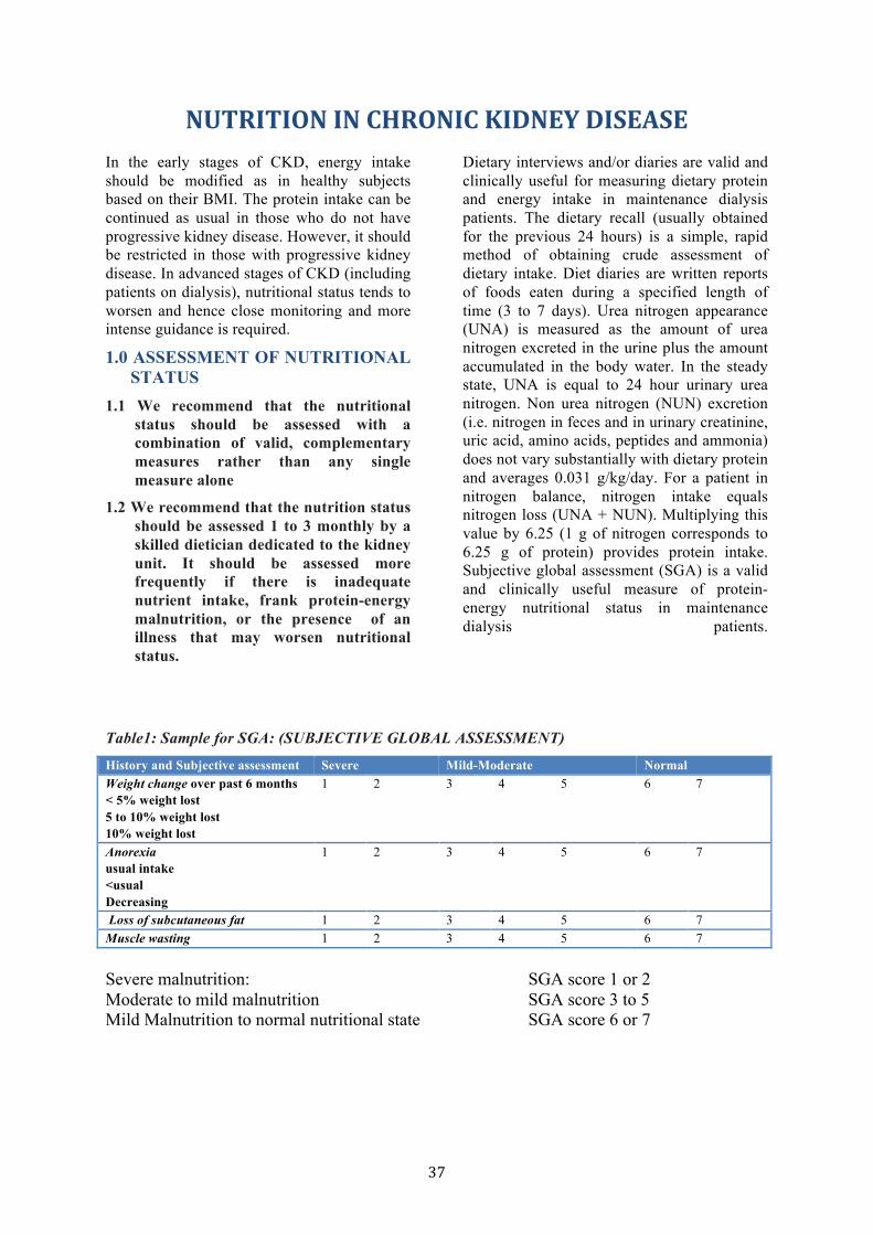
37
NUTRITION IN CHRONIC KIDNEY DISEASE In the early stages of CKD, energy intake should be modified as in healthy subjects based on their BMI. The protein intake can be continued as usual in those who do not have progressive kidney disease. However, it should be restricted in those with progressive kidney disease. In advanced stages of CKD (including patients on dialysis), nutritional status tends to worsen and hence close monitoring and more intense guidance is required.
1.0 ASSESSMENT OF NUTRITIONAL STATUS
1.1 We recommend that the nutritional status should be assessed with a combination of valid, complementary measures rather than any single measure alone
1.2 We recommend that the nutrition status should be assessed 1 to 3 monthly by a skilled dietician dedicated to the kidney unit. It should be assessed more frequently if there is inadequate nutrient intake, frank protein-energy malnutrition, or the presence of an illness that may worsen nutritional status.
Dietary interviews and/or diaries are valid and clinically useful for measuring dietary protein and energy intake in maintenance dialysis patients. The dietary recall (usually obtained for the previous 24 hours) is a simple, rapid method of obtaining crude assessment of dietary intake. Diet diaries are written reports of foods eaten during a specified length of time (3 to 7 days). Urea nitrogen appearance (UNA) is measured as the amount of urea nitrogen excreted in the urine plus the amount accumulated in the body water. In the steady state, UNA is equal to 24 hour urinary urea nitrogen. Non urea nitrogen (NUN) excretion (i.e. nitrogen in feces and in urinary creatinine, uric acid, amino acids, peptides and ammonia) does not vary substantially with dietary protein and averages 0.031 g/kg/day. For a patient in nitrogen balance, nitrogen intake equals nitrogen loss (UNA + NUN). Multiplying this value by 6.25 (1 g of nitrogen corresponds to 6.25 g of protein) provides protein intake. Subjective global assessment (SGA) is a valid and clinically useful measure of protein-energy nutritional status in maintenance dialysis patients.
Table1: Sample for SGA: (SUBJECTIVE GLOBAL ASSESSMENT)
History and Subjective assessment Severe Mild-Moderate Normal Weight change over past 6 months < 5% weight lost 5 to 10% weight lost 10% weight lost
1 2 3 4 5 6 7
Anorexia usual intake <usual Decreasing
1 2 3 4 5 6 7
Loss of subcutaneous fat 1 2 3 4 5 6 7 Muscle wasting 1 2 3 4 5 6 7
Mild-Moderate Normal Severe malnutrition: SGA score 1 or 2 Moderate to mild malnutrition SGA score 3 to 5 Mild Malnutrition to normal nutritional state SGA score 6 or 7
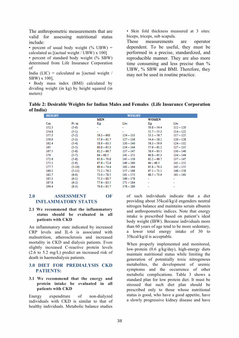
38
The anthropometric measurements that are valid for assessing nutritional status include: • percent of usual body weight (% UBW) = calculated as [(actual weight / UBW) x 100] • percent of standard body weight (% SBW) determined from Life Insurance Corporation of India (LIC) = calculated as [(actual weight / SBW) x 100], • Body mass index (BMI) calculated by dividing weight (in kg) by height squared (in meters)
• Skin fold thickness measured at 3 sites: biceps, triceps, sub scapula. These measurements are operator dependent. To be useful, they must be performed in a precise, standardized, and reproducible manner. They are also more time consuming and less precise than % UBW, % SBW and BMI. Therefore, they may not be used in routine practice.
Table 2: Desirable Weights for Indian Males and Females (Life Insurance Corporation of India)
2.0 ASSESSMENT OF
INFLAMMATORY STATUS 2.1 We recommend that the inflammatory
status should be evaluated in all patients with CKD
An inflammatory state indicated by increased CRP levels and IL-6 is associated with malnutrition, atherosclerosis and increased mortality in CKD and dialysis patients. Even slightly increased C-reactive protein levels (2.6 to 5.2 mg/L) predict an increased risk of death in haemodialysis patients.
3.0 DIET FOR PREDIALYSIS CKD PATIENTS:
3.1 We recommend that the energy and protein intake be evaluated in all patients with CKD
Energy expenditure of non-dialyzed individuals with CKD is similar to that of healthy individuals. Metabolic balance studies
of such individuals indicate that a diet providing about 35kcal/kg/d engenders neutral nitrogen balance and maintains serum albumin and anthropometric indices. Note that energy intake is prescribed based on patient’s ideal body weight (IBW). Because individuals more than 60 years of age tend to be more sedentary, a lower total energy intake of 30 to 35kcal/kg/d is acceptable.
When properly implemented and monitored, low-protein (0.6 g/kg/day), high-energy diets maintain nutritional status while limiting the generation of potentially toxic nitrogenous metabolites, the development of uremic symptoms and the occurrence of other metabolic complications. Table 3 shows a standard plan for low protein diet. It must be stressed that such diet plan should be prescribed only to those whose nutritional status is good, who have a good appetite, have a slowly progressive kidney disease and have

39
protein intake > 0.6 g/kg/day (as calculated from urea nitrogen appearance).
Table 3: A standard plan for a low protein diet (0.6gm/kg/day) B’fast: Tea 1 cup and Poha 1 katori OR Upma 1 katori 10 am: Fruit 1 Lunch: 3 Chapati (Wheat Flour 60gm + Arrowroot 60gm),
Rice 1 Katori, Vegetable 1 katori, Thin Dal 1 Katori (Raw 15 gm), Buttermilk 1glass (Curd 50gm), Salad
Teatime: Tea 1 cup Evening: Apple 1 Dinner: 3 Chapati (Wheat Flour 60gm + Arrowroot 60gm), Rice
1Katori,Vegetable 1 katori, Thin Dal 1 Katori (Raw 15 gm), Buttermilk 1glass (Curd 50gm), Salad
Use Cow Milk Oil and Ghee 6 tsp/day Use Root Vegetables Wt: 60 kg Calories: 2000 Kcal (33 Kcal/kg) Protein: 37.8 gm (0.63gm/kg)
Beheray and Shah estimated dietary protein intake in 20 stable patients with CKD who were on an unrestricted vegetarian diet. The mean protein intake was 0.64+0.15 g/kg/day. In these patients, dietary protein should not be restricted unless they are supplemented with ketoanalogues
3.2: We recommend special diet in consultation with the dietician, to be adopted in diabetic patients and those with nephrotic range proteinuria
• In diabetic patients, energy intake should be the same (30-35 kcal/kg of IBW/day) as for non-diabetic subjects. About 60% of calories should be from carbohydrates, 30% from fats < 10% from saturated fats, < 10% from polyunsaturated fats and about 15% from monounsaturated fats.
• In patients with nephrotic range proteinuria, it is a common misconception to provide high protein diet to patients with nephrotic range proteinuria. In fact, doing so increases
proteinuria and worsens hypercholesterolemia. A diet providing 0.8 g/kg/day protein (plus 1 g protein/g of proteinuria) and 30-35 kcal/kg of IBW/day maintains nitrogen balance.
3.3 We suggest the essential amino acid (EAA) and ketoacid (KA) supplemented diet regimens may be used in patients for prevention of progression of CKD
The basis of prescribing dietary protein restriction is to minimize adaptive changes that play some role in progression of CKD, and to diminish the production of nitrogenous wastes. Attempts have been made to prescribe very low protein diet (VLPD) containing about 0.3 g /kg/d of unrestricted quality protein plus a supplement of EAAs or KAs. In our experience restricting protein to 0.3 g/kg/day is difficult. The best we achieve is protein intake of 0.4g/kg/day. Table 4 shows a standard plan for supplemented very low protein diet.
Table 4: A standard plan for very low protein diet (0.4/kg/day)
B’fast: Tea 1 cup and Poha 1 katori OR Rice Upma 1 katori 10 am: Banana 1 Lunch: 3 Rice Chapati (Rice 60gm + Arrowroot 60gm), 2 Vegetable
1 katori, Thin Dal 1 Katori (Raw 10 gm) OR Buttermilk 1 glass (Curd 50gm), Salad
Teatime: Tea 1 cup Evening: Apple 1
40
Dinner: 3 Rice Chapati (Rice 60gm + Arrowroot 60gm), 2Vegetable 1 katori, Thin Dal 1 Katori (Raw 10 gm) OR Buttermilk 1 glass (Curd 50gm), Salad
Use Cow Milk Oil and Ghee 6 tsp/day Use Root Vegetables Wt: 60 kg Calories: 1900 Kcal (32Kcal/kg) Protein: 23.4 gm (0.39gm/kg)
In a study done by Nayak, out of a total number of 132 subjects, 92 were in the sLPD group (0.6 g/kg/day of protein) and 40 in the Svlpd group (0.3g/kg/day of protein). They were followed up for 6months, both were supplemented with keto-analogues. Both groups showed significant improvement in renal parameters, anthropometric measures and biochemical nutritional parameters such as serum albumin. Those who started the ketodiet early (Stage 3 and 4 CKD) and at the right dosage (one tablet for every 5 kg/body wt.) with regular monitoring by a skilled renal dietician did much better compared to others.
Prakash et al conducted a randomized double blind, placebo controlled trial to evaluate efficacy of VLPD supplemented with KA in patients with CKD. Thirty-four patients were randomized to 2 comparable groups in terms of age, sex distribution, etiology of CKD, blood pressure control, use of angiotensin converting enzyme inhibitors, GFR and body mass index (BMI). Subjects randomly received either 0.6 gm/kg/day protein plus placebo (n==16) or 0.3 gm/kg/day protein plus tablets of KA for 9 months. The mean GFR at baseline in the KA group and control group was 28.1 + 8.8 and 28.6 + 17.6 ml/min/1.73 m2 respectively. At the end of the study it was 27.6 + 10.1 and 22.5 + 15.9 ml/min/1.73 m2 respectively. Thus there was a significant drop in GFR in the control group. In both groups there was no significant change in the BMI after the study.
3.4. We recommend that fluid and electrolytes be advised according to the individual patient status in consultation with the treating nephrologist
In patients with tendency to become edematous (usually those with proteinuric CKD), fluid and salt intake should be restricted to the maximum extent tolerated by the patient, ensuring that it does not
compromise their calorie and protein intake. Diuretics will also have to be used to maintain patient edema free. Potassium intake has to be advised according to serum potassium levels.
3.5 We recommend that in all CKD patients, make all efforts to maintain acid-base balance to near normal state:
• Serum bicarbonate should be measured in advanced CKD once monthly. The aim is to maintain serum bicarbonate levels at or above 22mmol/L.
• This can be achieved by oral supplement with bicarbonate (sodamint tablets. Each tablet provides approximately 4 meq of bicarbonate)
4.0 INDICATIONS FOR RENAL REPLACEMENT THERAPY
4.1 We strongly recommend that in patients with advanced CKD, (e.g. GFR < 10 mL/min) if protein-energy malnutrition develops or persists despite vigorous attempts to optimize protein and energy intake and there is no apparent cause for malnutrition other than low nutrient intake, initiation of maintenance dialysis or a renal transplant should be commenced.
Malnutrition in CKD stage 5 is due to uraemia per se with associated anorexia. Nutritional supplements at this low GFR will not help in alleviating malnutrition. Dialysis would correct uremic milieu, improve appetite and consequently improve the nutritional status.
5.0 DIET FOR MAINTAINENCE HEMODIALYSIS AND CONTINUOUS AMBULATORY PERITONEAL DIALYSIS PATIENTS:
5.1.1 We recommend that the daily protein intake (DPI) for clinically stable maintenance hemodialysis (MHD)

41
patients is 1.2g/kg body weight / day with at least 50% of the dietary protein to be of high biological value.
5.1.2 We recommend that the DPI for clinically stable CAPD patients is 1.2 to 1.3 g/kg body weight / day with the dietary protein intake to be no less than 1.2g/kg/day and at least 50% of the dietary protein should be of high biological value
The energy expenditure of patients undergoing maintenance HD or CAPD is similar to that of normal healthy individuals. Metabolic balance studies of people undergoing maintenance HD indicate that a diet providing about 35kcal/kg/d engenders neutral nitrogen balance and maintains serum albumin and anthropometric indices. Because individuals more than 60 years of age tend to be more sedentary, a lower total energy intake of 30 to 35kcal/kg/d is acceptable.
5.2.1 We recommend that in patients on MHD, fluid and salt intake should be such that inter dialytic weight gain does not exceed 1 to 1.5 kg.
5.2.2 We recommend that in patients on CAPD, fluid and salt restriction will have to be adjusted according to negative balance achieved.
6.0 INDICATIONS FOR NUTRITIONAL SUPPORT
6.1 We recommend that individuals undergoing maintenance dialysis who are unable to meet their protein and energy requirement with food intake for an extended period of time should receive nutrition support
Before considering nutrition support, the patient should receive a complete nutritional assessment. Any potentially reversible or treatable condition or medication that might interfere with appetite or cause malnutrition should be eliminated or treated. For nutrition support, the oral diet may be fortified with energy and protein supplements (table 5). If oral nutrition (including nutrition supplement) is inadequate, tube feeding should be offered. If tube feeding is not possible, Intra dialytic parenteral nutrition (IDPN) for hemodialysis patients and intra peritoneal amino acids (IPAA) for peritoneal dialysis patients should be considered. If the combination of oral intake and IDPN or IPAA does not meet protein and energy requirements, daily total or partial parenteral nutrition should be considered as last resort.
7.0 We suggest that the routine use of L-carnitine in maintenance dialysis patients is to be avoided.
Table 5: A list of nutritional supplements available in India
* Per 100gm FURTHER READING: • Gersovitz M, Maddon JP, Smiciklas-
Wright H;validity of the 24-hr dietary recall and seven-day record for group
comparisons. J Am Diet Assoc. 73: 48-55, 1978.
• Baker JP, Detsky AS, Wesson DE, Wolman SL, Stewart S, Whitewell J, Langer B, Jeejeebhoy KN: Nutritional
42
assessment: A comparison of clinical judgment and objective measurements. N Engl. J Med. 306: 969-972,1982.
• Keshaviah P:Lean body mass estimation by creatinine kinetics. J Am Soc. Nephrol. 4: 1475 – 1485, 1994.
• K/DOQI Clinical Practice guidelines for nutrition in chronic renal failure. Am J Kidney. 2002; 17: 3, 35 Suppl. 2: S17-S104
• Kaysen GA, Stevenson FT, Depner TA. Determinants of albumin concentration in hemodialysis patients. Am J Kidney Dis. 29: 658 – 668, 1997.
• Jacob V, Marchant PR, Wild G, Brown CB, Moorhead PJ, el Nahas AM: Nutritional profile of continuous
ambulatory peritoneal dialysis patients. Nephron, 71: 16-22, 1995.
• Oksa H., Ahonen K., Pasternack A., Marnela KM: Malnutrition in hemodialysis patients. Scand J UrolNephrol. 25: 157 – 161, 1991.
• Food and Nutrition Board NRCN: Recommended Daily Allowances (ed 10). Washington, DC, National Academy Press, 1989.
• Guidelines for the use of parenteral & enteral nutrition in adult &pediatric patients. J Parenter Enteral Nutr: 17: S1A-S52A, 1993 (abstr, suppl).
• Barton RG: Nutrition support in critical illness. NutrClinPract: 9: 127-139, 1994.
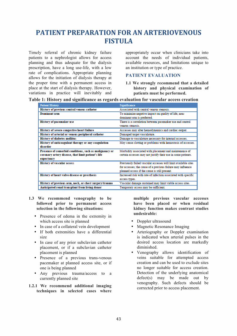
43
PATIENT PREPARATION FOR AN ARTERIOVENOUS FISTULA
Timely referral of chronic kidney failure patients to a nephrologist allows for access planning and thus adequate for the dialysis prescription, have a long use-life, with a low rate of complications. Appropriate planning allows for the initiation of dialysis therapy at the proper time with a permanent access in place at the start of dialysis therapy. However, variations in practice will inevitably and
appropriately occur when clinicians take into account the needs of individual patients, available resources, and limitations unique to an institution or type of practice.
PATIENT EVALUATION 1.1 We strongly recommend that a detailed
history and physical examination of patients must be performed.
Table 1: History and significance as regards evaluation for vascular access creation
Patient History Significance 1.3 We recommend venography to be
followed prior to permanent access selection in the following situations:
• Presence of edema in the extremity in which access site is planned
• In case of a collateral vein development • If both extremities have a differential
size • In case of any prior subclavian catheter
placement, or if a subclavian catheter placement is planned
• Presence of a previous trans-venous pacemaker at planned access site, or if one is being planned
• Any previous trauma/access to a currently planned site
1.2.1 We recommend additional imaging techniques in selected cases where
multiple previous vascular accesses have been placed or when residual kidney function makes contrast studies undesirable:
• Doppler ultrasound • Magnetic Resonance Imaging • Arteriography or Doppler examination
is indicated when arterial pulses in the desired access location are markedly diminished.
• Venography allows identification of veins suitable for attempted access creation and can be used to exclude sites no longer suitable for access creation. Detection of the underlying anatomical defect(s) may be made out by venography. Such defects should be corrected prior to access placement.
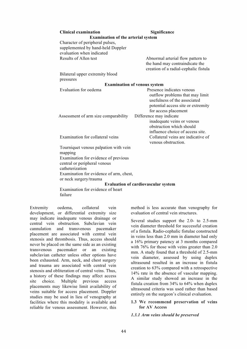
44
Clinical examination Significance Examination of the arterial system
Character of peripheral pulses, supplemented by hand-held Doppler evaluation when indicated Results of Allen test Abnormal arterial flow pattern to
the hand may contraindicate the creation of a radial-cephalic fistula
Bilateral upper extremity blood pressures
Examination of venous system Evaluation for oedema Presence indicates venous
outflow problems that may limit usefulness of the associated potential access site or extremity for access placement
Assessment of arm size comparability Difference may indicate inadequate veins or venous obstruction which should influence choice of access site.
Examination for collateral veins Collateral veins are indicative of venous obstruction.
Tourniquet venous palpation with vein mapping Examination for evidence of previous central or peripheral venous catheterization Examination for evidence of arm, chest, or neck surgery/trauma
Evaluation of cardiovascular system Examination for evidence of heart failure
Extremity oedema, collateral vein development, or differential extremity size may indicate inadequate venous drainage or central vein obstruction. Subclavian vein cannulation and transvenous pacemaker placement are associated with central vein stenosis and thrombosis. Thus, access should never be placed on the same side as an existing transvenous pacemaker or an existing subclavian catheter unless other options have been exhausted. Arm, neck, and chest surgery and trauma are associated with central vein stenosis and obliteration of central veins. Thus, a history of these findings may affect access site choice. Multiple previous access placements may likewise limit availability of veins suitable for access placement. Doppler studies may be used in lieu of venography at facilities where this modality is available and reliable for venous assessment. However, this
method is less accurate than venography for evaluation of central vein structures.
Several studies support the 2.0- to 2.5-mm vein diameter threshold for successful creation of a fistula. Radio-cephalic fistulae constructed in veins less than 2.0 mm in diameter had only a 16% primary patency at 3 months compared with 76% for those with veins greater than 2.0 mm. A study found that a threshold of 2.5-mm vein diameter, assessed by using duplex ultrasound resulted in an increase in fistula creation to 63% compared with a retrospective 14% rate in the absence of vascular mapping. A similar study showed an increase in the fistula creation from 34% to 64% when duplex ultrasound criteria was used rather than based entirely on the surgeon’s clinical evaluation.
1.3 We recommend preservation of veins for AV Access
1.3.1 Arm veins should be preserved
45
• Arm veins suitable for placement of vascular access should be preserved, regardless of arm dominance • Arm veins, particularly the cephalic veins of the non-dominant arm, should not be used for venipuncture or intravenous catheters • The dorsum of the hand should be used for intravenous lines • When venipuncture of the arm veins is necessary, sites should be rotated. 1.3.2 We strongly recommend that subclavian
vein catheterization should be avoided for temporary access in all patients with kidney failure
The hospital staff, and patients with progressive kidney disease (creatinine >3 mg/dL), and all patients with conditions likely to lead to ESRD are to protect the arms from venipuncture and intravenous catheters. A bracelet should be worn to inform hospital staff to avoid IV cannulation of essential veins. Venipuncture complications of veins that are potentially available for vascular access, may render such vein sites unsuitable for construction of a primary AV fistula.
Subclavian vein catheterization is associated with central venous stenosis and precludes the use of the entire ipsilateral arm for vascular access. Thus, subclavian vein catheterization should be avoided for temporary access in patients with kidney failure.
2.0 TIMING OF PLACEMENT OF VASCULAR ACCESS
2.1 We recommend that patients with CKD should be referred for surgery to attempt construction of a primary AV fistula when their creatinine clearance is <25 mL/min, their serum creatinine level is >4 mg/dL, or within 1 year of an anticipated need for dialysis
2.2.We recommend that the patient should be referred to a nephrologist prior to the need for access to facilitate kidney failure treatment and for counselling about modes of ESRD care, including hemodialysis, peritoneal dialysis, and renal transplantation
2.3 We recommend that a new primary fistula should be allowed to mature for at least 1 month, and ideally for 3 to 4 months, prior to cannulation
2.4 We recommend that AV grafts should be placed at least 3 to 6 weeks prior to an anticipated need for hemodialysis in patients who are not candidates for primary AV fistulae
2.5 We recommend that hemodialysis catheters should not be inserted until hemodialysis is needed
Both the size and anatomical qualities of venous and arterial components of primary AV fistulae can influence the fistulae maturation time. An aggressive policy of primary AV fistulae creation may result in failures in patients with marginal anatomy. However, timely attempts to create primary AV fistulae before the anticipated need for dialysis will allow adequate time for the fistulae to mature, and will allow sufficient time to perform another vascular access procedure if the first attempt fails, thus avoiding the need for temporary access.
3.0 MATURATION OF VASCULAR ACCESS
3.1 We recommend that a primary AV fistula is mature and suitable for use when the vein’s diameter is sufficient to allow successful cannulation, but not sooner than 1 month (and preferably 3 to 4 months after construction.
3.1.1 Cuffed and non-cuffed hemodialysis catheters are suitable for immediate use and do not require maturation time.
A vein must be mature, both physically and functionally, prior to use for vascular access. The time required for fistula maturation varies among patients. It is not advisable to use the fistula within the first month after construction because premature cannulation of a fistula may result in a higher incidence of infiltration with associated compression of the vessel by hematoma and permanent loss of the fistula. Allowing the fistula to mature for 3 to 4 months before use may be considered ideal.
The following procedures may enhance maturation of AV fistulae:
• Fistula hand-arm exercise (e.g., squeezing a rubber ball with or without a lightly applied tourniquet) will increase blood flow and speed maturation of a new native AV fistula

46
• Selective obliteration of major venous side branches will speed maturation of a slowly maturing AV fistula • When a new native AV fistula is infiltrated (that is, presence of hematoma with associated induration and edema), it should be rested until swelling is resolved. In general, allowing the fistula to mature for 6 to 8 weeks before investigating the reason for failure to mature is appropriate. For a fistula to be considered successful, it must be usable. In general, a working fistula must have all the following characteristics: blood flow adequate to support dialysis, which usually equates to a blood flow greater than 600 mL/min; a diameter greater than 0.6 cm, with location accessible for cannulation and discernible margins to allow for repetitive cannulation; and a depth of approximately 0.6 cm (ideally, between 0.5 to 1.0 cm from the skin surface). This combination of characteristics can be remembered easily as the Rule of 6s.
FURTHER READING: • Mehta S: Statistical summary of clinical
results of vascular access procedures for
haemodialysis, in Sommer BG, Henry ML (eds): Vascular Access for Hemodialysis–II. W.L. Gore & Associates, 1991, pp 145-157.
• National Kidney Foundation: DOQI Clinical Practice Guidelines for Vascular Access. Am J Kidney Dis 30:S150–S191, 1997 (suppl 3).
• Golper T. patient education: can it maximise success of therapy? Nephro Dial Transplant 2011;16(suppl7):20-24..
• Harland RC: Placement of permanent vascular access devices: Surgical considerations. Adv Ren Replace Ther 1:99-106, 1994.
• Marx AB, Landerman J, Harder FH: Vascular access for hemodialysis. Curr Probl Surg 27:15-48, 1990.
• Nonnast-Daniel B, Martin RP, Lindert O, et al. Colour doppler ultrasound assessment of arteriovenous haemodialysis fistulas. Lancet 339:143-145, 1992.
47
PREVENTION, DIAGNOSIS, EVALUATION, AND TREATMENT OF HEPATITIS C INFECTION IN CHRONIC
KIDNEY DISEASE Testing for HCV can be done by two methods: serologic assays and nucleic acid tests (NAT). Immunoblots and Enzyme linked Immunoassays (ELIAs) are used to detect antibodies in circulation against HCV proteins. Third generation ELIAs which are in current use have high sensitivity and specificity, making other serologic tests obsolete. This test is extensively used to screen for the infection in blood components. The one disadvantage is the so called window period, i.e., the time between acquisition of infection and the development of anti-HCV antibodies, which is about 60-90 days, and failure of development of antibodies in immune suppressed individuals. Improved testing methods currently in development are likely to reduce this window period. The presence of hyper gamma globulinemia may give rise to a false positive test.
NAT is can be qualitative, that simply detects whether the HCV RNA is present or not. Quantitative tests give an idea of the RNA copy number, which allows estimation of infection load and treatment monitoring. Commercially available qualitative assays can detect 50 IUml1 of HCV RNA. The detection limits of quantitative assays are higher than those of qualitative detection assays.
The prevalence of HCV in dialysis population varies widely in India. It is as low as 3% in some centres but goes as high as 70% in others. There is tremendous variation in the prevalence from unit to unit even within the same city. Not much is known about the prevalence in the earlier stages of CKD.
The following guidelines are suggested for the management of HCV infection in patients with kidney disease. These have been based largely on the 2008 KDIGO Guidelines for Prevention, Diagnosis, Evaluation, and Treatment of Hepatitis C in Chronic Kidney Disease with modifications based on the prevailing practices and Indian data. The strength of recommendation are based on rigorous evidence review done by the KDIGO workgroup.
1.0: DETECTION AND EVALUATION OF HCV IN CKD
1.1 Determining which CKD patients should be tested for HCV
1.1.1 We suggest that CKD patients be tested for HCV
1.1.2 We strongly recommend that testing for HCV should be performed in patients on maintenance hemodialysis (CKD Stage 5D) and kidney transplant candidates.
1.2 HCV testing for patients on maintenance hemodialysis:
1.2.1 We strongly recommend that patients on hemodialysis should be tested when they first start hemodialysis or when they transfer from another hemodialysis facility.
1.2.2. We recommend in hemodialysis units with a low prevalence of HCV, initial testing with EIA (if positive, followed by NAT) should be considered
1.2.3. We suggest that in hemodialysis units with a high prevalence of HCV, initial testing with NAT should be considered
1.2.4 We recommend for patients on hemodialysis who test negative for HCV, retesting every 6–12 months with EIA should be considered
1.2.5 We strongly recommend that testing for HCV with NAT should be performed for hemodialysis patients with unexplained abnormal aminotransferase(s) levels.
1.2.6 We strongly recommend that if a new HCV infection in a hemodialysis unit is suspected to be nosocomial, testing with NAT should be performed in all patients who may have been exposed.
1.2.7. We suggest that repeat testing with NAT is suggested within 2–12 weeks in initially NAT-negative patients.

48
In view of the reported high prevalence of HCV infection in many Indian dialysis units, it is imperative that all units test the patient using a sensitive test before starting dialysis for the first time. It is even more important to test when a patient transfers from another unit, since the likelihood of nosocomial exposure is relatively high. Patients frequently move from unit to unit in India, and the need for screening must be emphasized.
Another issue is the method of testing. Because of the high prevalence, and the likelihood of the patient being in the window period especially if they have contracted the infection in one unit and move to another, the method of choice for testing should be NAT.
In view of the cost, we suggest that qualitative methods be used for detection. They have the added advantage of having a lower detection threshold compared to quantitative methods. Quantitative methods should be used, however, when treatment is being contemplated since they allow monitoring of load and treatment response. The recommended frequency of rescreening is not based on a strong evidence base. However, since the likelihood of transmission increases with increase in prevalence, we suggest that units with high prevalence increase the frequency of retesting to once every 3-4 months.
The following algorithm suggested by the KDIGO is very useful.
Algorithm 1. CKD Stage 5 Hemodialysis diagnostic algorithm. ALT, alanine aminotransferase; AST, aspartate aminotransferase; CKD: Chronic Kidney Disease; EIA: enzyme immunoassay; HCV: hepatitis C virus; NAT: nucleic acid test.
2.0: TREATMENT OF HCV INFECTION IN PATIENTS WITH CKD (BASED ON KDIGO HEP C GUIDELINES)
2.1 We suggest the following in CKD patients with HCV infection
2.1.1 Patients be evaluated for antiviral treatment.
49
2.1.2 The decision to treat be based on the potential benefits and risks of therapy, including life expectancy, candidacy for kidney transplantation, and co-morbidities.
2.1.3 All CKD patients except kidney transplant recipients who develop an acute HCV infection, a waiting period beyond 12 weeks to observe spontaneous clearance (by NAT) is not justified and that antiviral treatment should be started
2.1.4 HCV-infected patients accepted for kidney transplantation be treated.
2.1.5 Treatment of HCV-infected kidney transplant recipients be considered only when the benefits of treatment clearly outweigh the risk of allograft rejection due to IFN-based therapy (for example, fibrosing cholestatic hepatitis, life threatening vasculitis).
2.1.6 Antiviral therapy be considered for patients with HCV-related GN.
2.1.7 For HCV-infected patients with CKD Stages 1 and 2, using combined antiviral treatment using pegylated IFN and ribavirin is suggested, as in the general population. The ribavirin dose to be titrated according to patient tolerance.
2.1.8 For HCV-infected patients with CKD Stages 3, 4, and 5 not yet on dialysis, monotherapy with pegylated IFN with doses adjusted to the level of kidney function is suggested. For HCV-infected patients with CKD Stage 5D on maintenance hemodialysis, monotherapy with standard IFN that is dose-adjusted for a GFR of 15 ml per min per 1.73m2 is suggested
2.1.9 For HCV-infected kidney transplant recipients in whom the benefits of antiviral treatment clearly outweigh the risks, monotherapy with standard IFN is suggested. SVR, defined as HCV RNA clearance 6 months after completion of antiviral treatment, is suggested for assessing response to antiviral treatment. If SVR is achieved, it is suggested that testing with NAT be performed annually to ensure that the
patient remains non viremic. For patients on maintenance hemodialysis, repeat testing with NAT every 6 months is suggested.
2.2 We recommend that all patients with HCV infection, regardless of treatment or treatment response, should be followed for HCV-associated co morbidities.
2.3 We recommend that patients who have evidence of clinical or histologic cirrhosis should have follow-up every 6 months.
2.3.1 Annual follow-up for patients without cirrhosis is suggested
Since treatment for HCV is expensive and carries high morbidity, the decision to go ahead should be made only after careful discussion of benefits with the patient and family. Although observational studies have shown an independent and significant association between anti-HCV-positive status and diminished survival in dialysis patients, there is no or poor data to suggest that the outcome can be improved by treating the HCV infection in these patients. The profile of side effects to IFN therapy in dialysis patients seems different from normal controls. In addition to flu-like symptoms, CKD patients also develop neurologic and cardiovascular disorders that necessitate interruption in treatment. Therefore, while it seems reasonable to recommend treatment for HCV infected dialysis patient, the latter caveat must be kept in mind.
The decision to treat patients with advanced stages of CKD for their HCV infection must take into consideration the significant mortality associated with CKD, a burden of disease that can only be made worse by the added co morbid condition of HCV infection.
There is clear data that shows that compared to non-HCV infected; HCV-infected kidney transplant recipients have diminished long-term graft and patient survival. Therefore, wherever possible, treatment is recommended for patients being considered for transplant, especially those infected with genotypes 2/3 before transplantation. Because of uncertainty about adherence to universal precautions in some dialysis units, patients should be

50
counselled to switch over to CAPD when this cannot be ensured.
The decision is more difficult when treatment affordability is a problem. Many times, patients have to travel to a distant place to get a transplant and are discovered to have HCV infection before transplant. At that time, they will be unable to stay back in the hospital for several months for treatment of HCV infection. Moreover, continuing dialysis is a problem because of high likelihood of transmission of infections in HD units. In view of the fact that HCV-infected ESRD patients have a better survival following transplantation compared to while on dialysis, at least for the first 8-10 years, and considering the unevenness in the quality of dialysis, transplantation can be recommended even without treatment if the liver disease is not advanced. In most centres, patients with cirrhosis, even if it is well-compensated, will not be considered reasonable transplant candidates.
The goal of treatment for HCV infection in patients with chronic kidney diseases is viral eradication in order to improved patient survival and morbidity associated with HCV infection. Treatment options include standard interferon or pegylated interferon alone or in combination with ribavirin. Since the half life of standard interferon is increased in patients with renal failure, most guidelines recommend treatment with this agent. Evidence for use of Peg–interferon is increasing. The dose of the ribavirin needs to be adjusted according to the kidney function because of the risk of development of hemolytic anemia. The dose of erythropoietin may need to be increased. Viral response should be assessed at the end of 3 months since failure to achieve response at this point is associated with low likelihood of long-term response.
3.0 PREVENTING HCV TRANSMISSION IN HEMODIALYSIS UNITS
3.1 We strongly recommend that hemodialysis units should ensure implementation of, and adherence to, strict infection-control procedures designed to prevent transmission of blood-borne pathogens, including HCV
3.1.1 Isolation of HCV-infected patients is not recommended as an alternative to strict infection-control procedures for preventing transmission of blood-borne pathogens.
3.1.2 The use of dedicated dialysis machines for HCV infected patients is not recommended.
3.1.3 Where dialyzer reuse is unavoidable, it is suggested that the dialyzers of HCV infected patients can be reused provided there is implementation of, and adherence to, strict infection-control procedures.
3.2 We strongly recommend that infection control procedures should include hygienic precautions that effectively prevent the transfer of blood-or fluids contaminated with blood-between patients, either directly or via contaminated equipment or surfaces.
3.2.1 It is suggested to integrate regular observational audits of infection-control procedures in performance reviews of hemodialysis units.
There is sufficient evidence to support Nosocomial transmission of HCV in dialysis units. These include a strong correlation between HCV infection and time on dialysis, higher prevalence in hemodialysis than peritoneal dialysis or home hemodialysis, and the highly variable prevalence from unit to unit, and identification of clusters of closely related isolates of HCV. The only proved method of control is the implementation of universal hygienic practices (Tables 1, 2) in dialysis units. The strict implementation of these practices include a regular performance audit. Unfortunately, these practices are lacking in most HD units in India. Whether or not patients with HCV infections should be isolated as is done for HBV infected patients generates a lot of debate. According to most international guidelines, this practice is not recommended. This is based on the fact that the infectivity of HCV is low, and that isolation is likely to restrict the availability of dialysis or restrict the choice of dialysis location, shift, or treatment modality of HCV-positive patients compared to uninfected patients and increase the risk of infection with multiple genotypes complicating treatment.

51
However, several observational studies including one from India have shown that isolation of HCV infected patients or allocation of specific machines to positive patients can reduce the transmission of infection in dialysis units. It is therefore at the discretion of the units to follow this practice if they can achieve reduction in transmission and if they have enough resources to provide machines to HCV-infected in a separate area. It is unethical to deny HD to patients because they are HCV-positive on the grounds that the unit does not have the resources to dialyze them in a separate area. It needs to be emphasized that implementation of the policy of isolation should be based on NAT as only serologic testing will miss a substantial number of patients who are in the window period. Also, wherever possible, the virus should be genotyped and patients with infection with the same genotype should be grouped together.
4.0 MANAGEMENT OF HCV-INFECTED PATIENTS BEFORE AND AFTER KIDNEY TRANSPLANTATION
4.1 We strongly recommend that all kidney transplant candidates should be evaluated for HCV infection.
4.1.1 In low-prevalence settings, initial testing with EIA and follow-up of positive EIA with NAT should be considered. In high prevalence settings, initial testing with NAT should be considered.
4.2 We suggest that HCV infection should not be considered a contraindication for kidney transplantation.
4.2.1 We suggest that HCV-infected kidney transplant candidates be considered for treatment with standard IFN before transplantation
4.3 We suggest that HCV-infected kidney transplant candidates undergo a liver biopsy before transplantation.
4.3.1 HCV-infected patients with cirrhosis confirmed by liver biopsy, but clinically compensated liver disease, be considered for kidney transplantation only in an investigational setting.
4.4 We strongly recommend that all kidney donors should be tested for HCV infection.
4.4.1 Testing with both EIA and NAT (if NAT is available) is suggested.
4.4.2 It is suggested that transplantation of kidneys from donors infected with HCV be restricted to recipients with positive NAT. All conventional current maintenance immunosuppressive regimens can be considered for use in HCV-infected kidney transplant recipients.
4.4.3 It is suggested that HCV-infected kidney transplant recipients more than 6 months after transplant have their liver disease evaluated at least annually.
4.4.4 For HCV-infected kidney transplant recipients in whom the benefits of antiviral treatment clearly outweigh the risks, monotherapy with standard IFN is suggested.
In order to increase the availability of organs, it has been suggested that kidneys from HCV +ve donors may be considered for HCV +ve recipients. Transplantation of kidneys from HCV positive donors to HCV positive recipients does not lead to any worsening of patient and allograft survival. On the basis of this evidence, current recommendations support the use of such an approach. However, it is recommended that so far as possible, one should ensure that the same HCV genotype is present in both donor and recipient.
4.5 We suggest that HCV-infected kidney transplant recipients be screened for the development of hyperglycemia after transplantation.
4.6 We suggest that HCV-infected kidney transplant recipients be tested at least every 3-6 months for proteinuria
4.6.1 Patients who develop new onset proteinuria (either urine protein/creatinine ratio, or 24-h urine protein greater than 1 g on two or more occasions) have an allograft biopsy with immune fluorescence and electron microscopy be included in the analysis
4.6.2 Because of the risk of rejection, it is suggested that kidney transplant
52
recipients with HCV-associated glomerulopathy not receive IFN-based therapy, unless it is determined that the benefits of therapy outweigh the risks of treatment.
HCV-infected ESRD patients fare better with kidney transplantation than with maintenance dialysis, but have worse patient and allograft survival after transplantation compared to their uninfected counterparts. This is both due to progressive liver disease after transplantation and extra hepatic complications of HCV infection such as NODAT and post-transplant glomerulopathy. In India, where long-term dialysis may not be available to a large number, clearly transplantation offers the most reasonable chance of long-term survival. However, patients need careful pre-transplant evaluation and post-transplant follow up to assess risk status and detect complications early if they develop. Special vigil should be for NODAT and proteinuria.
As mentioned before testing should be with NAT because of the high-prevalence setting in India. Ideally, all positive patients should undergo a liver biopsy but because of the constrained mentioned above, it is not practiced rigorously in India and transplantation is recommended if advanced liver disease such as cirrhosis can be ruled out by non-invasive tests such as biochemistry, imaging (ultrasound, CT scan), UGI endoscopy and fibroscan. Patients with cirrhosis are usually not offered a transplant.
FURTHER READING • Younossi Z, McHutchison J. Serological
tests for HCV infection. Viral Hepatitis Reviews 1996;2:161.
• Jasuja S, Gupta AK, Choudhry R, et al. Prevalence and associations of hepatitis C viremia in hemodialysis patients at a tertiary care hospital. Indian J Nephrol 2009;19(2):62-7.
• Agarwal SK, Dash SC, Gupta S, et al. Hepatitis C virus infection in haemodialysis: the 'no-isolation' policy should not be generalized. Nephron Clin Pract 2009;111(2):c133-40.
• Reddy AK, Dakshinamurty KV, Lakshmi V. Utility of HCV core antigen ELISA in the screening for hepatitis C virus infection in patients on hemodialysis. Indian J Med Microbiol 2006;24(1):55-7.
• Agarwal SK, Dash SC, Irshad M. Hepatitis C virus infection during haemodialysis in India. J Assoc Physicians India 1999;47(12):1139-43.
• Busch MP. Insights into the epidemiology, natural history and pathogenesis of hepatitis C virus infection from studies of infected donors and blood product recipients. Transfus Clin Biol 2001;8(3):200-6.
• Al-Freah MA, Zeino Z, Heneghan MA. Management of hepatitis C in patients with chronic kidney disease. Curr Gastroenterol Rep 2012;14(1):78-86.
• Esforzado N, Campistol JM. Treatment of hepatitis C in dialysis patients. Contrib Nephrol 2012;176:54-65.
• Kidney Disease: Improving Global Outcome. KDIGO clinical practice guidelines for the prevention, diagnosis, evaluation, and treatment of hepatitis C in chronic kidney disease. Kidney Int Suppl 2008(109):S1-99.
• Veroux P, Veroux M, Puliatti C, et al. Kidney transplantation from hepatitis C virus-positive donors into hepatitis C virus positive recipients: a safe way to expand the donor pool? Transplant Proc 2005;37(6):2571-3.
53
PSYCHOLOGICAL MANAGEMENT OF PATIENTS WITH CKD
A multidisciplinary team is required to look after the welfare of the patients, namely the nephrologist, nurse, dialysis technician, dietician, medical social worker, health psychologist/ human development specialist. Any or all of these personnel can function as patient educators depending on their knowledge in the field and their communication skills. However, psychological counselling can be undertaken only by those trained in the field.
This section deals with our recommended suggestions for the psychological management aspects of patients with chronic kidney disease (CKD) for the nephrologist, his team members, and for the health psychologist.
1.1 We suggest that management of patients during the diagnostic phase requires the following considerations:
• Understand individual differences in the patient symptom report
• Maintain a balanced attitude while examining the patient
• Emphasize key aspects while indicating the diagnosis, depending on the severity of the medical problem
• Respect the patient’s intelligence and power of choice
• Have relatives support the patient and absorb the information and instructions given by the doctor.
1.2 We suggest that nephrologists to be sensitive to the fact that some patients over-report their symptoms, while others underreport the same. They should thus not get over-influenced with the patient’s mere report of the frequency, duration, and intensity of their symptoms. Probing is absolutely essential.
While examining the patient, striking a balance between an attitude of detachment and over involvement is imperative. If the diagnosis of the disease is of mild nature, there is a need to make patients understand to avoid more serious problems. If the diagnosis is grave, there is a need to ensure that the
information provided to the patient/relatives is indeed registered by them.
Today, quite a few patients are educated and intelligent. They read medical journals, browse through the internet and think that they have diagnosed their problem accurately, which is most often not the case. Nephrologists need to respect the intelligence of their patients. Rather than getting angry with them, they need to explain to them why their (the doctor’s) diagnosis is accurate and how though the internet can provide a wealth of information, it cannot make interpretations or take into account the uniqueness of each individual case. This behaviour on the part of the physician is important to obtain respect, gain credibility and elicit the cooperation of the patient. Unless the patient is intellectually satisfied, he/she cannot move towards the acceptance of the diagnosis. Once the diagnosis is made, the choices of the patient need to be respected, as choices empower the patient. However, the patient needs to be made aware of the consequences of each course of action. Also, at the time of the diagnosis, relatives can be present to give support and absorb the information provided thereby easing the burden on the patient. Having close relatives attend the diagnostic session is additionally helpful to the doctor as often relatives drop in at different intervals of time and ask the same questions thereby wasting the precious time of the practitioner. People vary along the dimension of monitoring/blunting, which relate to how quickly they recognize symptoms and what they make of them. ‘Monitors’ are very vigilant to the experience of physical change in their bodies, while ‘blunters’ ignore such experiences. Thus, ‘monitors’ come with less severe medical problems than ‘blunters’, but with equivalent levels of discomfort, dysfunction and distress. Most practitioners prefer to adopt, while examining the patient, a depersonalized attitude, as it helps in concentration and also provides emotional protection. Depersonalization can however be detrimental and can cause excessive anxiety to the patient, as when medical jargon is used in front of the patient. Over involvement is also
54
to be avoided because when the practitioner exhibits anxiety, if the problem is serious, this can trigger a severe anxiety in the patient as well.
If the diagnosis is minor, patients are relieved, but not motivated to adhere to any instructions that may follow. If the verdict is grave, patients are likely to become anxious, which may then interfere with their concentration on subsequent medical advice.
When patients’ choices are respected, they are more willing to adhere to the treatment than if they are told or expected to take certain decisions. Also when patients are anxious, they are not receptive to information.
1.3 We suggest the nephrologist should adequately deal with post-diagnosis reactions (feelings, thoughts and behaviour)
• Help the patient accept the diagnosis by providing information and inviting questions about the disease and treatment.
• Reflect the patient’s feelings and reassure the patient realistically.
• Inform the patient that seeking multiple opinions about the diagnosis may only interfere with the acceptance of the disease and treatment.
• Understand that the anger of the patient is a sign of grief, not to be taken personally, but to be responded to with sensitivity.
• Identify signs of depression and impending suicide and refer to a mental health specialist.
• Always leave the patient with some degree of hope, to help him/her fight the disease.
Different patients react differently to the diagnosis. Some are anxious and reveal their anxiety by either repeatedly asking the same questions or keeping silent and passive. Others reveal anger or depression, reactions which stem from grief. Still others engage in doctor shopping, in the hope that they find one who offers them a palatable diagnosis with greater hope. Elizabeth Kubler - Ross has highlighted the various phases that the patient goes through, particularly if the diagnosis is a grave one, namely, denial [“No, it can’t be me”], anger [“Why me? I don’t deserve this”],
bargaining [“I’ll do anything, just let this not happen”], depression [“I’ll never get over this”], acceptance [“I can make it”] and hope [“I can move on”].
1.4 We suggest that information be provided to patients about the steps in the medical procedure and the sensations likely to be experienced.
It is suggested that giving prior information to the patient about the steps in the medical procedure and sensations they would experience during this procedure is helpful, if the patient uses the information to cope. This strategy is not helpful however for those who adopt an avoidance coping style [avoid thoughts about the procedure]. Also an avoidance coping style is more helpful for procedures involving passivity, while a vigilant style is more applicable for procedures that require patient activity.
1.5 We suggest that patients be explained how to deal with the impact of hospitalization
Hospitalization stresses patients in a number of ways a) dealing with pain, discomfort of physical symptoms and immobility, b) undergoing frequent medical assessments and interventions c) adjusting to the hospital routine and environment d) incurring additional expenditure etc. Hospitalization is a stressful experience for most patients. Practitioners should therefore show care and concern. They should realize that patients who incessantly ask questions, insist on getting answers and frequently demand attention are attempting to cope and can be helped to use this challenging attitude to understand the treatment regimen and gain control over their lives. Capitalizing on the strengths and interests of the patients is important to facilitate their recovery.
1.6 We suggest that nephrologist discourage patients from secondary gains from the illness
Illness brings in its wake many benefits which are referred to as secondary gains, such as the ability to rest, to be freed from unpleasant tasks, to be cared for by others. If such attempts at seeking secondary gains are inadvertently reinforced by the physician, then the desire to get well can be stalled and signs of poor mental health such as immaturity,
55
irresponsibility, and poor frustration tolerance are encouraged.
1.7 We recommend that nephrologists must facilitate patient adherence to treatment
• Provide information in small doses periodically and asking patients to repeat the same.
• Keep prescriptions simple, instructions clear, and sentences short.
• Alert patients to their increased vulnerability to medical complications
• Explain to them, in detail, the benefits of every treatment regimen prescribed.
• Provide medication that easily fits into the patient’s daily schedule
• Break down broad goals into manageable subgoals, for e.g., instead of saying try to lose weight, say, try to lose 1 kg this week
• Involve the patient’s spouse/family members in every phase of the treatment process
• Reinforce good adherence by decreasing frequency of visits to the doctor
• Listen to the patients, addressing their fears and barriers to adherence
• Give reassurance to reduce patient anxiety, but take care to give the factual basis for the
reassurance • Display warmth and friendliness,
verbally and nonverbally
Research studies have found some evidence for each of the predictors of the lack of adherence. Lack of information about the disease and treatment regimen, patients perception of lack of severity of the illness, lack of vulnerability to medical complications and lack of treatment benefits, long and complicated treatment regimen, lack of social support for adherence, poor practitioner communication/relationship/management/in relation to the patient are some of the main factors that have been cited.
1.8 We suggest that nephrologist and his team members help patients develop better health by providing
• Strength • Positive self-esteem • A fighting spirit
• Faith, prayer and forgiveness • Hope
Practitioners can facilitate the development of hardiness in patients by helping them control the course of their illness via different ways, namely cognitive control (help patients think about the benefits of a medical procedure rather than current discomfort), behavioural control (help patients believe in their ability to take steps to reduce the intensity of their illness), decision control (help patients believe in their ability to make decisions about the future course of action).
They can identify patients with low self-esteem by looking for signs, such as feelings of worthlessness and inferiority, belief that one has no good qualities and is a failure, dissatisfaction with and disrespect for self. They can increase the self-esteem of patients whose self-esteem has decreased by helping them dispute irrational beliefs which negate their sense of worth e.g.; the belief “If I am not able to accomplish things as before, I am worthless “can be disputed by the statement “I am a person of worth, regardless of my accomplishments”. They can help patients develop a fighting spirit by a) checking the level of optimism of the patients (optimistic people expect the best, see the bright side of life and dwell on the good things happening to them) and by boosting it, if necessary, b) capitalizing on assets to overcome crises or challenges. They can encourage their patients, regardless of their religious affiliation, to have faith in the being they consider Supreme, to engage in prayer and to attain healing via forgiveness of those who have done wrong to them. They can instil in their patients a sense of hope, that regardless of the severity of the problem, the outcome can still be perceived as being optimistic in nature.
1.9 We suggest that if the patient is a child, he/she should be treated with respect as well
• Providing the child with information about the illness and treatment and especially the need for adherence
• Encouraging the child to make choices and participate actively in the treatment process
• Reinforcing the child for managing his/her self- care activities

56
1.10 We suggest that the nephrologist and his team members who actively deal with very serious patients
• Help patients/ family members to take steps to stabilize the condition
• If there is no hope of recovery, the patient should be encouraged to:
• Accept death by gaining closure on several issues, such as making a will, dividing the property, making peace with ones loved ones and praying to God.
After the patient dies, the team should not hold oneself responsible for the patient’s death, as life and death is not in ones hands but in. One should tell oneself that if history were repeated, one would have dealt with this patient in just the same way.
1.11 It is suggested that the nephrologist and his team members also deal with bereaved relatives
• Try and be supportive towards the relatives
• Relatives should be made to understand how the whole medical team was doing their best
for the patient • Appreciation can also be expressed to
the relatives in terms of how much they cared for
the patient and how much they did for the patient
• Relatives should be made to understand that only God gives life and takes life and that one doesn’t have a choice in this respect, but that, one has a choice to accept or not the death of a loved one.
1.12. It is suggested that the nephrologist should refer the patient to the psychologist/ mental health practitioner when the patient:
• fails to accept the fact that he has a chronic health problem
• undergoes severe depression post-diagnosis
• is unduly scared of medical procedures, especially dialysis
• fails to adhere to the treatment regimen • has major difficulties in adjusting to
hospitalization
• is experiencing a significant decrease in self-esteem
• is unable to find any purpose in living • is employing inappropriate strategies for
coping with the illness • has no hope of recovery and is scared of
impending death
1.13 It is suggested that the nephrologist should refer the family member to the psychologist/ mental health practitioner when the family member
• fails to accept the fact that the patient has a chronic health problem
• has great difficulty getting the patient to adhere to treatment recommendations
• is employing inappropriate strategies for coping with the illness
• is experiencing a breakdown in communication with the patient
• is experiencing severe marital and family problems as a result of the patient's illness
• is experiencing burn out because of the stress of caring for the patient
• is not accepting of the severe deterioration of the patient and impending death
• does not deal with the demise of the patient in a healthy and appropriate manner
Nephrologists should be able to differentiate the situations wherein they can deal with the patients’/ family members’ psychological issues and when they need to refer these patients/ family members to the mental health practitioner so that the patient gets the best possible help and support.
FURTHER READING: • Baum A, Taylor S.E, & Singer JE (Eds).
Handbook of psychology and health. Vol 4 Social psychological aspects of health. 1984. Hillsdale NJ: Earlbaum
• Brannon L, Feist J. Health Psychology. An introduction to behaviour and health. 4th edition. 2000. Wadsworth, Australia.
• Edwards, S, Davis, P. Counseling children with chronic medical conditions. Communication and Counseling in Health Care Series.1997 .130:56-79.
57
• Grumke J, King K. Missouri Kidney Program. Patient Education Program – a 10 year review. Dialysis and Transplantation 1994;9:978-87
• Hoeger WWK, Turner LW, Hafen BQ. Wellness guidelines for a healthy lifestyle. 3rd Edition, 2002, Wadsworth, Australia.
• K/DOQI Clinical Practice Guidelines for Chronic Kidney Disease: Evaluation, Classification, and Stratification. 2002;39(2), Suppl 1: 161-9.
• Taylor S. Health Psychology. 1995 McGraw-Hill Inc. New York

58
NEPHROLOGY SOCIAL WORKER (NSW) IN CKD MANAGEMENT
The availability of a qualified Nephrology Social Worker (NSW) helps to support the patient & his/her family members with CKD to adjust well with the disease outcome. The NSW forms a very important member of multidisciplinary team involved in the treatment of CKD. “To add life to years, not just years to life” might just appropriately become the watchword for those of us who are concerned with the patient who suffers from a prolonged illness. Understanding of the psychological aspects of CKD has grown rapidly over the past decade. This increased understanding has resulted in the opportunity to offer informed psychological support as an integral part in management of CKD patients. The course and outcome of severe chronic illness are decisively affected by social, psychological & emotional variables. It is important to understand the individual & the family as all face a series of adaptive tasks in relation to the illness. The family as such contributes an essential ego sustaining function, for it is the intimate give and take of interpersonal interaction. The family more than any other groups provides the individual with affection, understanding, sympathy, sense of worth and identity. Many other factors play a key role in tackling illness, such as economic background, education & emotional interaction between family members. Each of the three phases of the illness, i.e. the diagnostic, chronic and end stage poses special tasks and requires different defences and coping capacities from the patient, family & health care personnel.
ORGANIZING SOCIAL WORK REFERRALS
In a Nephrology Centre one can include Nephrology Social Worker as an essential team member for delivering care to patients having CKD. Nephrology Social Worker provides services that support maximize the psychosocial functioning, adjustment to chronic kidney disease for patients & their families. These services are provided to ameliorate social & emotional stresses resulting from the interaction between the physical, social & psychological aspects of
CKD. The NSW to patient ratio should be 1:125 patients. Every new CKD patient should be referred to NSW for psychosocial assessment, which will help in identifying psychosocial problems. NSW interventions are planned to solve identified problems, educate & empower patients to deal with the disease outcome.
ROLE OF NSW IN OPTIMIZING TREATMENT OUTCOMES FOR CKD PATIENTS.
An Outcome – Driven Practice Perspective describes five point explanations for effectively managing all phases of CKD. The NSW role highlights five major objectives while working with patients facing ramification of CKD.
1) Disease Management a) CKD information/ knowledge b) Trust/ confidence in treatment c) Satisfaction with care 2) Adjusting to physical discomfort hospital visits and adherence a) Continuation of life goals b) Living long and living better c) Support for follow-up 3) Family counselling to enhance patient Adaptation to illness a) Understanding the emotional impact of the disease on patient and family b) Family support - helping the spouse / significant others for navigating care c) Strength & weakness d) Coping mechanisms 4) Understanding social financial barriers and mobilizing resources a) Tapping community resources b) Fund raising for underprivileged patients for continuation of treatment 5) Quality of Life a) Hope b) Set Goals to get back feelings of wellness We suggest that nephrologists may refer the patient to NSW for assessment & intervention as mentioned below:
• Patient/ family need information.

59
• Patient shows decreased functional level due to onset of renal disease or other medical complication. • Patient finds difficulty in performing activities of daily living and looking after himself. • Patient is experiencing depression and anxiety. • Patient is having pre-existing psychiatric disorder or organic brain disorder. • Patient has poor relationship with health care team that impedes adherence to treatment plan. • If patient is irregular in follow-up/ has missed scheduled visit to the clinic. • Patient is not compliant with diet & medications. • Patient finds difficulty in understanding the treatment due to language barriers. • If patient/ family is uneducated he/they find difficulty in understanding CKD implications. • Patient’s cultural or religious beliefs interfere with adherence to treatment. • Patient has financial problem & requires assistance. • Requires special certificates for medical reimbursements.
OUT PATIENT SERVICES All the above are some of the conditions which should be addressed by NSW in out- patient clinic. In addition to problem solving NSW should focus on motivating & encouraging patients for following advised treatment plan to manage or delay onset of CKD. During Phase III to VI patients requires psychological preparation for initiation of renal replacement therapy. Emotional & physical stabilization are the primary goals of managing patient & family.
HOSPITAL ADMISSIONS: Patient experiences psychosocial stresses and changes in self-image. Some of the emotionally disturbing conditions are mentioned below.
• Loss of sense of well being • Denial reaction towards disease • Loss of physical integrity • Possibility of death • Uncertainty of the future/ defeat of plans & hopes for the future • Loss of independence • Sense of being a burden
• Coping with anxiety, discomfort associated with treatment • Trying to find doctors/ paramedical, other pathies who “promise cures” • Coping with tests & hospitalization, surgery for access for haemodialysis (AVF)/CAPD • Changes in mobility/ freedom • Changes in employment/ income, exhaustion of savings/ mounting debts • Changes in financial security/ lowering of standards • Changes in roles/ responsibility in family • Decreased time/ energy for family/ socializing
SELF MANAGEMENT: Success & effectiveness of the treatment for CKD depends on the active co-operation of the patient with the therapeutic regimen. Empowering patient with the disease management is the key to improving outcomes. One of the effective approaches of “setting goals” as an integral part of self-management. This is the way to involve patients by assisting them to set goals through the use of patient centred care plans. Helping patients set goals & work towards achieving them empowers them to take control over various aspects of their lives & manage the impact of chronic illness more effectively. Thus it is important how to engage patient to facilitate adherence to achieve desired clinical outcome.
1. It is recommended that the services of a qualified nephrology social worker (NSW) to be provided for CKD patient benefit.
1.1 A planned Nephrology Social Work Department is established to meet the needs of Chronic Kidney Disease (CKD) patients. A professionally trained qualified Nephrology Social Worker heads the NSW Department, by delegation of ancillary tasks (administrative social welfare, clerical etc.) to another staff member. These tasks should be implemented by a written plan to ensure that patient’s needs are being met by documentation of delegated tasks and evaluation of outcome.

60
1.2 Availability of sufficient, suitable space for privacy and confidentiality in working with the patients.
2 It is recommended that NSW should aim to fulfill major functions for helping CKD patients & by providing appropriate services to CKD patients.
2.1 Patient, family & staff education & support.
2.2 Ongoing psychosocial assessment
2.3 Casework: Goal Directed counselling with patients & family members, interventions during Out Patient Department (OPD) visits, Problems related to chronic illness and treatment, related to Quality of Life (QOL), Physical, sexual & emotional relationship, activities of daily living, educational, vocational/ employment, special referrals for consultations & tests, finances. Indoor admissions: crisis intervention, discharge planning
2.4 Group work, education, peer monitoring & emotional support.
2.5 Multidisciplinary team care planning and collaboration, provide information to understand adherence to treatment, develop behavioural contract. Managing difficult patients through multidisciplinary care planning to achieve optimal treatment outcomes.
2.6 Patient advocacy & mobilizing resources within the hospital and appropriate local, state & centre agencies.
2.7 Rehabilitation assessment and intervention.
2.8 Quality of life: NSW to assess patient’s quality of life through psychosocial tests or instruments like SF36, Marital Adjustment, Subjective Global Assessment or any other relevant psychological tool.
3. It is recommended that NSW should plan intervention strategies for CKD patient benefit, to cope with CKD stages 1 – 5
3.1 Education & counselling
3.2 Acute medical crisis: NSW to help patient to manage ups & downs of his health status. Uncertainty of health status:
Patient experiences physical decline, suffering, discomfort, dependency, caregiver burnout, hopelessness, sadness/ despair.
3.3 End of life decisions, Family preparation for end of life, Peace & integrity
3.4 Treatment planning: CKD stage V, preparation for Dialysis or transplant.
3.5 Lifestyle changes: Life review, encouragement for “My wellness Goals”
3.6 Bio-psychosocial functioning & vocational rehabilitation.
Emotional & physical stabilization is the key to the adjustment with the diagnosis & treatment of CKD. Though the aim of treating patients is managing the disease & retard the progression of disease, during the course of the treatment patients having CKD experience “Chronic Fluctuating Illness” manifesting “chronicity” and “unpredictability”. NSW to support patient to achieve maximum psychosocial functioning and adjustment to ramifications of CKD for patient & his family members. Medical record documentation will include a review of patient’s rehabilitation goal once in 6 months.NSW helps set goals to get back to the feeling of wellness. Moral & emotional support before & during the death process, it’s a very important & sensitive issue to be dealt by NSW. NSW may express how much you enjoyed that person & what he/she meant to NSW, a card to be sent.
4. It is recommended that NSW should take initiative in organizing community based programs for fund raising, prevention of kidney diseases & spreading appropriate & effective awareness
4.1 Maintaining liaison with charitable organizations/ NGO’s and other philanthropic sources & should raise funds for patient’s treatment.
4.2 NSW maintains contact and seeks cooperation from various community services & social organizations to organize kidney disease awareness programs.
Funds are required to undergo continuous treatment. To help underprivileged patients,

61
funds should be raised by appealing to charitable organizations. NSW to make use of media to bring to the notice of lay people that they may be harnessing a cause of kidney disease. To know their kidneys, they should undergo check-up.
5 It is recommended that NSW should participate in research work through presentation and publication.
5.1 NSW should maintain records, statistics as applicable & participate in continuous quality improvement (CQI) activities.
5.2 NSW to participate in local, national & international scientific meetings.
5.3 To undertake clinical practice research projects focusing on social work assessment & treatment strategies with patient, families & staff.
NSW research will serve to test the effectiveness of interventions for future students and benefit the multidisciplinary team members. Research will help policy makers to formulate & implement programs for improving services for CKD patients.
FURTHER READING: • National Kidney Foundation (NKF),
Standards of Practice of Social Work,
Council of Nephrology Social Workers, 1998, 4th Edition, NY, 64 - 65.
• National Kidney Foundation (NKF), Professional Advocacy for the Nephrology Social Worker, CNSW, 2002, 1st Edition, NY, 50-53.
• National Kidney Foundation (NKF), Nephrology Social Work: An Outcome – Driven Practice Model, Council of Nephrology Social Workers, 2008, NKF Spring Clinical Meetings.
• Thomas Charlie, Achieving Improved Patient Outcomes : The role of the Nephrology Social Worker,1996, The Journal of Nephrology Social Work, 16, 37–42.
• Teri Arthur Browne, Nephrology Social Work, In : Ed. Sarah Gehlert, Hand book of Health Social Work, 2006, John Wiley & Sons, New Jersy, Ch. 16 : 482.
• Tramo, J.G. The Nephrology Social Worker As The Primary Psychological Practitioner. In: Ed. Levy, N.B, Psychonephrology, I, NY, 1981, Plenum Publishing Corporation, 71-78.
• Vasta P.L. et al, Setting Goals : An Integral Part of Self-Management, 2003, The Journal of Nephrology Social Work, 22, 31-35.

62
ETHICAL ISSUES IN THE CARE OF PATIENTS WITH CHRONIC KIDNEY DISEASE
We need to preface this section with the statement that none of us, unfortunately are trained in medical ethics, and this is due to the fact that this subject did not find any place in our undergraduate medical curriculum. We all practice the art and science of medicine-based on the Hippocratic oath which we had to take upon graduation. We also follow the dictum that, above all, we shall do no harm. At the individual level, all of us make decisions every day based on what we perceive to be right or wrong. Until recently, the attitude of the medical profession towards patients and their family was a paternalistic one. It is only in the past couple of decades, and, more so, with so much information available in the public domain that participatory decision making has become more common. This has led to the principles of: a) ‘informed consent’ and its extension to the further principle of: b) ‘patient comprehended choice’. Since ESKD is only the tip of the iceberg in Chronic Kidney Disease (CKD), the number of patients with CKD is huge. Sakhuja and Kohli from PGI Chandigarh have noted that 65.7% of patients with ESKD have no treatment or stop treatment for financial reasons, 16.3% have maintenance hemodialysis, 12.5% undergo a kidney transplant, and 5% are on Continuous Ambulatory Peritoneal Dialysis (CAPD). Thus only one third of patients with ESKD have definitive treatment. The reasons for this that have been cited by the authors include financial, inability to access nephrology services combined with a lack of insight and knowledge.
In countries like Australia and New Zealand the decision regarding accepting a patient on dialysis for ESKD depends on: 1) whether dialysis is likely to benefit the patient; 2) the patient or legal guardian giving consent for the treatment or refusing the treatment, and 3) an expectation of survival with an acceptable quality of life as a starting point for recommending dialysis. When the patient or guardian opts not to have dialysis, supportive care should be offered or continued, since supportive care is a recognized option for patients with ESKD. When the physician or any other health professional, patient or the
family disagree about acceptance on to the dialysis program, mechanisms should be available for access without difficulty to a second opinion, referral to other units or physicians of the patient’s choosing. An alternative is to appoint ‘patient advocates’ who can be approached. This part is generally not relevant to our context since in the majority of instances the patient or family do have the option to obtain another opinion since they are likely to have to pay for their treatment. The UK Renal Association guidelines states that Nephrology units should provide or facilitate optimal management of patients with established renal failure who opt for non-dialytic treatment. Should the decision be made that the patient be taken on to maintenance dialysis, one need to discuss the quality of life (QOL) with the patient and his/her relatives. Unfortunately no evidence is available to guide to use of QOL data for acceptance onto dialysis, in particular, the transition from pre-dialysis to dialysis, therefore the impact of dialysis on QOL cannot be easily assessed. QOL reduces as GFR reduces. Although age has a significant influence on physical functions, older patients report less loss of health- related QOL and greater satisfaction with life than do younger patients.
Some useful information to share with patients and family
It is important to share with patient and family the anticipated outcome to enable a fully informed and considered decision to be made, as in most instances if renal replacement (RRT) is chosen large investment in time and finances has to be made by the family.
Role of palliative care
Given the constraints of resources, both financial and human, and in the situation where the majority of patients with CKD will not be able to access definitive therapy, there is a very definite role for palliative care.
In an article in Seminars in Dialysis 2008, Jablonski states “There is an urgent need to incorporate palliative care into the treatment of patients with ESRD. These patients have a
63
shortened lifespan and face end-of- life decisions as renal function declines and RRT becomes necessary. They also experience a high symptom burden as a result of illness as well as its treatment”. Then, why do patients with ESKD rarely receive expert palliative care services that have been shown to enhance the QOL with other life limiting illnesses? The lack of access to palliative care can be attributed, in part, to misconceptions about its philosophy and goals.
It is hoped that clarification of these misconceptions will facilitate integration of palliative care into routine nephrology practice. Tamurra and Cohen state that there is a large unmet need to alleviate the physical, psychosocial and existential suffering of patients with ESKD. More fully integrating palliative care for ESKD management by improving end of- life care training, eliminating structural and financial barriers to hospice use and identifying optimal methods
to deliver palliative care are necessary if we are to successfully address the needs of an aging ESKD population.
Advance care directives
Decision making n the care of CKD becomes easier if there are advance care directives. These are specific instructions prepared in advance that are intended to direct a person’s medical care if he or she is unable to decide so in the future. They can also include designating a trusted person to make the decisions at the appropriate time. The process of creating advance care directives may be difficult. It requires one to think about priorities regarding QOL and death. Treatment options and their possible influence on QOL need to be fully understood and considered. Patients should know the potential implications of choosing or refusing specific forms of care, and discuss their wishes regarding advance care directions with their health care providers, family and friends.