ingrid kreissig - oculist.net€¦ · primary retinal detachment options for repair with 55...
TRANSCRIPT


Ingrid Kreissig (Ed.)
Primary Retinal Detachment

Ingrid Kreissig (Ed.)
Primary Retinal DetachmentOptions for Repair
With 55 Figures, Mostly in Colorand 20 Tables

Professor Dr. med. Ingrid KreissigDepartment of OphthalmologyFaculty of Clinical Medicine MannheimUniversity of HeidelbergTheodor-Kutzer-Ufer 1–368167 Mannheim, GermanyAdjunct Professor of Clinical OphthalmologyThe Weill Medical College of Cornell UniversityNew York, USA
Library of Congress Control Number: 2004105922
isbn 3-540-21132-2 Springer Berlin Heidelberg New York
This work is subject to copyright. All rights are reserved, whether the whole or part of the materialis concerned, specifically the rights of translation, reprinting, reuse of illustrations, recitation, broad-casting, reproduction on microfilm or in any other way, and storage in data banks. Duplication ofthis publication or parts thereof is permitted only under the provisions of the German CopyrightLaw of September 9, 1965, in its current version, and permission for use must always be obtainedfrom Springer-Verlag. Violations are liable for prosecution under the German Copyright Law.
Springer is a part of Springer Science+Business Mediaspringeronline.com
© Springer-Verlag Berlin Heidelberg 2005Printed in Germany
The use of general descriptive names, registered names, trademarks, etc. in this publication doesnot imply, even in the absence of a specific statement, that such names are exempt from the rele-vant protective laws and regulations and therefore free for general use.
Product liability: The publisher cannot guarantee the accuracy of any information about theapplication of operative techniques and medications contained in this book. In every individualcase the user must check such information by consulting the relevant literature.
Editor: Marion Philipp, HeidelbergDesk editor: Martina Himberger, HeidelbergProduction editor: Andreas Gösling, HeidelbergCover design: eStudio Calamar, Pau/GironaTypesetting: Fotosatz-Service Köhler GmbH, Würzburg
Printed on acid-free paper 22/3150ag 5 4 3 2 1 0

Preface
I wish to express my gratitude to the experts in retinal and vitreoussurgery who were generous enough to provide chapters for thisbook.
The book aims to enable the retinal and vitreous surgeon toparticipate in the ongoing discussion regarding the best surgicaltechnique for primary retinal detachment. The chapters of the bookare written by experts in the field. Four separate chapters describethe four principle techniques available for repair of primary retinaldetachment at the beginning of the twenty-first century. Attentionis given to pharmaceutical interventions that might improve surgi-cal outcome.
Each of the four surgical techniques can be successful in thehands of an expert on the procedure. The difference lies in postope-rative morbidity, rate of reoperation and long-term visual function.
Chapter 9 takes up the preceding chapters and presents an example of a primary three quadrant detachment with one breaktreated by each of the four surgical techniques. The reader is in-vited to draw his or her own conclusion about which procedure isthe better one and what to do and what not to do.
The last chapter, subtitled “Outlook for the Future”, representsspeculation about future developments in the field of retinal de-tachment surgery.
The book is intended as a “hands-on” guide for the retina andvitreous surgeon who is confronted with a primary retinal detach-ment and wishes to select a surgical technique with a minimum ofmorbidity and an optimum of long-term visual outcome.

Contents
1 The History of Retinal Detachment Surgery . . . . . . 1Kourous A. Rezaei, Gary W. Abrams
2 Prophylaxis in Fellow Eye of Primary Retinal Detachment: What Not to Do and What to Do . . . . . . 25Norman Byer
3 Encircling Operation with Drainage for Primary Retinal Detachment . . . . . . . . . . . . 35Hermann D. Schubert
4 Pneumatic Retinopexy for Primary Retinal Detachment 55Eric R. Holz, William F. Mieler
5 Vitrectomy for the Primary Management of Retinal Detachment . . . . . . . . . . . . . . . . . 81Stanley Chang
6 Minimal Segmental Buckling With Sponges and Balloons for Primary Retinal Detachment . . . . . 95Ingrid Kreissig
7 Pharmacological Approaches to Improve Surgical Outcomes After Retinal Reattachment Surgery . . . . . 145Mark S. Blumenkranz

ContentsVIII
8 Systematic Review of Efficacy and Safety of Surgery for Primary Retinal Detachment . . . . . . . . . . . . 161Harvey Lincoff, Anne Lincoff, Marcin Stopa
9 Repair of Primary Retinal Detachment:The Present State of the Art and How It Came About . . 177Ingrid Kreissig, Harvey Lincoff
10 Retinal Detachment Repair: Outlook for the Future . . 193William R. Freeman
Subject Index . . . . . . . . . . . . . . . . . . . . . . . . . 209

List of Contributors
Gary W. Abrams, M.D.Professor and Chair, Department of Ophthalmology, Wayne StateUniversity, Director, Kresge Eye Institute, 4717 St. Antoine, Detroit,MI 48201, USA
Mark S. Blumenkranz, M.D.Professor and Chairman, Department of Ophthalmology,Stanford University, Med. Center, 300 Pasteur Drive, Room A157,Boswell Building, Stanford, CA 94305, USA
Norman E. Byer, M.D.Clinical Professor of Ophthalmology Emeritus UCLA School of Medicine, Los Angeles P.O. Box 1036, Torrance, CA 90505, USA
Stanley Chang, M.D.Edward Harkness Professor and ChairmanKK Tse and Ku Teh Ying Professor of Ophthalmology,Department of Ophthalmology, Columbia University,635 West 165th Street, New York, NY 10032, USA
William R. Freeman, M.D.Professor of OphthalmologyDirector Joan and Irwin Jacobs Retina Center,UCSD Department of Ophthalmology,Shiley Eye Center, University of California San Diego,9415 Campus Point Drive, La Jolla, CA 92093-0946, USA

X List of Contributors
Eric R. Holz, M.D.Cullen Eye Institute, Baylor College of Medicine,6565 Fannin, NC-205 Houston, TX 77030, USA
Ingrid Kreissig, M.D.Professor of Ophthalmology, University Mannheim/Heidelberg,Theodor-Kutzer Ufer 1-3, 68167 Mannheim, GermanyAdjunct Professor of Clinical Ophthalmology,Weill Medical College of Cornell UniversityThe New York Presbytarian Hospital, New York, NY 10021, USA
Anne Lincoff, M.D.Department of Ophthalmology,Weill Medical College of Cornell UniversityThe New York Presbytarian Hospital,525 East 68th Street, New York, NY 10021, USA
Harvey Lincoff, M.D.Professor of Ophthalmology,Weill Medical College of Cornell UniversityThe New York Presbytarian Hospital, 525 East 68th StreetNew York, NY 10021, USA, Newhouse Clinical Scholar
William F. Mieler, M.D.Professor and Chairman,Department of Ophthalmology and Visual ScienceUniversity of Chicago, 5841 So. Maryland,MC 2114 Chicago, Illinois 60637, USA
Kourous A. Rezaei, M.D.Head of Vitreoretinal Service,Department of Ophthalmology and Visual ScienceUniversity of Chicago, 5841 So. Maryland,MC 2114 Chicago, Illinois 60637, USA
Hermann D. Schubert, M.D.Professor of Clinical Ophthalmology and Pathology,Columbia University New YorkE. S. Harkness Eye Institute,635 W 165th Street, New York, NY 10032, USA
Marcin Stopa, M.D.Department of Ophthalmology,Weill Medical College of Cornell UniversityThe New York Presbytarian Hospital525 East 68th Street, New York, NY 10021, USA
List of Contributors XI

The History of Retinal Detachment Surgery
Kourous A. Rezaei, Gary W. Abrams
Chapter 1
The history of retinal detachment surgery is one of the greatsuccess stories in the history of medicine. The first descriptions ofretinal detachment were by Ware in 1805, Wardrop in 1818, andPanizza in 1826 [1–3]. These descriptions relied mainly on patho-logical observations. The introduction of the ophthalmoscope by
Fig. 1.1. Jules Gonin. (Reproduced with permission; Wilkinson CP, Rice TA (1997) Michels retinal detachment, 2nd edn. Mosby St. Louis MO.pp 241–333 [10])

Helmholz in 1850 made an accurate and reliable clinical diagnosispossible [4]. Coccius in 1853 followed by von Graefe in 1854, whoalso portrayed the course of retinal detachment, observed the firstretinal tear [5, 6]. The history of retinal detachment surgery can bedivided into pre- (before 1920) and post-Jules Gonin’s era (after1930).
In 1920, Gonin reported the first successful treatment of retinaldetachment by sealing the retinal break to the underlying retinalpigment epithelium (RPE) and the choroid (Fig. 1.1) [7, 8]. Duringand after the time of Gonin’s contributions, many surgeons con-tributed to the advancement and success of retinal surgery. Prior to this time, however, there was little or no successful treatment for retinal detachment but a large number of treatments were pro-posed and are mentioned here for historical interest. Some ofthis work has been adapted from the great historical collection ofDuke Elder’s System of Ophthalmology and from Michels’ RetinalDetachment [9, 10].
Pre-Gonin Era
Medical Treatment of Retinal Detachment
Stellwag in 1861 and Donders in 1866 proposed rest as essential fortreatment of retinal detachment [11, 12]. By rest, it was meant theimmobility of the body and the eyes, with the latter being the moreimportant component; both eyes were bandaged, atropine was ap-plied for intraocular immobility, and complete immobility of thebody was achieved by laying on the back with the head sandwichedbetween sandbags. Samelsohn in 1875 suggested compressionbandaging combined with rest for many weeks [13]. Mendoza in1920 recommended a plaster mould that would fit the eye and theorbital ridges and therefore apply even pressure to the eye [14].Further, Marx in 1922 advised a salt-free diet to promote the ab-sorption of subretinal fluid [15].
1 The History of Retinal Detachment Surgery2

Surgical Treatment of Retinal Detachment
The first operation attempted for treatment of retinal detachmentwas by James Ware in 1805 who drained the subretinal fluid bypuncturing the sclera with a knife [16]. In 1863, von Graefe modifiedthis method by also puncturing the retina and creating a secondhole for the drainage of the subretinal fluid into the vitreous cavity[17].G.Martin in 1881 and de Wecker in 1882 introduced the thermo-cautery (later popularized by Dor (1895–1907) as the method ofpuncture [18–20]).
Permanent drainage of subretinal fluid using trephining wasadvocated by de Wecker in 1872 and Argyll Robertson in 1876 [21,22]. The introduction of Elliot’s operation for glaucoma popular-ized trephining between 1915 and 1920 [9]. Groenholm in 1921 ad-vocated the Holt pre-equatorial sclerectomy: the removal of a largedisc of sclera so that the suprachoroidal space is in communicationwith subtenon’s space [23]. In 1924, Wiener made two trephineholes 1 mm apart and threaded a strand of horse-hair into one holeand out of the other [24].
There were numerous other surgical methods attempted forretinal detachment. Subconjunctival injections were first suggest-ed by Grossman in 1883 and then popularized by Mellinger in 1896who used hypertonic saline to extract the subretinal fluid by osmotic forces [25, 26]. Division of vitreous fibers to treat retinaldetachment was attempted by Deutschmann in 1895 [27]. Reduc-tion of the globe capacity on the basis of von Graefe’s theory thatthe cause of detachment was an increase in the volume of the eyein myopia was advocated by Leopold Mueller in 1903 [28]. Torokcollected reports of 50 such procedures and found that none hadpermanent success [29]. Raising the intraocular pressure was advo-cated, postulating that the retina would be re-apposed by the highpressure in the eye. Lagrange in 1912 introduced colmatage, where-by triple rows of cautery were made underneath a conjunctival flap [30]. Carbone in 1925 recommended the injection of material
Pre-Gonin Era 3

(vitreous, gelatin) into the anterior chamber to raise the intraocu-lar pressure [31]. Others attempted to push the retina back towardsthe choroid by injecting various materials into the vitreous cavity.Deutschmann injected rabbit vitreous in 1895, Nakashima injectedprotein solutions in 1926, and Ohm (1911), Rohmer (1912), Jean-delize and Baudot in 1926, and Szymanski in 1933 injected air [27,32–36]. Meyer in 1871 attempted suturing of the retina to an open-ing in the scleral wall and Galezowski in 1890 practiced suturingthe retina to the choroid [37, 38].
Many possible methods of retinopexy were attempted (cautery,electrolysis, and injection of irritant substances under the retina);however, they were all unsuccessful since there was no attentiongiven to the closure of retinal breaks.
Although many procedures were proposed for the treatment ofretinal detachment, the success rate was low. In 1912, Vail surveyedthe ophthalmologists in the United States to report their successrate in treating retinal detachment. He concluded that the successrate was 1 in 1,000 and that the treatment modalities were ineffec-tive [39].
Post-Gonin Era
Among many competing theories on the cause of retinal detach-ment prior to Gonin were suggestions that retinal breaks were nec-essary for the retina to detach and vitreous traction caused retinalbreaks. de Wecker in 1870 argued that “retinal ruptures” were neces-sary for fluid to pass beneath the retina to cause a retinal detach-ment [40]. He subscribed to Iwanoff ’s theory that distention of theeye, caused by exudation of fluid behind the vitreous, led to devel-opment of the ruptures [40]. Leber and Nordenson in 1882 and 1887,respectively, originated the vitreous retraction or shrinkage theory.They thought that retraction of the shrinking vitreous placed trac-tion on the anterior retina that caused tearing of the retina. Theytheorized that serous vitreous fluid then entered through the tears
1 The History of Retinal Detachment Surgery4

into the subretinal space to detach the retina. The major contribu-tion of Jules Gonin was to show that retinal breaks are the maincause of retinal detachments and that successful reattachment ofretinas was dependent on the sealing of such breaks [7, 8, 41]. Hisprocedure required a meticulous retinal examination and search forbreaks. In 1918, he told the Swiss Ophthalmologic Society that thecause of idiopathic retinal detachment was the development of reti-nal tears due to tractional forces caused by the vitreous [42, 43]. In1920, he reported to the French Ophthalmologic Society that he hadcured retinal detachments by application of cautery to the scleraover retinal breaks (first operations in 1919) [8]. Many did not be-lieve him. In 1929, at the International Congress of Ophthalmologyin Amsterdam, Gonin (along with his disciples Arruga, Weve, andAmsler) conclusively proved to his audience that retinal breaks werethe cause of retinal detachment and that closure of retinal breakscaused the retina to reattach [42, 43]. During Gonin’s era, the successrate exceeded 50%. At this time, many procedures were proposedwhich we will summarize here from the historical standpoint.
Gonin’s original procedure was to accurately localize the retinalbreak on the sclera [44]. Localization required estimating the dis-tance of the break from the ora serrata in disc diameters, multi-plying that figure by 1.5, then adding 8 mm to determine the dis-tance of the break from the limbus. After measurement in themeridian of the break, a Paquelin thermocautery, heated till be-coming white, was inserted into the vitreous. When the needle waswithdrawn, there was drainage of subretinal fluid and incarcera-tion of the edges of the break in the drainage site. In successfulcases, there was subsequent closure of the edges of the break in thedrainage site. During this procedure, subretinal fluid was some-times only partially drained and he observed that, if breaks weresealed, the residual fluid would usually absorb. The majority ofprocedures for the next 20 years were variants of Gonin’s operationwith modifications in the method of treatment of breaks and themethod of drainage. Significant advances were the use of intra-ocular air to close retinal breaks and the early experimentation
Post-Gonin Era 5

with scleral resection that set the stage for scleral buckling proce-dures [45–50].
Modern surgical techniques for repair of retinal detachmenthave evolved from the methods developed by pioneers who firstlearned to close retinal breaks. These techniques can be mainly divided into retinopexy, scleral buckling, vitreous surgery, and intraocular tamponade.
Retinopexy
Many techniques were proposed for the creation of chorioretinaladhesions. Diathermy became the worldwide standard for retino-pexy until the adoption of cryopexy in the 1960s. However, othermethods were transiently used. In 1931, Guist cauterized thechoroid around the break by touching it with a caustic potash stickin several places after it had been exposed with trephine openingsthrough the sclera and the subretinal fluid drained [51]. Thismethod was further modified by Lindner [52]. Passage of a galvan-ic electric current to produce a chorioretinal scar was proposed byImre in 1930 followed by von Szily and Helmut Machemer in 1934[53–55].1 The technique of diathermy was originally proposed by Larsson, Weve, and Safar and was further modified by Walkerwho developed a small, compact diathermy device [56–59]. Later,Weve employed both surface and puncture applications by uni-polar electrodes while viewing with an indirect ophthalmoscope.Three methods of diathermy were utilized: (1) surface diathermyfollowed by drainage of subretinal fluid, (2) penetrating diathermywith drainage of subretinal fluid through the needle tracts, and (3)partial penetrating or surface diathermy with penetrating appli-cations (the penetrating applications were used for drainage andwere surrounded by non-penetrating applications) [10]. Dellaporta
1 The History of Retinal Detachment Surgery6
1 Helmut Machemer was the father of Robert Machemer, the originator ofvitreous surgery.

in 1954 closed retinal breaks with intraocular diathermy throughthe pars plana; he used a needle that was insulated except at its tip[60]. Although diathermy alone (with or without drainage of sub-retinal fluid) was the treatment of choice for retinal detachmentprior to 1950, between 1955 and 1960, in most cases an indentationby a scleral buckle or scleral resection was added [61].
Light photocoagulation was first described by Czerny in 1867who used a concave mirror and convex lens to focus sun light to induce retinal burns in animals [62]. Maggiore, in 1927, did the first experimental photocoagulation of the human retina when he focused sunlight for 10 min on the retina of a patient prior to enucleation for a malignant tumor [63]. Moran-Sales first used photocoagulation therapeutically in humans; however,Meyer-Schwickerath, in 1949, was the first to publish this techni-que [64, 65]. Due to his pioneering work, Meyer-Schwickerath isconsidered the father of photocoagulation. His work originatedfrom his observation of chorioretinal scars secondary to eclipseburns [64]. He first tried to photocoagulate the retina with acarbon arc lamp and, then, through a series of mirrors and lenseswith the sun as the source of light [66]. In cooperation with HansLittmann,he subsequently developed a xenon-arc photocoagulationsystem that became available in 1958 and was used for the next15 years. Following the development of the first laser (the rubylaser) in 1960 by Maiman, Zaret, in 1961, first published his ex-perience with ruby laser photocoagulation of the animal iris andretina [67, 68]. Campbell and coworkers, in 1963, first reported rubylaser photocoagulation of the human retina [69]. They treated aretinal tear with a combination of ruby laser and xenon-arc photo-coagulation.Argon laser treatment in humans was first reported in1969 by L’Esperance followed by Little et al. in 1970 [70, 71]. At thistime, point argon laser widely replaced xenon photocoagulationfor treatment of retinal diseases.
Cryotherapy was introduced in 1933 by Deutschmann, who used solid carbon dioxide snow, and Bietti (1933–34), who used amixture of this substance with acetone, to induce adhesive choroi-
Post-Gonin Era 7

ditis [72–74].Temperatures up to –80°C could be reached using thistechnique. Three decades later in 1961, cryotherapy was re-intro-duced for intracapsular removal of cataracts by Krwawicz [75]. Thecooling mechanism was a mixture of alcohol and solid carbondioxide. In 1963, Kelman and Cooper created cryogenic chorioreti-nal scars in rabbits using a cryosurgical unit designed for treat-ment of neurological movement disorders that utilized liquid ni-trogen to reach temperatures as low as –196°C [76]. Lincoff andcoworkers, in 1964, using a similar neurosurgical Cooper-Lindecryosurgical unit, designed and built a probe for trans-scleraltreatment of retinal diseases that would produce temperatures aslow as –90°C [77]. In experimental work in animals and early ex-perience in humans, they found that –20°C to –40°C were the re-quired temperatures for clinical use. Lincoff first treated humanswith cryopexy in 1963, and reported the following year on his first30 cases with retinal tears with or without retinal detachment [77].Lincoff observed that cryotherapy did not cause scleral complica-tions, such as those seen following diathermy application to full-thickness sclera, and led the popular transition from diathermy tocryotherapy for retinal detachment repair. Smaller, lighter, less-complicated instruments for cryopexy that are safe and easilymaintained were developed that use the Joule-Thomson effect incooling of gases such as nitrous oxide or carbon dioxide [78].
Scleral Buckling
Mueller introduced shortening of the sclera in 1903 for reducingthe volume of the globe [79]. Lindner, in 1931, revived this tech-nique by performing a perforating sclerectomy and removing ameridional section of sclera [9]. Due to its difficulty and high com-plication rate it was replaced by lamellar scleral resection that wasoriginally introduced by Blascovics in 1912 and later popularizedby Shapland (1951–1953), Dellaporta (1951–1957), and Paufique(1952) [47, 48, 61, 80, 81]. Using this technique, two-thirds of the out-
1 The History of Retinal Detachment Surgery8

er sclera over the retinal breaks was dissected in a circumferentialdirection and removed. The edges were opposed with sutures andthe inversion of the scleral bed caused by the sutures created a scle-ro-choroidal ridge. Diathermy was applied to the retinal hole, butwas later replaced by cryotherapy or photocoagulation. This pro-cedure not only induced shortening of the sclera but also induceda buckling effect that led to the later development of encirclingscleral buckles.
In 1937, Jess was the first to use a foreign substance to create ascleral buckle when he inserted a temporary tampon of gauze be-neath Tenon’s capsule over the retinal break [82]. Lindner in 1949and Weve in 1949–1950 used a reefing stitch in the sclera to inducea similar effect [83, 84]. The first scleral buckling procedure with aretained exoplant was performed by Custodis in 1949 (Fig. 1.2) [85].
Post-Gonin Era 9
Fig. 1.2. Ernst Custodis. (Reproduced with permission; Wilkinson CP,Rice TA (1997) Michels retinal detachment, 2nd edn. Mosby St. Louis MO.pp 241–333 [10])

After applying surface diathermy to the full-thickness sclera overthe break, he sutured a polyviol material to the sclera. The eye wallwas indented at the area of the break so that the retina would ap-pose the RPE and close the break. In 1956, he reported his experi-ence with 515 consecutive patients with an 83.3% successful reat-tachment rate [85]. He did not believe that subretinal fluid neededto be drained and, if the subretinal fluid was not absorbed by day 4,he recommended re-operation.Schepens in 1951 performed the firstscleral buckling procedure with an exoplant in the United States(Fig. 1.3) [86–93]. In 1956, he described the use of an encirclingpolyethylene tube that was placed under the flap of a lamellarscleral dissection [88]. Using the indirect ophthalmoscope intro-duced by Schepens, he and his colleagues were able to identify andmeticulously localize the posterior edge of retinal breaks [94]. The
1 The History of Retinal Detachment Surgery10
Fig. 1.3. Charles L. Schepens. (Reproduced with permission; WilkinsonCP, Rice TA (1997) Michels retinal detachment, 2nd edn. Mosby St. LouisMO. pp 241–333 [10])

midpoint of the scleral dissection was slightly posterior to thebreaks and surface diathermy was placed in the bed of the lamellardissection along this line at the posterior edge of the breaks and ex-tended anterior, at each end of the retinal detachment. The goal ofthe operation was to form a permanent barrier with the buckle andthe diathermy-induced adhesion to prevent residual anterior sub-retinal fluid from extending posteriorly. Contrary to the practice ofCustodis, Schepens and his colleagues would drain the subretinalfluid. The rigid polyethylene tubes, though effective, sometimeseroded through the sclera into the eye. Schepens further modifiedthe scleral buckling procedure using silicone rubber implants,originally recommended by McDonald, that were less likely toerode because they were softer and less rigid than the polyethylenetubes, but retained the barrier concept [93]. Because the anterioredge of the breaks often remained open, subretinal fluid wouldsometimes leak anteriorly and extend through the barrier to detachthe posterior retina. Their next step was to modify the encirclingprocedure to close the retinal breaks. In 1965, Brockhurst and col-leagues described the now-classic scleral buckling technique oflamellar dissection,diathermy of the scleral bed,and the use of sili-cone buckling materials of various shapes, widths and thicknessesin conjunction with an encircling band to close the breaks [95].
In 1965, Lincoff modified the Custodis procedure using siliconesponges instead of polyviol explants, better needles for scleralsuturing, and cryopexy instead of diathermy (Fig. 1.4) [96]. Lincoffbecame the major advocate of non-drainage procedures and ledthe movement from diathermy to cryotherapy for retinopexy. ByKreissig in subsequent years, the non-drainage technique with seg-mental buckling was further refined to so-called minimal surgeryfor retinal detachment [97].
A number of absorbable materials, such as sclera, gelatin, fascialata, plantaris tendon, cat gut, and collagen were introduced [98–108]. However, some absorbable materials were complicated byerosion, intrusion, and infection and none is currently used. Sili-cone rubber and silicone sponges have proven reliable and safe for
Post-Gonin Era 11

many years and are the standard for scleral buckles. However, an-other material that was used for scleral buckles proved problemat-ic. A form of hydrogel, co-poly (methylacrylate-2-hydroxyethylacrylate) (MAI) (Miragel) can undergo microstructural change ofthe architecture of the porous material when left in place for5 years or more and require removal [109]. MAI can swell, fragmentand cause a granulomatous foreign body reaction. A patient candevelop irritation, disturbance of ocular motility, an extraocularmass and rarely intrusion of the buckle through the sclera. It hasbeen necessary to remove many MAI scleral buckles.
1 The History of Retinal Detachment Surgery12
Fig. 1.4. Harvey Lincoff. (Reproduced with permission; Wilkinson CP,Rice TA (1997) Michels retinal detachment, 2nd edn. Mosby St. Louis MO.pp 241–333 [10])

Vitreous Surgery
Von Graefe and Deutschmann were the first clinicians to advocatecutting vitreous and/or retina in order to treat retinal detachment;however, they did not cut vitreous gel, but mainly cut vitreousmembranes with a knife [17, 110]. Von Hippel in 1915 cut a vitreousmembrane and successfully treated a tractional retinal detachment[111]. The first modern intraocular instruments, made specificallyfor cutting vitreal membranes, were developed in the second halfof the twentieth century. Neubauer in 1963 described intravitrealscissors that were activated by finger pressure [112, 113]. Cibis in1965 devised a tissue cutter that consisted of a hook and a trephine[114]. Kasner in 1962 was the first to advocate open-sky vitrectomyto remove vitreous gel for the treatment of eye diseases [115–118].Kasner engaged the vitreous with cellulose sponges and cut it withscissors. He proved that the eye can tolerate the removal of thevitreous gel. Stimulated by the pioneering work of Kasner, RobertMachemer initiated and developed closed vitreous surgery (Fig. 1.5)[119–121]. He and Parel developed instruments that could, throughthe pars plana, suction and cut vitreous and infuse replacementfluid all in one single probe [122]. His original instrument wascalled the VISC (Vitreous Infusion Suction Cutter). Machemer per-formed the first pars plana vitrectomy in April 1970 and first pub-lished the technique in 1971 [119]. In a remarkable series of publi-cations from 1971–1976, Machemer and coworkers described theoriginal instrumentation and technique, initial indications and re-sults, new instrumentation, and expanded indications, techniques(such as bimanual dissection techniques and relaxing retinecto-my), and results [123–134]. Independently, Peyman et al. reportedtheir experience with vitrectomy in 1971 [135]. The next step in thedevelopment of the instrumentation was reduction of the diameterof the probes by separation of the infusion, the endo-illumination,and cutting/aspiration probes. The Ocutome system was intro-duced by O’Malley and Heintz in 1975 [136]. Another milestone invitreous surgery was improvement in the operating microscope.
Post-Gonin Era 13

Littmann in 1954 first described a telecentric device with a paraxialillumination source [137]. Parel et al. in 1974 developed an operat-ing microscope with foot control and X-Y movement that led to thedevelopment of the modern operating microscope [129]. Many dif-ferent intraocular instruments, infusion systems, and illuminationsources have been developed.Vitrectomy is now the standard treat-ment for many forms of retinal detachment including traction reti-nal detachment, retinal detachment due to giant retinal tears, anyretinal detachment associated with opaque vitreous, retinal de-tachment with posterior retinal breaks (including macular holes),proliferative vitreoretinopathy, and other forms of complicatedretinal detachment.Although studies have yet to show a conclusiveadvantage, some surgeons favor vitrectomy over other methods forrepair of primary retinal detachments [138].
1 The History of Retinal Detachment Surgery14
Fig. 1.5. Robert Machemer. (Reproduced with permission; Wilkinson CP,Rice TA (1997) Michels retinal detachment, 2nd edn. Mosby St. Louis MO.pp 241–333 [10])

Intraocular Tamponade
Another technique to help appose retina and choroid that could beused in conjunction with other procedures was injection of air intothe vitreous cavity. Originally described by Ohm in 1911 and thenby Rohmer in 1912, injection of air at the end of the operation wasadopted by Arruga in 1935,and to close retinal breaks by Rosengrenin 1938 [32, 34, 139]. Rosengren carefully localized retinal breaks,then placed penetrating diathermy in a pattern covering an area6–7 mm in diameter with drainage of subretinal fluid. He injectedair into the vitreous cavity, then positioned the patient postopera-tively such that the air bubble closed the retinal breaks and ap-posed the retina to the RPE. Rosengren reported successful retinalreattachment in 75% of 300 cases with the technique [45].
Later Norton concluded that large breaks may respond better totamponade by air than by a scleral buckle alone; however, air didnot persist long enough in the eye [140]. He introduced sulfurhexafluoride (SF6) gas for internal tamponade of retinal breaks.Pure SF6 expands approximately twice its injected volume in theeye and persists twice as long as a comparable air bubble. Inert per-fluorocarbon gases, introduced by Vygantas (C4F8) and Lincoff(C2F6, C3F8, C4F10), expanded more and lasted even longer thanSF6 in the eye [141, 142].
Cibis in 1962 was the first to report the use of silicone oil fortreatment of retinal detachment [143]. The complications of sili-cone oil made its usage unfavorable at that time. Haut in 1978 in-troduced the use of silicone oil with vitrectomy [144]. Zivojnovicbecame the major advocate of silicone oil in combination with“retinal surgery” (relaxing retinectomy) to treat severe prolifera-tive vitreoretinopathy and traumatic retinal detachments [145].Parke and Aaberg first reported the technique of argon laser endo-photocoagulation in conjunction with vitrectomy, retinectomy,and intraocular gas for the management of PVR [146]. Develop-ment of air pumps was also an important landmark, so retinascould be reattached with a fluid-air exchange in a controlled fash-
Post-Gonin Era 15

ion [147]. Perfluorocarbon liquids which were originally evaluatedas blood substitutes were first used as a vitreous substitute byHaidt in 1982 [148]. Chang later popularized the use of perfluoro-carbon liquids for the clinical management of certain types of reti-nal detachments and giant tears [149, 150].
Retinal detachment surgery has come a long way since it wasfirst successfully performed by Gonin. The past 50 years mark theevolution of this surgery, reaching success rates of 90% or higher.The future of retinal surgery most likely will be reduction in themorbidity of surgery and improving the visual outcome in eyeswith successfully reattached retinas.
References
1. Ware J (1805) Surgical observations relative to the eye,2nd edn.London,pp 1–510
2. Wardrop J (1818) Essays on the morbid anatomy of the human eye.Edinburgh, pp 2–64
3. Panizza B (1826) Sul fungo midollare dell’occhio. Pavia4. Rucker CW (1971) A history of the ophthalmoscope. Whiting Print-
ers, Rochester, MN, p 235. Coccius A (1853) Ueber die Anwendung des Augenspiegels nebst An-
gabe eines neuen Instruments. Immanuel Mueller, Leipzig, p 1316. von Graefe A (1854) Notiz ueber die im Glaskoerper vorkommenden
Opacitaeten. Arch f Ophthalmol 1:3517. Gonin J (1919) Ann Oculist Paris 156:2818. Gonin (1920) Bull Soc Franc Ophthal 33:19. Duke-Elder S,Dobree JH (1967) Detachment and folding of the retina.
In: Duke-Elder S (ed) System of ophthalmology, vol 10: diseases ofthe retina, Mosby-Year Book, St. Louis, pp 816–822
10. Wilkinson CP, Rice TA (1997) Michels retinal detachment, 2nd edn.Mosby, St. Louis, MO, pp 241–333
11. Stellwag C (1861) Lehrbuch der praktischen Augenheilkunde. Wil-helm Braumueller, Vienna
12. Donders (1866) Die Anomalien der Refraction und Accomodationdes Auges. Wien
1 The History of Retinal Detachment Surgery16

13. Samelsohn J (1875) Ueber mechanische Behandlung der Netzhaut-abloesung. Zentrablatt fuer die Medizinischen Wissenschaften 49:833
14. Mendoza R (1920) Rev Cubana Oftal 2:14315. Marx V (1922) von Graefe’s Arch Ophthalmol 108:23716. Ware J (1805) Chirurgical observations relative to the eye, vol 2, 2nd
edn. J Mawman, London, p 23817. von Graefe A (1863) Perforation von abgeloesten Netzhaeuten and
Glaskoerpermembranen. Arch f Ophthalmol 9:8518. Martin G (1881) VII Int Cong Med 3:11019. de Wecker, M (1882) Ann Oculist Paris 87:3920. Dor H (1907) Meine frueheren Erfolge in der Behandlung der
Netzhautabloesung. Ophthalmol Klin 11:61821. de Wecker (1872) Ann Oculist Paris 68:13722. Robertson A (1876) Roy Lond Ophthal Hosp Rep 8:10423. Groenholm (1921) Von Graefes Arch Ophthalmol 105:89924. Wiener (1924) Arch Ophthalmol 53:36825. Grossman K (1883) On the mechanical treatment of detached retina.
Ophthalmic Rev 2:28926. Mellinger JB (1896) Augenheilanstalt in Basel. 32:7927. Deutschmann (1895) Beitr Augenheilk 2:85028. Mueller L (1903) Eine neue operative Behandlung der Netzhautab-
hebung, Klin Monatsbl Augenheilkd 41:45929. Torok E (1920) Results obtained with Mueller’s resection of the sclera
in detachment of the retina due to high myopia.Arch Ophthalmol 49:506
30. Lagrange JL (1912) Moyens chirurgicaux pour combattre l’hypotonieoculaire. Rev Gen d’Ophtalmol 32:379
31. Carbone (1925) Atti Cong Soc Oftal Ital, p 30132. Ohm J (1911) Ueber die Behandlung der Netzhautabloesung durch
operative Entleerung der subretinalen Fluessigkeit und Einspritzungvon Luft in der Glaskoerper. Arch f Ophthalmol 79:442
33. Nakashima (1926) Von Graefes Arch Ophthalmol 116:40334. Rohmer L (1912) Effets des injections d’air sterilize dans le vitré con-
tre le décollement de la rétine. Arch d’Ophthalmol (Paris) 32:25735. Jeandelize B (1926) Arch Ophthalmol 43:41336. Szymanski (1933) XIV Int Cong Ophthal Madrid 2:5137. Meyer E (1871) Traite des operations que se pratiquent sur l’oeil.Paris38. Galezowski X (1890) Du traitement du décollement de la rétine.Arch
f Augenheilkd 21:146
References 17

39. Vail DT (1912) An inquiry into results of the established treatment ofdetachment of the retina and a new theory.Trans Am Acad OphthalmolOtolaryngol 17:29
40. Colyear BH, Pischel DK (1956) Clinical tears in the retina without detachment. Am J Ophthalmol 41:773–792
41. Gonin J (1930) The treatment of detached retina by sealing the retinaltears. Arch Ophthalmol 4:621
42. Duke-Elder S, Dobree JH (1967) Detachment and folding of the retina.In: Duke-Elder S (ed) System of ophthalmology, vol 10: diseases of theretina. Mosby-Year Book, St. Louis, p 772
43. Rumpf J (1976) Jules Gonin. Inventor of the surgical treatment forretinal detachment. Surv Ophthalmol 21:276
44. Brown AL (1930) Gonin’s cautery puncture for detached retina. Re-port of three cases. Trans AMA Sect Ophthalmol 81:236
45. Rosengren B (1952) 300 cases operated upon for retinal detachment;method and results. Acta Ophthalmol 30:117–122
46. Lindner K (1933) Heilungsversuche bei prognostisch unguenstigenFaellen von Netzhautabhebung. Z Augenheilkd 81:227
47. Bordley W (1949) The scleral resection (eyeball-shortening) opera-tion. Trans Am Ophthalmol Soc 47:462
48. Paufique L, Huggonnier R (1951) Traitement du decollement deretina par la resection sclerale: technique personelle, indications, etresultats. Bull Mem Soc Fr Ophthalmol 64:435
49. Dellaporta AN (1951) Die Verkuerzung des Bulbus mittels Sklera-faltung. Klin Monatsbl Augenheilkd 119:135
50. Berliner ML (1952) Lamellar resection of sclera in treatment of retinaldetachment. Preliminary report. Arch Ophthalmol 48:596
51. Guist G (1931) Eine neue Ablatiooperation. Z f Augenheilkd 74:232
52. Lindner K (1931) Ein Beitrag zur Entstehung und Behandlung der idiopathischen und der traumatischen Netzhautabhebung. Arch fOphthalmol 127:177
53. Imre J (1930) Ber. Deutsch Ophthalmol Ges 48:32154. Imre J (1932) Surgical therapy of detachment of retina: Die operative
Therapie der Ablatio retinae. Orv Hetil 76:24555. von Szily A, Machemer H (1934) Vergleichende Untersuchungen
ueber die Wirkung der verchiedenen operativen Behandlungsme-thoden der Netzhautabloesung im Tierexperiment. Klin MonatsblAugenheilkd 92:44
1 The History of Retinal Detachment Surgery18

56. Larsson S (1932) Electro-diathermy in detachment of the retina.ArchOphthalmol 7:661
57. Weve HJ (1932) Zur Behandlung der Netzhautabloesung mittelsDiathermie. Abhandlungen aus der Augenheilkunde no 14
58. Safar K (1932) Behandlung der Netzhautabhebung mit Elektroden fuermultiple diathermische Stichelung. Ber Dtsch Ophthalmol Ges 49:119
59. Walker CB (1934) Retinal detachment. Technical observations andnew devices for treatment with a specially arranged diathermy unitfor general ophthalmic service. Am J Ophthalmol 17:1
60. Dellaporta A (1954) Endodiathermy. A method of sealing macularholes by transbulbar coagulation. Am J Ophthalmol 37:649
61. Shapland CD (1961) Developments in detachment surgery during thepast thirty years. Trans Ophthalmol Soc Aust 21:19
62. Czerny V (1867) Ueber Blendung der Netzhaut durch Sonnenlicht.Ber Wien Acad Wiss 56:2
63. Maggiore L (1927) Contributo sperimentale alle alterazioni retinichenegli occhicemani esposti a luce intense. Atti Congr Soc Ital Oftal,Rome, p 42
64. Meyer-Schwickerath G (1949) Koagulation der Netzhaut mit Sonnen-licht. Ber Dtsch Ophthalmol Ges 55:256
65. Moran-Sales J (1950) Obliteracion de los desgarvos retinianos porquemadura con luz. Arch Soc Oftal Hispano-am 10:566
66. Meyer-Schwickerath G (1960) Light coagulation. Mosby-Year Book,St. Louis, p 18
67. Maiman TH (1960) Stimulated optical radiation in ruby. Nature 493:493–494
68. Zaret MM, Breinin GM, Schmidt H, et al (1961) Ocular lesions pro-duced by an ocular maser (laser). Science 134:1525
69. Campbell CJ, Rittler CM, Koester CJ (1963) The optical laser as aretinal coagulator. Trans Amer Acad Ophthal Otolaryng 67:58–67
70. L’Esperance FA Jr (1969) Clinical applications of argon laser photo-coagulation. Trans Ophthal Soc U K 89:557–573
71. Little HL, Zweng HC, Peabody RR (1970) Argon laser slitlamp retinalphotocoagulation. Trans Amer Acad Ophthal Otolaryng 74:85–97
72. Deutschmann R (1933) Ueber zwei Verfahren bei Behandlung derNetzhautabloesung (eines davon der Diathermie scheinbar entge-gengesetzt) nebst Bemerkungen zur Genese des Netzhautrisses undseines Verhaeltnisses zur Entstehung der Abloesung. Klin MonatsblAugenheilkd 91:450
References 19

73. Bietti GB (1933) Corioretiniti adhesive da crioapplicazioni episleral.Acta XIV Conc Ophthalmol, (Madrid) 2:12
74. Bietti GB (1934) Criocausticazioni episcleral: con mezzo di terapia neldistacco retinico. Boll Oculist 13:576
75. Krwawicz T (1961) Intracapsular extraction of intumescent cataractby application of low temperature. Br J Ophthalmol 45:279
76. Kelman CD, Cooper TS (1963) Cryogenic ophthalmic surgery. Am JOphthalmol 56:731
77. Lincoff HA, McLean JM, Nano H (1964) Cryosurgical treatment ofretinal detachment. Trans Am Acad Ophthalmol Otolaryngol 68:412
78. Amoils SP (1965) Technique for round pupil cryogenic cataract ex-traction. Am J Ophthalmol 60:846
79. Mueller L (1903) Eine neue operative Behandlung der Netzhautab-hebung. Klin Monatsbl Augenheilkd 41:459
80. von Blaskovic L (1912) Erfahrungen ueber die Muellersche Leder-hautresektion gegen Netzhautabloesung. Z Augenheilkd 27:88
81. Paufique, Hugonnier, Moreau (1952) Ann Oculist Paris 185:11382. Jess (1937) Klin Mbl Augenheilk 99:31883. Lindner (1949) Wien Klin Wschr 61:20684. Weve H (1949) Bulbusverkuerzung durch Raffung der Sklera. Oph-
thalmologica 118:66085. Custodis E (1956) Die Behandlung der Netzhautabloesung durch um-
schriebene Diathermiekoagulation und einer mittels Plombenauf-naehung erzeugten Eindellung der Sklera im Bereich des Risses. KlinMonatsbl Augenheilkd 129:476
86. Schepens CL (1953) Prognosis and treatment of retinal detachment.The Mark J. Schoenberg Memorial Lecture. A review by KronenbergB, New York Society for Clinical Ophthalmology. Am J Ophthalmol36:1739
87. Schepens CL (1954) New England Ophthalmological Society, 409thMeeting, Dec 17, 1952. Am J Ophthalmol 38:410
88. Schepens CL (1956) Symposium: retinal detachment. In: Lyle D (ed)Society proceedings. Am J Ophthalmol 41:698
89. Schepens CL, Okamura ID, Brockhurst RJ (1957) The scleral bucklingprocedures. I. Surgical techniques and management. Arch Ophthal-mol 58:797
90. Schepens CL, Okamura ID, Brockhurst RJ (1958) The scleral bucklingprocedures. II. Technical difficulties of primary operations. ArchOphthalmol 60:84
1 The History of Retinal Detachment Surgery20

91. Brockhurst RJ, Schepens CL, Okamura ID (1958) The scleral bucklingprocedures. III. Technical difficulties of reoperations. Arch Ophthal-mol 60:1003
92. Okamura ID, Schepens CL, Brockhurst RJ (1959) The scleral buckingprocedures. IV. Reoperations following scleral buckling. Arch Oph-thalmol 62:445
93. Schepens CL, Okamura ID, Brockhurst RJ (1958) The scleral bucklingprocedures.V.Synthetic sutures and silicone implants.Arch Ophthal-mol 60:1003
94. Schepens CL (1947) A new ophthalmoscope demonstration. TransAm Acad Ophthalmol Otolaryngol 51:298–301
95. Brockhurst RJ (1965) Scleral buckling techniques. In: Schepens CL,Regan CDJ (eds) Controversial aspects of the management of retinaldetachment. Little, Brown, Boston, p 111
96. Lincoff HA, Baras I, McLean J (1965) Modifications to the Custodisprocedure for retinal detachment. Arch Ophthalmol 73:160
97. Kreissig I, Rose, D, Jost B (1992) Minimized surgery for retinal detach-ments with segmental buckling and nondrainage: an 11-year follow-up. Retina 12: 224–233
98. Strampelli B (1954) Introduzione di spugna de gelatina nello spaziosopracoroideale nella operazions del distacco di retina non riduciblecon il riposo. Ann Ottalmol Clin Oculist 80:275
99. Borrás A (1961) Inclusion of absorbable gelatin film between thescleral lamellae in the treatment of retinal detachment. Am J Oph-thalmol 52:561
100. Jacklin HN,Freeman HM,Schepens CL,Tablante RT (1968) Gelatin asan absorbable implant in scleral buckling procedures.Arch Ophthal-mol 79:286
101. Wilson RS (1983) New absorbable exoplants using gelatin and syn-thetic materials. Trans Am Ophthalmol Soc 81:966
102. Miller HA, Laroche M (1961) Les techniques du bourrelet scleral dansle décollement de la rétine. Bull Soc Ophtalmol Fr 61:946
103. Paufique L (1965) Technique de compression intra-sclerale. ModProbl Ophthalmol 3:152
104. Spira C (1963) Report on two years of scleral inclusion. Presentationat third meeting of Club Jules Gonin, 1963. Cited in Cibis PA, Knob-loch WH (1968) Scleral buckling procedures with preserved humansclera. In: McPherson A, Shelton JA, (eds) New and controversial aspects of retinal detachment. Harper & Row, New York, p 318
References 21

105. Knobloch WH, Cibis PA (1965) Retinal detachment surgery with preserved human sclera. Am J Ophthalmol 60:191
106. Thorpe H (1960) Fascia lata girdle. In: Schepens CL (ed) Importanceof the vitreous body in retinal surgery with special emphasis on re-operations. Mosby-Year Book, St. Louis, p 211
107. Kloeti R (1962) Le fascia lata nouveau matériel en chirurgie reti-nienne. Bull Mem Soc Fr Ophtalmol 75:414
108. Havener WH, Olson RH (1962) Encircling fascia lata strips for retinaldetachment. Arch Ophthalmol 67:721
109. Oshitari K, Hida T, Okada AA, Hirakata A (2003) Long-term com-plications of hydrogel buckles. Retina 23:257–261
110. Deutschmann (1913) Zur Heilung von Netzhautabloesung. KlinMonatsbl Augenheilkd 51:762
111. von Hippel E (1915) Erfolgreiche Operation bei posttraumatischerNetzhautabloesung. Klin Monatsbl Augenheilkd 55:146
112. Neubauer H (1963) Der amagnetische intraokulare Fremdkoerper. InSautter H (ed) Entwicklung und Fortschritt in der Augenheilkunde,Ferdinand Enke, Stuttgart, p 292
113. Neubauer H (1965) Der intraoculare Fremdkoerper. Ber Dtsch Oph-thalmol Ges 67:297
114. Cibis PA (1965) Vitreoretinal pathology and surgery in retinal detach-ment. Mosby-Year Book, St. Louis, pp 223–229
115. Kasner D (1969) Vitrectomy: a new approach to management ofvitreous. Highlights Ophthalmol 11:304
116. Kasner D (1970) Vitrectomy. Highlights Ophthalmol 12:124117. Kasner D (1969) The technique of radical anterior vitrectomy in
vitreous loss. In: Welsh RC, Welsh J (eds) The new report on cataractsurgery. Miami Education Press, Miami, p 1
118. Kasner D, Miller GR, Taylor WH, et al (1971) Surgical treatment ofamyloidosis of the vitreous. Trans Am Acad Ophthalmol Otolaryngol75:813
119. Machemer R, Buettner H, Norton EWD, Parel JM (1971) Vitrectomy:a pars plana approach. Trans Am Acad Ophthalmol Otolaryngol75:813
120. Machemer R, Parel JM, Buettner H (1972) A new concept for vitreoussurgery. I. Instrumentation. Am J Ophthalmol 73:1
121. Machemer R, Parel JM, Norton EWD (1972) Vitrectomy: a pars planaapproach.Technical improvements and further results.Trans Am AcadOphthalmol Otolaryngol 76:462
1 The History of Retinal Detachment Surgery22

122. Parel JM, Machemer R, Aumayr W (1974) A new concept for vitreoussurgery. 4. Improvements in instrumentation illumination. Am JOphthalmol 76:6–12
123. Buettner H, Machemer R, Parel JM (1972) Vitrectomy. 1. Instrumen-tarium and operative technique. Ber Zusammenkunft Dtsch Oph-thalmol Ges. 71:424–428
124. Machemer R, Parel JM, Buettner H (1972) A new concept for vitreoussurgery. I. Instrumentation. Am J Ophthalmol 73:1–7
125. Machemer R, Parel JM, Buettner H, Parel JM (1972) Vitrectomy, a parsplana approach. Instrumentation. Mod Probl Ophthalmol 10:172–177
126. Machemer R,Norton EW (1972) Vitrectomy,a pars plana approach. II.Clinical experience. Mod Probl Ophthalmol 10: 178–185
127. Aaberg TM, Machemer R (1972) Vitreous band surgery. Instrumen-tation and technique. Arch Ophthalmol 87:542–544
128. Machemer R, Norton EW (1972) A new concept for vitreous surgery.2. Surgical technique and complications. Am J Ophthalmol 74:1022–1033
129. Parel JM, Machemer R, Aumayr W (1974) A new concept for vitreoussurgery. 5. An automated operating microscope. Am J Ophthalmol77:161–168
130. Machemer R, Uffenorde T (1974) Hand support rail and surgicalchair for microsurgery. Am J Ophthalmol 78:332–334
131. Machemer R (1974) Automated ophthalmoscopy hanger for operat-ing room. Am J Ophthalmol 78:534
132. Machemer R (1974) A new concept for vitreous surgery. 7. Two instrument techniques in pars plan vitrectomy. Arch Ophthalmol92:407–412
133. Machemer R (1976) Pars plana vitrectomy. Introduction. Trans AmAcad Ophthalmol Otolaryngol. 81:350–351
134. Machemer R (1976) Pars plana vitrectomy. Removal of preretinalmembranes. Trans Am Acad Ophthalmol Otolaryngol 81:420–425
135. Peyman GA, Dodich NA (1971) Experimental vitrectomy. Instrumen-tation and surgical technique. Arch Ophthalmol 86:548–551
136. O’Malley C, Heintz RM (1975) Vitrectomy with an alternative instru-ment system. Ann Ophthalmol 7:585–594
137. Littmann H (1954) Ein neues Operations-Mikroskop. Klin MonatsblAugenheilkd 24:473
138. Escoffery RF, Olk RJ, Grand MG, Boniuk I (1985) Vitrectomy withoutscleral buckling for primary rhegmatogenous retinal detachment.Am J Ophthalmol 99:275–281
References 23

139. Rosengren B (1938) Ueber die Behandlung der Netzhautabloesungmittelst Diathermie und Luftinjektion in den Glaskoerper.Acta Oph-thalmol 16:3
140. Norton EW (1973) Intraocular gas in the management of selectedretinal detachments. Ophthalmology 77:85
141. Vygantas CM, Meymen GA, Daily MJ, Ericson ES (1973) Octafluoro-cyclobutane and other gases for vitreous replacement. Arch Ophthal-mol 90:235
142. Lincoff H, Maisel JM, Lincoff A (1984) Intravitreal disappearancerates of four perfluorocarbon gases. Arch Ophthalmol 102:928
143. Cibis PA, Becker B, Canaan S (1962) The use of liquid silicone in retinal detachment surgery. Arch Ophthalmol 68:590
144. Haut J, Ullern M, Boulard ML et al (1978) Utilisation du silicone intra-oculaire apres vitrectomie comme traitement des retractions mas-sive du vitre (note preliminaire). Bull Soc Ophtalmol Fr 78:361
145. Zivojnovic R (1987) Silicone oil in vitreoretinal surgery. Martinus Nijhoff/Dr W Junk Publishers, Dordrecht, The Netherlands
146. Parke DW 2nd, Aaberg TM (1984) Intraocular argon laser photo-coagulation in the management of severe proliferative vitreoretino-pathy. Am J Ophthalmol 97:434–443
147. Hueneke RL,Aaberg TM (1983) Instrumentation for continuous fluid-air exchange during vitreous surgery. Am J Ophthalmol 96:547–548
148. Haidt SJ, Clark LC, Ginsberg J (1982) Liquid perfluorocarbon replace-ment in the eye (abstr). Invest Ophthalmol Vis Sci 22[Suppl]:233
149. Chang S, Zimmerman NJ, Iwamoto T, et al (1987) Experimentalvitreous replacement with perfluorotributylamine. Am J Ophthal-mol 103:29
150. Chang S (1987) Low viscosity liquid fluorochemicals in vitreous surgery. Am J Ophthalmol 103:38
1 The History of Retinal Detachment Surgery24

Prophylaxis in Fellow Eye of Primary RetinalDetachment: What Not to Do and What to Do
Norman Byer
Chapter 2
It is generally very helpful in understanding the present to make a retrospective survey of the thinking of the past, which has led usto our present concepts. The progression of ideas in the case ofprophylactic treatment of retinal detachment first developed froma few correct elementary clinical observations, but then proceededon the basis of mostly theoretical reasoning because of the pro-found dearth of empirical data.
Along with the early realization that some retinal detachmentscould be successfully treated, other observations also began to bemade. Certain associated pre-existing retinal lesions began to beobserved in eyes in which causative retinal tears had led to retinaldetachment. It was thought that perhaps these associated lesionswere responsible for the onset of the retinal tears and, therefore,were of prognostic importance. With the advance of more carefulretinal examination, more of these lesions began to be discoveredin the “fellow eyes” of patients who had had a retinal detachment in their primary eyes and also in the eyes of patients who had notsuffered a retinal detachment.
The idea soon began to be entertained that perhaps retinal de-tachments could be prevented, in the first instance, by “treating”these associated lesions before the initiating retinal tears occurred.Because natural history information regarding these various reti-nal lesions was almost non-existent at that time, the concept of thevalue of such “prophylactic” treatment rested on purely theoreticalreasoning. This gulf between theoretical expectations and actualempirical data was easily, and unknowingly, bridged by several

broad assumptions which were as follows: (1) the occurrence ofbilateral retinal detachment was thought to be in the range of20–50% of patients who had suffered a primary detachment, (2)the associated pre-existing “suspect” retinal lesions were thoughtto represent the precursor sites from which retinal tears would later arise, and (3) the pretreatment of these pre-existing visibleretinal lesions was thought to prevent later retinal tears and de-tachment.
This thinking, which was believed to justify the concept of“prophylactic” treatment, was greatly advanced and crystallized inthe 1950s by the bringing together of two important developmentsin ophthalmology. The first of these was the popularization bySchepens [1] of a definitive method of retinal examination, usingbinocular indirect ophthalmoscopy combined with simultaneouslocalized scleral indentation. This method opened the possibilityof examining in detail all areas of the retina in multiple, stereo-scopically viewed images. This development was a vast advanceover previous methods and eventually led to the accurate charac-terization of various peripheral retinal lesions and to the quanti-tative collection of natural history data that had previously re-mained unknown.
The second very significant event that influenced the thinkingregarding “prophylactic” treatment was the invention by Meyer-Schwickerath [2] of an effective method to deliver controlled pho-tocoagulation energy to the retina to produce discrete retinalburns which later became converted into small scars.
The concurrence of these two events had the momentous effectof opening up the vastly improved possibilities of finding and oftreating many peripheral retinal lesions which heretofore had re-mained hidden. In a relatively short time, as the result of very suc-cessful promotion and distribution, photocoagulation instrumentsbecame available throughout the world. It is very easy to under-stand, based on the uncritical acceptance of the previously men-tioned three assumptions, that a new “standard of care” soonemerged throughout the world that prevention of retinal detach-
2 Prophylaxis in Fellow Eye of Primary Retinal Detachment26

Table 2.1. Incidence of bilateral retinal detachment
Author(s) Incidence (%)
Toernquist 1963 [5] 11.2Edmund 1964 [6] 9.3Boeke 1966 [7] 6.6Michaelson et al. 1969 [8] 10.9Davis et al. 1974 [9] 7.9Bleckman and Engels 1975 [10] 8.1Haut and Massin 1975 [11] 11.4Laatikainen and Harju 1985 [12] 10.0Toernquist et al. 1987 [13] 11.0
ment could (and should) be achieved by systematic “prophylactic”treatment of various pre-existing asymptomatic retinal lesions.
Two large long-term surveys of reports in the literature, pur-porting to substantiate the correctness of this view were publishedby Meyer-Schwickerath and Fried in 1980 [3] and by Haut et al. in1988 [4] all of whom were staunch advocates of this standard ofcare and believed that it provided substantial success in achievingthe goal of preventing retinal detachment. Both surveys revealedthat there was a residual risk of retinal detachment, even afterthose attempts to prevent it, amounting to 5% in the first report [3]and 2–5.5% in the second report [4], depending on the modalityused.
Eventually, however, various reports began to appear whichtended to agree in showing that the three underlying assumptionswhich formed the basis of the new standard of care were not accu-rate. With regard to the first assumption, the bilaterality of retinaldetachment had been considerably overestimated, and instead ofbeing 20–50%, was in the range of 6–11% [5–13] (Table 2.1).
With regard to the second assumption, it has been reported that72% of new symptomatic retinal tears occur in retinal areas thatappear clinically normal [14]; and, in a large autopsy study of eyes
2 Prophylaxis in Fellow Eye of Primary Retinal Detachment 27

Table 2.3. Incidence of retinal detachment in fellow eyes of comparisongroups of patients with “dangerous” lesions without and with “prophylac-tic” treatment
Author(s) Without Rx (%) With Rx (%)
Dralands et al. 1980 [18] 3.7 2.9Girard et al. 1982, 1983 [19, 20] 0.0 4.4Folk et al. 1989 [21] 5.1 2.9
Table 2.2. Remaining risk of retinal detachment (RD) following “prophy-lactic” treatment of fellow eyes with predisposing lesions
Author(s) Risk of RD (%)
Michaelson et al. 1972 [16] 9.1Morax et al. 1974 [17] 8.6Dralands et al. 1980 [18] 2.9Meyer-Schwickerath and Fried 1980 [3] 5.0Girard et al. 1982, 1983 [19, 20] 4.4Haut et al. 1988 [4] 2.0–5.5Folk et al. 1989 [21] 2.9
with lattice degeneration, 79% of the tears were located in such areas [15].
As for the third assumption, various reports have shown thestill remaining rate of detachment following “prophylactic” treat-ment of fellow eyes to be from 2% to 9% [3, 4, 16–21] (Table 2.2).
It is especially helpful in this discussion to present data report-ed by authors who compared two parallel groups of patients – onebeing treated and one not being treated [18–21]. These are sum-marized in Table 2.3.
This led Michaelson et al. [16] to say that “no notable drop infellow eye detachment had occurred”, and they officially discon-tinued the practice of “prophylactic” treatment. Dralands et al. [18]
2 Prophylaxis in Fellow Eye of Primary Retinal Detachment28

also concluded that “the incidence of second eye detachments does not decrease as the result of preventive treatment”. These data aresummarized in Table 2.2.
The special category of aphakic or pseudophakic fellow eyeshas very little pertinent data from comparative studies in the liter-ature comparing treated and untreated groups. However, such astudy was reported in 1989 by Herzeel et al. [22] in which one of thegroups was treated with encircling circumferential cryotherapy.They found that the treated group developed retinal detachment in2.3%; whereas, this outcome occurred in only 1.3% of the untreatedgroup, leading the authors to say that “these results lead us to con-clude that ‘prophylactic’ treatment does not necessarily preventthis complication”.
A further category of fellow eyes that simultaneously harbormultiple risks has always represented a special group which hasbeen thought to have a more marked vulnerability to detachmentand therefore to be pre-eminently eligible for “prophylactic” treat-ment. However, Folk et al. [21] in 1989 reported comparisongroups of fellow eyes, all of which had three simultaneous risk fac-tors for retinal detachment (fellow eye status, lattice degenerationand high myopia). They found that “prophylactic” treatment ofthe lattice lesions did not confer any advantage in lowering therate of detachments. Instead, apparently what happens is that,although the risk of detachment is higher with existing multiplerisk factors, so also is the risk of secondary detachment followingtreatment.
In fact the incidence of retinal detachment following “prophy-lactic” treatment is in approximately the same range as the rate ofdetachment in fellow eyes left untreated.
Therefore, we may conclude that the earlier hope of preventingretinal detachment in fellow eyes by some form of “prophylactic”treatment has not been significantly substantiated, and this ap-proach offers no more than a slight benefit.
The significant visible predisposing lesions of the peripheralretina, related to retinal detachment, which are primarily lattice
2 Prophylaxis in Fellow Eye of Primary Retinal Detachment 29

degeneration, senile retinoschisis, retinal breaks, and cystic retinaltufts also have a significant prevalence rate in primary eyes. Here,also the natural histories as well as the futility of applying so-called“prophylactic” treatment has been amply documented [23–26].
In summary, we may say that the well-established practice ofapplying “prophylactic” treatment to visible predisposing peri-pheral retinal lesions, whether in primary eyes or in fellow eyes,before any detachment has occurred, and specifically for the pur-pose of preventing this outcome has by now been thoroughly discredited and must be discarded as the “standard of care”. Thisanswers the first major question of this chapter, and constitutes,“What NOT to do”.
This does not mean however that retinal detachments cannotbe prevented. However, our clinical attention must be directed to acompletely different matter.We should not treat asymptomatic eyes(whether primary or fellow eyes), but we should be on the lookoutfor patients who complain of recent visual symptoms that suggestthe occurrence of a posterior vitreous detachment. Such symptomsconsist of the sudden appearance of vitreous floaters or light flash-es. It has been reported that among patients older than 50 years ofage, the symptom of suddenly appearing floaters is known to becaused by posterior vitreous detachment in 95% of cases [27].
Patients with this complaint all should be thoroughly and con-scientiously examined with indirect ophthalmoscopy and simulta-neous scleral indentation in both eyes, to search for any new trac-tional retinal tear or tears that may be present. If such are dis-covered they should be promptly treated by surrounding themwith either laser photocoagulation or cryotherapy. It has beenfound in several clinical series [14, 28–31] and in an autopsy seriesby Foos [32] that about 15% of eyes that have had a vitreous detach-ment also have a tractional retinal tear or tears. It has been report-ed that around 28% of these will progress to a retinal detachmentbefore the patient first consults an ophthalmologist [33].
If the remaining 72% of eyes with fresh retinal tears are notpromptly discovered and treated, about one-third of this number
2 Prophylaxis in Fellow Eye of Primary Retinal Detachment30

will progress to a retinal detachment either very soon or within2–3 months. Among eyes discovered with fresh tractional retinaltears but which have not yet led to retinal detachment, it has beenwell established that about 35% will lead to retinal detachment[34–36].
Therefore, prompt examination of patients with suggestivesymptoms of vitreous detachment, followed by prompt treatmentof any new tractional retinal tears, is mandatory and will provide a high probability of preventing further progress to a retinal detachment, which is a much more serious event. Altogether theseaccount for approximately 95% of all retinal detachments. If allfresh retinal tears resulting from posterior vitreous detachmentwere discovered at the time when no retinal detachment was yetpresent, and were successfully treated at that time, then approxi-mately 44% of retinal detachment in general could be prevented(Table 2.4). We must remember that about two-thirds of the eyeswith such tears would never progress to detachment even withouttreatment. Herein lies a very great opportunity for the successfulprevention of this historic and continuing scourge of vision.
It may be thought that eyes that undergo a sudden posteriorvitreous detachment may have a still higher risk if they containpre-existing visible predisposing lesions related to retinal detach-ment. However, in a large prospective study of such eyes [14] it was
2 Prophylaxis in Fellow Eye of Primary Retinal Detachment 31
Table 2.4. Fate of new retinal tears resulting from posterior vitreous detachment
Progressing promptly to retinal detachment 28%(before consulting ophthalmologist)Expected to progress to detachment 24%(without treatment) = 72/3Total progressing to retinal detachment 52%(without treatment)Proportion of retinal detachments prevented (in general) 44%

found that eyes with pre-existing retinal breaks do not have any in-creased risk of retinal detachment following PVD. The same wastrue of eyes with pre-existing senile retinoschisis (N.E. Byer, un-published observations). In the group of eyes with pre-existing lat-tice degeneration the onset of sudden PVD did lead to new retinaltears in 24% (N.E. Byer, unpublished observations). In 76% of theeyes with lattice degeneration, the occurrence of a PVD did notproduce any complication of any kind (N.E. Byer, unpublished ob-servations). However, in 50% of the lattice-eyes the new tears ap-peared in “normal”-appearing retinal areas and, thus, could nothave been pretreated (using the “prophylactic” method) becausethey would not have been detected.
In summary, the conscientious examination of patients with re-cent symptoms of posterior vitreous detachment, followed by dis-covery and treatment of new retinal tears, provides the capabilityof preventing around 44% of rhegmatogenous retinal detach-ments. The older practice of “prophylactically” treating eyes withpredisposing retinal lesions has led only to a disappointing andquestionable clinical benefit. That procedure should be officiallydiscarded, and the above much-preferred method should be recog-nized as the proper standard of care. This then is the obvious andavailable answer to the question of “What to DO” to prevent retinaldetachment.
References
1. Schepens CL (1947) A new ophthalmoscope demonstration. Trans AmAcad Ophthalmol Otolaryngol 51:298–301
2. Meyer-Schwickerath G (1949) Ber. 55,Vers Deutsch Ophth Ges Heidel-berg
3. Meyer-Schwickerath G, Fried M (1980) Prophylaxis of retinal detach-ment. Trans Ophthalmol Soc UK 100:56–65
4. Haut J, Arne J, Khairallah M (1988) La prevention du decollementidiopathique de la retine. Bull Soc Ophtalmol Fr 1:173
2 Prophylaxis in Fellow Eye of Primary Retinal Detachment32

5. Toernquist R (1963) Bilateral retinal detachment.Acta Ophthalmol 41:126–133
6. Edmund J (1964) The clinical picture and prognosis of retinal detach-ment. Acta Ophthalmol 42:980–1014
7. Boeke W (1966) Contribution to the discussion on the prophylaxis ofretinal detachment. Modern Problems in Ophthalmology. Karger,Lausanne, pp 141–144
8. Michaelson IC, Stein R, Barkai S et al (1969) A study in the preventionof retinal detachment. Ann Ophthalmol 1:49–55
9. Davis MD,Segal PP,MacCormick A (1974) The natural course followedby the fellow eye in patients with rhegmatogenous retinal detach-ment. In: Pruett RC, Regan CDJ (eds) Retina Congress.Appleton-Cen-tury-Crofts, New York pp 643–659
10. Bleckmann H, Engels T (1975) Amotio retinae: Netzhautbefunde desPartnerauges. Ber Deutsch Ophthalmol Gesellsch 74:329–333
11. Haut J, Massin M (1975) Frequence des decollements de retine dans lapopulation francaise. Pourcentage des decollements bilateraux. ArchOphtalmol (Paris) 35: 533–536
12. Laatikainen L, Harju H (1985) Bilateral rhegmatogenous retinal de-tachment. Acta Ophthalmol 63:541–545
13. Toernquist R, Stenkula S, Tornquist P (1987) Retinal detachment, astudy of population-based patient material in Sweden 1971–1981. I.Epidemiology. Acta Ophthalmol 65:213–222
14. Byer NE (1994) Natural history of posterior vitreous detachment withearly management as the premier line of defense against retinal de-tachment. Ophthalmology 101:1503–1513
50. Foos RY (1974) Postoral peripheral retinal tears. Ann Ophthalmol6:679–687
16. Michaelson IC, Stein RC, Neumann E, Hyams S (1972) A national co-operative study in the prevention of retinal detachment. In: Pruett RC,Regan CDJ (eds) Retina Congress. Appleton-Century-Crofts, NewYork, pp 661–667
17. Morax S et al (1974) Avenir du deuxieme oeil apres traitement eventuelde ses lesions retiennes, chez 571 malades ayant presente un decolle-ment retinien du premier oeil. I. Etude statistique. Arch Ophtalmol(Paris) 34:193–200
18. Dralands L et al (1980) Evolution des lesions de la peripherie de la retine dans l’oeil congenere d’un decollement retinien. Bull Mem SocFr Ophtalmol 92:73–77
References 33

19. Girard P et al (1982) Le devenir de l’oeil adelphe dans le decollementde retine. Etude sur 1148 patients. J Fr Ophtalmol 5:681–685
20. Girard P et al (1983) Le ecollement de retine du deuxieme eil. Facteursde risque. J Fr Ophtalmol 6:975–979
21. Folk JC et al (1989) The fellow eye of patients with phakic lattice reti-nal detachment. Ophthalmology 96:72–77
22. Herzeel R et al (1989) Cryotherapie preventive de la retine et chirurgiede la cataracte. J Fr Ophtalmol 12:433–437
23. Byer NE (1989) Long-term natural history of lattice degeneration ofthe retina. Ophthalmology 96:1396–1401
24. Byer NE (1986) Long-term natural history study of senile retinoschi-sis with implications for management. Ophthalmology 93:1127–1136
25. Byer NE (1998) What happens to untreated asymptomatic retinalbreaks, and are they affected by posterior vitreous detachment? Oph-thalmology 105: 1045–1049
26. Byer NE (1981) Cystic retinal tufts and their relationship to retinal detachment. Ophthalmology 99:1788–1790
27. Murakami K et al (1983) Vitreous floaters.Ophthalmology 90:1271–127628. Linder B (1966) Acute posterior vitreous detachment and its retinal
complications: a clinical biomicroscopic study. Acta Ophthalmol87[Suppl]:65–68
29. Tasman WS (1968) Posterior vitreous detachment and peripheral reti-nal breaks. Trans Am Acad Ophthalmol Otolaryngol 72:217–224
30. Jaffe NS (1968) Complications of acute posterior vitreous detachment.Arch Ophthalmol 79:568–571
31. Tabotabo MM, Karp LA, Benson WE et al (1980) Posterior vitreousdetachment. Ann Ophthalmol 12:59–61
32. Foos RY (1972) Posterior vitreous detachment. Trans Am Acad Oph-thalmol Otolaryngol 76:480–497
33. Byer NE (2000) Kann die rhegmatogene Netzhautabloesung verhin-dert werden? Ueberlegungen zur der “prophylaktischen” Behandlungder Netzhautabloesung. Ophthalmologie 97:696–702
34. Colyear BH, Pischel DK (1956) Clinical tears in the retina without detachment. Am J Ophthalmol 41:773–792
35. Colyear BH, Pischel DK (1960) Preventive treatment of retinal detach-ment by means of light coagulation. Trans Pac Coast Oto-OphthalmolSoc 41:193–215
36. Davis MD (1973) The natural history of retinal breaks without detach-ment. Trans Am Ophthalmol Soc 71:343–372
2 Prophylaxis in Fellow Eye of Primary Retinal Detachment34

Encircling Operation with Drainage for Primary Retinal Detachment
Hermann D. Schubert
Chapter 3
Encircling circumferential buckles and drainage, also known as“scleral buckling”, were introduced by Arruga and Schepens [1, 2]in the 1950s. Encircling traces its roots to circumferential dis-section [3], barrier diathermy [4] and the buckle as practiced byJess [5] and Custodis [6]. Drainage can be traced back to repeatedscleral punctures to flatten retinal elevations (ponction sclerale)[7] and Jules Gonin’s ignipuncture technique [8]. This chapter willreview the reasons why eyes were originally encircled, why the procedure works in a majority of cases, and why eyes are still beingencircled and drained today, despite the procedure’s inherent morbidity.
Origins of Encircling: Arruga and Schepens
Arruga devised a simple procedure using a nylon, silk or supramidsuture to encircle the equator of the eye (Fig. 3.1). Breaks wereeither diathermized according to Gonin’s principle or isolated by abarrier: “Je diathermise la region des dechirures, ou aux endroitsqu’il faut isoler par un barrage” [1].
Arruga’s operation consisted of a treatment of breaks, creationof barriers, and volume reduction. Fluid was drained from and airwas injected into the eye to replace lost volume. Tying a suture atthe equator (14 mm posterior to the limbus) both reduced the vol-ume of the ocular cavity, relieving vitreous traction, and protectedthe posterior segment from the torn anterior segment, at the price

of constriction. Constriction could lead to intrusion of the sutureinto the eye and narrowing of the lid fissure [9], but more often itwould lead to ocular ischemic symptoms: lid edema, chemosis,uveitis and ocular hypotension, also described as the “string syn-drome” [10].
At about the same time, Schepens recognized the imperfectlocation of an equatorial circling suture, which walled off anteriorbreaks without really buckling or closing them. He wrote that“Such a barrage forms a dyke, which limits the detachment to thearea surrounding the untreated retinal breaks and protects theportion of the retina which has potential usefulness” [2]. The loca-tion (latitude) of the circling polyethylene tube was determined by the posterior edge of the most posterior retinal break. Ideally,all breaks of similar latitude would come to lie on the anterior slopeor the crest itself, which would follow as closely as possible “thegreat circle of the eyeball” [2].
3 Encircling Operation with Drainage for Primary Retinal Detachment36
Fig. 3.1. An Arruga suture was placed 14 mm posterior to the limbus toprotect the posterior segment from the “porous” anterior retina

Breaks posterior to the crest would lead to failure, whereasthose too far anterior to be buckled could be walled off by dia-thermy barriers.After drainage, the encircling band was shortenedup to 25–30 mm in severe cases of massive vitreous retraction.Since the retinal circumference at the equator measures 72 mm inthe emmetrope, such reduction was 40%, complicated by merid-ional folds or fishmouthing of the horseshoe tear(s) and sub-sequent redundant folds across the crest of the buckle (Fig. 3.2).
The 1957 encircling operation closed breaks in the chosen lati-tude and walled off anterior breaks, but it did not reliably supportthe anterior horns of horseshoe tears [11, 12].Anterior buckling wasimproved by the addition of myriad shapes of wider explants,including “accessories”, “radial wedges” and “meridionals,” whichare still available commercially today [13].
Origins of Encircling: Arruga and Schepens 37
Fig. 3.2. A 32-year-old myopic woman whose eye was constricted with a55-mm overlapping band. The fundus drawing shows the “purse-stringsign”, meridional folds and the barrier function which is incomplete in-feriorly of the 2.5-mm band. The eye was uncomfortable with signs ofiritis as part of the “string syndrome”

How Encircling Works
Similar to a circumferential buckle, encircling closes breaks bycorking them functionally at the crest, interrupting the conduit offluid through the hole. The constriction permanently reduces trac-tion on the break and volume reduction concentrates the vitreousmass, both facilitating apposition of the retina and plugging of thetear. This may be particularly beneficial in small undetected fullthickness holes, which remain relaxed, supported and corked and,therefore, may never become functional. As Gonin said,“La massepulpaire du vitre, formant elle-meme bouchon au devant de l’ouver-ture, favorise cette obliteration” [7].
With and without intraocular injection of gas the inferior reti-na has been prone to break formation. In 1921, Gonin pointed to thetraction-reducing effect of the weight of the vitreous mass on theinferior retina. He felt, however, that the inferior vitreous attach-ment was firmer and of larger surface, “En revanche, par suite ducontact plus direct et plus durable qui existe entre la pulpe du vitreet la retine dans les parties declives, les adherences ont chances d’yetre plus intimes et plus etendues” [7].
Buckling of the vitreous base, as in encircling [14], might pro-vide protection against increased vitreous traction, particularly in-ferior. Most vitreous proliferation starts inferiorly and is stimulat-ed by trauma or vitreous manipulation. Gas bubbles, depending onbuoyancy, may contribute to inferior traction directly. Encirclingprotects the vulnerable vitreous base, as seen by the paucity ofretinal breaks after encircling as opposed to pneumatic retinopexy[15], and the barrier effect of prophylactic laser treatments, whichinclude the inferior periphery [16].
Complications of Encircling
Complications of encircling include surgery in all quadrants in allcases, increased myopia, strabismus, internal erosion, the string and
3 Encircling Operation with Drainage for Primary Retinal Detachment38
Complications of Encircling 39
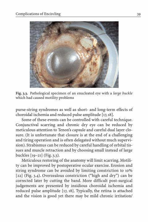
Fig. 3.3. Pathological specimen of an enucleated eye with a large bucklewhich had caused motility problems
purse-string syndromes as well as short- and long-term effects ofchoroidal ischemia and reduced pulse amplitude [17, 18].
Some of these events can be controlled with careful technique.Conjunctival scarring and chronic dry eye can be reduced bymeticulous attention to Tenon’s capsule and careful dual layer clo-sure. (It is unfortunate that closure is at the end of a challengingand tiring operation and is often delegated without much supervi-sion). Strabismus can be reduced by careful handling of orbital tis-sues and muscle retraction and by choosing small instead of largebuckles [19–21] (Fig. 3.3).
Meticulous restoring of the anatomy will limit scarring. Motili-ty can be improved by postoperative ocular exercise. Erosion andstring syndrome can be avoided by limiting constriction to 10%[22] (Fig. 3.4). Overzealous constriction (“high and dry”) can becorrected later by cutting the band. More difficult post-surgicaljudgements are presented by insidious choroidal ischemia and reduced pulse amplitude [17, 18]. Typically, the retina is attachedand the vision is good yet there may be mild chronic irritation/

inflammation and macular pigmentary changes that could also be ascribed to the original detachment or aging. It is difficult topropose to a patient that cutting the band at a small risk of re-detachment might prevent future macular changes and ill-defineddegeneration; however, the intervention might prove more effectivethan daily antioxidants.
How Drainage Works
Although fluid drainage was part of Gonin’s ignipuncture tech-nique, it had been performed without attention to retinal breaksand accordingly with predictably poor results. Rosengren, who injected air intravitreally, also drained [23]. Arruga and Schepensboth drained subretinal fluid to bring the neuroepithelia into apposition and to make space for the volume reduction that
3 Encircling Operation with Drainage for Primary Retinal Detachment40
Fig. 3.4. Pathological specimen of an enucleated eye that had a tight en-circling buckle which had covered the vortex vein ampullas.The retina wasattached at the price of constriction and ischemia

encircling entailed. Drainage also shortened recovery time,which both relieved the unease of the surgeon and shortened hospital stays from weeks and months (Treatment of Samelsohn,[7]).
Schepens reserved bed rest for macula-on retinal detachments.For all other detachments, subretinal fluid was desirable, since itmade volume reduction more effective and safer: “In all other cas-es, the patients are encouraged to be up and about, in order to keepthe retina detached prior to surgery, because the scleral bucklingoperation, which necessarily decreases the volume of the eye, re-quires the loss of ample subretinal fluid at the time of operation”[2]. The amount of fluid to be removed from the eye was inves-tigated by Thompson and Michels. The volume displacement of a2.5-mm-wide band was measured to be 0.5 ml; with explants, thedisplaced volume could be close to 2 ml or up to 45% of the vitre-ous cavity [24].
Complications of Drainage
Complications of drainage include hemorrhage, choroidal detach-ment, retinal incarceration, iatrogenic retinal holes, and infection.Cibis wrote about the uncertain nature of a release of subretinalfluid:“This is perhaps the most dangerous step in any of the retinaldetachment procedures presently employed,except where Custodis’technique is used” [25].
Intraocular bleeding (major retinal, subretinal, or vitreal) wasseen in 14.4% of drained versus 3.3% of undrained cases [26].Choroidal detachment occurred in 8.6% of drained versus 1.6% ofundrained cases. Both bleeding and choroidal detachment reducedthe probability of reattachment and good postoperative vision in this population-based study [26]. Choroidal edema was foundmore frequently in older patients after drainage, hypotony, encir-cling, larger buckles, more extensive cryopexy and more completevortex vein obstruction [19].
Complications of Drainage 41

Kreissig, who drained 98.7% of cases from 1966 to 1969, report-ed intraocular hemorrhage (smallest or larger) in 15.6% [28]. In1975, Blagojevic, who drained 96% of cases, found intraocularhemorrhages in 16% and retinal incarceration in 1% [29]. Huebner,who drained 89% of cases, found intraocular hemorrhages in 6.9%and retinal incarceration in 0.7% [30]. Also in 1975, Spalter report-ed iatrogenic retinal holes related to drainage that had occurred in2.3–14.8%, often accompanied by vitreous loss, depending on thetechnique used [31].
Improvements in the technique of drainage included transil-lumination of the choroid to identify large vessels, diathermy [32],puncturing away from vortices and long posterior ciliary vessels[29], using the microscope [33], using traction [31], or incompletedrainage [34].
All of the reports on drainage and its complications are retro-spective. It is hard to imagine a detailed report about the exact sub-macular distribution of blood in the operative note. Yet, as Cibiswryly remarked,“As you all know, surgeons, as a rule, do not reporttheir complications and mistakes unless they are related to a tech-nique devised by another surgeon” [25]. Nevertheless, reports offailures exist, relating failure to complications of drainage in amajority of cases [35]. Underreporting of less than a “major hem-orrhage” is likely. Blagojevic, who noted intraocular hemorrhagesin 16% wrote:“It is important to stress that [the intraocular hemor-rhages] were never widespread, and that they did not unfavorablyaffect the results of the operation. Hemophthalmus and choroidalhematoma were not noticed” [29]. There have been many modifi-cations of the drainage technique [27–34], attesting to the difficultyof producing a bloodless perforating injury in a highly perfused,inflamed and hypotonous vascular layer. Added risks over the pastseven decades have been increasing patient age and widespreaduse of anticoagulants. Possibly, poor visual acuity found after “scleral buckling” might be related to underreported complica-tions of drainage and layers of barely visible subretinal blood under the fovea (Fig. 3.5).
3 Encircling Operation with Drainage for Primary Retinal Detachment42

Are Encircling and Drainage Still Popular Methods?
In light of the above tales of both success and morbidity, are encircling and drainage still done? The short answer is that en-circling with drainage, as opposed to minimal segmental bucklingwithout drainage [36], requires less-accurate meridional local-ization of all breaks, is subsequently somewhat easier to do, andworks at the short-term risk of hemorrhage and long-term risk ofchoroidal ischemia.
In more recent (1982–2002) clinical series on scleral buckling[15, 26, 37, 38], encircling was practiced in 43–100% (average 74%)and was combined with drainage in 72–85% (average 78%) of totalcases. Gas was injected in 26–32% in two series; this procedure isalso known as “pneumatic buckle” [15, 37]. Primary success rangedfrom 78% to 96% (average 85%). Obstacles to success wereaphakia/pseudophakia, whereas the main benefit of encircling relative to alternate procedures was found to be the low incidenceof secondary tears (1.3% versus 18–20%) [15].
Are Encircling and Drainage Still Popular Methods? 43
Fig. 3.5. Subretinal blood after drainage. It is parafoveal, but vision re-mained poor

It is of note that two of the series [15, 37] compared scleral buck-ling (78–100% encircling and 72–85% drainage) to pneumaticretinopexy. Scleral buckling was therefore used synonymouslywith encircling and drainage in the recent literature and we can as-sume that it represents the standard of care.
Encircling at Wills Eye Hospital in 1985
To test this hypothesis, that encircling and drainage are still theprimary procedure, the author reviewed 100 consecutive scleralbuckling procedures done at Wills Eye Hospital from 1985 to 1986.Eleven members of the retina service encircled primary detach-ments in 83% and drained in 73% of cases, consistent with theliterature.
Air or gas was injected in 6%. The extent of the detachment wasone quadrant in 10%, two quadrants in 52%, three quadrants in 21%and four quadrants in 17%. The average area of detachment was 2.9quadrants.
The preferred buckling procedure consisted of a 3-mm encir-cling band used in 83%, combined with a 7-mm explant which wasused in 73%. The explant covered 2.3 quadrants on average so thatin 49% of all cases it covered the entire extent of the detachment(Fig. 3.6). The primary success rate was around 90%. The authorhad no follow-up after discharge from the hospital, except for re-admissions.
It is easy to see why this skillfully executed procedure had a highsuccess rate. Careful preoperative study was mandatory as was adetailed retinal drawing. (Encircling and drainage did not meanthat the study of the retina was optional). Patients were admittedthe day before surgery, were studied in the evening and stayedovernight to help flatten the detachment. During surgery, all breakswere carefully marked on sclera to ensure their placement on thecrest or anterior slope of the buckle. Cryopexy was applied tobreaks, lattice and suspicious retinal lesions. Since the majority of
3 Encircling Operation with Drainage for Primary Retinal Detachment44

breaks were located at a latitude 13–14 mm posterior to the lim-bus, detected, undetected and anticipated future breaks would be covered.
How Buckling Was Modified in One Practice
The author reviewed his last 100 scleral buckling procedures comprising his personal experience at the Harkness Eye Institutebetween 2000 and 2003. Encircling was done in 17%, drainage in6%, and air was injected in 11%. Gas was not used. The detachmentsextended one quadrant in 14%, two quadrants in 45%, three quad-rants in 35% and four quadrants in 6%. The detachments averaged2.3 quadrants as opposed to 2.9 quadrants at Wills Eye Hospital.
How Buckling Was Modified in One Practice 45
Fig. 3.6. Example of routine encircling and drainage: The eye was buck-led with a 7-mm explant for the entire extent of the detachment. Drainagewas made near a rectus muscle in the bed of the buckle. A 2.5-mm bandwas used to encircle the eye to barricade undetected breaks

Double patching and bed rest were encouraged whenever possible.After localization of all breaks and cryopexy, 5-mm sponges wereapplied in 82%. They were placed radially in 49% and circum-ferentially in 33%. The primary success rate was 87%.
Reasons for Modification
As indicated above, the success rate depends on case selection,length of follow-up, whose cases they are, who evaluates them, etc.The impetus for change in the latter series was the unavoidablemorbidity associated with encircling and drainage. In order tomake sure that the overall success rate corresponded to the pub-lished norm of 80–90%, modifications occurred gradually – firstby tying the band loosely, then by omitting it. Modifications werealso made by shortening the circumferential 7-mm explant and finally by just buckling the breaks, preferably in a radial orienta-tion. Fewer and fewer cases were drained, sometimes at the cost ofsleep.
This experience somewhat parallels the trend described by Lincoff,who drained 48% of cases in 1963 and only 13% by 1971 [39],or by Kreissig who drained almost all cases in 1966, 6% of cases in1972 [40] and none in 1992 [36]. The reasons for drainage were (1)giant tears, (2) severe preretinal retraction, (3) uncertain local-ization of the break, (4) defective choroid, (5) thin sclera, or (6)glaucoma [39]. Reasons 1–3 might be managed by vitrectomy tech-niques today including encircling with the ubiquitous 3.5-mmband, which reduces indications for drainage.
Are Encircling and Drainage Still Worth Doing?
As every detachment surgeon knows,there are detachments of goodand of poor prognosis. Good prognostic signs include: fewerbreaks, less extensive area of detachment, shallow detachments and
3 Encircling Operation with Drainage for Primary Retinal Detachment46

phakic status (Fig. 3.7). Indicators of poor prognosis include: manybreaks, undetectable breaks, a large area of detachment, bullous de-tachment, aphakic/pseudophakic status and proliferations of vitre-ous and retina (Fig. 3.8). Not surprisingly, these indicators are simi-lar in the literature on both buckling and pneumatic retinopexy [16].
Minimal procedures are appropriate to repair more favorablecases, whereas procedures associated with higher morbidity are re-served for the complex case. Schepens corroborated this finding.“The circling element was at first used in cases with an unfavorableprognosis.As experience with this procedure increased it was usedon more and more favorable cases and it was found to be the mostdependable operation”[2]. This misconception, that if it is good forcomplicated cases it is even better for uncomplicated ones, hasproved to be a common reasoning in clinical practice, as observedby Lincoff and Kreissig [41].
Are Encircling and Drainage Still Worth Doing? 47
Fig. 3.7. A detachment with a good prognosis. A one-quadrant detach-ment, one break, in a 50-year-old male patient, flattened with bed rest.A radial 5-mm sponge was placed, followed by laser

Encircling and drainage may still be of value in highly bullousdetachments not responding to bed rest, many breaks of similarlatitude, anterior vitreoretinopathy, need for a higher and morepermanent buckle and thin sclera interfering with suturing. Theauthor prefers encircling for most revisions of failed segmentalbuckles. The reasons are psychological and practical: the minimalprocedure has failed and the revision includes the preparation forpossible vitreous surgery. The band is in place, but may not be tied.
Conclusion
Whereas modern examination techniques facilitate localization ofbreaks and allow for precise treatment of the holes, drainage and
3 Encircling Operation with Drainage for Primary Retinal Detachment48
Fig. 3.8. A detachment with a poor prognosis in a 42-year old male pa-tient. There were eight breaks. It was a reoperation with PVR stage B,unresponsive to bed rest. Five segmental buckles were placed. The retinaattached without drainage. After 3 months the retina redetached due toPVR. This may represent the limit of the segmental buckling technique

encircling continue to be popular and are used by a majority ofsurgeons. Careful preoperative examination including a detailedfundus drawing was advocated by Schepens and should still bedone, irrespective of the surgical method.Examination is time con-suming in the age of managed care and even the best effort cannotalways identify all breaks. For the buckling procedure to be suc-cessful, all breaks have to be identified and closed, encircled or not.
Encircling and drainage were successful in 78–96% and havebecome synonymous with scleral buckling [15, 37]. Since the 1950s,at least two generations of surgeons have been well trained in this procedure. It is “dependable” and incorporates the barrier concept [2]. Intraoperative localization as to latitude is critical,but meridional localization may be less precise compared withminimal radial buckling. The vitreous base is ring-like; supportingit treats the hidden break and the anticipated traction. Broad buck-les support anterior PVR and circumferential retinotomies [42].This “ring” concept is behind prophylactic buckling and lasercircling for 360 degrees, as they are meant to barrage and reducethe incidence of secondary breaks in alternate techniques [14, 16].Most encircling is reversible: a band can be cut in a timely fashionwithout re-detachment or permanent damage from ischemia.
Can the surgeon sleep better after the retina has been drainedflat? It depends: a non-drainage procedure increases the chance of primary failure, but the eye will survive the attempt almost intact. By draining, the retina may be attached on the table, yetmorbidity (blood under the macula etc.) may forever preclude visual recovery. Who could sleep well after the latter? From apathologist’s viewpoint, drainage will always be a penetrating in-jury to a vascular tissue in an inflammatory and hypotonous setting. The data reporting intraocular hemorrhage attest to thissimple fact that cannot be changed by even the most sophisticatedtechnique. The fear of anatomic failure (first operation success orlack thereof ) apparent to both physician and patient has helped the propagation of techniques that flatten the retina under the surgeon’s eye, like external drainage or internal drainage during
Conclusion 49

vitrectomy. Both procedures share the complications of penetrat-ing ocular injury.
After careful examination, a skillfully executed encircling anddrainage operation has a high rate of success. However, morbidityleads to a gradual change in the author’s practice. Encircling anddrainage are not necessary conditions for a high rate of first opera-tion success after scleral buckling; in fact, one can be equally suc-cessful employing less-morbid procedures. An editorial concludedthat “accurate localization of all retinal holes and precise placing ofthe buckle are of course essential, and confidence in the success ofthe procedure is necessary to outweigh a fear of failure induced in the surgeon by the presence of fluid under the retina at the end of the operation. The temptation to drain and ‘make sure’ must beresisted.Yielding may well court complications” [43].
� Acknowledgements. Illustrations by Daniel Casper, MD, PhD.This chapter was supported by an unrestricted grant generouslyprovided by Mr. Walter Klein.
References
1. Arruga MH (1958) Le cerclage equatorial pour traiter le decollementretinien. Bull Soc Franc Ophtal 71:571–580
2. Schepens CL, Okamura ID, Brockhurst RJ (1957) The scleral bucklingprocedures. 1. Surgical techniques and management. Arch Ophthal-mol 58:797–811
3. Janssens GJA (1958) The treatment of retinal detachment by means of“eye-ball reducing” operations. Dekker & van de Vegt N.V., Utrecht
4. Safar K (1932) Behandlung der Netzhautabloesung mit Elektrodenfuer multiple diathermische Stichelung. Ber Dtsch Ophthalmol Ges49:119
5. Jess A (1937) Temporaere Skleraleindellung als Hilfsmittel bei der Operation der Netzhautabloesung. Klin Monatsbl Augenheildlk 99:318–319
3 Encircling Operation with Drainage for Primary Retinal Detachment50

6. Custodis E (1953) Bedeutet die Plombenaufnaehung auf die Skleraeinen Fortschritt in der operativen Behandlung der Netzhautabloe-sung? Ber Dtsch Ophthalmol Ges 58:102–105
7. Gonin J (1921) Le traitement du decollement retinien. Ann D’Oculis-tique 158:175–194
8. Gonin (1928) J Mes plus recentes Experiences touchant le DecollementRetinien. Arch d’Ophtalmologie 55:554–561
9. Dark AJ, Rizk SNM (1965) Untoward sequels of arruga encirclementfor retinal detachment. Br J Ophthal 49:259
10. Manson N (1964) The “String Syndrome” seen as a complication ofArruga’s cerclage suture. Br J Ophthal 48:70
11. Schepens CL, Okamura ID, Brockhurst RJ (1958) The scleral bucklingprocedures. II. Technical difficulties of primary operations.Arch Oph-thalmol 60: 84–92
12. Schepens CL (1964) Scleral buckling with circling element. Trans AmAcad Ophthalmol Otolarnygol 68:959–979
13. MIRA Vitreoretinal Implants. Mira, Inc., Waltham, MA14. Hudson JR, Kanski JJ, Elkington AR (1973) Prophylactic encirclement.
Br J Ophthal 57:531–53515. McAllister IL, Meyers SM, Zegarra H, Gutman FA, Zakov ZN, Beck GJ
(1988) Comparison of pneumatic retinopexy with alternative surgicaltechniques. Ophthalmology 95:877–883
16. Tornambe PE (1997) Pneumatic retinopexy: the evolution of case se-lection and surgical technique. A twelve-year study of 302 eyes. TransAm Ophthalmol Soc 95:551–578
17. Dobbie JG (1980) Circulatory changes in the eye associated with reti-nal detachment and its repair. Trans Am Ophthalmol Soc 78:503–563
18. Yoshida A, Feke GT, Green GJ, Coger DG, Matsuhashi M, Jalkh AE,McMeel JW (1983) Retinal circulatory changes after scleral bucklingprocedures. Am J Ophthalmol 95:182–188
19. Tornambe PE, Hilton GF, the Retinal Detachment Study Group (1989)Pneumatic retinopexy. A multicenter randomized controlled clinicaltrial comparing pneumatic retinopexy with scleral buckling. Ophthal-mology 96:772–784
20. Lagreze W, Lieb B, Schrader W, Kommerell G. Periphere (1993) Netz-hautkorrespondenz nach Amotio retinae. Klin Monatsbl Augenheilkd203:347–350
21. Schrader WF, Hamburger G, Lieb B, Hansen LL, Kommerell G (1995)Motilitaet und Binokularfunktion nach radiaerer episcleraler Plombe.Klin Monatsbl Augenheilkd 207:224–231
References 51

22. Lincoff H, Kreissig I, Parver L (1976) Limits of constriction in thetreatment of retinal detachment. Arch Ophthalmol 94:1473–1477
23. Rosengren B (1938) Ueber die Behandlung der Netzhautabloesungmittelst Diathermie und Luftinjektion in den Glaskoerper. Acta Oph-thalmologica 16:3
24. Thompson JT, Michels RG (1985) Volume displacement of scleralbuckles. Arch Ophthalmol 103:1822–1824
25. Cibis PA (1965) Errors in the management of retinal detachmentscomplications avoidable and unavoidable. Mod Probl Ophthalmol3:284–308
26. Toernquist R, Toernquist P (1988) Retinal detachment. III. Surgical results. Acta Ophthalmologica 66:630–636
27. Aaberg TM, Maggiano JM (1979) Choroidal edema associated withretinal detachment repair: experimental and clinical correlation. ModProbl Ophthalmol 20:6–15
28. Kreissig I (1971) Kryopexie in der Ablatio-Chirurgie. Klin MonatsblAugenheilk 159:737–741
29. Blagojevic M, Ilic R (1975) Drainage of the subretinal fluid and injec-tion of saline solution in the vitreous body. Mod Probl Ophthalmol15:169–173
30. Huebner H, Boeke W (1975) Klinische und technische Aspekte dersubretinalen Drainage. Mod Probl Ophthalmol 15:137–142
31. Spalter HF (1975) Traction release of subretinal fluid. Mod Probl Ophthalmol 15:163–168
32. MacKenzie Freeman H, Schepens CL (1975) Innovations in the tech-nique for drainage of subretinal fluid transillumination and choroidaldiathermy. Mod Probl Ophthalmol 15:119–126
33. Gaertner J (1975) Release of subretinal fluid with the aid of the micro-scope. Report on 100 cases. Mod Probl Ophthalmol 15:127–133
34. Martin B (1975) Controlled release of subretinal fluid. Mod Probl Ophthalmol 15:149–153
35. Chignell AH, Fison LG, Davies WG, Hartley RE, Gundry MF (1973)Failure in retinal detachment surgery. Br J Ophthalmol 57:525–530
36. Kreissig I, Rose D, Jost B (1992) Minimized surgery for retinal detach-ments with segmental buckling and nondrainage. Retina 12:224–231
37. Han DP, Mohsin NC, Guse CE, Hartz A, Tarkanian CN, SoutheasternWisconsin Pneumatic Retinopexy Study Group (1998) Comparison of pneumatic retinopexy and scleral buckling in the management ofprimary rhegmatogenous retinal detachment. Am J Ophthalmol 126:658–668
3 Encircling Operation with Drainage for Primary Retinal Detachment52

38. Schwartz SG, Kuhl DP, McPherson AR, Holz ER, Mieler MF (2002)Twenty-year follow-up for scleral buckling. Arch Ophthalmol120:325–329
39. Lincoff H, Kreissig I, Goldbaum M (1974) Reasons for failure in non-drainage operations. Mod Probl Ophthalmol 12:40–48
40. Kreissig I (1978) Der gegenwaertige Stand der Ablatiochirurgie ohnePunktion. Klin Monatsbl Augenheilk 173:140–149
41. Lincoff H, Kreissig I (2000) Changing patterns in the surgery for reti-nal detachment: 1929 to 2000. Klin Monatsbl Augenheilkd 216:352–359
42. Charles S, Katz A,Wood B (2002) Vitreous microsurgery, 3rd edn. Lip-pincott Williams & Wilkins, Philadelphia
43. Anonymous (1975) Editorial: non-drainage of subretinal fluid. Br JOphthalmol 59:251
References 53

Pneumatic Retinopexy for Primary Retinal Detachment
Eric R. Holz, William F. Mieler
Chapter 4
Introduction
Pneumatic retinopexy (PR) is a minimally invasive surgical techni-que utilized for the repair of rhegmatogenous retinal detachment(RRD).It is composed of intravitreal gas injection,either cryopexy orlaser, and postoperative patient positioning. Historically, the first report of the use of intraocular gas injection for the treatment ofRRD appeared in 1911 [1]. Rosengren, in 1938, reported the first largeseries of patients treated with intraocular air injection, drainage ofsubretinal fluid (SRF), and diathermy [2]. The modern PR techniquewas born in 1985 with simultaneous reports by Hilton and Grizzardin the United States [3] and Dominguez in Spain [4, 5].
The PR procedure appears to be gaining in popularity, althoughthere is considerable variation in its use based on geography andpractitioner’s years in practice. Surveys of American vitreoretinalspecialists in 1990 and 1997 revealed an increase in use from 38% to55% for a given “ideal” clinical scenario. There was significant va-riation in the choice of surgical technique based on surgeon age.Those in practice less than 10 years chose PR 65% of the time com-pared with 35% of those in practice 20 years or more. Within theUnited States, there was regional variation with 74% of survey respondents in western states choosing PR compared with 43% innorth central states [6].
The rise in the popularity of PR may be due to the perceivedbenefits to the patient, the attending physician, and society as awhole. Well-informed patients typically prefer PR because of its

minimally invasive nature. There is little pain, more rapid return ofvision, and fewer postoperative complications, resulting in a moreexpedient return to daily activities. For the surgeon, the procedureis quick, technically easy, and performed in the outpatient or officesetting. These advantages, combined with the procedure’s low cost,have led to increased interest and usage of PR for the treatment ofprimary RRD.
Technique
Pneumatic retinopexy consists of several key elements, includingproper preoperative patient and case selection, gas injection, laseror cryopexy, and postoperative positioning.
Patient Selection
The use of PR demands a high degree of patient understandingand compliance in order to succeed. The most frequent cause offailed closure of the primary break(s) is inadequate tamponadedue to patient non-compliance.As a result, the patient must be ableto understand the vital role of bubble positioning during the early postoperative course. The surgeon should take extra careeducating the patient preoperatively about the desired head posi-tion. Taking the patient’s head in his/her hands and turning or tilting it to the proper orientation is frequently helpful. Involvingaccompanying family or friends in the discussion may reinforcethe message. Physically, the patient must be able to comply. Thosewith arthritis, neck or back problems, or other physical constraintsare less desirable candidates for PR. In general, a young, bright,highly motivated individual is the best candidate for the PR technique.
4 Pneumatic Retinopexy for Primary Retinal Detachment56

Case Selection
Proper case selection is critical to success with PR. The “ideal” sce-nario involves an acute, phakic retinal detachment due to a singlebreak or small cluster of breaks located in the superior 8 clockhours of the fundus. Careful preoperative examination is exceed-ingly important when considering PR. Clear ocular media are es-sential to allow visualization of all breaks. Sector cataract, vitreoushemorrhage, and pseudophakic lens capsular opacification are relative contraindications. In general, pseudophakic and aphakicdetachments are more prone to multiple small breaks than phakiccases. However, if the view to peripheral retina affords a view suffi-cient to disclose all the breaks, these detachments can be managedwith PR. A single break is most easily covered with bubble tam-ponade. If multiple, the breaks must be close enough together to be covered by a single bubble.Breaks greater than 90–120° apart re-quire large volume injections and, as a result, are relative contrain-dications. Retinal tears located in the superior 8 clock hours areeasier to treat because gas bubbles float in the fluid vitreous. Al-though retinal tears in attached retina located inferiorly are easilymanaged with barricade laser, breaks in detached retina in the inferior 4 clock hours present a relative contraindication for PR(see below – New possibilities).
There are several other contraindications to PR. Required pa-tient air travel while the bubble is in place is an absolute con-traindication. There is no relief of vitreoretinal traction with PR;the adhesion formed must be stronger than the tractional forcesgenerated to achieve long-term success. As a result, patients withsevere traction due to proliferative vitreoretinopathy (PVR) are notgood candidates. Subretinal fluid is removed by the pigment epi-thelial pump. This process is much more efficient with the liquidSRF of acute detachments than with the viscid proteinaceous fluidencountered with chronic detachments. Pneumatic retinopexy canbe successfully utilized with the latter, but there may be loculatedpockets of chronic SRF that persist for months due to delayed
Technique 57

resorption. The combination of glaucoma with retinal detachmentleads to several considerations with respect to PR. Patients with a functioning bleb or tube shunt device in place may be bettermanaged by PR than scleral buckle (SB). Although an expandinggas bubble has the potential to dramatically raise the intraocularpressure (IOP), bubble expansion typically occurs simultaneouslywith resorption of SRF. The resolution of SRF provides potentialspace for bubble expansion without perturbations in the IOP. Onlydetachments with scant SRF or chronic, thick SRF are more proneto IOP problems and, as a result, are relative contraindications toPR in patients with coexisting glaucoma.
Gas Selection
Intraocular gas works by temporarily closing retinal breaks via thesurface tension properties of the bubble meniscus. Blocking themovement of liquid vitreous into the subretinal space allows theretinal pigment epithelium to actively pump fluid from the sub-retinal space and flatten the detachment. Once the neurosensoryretina is in apposition to the pigment epithelial layer, the adhesiveproperties of cryopexy or laser retinopexy permanently close thebreak(s). The most commonly utilized gases are air, sulfur hexa-fluoride (SF6), and perfluoropropane (C3F8). Choice of gas is basedupon volume issues, arc length of contact/bubble size require-ments, and bubble duration (Table 4.1) [7]. The tamponade mustlast until the laser or cryopexy adhesion is strong enough to resistreopening – generally 3–5 days for laser and 5–7 days for cryopexy.Air is non-expansile and quickly disappears from the eye. Sulfurhexafluoride and C3F8 are expansile and have longer half-lives. Ingeneral, a 1-ml final bubble size tamponades a 120° arc length,which is sufficient for most cases of PR (Table 4.2) [8]. There is,however, considerable variation based on the axial length/ size ofthe globe, so that larger bubbles are required in myopic individualsto achieve the desired arc length of contact.
4 Pneumatic Retinopexy for Primary Retinal Detachment58

The volume of the initial injection is a key consideration in gasselection. Intravitreal injections of 0.1–0.2 ml usually result inmodest IOP rises, but increasing the initial volume beyond thispoint dramatically increases pressure. Paracentesis provides po-tential space within the globe, but is usually limited to 0.1–0.2 ml.As a result of these considerations, the authors routinely use0.25 ml of pure (100%) C3F8 gas, which provides a small initial in-jected volume and a large final volume (~1 ml) sufficient to tam-ponade most breaks.
Technique
Pneumatic retinopexy is either performed in one or two steps,depending on whether cryopexy or laser is utilized to form thechorioretinal adhesion (Tables 4.3, 4.4). Anesthesia may be applied
Technique 59
Table 4.1. Gas characteristics (from [7])
Expansion Half-life Isoexpansile Standard volume(days) concentration (pure gas)
Air 0 1 100% 0.5–1.0 mlSF6 2.0–2.5¥ 2.5 18% 0.5 mlC3F8 4¥ 4 14% 0.3 ml
Table 4.2. Bubble size/tamponade arc. Arc varies with axial length/size of globe (from [8])
Final bubble size Arc of contact
0.30 ml 90°1.0 ml 120°3.0 ml 180°

by topical, subconjunctival, peribulbar, or retrobulbar routes. Aretrobulbar or parabulbar injection is given when cryopexy isplanned, while topical or subconjunctival administration is suffi-cient for gas injection alone. The globe, eyelids, and periocular skinare prepared with a 5% povidone–iodine solution applied twice at5-min intervals. Some surgeons choose to add a topical antibiotic
4 Pneumatic Retinopexy for Primary Retinal Detachment60
Table 4.4. Pneumatic retinopexy technique steps
Apply anesthetic of choicePrepare eye – povidone–iodine 5% – 5 min ¥2Prepare chosen gasPlace eyelid speculumPerform cryopexy (one-step procedure)Paracentecis (surgeon’s choice)Position patient, headCaliper measurement – 4 mmInject gas bubble/locationInspect gas location, central retinal artery perfusionCheck intraocular pressure until normalizesPatch with arrow drawnLaser (next day, if two-step procedure)
Table 4.3. Required materials
Anesthetic of choicePovidone–iodine 5% solutionEyelid speculum and caliperSeveral sterile cotton-tip applicators1-ml syringe/30-gauge needle – plunger removed (if paracentesis)Gas cylinder/tubingMillipore filter1-ml or 3-ml syringe/30-gauge needleCryotherapy unit or laserAntibiotic/steroid combination ointmentPatch materials

solution; however, these need to be started 24–48 h prior to the pro-cedure with frequent use to substantially affect the ocular flora,limiting their usefulness as a true prophylactic method in this set-ting. A wire-lid speculum is used to open the lids, avert the lashesfrom the field, and protect the lid margin from cryopexy damage.If the patient is to undergo a one-step procedure, then cryopexy isperformed prior to gas injection, as small breaks may be difficultto visualize following gas injection. Cryopexy is the preferredmethod in cases where media opacities limit the view, when thebreak(s) are located in the far periphery, or when there is under-lying pigment epithelial atrophy. Laser, via a two-step method, ispreferred with bullous superior detachments with large retinalbreaks, when breaks occur over a previously placed buckle element,and with posteriorly located tears. Some surgeons feel that there is a lower incidence of PVR with laser retinopexy compared withcryopexy. Laser can be difficult to perform through or around thegas bubble and will not provide adequate adhesion if there is still asmall amount of fluid present near the tear site. Small breaks maybe difficult to find with the laser once the retina is reattached. Lasermight form a chorioretinal adhesion faster than cryopexy, whichdecreases the critical duration of required post-operation posi-tioning. Multiple rows of confluent laser are placed around eachtear with careful attention to carry the treatment anterior to theora serrata. It should be noted that the two methods are notmutually exclusive, and many instances are best managed by acombination of both modalities.
Preparation of the chosen gas is performed by withdrawing gasfrom a cylinder via a valve system through a Millipore filter into a1-ml or 3-ml syringe. The valve, tubing, filter, and syringe areflushed with gas once, and the process is repeated to eliminateroom air from the system. High pressures may damage Milliporefilters, so care must be taken to maintain lower pressures duringgas filtration. The filter is replaced with a 30-gauge needle, and excess gas is pushed out of the syringe until the desired amount forinjection remains behind.
Technique 61

Paracentesis of the anterior chamber is a controversial step inthe procedure. Some surgeons routinely soften all eyes prior to in-jection, while some perform the step only rarely. Others performparacentesis after the gas injection as required by the IOP. Para-centesis is less important with one-step procedures where thescleral depression associated with cryopexy softens the globe andin cases where smaller volumes of expansile gas are utilized. Para-centesis is most often required in two-step (laser) cases and wheninjecting large gas volumes. The step is performed by entering the anterior chamber with a 30-gauge needle affixed to a 1-mlsyringe without the plunger.Aqueous humor is allowed to passive-ly egress until the anterior chamber shallows. A sterile cotton tipapplicator is rolled onto the needle track as the needle is with-drawn to avoid additional fluid egress. Care is given to avoid needletip-lens touch. Paracentesis is contraindicated in aphakic andpseudophakic patients with vitreous prolapse into the anteriorchamber.
Gas injection is the most important component step of PR, andmany postoperative complications can be avoided with propertechnique. The surgeon utilizes the indirect ophthalmoscope forlighting, visualization of needle tip, and later to assess gas locationand patency of the central retinal artery. The patient is placed in arecumbent position with the head tilted 45° away from the opera-tive eye. This places the temporal pars plana as the highest point on the globe. The injection is given 4 mm posterior to the limbus,usually in the temporal quadrant, unless the retina is bullously de-tached in the area. The needle tip is advanced into the mid-vitreouscavity, under direct visualization with the indirect ophthalmo-scope to penetrate the anterior hyaloid face. Then the needle iswithdrawn until just the tip is visible, 2–3 mm through the parsplana epithelium. Gas is injected in a brisk but controlled manner.Following gas injection, the head is carefully rotated to a neutralposition in order to move gas away from the injection site andavoid egress of gas out the needle track. A sterile cotton tip appli-cator is rolled over the track as the needle is removed to minimize
4 Pneumatic Retinopexy for Primary Retinal Detachment62
gas reflux. Following gas injection, the bubble size and position are assessed, and central retinal artery perfusion is assured withindirect ophthalmoscopy.
IOP rises abruptly in most patients who receive greater than0.2 ml gas. The IOP is checked immediately following gas place-ment. Frequently, pressure measurements fall in the 50–70 mmHgrange. If the patient has a normal aqueous outflow mechanism, heor she can be monitored with serial tonometry every 10–15 min,which frequently demonstrates a return to more normal pressuresover 15–30 min. Paracentesis, as noted above, may be performedbefore or after gas injection (or both) to normalize IOP. The pres-sure should be near normal and the central retinal artery perfusedprior to the patient’s departure.
Postoperatively, an antibiotic/steroid combination ointment isplaced in the eye, and a patch is applied. An arrow is drawn on thepatch, such that the arrow points straight at the ceiling when thepatient is properly positioned (break in uppermost position of theglobe). The patient and caregiver are reminded of the required po-sition with special emphasis on the need for compliance, especial-ly at night while asleep. The patient returns for follow-up on thefirst postoperative day. The SRF is usually substantially improvedor entirely resolved. The gas bubble size and location are assessed,and the IOP is measured. Laser may be performed as part of astaged procedure (see above).For patients with extensive cryopexy,antibiotic and steroid drops may be prescribed for a few days.The importance of proper position is stressed yet again. In caseswhere there is little or no change in the SRF, patient compliance isreassessed, and an exhaustive exam for new or missed breaks is undertaken. In the typical scenario where the fluid is substan-tially better, the patient is re-examined on the third to fifth post-operative day.
Technique 63

Complications: Prevention and Management
Intraoperative
There are limited complications associated with the use of PR. Themost common set of problems arise from difficulties with the gasbubble itself, particularly migration into unintended potentialspaces. Subconjunctival gas is the most common location, being re-ported in 0–10% of cases (Table 4.5) [3, 9–11]. Gas has also been re-ported in the subretinal space (0–4%) [12–15], anterior to the ante-rior hyaloid (0–9%) [11, 13, 14], in the suprachoroidal space (0–5%)[13–15], and exterior to pars plana epithelium (0–1%) [9]. Followinginjection, the gas inside the eye may form multiple small “fish egg”bubbles rather than a single large one. Fish eggs provide inade-quate tamponade, as they do not occlude breaks with the same ef-ficiency as a large smooth meniscus. This same feature also makesthem more likely to migrate into the subretinal space. Multiplesmall bubble formation can usually be avoided through proper in-jection technique (see above).When this does occur, the eye may beforcefully tapped or “thumped” with the surgeon’s finger, whichcan lead to coalescence (a technique that has been described,
4 Pneumatic Retinopexy for Primary Retinal Detachment64
Table 4.5. Reported intraoperative complications
Subconjunctival hemorrhage [3] Up to 60%Subconjunctival gas [3, 9–11] 0–10%Anterior hyaloid gas [11, 13, 14] 0–9.7%Vitreous hemorrhage [3, 9, 13, 15] 0–5%Choroidal detachment [13–15] 0–5%Subretinal gas [12–15] 0–4%Vitreous incarceration [9, 10, 12, 13] 0–3.6%Sub pars plana gas [9] 0–1%Subretinal hemorrhage [13] 0–1%Hyphema [13, 14] 0–1%Lens touch [13] 0–1%

though perhaps not employed by all). If this maneuver fails, the patient should be positioned face down for 6–12 h in order to prevent subretinal migration. During this time period, fish eggs inevitably unite to form the desired, effective single large bubble.Migration of bubbles, especially with expansile gas, into the sub-retinal space is a substantial complication. This event can be avoid-ed by visualizing the needle within the vitreous cavity prior toinjection, achieving a single bubble rather than fish eggs, and by avoiding case selection involving large tears with severe trac-tion. Once gas enters the subretinal space, it may be managed bymaneuvering the patient’s head and eye in such a way that it rollsthe bubble back through the tear into the vitreous cavity. This is often aided by simultaneous scleral depression. These maneuversare often unsuccessful, and vitrectomy surgery is necessary for removal. During vitrectomy, the bubble will displace the detachedretina anteriorly toward the lens – making infusion line placement,sclerotomy incisions, and instrument entry into the eye problem-atic. A small retinotomy performed with the vitreous cutter probelocated at the most anterior, superior pole of the subretinal bubbleusually works well for evacuation.
Postoperative
The most common postoperative complication of PR is new and/ormissed retinal breaks (Table 4.6) [3, 9, 11–18]. Most of these are dis-covered during the first postoperative month, with between 61%and 86% being identified during this time period [19, 20]. Of newand/or missed breaks, 76% occur in the superior two-thirds of theretina. They almost invariably occur anterior to the equator andare more common in pseudophakic or aphakic eyes [20]. Missedbreaks can be avoided by performing a very thorough preoperativeretinal examination. The authors have found that a 78D or 90Dexam of the peripheral retina is invaluable for discovering smallbreaks preoperatively. Additionally, cases with media opacities,
Complications: Prevention and Management 65

such as sector or spoke cortical cataracts, peripheral capsularopacification, and vitreous hemorrhage, which preclude a clearview, may be less well suited to PR. The risk of new break formationis minimized using prophylactic treatment of at-risk lesions, suchas lattice patches, cystic tufts, and meridional complexes, with laserprior to gas injection. Consideration should be given to prophylac-tic 360° laser at the vitreous base, which has been reported to low-er the rate of new/missed breaks and increase surgical success [14].Delayed resorption of SRF is encountered in between 0% and 6%of cases [10, 14, 16]. In most instances, the original detachment wassubacute or chronic, and the SRF was shifted away from the origi-nal break, trapping it in the subretinal space.
Additional reported posterior segment complications includedepiretinal membrane (ERM) formation in 0% to 11% of cases [9,11–17], PVR in 3% to 13% [3, 9, 11–18], cystoid macular edema (CME)in 0% to 8% [14, 18], macular hole in 0% to 3% [13–15, 17], anteriorischemic optic neuropathy in four cases [14, 17], and endoph-thalmitis in one case [13].
Late anterior segment complications include cataract forma-tion.Although lens injury during injection is rare, late cataract pre-sumably due to gas-lens touch is much more common. Cataract isreported in 0% to 20% of cases [11, 13–15, 17, 18], with rates depend-
4 Pneumatic Retinopexy for Primary Retinal Detachment66
Table 4.6. Postoperative complications
New/missed breaks [3, 9, 11–18] 7–33%Break re-opened [13, 15] 0–14%Epiretinal membrane [9, 11–14, 16, 17] 2–11%Proliferative vitreoretinopathy [3, 9, 11–18] 3–13%Cataract [11, 13–15, 17, 18] 0–20%Cystoid macular edema [14, 18] 0–8%Delayed resorption of subretinal fluid [10, 14, 16] 0–6%Macular hole [13, 14, 15, 17] 0–3%Anterior ischemic optical neuropathy [14, 17] 0–2%Endophthalmitis [13] <1%

New Possibilities 67
ing on type and amount of gas used as well as duration of follow-up postoperatively.
New Possibilities
Surgeons continue to push the limits for detachments amenable to treatment using PR. Detachments with breaks in more than onequadrant may be repaired by augmenting the bubble size via a second injection on the first or second postoperative day, or by flattening one break over a 72-h period, then changing patient positioning to address the second area in another quadrant [21].The treatment of detachments with large breaks has been contro-versial. Gas is more prone to migrate into the subretinal space, andthe arc of contact may not be broad enough to tamponade the en-tire break. Nevertheless, reports exist of the successful use of PR forRRDs due to giant retinal tear (4 of 5–80%), retinal dialysis (4 of4–100%), and other large breaks [22–24]. These reports demon-strate that PR can be effective for cases with large breaks if they arelocated superiorly and lack significant vitreoretinal traction.Pneumatic retinopexy has generally been avoided for RRD withbreaks in the inferior 4 clock hours of the fundus. Inverted PR hasbeen reported in phakic detachments. Utilizing 8 h of “head dan-gling” positioning followed by laser retinopexy or cryopexy, thesingle surgery reattachment rate was 9 of 11 (82%) [25]. It is evidentthat although PR has an “ideal” scenario for its chief indication, thetechnique is more widely applicable in certain select cases for thosewith multiple breaks, large breaks, and even breaks located in theinferior four clock hours.

Results
Anatomic Results of PR
The reported primary anatomic success rates of PR vary widely inpublished series, ranging from 61% to 90% with an overall singleprocedure combined rate of 75.5% – 918/1,215 cases (Table 4.7) [3,9–18, 26–29]. There does not appear to be a difference between theselection of tamponade agents with respect to anatomic outcome –with filtered air, SF6, and C3F8 having similar reported results.There is no trend toward higher success rates over time. The 84%primary anatomic success rate reported by Hilton [9] is similar to the reports by Abecia [15] and Eter [28], with success rates of82% and 86%, respectively. The overall final anatomic (with re-operations) success varies between 87% and 100% in this group of
4 Pneumatic Retinopexy for Primary Retinal Detachment68
Table 4.7. Anatomic and visual outcomes
Author Year Number Technique Primary Final Visualof success success outcome patients (for macula-
detached subset)
Hilton 1986 20 0.3 cc 90% 100%[3] C3F8
0.6 cc SF6
Hilton 1987 100 C3F8 or 84% 98% 65%[9] SF6 20/20–20/50
Chen 1988 51 0.3 cc 63% – 35% [16] C3F8 20/20–20/50
Lowe 1988 55 0.3–0.5 cc 82% 98%[12] C3F8

Results 69
Table 4.7 (continued)
Author Year Number Technique Primary Final Visualof success success outcome patients (for macula-
detached subset)
Algvere 1988 58 C3F8 64.0% 95% 74% [26]
Tornambe 1989 103 C3F8 or 73.0% 99% 80% [13] SF6 20/20–20/50a
Sebag 1993 45 0.8 cc Air 86.7% 100% 64% [27] 20/20–20/50
Trillo 1993 55 0.6 cc SF6 83.6% 100%[10] 0.3 cc C3F8
Tornambe 1997 302 C3F8 or 68% 95%[14] SF6
Han [17] 1998 50 SF6, C3F8, 62% 98% 63%Air 20/20–0/50
Lisle [18] 1998 48 0.6 cc SF6 83% 100% 65% >0.40.3 cc C3F8
Assi [11] 1999 31 SF6 or C3F8 61% 87% 45% >6/12a
Abecia 2000 219 0.5 cc SF6 82% 98.9%[15]
Eter [28] 2000 78 0.4 cc SF6 86% 98.7%
Overall 75.5% 97.4%average
a Macula detached <2 weeks

reports, with a cumulative success rate of 97.4%.When PR fails, themost common second technique employed was SB; however, anumber of patients undergo either pars plana vitrectomy (PPV)alone or PPV with SB.
Visual (Functional) Results
The functional results of PR reported in the literature vary widely,so that it is difficult to compare and summarize the data. For thosepatients with macula-involved detachments, final best-correctedvisual acuity greater than or equal to 20/50 was achieved in be-tween 35% and 80% of cases. The wide variation in these numbersprobably represents variation in the duration of macular detach-ment. Studies that categorized detachments of less than 2 weeks ofmacular involvement tended to have better outcome averages.Overall, most series reported averages for best-corrected visualacuity greater than or equal to 20/50 following macula-involvingdetachments to be about 65%.
When examining the data for patients without preoperative detachment of the macula, 86% to 88% will have the same or im-proved best-corrected visual acuity [9, 13]. However, between 12%and 14% of patients will lose two or more lines of best-corrected visual acuity. Surgical failures and complications were the reasonfor vision loss in most instances.
Reasons for Failure
The causes of anatomic failure following PR have been examinedin several series [19, 30]. New retinal breaks are the most common-ly cited reason for failure; however, missed pre-existing breaks arecommonly grouped together because of the difficulty differentiat-ing the two. New and/or missed breaks occur in between 7% and33% of reported cases [3, 9, 11–18] and account for 48% to 73% of
4 Pneumatic Retinopexy for Primary Retinal Detachment70

surgical failures. Failure to close the initial break and re-opening ofthe initial break are typically grouped together. This problem is encountered in 5% to14% of cases [13, 15] and is responsible for 25%to 51% of surgical failures. PVR occurs postoperatively in 3% to 13%of cases in reported series [3, 9, 10–18], though fortunately, it is arare cause of failure.
Discussion
Rhegmatogenous retinal detachment is a very heterogeneous disease state, and, as a result, comparison of surgical results of dif-ferent techniques is difficult. Certainly PR, primary PPV, and SBeach have a place in a surgeon’s armamentarium of treatmentmodalities. The use of PR is limited by anatomic considerations –number, location and size of breaks, chronicity, preoperative PVR,and lens status – while primary PPV and SB techniques can be usedfor most cases of RRD. Nevertheless, PR has advantages in certainclinical situations.
Advantages of PR
Given an optimal clinical scenario, PR has several advantages overprimary PPV and/or SB for the repair of a RRD. Pneumaticretinopexy is usually performed in the office or as a brief proce-dure in an outpatient surgical facility. In a multicenter trial report-ed by Tornambe [13], the average number of hospital days in-cluding re-operations was 0.6 days for the PR group and 2.7 daysfor the SB group. The physician spends less time waiting for avail-ability of the operating room, performing the procedure, and per-forming post-operative hospital rounds. It should be noted, how-ever, that since this publication in 1989, the majority of procedures,including PR, primary PPV, and SB, are now performed in an out-patient setting.
Discussion 71

With PR, the patient generally experiences less pain, and thereis a quicker recovery in the more comfortable setting of home.There is also a significant economic advantage to the patient andthe insurer in terms of cost savings by avoiding the operatingroom, anesthesia, and hospitalization expenses. It is estimated thatthe cost of PR is between 25% and 50% of that of SB, including re-operations [14].
Pneumatic retinopexy is a technically easy procedure. There arevery few significant intraoperative complications. When they dooccur, they generally involve improper location of the injected airor gas, generally into the subretinal space. This is seen in only 0%to 4% of cases [12–15], however, postoperative complications of PRare rare, with the exception of new and/or missed retinal breaks.But ERM, CME, macular hole, and PVR rates are more than orequal to published risk rates for SB and PPV [3, 9, 11–17, 31–60].
Functional visual results of the three techniques is an area ofsignificant controversy. It is well recognized that PR and primaryPPV both avoid the significant induced myopia and astigmatismassociated with SBs. The induced changes in refractive error can, insome cases, produce significant anisometropia, requiring contactlens use or even refractive surgery. A large multicenter trial com-paring SB and PR found a significant visual benefit with PR. Foreyes with preoperative macular detachment of less than 2 weeksduration, the percentage of patients achieving 20/50 or better best-corrected visual acuity was 80% for PR and 56% for SB [13]. Tworetrospective, comparative series by Han [17] and McAllister [61],however, found no statistically significant difference in visual out-comes between the two procedures. Similar data for primary PPVis unavailable for a meaningful comparison; however, the positiveimpact of the clearance of vitreous floaters and debris cannot beunderestimated.
4 Pneumatic Retinopexy for Primary Retinal Detachment72

Disadvantages of PR
Anatomical success is one key issue regarding the outcome of sur-gical techniques for the repair of RRD. The cumulative initial suc-cess rate for the surveyed papers was 75.5% (Table 4.7) [3, 9–18,26–29], with a final overall success of 97.4%. This is lower than re-ported rates for PPV (85%) [31] and SB (88% – 1,440/1,630) [62–67].The case selection for PR typically involves simple anatomy, somatching for similar cases done with PR or SB might uncover alarger disparity in success rates. A prospective, randomized multi-center trial comparing PR with SB found a lower primary successrate with PR (73%) versus SB (82%), but a similar final success rateof 99% versus 98%, respectively [13]. A retrospective comparativeseries by McAllister [61] found a higher success rate for SB (96%)compared with PR (71%). However, when aphakic and pseudopha-kic eyes with open posterior capsule were excluded, the successrate for PR improved to 81%. A similar study by Han [17] found ahigher anatomic success rate for SB (84% versus 62%), but an equalfinal success rate of 98%. The data does support the fact that forRRD, SB and primary PPV offer superior initial success rates, yetequivalent final anatomic success rates.
Pneumatic retinopexy has a lower initial success rate for twomajor reasons: (1) re-opening of the original break; and (2) newand/or missed retinal breaks. Both SB and primary PPV perma-nently relieve vitreoretinal traction, and, therefore, break re-open-ing is a relatively rare phenomenon. With PR, there is no relief oftraction, so that the laser- or cryopexy-induced chorio-retinal ad-hesion must be strong enough to overcome this tractional force onthe retina. New and/or missed retinal breaks are more commonlyencountered with PR than with PPV or SB. One contributing factorfor missed breaks may be the extent of retinal examination per-formed with each technique.All patients undergo extensive retinalexamination prior to any of the three surgical procedures, but SBand primary PPV provide additional examination opportunities.During SB, an exam under anesthesia with open conjunctival
Discussion 73

scleral depression is routinely performed, thereby allowing discov-ery of previously missed breaks. PPV, especially when performedunder wide-field viewing, allows extensive, high-magnification,peripheral examination under anesthesia. In addition, PPV will re-move media opacities, such as an opacified posterior capsule, vit-reous hemorrhage, or vitreous debris, resulting in a superior viewof the retinal periphery.
New retinal breaks do occur following PR. It is postulated that agas bubble within the vitreous cavity creates additional vitreoreti-nal traction, particularly when the bubble is positioned betweenthe retina and posterior hyaloid face. These breaks may occur inany quadrant, but 76% are located in the superior two-thirds of theretina, and 52% are located within 3 clock hours of the originalcausative break. The majority (59%) of new breaks occur duringthe first postoperative month [13]. Prophylactic 360° peripheralbarricade laser has been advocated to reduce the risk of new and/or missed retinal breaks. Tornambe [14] found a single operationsuccess rate of 55% when focal retinopexy was employed comparedwith 85% for patients following 360° retinopexy. Presumably, thisdifference was due to a lower number of failures due to new and/ormissed breaks in the 360° retinopexy group.
Conclusion
Pneumatic retinopexy certainly has a place alongside SB and pri-mary PPV in the constellation of surgical techniques for the repairof RRD. It is evident that PR has a lower single procedure primaryanatomic success rate than the other techniques in most clinicalsituations. During informed consent discussions with patients,when describing the risks and benefits of the various treatment op-tions, PR shines forth. Patients frequently choose PR, in spite of itslower primary success rate, because of the perceived benefit of lesspain, less time away from work and favored activities, less surgicalrisk, and lower costs. It is reasonable to try PR first in favorable
4 Pneumatic Retinopexy for Primary Retinal Detachment74

situations, then to proceed with SB or PPV, if and when success isnot obtained. The final overall success rate with re-operations forthe three procedures is equivalent. As a result, PR should have aplace in each vitreoretinal surgeon’s repertoire of techniques forthe repair of RRD.
References
1. Ohm J (1911) Ueber die Behandlung der Netzhautabloesung durch operative Entleerung der subretinalen Fluessigkeit und Einspritzungvon Luft in den Glaskoerper. Graefes Arch Clin Exp Ophthalmol79:442–450
2. Rosengren B (1938) Results of treatment of detachment of the retinawith diathermy and injection of air into the vitreous. Acta Ophthal-mol 16:573–579
3. Hilton GF, Grizzard WS (1986) Pneumatic retinopexy: a two-step outpatient operation without conjunctival incision. Ophthalmology93:626–641
4. Dominguez DA, Boyd BF, Gordon S (1986) Repeated insufflation ofexpansive gas. Highlights Ophthalmol Lett 14:1–14
5. Dominguez DA, Fonseca A, Gomez-Montana J (1987) Gas tamponadefor ambulatory treatment of retinal detachment. In: Proceedings ofthe Twenty-fifth International Congress of Ophthalmology, Rome,4–10 May, 1986. Kugler and Ghedini, Amsterdam
6. Benson WE, Chan P, Sharma S, Snyder WB, Bloome MA, Birch DG(1999) Current popularity of pneumatic retinopexy. Retina 19:238–241
7. Chang S (2001) Intraocular gases. In: Ryan SJ, Wilkinson CP (eds)Retina. Mosby, St. Louis, MO, pp 2147–2161
8. Parver LM, Lincoff H (1977) Geometry of intraocular gas used in retinal surgery. Mod Probl Ophthalmol 18:338–343
9. Hilton GF, Kelly NE, Salzano TC, Tornambe PE, Wells JW, Wendel RT(1987) Pneumatic retinopexy: a collaborative report of the first 100cases. Ophthalmology 94:307–314
10. Trillo M, Facino M, Terrile R, Corazza M, Mosci C, Balde F, Trillo CA(1993) Treatment of uncomplicated cases of rhegmatogenous retinaldetachment with an expanding gas bubble. Ophthalmologica 207:140–143
References 75

11. Assi AC, Charteris DG, Pearson RV, Gregor ZJ (1999) Pneumatic retino-pexy in the treatment of primary rhegmatogenous retinal detach-ment. Eye 13:725– 728
12. Lowe MA, McDonald HR, Campo RV, Boyer DS, Schatz H (1988) Pneumatic retinopexy: surgical results. Arch Ophthalmol 106:1672–1676
13. Tornambe PE, Hilton GF, The Retinal Detachment Study Group (1989)Pneumatic retinopexy: a multicenter randomized controlled clinicaltrial comparing pneumatic retinopexy with scleral buckling. Ophthal-mology 96:772–784
14. Tornambe PE (1997) Pneumatic retinopexy: the evolution of caseselection and surgical technique. A twelve-year study of 302 eyes.Trans Am Ophthalmol Soc 95:551–578
15. Abecia E, Pinilla I, Olivan JM, Larrosa JM, Polo V, Honrubia FM (2000)Anatomic results and complications in a long-term follow-up of pneu-matic retinopexy cases. Retina 20:156–161
16. Chen JC, Robertson JE, Coonan P, Blodi CF, Klein ML,Watzke RC, FolkJC, Weingeist TA (1988) Results and complications of pneumaticretinopexy. Ophthalmology 95:601–608
17. Han DP, Mohsin NC, Guse C, Hartz A, Tarkanian CN, The Southeast-ern Wisconsin Pneumatic Retinopexy Study Group (1998) Compari-son of pneumatic retinopexy and scleral buckling in the managementof primary rhegmatogenous retinal detachment. Am J Ophthalmol126:658–668
18. Lisle C, Mortensen KK, Sjølie AK (1998) Pneumatic retinopexy: a longterm follow-up study. Acta Ophthalmol Scand 76:486–490
19. Grizzard WS, Hilton GF, Hammer ME, Taren D, Brinton DA (1995)Pneumatic retinopexy failures: cause, prevention, timing, and manage-ment. Ophthalmology 102:929–936
20. Hilton GF, Tornambe PE, The Retinal Detachment Study Group (1991)Pneumatic retinopexy: an analysis of intraoperative and postopera-tive complications. Retina 11:285–294
21. McAllister IL, Zegarra H, Meyers SM, Gutman FA (1987) Treatment ofretinal detachments with multiple breaks by pneumatic retinopexy.Arch Ophthalmol 105:913–916
22. Irvine AR, Lahey JM (1994) Pneumatic retinopexy for giant retinaltears. Ophthalmology 101:524–528
23. Melgen SE,Michaels M (1994) Pneumatic retinopexy for the treatmentof giant retinal dialyses. Am J Ophthalmol 118:762–765
4 Pneumatic Retinopexy for Primary Retinal Detachment76

24. Tornambe PE, Hilton GF, Kelly NF, Salzano TC, Wells JW, Wendel RT (1988) Expanded indications for pneumatic retinopexy. Ophthal-mology 95:597–600
25. Chang TS, Pelzek CD, Nguyen RL, Purohit SS, Scott GR, Hay D (2003)Inverted pneumatic retinopexy: a method of treating retinal detach-ments associated with inferior retinal breaks. Ophthalmology 110:589–594
26. Algvere P, Hallnaes K, Palmqvist BM (1988) Success and complicationsof pneumatic retinopexy. Am J Ophthalmol 106:400–404
27. Sebag J, Tang M (1993) Pneumatic retinopexy using only air. Retina13:8–12
28. Eter M. Boeker T, Spitznas M (2000) Long-term results of pneumaticretinopexy. Graefes Arch Clin Exp Ophthalmol 238:677–681
29. Holz ER, Mieler WF (2003) Controversies in ophthalmology: view 3:the case for pneumatic retinopexy. Br J Ophthalmol 87:787–789
30. Ambler JS, Meyers SM, Zegarra H, Paranandi L (1990) Reoperationsand visual results after failed pneumatic retinopexy. Ophthalmology97:786–790
31. The SPR Study Group (2003) Controversies in ophthalmology: view 2:the case for primary vitrectomy. Br J Ophthalmol 87:784–787
32. Brazitikos PD (2000) The expanding role of primary pars plana vitrectomy in the treatment of rhegmatogenous retinal detachment.Semin Ophthalmol 15:65–77
33. Campo RV, Sipperley JO, Sneed SR, Park DW, Dugel PU, Jacobsen J,Flindall RJ (1999) Pars plana vitrectomy without scleral buckle forpseudophakic retinal detachments. Ophthalmology 106:1811–1816
34. Speicher MA, Fu AD, Martin JP, von Fricken A (2000) Primary vitrec-tomy alone for repair of retinal detachments following cataract sur-gery. Retina 20: 459–464
35. Bartz-Schmidt KU, Kirchhof B, Heimann K (1996) Primary vitrec-tomy for pseudophakic retinal detachment. Br J Ophthalmol 80:346–349
36. Newman DK, Burton RL (1999) Primary vitrectomy for pseudophakicand aphakic retinal detachments. Eye 13:635–639
37. Sharma T, Gopal L, Badrinath SS (1998) Primary vitrectomy for rheg-matogenous retinal detachment associated with choroidal detach-ment. Ophthalmology 105:2282–2285
38. Roth AM, Sypnicki BA (1975) Motility dysfunction following surgeryfor retinal detachment. Am Orthoptics J 25:118–121
References 77

39. Mets MB, Wendell ME, Gieser RG (1985) Ocular deviation after retinaldetachment surgery. Am J Ophthalmol 99:667–672
40. Smiddy WE, Loupe D, Michels RG, Enger C, Glaser BM, deBustros S(1989) Extraocular muscle imbalance after scleral buckling surgery.Ophthalmology 96:1485–1490
41. Theodossiadis G, Nikolakis S, Apostolopoulos M (1979) Immediatepostoperative muscular disturbance in retinal detachment surgery.Mod Probl Ophthalmol 20:367–372
42. Wilkinson CP, Rice TA (eds) (1997) Complications of retinal detach-ment surgery and its treatment. In: Michels retinal detachment, 2ndedn. Mosby, St. Louis, pp 979–1079
43. Desai UR, Strassman IB (1997) Combined pars plana vitrectomy andscleral buckling for pseudophakic and aphakic retinal detachments inwhich a break is not seen preoperatively. Ophthalmic Surg Lasers28:718–722
44. Devenyi RG, de Carvalho Nakamura H (1999) Combined scleral buckleand pars plana vitrectomy as a primary procedure for pseudophakicretinal detachments. Ophthalmic Surg Lasers 30:615–618
45. Escoffery RF, Olk RJ, Grand MG, Boniuk I (1985) Vitrectomy withoutscleral buckling for primary rhegmatogenous retinal detachment.AmJ Ophthalmol 99:275–281
46. Gastaud P, Rouhette H, Negre F, Leguay JM, Durafoury F (2000) Place of “exploratory vitrectomy” in the treatment of retinal detach-ment without proliferative vitreoretinopathy. J Fr Ophthalmol 23:482–487
47. Heimann H, Bornfeld N, Friedrichs W, Helbig H, Kellner U, Korra A,Foerster MH (1996) Primary vitrectomy without scleral buckling forrhegmatogenous retinal detachment. Graefes Arch Clin Exp Ophthal-mol 234:561– 568
48. Hoerauf H, Roider J, Herboth T, Hager A, Laqua H (1997) Out-come after vitrectomy in rhegmatogenous retinal detachment anddense vitreous opacities. Klin Monatsbl Augenheilkd 211:369–374
49. Miki D, Hida T, Hotta K, Shinoda K, Hirakata A (2001) Comparison ofscleral buckling and vitrectomy for retinal detachment resulting fromflap tears in superior quadrants. Jpn J Ophthalmol 45:187–191
50. Oshima Y, Emi K, Motokura M, Yamanishi S (1999) Survey of surgicalindications and results of primary pars plana for rhegmatogenousretinal detachments. Jpn J Ophthalmol 43:120–126
4 Pneumatic Retinopexy for Primary Retinal Detachment78

51. Pournaras CJ, Donati G, Sekkat L, Kapetanios AD (2000) Pseudo-phakic retinal detachment: treatment by vitrectomy and scleralbuckling: pilot study. J Fr Ophthalmol 23:1006–1011
52. Meredith TA, Reeser FH, Topping TM, Aaberg TM (1980) Cystoidmacular edema after retinal detachment surgery. Ophthalmology87:1090–1095
53. Lobes LA Jr, Grand MG (1980) Incidence of cystoid macular edemafollowing scleral buckling procedure. Arch Ophthalmol 98:1230–1232
54. Miyake K, Miyake Y, Maekubo K, Asakura M, Manabe R (1983) In-cidence of cystoid macular edema after retinal detachment surgeryand the use of topical indomethacin. Am J Ophthalmol 95:451–456
55. Francois J,Verbraeken H (1979) Relationship between the drainage ofthe subretinal fluid in retinal detachment surgery and the appearanceof macular pucker. Ophthalmologica 179:111–114
56. Lobes LA Jr, Burton TC (1978) The incidence of macular pucker afterretinal detachment surgery. Am J Ophthalmol 85:72–77
57. Ryan SJ (1979) The development of an experimental model of subreti-nal neovascularization in disciform macular degeneration. Trans AmOphthalmol Soc 77:707–745
58. Chignell AH (1977) Retinal detachment surgery without cryotherapy.Trans Ophthalmol Soc U K 97:30–32
59. Fetkenhour CL, Hauch TL (1980) Scleral buckling without thermaladhesion. Am J Ophthalmol 89:662–666
60. Schwartz SG, Kuhl DP, McPherson AR, Holz ER, Mieler WF (2002)Twenty-year follow-up for scleral buckling. Arch Ophthalmol120:325–329
61. McAllister IL, Meyers SM, Zegarra H, Gutman FA, Zakov ZN, Beck GJ(1988) Comparison of pneumatic retinopexy with alternative surgicaltechniques. Ophthalmol 95:877–883
62. Freeman HM, Dobbie JG, Friedman MW, Johnston GP, JungschafferOH, McPherson AR, Nicholson DH, Spalter HF,Wilkinson CP, TasmanWS (1979) Pseudophakic retinal detachment. Mod Probl Ophthalmol20:345–353
63. Yoshida A, Ogasawara H, Jalkh AE, Sanders RJ, McMeel W, SchepensCL (1992) Retinal detachment after cataract surgery: surgical results.Ophthalmology 99:460–465
64. Ho PC, Tolentino FI (1984) Pseudophakic retinal detachment; surgicalsuccess rate with various types of IOLs. Ophthalmology 91:847–852
References 79

65. Wilkinson CP (1985) Pseudophakic retinal detachments. Retina 5:1–466. Cousins S, Boniuk I, Okun E, Johnston GP, Arribas NP, Escoffery RF,
Burgess DB, Grand MG, Olk RJ, Schoch LH (1986) Pseudophakic reti-nal detachments in the presence of various IOL types. Ophthalmology93:1198–1208
67. Greven CM, Sanders RJ, Brown GC, Annesley WH, Sarin LK, TasmanW, Morgan TM (1992) Pseudophakic retinal detachments: anatomicand visual results. Ophthalmology 99:257–262
4 Pneumatic Retinopexy for Primary Retinal Detachment80

Vitrectomy for the Primary Management of Retinal Detachment
Stanley Chang
Chapter 5
Introduction
Treatment options for the primary management of rhegmatogenousretinal detachment have increased in recent years. The “goldstandard” approach has been the use of scleral buckling. Thesuccess of the scleral buckle operation depends on two factors – theability of the surgeon to find and to localize all of the retinal breaksand the surgical procedure to successfully close them on the bucklewithout surgical complications. However, there are even varyingapproaches and differing surgical techniques in the scleral buckleoperation. Controversy regarding surgical aspects, such as encir-clement versus localized buckle and drainage of subretinal fluidversus non-drainage, persist among surgeons. In the end, the success rates for anatomic retinal reattachment are high, ranging inthe 83–95% range after a single operation. Careful examination ofthe retina combined with a compact surgical explant operationthat closes the retinal breaks as pioneered by Harvey Lincoff andIngrid Kreissig [1–3] is a very effective method for the treatment ofretinal detachment.
Newer techniques have sought to minimize the role of thescleral buckle by either closing the retinal break temporarily inter-nally or externally until a chorioretinal adhesion can form aroundit. These techniques include pneumatic retinopexy, temporaryballoon buckling, or vitrectomy. Both pneumatic retinopexy andballoon buckling may be useful and most successful in selectedcases, offering a less invasive surgical procedure and avoid per-

manently implanted material around the globe. The improvedvisual outcomes in patients with macula-off retinal detachmentstreated with pneumatic retinopexy compared with those treatedwith scleral buckling is debatable. These two procedures do not relieve the vitreous traction permanently and, thus, inherently have a higher primary failure rate compared with scleral buckling.Vitrectomy has appeal for retinal surgeons because of the ability toremove vitreous traction internally, reducing the forces that causesubretinal fluid to develop. It is usually easier to be sure that all ofthe retinal breaks are found intraoperatively. Annoying vitreousfloaters are removed, and, in pseudophakic eyes, the refractive error is changed minimally. These are attractive benefits that seemto result from vitrectomy, and, increasingly, this approach is takenby younger vitreoretinal surgeons in practice.
The choice of the surgical procedure will be dependent on thesurgeon’s comfort and experience with each of the available proce-dures. The preference for the procedure should lead to the bestchances for the optimal outcomes – an attached retina with excel-lent final visual result that synchronizes with the fellow eye. Thischapter will discuss my personal views on the indications, surgicaltechniques, and published results of the management of primaryretinal detachment with vitrectomy.
Indications
The indications for the choice of vitrectomy as the primarymethod for managing retinal detachment is quite varied amongsurgeons. Some believe that it should be used in every case, andothers feel that a scleral buckle should be attempted first in all cases before vitrectomy is done. Until the clinical evidence can be established for each end of the spectrum, I have chosen an ap-proach that is somewhat more conservative and that balances therisks of vitrectomy with its benefits.
5 Vitrectomy for the Primary Management of Retinal Detachment82

Indications 83
Table 5.1. Indications for vitrectomy in primary retinal detachment
1. Vitreous opacity – hemorrhage, pigment/debris, uveitis,asteroid hyalosis
2. Undetected retinal breaks3. Large posterior retinal tears usually associated with lattice
degeneration4. Posterior retinal breaks in high myopia, colobomas,
and staphylomas5. Failed pneumatic retinopexy6. Subretinal gas7. Selected cases of retinoschisis8. Giant retinal tears9. Proliferative vitreoretinopathy
10. Retinal detachment following open globe injury
Primary management with vitrectomy is reserved for selectedtypes of retinal detachment that are more difficult to manage withscleral buckling alone. These types of retinal detachment are oftenmore complicated using an external episcleral approach and arelisted in Table 5.1. An internal approach allows better visualizationof the retinal breaks, better removal of traction on the retina, orbetter repositioning of the detached retina (Figs. 5.1, 5.2). In somecases, an encircling scleral buckle may also be necessary to supportthe area of the vitreous base. A more detailed discussion of eachsituation follows below.
Opacification of the vitreous may be sufficient to prevent ade-quate examination of the peripheral retina.These opacities may re-sult from vitreous hemorrhage, pigment or debris, uveitis, or aster-oid hyalosis.When a dense vitreous hemorrhage is present, there isa higher chance that proliferative vitreoretinopathy (PVR) will de-velop. The reason for this is not completely understood. Does thehemorrhage introduce cytokines that activate the proliferativeprocesses, or is it the type of retinal tears (usually large flap tears)

that allow more retinal pigment epithelial cells to be liberated?Clearing the vitreous opacity allows more accurate identificationof the retinal breaks and removal of stimulatory factors for PVR.
In approximately 1–4% of retinal detachments, retinal breaksare not visualized. There are several reasons. In some cases, despitecareful funduscopic examination with indirect ophthalmoscopyand contact lens examination, retinal breaks causing the retinal detachment cannot be found. Eyes that have undergone cataractsurgery (aphakic or pseudophakic) are more likely to have smallretinal breaks in the vicinity of the vitreous base. In other cases,anterior segment changes limit the visualization of the fundus.These include cortical lens opacities in phakic eyes, or capsularphimosis or peripheral capsular opacities in pseudophakic eyes.Microcornea or a small pupil may also prevent adequate evaluationof the retina. There is a worse prognosis in cases where a retinalbreak cannot be found when treated with scleral buckling alone.
5 Vitrectomy for the Primary Management of Retinal Detachment84
Fig. 5.1. When the retinal detachment is bullous (left), adding perfluoro-carbon liquid after removing the central cortical vitreous can help to flat-ten and immobilize the retina, creating additional space to excise the pe-ripheral vitreous. There is less likelihood of damage of the peripheral reti-na as instruments enter through the sclerotomy incisions

Indications 85
Fig. 5.2. The vitreous is excised along the vitreous base and tractionaround the flap of the retinal tear is removed. Scleral depression is used toassist in visualizing the anterior vitreous base
Failure of retinal reattachment has been reported for 31–43% ofthese cases [4, 5].Vitrectomy allows better visualization of the peri-pheral retina intraoperatively and offers a better chance to identifythe breaks and treat them.
Some retinal detachments are associated with large posteriorretinal breaks, usually in lattice degeneration (Fig. 5.3). Thesebreaks may occur in lattice degeneration with differing antero-posterior levels. Sometimes a portion of the retinal tear extendsposterior to the equator. When the breaks are multiple, with longpatches of lattice degeneration, a wide posterior scleral bucklingelement may be difficult to suture to the sclera and may deform theshape of the globe, resulting in diplopia and anisometropia. Oftenthese eyes are highly myopic with thin scleral tissue. Thus, it maybe preferable to select vitrectomy and endophotocoagulation to reduce the amount of surgical trauma in these cases.

Posterior retinal breaks, such as macular holes in highly myopiceyes and retinal breaks within the colobomatous area, are bestmanaged initially with vitrectomy and gas tamponade. Placing ascleral buckle in these eyes may be difficult and more likely to havecomplications.
Our experience in cases of failed pneumatic retinopexy oftenreveals that vitrectomy with or without scleral buckling is neces-sary. There may be persistent vitreous traction or even new retinalbreaks that are better managed with vitrectomy. In cases that failfrom gas bubbles expanding in the subretinal space, the best way tomanage this situation is vitrectomy with the use of perfluoro-carbon liquids to express the bubble from the subretinal space.
Full thickness retinal detachments are seen in patients withretinoschisis when both, an inner layer and an outer layer retinalbreak, are present. In selected cases where the outer layer breaks are posteriorly located, vitrectomy may be preferable to scleralbuckling. In cases where the breaks are peripherally located, scleralbuckling is effective in reattaching the retina.
Giant retinal tears and retinal detachment with PVR are com-plex forms of retinal detachment that are routinely managed with
5 Vitrectomy for the Primary Management of Retinal Detachment86
Fig. 5.3. A large posterior retinal tear developing along postequatorial lattice degeneration (left). Postoperatively after the gas bubble has re-absorbed, the retinal tear is sealed by laser photocoagulation (right).No scleral buckle was placed, because of the tear’s posterior location

vitrectomy and scleral buckling. Giant tears with an inverted pos-terior retinal flap are best repositioned with perfluorocarbon liquids after core vitrectomy. Giant tears that do not have a rolledposterior flap might be managed with scleral buckling alone.WhilePVR usually develops as a complication of prior retinal surgery,it is occasionally seen primarily. Such situations might result from a delay in diagnosis, or in eyes with vitreous hemorrhage orchoroidal detachment. Vitrectomy is necessary if the epiretinaltraction prevents the retinal breaks from flattening on the scleralbuckle.
Surgical Technique
Advances in surgical instrumentation and technique have madevitrectomy a safer and more effective procedure in an eye with adetached, mobile, elevated retina. Critical components of the sur-gical instrumentation should include a high-speed vitreous cutter(2,500 cuts/min), a panoramic viewing system, and perfluoro-carbon liquids. High-speed vitreous cutters allow shaving of the vitreous near mobile retina. The vitreous traction can be relievedaround the tear, and it is possible to shave vitreous around areas oflattice degeneration, even with a mobile retinal detachment. Theintraoperative use of perfluorocarbon liquids flattens the retinaldetachment and reduces the potential for iatrogenic retinal breaks,as the vitreous instruments pass in and out of the sclerotomy sites.Also, the perfluorocarbon liquids reduce the mobility of the retina,as the cortical vitreous is shaved near the vitreous base (Figs. 5.1,5.2). Panoramic viewing allows better visualization of the peri-phery and helps to localize the retinal tears or breaks. This isparticularly useful in pseudophakic eyes with a small optical aper-ture, or in eyes with microcornea.
The surgical algorithm starts with a decision about the neces-sity for a concomitant scleral buckle. In aphakic or pseudophakiceyes, where the retinal breaks are small and located in the vitreous
Surgical Technique 87

base, a low encircling scleral buckle is used to reduce vitreous trac-tion that inherently cannot be removed. A scleral buckle shouldalso be used to support inferiorly located retinal breaks. In general,scleral buckling is not needed if the vitreous attachments can becompletely relieved around the retinal break. These are usuallyretinal tears that are located posterior to the vitreous base.A 2.0-mm or 2.5-mm encircling band with low to moderate eleva-tion can be placed to support the vitreous base region.
After preplacing the scleral buckle, the vitrectomy proceedswith removal of the central vitreous. If the retinal detachment isvery bullous and close to the posterior surface of the lens, it is pos-sible to drain subretinal fluid before entering with the vitrectomyinstruments. In my experience, this has never been required. Incases where the retinal detachment is bullous or the detachmentthreatens to involve the macula, it is helpful to use some perfluoro-carbon liquid to flatten the posterior retina.Approximately 1–1.5 mlof liquid is used, and this can prevent the retinal detachment frombecoming more bullous or a further detachment anteriorly due to the introduction of the surgical instruments. The vitrectomy instrument is set using high cutting rates (2,000–2,500 cuts/min)with relatively low aspiration settings to reduce the chance ofcausing iatrogenic retinal breaks during the vitrectomy. At thispoint, a wide-field contact lens is used to examine the retina and tolocalize the retinal breaks. In most instances, the breaks are readilyappreciated, but occasionally, the retinal breaks will be found afterscleral depression and shaving of the basal vitreous. Occasionallyretinal breaks can also be seen by observing the “schlieren” fromsubretinal fluid passing through the retinal break as additionalperfluorocarbon liquid is injected.
It is important to excise much of the peripheral vitreous at thevitreous base to reduce remaining anterior tractional forces on the retinal break. It is contraction of residual basal vitreous thatleads to anterior foreshortening of the retina. After careful scleraldepression and peripheral vitreous excision, it may be helpful tolightly mark the retinal breaks with endodiathermy so that they
5 Vitrectomy for the Primary Management of Retinal Detachment88

will be visible under air. The scleral buckle is then adjusted to thedesired height, and perfluorocarbon liquid is added until the levelreaches the level of the retinal tears. Endophotocoagulation isplaced around the retinal breaks through perfluorocarbon liquid.In some cases, the anterior portion of the retinal break may still bedetached, and the laser treatment around the anterior locationscan be completed after fluid-air exchange.
Fluid-air exchange is done by placing an extrusion needle in thesaline compartment near the retinal breaks as the air bubble entersthe eye. The anterior retina is flattened by the air bubble, and thenthe perfluorocarbon liquid is passively aspirated. At the end of theexchange, the retinal tears are flattened under air with minimal orno visible subretinal fluid remaining.There should be visible photo-coagulation treatment surrounding the retinal breaks. Usually twoto three rows of treatment are sufficient. The eye is then flushedwith a mixture of sulfur hexafluoride (25%) and air, or perfluoro-ethane (15%) and air, depending on the location of the retinalbreaks. When retinal breaks are present inferiorly, a longer-lastinggas is used. Air alone can also be used for easier cases.
Outcomes
The increasing use of vitrectomy for the primary management ofretinal detachment was studied at one hospital that compared thecharacteristics of the surgical procedures used in 1979–1980 withtheir cases 20 years later (in 1999) [6]. Of 124 eyes managed in1979–1980, only one had vitrectomy as the primary mode of treat-ment. In 1999, 79 of 126 (63%) were managed with vitrectomy. Theseverity of cases did also differ, however, with more complex cases,such as pseudophakic retinal detachments, giant retinal tears, andproliferative vitreoretinopathy cases managed in 1999.
The rates of retinal reattachment after vitrectomy vary from64% to 100% after a single operation [7–11]. When all the cases arecombined, the retina was reattached in 87.7% (421 of 480 eyes) of
Outcomes 89

eyes after one operation and 96.7% after multiple operations. Thesurgical techniques did vary and might explain some of the dif-ferences in anatomic outcomes. In some series, concomitant scleralbuckling was done [10], but in others, a scleral buckle was not usedin any of the eyes [9]. Also, vitrectomy was done only in pseudo-phakic eyes, compared with other series that were operated withoutregard to the lens status. The most common causes of failure of theprimary operation were missed retinal breaks or the developmentof PVR.In a series of 25 failed cases analyzed for the cause of failure,missed retinal breaks were responsible for 64.3% of failures [12].These cases were managed with vitrectomy revision with or with-out scleral buckling. The visual acuity outcomes are not stated inmany of these reports, and it is not possible to determine if the outcomes are more favorable than in eyes treated with scleral buckling alone. Some retrospective reviews and randomized clini-cal studies comparing the anatomic outcomes between vitrectomyand scleral buckling as the primary operation have been reported[13–15]. In general, they report similar anatomic and visual out-comes and probably have too small a number of eyes to have suffi-cient power to detect a difference. A large prospective randomizedstudy with matched retinal detachments would be required to findif there is a difference in visual acuity outcomes. Such a study wouldprobably require hundreds of patients, since the anatomic reattach-ment rates are high both with scleral buckling and with vitrectomyand is currently in progress in Germany (SPR Study) [16].
There are several advantages to the use of vitrectomy in the primary management of retinal detachment. Intraocular visualiza-tion is much improved, reducing the possibility of missing retinalbreaks. In one prospective study of 51 eyes undergoing vitrectomy,7 (13.7%) eyes were found to have previously undetected retinalbreaks, and additional holes were found in 21 (41%) eyes [17]. Thetraction on the tear is removed with vitrectomy with any vitreous debris or hemorrhage. Thus, the patient rarely sees “floaters” post-operatively. The retina is almost completely flattened at the time of surgery with the help of perfluorocarbon liquids, and there is
5 Vitrectomy for the Primary Management of Retinal Detachment90

almost no persistent subretinal fluid involving the macula. Thus, therecovery of macular function starts immediately postoperatively.Choroidal detachment is rarely seen in vitrectomized eyes. Endo-laser photocoagulation is more comfortable for the patient and maycause less surgically-induced inflammation compared with cryo-therapy. If a scleral buckle is used in conjunction with vitrectomy,it tends to be a smaller buckling element, especially with large posterior retinal tears. In pseudophakic eyes, there is less change inpostoperative refractive error when no scleral buckle is used.
The complications of vitrectomy may affect the visual andanatomic outcome. The possibility of iatrogenic retinal breaks as aresult of cutting near mobile retina or from vitreous incarcerationat sclerotomy sites increases the risk of failure. A large gas bubblemay be associated with glaucoma or with iris capture of an intraocular lens. Unexplained visual field defects may also occurafter vitrectomy. Rarely, a retinal fold that involves the macula isseen postoperatively. The patient complains of marked distortion.Endophthalmitis is a rare but devastating complication. In phakiceyes, the postoperative progression of nuclear cataract may be thesingle reason that vitrectomy is not recommended routinely forevery retinal detachment. In a young patient with a clear lens in thefellow eye, the loss of accommodation resulting from pseudopha-kia can be quite disabling. Thus, whenever possible, it is preferableto use an operation that will not increase the rate of cataract pro-gression.
There are also economic considerations that may play a role inthe choice between vitrectomy and scleral buckling as the primarytreatment for retinal detachment. The cost of supplies for a scleralbuckling procedure is significantly less than that of a vitrectomy.If cataract surgery is also required later, the cost difference is multiplied. The rehabilitation time is increased after vitrectomycompared with scleral buckling. In general, most of my patients are able to return to work 1 week after scleral buckling. After vitrectomy, most patients are incapacitated for 2–4 weeks becauseof head positioning and inability to drive. There is a prolonged
Outcomes 91

recovery time, with lengthened disability. While these factorsmight seem small for the individual case, it is estimated that up to30,000–50,000 retinal detachments are treated annually in theUnited States.
Further data will be required to define the role of vitrectomy forthe management of uncomplicated retinal detachments. In par-ticular, these studies should provide strong clinical evidence forsituations where the benefits from vitrectomy are superior toscleral buckling. At the present time, the main limitation forvitrectomy in phakic eyes is the progression of nuclear cataract.Until there are better treatments developed to prevent this com-plication, scleral buckling remains the main treatment modalityfor most retinal detachments with vitrectomy as an adjunct formore complex cases.
References
1. Lincoff H, Baras I, McLean J (1965) Modifications to the Custodis pro-cedure for retinal detachment. Arch Ophthalmol 73:160–163
2. Kreissig I (2000) A practical guide to minimal surgery for retinal detachment: volume 1. Diagnostics – segmental buckling withoutdrainage – case presentations. Thieme Stuttgart, New York, pp 1–300
3. Kreissig I (2000) A practical guide to minimal surgery for retinal detachment: volume 2. Temporary tamponades with balloon and gases without drainage – reoperation – case presentations. ThiemeStuttgart, New York, pp 1–368
4. Cousins S, Boniuk I, Okun E, Johnston GP, Arribas NP, Escoffery RF,Grand MG, Olk RJ, Schoch LJ (1986) Pseudophakic retinal detach-ments in the presence of various IOL types. Ophthalmology 93:1198–1208
5. McHugh D, Wong D, Chignell A, Leaver P, Cooling RJ (1991)Pseudophakic retinal detachment. Graefes Arch Clin Exp Ophthalmol229:521–525
6. Minihan M, Tanner V, Williamson TH (2001) Primary rhegmato-genous retinal detachment: 20 years of change. Br J Ophthalmol 85:546–548
5 Vitrectomy for the Primary Management of Retinal Detachment92

7. Heimann H, Bornfeld N, Friedrichs W, Helbig H, Kellner U, Korra A,Foerster MH (1996) Primary vitrectomy without scleral buckling forrhegmatogenous retinal detachment. Graefes Arch Clin Exp Ophthal-mol 234:561–568
8. Bartz-Schmidt KU, Kirchoff B, Heimann K (1996) Primary vitrectomyfor pseudophakic retinal detachment. Br J Ophthalmol 80:346–349
9. Campo RV, Sipperley JO, Sneed SR, Park DW, Dugel PV, Jacobsen J,Flindall RJ (1999) Pars plana vitrectomy without scleral buckle forpseudophakic retinal detachments. Ophthalmology 106:1811–1815
10. Devenyi RG, de Carvalho Nakamura H (1999) Combined scleralbuckle and pars plana vitrectomy as a primary procedure for pseudo-phakic retinal detachments. Ophthalmic Surg Lasers 30:615–618
11. Newman DK, Burton RL (1999) Primary vitrectomy for pseudophakicand aphakic retinal detachments. Eye 13:635–639
12. Richardson EC, Verma S, Green WT, Woon H, Chignell A (2000)Primary vitrectomy for rhegmatogenous retinal detachment: an ana-lysis of failure. Eur J Ophthalmol 10:160–166
13. Oshima Y,Yamanishi S, Sawa M, Motokura M, Harino S, Emi K (2000)Two-year follow-up study comparing primary vitrectomy with scleralbuckling for macula-off rhegmatogenous retinal detachment. Jpn JOphthalmol 22:538–549
14. Miki D, Hida T, Hotta K, Shinoda K, Hirakata A (2001) Comparison ofscleral buckling and vitrectomy for retinal detachment resulting fromflap tears in superior quadrants. Jpn J Ophthalmol 45:187–191
15. LeRouic JF, Behar-Cohen F,Azan F, Bertin S, Bettembourg O, Rumen F,Caudron C, Renard G, Chauvaud D (2002) Vitrectomy without scleralbuckle versus ab-externo approach for pseudophakic retinal detach-ment. J Fr Ophthalmol 24:240–245
16. Heimann H, Hellmich M, Bornfeld N, Bartz-Schmidt KU, Hilgers RD,Foerster MH (2001) Scleral buckling versus primary vitrectomy inrhegmatogenous retinal detachment (SPR Study): design issues andimplications, SPR Study report no. 1. Graefes Arch Clin Exp Ophthal-mol 239:567–574
17. Pournaras CJ, Kapetanios AD (2003) Primary vitrectomy for pseudo-phakic retinal detachment: a prospective, non-randomized study. EurJ Ophthalmol 13:298–306
References 93

Minimal Segmental Buckling With Sponges and Balloons for Primary Retinal Detachment
Ingrid Kreissig
Chapter 6
Introduction
We have known for more than 70 years that a retinal detachment iscaused by a break, as Gonin postulated in 1929 [1]. The postulate isno longer in doubt; however, the discussion on how to close it is on-going. Therefore, the best procedure to repair a rhegmatogenousretinal detachment should be one with a minimum of trauma, amaximum of primary attachment, a minimum of reoperationswith a minimum of secondary operations, e.g., cataract, glaucoma,etc., and a maximum of long-term visual function.
By the beginning of the twenty-first century, four main surgicaltechniques had evolved to attach a primary rhegmatogenous reti-nal detachment, i.e., cerclage with drainage, pneumatic retinopexy,primary vitrectomy, and minimal segmental buckling withoutdrainage (extraocular minimal surgery). All four procedures haveone issue in common: to find and close the leaking break thatcaused the retinal detachment and that would cause a redetach-ment if not closed. This issue is independent of (1) whether thesurgery is limited to the area of the break or extends over the entiredetachment and (2) whether it is performed as an extraocular orintraocular procedure.
Since the rhegmatogenous detachments present a wide range offindings, each of the four procedures could cover a specific type ofdetachment. However, the indication of each is somehow in a grayzone, since de facto it depends on the expertise of the individualdetachment surgeon.

In the following, minimal segmental buckling without drainageof subretinal fluid will be described: its origin, subsequent develop-ment of two variations and their specifics, applicability, limits, com-plications, anatomical and functional results, and the disadvantagesand advantages of this extraocular minimal surgery.
Origins of Minimal Segmental Buckling Without Drainage
Minimal segmental buckling without drainage for repair of aprimary rhegmatogenous retinal detachment is an extraoculartechnique in which the buckle and the coagulations are limited tothe area of the break. The two components of this procedure can betraced back over several decades. The present minimal surgery developed in small steps over the years. Eliminating drainage ofsubretinal fluid and applying the surgery only in the area of thebreak was a change from treating the entire extent of the detach-ment to a surgery of the retinal break (Fig. 6.1).
For the first time since Gonin (1929), the coagulations were lim-ited to the break [1]. However, the advantage of this limited and focused treatment was given up over the years, and coagulation ofthe entire periphery of the detached retina again was recommend-ed to create a secure barrier against redetachment [2, 3]. Coagulat-ing limited to the leaking break was taken up again – for the secondtime – by Rosengren in 1938 [4]. However, with this procedure,redetachments occurred because the intraocular duration of theair bubble was sometimes too short for a sufficient adhesion to develop around the break. Consequently, the barrier concept wasintegrated again into the treatment of retinal detachment. This resulted in coagulations over the entire retinal periphery, and,subsequently, a circumferential buckle was added by Schepens in1957 [5] and Arruga in 1958 [6]. Thus, the cerclage operation withdrainage of subretinal fluid evolved.
Yet,drainage,which was applied with this procedure, representeda dangerous and vision-threatening complication. The procedure
6 Minimal Segmental Buckling With Sponges and Balloons96

Minimal Segmental Buckling Without Drainage 97
Fig. 6.1. Minimal segmental buckling without drainage, so-called extra-ocular minimal surgery. The treatment is limited to the area of the breakand not determined by the extent of the detachment. The small (top left)and the more extensive detachment (top right) are caused by the samehorseshoe tear at 1:00. The treatment of both is the same, consisting ofbuckling the tear either by a segmental sponge (as depicted) or a tem-porary balloon without drainage of subretinal fluid

was accompanied by serious complications, such as intraocularhemorrhages, which occurred in 15.6% of patients in our series, asreported in 1971 [7], in 16% as published by Blagojevic in 1975 [8],and in 6.9% as reported by Huebner and Boeke [9]. Additionalcomplications consisted of choroidals in 8.6%, as reported by Toernquist and Toernquist in 1988 [10] and intraocular infectionand incarceration of vitreous and retina, as described by Lincoffand Kreissig [11].
A procedure without drainage to attach the retina would elimi-nate two major hazards of drainage: (1) perforation of choroid withits serious complications and (2) the subsequent intravitreal injec-tion to restore lost volume, which adds the risk of an intraocularinfection.
The needed change was already “ante portas” in 1953 when Custodis [12] introduced a different approach to attach a retina.The procedure (1) eliminated drainage of subretinal fluid and theaccompanying complications and (2) limited the coagulations andthe buckle to the area of the break. The operation was in completecontrast to cerclage with drainage. Nondrainage of the Custodistechnique was made feasible by the use of an elastic explant, thepolyviol plombe, which was compressed by an intrascleral mattresssuture over the detached retinal break. However, the sclera wastreated by full-thickness diathermy, which subsequently proveddetrimental to this exceptional technique. Due to the subsequentexpansion of the compressed elastic plombe, the retinal breakwould be closed, and subretinal fluid would be absorbed. Thus,drainage was eliminated, and the intraoperative complicationswere reduced to a minimum. The simplicity of this Custodis prin-ciple was a concept of genius: “After the leaking break is closed,the pigment epithelium will pump out subretinal fluid and attachthe retina.” But despite all, this exceptional technique was nearlyabandoned, not because it did not work, but because of unexpect-ed serious postoperative complications caused by the polyviolplombe compressed over full-thickness and diathermized sclera.The diathermized sclera became necrotic, and, if bacteria were
6 Minimal Segmental Buckling With Sponges and Balloons98

present under the compressed explant (polyviol), a scleral abscessand perforation could result. In 1960, the Boston group [13] report-ed serious postoperative complications after the Custodis proce-dure, i.e., scleral abscess and endophthalmitis requiring evenenucleation.As a result, this exceptional procedure was abandonedin the United States and in Europe.
Actually, this was not true for everybody in the United States –not for Lincoff in New York.He had observed complications as well,but, on the contrary, did not give up the Custodis method. Instead,he was convinced of the logical approach and simplicity of the newCustodis procedure. Therefore, in the subsequent years, he with hisgroup replaced diathermy with cryopexy [14, 15] and the polyviolplombe with a tissue-inert silicone plombe – the Lincoff sponge[16]. The operation was called the modified Custodis procedureand was subsequently named the cryosurgical detachment oper-ation. The technique represents an extraocular approach, sincedrainage was eliminated, and the cryosurgery and the buckle werelimited to the area of the break. The procedure is the basis for today’s extraocular minimal surgery for a retinal detachment.
Why Was Acceptance of the Cryosurgical Operation Delayed?
There were doubts that limited its acceptance:
1. Was the cryosurgical adhesion strong enough? This was even-tually confirmed by extensive animal experiments by Kreissigand Lincoff [17, 18]. It was proved that cryopexy induces a suffi-ciently strong adhesion in 5 days and reaches maximum strengthafter 12 days.
2. Would this spontaneous or “magical” disappearance of subreti-nal fluid occur by tamponading the leaking break ab externowith an elastic buckle, even if the break is still detached over thebuckle at the end of surgery? This was the most difficult issue toaccept. Why? Because in this situation, the surgeon has to leave
Why Was Acceptance of the Cryosurgical Operation Delayed? 99
the operating table with the retina still detached, in contrast tothe one after drainage or the injection of a gas bubble afterdrainage, in which case the retina is already attached at thetable. Following such an operation, the surgeon can feel relaxedand, as often said,“sleep better.” However, the secrets of successwith nondrainage are: first, the surgeon has to be convincedthat all of the breaks have been found and tamponaded suffi-ciently, and, second, a spontaneous attachment on the next daywill confirm that all of the breaks were found and tamponadedsufficiently. However, this will be the case only in retrospect, i.e.,hours after surgery combined with postoperative concern onthe part of the surgeon. However, by performing drainage, oftenexplained as being done for the sake of the surgeon or the pa-tient, the retina might be attached at the table only temporarily,due to the drainage alone.
As a consequence, the “conditio sine qua non” for spontaneousattachment after nondrainage is that all of the leaking breaks havebeen found and tamponaded sufficiently intraoperatively. Other-wise, the spontaneous or “magic” disappearance of subretinal fluidwill not occur. Other questions were:3. Will a buckle that is unsupported by an encircling band persist? 4. Is the prophylactic value of a cerclage needed for long-term
retinal attachment?
Minimal Segmental Buckling With Sponges and Balloons Without Drainage (Extraocular Minimal Surgery)
Specifics
This surgery is derived from the cryosurgical detachment opera-tion of Lincoff, introduced in 1965 [16], which brought about twomajor changes: (1) the change from intraocular to extraocularsurgery, since drainage of subretinal fluid was omitted and (2) the change from a surgery of the detachment to a surgery of the
6 Minimal Segmental Buckling With Sponges and Balloons100

Optimal Orientation of a Segmental Buckle 101
retinal break. The retinal break represented the only issue of thenew surgery.
In subsequent years, the preconditions for this specific breaksurgery were further improved by better fundus examination techniques: binocular indirect ophthalmoscopy, as developed bySchepens, biomicroscopy, as introduced by Goldmann, develop-ment of various direct and indirect contact lenses, the 4 Rules forfinding the primary break [19, 20], and the subsequent 4 Rules forfinding a missed break in an eye requiring reoperation [21, 22].Today, these 8 Rules represent essential guidelines for the detectionof the leaking break in a detachment, which is the precondition forsurgery limited to the area of the break. By performing this kind of a minimal extraocular surgery, the time required for a retinaldetachment operation became dramatically reduced; however, thetime needed for preoperative study increased.
If retinal attachment did not result within days followingsurgery, the logical questions had to be: (1) Has a break been over-looked? (2) Is the break that was buckled still leaking due to an inadequate tamponade? Both causes of failure are iatrogenic. Thus,one can understand why an operation that would provide retinalattachment on the table and additional prophylaxis for overlookedbreaks by encircling might be preferred by some surgeons.
Optimal Orientation of a Segmental Buckle
Because the retina is fixed at the ora serrata and at the disk, whendetaching, it tends to form radial folds. A circumferentially orient-ed buckle will augment, or at least preserve, the radial folds be-cause it shortens the circumference of the globe, causing redun-dant retina circumferentially. The resulting radial folds tend to ag-gravate and align with a retinal break, producing what is termed“fishmouthing” of the retinal break (Fig. 6.2). The fishmouthing, inturn, provides a path for vitreous fluid to enter the subretinalspace, causing failure. The logical approach to filling the potential

6 Minimal Segmental Buckling With Sponges and Balloons102
Fig. 6.2. Optimal orientation of segmental buckle as tamponade of horse-shoe tear. Left: Using a circumferential buckle, the horseshoe tear is nottamponaded adequately. The operculum, an area of future traction, is noton the ridge of the buckle, but on the descending slope. In addition, thereis a risk of posterior radial folds (“fishmouthing”) with subsequent leak-age of the tear. Right: A short radial buckle provides an optimal tampon-ade for the horseshoe tear. The entire tear is placed on the ridge of thebuckle, i.e., this counteracts posterior “fishmouthing” of the tear and pro-vides an optimal support for the operculum, counteracting at the sametime future anterior vitreous traction
Fig. 6.3. Limit of an optimal circumferential buckle. When applying acircumferential buckle, radial folds are less likely if the buckle is notlonger than 90°. If the circumferential buckle is less than 90°, the inducedradial folds, caused by the constriction of the globe, will be just compen-sated by the two sloping ends of the buckle

Some Basics of Surgical Technique 103
fold at the posterior edge of a horseshoe tear is a radial buckle. Aradial buckle supports the operculum and, at the same time, closesthe posterior edge of the break, avoiding fishmouthing [23]. Gold-baum et al. [24] calculated that when applying a circumferentialbuckle, radial folds are less likely if the buckle is not longer than90° (Fig. 6.3). If the circumferential buckle is less than 90°, the in-duced radial folds, caused by constriction of the globe, will be com-pensated by the sloping ends of the buckle.
The radial buckle is advantageous because it: (1) places the en-tire break on the ridge of the buckle; (2) counteracts fishmouthingof the break and the risk of posterior leakage; and (3) provides op-timal support for the operculum, counteracting future traction andthe risk of anterior leakage. Therefore, whenever possible, thesponge should be oriented with its long axis in a radial direction ofthe break. Multiple radial buckles can be used if the breaks are sep-arated by approximately 11/2 clock hours. When a circumferentialbuckle is necessary, the greater the length of the buckle, the morelikely radial folds will result. Consequently, the shorter the circum-ferential buckle, the better it is.
Thus, minimal segmental buckling or so-called extraocularminimal surgery had evolved [25, 26]. It is one of the four optionstoday in use for treating a primary rhegmatogenous retinal de-tachment.
Some Basics of Surgical Technique
This surgery, performed under local anesthesia, is suitable for pri-mary retinal detachments caused by one or several breaks. It con-sists of cryosurgery under ophthalmoscopic control and a sponge,preferably radially oriented, to the break. Consequently, the size ofthe buckle is determined only by the size of the break(s) and not bythe extent of the detachment. The treatment of the two detach-ments, presented in Fig. 6.1, is the same and consists of a spongebuckle of equal size. After an analysis of 1,000 detachments, we

6 Minimal Segmental Buckling With Sponges and Balloons104
found that there is one break in 50% of detachments, two breaks in30%, and three or more breaks in 20%. Thus, after a meticulouspreoperative and intraoperative search for the break, in about 50%,not more than one break has to be buckled. In addition, in general,multiple breaks are not distributed over the entire retinal circum-ference of the eye, but tend to be located within the quadrant of theprimary break.
Optimum segmental buckling aims to fit the various sizes and configurations of retinal breaks. There can be three or foursegmental buckles to circumvent a cerclage. The complications of a cerclage tend to be: anterior segment ischemia, hypertensionor hypotension, uveitis, decreased retinal and choroidal blood flow and eventual unilateral pigmentary dystrophy, constricted visual field, reduction in two-point discrimination,and diminished retinal activity or a reduced amplitude of the electroretinogram[27–32].
Detachments with multiple breaks and even with proliferativevitreoretinopathy (PVR) grade C1–C2 can be treated with segmen-tal buckling and without drainage (Figs. 6.4–6.8). The segmentalbuckles may consist of radials, short circumferentials, or a combi-nation of both, but without a cerclage.

Minimal Segmental Buckling With Sponge(s) 105
Fig. 6.4. Total detachment in pseudophakic eye. a There is a horseshoetear at 11:00, a round hole at 12:00, and traction lines (insertion of the vitre-ous base) in the entire periphery. b After buckling of the two breaks by ashort circumferential sponge buckle, the retina had attached without drain-age the next morning.After 1 week, the cryopexy lesions had pigmented andsubsequently the so-called traction lines disappeared. The retina remainedattached during the next 15 years
a
b

6 Minimal Segmental Buckling With Sponge(s)106
Fig. 6.5. Multiple breaks in 11/2-quadrant detachment.a In this detachment,there are three horseshoe tears and a round hole at the same latitude. The“round hole” in lattice degeneration at 10:00 is a pseudohole. b After thebreaks had been tamponaded with a short circumferential sponge buckle(4 mm in diameter) and with precise localization of each break, the retinaattached without drainage. Pigmentation around the breaks was completedafter 1 week (the lattice degeneration was not treated)
a
b

Minimal Segmental Buckling With Sponge(s) 107
Fig. 6.6. Multiple breaks of various sizes in 11/2-quadrant detachment.a The breaks from 8:15 to 9:30 are at the same latitude,and the “hole”withinthe lattice is a pseudohole. However, the horseshoe tear at 10:45 extendsmore posteriorly than the other breaks. b The breaks at the same latitudewere supported by a circumferential 4-mm sponge cylinder, and the horse-shoe tear at 10:45 was supported by a radial 5-mm sponge cylinder withoutdrainage. The retina attached, and the cryopexy lesions had pigmented after 1 week. The lattice with the pseudohole was not coagulated
a
b
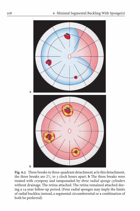
6 Minimal Segmental Buckling With Sponge(s)108
Fig. 6.7. Three breaks in three-quadrant detachment.a In this detachment,the three breaks are 21/2 to 3 clock hours apart. b The three breaks weretreated with cryopexy and tamponaded by three radial sponge cylinderswithout drainage. The retina attached. The retina remained attached dur-ing a 14-year follow-up period. (Four radial sponges may imply the limitsof radial buckles; instead, a segmental circumferential or a combination ofboth be preferred)
a
b

Minimal Segmental Buckling With Sponge(s) 109
Fig. 6.8. Group of breaks in 3-quadrant PVR-C1 detachment. a The breaks,between 1:00 and 2:00, are with adjoining starfolds from 2:30 to 6:00 poste-rior to equator. No additional break was found between 10:00 over 12:00 to1:00. b After buckling of breaks with 7-mm radial sponge without drainage,the retina and macula attached, except for a residual tractional detachmentlocated at starfolds still present 2 months postoperatively; it disappearedover time.The retina remained attached during follow-up period of 17 years
a
b

Limits of Minimal Segmental Buckling Without Drainage
There are limits to minimal segmental buckling; however, morethan 90% of rhegmatogenous detachments can be treated by extra-ocular minimal buckling alone – the remaining 10% can be dividedinto three major categories.
First Category of Difficult Detachments
In this type of detachments, the limits of the minimal procedureare exceeded if the tears are posterior (in about 1%),multiple at dif-ferent latitude (in 2–3%), or with a circumferential extent greaterthan 70° (in 1–2%). A tamponade with an expanding gas bubblewithout drainage represents the next level of a minimal surgery forthese conditions, but it requires an intraocular injection. Conse-quently, in this first category, a gas tamponade will suffice.A vitrec-tomy will be needed for the rare situation of a giant tear of less thanor equal to 150° with an overhanging flap or, as in some hands, ifthe tear is greater than 90°.
Second Category of Difficult Detachments
These are detachments with local vitreous traction that caused theredetachment of a horseshoe tear that had been buckled or detach-ments with proliferative vitreoretinopathy in more than two quad-rants. In these detachments, a primary vitrectomy may be indicat-ed. However, it is not indicated per se if the starfolds are more than1 clock hour from the tear to be buckled. In these difficult detach-ments, buckling first is advisable (Fig. 6.8) [33–35]. However, if onedecides upon a vitrectomy, it has to be combined with a meticulousremoval of proliferative preretinal membranes and the anterior vit-reous. Why? Because the additional gas tamponade, combined withthe vitrectomy, can provoke anterior vitreoretinal proliferation.
6 Minimal Segmental Buckling With Sponges and Balloons110

Third Category of Difficult Detachments
These are represented by pseudophakic detachments; the problemlies in the opacities of the optic media after an anterior segmentsurgery, which precludes finding the break and the application ofminimal external buckling. However, the limits of external mini-mal buckling in this third category are in a gray zone.
Actually, the indication for a vitrectomy in these eyes, combinedwith a preceding anterior segment surgery, does not depend onlyon the size of the tear or the presence of vitreoretinal proliferation,but also on the expertise of the surgeon in diagnostics in the pres-ence of optical interferences. Biomicroscopy with the use of the newindirect wide-field contact lenses combined with simultaneous depression of the retinal periphery and application of the 4 Rulesfor finding the primary break [19, 20] might enable a limitedprospective buckling and reduce the need for an invasive surgery.
Complications
There are no intraocular complications after minimal segmentalbuckling without drainage, except for a rare choroidal in a highlymyopic eye (about 0.3%). The remaining complications are extra-ocular and reversible: in less than 0.5%,a buckle infection or extru-sion may occur, and, in about 1%, diplopia may occur. However,diplopia after segmental buckles can be reduced to a minimum byavoiding trauma to the perimysium of the muscles during surgery,i.e., by avoiding grasping muscles with sharp instruments or un-controlled pulling on the traction sutures. In addition, starting onthe first day postoperatively, the patient should practice binocularmotility exercises in all directions several times a day; this shouldbe done independent of the state of the retina. This will avert thedevelopment of muscular adherences to the sclera or neighboringtissues and postoperative diplopia.
Complications 111

Temporary Balloon Buckle Without Drainage
To reduce the surgical trauma of minimal segmental bucklingwithout drainage even further, in 1979, Lincoff, Hahn, and Kreissig[36] replaced the segmental sponge sewed onto sclera by a tempo-rary buckle. Subsequently the Lincoff-Kreissig balloon evolved(Fig. 6.9) [37, 38]. In contrast to the sponge buckle, (1) the applica-tion of the balloon buckle is limited to detachments with one breakor a group of breaks within one clock hour, (2) the balloon is notfixated by sutures, and (3) it is withdrawn after 1 week. The ratio-nales for removing the balloon after 1 week were the results of our
6 Minimal Segmental Buckling With Sponges and Balloons112
Fig. 6.9. Lincoff-Kreissig Balloon. The presented balloon has (1) a metalstylette to facilitate insertion into the parabulbar space and (2) calibra-tions (black marks) on the tube to enable a more precise determination ofthe position of the balloon in the parabulbar space. Top: Deflated ballooncatheter with stylette in place; beneath it the adapter. Bottom: Inflated bal-loon (0.75 ml of sterile water) with self-sealing valve in place; beneath itthe withdrawn stylette

earlier animal experiments on the strength of the cryosurgical adhesion and the time it takes to develop a sufficiently strong ad-hesion. Thus, 10 years after the experimental data on the strength ofthe cryosurgical retinal adhesion were obtained, it was confirmedby the temporary balloon buckle, placed under the break surround-ed by cryosurgical lesions and removed after a week. The balloonoperation is performed under topical or subconjunctival anesthesia.
No sutures have to be placed to fixate the balloon buckle, andthe small conjunctival wound of 1–2 mm needed to insert the balloon catheter will close by itself after withdrawal of the balloon.After that, sustained attachment will depend exclusively on thestrength of the retinal adhesion, induced by transconjunctival cryo-pexy prior to insertion of the balloon, or by laser, applied post-operatively, after attachment of the break on the balloon buckle.
The balloon operation represents the ultimate refinement ofclosing a leaking break ab externo and without leaving a buckle atthe wall of the eye. The break is sealed off by surrounding retinaladhesions. It represents a procedure with a minimum of surgicaltrauma. The balloon operation follows the postulate of Gonin – tofind the break and to limit the treatment to the area of the leakingbreak – and the principle of Custodis – not to drain subretinal fluid.With the balloon, the last complications of segmental buckling,infection or extrusion, and diplopia are eliminated.
Some detachments, which were treated with the temporaryballoon buckle, will be depicted:
1. A detachment with a break under a rectus muscle is an optimal in-dication (Fig. 6.10), since after withdrawal of the balloon, diplopiadisappears.
2. A total pseudophakic detachment with an apparent circular an-terior traction line (which is, in fact, the vitreous base), capsularremnants,and no certain break (Fig.6.11).The treatment consistshere as well of a temporary balloon buckle in the suspected areato test for the presence of a break; after attachment, the so-calledtraction line tends to disappear.
Temporary Balloon Buckle Without Drainage 113
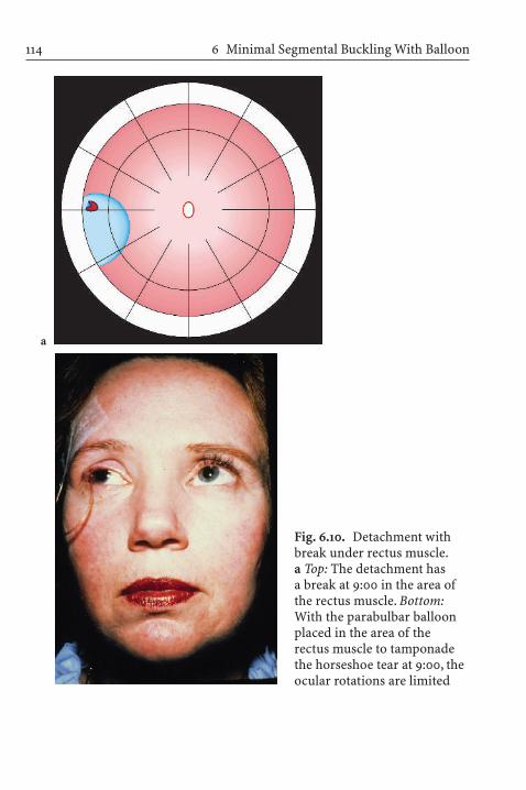
6 Minimal Segmental Buckling With Balloon114
Fig. 6.10. Detachment withbreak under rectus muscle.a Top: The detachment has a break at 9:00 in the area ofthe rectus muscle. Bottom:With the parabulbar balloonplaced in the area of the rectus muscle to tamponadethe horseshoe tear at 9:00, theocular rotations are limited
a

Temporary Balloon Buckle Without Drainage 115
Fig. 6.10. b Top: After 1 weekthe balloon was withdrawn;after that only pigmented cryopexy lesions surroundingthe horseshoe tear at 9:00were visible. Bottom: Withinhours after withdrawal ofthe balloon, the diplopia haddisappeared, because the eyemuscles function normallyagain
b

6 Minimal Segmental Buckling With Balloon116
Fig. 6.11a,b. Legend see page 117
a
b

Temporary Balloon Buckle Without Drainage 117
Fig. 6.11. A total pseudophakic detachment with capsular remnants.a In the detachment in the anterior so-called traction line at 11:30 a littletit was discovered. To find this area of suspicion at the operating table, inthe radian of the tit a laser mark was placed in the pars plana prior to sur-gery. b After balloon operation (1 day): The balloon was inserted beneaththe tit at 11:30. The localizing cryopexy lesion is visible on the balloonbuckle. The retina is attached. Since a break was not found for certain, ithas, however, to be located in the area of the balloon buckle. Therefore, theentire buckle had to be secured with interrupted laser lesions. The latticedegeneration with a pseudohole was not treated, not even at a later time.c After balloon operation at day 9: The balloon was withdrawn, and the entire area of suspicion, formerly placed on the buckle, is covered withpigmented thermal lesions. The retina remained attached during the entire follow-up of 7 years
c
Fig. 6.11c

3. An old detachment with a pigment demarcation line and anintraretinal cyst (Fig. 6.12); here too, a balloon buckle sufficed.
4. The balloon can also be used as a diagnostic tool to test forpresence of only one break in two separate detachments(Fig. 6.13).
5. Or, the balloon can be used even in a detachment up for re-operation with PVR stage C2 (Fig. 6.14).
Why Is the Balloon Operation so Difficult to Accept?
The premises for success are: (1) a maximum of preoperative diag-nostics, so as not to overlook a break; (2) a precise localization of the break at the table without prior drainage; (3) marking the detached break on mobile conjunctiva (in contrast to the segmen-tal sponge operation, in which the break can be marked preciselyon the sclera); and (4) localization of a highly elevated break on ayet slightly indenting parabulbar balloon in presence of a bullousdetachment with a pronounced and misleading parallax.
Advantages of the Balloon Operation
Advantages of the balloon operation are as follows:
1. The surgery is short, ranging between 10 min and 20 min2. The anesthesia is topical or subconjunctival3. The recovery of vision is fast and optimal4. The last complications of segmental buckling are eliminated,
i.e., there is no buckle infection, because the balloon is removed,and no diplopia. Diplopia, if present, disappears after the bal-loon is withdrawn.
6 Minimal Segmental Buckling With Sponges and Balloons118

Temporary Balloon Buckle Without Drainage 119
Fig. 6.12. Old inferior detachment. a The detachment has several pigmentdemarcation lines, an intraretinal cyst, and a round hole at 5:30. b Afterballoon operation at day 9: After balloon insertion (1 day) the retina hadattached. After pigmentation of the cryopexy lesions around the break,the balloon was withdrawn after a week. There are still visible the pigmentdemarcation lines and the intraretinal cyst
a
b

6 Minimal Segmental Buckling With Balloon120
Fig. 6.13. Two separate detachments with only one questionable break.a There is a superior detachment with two lattice degenerations and “erosions”. A most likely break is located at 11:00 with obvious vitreoustraction at the lateral edge of lattice degeneration. The convex pigment demarcation line beneath the superior detachment posterior to the latticedegeneration indicates that a full thickness break might be present at11:00. In the inferior detachment, there is a questionable tear at 8:00 at thelateral edge of lattice degeneration. When lying the patient flat, no com-munication between the two separate detachments was detected. b Afterinsertion of a diagnostic balloon (1 day) beneath the suspected break at11:00: The break and the entire lattice is surrounded with cryopexy lesions.The superior retina had attached and the inferior detachment diminishedin size, indicating that its fluid is originating from the superior break nowbeing tamponaded. c After balloon operation (10 days): The balloon waswithdrawn after 8 days when the cryopexy lesions were pigmented. Thelattice degeneration at 12:00 was surrounded with laser lesions. There isstill residual fluid around the inferior lattice degenerations. d After bal-loon operation (4 weeks): The residual fluid had disappeared. No furthertreatment was added

Temporary Balloon Buckle Without Drainage 121
a
b
Fig. 6.13a,b. Legend see page 120

6 Minimal Segmental Buckling With Balloon122
Fig. 6.13c,d. Legend see page 120
c
d

Temporary Balloon Buckle Without Drainage 123
Fig. 6.14a,b. Legend see page 124
a
b

6 Minimal Segmental Buckling With Balloon124
Fig. 6.14. Balloon as late reoperation of proliferative vitreoretinopathy(PVR)-C2 detachment. a A three-quadrant detachment with PVR andstarfolds in two quadrants in an eye with a previous buckle operation withdiathermy and a tear anterior to the buckle at 9:00. As minimal approachfor reoperation in presence of diathermized sclera, a parabulbar balloonwas selected.b After balloon operation (6 months): The macula had alreadyattached 2 days after surgery. At 6 months, a residual traction detachmentwith concave contours and concave surface is persisting; the macula is stillattached. c After balloon operation (4 years): The traction detachment hasflattened, only faint remnants of starfolds are visible
c
All of the listed advantages can be achieved by applying a temporaryand not suture-fixated balloon buckle without drainage as treat-ment for a primary retinal detachment. But the “conditio sine quanon” for success with this minimal procedure is experience in thenon-drainage operation.The surgeon has to be an expert in indirectbinocular ophthalmoscopy and has to be able to locate a ballooncorrectly in the parabulbar space beneath a highly detached break.
Fig. 6.14c

Minimal Segmental Buckling With Sponges or Balloons WithoutDrainage: A Medline Analysis
Materials and Methods
A Medline search was made of all reports that were identified bythe search terms “retinal detachment,” “segmental buckling,”“minimal extraocular surgery,” and “nondrainage.” Reports pub-lished in English, German, Italian, French, Spanish, and in someEast European journals were reviewed and analyzed. The majorityof reports did not contain homogenous data suitable for the ana-lysis. Many included both complicated and uncomplicated detach-ments, primary detachments and reoperations, or no preoperativecharacteristics. In many series, primary segmental buckling wascombined with a cerclage. The buckling was performed with orwithout drainage of subretinal fluid. Primary segmental bucklingwas sometimes combined with an injection of air, an expandinggas, or silicone oil.
After excluding these reports, an analysis was made of a relati-vely homogenous series of mostly primary retinal detachments,some with preoperative PVR stage C1–C2. The primary procedurein all eyes was minimal segmental buckling without drainage ofsubretinal fluid. The analysis consists of five reported series with acombined total of 1,462 retinal detachments [26, 37, 39–42].
The preoperative characteristics of the 1,462 primary retinal detachments were: aphakia/pseudophakia in 8.3% and preopera-tive PVR stage C1–C2 in 2.9% (Table 6.1). All operations were doneunder local anesthesia. Coagulation was limited to the area of thebreak(s) and performed with intraoperative cryopexy under oph-thalmoscopic control or with laser coagulation on a subsequentday after the break was attached. The buckle was limited to the areaof the break(s) and was obtained with an elastic silicone sponge ora temporary balloon. Subretinal fluid was not drained in any eye.Some of the detachments treated are represented in Figs. 6.4–6.13,recruited from series 2 and 5. All patients were mobilized after
Minimal Segmental Buckling With Sponges or Balloons 125

6M
inimal Segm
ental Buckling With Sponges and Balloons
126
Table 6.1. Preoperative characteristics of 1,462 primary retinal detachments treated with minimal segmentalbuckling (sponges or balloons) without drainage
Series Detach- Aphakia Perforating Reoperation Proliferative Myopiament pseudo- injury vitreoretino-
phakia pathy stage
C1 C2 > –7 to –25 dpt
First [21, 39] 752 30 – 7 5 – n.a.a
Second [26, 40] 107 22 – – 12 4 9Third [41] 35 3 – – 5 1 5Fourth [42] 68 5 1 – 11 – 5Fifth [37]b 500 62 3 30 3 2 71
122 (8.3%)43 (2.9%)
a Not availableb Treated with balloon
�
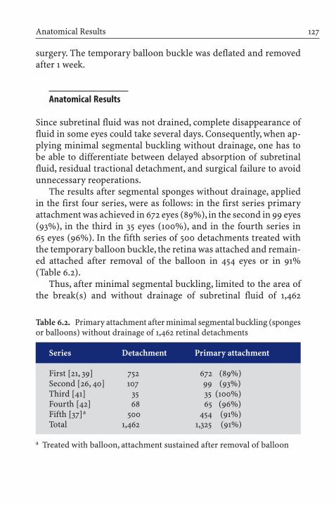
surgery. The temporary balloon buckle was deflated and removedafter 1 week.
Anatomical Results
Since subretinal fluid was not drained, complete disappearance offluid in some eyes could take several days. Consequently, when ap-plying minimal segmental buckling without drainage, one has tobe able to differentiate between delayed absorption of subretinalfluid, residual tractional detachment, and surgical failure to avoidunnecessary reoperations.
The results after segmental sponges without drainage, appliedin the first four series, were as follows: in the first series primary attachment was achieved in 672 eyes (89%), in the second in 99 eyes(93%), in the third in 35 eyes (100%), and in the fourth series in 65 eyes (96%). In the fifth series of 500 detachments treated withthe temporary balloon buckle, the retina was attached and remain-ed attached after removal of the balloon in 454 eyes or in 91%(Table 6.2).
Thus, after minimal segmental buckling, limited to the area ofthe break(s) and without drainage of subretinal fluid of 1,462
Anatomical Results 127
Table 6.2. Primary attachment after minimal segmental buckling (spongesor balloons) without drainage of 1,462 retinal detachments
Series Detachment Primary attachment
First [21, 39] 752 672 (89%)Second [26, 40] 107 99 (93%)Third [41] 35 35 (100%)Fourth [42] 68 65 (96%)Fifth [37]a 500 454 (91%)Total 1,462 1,325 (91%)
a Treated with balloon, attachment sustained after removal of balloon

6M
inimal Segm
ental Buckling With Sponges and Balloons
128
Table 6.3. Final attachment after minimal segmental buckling (sponges or balloons) without drainage and reoperation of 1,462 primary retinal detachments during 2-year follow-up
Series Detach- Preoperative Primary Reopera- Final Cause of final failurement proliferative attach- tion attach-
vitreoretino- ment ment Proliferative Missed Choroidalpathy C1–C2 vitreoretino- break
pathy C1–C2
First 752 5 672 60 732 14 4 3[21, 39]Second 107 16 99 5 104 4 3 –[26, 40]Third 35 6 35 – 35 – – –[41]Fourth 68 11 65 3 60 8 – –[42]Fifth 500 5 454 39 493 2 5 1[37]a
Total 1,462 43 1,325 107 1,424 28 12 4 (2.9%) (91%) (7.3%) (97.4%) (1.9%) (0.8%) (0.3%)
a Treated with balloon

detachments, primary retinal attachment was obtained in 1,325eyes or in 91%.After reoperation, attachment resulted in 97.4% andpersisted during a 2-year follow-up (Table 6.3).
Reasons for Primary and Final Failure
Missed Breaks
With minimal segmental buckling, if a break is missed anddrainage not performed, the retina will not become attached at anytime – not at the table, nor spontaneously in the days after surgery.A missed break was the cause of primary failure in 62 eyes or in4.2% and, after reoperation, in 12 eyes or in 0.8% (Table 6.3).
Inadequate Buckles
The second most frequent cause of primary failure was an inade-quate buckle, which was present in 51 eyes or in 3.5%. This waseasily corrected by either moving the buckle or enlarging it. Afterreoperation, the buckle was no longer a reason for final failure.
Proliferative Vitreoretinopathy
Despite the fact that PVR stage C1–C2 was already present preop-eratively in 43 eyes or in 2.9% of the 1,462 detachments treated, itwas the cause of primary failure in only 20 eyes (1.4%) and, after re-operation, the cause of final failure in 28 eyes or in 1.9%.
Thus, the presence of PVR did not preclude applying minimalsegmental buckling with sponges or balloons without drainage. Byomitting drainage, a breakdown of the blood–aqueous barrier waseliminated. By performing cryopexy and buckling with a mini-mum of trauma to these vulnerable eyes, a progression of PVRcould be circumvented.
Reasons for Primary and Final Failure 129

Choroidals
In 4 of the 1,462 detachments, a postoperative choroidal effusiondeveloped. These four eyes were highly myopic. Choroidals werethe cause of primary failure and final failure in 0.3%. This is lessthan the reported 2% to 8.6% after cerclage with drainage [10].
Complications
Intraocular
Minimal extraocular surgery is performed without drainage of sub-retinal fluid. As a result, the intraocular complications of drainage,such as hemorrhage, intraocular infection, incarceration of retinaor vitreous, do not occur. In addition, the need for a subsequent intraocular injection of gas or saline to restore intraocular pres-sure with its complications is also eliminated.
Secondary glaucoma, cataract, or anterior ischemia did not occur, and because the procedure was extraocular, there were noiatrogenic breaks. Intraocular complications from cryopexy didnot occur,because all applications were monitored by indirect oph-thalmoscopy for a medium lesion (first appearance of white in theretina) [43]. There was a rare choroidal in highly myopic eyes.
Extraocular
The extraocular complications that might occur with minimal seg-mental buckling without drainage are reversible. After using theLincoff sponge-2 in 210 eyes (series 2 to 4), one sponge (<0.5%) hadto be removed because of exposure: the retina remained attachedafter the sponge was removed. Diplopia did not occur because aneffort was made to avoid trauma to the rectus muscles during theoperation. If a break was localized in the area of a rectus muscleand a radial sponge intended, the muscle was split and re-sutured
6 Minimal Segmental Buckling With Sponges and Balloons130

to either side of the sponge. When a very anterior circumferentialbuckle was needed, it was placed external to the muscle [44]. Mostimportant were daily ocular motility exercises during the firstpostoperative week to counteract the development of adhesionsaround the muscle. This was done independent of the state of theretina.As a result, no sponge had to be removed because of diplopia.
In the fifth series (n=500), a temporary balloon buckle was ap-plied, which was removed after 1 week. In this series, there was nobuckle infection or exposure. Diplopia was sometimes presentwhen the balloon was in place, but diplopia remitted in all eyeswithin hours after the balloon was removed.
Functional Results
Since the end of the 1960s, detachment surgery has been concen-trating not only on anatomical results, but also on visual function[45–49] – both short-term recovery after surgery and long-term visual acuity.
In the first series, visual function was not described. In the remaining four series, treated with sponges or balloons, the meanvisual acuity was 0.67 after 2 years (Table 6.4). The two questionsthat arise are: (1) Would visual acuity decline over years? (2) Wouldthe presence of a segmental buckle over years cause a secondarydeterioration of visual function?
These pending questions can be answered by the second series of107 primary retinal detachments treated by minimal segmental spongebuckle(s) without drainage and with a complete follow-up of 15 years[40]. The mean preoperative visual acuity was 0.3; it improved to 0.5 during the first 6 months after surgery, and reached a maximumof 0.6 at 1 year. The increase was statistically significant (P<0.001).After 1 year, a slight linear decrease of 0.07 lines on the Snellen chartper year was observed over 15 years. In one patient, a cataract operation was done during the follow-up. The long-term functionalresults, including the surgical failures, are depicted in Fig. 6.15.
Functional Results 131

6M
inimal Segm
ental Buckling With Sponges and Balloons
132
Table 6.4. Visual acuity at 2 years postoperatively after minimal segmental buckling (sponges or balloons)without drainage and reoperation of 1,462 primary detachments
Series Detachment Preoperative Reoperation Final Visual acuity proliferative attachment 2 years postoperativevitreoretino-pathy C1–C2
First [21, 39] 752 5 60 732 n.a.a
Second [26, 40] 107 16 5 104 0.6Third [41] 35 6 – 35 0.6Fourth [42] 68 11 3 60 0.3Fifth [37]b 500 5 39 493 0.7Total 1,462 43 (2.9%) 107 (7.3%) 1,424 (97.4%) 0.67 (mean)
a Not availableb Treated with balloon

Functional Results
133
Fig. 6.15. Course of mean visual acuity of fellow and operated eyes during 15 years. Left: Course of mean visualacuity in the 107 unoperated fellow eyes during the 15-year follow-up. Right: Course of mean visual acuity in the107 eyes with retinal detachment operated with extraocular minimal surgery (segmental sponge buckle(s) without drainage) during the 15 years after surgery. Preoperative visual acuity of 0.3 had increased to 0.5 at6 months and to 0.6 at 1 year and decreased to 0.5 after 15 years. Of the patients, 72 were alive 15 years after theoperation. During the study period of 15 years, the difference in decrease of visual acuity was not significant be-tween the unoperated fellow eyes and the operated eyes (with the segmental buckle(s) in place) at any interval
1/21/2

To analyze whether this slight decrease in visual function could bedue to the presence of the segmental buckle or to secondary compli-cations, the visual acuity of the fellow eyes was compared with theoperated eyes at all intervals. There was no significant difference(P=0.079) in visual acuity over the 15 years. In this context, the data of Slataper are of value [50]. He had plotted the visual acuityof 17,349 individuals as a function of age and found that after the age of 60, a linear decrease of 0.07 lines per year on the Snellenchart occurs and that this decrease is age-dependent. There is nostatistically significant difference between the observed decreaseover the years, determined by Slataper, and the one in the analyzed operated eyes.
Thus, postoperative visual acuity (even when including the surgical failures) with a maximum of 0.6 at 1 year after surgery hadonly slightly decreased to 0.5 during 15 years. There was no statisti-cally significant difference from the course in the fellow eyes overthe ensuing 15 years; it was due to aging.
Series 2 through 5, with a combined total of 710 retinal detach-ments treated with segmental sponges or balloons without drain-age, provide useful data on postoperative visual acuity and itscourse over 2 years (Table 6.4) to 15 years (Fig. 6.15). The long-termpostoperative visual acuity of the eyes treated with sewed-on buck-les, limited to short segmental sponges to close the retinal breaks,confirm that minimal segmental buckling has no negative effect onlong-term visual function.
Disadvantages of Primary Minimal Segmental Buckling Without Drainage
Disadvantages of primary minimal segmental buckling withoutdrainage are as follows:
1. Preparation for a minimal buckle operation, limited to thebreak and without drainage, requires extensive preoperative
6 Minimal Segmental Buckling With Sponges and Balloons134

study. When a retinal break is not obvious, intensive biomicro-scopic study of the peripheral retina can be time-consuming,but is necessary.
2. A prerequisite for minimal segmental buckling without drain-age is having experience in indirect ophthalmoscopy and bio-microscopy to be able to find all of the breaks.
3. Experience with the special guidelines (the 8 Rules) and indirectwide-field contact lenses can help the biomicroscopic search forsmall breaks in a pseudophakic eye [19, 20–22, 51].
4. There is a learning curve to localizing posterior breaks in a bullous detachment and buckling them adequately withoutdrainage of subretinal fluid.
5. If a radial sponge is placed in the area of a rectus muscle, diplo-pia may occur. But diplopia can be averted with the use of atemporary balloon for breaks located beneath a rectus muscle.
6. Exposure or infection of the sponge buckle can occur, but is infrequent and ranges at <0.5%. After removal of the sponge,redetachment is rare, if a week or more has elapsed and the coagulation-induced adhesions have matured. In some cases,supplemental laser around the break may be applied prior to removal of the buckle.
7. The concept of minimal buckling without drainage can be dif-ficult to accept because the retina is not attached at the operat-ing table. Instead, the surgeon must wait for 24 hours or morefor the retina to attach spontaneously. This may be a strain onthe surgeon and “disturb his sleep,” as often described. How-ever, postoperative spontaneous attachment is an absolute con-firmation that the operation was correct.
Advantages of Primary Minimal Segmental Buckling Without Drainage
Advantages of primary minimal segmental buckling withoutdrainage are as follows:
Dis-/Advantages of Primary Minimal Segm. Buckling Without Drainage 135

1. The procedure is extraocular.2. It is performed under local or topical anesthesia.3. It can be done in an outpatient setting.4. It can be performed on a low budget, because it uses less oper-
ating time (rarely more than 45 min and 10–20 min with theballoon), inexpensive equipment, and few trained personnel inattendance. For the surgery itself, no costly disposable instru-ments or expensive intraocular tamponades are required.
5. It can be applied for superior and inferior breaks.6. There are, except for a rare choroidal in 0.3%, no intraocular
complications, such as: secondary glaucoma, cataract, intraocu-lar hemorrhage, intraocular infection, incarceration of retina or vitreous, or iatrogenic tears, because the procedure is extra-ocular.
7. No postoperative head positioning of the patient is requiredduring the day or at night while asleep, and traveling by airplaneis not restricted because intraocular gas is not injected.
8. The primary attachment rate of 1,462 primary retinal de-tachments treated with minimal segmental buckling and with-out drainage is 91% and after reoperation 97.4% over 2-year follow-up.
9. The low rate of redetachment: over a 2-year follow-up the rateof redetachment in the 1,462 eyes treated with minimal segmen-tal buckling averages 0.6% per year (series 1 to 5) and over a follow-up between 2 years and 15 years (series 2) 0.5% per year.
10. The recovery of visual acuity is optimal after minimal segmen-tal buckling without drainage. In the series of 107 detachmentswith a mean preoperative visual acuity of 0.3, the mean value at1 year is 0.6 and 0.5 at 15 years. The observed slight decreaseover years is an effect of aging and not of secondary complica-tions.
11. This low rate of intraoperative and postoperative complications,combined with optimal long-term visual results, is of benefit tothe elderly patient and to the decreasing financial resources.This becomes more relevant because new treatments for various
6 Minimal Segmental Buckling With Sponges and Balloons136

macular and retinal diseases are increasingly available, how-ever, at very high costs.
Discussion
In recent publications, the results of primary vitrectomy have beencompared with the results obtained with scleral buckling. How-ever, the comparison was done with scleral buckling consisting ofa cerclage with extensive coagulations, with drainage of subretinalfluid, and, frequently, with an intraocular gas tamponade.With thatcomparison, it was concluded that scleral buckling has a highermorbidity than primary vitrectomy [52–54]. Yet, had they com-pared primary vitrectomy with minimal segmental buckling with-out drainage, they would have concluded that segmental bucklingwithout drainage has less morbidity than primary vitrectomy [55].
Despite the excellent results that can be obtained with the mini-mal buckling technique, vitrectomy as a primary procedure is in-creasingly used. This is the case even though a recent analysis of595 detachments treated with vitrectomy, performed by experts,found that the rate of reoperation was 24.5% and PVR 11.5% [56] incontrast to minimal segmental buckling with a rate of reoperationat 7.3% and PVR at 1.9% (Table 6.3).
Retinal breaks, even small breaks, should be found preopera-tively. Postponing the search to the time of surgery should be a lastresort. Parenthetically, detecting breaks preoperatively is less cost-ly because the search uses only the time of the surgeon; a search forbreaks during surgery uses the time of additional personnel in at-tendance in an expensively equipped operating room.
It is of interest that failing to close the break is still the maincause of failure, regardless of the procedure employed. A recentpublication about 171 primary detachments treated with a primaryvitrectomy [57] demonstrated that the reason for primary failurewas a missed or leaking break in 64.3% of failures. In comparison,the causes of the 91 primary failures after 962 sponge buckles
Discussion 137

(series 1–4) was 39 times a missed break and 32 times an inade-quate buckle. Thus, failure to close the leaking break accounted for71 of 91 failures or 78% (Table 6.5).
Thus, a leaking break still remains the main cause of primaryfailure, and vitrectomy has not significantly altered this. This vali-dates the postulate of Gonin, defined more than 70 years ago: theretinal break causes the detachment.
However, in the future, the requirements of an optimal surgeryfor repair of a primary retinal detachment might be more specific:
1. Just one operation should attach the retina once and for all2. The surgery should have a minimum of morbidity3. The procedure should be done on a small budget and under
local anesthesia
6 Minimal Segmental Buckling With Sponges and Balloons138
Table 6.5. Reasons of primary failure (n=91) after one operation withminimal segmental sponge buckle(s) without drainage of 962 primaryretinal detachments
Series Detach- Cause of primary failure Totalment
Missed Inadequate PVR Choroidalsbreak buckle
First 752 31+2a 27 17 3 80[21, 39]Second 107 4 4 – – 8[26, 40]Third 35 – – – – –[41]Fourth 68 2 1 – – 3[42]Total 962 39 32 17 3 91
71 (78%)
a Macular hole
�

4. The operation should provide a maximum of long-term visualfunction, not jeopardized by secondary complications duringthe prolonged life expectancy of the patient treated
Outlook
Minimal segmental buckling by a sponge or a balloon and withoutdrainage of subretinal fluid is the ultimate development of scleralbuckling introduced by Schepens and Custodis and subsequent-ly refined by Lincoff, Kreissig, and others. Minimal segmentalbuckling without drainage provides an optimum of early and lateanatomical and visual results: retinal attachment results after oneoperation in 91% and after reoperation in 97%. However, to obtainthis rate of attachment requires an expertise in biomicroscopy and binocular indirect ophthalmoscopy to find the breaks pre-operatively and at surgery to localize these breaks correctly and toadequately position the segmental buckle beneath them withoutdrainage of subretinal fluid. However, this “Art of minimal seg-mental buckling” [25, 26] has a learning curve.
Detachments in pseudophakic eyes today are almost routinelyassigned to vitrectomy for primary repair. This may occur evenwhen the break can be visualized preoperatively and would re-spond to a segmental buckle without drainage. For these eyes inwhich the break cannot be found because the peripheral retina isobscured by a narrow pupil or capsule opacities, a vitrectomy toprovide better access for viewing the anterior retina may provide abetter prognosis than prospective buckling, being based on thecontour of the detachment, or a cerclage.
However, we have to keep in mind that the resources availablefor ophthalmology are diminishing as life expectancy increasesand new treatments for various macular and retinal diseases be-come available. This expanding spectrum includes invasive andnoninvasive, but expensive, treatment modalities. All this mayforce us to reconsider how to spend the limited resources for theincreasing number of patients.
Outlook 139

Probably, the future question no longer will be: which methodis better for attaching a primary detachment:
1. A limited or a prospective buckle?2. An intraocular or extraocular surgery?
Rather, it will be:
3. Which method is applied at its optimum with a minimum ofstrain on our financial resources?
And this could mean treating a break in a primary detachment byan extraocular surgery, limited to the break, i.e., a surgery, per-formed under local anesthesia and on a small budget, with a lowrate of morbidity and reoperations, and with optimal long-term visual results. Perhaps in the future a less morbid procedure toattach the retina will be developed, or the pendulum of detachmentsurgery, as witnessed already during the past 75 years, might swingback to an extraocular minimal surgery. And in this case, we againmight have to train surgeons skilled in preoperative diagnostics to find the break(s) and in the art of applying a minimum of seg-mental buckling without drainage to attach a retina.
References
1. Gonin J (1930) Le traitement opératoire du décollement rétinien.Conférence aux journées médicales de Bruxelles. Bruxelles-Médical23:No. 17
2. Guist E (1931) Eine neue Ablatiooperation. Ztsch Augenheilk 74:232–242
3. Lindner K (1931) Ein Beitrag zur Entstehung und Behandlung deridiopathischen und der traumatischen Netzhautabloesung. GraefesArch Ophthalmol 127:177–295
4. Rosengren B (1938) Ueber die Behandlung der Netzhautabloesungmittelst Diathermie und Luftinjektion in den Glaskoerper. Acta Oph-thalmol 16:3–142
6 Minimal Segmental Buckling With Sponges and Balloons140

5. Schepens CL, Okamura ID, Brockhurst RJ (1957) The scleral bucklingprocedures. 1. Surgical techniques and management. Arch Ophthal-mol 58:797–811
6. Arruga MH (1958) Le cerclage equatorial pour traiter le decollementretinien. Bull Soc Franc Ophtal 71:571–580
7. Kreissig I (1971) Kryopexie in der Ablatio-Chirurgie (2. Mitteilung).Klin Mbl Augenheilk 159:737–741
8. Blagojevic M, Ilic R (1975) Drainage of the subretinal fluid and injec-tion of saline solution in the vitreous body. Mod Probl Ophthal15:169–173
9. Huebner H, Boeke W (1975) Klinische und technische Aspekte dersubretinalen Drainage. Mod Probl Ophthal 15:137–142
10. Toernquist R, Toernquist P (1988) Retinal detachment. III Surgical re-sults. Acta Ophthalmologica 66:630–636
11. Lincoff H, Kreissig I (1972) The treatment of retinal detachment with-out drainage of subretinal fluid (modifications of the Custodis proce-dure, Part VI). Trans Am Acad Ophthalmol Otolaryngol 76:1221–1232
12. Custodis E (1953) Bedeutet die Plombenaufnaehung auf die Skleraeinen Fortschritt in der operativen Behandlung der Netzhautabloe-sung? Ber Dtsch Ophthalmol Ges 58:102–105
13. Schepens CL, Okamura ID, Brockhurst RJ et al (1960) Sceral bucklingprocedures. V. Sutures and silicone implants. Arch Ophthalmol 64:868–881
14. Lincoff H, O’Connor P, Kreissig I (1970) Retinal adhesion aftercryosurgical applications. Klin Monatsbl Augenheilkd 156:771–783
15. Kreissig I, Lincoff H (1971) Ultrastructure of cryopexy adhesion. In:DOG Symp.“Die Prophylaxe der idiopathischen Netzhautabhebung.”Bergmann, Muenchen, pp 191–205
16. Lincoff H, Baras I, McLean J (1965) Modifications to the Custodis pro-cedure for retinal detachment. Arch Ophthalmol 73:160–163
17. Kreissig I (1972) Ultrastructure of the cryopexy adhesion in retinalsurgery. Postdoctorial thesis. University of Bonn, pp 1–172
18. Kreissig I, Lincoff H (1975) Mechanism of retinal attachment aftercryosurgery. Trans Ophthalmol Soc U K 95:148–157
19. Lincoff H, Gieser R (1971) Finding the retinal hole. Arch Ophthalmol85:565– 569
20. Kreissig I (2000) A practical guide to minimal surgery for retinal detachment: vol. 1. Thieme Stuttgart, New York, pp 14–15, and back cover of book
References 141

21. Lincoff H, Kreissig I (1996) Extraocular repeat surgery of retinal de-tachment. A minimal approach. Ophthalmology 103:1586–1592
22. Kreissig I (2000) A practical guide to minimal surgery for retinal de-tachment: vol. 2. Thieme, Stuttgart, New York, pp 320–321, and backcover of book
23. Lincoff H, Kreissig I (1975) Advantages of radial buckling. Am J Oph-thalmol 79:955–957
24. Goldbaum MH, Smithline M, Poole TA, Lincoff HA (1975) Geometricanalysis of radial buckling. Am J Ophthalmol 79:958–965
25. Kreissig I (1989) Minimization of detachment surgery. Klin Mbl Augenheilk 195:126–134
26. Kreissig I, Rose D, Jost B (1992) Minimized surgery for retinal detach-ments with segmental buckling and nondrainage. An 11-year follow-up. Retina 12:224–231
27. Dobbie G (1980) Circulatory changes in the eye associated with reti-nal detachment and its repair. Trans Am Ophthalmol Soc 78:504–506
28. Diddie KR, Ernest JT (1980) Uveal blood flow after 360° constrictionin the rabbit. Arch Ophthalmol 98:129–130
29. Yoshida A, Feke G, Green GJ et al (1983) Retinal circulatory changes after scleral buckling procedures. Am J Ophthalmol 95:182–191
30. Scheider A, Korabjelnikoff E (1991) Bestimmung der arteriovenoesenPassagezeit in der Aderhaut mit Indozyaningruen. Klin Monatsbl Augenheilkd 199: 251–255
31. Santos L, Capeans C, Gonzalez F, Lorenzo J, Codesido J, Salorio MS(1994) Ocular blood flow velocity reduction after buckling surgery.Graefes Arch Clin Exp Ophthalmol 232:666–669
32. Winter W, Lipka P (1987) Untersuchungen der Lichtunterschieds-empfindlichkeit nach Operationen von Netzhaut-Abloesungen. FoliaOphthalmol 12: 315–319
33. Kreissig I, Rose D (1989) Scleral buckling without drainage in treat-ing PVR detachments stage B and C. In: Heimann K, Wiedemann P(eds) Proliferative vitreoretinopathy. Kaden Verlag, Heidelberg, pp182–190
34. Kreissig I, Simader E, Rose D (1994) The place of segmental bucklingin the treatment of PVR detachments stage B and C.A longterm study.Klin Monatsbl Augenheilkd 205:336–343
35. Sivkova N, Kreissig I (2002) Rhegmatogenous PVR detachment:long-term results after extraocular minimal scleral buckling. KlinMonatsbl Augenheilkd 219:519–522
6 Minimal Segmental Buckling With Sponges and Balloons142

36. Lincoff HA, Kreissig I, Hahn YS (1979) A temporary balloon buckle forthe treatment of small retinal detachments. Ophthalmology 86:586–592
37. Kreissig I, Failer J, Lincoff H, Ferrari F (1989) Results of a temporaryballoon buckle in the treatment of 500 retinal detachments and a com-parison with pneumatic retinopexy. Am J Ophthalmol 107:381–389
38. Kreissig I (2000) A practical guide to minimal surgery for retinal detachment: vol. 2. Thieme, Stuttgart, New York, pp 2–107
39. Lincoff H, Kreissig I, Goldbaum M (1974) Reasons for failure in non-drainage operations. Mod Probl Ophthal 12:40–48
40. Kreissig I, Simader E, Fahle M, Lincoff H (1995) Visual acuity after segmental buckling and non-drainage: a 15-year follow-up. Eur J Oph-thalmol 5:240–246
41. Sivkova N, Katsarov K, Kreissig I, Chilova-Atanassova B (1997) Our experience in minimized surgery for retinal detachment: first results.Folia Medica 34: 44–47
42. Sirtautiene R, Bagdoniene R (1997) Minimised surgery for retinal detachments with segmental buckling and non drainage. In: XIthCongress of the European Society of Ophthalmology, Budapest 1997.Monduzzi Editore S.p.A., Bologna, pp 1161–1165
43. Kreissig I, Lincoff H (2001) Cryosurgery of the Retina. In: Peyman GA,Meffert SA, Conway MD, Chou F (eds) Vitreoretinal surgical techni-ques. Martin Dunitz Ltd, London, pp 33–48
44. Kreissig I (1988) Episklerale Plomben und gerade Augenmuskeln:Pilotstudie zur Reduzierung postoperativer Muskelfunktionsstoerun-gen. Ber Dtsch Ophthalmol Ges 132
45. Kishimoto N, Nakamuro S, Fujino T (1967) Visual acuity after success-ful retinal detachment surgery. Jap J Clin Ophthal 21:173–180
46. Davies EWG (1972) Factors affecting recovery of visual acuity follow-ing detachment of the retina. Trans Ophthal Soc UK 92:335–344
47. Kreissig I, Lincoff H (1974) Urgency in the repair of retinal detach-ment. Klin Mbl Augenheilk 165:315–318
48. Kreissig I (1977) Prognosis of return of macular function after retinalreattachment. Mod Probl Ophthalmol 18:415–429
49. Kreissig I, Lincoff H, Witassek B, Kolling G (1981) Color vision andother parameters of macular function after retinal reattachment. DevOphthal 2:77–85
50. Slataper FJ (1950) Age norms of refraction and vision. Arch Ophthal-mol 43:466–479
References 143

51. Lincoff H, Kreissig I (1994) Finding the retinal hole in the pseudopha-kic eye with detachment. Am J Ophthalmol 117:442–446
52. Framme C, Roider J, Hoerauf H et al (2000) Complications after external retinal surgery in pseudophakic retinal detachment – arescleral buckling operations still current? Klin Monatsbl Augenheilkd216:25–32
53. Tornambe PE, Hilton GF, Brinton DA et al (1991) Pneumatic retino-pexy.A two-year follow-up study of the multicenter clinical trial com-paring pneumatic retinopexy with scleral buckling. Ophthalmology98:1115–1123
54. Bartz-Schmidt KU,Kirchhof B,Heimann K (1996) Primary vitrectomyfor pseudophakic retinal detachment. Br J Ophthalmol 80:346–349
55. Lincoff H, Kreissig I (2000) Changing patterns in the surgery for retinal detachment: 1929 to 2000. Klin Monatsbl Augenheilkd 216:352–359
56. Kreissig I (2000) A practical guide to minimal surgery for retinal detachment: vol. 2. Thieme, Stuttgart, New York, p 305
57. Richardson EC, Verma S, Green WT, Woon H, Chignell AH (2000) Primary vitrectomy for rhegmatogenous retinal detachment: an ana-lysis of failure. Eur J Ophthalmol 10:60–166
6 Minimal Segmental Buckling With Sponges and Balloons144

Pharmacological Approaches to Improve SurgicalOutcomes After Retinal Reattachment Surgery
Mark S. Blumenkranz
Chapter 7
Introduction
Prior to the modern era ushered in by Gonin, with the recognitionthat retinal detachment is caused by retinal breaks, a variety ofnonspecific and pharmacological methods were employed, with-out predictable success, for the treatment of retinal detachment.It remains true today that the most important pharmacologicalagent in the diagnosis and treatment of retinal detachment is themydriatic drop, since without adequate visualization of the fundusand identification of the break, no definitive treatment is possible.Other opportunities still exist for the pharmacological enhance-ment of retinal reattachment, by improvement in both anatomicaland functional results. These include (1) improvement in the finalretinal reattachment rate, (2) improvement in visual acuity follow-ing successful retinal reattachment, and (3) reduction in complica-tions.
Inasmuch as the majority of routine or uncomplicated retinaldetachments can now be accomplished safely by utilizing modernscleral buckling techniques, the risk–benefit ratio of utilizing poten-tially toxic pharmacological agents under routine circumstanceshas to be carefully considered in light of the opportunities for mar-ginal potential improvement [1]. However, given the increasingutilization of pneumatic retinopexy, which seems to have a lowerinitial retinal reattachment rate than conventional scleral buckling[2], the justification for the use of pharmacological agents may beincreased. Some authors now propose primary vitrectomy without

scleral buckling as a first operation, and although the immediatepostoperative morbidity may be lower, the final rate of proliferativevitreoretinopathy (PVR) may be higher than scleral buckling alone[3–5]. Similarly, when scleral buckling alone is considered for eyeswith early or late forms of proliferative vitreoretinopathy, successrates are considerably lower, and the case for use of an adjuvantagent, either to facilitate intraoperative flattening, such as perfluoro-carbon, or one of several different anti-proliferative agents seemsbetter justified [7–9]. Recently, a British group has developed a pre-dictive formula to calculate the risk of PVR following primaryretinal reattachment, which may aid in the selection of patientswho might benefit most from the use of adjuvant pharmacologicalmethods [10].
Improvement in Visual Acuity
Improvements in visual acuity following reattachment surgery maybe achievable using several different methods. The first would be areduction in the rate of complications associated with macular dys-function. These include both macular edema and macular pucker.Both are well-described complications of retinal reattachment sur-gery as well as other vitreoretinal conditions and may be amenableto pharmacological intervention. Recent reports suggest that intra-vitreal steroids, principally triamcinolone acetonide, and also dexa-methasone in a bioerodable polymer may reduce macular edemaassociated with several conditions, including diabetic retinopathy,uveitis, and retinal vein occlusion [11, 12]. The use of steroids byother routes of administration, including oral and parenteral, mayalso be of benefit in preventing macular complications, such aspucker [13].
It may also be possible to improve visual acuity through the en-hancement of photoreceptor regeneration in eyes with macula-offretinal detachments. Processes governing photoreceptor renewaland normal alignment of the outer segments following retinal
7 Pharmacological Approaches to Improve Surgical Outcomes 146

reattachment are still poorly understood, and the potential use of pharmacological agents and, specifically, cytokines may be apromising avenue to restoring visual function above and beyondthat which might be expected simply by reattachment of the macula,particularly in eyes with more longstanding retinal detachments.To date, there are no well-defined or well-controlled clinical trialsthat would suggest such an agent is available or on the near hori-zon, but it is anticipated that in the future this approach may befeasible.
Reduction in Other Non-Macular Complications Limiting Either Anatomic Retinal Reattachment or Normal PhysiologicalFunction
Well-known complications of retinal reattachment surgery includealterations in the intraocular pressure, both glaucoma and hypo-tony, postoperative inflammation, cataract, motility disorders, andPVR. Pharmacological therapy, both intraoperatively as well aspostoperatively, may play a role in the reduction of several of thesecomplications, either singly or in combination. The remainder ofthis chapter is devoted to a discussion of the principal cause ofretinal reattachment failure after primary surgery, PVR, and themethods by which pharmacological therapy might favorably in-fluence this condition.
The Cell Biology of PVR
PVR is the most common cause of failure following attempted reti-nal reattachment repair in primary cases, as well as complex formsof retinal detachment. The precise initiating events remain poorlyunderstood, but it is known that the disease is characterized bycell-mediated tractional forces exerted on both preretinal andsubretinal membranes, as well as more diffusely to the vitreous gel
The Cell Biology of PVR 147

itself [9, 14, 15]. These contractile forces lead to stiffening and eleva-tion of the retina, leakage through retinal breaks, and retinal re-detachment typically between 4 weeks and 8 weeks following attempted repair. The condition is more commonly encountered ineyes with large or multiple breaks and is more frequently seen inmales and in patients with a history of trauma, hemorrhage,choroidal detachment, or giant retinal tear [5, 10]. The membranesassociated with this condition are composed of a variety of celltypes, principally pigment epithelial, glial and myofibroblastic elements, either interspersed within the gel producing compactionof the collagen (hypocellular gel contraction) [15] or defined hy-percellular membranes on either the anterior or posterior surfaceof the retina [14]. Two of the earliest findings in eyes that go on to develop PVR are breakdown of the blood–ocular barriers with in-creased amounts of intravitreal protein and free-floating dispersedcells, principally retinal pigment epithelium (RPE). The cells ap-pear to enter the growth cycle and actively proliferate in responseto chemotactic and mitogenic stimuli and, subsequently, undergoan orderly sequence of steps, including attachment, contraction,and secretion of newly formed extracellular matrix (ECM) [14–21].
Initially, attempts to treat this condition by scleral bucklingtechniques alone were largely unsuccessful, as were the earliesttechniques using vitrectomy without supplemental long-actingtamponades [6, 7, 14]. These initially unsuccessful surgical ap-proaches served as the principal stimulus for a search for anti-proliferative agents that might improve success rates. Early studieswere performed using triamcinolone acetonide, daunomycin, andfluorouracil in the early 1980s prior to the widespread availabilityof long-acting gases and silicone oil [8, 22–26].
The introduction of long-acting gases, particularly perfluoro-propane, and the re-emergence of silicone oil (polydimethylsilox-ane) were major steps in the improvement in surgical success rates.The definitive silicone oil study confirmed that while silicone oilwas superior to sulfur hexafluoride in providing visual acuity improvement and retinal reattachment in PVR, a companion study
7 Pharmacological Approaches to Improve Surgical Outcomes 148

further amplified the relative equivalence of perfluoropropane tosilicone oil in achieving retinal reattachment [27]. Approximately64–73% of patients in the latter study achieved complete posteriorretinal attachment and 43–45% achieved functional visual acuity ofgreater than 5/200 compared with success rates less than half thatprior to the utilization of both of these long-acting tamponadetechniques [28]. Paradoxically, with the improvement in anatomicreattachment associated with the long-acting tamponades of per-fluoropropane and silicone oil, both the medical need and the likelihood of demonstrating a statistically significant treatmentbenefit by incremental pharmacological methods were reduced,although studies actively continued throughout the 1980s and early1990s to develop new pharmacological therapies. In addition to reducing the driving force for development of new drugs due toimproved surgical success rates, the use of long-acting tamponades,including silicone oils and long-acting gases, also created uniqueproblems related to bio-availability because of the presence ofeither a gas-filled eye or an eye filled with an hydrophobic agent[29]. Additionally, subsequent to the publication of the results ofthe silicone oil study, another pharmaceutical compound used as atemporary intraoperative tamponade, perfluoro-n-octane, as wellas other liquid perfluorochemicals came into common use. The useof these compounds further improved the success rates for com-plex forms of retinal detachment repair in conjunction with long-acting tamponade even without the use of other pharmacologicalanti-proliferative agents with success rates reported in the range of78% [30].
The General Approach to Pharmacological Therapy
The search for drugs to inhibit vitreoretinal scarring either pre-ceding or following retinal reattachment surgery has proceededalong lines that target the specific steps of the vitreoretinal scarring response, including cellular activation, proliferation, ECM
The General Approach to Pharmacological Therapy 149

elaboration, and contraction. This process has been facilitated bythe use of cell-culture methods for initial screening [31, 32]. Somedrugs attack specific points within the cycle, whereas other agentsmay attack more than one, such as steroids or heparin-like com-pounds. The various agents have been divided into classes accord-ing to their mechanism of action. These include the following:(1) anti-inflammatory agents, (2) drugs that inhibit cellular pro-liferation, (3) drugs that act on the ECM and cell surface. A generalreview of these classes of drugs follows.
Anti-Inflammatory Agents
Corticosteroids were the first agents to be employed in the treatmentof experimental PVR and have recently regained currency basedupon their widely disparate effects [24]. It is known that steroids exhibit a bimodal effect on cultured fibroblasts, causing stimulationat low doses and inhibition at supraphysiological doses [31]. Triam-cinolone acetonide has been shown to reduce experimental PVR in a rabbit model after the injection of cultured fibroblasts [24]. Onehuman clinical study employing oral prednisone showed a reducedrate of macular pucker, a limited form of proliferative response, afterretinal reattachment surgery, although it did not affect the ultimatereattachment rate or rate of PVR [13]. Intravitreal steroids, whenincluded in the infusate with heparin, resulted in a lower rate ofretinal reoperation in one clinical study [33]. A novel use of intra-vitreal triamcinolone recently described by Peyman and colleaguesinvolves visualizing remnants of the residual vitreous cortex follow-ing injection of a suspension of triamcinolone acetonide and, there-by, enhancing a full removal of cortex and vitreous membranes in amore expeditious manner [34].
7 Pharmacological Approaches to Improve Surgical Outcomes 150

Drugs That Inhibit Cellular Proliferation
A variety of subclasses of anti-proliferative agents have beenshown to be effective in animal models as well as more recently inclinical trials. The greatest clinical experience has been obtainedwith fluoropyrimidines [8, 22, 23, 28, 29, 31, 35–42]. Fluoropyrimi-dines were first chosen because of their potency in inhibiting cellu-lar proliferation in vitro compared with a relative lack of toxicity athigh concentrations in toxicological studies [8, 31]. 5-Fluorouracil,the first agent to be tested in detail, has been found to have a medianinhibitory dose (ID 50) of between 0.35 µg/ml and 0.71 µg/ml formost ocular and vascular cell types tested [31]. 5-FU has beenfound to be non-toxic as an intravitreal injection and can be welltolerated both in animals and in humans following intravitreal injections of up to 1.0 mg [8]. The drug is thought to exert its effectby enzymatic conversion into the ribose nucleotide form, which inturn both effects protein synthesis and also the enzyme thymidyl-ate synthetase. Other fluoropyrimidine cogeners, particularly theribonucleoside (5 FUR), have not only anti-proliferative effects like5-FU, but also anti-contractile effects, which 5-FU does not seem toexhibit, at least in culture. These, however, are associated with agreater potential toxicity than 5-FU [31, 32, 39].
One of the theoretical problems associated with drug therapyemploying 5-FU in eyes undergoing surgery for PVR is interactionwith the long-acting tamponade, whether it is gas or silicone oil. Inone study, a sustained release of a co-drug of 5-FU and fluocino-lone pellet was tested in gas-filled eyes and found to be effective inreleasing drugs in a manner comparable with non-gas-filled eyes[40, 41]. Co-drugs of 5-FU linked to an alkyl side-chain are solublein silicone oil, whereas 5-FU itself is not and may be slowly releasedinto the vitreous cavity by hydrolysis of the alkyl side-chain and 5-FU bond. To date, no studies have been performed on humansemploying this technique, although it retains some promise [29].
The first randomized prospective clinical trial testing the effi-cacy of intravitreal 5-FU combined with low-molecular-weight
Drugs That Inhibit Cellular Proliferation 151

heparin was published in eyes undergoing retinal reattachmentand thought to be at high risk for the development of PVR ratherthan having already established PVR. In this study, 200 µg/ml of5-FU and 5 IU/ml of low-molecular-weight heparin were added tothe intravitreal infusate in a randomized fashion, with the remain-der of the patients receiving a placebo injection into the infusateduring vitrectomy. The incidence of post-operative PVR wasjudged to be lower at 6 months in the treatment group (12.6%) thanthe placebo group (26.4%), with those in the placebo group also requiring a higher re-operation rate than those in the 5-FU group.However, no differences were seen in the final complication rates[42]. Results of a similar protocol for the treatment of establishedPVR are still pending at the time of writing of this chapter.
Daunomycin
Another anti-proliferative agent, daunomycin, has been tested inpreclinical and clinical studies. The drug is an anthracycline anti-biotic with efficacy in an animal model. Daunomycins appear tohave a somewhat lower therapeutic index than 5-FU, principallydue to its greater toxicity; but it has been tolerated in animals andin humans as a continuous intravitreal infusion of 7.5 µg/ml for10 min. In pilot studies of patients undergoing vitrectomy, dauno-mycin was felt to be effective [26, 43].A variety of other agents havebeen employed for the treatment of experimental PVR, with no significant published data yet in human trials. These includeretinoids, which play an important role in the differentiation andproliferation of various cell types, including RPE. The treatment ofRPE cells with vitamin A (all-trans-retinol) significantly inhibitscellular proliferation migration in vitro as well as having effects on morphology [44]. Immunotoxins composed of a monoclonalantibody linked to a biological toxin have been employed in ex-perimental models, including an antibody against the humantransferrin receptors to the A chain of ricin [45].
7 Pharmacological Approaches to Improve Surgical Outcomes 152

Another chemotherapeutic agent useful in cancer and also inthe treatment of coronary restenosis, Taxol, has been tested forefficacy in experimental models in the eye. It appears to act as apromoter rather than an inhibitor of microtubular assembly andinhibits cell-mediated contraction of a collagen gel as well as ex-perimental retinal detachment in various animal models [46]. A related cytoskeletal agent, Colchicine, also inhibits RPE astrocyteand fibroblast proliferation in addition to migration and wasshown in one animal model to have some beneficial effects onPVR, although it has not yet been proven to be beneficial in any human studies [47].
Drugs Acting on the ECM
Drugs which act on the interface between cells and the ECM havethe potential to inhibit intraretinal scarring at a relatively earlierstep than simple proliferation. Heparin and related peptides have amultitude of effects on cells and their interaction with the ECM.Heparin is a glycosaminoglycan derived from heparin sulfate,whichbinds to several ECM proteins, including fibronectin, laminin,and vitronectin [48]. In addition to its antithrombotic properties,for which it was first discovered and processed, heparin clearly has important effects on a variety of growth factors. It activelybinds fibroblast growth factor, platelet-derived growth factor, andendothelial-cell growth factor. Soluble heparin causes an increasein cell spreading and produces changes in the cytoskeleton ofsmooth muscles. It also inhibits the polymerization of type-1 collagen and reduces cell-mediated contraction of collagen gelswhen cultured fibroblasts or RPE cells are interspersed within acollagen matrix. This may be a process analogous to hypocellulargel contraction,an important attack point in the prevention of PVR[15, 48].
Because heparins have significant anticoagulant affects, theymay result in hemorrhagic complications, and this stimulated the
Drugs Acting on the ECM 153

search for compounds with heparin-like qualities on growth fac-tors, but without the potential hemorrhagic issues. It is known thatfractionation of longer chains of heparin into smaller molecularweight fragments causes loss of some of the anticoagulant activitywhile preserving the ECM effects.As a result, low-molecular-weightfractions of heparin with a molecular weight of 5000 or less retaintheir ability to catalyze the inhibition of Factor Xa, but lose theirability to directly inhibit thrombin [49]. When low-molecular-weight heparin was introduced into the infusate during vitrectomyin an animal model, fibrin formation was markedly reduced with-out any coincident increase in intraocular hemorrhage [50]. Use of5 IU/ml of low-molecular-weight heparin in the infusate duringcreation of an experimental model of proliferative vitreoretino-pathy reduced the rate of traction detachment from 77% to 28% at3 months [51]. These encouraging results led to the inclusion of low-molecular-weight heparin in the infusate, along with 5-FU, whichwas studied in the prevention of PVR in humans [15, 42, 50, 51].
In another important PVR trial, when conventional heparin(1 IU/ml) was combined with a steroid dexamethasone (5 mg/ml)in the infusate, there was a slight increase in the retinal reattach-ment rate compared with controls from 65% to 80% in addition toa reduction in the rate of reproliferation from 26.5% to 16%.A mildincrease in the rate of hyphema and vitreous hemorrhage wasseen, although it was not judged to be clinically significant [33].
Summary
Pharmacological methods remain a promising potential adjunct tothe successful treatment of retinal detachment. In addition to con-ventional strategies, including adequate pupillary dilatation, con-trol of inflammation by anti-inflammatory agents, and intraocularpressure by ocular hypotensive agents, drugs may play a furtherrole by inhibiting other late complications. These include prolifer-ation and macular edema. Steroids, fluoropyrimidines, and he-
7 Pharmacological Approaches to Improve Surgical Outcomes 154

parin-like compounds, particularly low-molecular-weight heparin,all appear to be potentially useful agents either singly, or in com-bination. The ability to combine agents together with differingmechanisms of action, either as a single intravitreal injection, or,more recently, as a component of the infusate or with extended delivery devices, opens up new therapeutic avenues. Issues relatedto bioavailability, particularly in eyes with long-acting tampon-ades, either gas or silicone oil, are particularly challenging but intriguing. The use of modern bioerodable polymers and otherfixed extended delivery devices may further enhance the utiliza-tion of such agents. In the future, additional capabilities, includingthe use of intravitreal steroids for control of macular edema andcytokines for the improvement of photoreceptor recovery, may further improve visual acuity results beyond those that might be expected when anatomic reattachment reaches a high plateau.Further understanding of the cellular biology of retinal reattach-ment and macular function will undoubtedly lead to concomitantimprovements in therapeutic advances.
References
1. Lincoff H, Kreissig I (1972) The treatment of retinal detachment without drainage of subretinal fluid. Trans Am Acad of OphthalmolOtolaryngol 76:1221–1232
2. Han DP, Mohsin NC, Guse CE, Harz A, Tarkanian CN (1999) Com-parison of pneumatic retinopexy and scleral buckling in the manage-ment of primary rhegmatogenous retinal detachment. Southern Wis-consin Pneumatic Retinopexy Study Group. Am J Ophthalmol 127:741–743
3. Campo RV, Sipperley JO, Sneed SR, Park DW et al (1999) Pars planavitrectomy without scleral buckle for pseudophakic retinal detach-ments. Ophthalmology 106:1811–1815
4. Brazitikos PD, Androudi S, D’Amico DJ, Papadopoulis N et al (2003)Perfluorocarbon liquid utilization in primary vitrectomy repair ofretinal detachment with multiple breaks. Retina 23:615–621
References 155

5. Cowley M, Conway B, Campochiaro P, Kaiser D, Gaskin H (1989) Clini-cal risk factors for proliferative vitreoretinopathy. Arch Ophthalmol107:1147–1151
6. Grizzard WS, Hilton GF (1982) Scleral buckling for retinal detach-ments complicated by periretinal proliferation.Arch Ophthalmol 100:419–422
7. Sternberg P Jr, Machemer R (1985) Results of conventional vitreoussurgery for proliferative vitreoretinopathy. Am J Ophthalmol 100:141–146
8. Blumenkranz M, Hernandez E, Ophir A, Norton EWD (1984) 5-Fluorouracil: new applications in complicated retinal detach-ment for an established antimetabolite. Ophthalmology 91:122–130
9. Blumenkranz MS (1988) Management of complicated retinal detach-ment. In: Tso MOM (ed) Retinal diseases: biomedical foundations andclinical management. JB Lippincott, Philadelphia
10. Asaria RH, Kon CH, Bunce C, Charteris DC et al (2001) How to predictproliferative vitreoretinopathy: a prospective study. Ophthalmology108:1184–1186
11. Martidis A, Duker JS, Greenberg PB, Rogers AH et al (2002) Intra-vitreal triamcinolone for refractory diabetic macular edema. Ophthal-mology 109:920–927
12. Greenberg PB, Martidis A, Rogers AH, Duker JS, Reichel E (2002) Intravitreal triamcinolone acetonide for macular oedema due to central retinal vein occlusion. Br J Ophthalmol 86:247–248
13. Koerner F, Merz A, Gloor B, Wagner E (1982) Postoperative retinal fibrosis – a controlled clinical study of systemic steroid therapy.Graefes Arch Clin Exp Ophthalmol 219:268–271
14. Machemer R (1977) Massive periretinal proliferation: a logical ap-proach to therapy. Trans Am Ophthalmol Soc 75:556–586
15. Blumenkranz MS, Hartzer MK (2000) The mechanism of action ofdrugs for the treatment of vitreoretinal scarring. In: Retina, vol. 3.Ryan S, Glaser B (eds) CV Mosby, St. Louis, pp 2281–2293
16. Campochiaro PA, Bryan JA III, Conway BP, Jaccoma EH (1986) Intravitreal chemotactic and mitogenic activity implications of blood-retinal barrier breakdown. Arch Ophthalmol 104:1685–1687
17. Campochiaro PA, Glaser BM (1986) Mechanisms involved in retinal pigment epithelial cell chemotaxis. Arch Ophthalmol 104:277–280
7 Pharmacological Approaches to Improve Surgical Outcomes 156

18. Yeo JH, Sadeghi J, Campochiaro PA, Green WR, Glaser BM (1986) Intravitreous fibronectin and platelet-derived growth factor: new model for tractional retinal detachment.Arch Ophthalmol 104:417–421
19. Verdoorn C, Renardel de Lavalette VW, Dalma-Weizhaus J, Orr GM,Sorgente N, Ryan SJ (1986) Cellular migration, proliferation and con-traction: an in vitro approach to a clinical problem – proliferative vitreoretinopathy. Arch Ophthalmol 104:1216–1219
20. Vidaurri-Leal J, Glaser B (1984) Effect of fibrin on morphologiccharacteristics of retinal pigment epithelial cells. Arch Ophthalmol102:1376–1379
21. Vidaurri-Leal J, Hohman R, and Glaser BM (1984) Effect of vitreous onmorphologic characteristics of retinal pigment epithelial cells: a newapproach to the study of proliferative vitreoretinopathy. Arch Oph-thalmol 102:1220–1223
22. Blumenkranz MS, Ophir A, Claflin A, Hajek AS (1982) Fluorouracil forthe treatment of massive periretinal proliferation. Am J Ophthalmol94:458–467
23. Stern WH, Lewis GP, Erickson PA, Guerin CJ,Anderson DH, Fisher SK,O’Donnell JJ (1983) Fluorouracil therapy for proliferative vitreo-retinopathy after vitrectomy. Am J Ophthalmol 96:33–42
24. Tano Y, Sugita G, Abrams G, Machemer R (1980) Inhibition of intra-ocular proliferations with intravitreal corticosteroids. Am J Ophthal-mol 89:131–136
25. Van Bockxmeer FM, Martin CE, Constable IJ (1985) Models for assess-ing scar tissue inhibitors. Retina 5:47–60
26. Wiedemann P, Lemmen K, Schmiedl R, Heimann K (1987) Intraoculardaunorubicin for the treatment and prophylaxis of traumatic prolife-rative vitreoretinopathy. Am J Ophthalmol 104:10–14
27. Anonymous (1992) Vitrectomy with silicone oil or sulfur hexafluoridegas in eyes with severe proliferative vitreoretinopathy: results of arandomized clinical trial. Silicone Study Report 1. Arch Ophthalmol110:770–779
28. Anonymous (1992) Vitrectomy with silicone oil or perfluoropropanegas in eyes with severe proliferative vitreoretinopathy: results of arandomized clinical trial. Silicone Study Report 2. Arch Ophthalmol110:780–792
29. Jolimaitre P, Malet-Martino M, Martino R (2003) Fluorouracil pro-drugs for the treatment of proliferative vitreoretinopathy: formulationin silicone oil and in vitro release fluorouracil. Int J Pharm 259:181–192
References 157

30. Scott IU, Flynn HW Jr, Murray TG, Feuer WJ (2003) Perfluoron studygroup. Outcomes of surgery for retinal detachment associated withproliferative vitreoretinopathy using perfluoro-n-octane: a multi-center study. Am J Ophthalmol 136:454–463
31. Blumenkranz MS, Claflin A, Hajek AS (1984) Selection of therapeuticagents for intraocular proliferative disease: cell culture evaluation.Arch Ophthalmol 102:598–604
32. Blumenkranz MS, Hartzer MK, Hajek AS (1987) Selection of thera-peutic agents for intraocular proliferative disease. II. Differing anti-proliferative activity of the fluoropyrimidines. Arch Ophthalmol105:396–399
33. Williams RG, Chang S, Comaratta M, Simoni G (1996) Does the pres-ence of heparin and dexamethasone in the vitrectomy infusate reducereproliferation in proliferative vitreoretinopathy? Graefes Arch ClinExp Ophthalmol 234: 496–503
34. Peyman GA, Cheema R, Conway MD, Fang T (2000) Triamcinoloneacetonide as an aid to visualization of the vitreous and the posteriorhyaloid during pars plana vitrectomy. Retina 20:554–555
35. Barrada A, Peyman GA, Case J, Fishman G, Thomas A, Fiscella R (1984)Evaluation of intravitreal 5-fluorouracil, vincristine, VP16, doxorubi-cin, and thiotepa in primate eyes. Ophthalmic Surg 15:767–769
36. Berger AS, Cheng CK, Pearson A, Ashton P, Crooks P, Cynkowski T,Cynkowska G, Jaffe GJ (1996) Intravitreal sustained release cortico-steroid-5-fluorouracil conjugate in the treatment of experimental proliferative vitreoretinopathy. Invest Ophthalmol Vis Sci 37:2318–2325
37. Rubsamen PE, Davis P, Hernandez E, O’Grady G, Cousins SW (1994)Prevention of experimental proliferative vitreoretinopathy with a bio-degradable intravitreal implant for the sustained release of fluorouracil.Arch Ophthalmol 112: 407–413
38. Stern WH, Guerin CJ, Erickson PA, Lewis GP, Anderson DH, and FisherSK (1983) Ocular toxicity of fluorouracil after vitrectomy.Am J Ophthal-mol 96: 43–51
39. Hartzer MK, Blumenkranz MS, Hajek AS, Dailey WD, Cheng M,Margherio AS (1989) Selection of therapeutic agents for the treatmentof intraocular proliferative disease. Part 3. Effects of fluoropyrimi-dines on cell-mediated contraction. Exp Eye Res 48:321–328
40. Berger AS, Cheng CK, Pearson A, Ashton P, Crooks P, Cynkowski T,Cynkowska G, Jaffe GJ (1996) Intravitreal sustained release cortico-
7 Pharmacological Approaches to Improve Surgical Outcomes 158

steroid-5-fluorouracil conjugate in the treatment of experimental pro-liferative vitreoretinopathy. Invest Ophthalmol Vis Sci 37:2318–2325
41. Perkins SL, Galemore R, Yang CH, Guo H, Ashton P, Jaffe GJ (2000)Pharmacokinetics of the flucinolone/5-fluorouracil codrug in the gas-filled eye. Retina 20:514–519
42. Asaria RH, Kon CH, Bunce C, Charteris D,Wong D, Khaw P,Aylward G(2001) Adjuvant 5-fluorouracil and heparin prevents proliferativevitreoretinopathy: results from a randomized, double-blind, con-trolled clinical trial. Ophthalmology 108:1179–1183
43. Wiedemann P, Leinung C, Hilgers RD, Heimann K (1991) Daunomycinand silicone oil for the treatment of proliferative vitreoretinopathy.Graefes Arch Clin Exp Ophthalmol 229:150–152
44. Campochiaro PA,Hackett SF,Conway BP (1991) Retinoic acid promotesdensity-dependent growth arrest in human retinal pigment epithelialcells. Invest Ophthalmol Vis Sci 32:65–72
45. Mandava N, Blackburn P, Paul D, Wilson M, Read S, Alspaugh E,Tritz R, Barber J, Robbins J, Kruse C (2002) Ribozyme to proliferatingcell nuclear antigen to treat proliferative vitreoretinopathy. InvestOphthalmol Vis Sci 43:3338–3348
46. Van Bockxmeer FM, Martin CE, Thompson DE, Constable IJ (1985)Taxol for the treatment of proliferative vitreoretinopathy. Invest Ophthalmol Vis Sci 26: 1140–1147
47. Lemor M, Yeo JH, Slater B (1986) Oral colchicine for the treatment ofexperimental retinal detachment. Arch Ophthalmol 104:1226–1229
48. Blumenkranz M, Hartzer M, Iverson D (1992) An overview of poten-tial applications of heparin in vitreoretinal surgery. Retina 12[Suppl]:S71–S74
49. Bratt G, Tornebohm E, Lockner D, Bergstrom K (1985) A humanpharmacological study comparing conventional heparin and a lowmolecular weight heparin fragment. Thromb Haemost 53:208–211
50. Iverson DA, Katsura H, Hartzer MK, Blumenkranz MS (1991) Inhibi-tion of intraocular fibrin formation following infusion of low-molecular weight heparin during vitrectomy.Arch Ophthalmol Vis Sci109:405–409
51. Chapman C, Iverson D, Hartzer M, Minelli E, and Blumenkranz M(1992) The effect of low molecular weight heparin on proliferative vitreoretinopathy induced in the rabbit eye. Invest Ophthalmol Vis Sci33[Suppl]:818
References 159

Systematic Review of Efficacy and Safety of Surgery for Primary Retinal Detachment
Harvey Lincoff, Anne Lincoff, Marcin Stopa
Chapter 8
Introduction
There is currently a debate about whether the buckle operationshould be replaced by two intraocular procedures, pneumaticretinopexy and vitrectomy, for the repair of retinal detachment. Toobtain comparative results, we examined case series in the litera-ture from 1972 to 2003 that enrolled patients with primary retinaldetachment who were treated by either pneumatic retinopexy,vitrectomy, or segmental buckling.
Materials and Methods
The literature was reviewed from 1972 to 2003 for reports about re-pair of primary retinal detachment. The database (Medline) wassearched with keywords: primary retinal detachment and pneu-matic retinopexy, vitrectomy, and buckle. We found a total of 329trials (Tables 8.1–8.3). Inclusion criteria were that the detachmentswere uncomplicated; the extension of the detachments, number ofbreaks, and the age of the detachments were not a factor. Studieswere excluded if proliferative vitreoretinopathy (PVR) was greaterthan a C1. There were 25 studies enrolling 1,465 patients treatedwith primary vitrectomy [1–25], 29 studies enrolling 1,919 patientsfor pneumatic retinopexy [26–54], and 10 studies enrolling 1,854patients treated with segmental buckles [55–62]. Unsuccessful pri-mary attachment and the presence of PVR postoperatively were

8 Systematic Review of Efficacy and Safety of Surgery162
Table 8.1. Pneumatic retinopexy studies
Study Year Number of Second Proliferativepatients surgery vitreoretino-(total 1919) (total 484) pathy
(total 111)
Kleinmann [26] 2002 44 20% 7%Abecia [29] 2000 219 18% 3%Eter [27] 2000 78 35% 6%Assi [28] 1999 31 39% 13%Lisle [30] 1999 36 17% 3%Han 1998 50 36% 12%Tornambe [54] 1997 302 32% 10%Mulvihill [32] 1996 10 10% n.a.a
Grizzard [33] 1995 107 31% n.a.a
Gunduz [34] 1994 30 10% 3%Boeker [35] 1994 133 27% 5%Sebag [36] 1993 45 13% 2%Bochow [37] 1992 17 29% 12%Algvere [44] 1992 51 14% 4%Berrod [39] 1990 56 34% 16%Tornambe [31] 1989 103 27% 7%Termote [40] 1989 20 20% 5%Lemmen [41] 1989 54 50% 6%Skoog [42] 1989 50 16% n.a.a
Lowe [43] 1988 55 18% n.a.a
Algvere [38] 1988 58 36% 14%McAllister [45] 1988 56 29% 7%Chen [46] 1988 51 37% 4%Hilton [50] 1987 100 16% 6%Poliner [48] 1987 13 31% 8%Gnad [49] 1987 27 4% 0%van Effenterre [24] 1987 60 10% 2%Hilton [47] 1986 20 10% 5%Dominguez [51] 1986 43 7% 2%Weighted average 25.2% 6.5%
a Not available
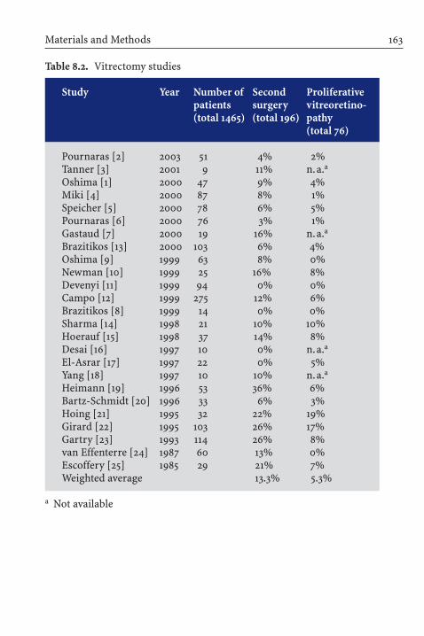
Materials and Methods 163
Table 8.2. Vitrectomy studies
Study Year Number of Second Proliferativepatients surgery vitreoretino-(total 1465) (total 196) pathy
(total 76)
Pournaras [2] 2003 51 4% 2%Tanner [3] 2001 9 11% n.a.a
Oshima [1] 2000 47 9% 4%Miki [4] 2000 87 8% 1%Speicher [5] 2000 78 6% 5%Pournaras [6] 2000 76 3% 1%Gastaud [7] 2000 19 16% n.a.a
Brazitikos [13] 2000 103 6% 4%Oshima [9] 1999 63 8% 0%Newman [10] 1999 25 16% 8%Devenyi [11] 1999 94 0% 0%Campo [12] 1999 275 12% 6%Brazitikos [8] 1999 14 0% 0%Sharma [14] 1998 21 10% 10%Hoerauf [15] 1998 37 14% 8%Desai [16] 1997 10 0% n.a.a
El-Asrar [17] 1997 22 0% 5%Yang [18] 1997 10 10% n.a.a
Heimann [19] 1996 53 36% 6%Bartz-Schmidt [20] 1996 33 6% 3%Hoing [21] 1995 32 22% 19%Girard [22] 1995 103 26% 17%Gartry [23] 1993 114 26% 8%van Effenterre [24] 1987 60 13% 0%Escoffery [25] 1985 29 21% 7%Weighted average 13.3% 5.3%
a Not available

the main outcome measures. Patient and surgical characteristicswere mostly homogeneous. The common denominator was thatthe detachment might have responded to scleral buckling.
Results
The pooled risk of second surgery for primary vitrectomy was 13.3% (196/1,465), for pneumatic retinopexy 25.2% (484/1,919), andfor segmental buckling 9.1% (170/1,854). The pooled risk of PVR after vitrectomy was 5.3% (76/1,417), after pneumatic retinopexywas 6.5% (111/1,697), and after scleral buckling 0.9% (17/1,702)(Fig. 8.1).
8 Systematic Review of Efficacy and Safety of Surgery164
Table 8.3. Scleral buckle and balloon studies
Study Year Number of Second Proliferativepatients surgery vitreoretino-(total 1854) (total 170) pathy
(total 17)
Oshima [1] 2000 55 9% 4%Green [61] 1996 162 12% 2%Kreissig [62] 1992 107 7% 4%Kreissig [56] 1989 500 7% 0%McAllister [45] 1988 28 36% 7%Richard [60] 1987 100 6% n.a.a
Binder [58] 1986 52 4% n.a.a
Schoch [59] 1986 45 7% 2%O’Connor [55] 1976 50 0% 0%Lincoff [57, 67] 1972 755 11% 0%Weighted average 9.1% 0.9%
a Not available

The odds ratio of the pooled procedures, i.e., 3384 intraoculargas operations and vitrectomies versus 1854 extraocular spongeand balloon buckles, for the risk of second surgery, was:
(680/2704)08 = 2.5 to 1(107/1684)
and for PVR:
(187/2927)07 = 6 to 1 .(17/1685)
Discussion
In the latter half of the twentieth century, the buckle operations in-vented by Charles Schepens in 1951 [63] and Ernst Custodis in 1953[64] were modified and refined. Diathermy was replaced with cryo-
Discussion 165
30%
25%
20%
15%
10%
5%
0%second surgery PVR
pneumaticretinopexyvitrectomybuckling
Fig. 8.1. Pool risk of second surgery and proliferative vitreoretinopathy(PVR)

pexy; scleral resections and implants were replaced with explantssutured over full thickness sclera [65, 66]. Extensive circumferen-tial buckles and encircling operations – barrier procedures thatwere intended to wall away undetected breaks in the periphery –were replaced with segmental buckles confined to the breaks. Theundetected break was less frequent because preoperative indirectophthalmoscopy was augmented by binocular microscopy of theretinal periphery through the mirrors of the Goldmann lens.
Closing the retinal breaks became the sole surgical problem;the extent of the detachment was a lesser factor. If the breaks wereeffectively buckled, the large detachment would attach withoutdrainage after only a few additional hours (Fig. 8.2). Not drainingsubretinal fluid was increasingly adopted. At the New York Hospi-tal, the incidence of not draining rose from 50% to 90% in thecourse of the first 1,000 cases after the senior author (HL) met withErnst Custodis and adopted his method [67].
Diminished morbidity was the Holy Grail. The external buckleoperation with a segmental sponge and without perforation fordrainage had no intraocular complications and only infrequent ex-traocular ones. There was a buckle infection initially of 3% thatdropped to 1% with the development of the closed-cell sponge andthe use of parabulbar antibiotic [68]. Diplopia might occur if asponge intruded on a rectus muscle. The substitution of a tempo-rary balloon for breaks beneath a rectus muscle eliminated post-operative diplopia because, within hours after the balloon waswithdrawn, the muscle functioned normally again [55]. A secondoperation after the sponge procedure was required in 11% and afterthe balloon procedure in 7%. Failure with either the sponge or theballoon was due to an undiscovered break or an inaccurately placedsponge or balloon. Final attachment for the sponge operation aftera second buckle was 97% and for the balloon was 99%. Less than2% developed PVR postoperatively after either procedure. The lowincidence of PVR was a positive affect of diminished operativetrauma. The greater incidence of PVR as we knew it in prior yearswas iatrogenic, a product of trauma inflicted by extensive barrier
8 Systematic Review of Efficacy and Safety of Surgery166

coagulation, constriction by the encircling band, and the drainingof subretinal fluid – all of which contribute to a breakdown of theblood–aqueous barrier and the infusion of cells and protein thatprovoke PVR. Of the 2% that failed to attach because PVR prevent-ed closure of the break with a buckle, vitrectomy could attach halfof them. Thus, blindness from a retinal detachment, which was in-evitable before 1929, was a rare event at the end of the twentieth century. Why the current swing to an intraocular procedure, whichour analysis indicates has a greater morbidity in terms of requiring2nd surgeries and causing PVR? – We suggest that the reasons areexternal and not related to results.
Discussion 167
Fig. 8.2. The small detachment (upper left) and the more extensive one(upper right) both responded to a radial sponge buckle (lower center). Thelarger detachment attached after only a few additional hours

Inadequate Training
Vitrectomy is being increasingly employed for primary retinal de-tachments because the number of doctors trained to do vitrectomyhas markedly increased in the past decade, as have the indicationsfor vitrectomy. In addition to the original indications, traction de-tachment in the diabetic eye and PVR after failed retinal surgery,vitrectomy is done for macular holes, macular puckers, droppedlens and particles, to clear vitreous hemorrhage, and, more recently,to dissect subretinal proliferative lesions in the macula. The in-dications keep expanding. Retinal detachment can be a relativelyinfrequent indication for operation on a busy retinal service and ofsecondary interest. As a result, training for the treatment of retinaldetachment may be limited. There are few opportunities outside ofthe fellowship to learn about buckling. Retinal programs rarelyinclude papers on the subject; it has all been said. Except forWilliam Mieler’s short course at the Academy and Ingrid Kreissig’sRetinal Detachment Courses at various national and internationalmeetings, there are no workshops on the art of scleral buckling.As a consequence, the coming generation of retinal surgeons maynot be very skillful at performing a scleral buckle. In anotherdecade, it is possible that the buckling operation will have becomea lost art.
Market Forces
Preparation for a segmental buckle operation can require muchstudy time, frequently hours and sometimes days. A decade ago, ifthe senior author (HL) could not find a break that promised a 90%prognosis after an hour’s study, he would patch both eyes, put thepatient at rest, and re-study the eye the next day. Most detachmentschange with ocular rest, and the change is informative. Today, theinsurance companies and Medicare do not allow for an extra day,nor is there payment for study time. It is more expedient to do a
8 Systematic Review of Efficacy and Safety of Surgery168

vitrectomy and search for the retinal break at the operating table,where the wage rate is 37% higher (Medicare fees from New York,New Jersey, Connecticut). If one does not find the break, a surgeonmight be inclined to do a peripheral laser barricade augmented by an encircling band. The barrier operation, which was disap-pearing near the end of the twentieth century, is returning as a prophylactic supplement to primary vitrectomy and pneumaticretinopexy [54].
Peer Review
A third factor operating against the buckle operation for primarydetachment, beyond the lack of training and the limit of reim-bursement for the time spent, is the absence of peer review. Preop-erative surgical rounds, where the surgical plan for retinal detach-ment was open for review, suggestion, criticism, and even censure,have disappeared; with 1-day surgery, there is not time for it. Thesurgeon admits his patient on the day of the operation with asmuch preparation as his schedule allowed, and, when the patientleaves the hospital later in the day with the eye filled with gas, thereis no opportunity to evaluate the effort by his or her peers.
Conclusion
The proponents of primary vitrectomy claim that the final attach-ment rate after multiple procedures is 99%. In the series that we examined, it was 97%, equal to that of buckling. This is reassuring.They dismiss the morbidity of multiple operations and regard thefrequent provocation of cataract as acceptable.
We do not think it is possible to counteract the change in thetreatment of retinal detachment. Perhaps market forces will relax,and peer review will return, or maybe new and less morbid methodsto attach the retina will be invented. Admittedly, the incidence of
Conclusion 169

second surgery and PVR were beginning to diminish in the mostrecent of the case series that we examined, and perhaps a way to reduce the incidence of postoperative cataract will develop. If not,maybe in another 50 years, someone will rediscover the scleralbuckle.
� Acknowledgements. This study was supported by The Edward L. Grayson Retinal Research Fund, West Orange, NJ; Señora SaluaH. Kuri, Mexico City, Mexico; Caroline H. Newhouse and the New-house Foundation, New York, USA. The authors have no proprietaryinterest in any of the aspects of this study.
References
1. Oshima Y,Yamanishi S, Sawa M, Motokura M, Harino S, Emi K (2000)Two-year follow-up study comparing primary vitrectomy with scleralbuckling for macula-off rhegmatogenous retinal detachment. Jpn JOphthalmol 44:538–549
2. Pournaras CJ, Kapetanios AD (2003) Primary vitrectomy for pseudo-phakic retinal detachment: a prospective non-randomized study. EurJ Ophthalmol 13:298–306
3. Tanner V, Minihan M, Williamson TH (2001) Management of inferiorretinal breaks during pars plana vitrectomy for retinal detachment.Br J Ophthalmol 85:480–482
4. Miki D, Hida T, Hotta K, Shinoda K, Hirakata A (2000) Comparison ofscleral buckling and vitrectomy for superior retinal detachmentcausedby flap tears (in Japanese). Nippon Ganka Gakkai Zasshi 104: 24–28
5. Speicher MA, Fu AD, Martin JP, von Fricken MA (2000) Primaryvitrectomy alone for repair of retinal detachments following cataractsurgery. Retina 20:459–464
6. Pournaras CJ, Donati G, Sekkat L et al (2000) Pseudophakic retinal de-tachment:treatment by vitrectomy and scleral buckling. Pilot study(in French). J Fr Ophtalmol 23:1006–1011
7. Gastaud P, Rouhette H, Negre F et al (2000) Place of “exploratoryvitrectomy” in the treatment of retinal detachment without prolifera-tive vitreoretinopathy (in French). J Fr Ophtalmol 23:482–487
8 Systematic Review of Efficacy and Safety of Surgery170

8. Brazitikos PD, D’Amico DJ, Tsinopoulos IT et al (1999) Primary vitrectomy with perfluoro-n-octane use in the treatment of pseudo-phakic retinal detachment with undetected retinal breaks. Retina 19:103–109
9. Oshima Y, Emi K, Motokura M et al (1999) Survey of surgical indica-tions and results of primary pars plana vitrectomy for rhegmatoge-nous retinal detachments. Jpn J Ophthalmol 43:120–126
10. Newman DK, Burton RL (1999) Primary vitrectomy for pseudophakicand aphakic retinal detachments. Eye 13:635–639
11. Devenyi RG, de Carvalho Nakamura H (1999) Combined scleral buckle and pars plana vitrectomy as a primary procedure for pseudo-phakic retinal detachments. Ophthalmic Surg Lasers 30:615–618
12. Campo RV, Sipperley JO, Sneed SR et al (1999) Pars plana vitrectomywithout scleral buckle for pseudophakic retinal detachments. Oph-thalmology 106: 1811–1816
13. Brazitikos PD (2000) The expanding role of primary pars planavitrectomy in the treatment of rhegmatogenous noncomplicated reti-nal detachment. Semin Ophthalmol 15:65–77
14. Sharma T, Gopal L, Badrinath SS (1998) Primary vitrectomy for rheg-matogenous retinal detachment associated with choroidal detach-ment. Ophthalmology 105:2282–2285
15. Hoerauf H, Roider J, Herboth T et al (1997) Outcome after vitrectomyin rhegmatogenous retinal detachment and dense vitreous opacities(in German). Klin Monatsbl Augenheilkd 211:369–374
16. Desai UR, Strassman IB (1997) Combined pars plana vitrectomy andscleral buckling for pseudophakic and aphakic retinal detachments in which a break is not seen preoperatively. Ophthalmic Surg Lasers28:718–722
17. El-Asrar AM (1997) Primary vitrectomy for bullous rhegmatogenousretinal detachments due to complex breaks. Eur J Ophthalmol 7:322–326
18. Yang CM (1997) Pars plana vitrectomy in the treatment of combinedrhegmatogenous retinal detachment and choroidal detachment inaphakic or pseudophakic patients.Ophthalmic Surg Lasers 28:288– 293
19. Heimann H, Bornfeld N, Friedrichs W et al (1996) Primary vitrectomywithout scleral buckling for rhegmatogenous retinal detachment.Graefes Arch Clin Exp Ophthalmol 234:561–568
20. Bartz-Schmidt KU,Kirchhof B,Heimann K (1996) Primary vitrectomyfor pseudophakic retinal detachment. Br J Ophthalmol 80:346–349
References 171

21. Hoing C, Heidenkummer HP, Kampik A (1995) Primary vitrectomy inrhegmatogenous retinal detachment (in German). Ophthalmologe92:668–671
22. Girard P, Karpouzas I (1995) Vitrectomy in the treatment of simpleretinal detachment (in French). J Fr Ophtalmol 18:188–193
23. Gartry DS, Chignell AH, Franks WA et al (1993) Pars plana vitrectomyfor the treatment of rhegmatogenous retinal detachment uncom-plicated by advanced proliferative vitreoretinopathy. Br J Ophthalmol77:199–203
24. Van Effenterre G, Haut J, Larricart P et al (1987) Gas tamponade as asingle technique in the treatment of retinal detachment: is vitrectomyneeded? A comparative study of 120 cases. Graefes Arch Clin Exp Oph-thalmol 225:254–258
25. Escoffery RF, Olk RJ, Grand MG et al (1985) Vitrectomy without scler-al buckling for primary rhegmatogenous retinal detachment. Am JOphthalmol 99: 275–281
26. Kleinmann G, Rechtman E, Pollack A, Schechtman E, Bukelman A(2002) Pneumatic retinopexy: results in eyes with classic vs relativeindications. Arch Ophthalmol 120:1455–1459
27. Eter N, Boeker T, Spitznas M (2000) Long-term results of pneumaticretinopexy. Graefes Arch Clin Exp Ophthalmol 238: 677–681
28. Assi AC, Charteris DG, Pearson RV, Gregor ZJ (1999) Pneumaticretinopexy in the treatment of primary rhegmatogenous retinal de-tachment. Eye 13:725–728
29. Abecia E, Pinilla I, Olivan JM, Larrosa JM, Polo V, Honrubia FM (2000)Anatomic results and complications in a long-term follow-up of pneu-matic retinopexy cases. Retina 20:156–161
30. Lisle C, Mortensen KK, Sjolie AK (1998) Pneumatic retinopexy.A long-term follow-up study. Acta Ophthalmol Scand 76:486–490
31. Tornambe PE, Hilton GF (1989) The Retinal Detachment Study Group.Pneumatic retinopexy. A multicenter randomized controlled clinicaltrial comparing pneumatic retinopexy with scleral buckling. Ophthal-mology 96:772–784
32. Mulvihill A, Fulcher T, Datta V, Acheson R (1996) Pneumatic retino-pexy versus scleral buckling: a randomised controlled trial. Ir J MedSci 165:274–277
33. Grizzard WS, Hilton GF, Hammer ME, Taren D, Brinton DA (1995)Pneumatic retinopexy failures. Cause, prevention, timing and manage-ment. Ophthalmology 102:929–936
8 Systematic Review of Efficacy and Safety of Surgery172

34. Gunduz K, Gunalp I (1994) Pneumatic retinopexy with drainage ofsubretinal fluid. Int Ophthalmol 18:143–147
35. Boeker T, Schmitt C, Mougharbel M (1994) Results and prognostic factors in pneumatic retinopexy. Ger J Ophthalmol 3:73–78
36. Sebag J, Tang M (1993) Pneumatic retinopexy using only air. Retina13:8–12
37. Bochow TW, Olk JM, Hershey JM (1992) Pneumatic retinopexy per-fluoroethane (C2F6) in treatment of rhegmatogenous retinal detach-ment. Arch Ophthalmol 110:1723–1724
38. Algvere P, Hallnaes K, Palmqvist B (1988) Success and complicationsof pneumatic retinopexy. Am J Ophthalmol 106:400–404
39. Berrod JP, Bazard MC, Bodart E, Noye JF, Raspiller A (1990) Pneumaticretinopexy using SF6 or C3F8. Results and complications apropos of56 patients (in French). Bull des Soc d Ophtalmologie de Fr 90:117–121
40. Termote H (1989) Pneumatic retinopexy. Analysis of the first 20 cases(in French). Bull de la Soc Belge d Ophthalmologie 231:107–116
41. Lemmen KD, Heimann K (1989) Probleme der pneumatischenretinopexie. Klin Monatsbl Augenheilkd 195:150–153
42. Skoog KO, Textorius O, Wrigstad A (1989) Pneumatic retinopexy in 50 patients. Acta Ophthalmol 67:103–105
43. Lowe MA, McDonald HR, Campo RV, Boyer DS, Schatz H (1988) Pneu-matic retinopexy. Surgical results. Arch Ophthalmol 106:1672–1676
44. Algvere P, Gjoetterberg M, Olivestedt G, Fituri S (1992) Results ofpneumatic retinopexy with air. Acta Ophthalmol 70:632–636
45. McAllister IL, Meyers SM, Zegarra H, Gutman FA, Zakov ZN, Beck GJ(1988) Comparison of pneumatic retinopexy with alternative surgicaltechniques. Ophthalmology 95:877–883
46. Chen JC, Robertson JE, Coonan P et al (1988) Results and complica-tions of pneumatic retinopexy. Ophthalmology 95:601–608
47. Hilton GF, Grizzard WS (1986) Pneumatic retinopexy; a two-step outpatient operation without conjunctival incision. Ophthalmology93:626–641
48. Poliner LS, Grand MG, Schoch LH, Olk RJ, Johnston GP, Okun E,Boniuk I, Escoffery RF (1987) New retinal detachment after pneumaticretinopexy. Ophthalmology 94:315–318
49. Gnad HD, Skorpik C, Paroussis P, Menapace R, Kulnig W (1987) C3F8gas as internal tamponade in retinal surgery – a “pneumatic” retinaldetachment operation without opening the conjunctiva (in German).Fortschr Ophthalmol 84:462–464
References 173

50. Hilton GF, Kelly NE, Salzano TC, Tornambe PE, Wells JW, Wendel RT(1987) Pneumatic retinopexy. A collaborative report of the first 100cases. Ophthalmology 94:307–314
51. Dominguez A, Fonseca A, Gomez-Montana J (1987) Gas tamponadefor ambulatory treatment of retinal detachment. In: Proceedings ofthe XXVth International Congress of Ophthalmology. Rome, 4–10May, 1986. Kugler & Ghedini, Amsterdam, pp 2038–2045
52. Tornambe PE, Hilton GF (1989) Pneumatic retinopexy. A multicenterrandomized controlled clinical trial comparing pneumatic retinopexywith scleral buckling. The Retinal Detachment Study Group. Ophthal-mology 96:772–783
53. Tornambe PE, Poliner LS, Hilton GF, Grizzard WS (1999) Comparisonof pneumatic retinopexy and scleral buckling in the management ofprimary rhegmatogenous retinal detachment. Am J Ophthalmol 127:741–743
54. Tornambe PE (1997) Pneumatic retinopexy: the evolution of case se-lection and surgical technique. A twelve-year study of 302 eyes. TransAm Ophthalmol Soc 95:551–578
55. O’Connor PR (1976) External buckling without drainage for selecteddetachments in aphakic eyes. Am J Ophthalmol 82:358–364
56. Kreissig I, Failer J, Lincoff H et al (1989) Results of a temporary bal-loon buckle in the treatment of 500 retinal detachments and a com-parison with pneumatic retinopexy. Am J Ophthalmol 107:381–389
57. Lincoff H, Kreissig I (1972) The treatment of retinal detachment with-out drainage of subretinal fluid. (Modifications of the Custodis proce-dure. VI). Trans Am Acad Ophthalmol Otolaryngol 76:1121–1133
58. Binder S (1986) Repair of retinal detachments with temporary balloonbuckling. Retina 6:210–214
59. Schoch LH, Olk RJ, Arribas NP, Okun E, Johnston GP, Boniuk I,Escoffery RF, Grand MG (1986) The Lincoff temporary balloon buck-le. Am J Ophthalmol 101: 646–649
60. Richard G (1987) Indications, technic and results of the balloon opera-tion (in German). Klin Monatsbl Augenheilkd 190:484–488
61. Green SN, Yarian DL, Masciulli L, Leff SR (1996) Office repair of reti-nal detachment using a Lincoff Temporary balloon buckle. Ophthal-mology 103:1804–1810
62. Kreissig I, Rose D, Jost B (1992) Minimized surgery for retinal detach-ments with segmental buckling and nondrainage. An 11-year follow-up. Retina 12:224–231
8 Systematic Review of Efficacy and Safety of Surgery174

63. Schepens CL (1951) Progress in detachment surgery. Trans Am AcadOphthalmol Otolaryngol 55:607–615
64. Custodis E (1953) Bedeutet die Plombenaufnaehung auf die Skleraeinen Fortschritt in der operativen Behandlung der Netzhautabloe-sung? Ber Deutsch Ophth Ges 58:102
65. Lincoff HA, Baras I, McLean J (1965) Modifications to the Custodisprocedure for retinal detachment. Arch Ophthalmol. 73:160–163
66. Lincoff HA, McLean JM (1966) Cryosurgical treatment of retinal detachment. II. Trans Am Acad Ophthalmol Otolaryngol 70:202–211
67. Lincoff H, Kreissig I (1972) The treatment of retinal detachment with-out drainage of subretinal fluid. Modifications of the Custodis proce-dure: part VI. Trans Am Acad Ophthalmol Otolaryngol 76:1221–1232
68. Hahn YS, Lincoff A, Lincoff H (1979) Infection after sponge implanta-tion for scleral buckling. Am J Ophthalmol 87:180–185
References 175

Repair of Primary Retinal Detachment:The Present State of the Art and How It Came About
Ingrid Kreissig, Harvey Lincoff
Chapter 9
A major advance in the concept of treating a primary rhegmatoge-nous retinal detachment was the realization that the surgical prob-lem was solely closing the leaking retinal break and that the extentof the detachment or tractional configurations remote from thebreak are of no consequence. Let us share with you this change inconcept over time [1].
Recall, Gonin [2] postulated – for the first time – that a leakingbreak is the cause of a retinal detachment, and his treatment waslimited to the area of this break.With his operation, the attachmentrate increased from 0% to 57%. However, this localized procedurewas soon modified to coagulations of the entire quadrant of theleaking break. In 1931, Guist and Lindner [3, 4] circumvented fur-ther the need for localizing the leaking break by doing multiplecauterizations posterior to the estimated position of the break;Safar [5] applied a semicircle of coagulations posterior to the break.The intent was to create a “barrier” of retinal adhesions posteriorto the leaking break. As a result, the treatment was no longer limit-ed to the break, but was expanded over the quadrant in which thebreak or presumed breaks were located.
In 1938, Rosengren [6] again limited – now for the second time– the coagulations to the leaking break. In addition – and for thefirst time – he added an intraocular tamponade of air, which waspositioned in the area of the break to provide an internal supportduring the formation of retinal adhesion. Retinal attachment increased to about 77% with Rosengren’s procedure.
However, the precise placement of coagulations around thebreak was difficult, and the Rosengren technique was not widely

adopted. Instead, the pendulum swung back to an extensive co-agulation. Now, for the second time, the barrier concept was inte-grated into the treatment. Coagulations were placed posterior tothe break, but, in addition, the barrier of coagulations was rein-forced with a scleral resection. Subsequently, a polyethylene tubewas embedded into the resection to create a higher wall. Thus,for the first time, a buckle was applied in detachment surgery tomore effectively barricade the break (Figs. 9.1, 9.2). The break was
9 Repair of Primary Retinal Detachment178
Fig. 9.1. A three-quadrant detachment with a horseshoe tear at 1:15
Fig. 9.2. Scleral resection with an embedded polyethylene tube anddrainage for repair of the three-quadrant detachment in Fig. 9.1. a Thehorseshoe tear was attached, but positioned on the anterior edge of thebuckle and not sufficiently tamponaded. Diathermy coagulations wereadded around the tear, on the buckle, and additional lines of coagulationsextended to the ora serrata. b The horseshoe tear leaked anteriorly, brokethrough the lines of coagulations before the adhesions were secure, andcaused an anterior redetachment that progressed inferiorly and rede-tached the posterior retina

Repair of Primary Retinal Detachment 179
a
b
Fig. 9.2a,b. Legend see page 178

9 Repair of Primary Retinal Detachment180
a
b
Fig. 9.3a,b. Legend see page 181

positioned at the anterior edge of the buckle and larger breaks werenot adequately tamponaded and would leak anteriorly and cause ananterior redetachment, which descended behind the buckle, wentaround the buckle inferiorly, and redetached the posterior retina.
The consequence could have been a more sufficient tamponadeof the leaking break. Instead, a more effective barrier was devel-oped in 1953, the segmental buckle barrier was extended for 360° –for the first time – by Schepens [7] and in 1958 by Arruga [8]. Thecerclage operation with drainage of subretinal fluid represented amaximum barrier for the leaking break. But here, as well, redetach-ments developed (Fig. 9.3). Eventually the cerclage was widened inthe area of the tear with a polyethylene sleeve to buckle the anterioredge of the tear. Later, various silicone forms for buckling were
Repair of Primary Retinal Detachment 181
Fig. 9.3. Encircling buckle (cerclage), extensive diathermy coagulations,and drainage for repair of the detachment in Fig. 9.1. a The horseshoe tearwas attached, but positioned on the anterior edge of the buckle and notsufficiently tamponaded. b Anterior redetachment was confined by theencircling buckle. c Anterior redetachment eventually broke over the in-ferior buckle barrier and redetached the posterior retina
c

designed to fit the tear combined with coagulations limited to thetear or extending over 360° (Fig. 9.4). More retinas were attached –more than 80%.
The modified cerclage with drainage represents one of the fourtechniques still in use for repair of a primary retinal detachment atthe beginning of the twenty-first century (Fig. 9.4). Drainage, how-ever, required by this technique, has complications.
In 1953, Custodis [9] limited the treatment – now for the thirdtime – to the area of the leaking break, but – for the first time –omitted drainage of subretinal fluid. This exceptional techniquewas nearly abandoned, not because it did not work, but because ofunexpected postoperative complications caused by diathermy andthe polyviol plombe, which Custodis compressed over full-thick-ness and diathermized sclera, which sometimes caused scleralnecrosis. As a result, the technique was abandoned in the UnitedStates and in Europe.
Lincoff in New York, who was convinced of the logic and sim-plicity of the Custodis procedure, made the operation acceptable byreplacing diathermy with cryopexy [10, 11] and the polyviol plombewith the tissue-inert silicone sponge [12]. In the following years, thistechnique was further refined by smaller segmental buckles thatwere positioned more precisely [13] and by replacing the sclera-fixated sponge with a temporary balloon buckle [14, 15] that was notsutured onto sclera. The balloon operation was suitable for detach-ments with a single break. This minimal segmental buckling withsponges or a balloon represents an extraocular approach, limited,again, to the area of the leaking break.
However, the “conditio sine qua non” for a spontaneous attach-ment without drainage was that all of the leaking breaks had to befound and tamponaded adequately. Otherwise, the disappearanceof subretinal fluid would not occur. Finding all the breaks washelped by the development of binocular indirect ophthalmoscopy,biomicroscopy with contact lenses, the 4 Rules to find the break ina primary detachment [16, 17], and the 4 Rules to find the break inan eye up for reoperation [18, 19].
9 Repair of Primary Retinal Detachment182

Repair of Primary Retinal Detachment 183
Fig. 9.4. Modified cerclage: Encircling silicone band with a local bucklebeneath the tear and drainage for repair of the detachment in Fig. 9.1.With coagulations limited to the area of the tear (a) or with coagulationsextended for 360° (b)
a
b

As a result, minimal segmental buckling with sponge(s) or aballoon without drainage represents a second option for repair ofa primary retinal detachment in use at the beginning of the twenty-first century (Fig. 9.5).
Apart from these two options for closing the leaking break witha circular barrier (cerclage) or a segmental buckle (sponge or balloon) limited to the break, both of which are extraocular, twooptions for an intraocular approach developed. An intraocular gasbubble to tamponade a leaking break was introduced – now for thesecond time. Instead of air, SF6 was injected by Norton and Lincoff[20, 21]. This technique required drainage of subretinal fluid withits complications.
In 1979, Kreissig [22] applied – for the first time – intraocularSF6, an expanding gas, without prior drainage in selected de-tachments. The procedure was named the expanding-gas ope-ration without drainage. However, an increased rate of post-operative proliferative vitreoretinopathy (PVR) caused Kreissig to reserve intraocular gas for problematic breaks not suitable for buckling. Subsequently to reduce the morbidity of intraoculargas, Kreissig developed the balloon-gas procedure which enablesto inject primarily a larger bubble of a gas with a shorter intra-ocular duration [23]. To close a leaking break with a gas bubble andwithout prior drainage was introduced again – for the second time– by Hilton [24] and simultaneously by Dominguez [25] in 1986.
Hilton called the procedure pneumatic retinopexy which re-presents a third option for repair of a primary retinal detachmentin use at the beginning of the twenty-first century (Fig. 9.6a).When supplemented by 360° barrier coagulations, it is no longer aprocedure limited to the break (Fig. 9.6b) [26].
Pneumatic retinopexy has become a popular procedure, despitethe fact that it has a greater morbidity for closing the leaking breakthan minimal segmental buckling without drainage. Its popularityis due to its relative simplicity.
To reduce the postoperative complications of intraocular gas,a vitrectomy was added [27]. The rationale was that a vitrectomy
9 Repair of Primary Retinal Detachment184

Repair of Primary Retinal Detachment 185
Fig. 9.5. Minimal segmental buckling without drainage and coagulationslimited to the tear for repair of the detachment in Fig. 9.1. The buckle is obtained by a radial sponge (a) or by a temporary balloon beneath the tear(b). After withdrawal of the unsutured parabulbar balloon (after 1 week),the tear will be only secured by coagulations
a
b

9 Repair of Primary Retinal Detachment186
Fig. 9.6. Pneumatic retinopexy without drainage for repair of the detach-ment in Fig. 9.1. With coagulations limited to the tear (a) or with coagula-tions extended for 360° (b) [25]. An expanding gas was injected into the vitreous, and the patient’s head was positioned so that the gas bubbletamponaded the tear. Air travel will be restricted until the volume of thegas bubble is less than 10% of the ocular volume
a
b

Repair of Primary Retinal Detachment 187
Fig. 9.7. Primary vitrectomy with resection of the vitreous and internaldrainage for repair of the detachment in Fig. 9.1.With coagulations aroundthe tear (a) or with coagulations extended for 360°, a local buckle beneaththe tear and an encircling band (b). Gas was injected to replace the vitre-ous and the patient asked to avoid face up during sleep. Air travel will berestricted until the volume of the gas bubble is less than 10% of the ocularvolume
a
b

might eliminate traction on the break and reduce postopera-tive anterior and posterior vitreous proliferation. The analysis inChap. 8 indicates that this aim has not been achieved; nevertheless,the procedure is increasingly applied.
Primary vitrectomy has become a fourth option for repair of aprimary retinal detachment at the beginning of the twenty-firstcentury (Fig. 9.7). When supplemented by extensive barrier coagu-lations and a cerclage, it is no longer a procedure limited to thebreak.
Conclusion
In the beginning of the twenty-first century, the present state-of-the-art for repair of a primary retinal detachment has reverted froma local to a barrier concept of treatment – as has happened severaltimes during the past 75 years.
External buckling: local buckles with coagulations limited tothe break (Fig. 9.5a, b) are becoming replaced by local buckles supplemented by an encircling band with extended coagulations(Fig. 9.4a, b), applied as a barrier against redetachments.
The same applies to pneumatic retinopexy: the primary intentto limit treatment to the area of the tear (Fig. 9.6a) is given up –again – in favour of a barrier concept by applying 360° of coagula-tions (Fig. 9.6b).
A similar trend is becoming apparent with primary vitrectomy:initially aimed at removing traction on the tear and limiting the co-agulations to the area of the tear (Fig. 9.7a), the procedure has beenextended by a circular barrier of coagulations with an encirclingband supplemented by a local buckle beneath the tear to preventredetachments (Fig. 9.7b).
Of the four surgical techniques in use at present for repair of aprimary rhegmatogenous retinal detachment, two are extraocularoperations (minimal segmental buckling with sponges or a balloonwithout drainage and cerclage with drainage) and two are intra-
9 Repair of Primary Retinal Detachment188

ocular (pneumatic retinopexy and primary vitrectomy). To succeedwith any of these methods, the leaking break still has to be foundand sealed. Therefore, finding and closing the retinal break in arhegmatogenous retinal detachment will continue to be the pri-mary purpose of any surgical effort.
With any of the four presently applied surgical techniques, reti-nal attachment can result in 94–99% of primary rhegmatogenousretinal detachments, but with different degrees of morbidity. Atthis point in time, we must wait to see which of these four proce-dures or their extended modifications will prevail or whether abetter and less morbid method for repair of retinal detachmentwill evolve.
References
1. Lincoff H, Kreissig I (2000) Changing patterns in the surgery forretinal detachment. Klin Monatsbl Augenheilkd 216:352–359
2. Gonin J (1930) Le traitement opératoire du décollement rétinien.Conférence aux journées médicales de Bruxelles. Bruxelles-Médical23:No. 17
3. Guist E (1931) Eine neue Ablatiooperation. Ztsch Augenheilk 74:232–242
4. Lindner K (1931) Ein Beitrag zur Entstehung und Behandlung deridiopathischen und der traumatischen Netzhautabloesung. GraefesArch Ophthalmol 127:177–295
5. Safar K (1932) Behandlung der Netzhautabhebung mit Elektrodenfuer multiple diathermische Stichelung. Dtsch Ophthalmol Ges 39:119
6. Rosengren B (1938) Ueber die Behandlung der Netzhautabloesungmittelst Diathermie und Luftinjektion in den Glaskoerper. Acta Ophthalmol 16:3–42
7. Schepens CL (1953) Prognosis and treatment of retinal detachment.The Mark J Schoenberg Memorial Lecture.A review by Kronenberg B,New York Society for Clinical Ophthalmology. Am J Ophthalmol 36:1739–1756
8. Arruga MH (1958) Le cerclage equatorial pour traiter le decollementretinien. Bull Soc Franc Ophtal 71:571–580
References 189

9. Custodis E (1953) Bedeutet die Plombenaufnaehung auf die Sklera einenFortschritt in der operativen Behandlung der Netzhautabloesung?Berichte der Deutschen Ophthalmologischen Gesellschaft 58: 102–105
10. Lincoff H, Kreissig I (1971) The mechanism of the cryosurgical adhe-sion. Am J Ophthalmol 71:674–689
11. Kreissig I, Lincoff H (1971) Ultrastruktur der Kryopexieadhaesion. In:DOG Symp.“Die Prophylaxe der idiopathischen Netzhautabhebung.”Bergmann, Muenchen, pp 191–205
12. Lincoff H, Baras I, McLean J (1965) Modifications to the Custodis pro-cedure for retinal detachment. Arch Ophthalmol 73:160–163
13. Kreissig I, Rose D, Jost B (1992) Minimized surgery for retinal detach-ments with segmental buckling and nondrainage. An 11-year follow-up. Retina 12: 224–231
14. Lincoff HA, Kreissig I, Hahn YS (1979) A temporary balloon buckle forthe treatment of small retinal detachments. Ophthalmology86:586–592
15. Kreissig I, Failer J, Lincoff H, Ferrari F (1989) Results of a temporaryballoon buckle in the treatment of 500 retinal detachments and acomparison with pneumatic retinopexy.Am J Ophthalmol 107:381–389
16. Lincoff H, Gieser R (1971) Finding the retinal hole. Arch Ophthalmol85:565–569
17. Kreissig I (2000) A practical guide to minimal surgery for retinal detachment: vol. 1. Thieme, Stuttgart–New York, pp 14–15, and backcover of book
18. Lincoff H, Kreissig I (1996) Extraocular repeat surgery of retinal detachment. A minimal approach. Ophthalmology 103:1586–1592
19. Kreissig I (2000) A practical guide to minimal surgery for retinal detachment: vol. 2. Thieme, Stuttgart–New York pp 320–321, and backcover of book
20. Norton EWD (1973) Intraocular gas in the management of selectedretinal detachments. Trans Am Acad Ophthalmol Otolaryngol 77:85-98
21. Lincoff H (1974) Reply to Drs. Fineberg, Machemer, Sullivan and Norton. Mod Probl Ophthalmol 12:344–345
22. Kreissig I (1979) Clinical experience with SF6-gas in detachment surgery. Berichte der Deutschen Ophthalmologischen Gesellschaft76:553–560
23. Kreissig I (1986) The balloon-gas method. Fortschr. Ophthalmol83:219–223
9 Repair of Primary Retinal Detachment190

24. Hilton GF, Grizzard WS (1986) Pneumatic retinopexy. A two-step outpatient operation without conjunctival incision. Ophthalmology93:626–641
25. Dominguez DA, Boyd BF, Gordon S (1986) Repeated insufflation ofexpansive gas. Highlights Ophthalmol Lett 14:1–14
26. Tornambe PE (1997) Pneumatic retinopexy: the evolution of case se-lection and surgical technique. A twelve-year study of 302 eyes. TransAm Ophthalmol Soc 95: 551–78
27. Escoffery RF, Olk RJ, Grand MG, Boniuk I (1985) Vitrectomy withoutscleral buckling for primary rhegmatogenous retinal detachment.AmJ Ophthalmol 99:275–281
References 191

Retinal Detachment Repair: Outlook for the Future
William R. Freeman
Chapter 10
Due to the pioneering work of many ophthalmologists, includingGonin, Lincoff, and others, the basic pathophysiology of rhegma-togenous retinal detachment has been established. A retinal tear iscaused by a vitreous detachment and traction on the retinal tear,and peripheral retina is responsible for fluid currents, which gothrough the break and detach the retina. More fundamental ques-tions remain to be answered and will have important implicationsfor our ability to detect, prevent, and treat rhegmatogenous retinaldetachment and its complications.
Many controversies remain regarding surgical repair of thisdisease. This controversy is complicated due to a lack of prospec-tive randomized clinical trials comparing different methods of
Fig. 10.1. Radial sponge has cured a retinal detachment without the needfor drainage

treatment. For this reason, much of the debate revolves aroundtheory and philosophy, not hard clinical data. There is little doubtthat the most minimal operation that would be highly effectivewould be the procedure of choice. Highly effective should includeminimal complications and inconveniences, such as the inductionof refractive error (Lincoff, Kreissig) [1]. In this regard, the classicalradial sponge (Fig. 10.1) or the balloon buckle (Fig. 10.2) remainsthe gold standard for many, due to the extraocular nature of thesurgery and low complication rate. These procedures shared incommon extraocular placement of a bulky device,which reapposesor nearly apposes the neurosensory retina and retinal pigment epithelium/choroicapillaris. By bringing these two layers in closeproximity, the rate of fluid flow under the retina is limited and the pumping action of the pigmented epithelium overcomes theleakage of fluid through the retinal tear; thus the retina reattaches.The use of retinopexy is a backup procedure to further prevent fluid leakage, causing a permanent scar or adhesion of the two layers (Figs. 10.3 and 10.4). Unfortunately, segmental buckling is not
10 Retinal Detachment Repair: Outlook for the Future194
Fig. 10.2. Balloon buckle in place closing retinal break

applicable to all cases. For example, very large posterior tears aredifficult to externally tamponade.Giant retinal tears do not respondto external buckle procedures because one cannot reappose thelayers; the retina is merely pushed inward. In addition, the proce-dure can only work when all retinal breaks can be well visualized(Fig. 10.5). This may be difficult in eyes with media problems or
Retinal Detachment Repair: Outlook for the Future 195
Fig. 10.3. Horseshoe retinal tear with vitreous traction
Fig. 10.4. Laser retinopexy surrounding horsehoe retinal tear

when intraocular lenses, retained lens material, and other difficul-ties preclude identification of all retinal breaks [2].
Let us now consider the evolution of surgical techniques in thefuture. One of the many obstacles to performing minimal bucklingtechniques, as well as pneumatic retinopexy and the balloon buck-le, is the difficulty in finding all retinal breaks in certain eyes asoutlined above. New techniques will evolve to allow visualization ofthe peripheral retina, which will make break identification moreuniversally possible.
New imaging techniques, such as ballistic light imaging(Fig. 10.6), hold the promise of high-resolution trans-scleral opti-cal images [3]. Ordinarily, it is not possible to view clearly througha semi-transparent tissue, such as the thin sclera. A degraded im-age results due to scattered light. Light passes through the tissue,but scatter presents the ability to obtain a clear image. Scatteredlight is delayed compared with non-scattered light; however, theamount of delay is extremely small. The ability to fabricate fem-tosecond optical filters can theoretically gate out this scatteredlight based on time differences. In the future, such ballistic lightimaging promises to be a non-invasive way of examining the vitre-
10 Retinal Detachment Repair: Outlook for the Future196
Fig. 10.5. A well-visualized peripheral retinal tear
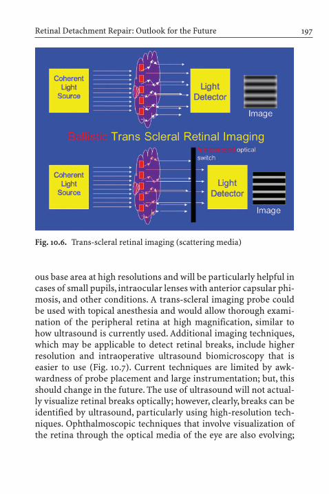
Retinal Detachment Repair: Outlook for the Future 197
Fig. 10.6. Trans-scleral retinal imaging (scattering media)
ous base area at high resolutions and will be particularly helpful incases of small pupils, intraocular lenses with anterior capsular phi-mosis, and other conditions. A trans-scleral imaging probe couldbe used with topical anesthesia and would allow thorough exami-nation of the peripheral retina at high magnification, similar tohow ultrasound is currently used. Additional imaging techniques,which may be applicable to detect retinal breaks, include higherresolution and intraoperative ultrasound biomicroscopy that iseasier to use (Fig. 10.7). Current techniques are limited by awk-wardness of probe placement and large instrumentation; but, thisshould change in the future. The use of ultrasound will not actual-ly visualize retinal breaks optically; however, clearly, breaks can beidentified by ultrasound, particularly using high-resolution tech-niques. Ophthalmoscopic techniques that involve visualization ofthe retina through the optical media of the eye are also evolving;

the wide-angle pseudo-color SLO and related techniques (Fig. 10.8)may allow noninvasive screening for retinal detachment by primarycare health care providers, which will allow us to treat detachmentsprior to macular involvement [4]. Such wide-angle SLO devicescurrently do not allow reliable imaging of structures anterior to the equator of the globe; however, they can detect retinal detach-ment early and, if set up as easy to use screening devices, will allow
10 Retinal Detachment Repair: Outlook for the Future198
Fig. 10.8. Pseudo-color wide-angle scanning laser may allow screeningfor retinal detachment
Fig. 10.7. Ultrasound biomicroscopy images peripheral retinoschisis

patients to be treated at earlier stages of the detachment, when themacula has not been affected, and the rate of success is higher.High-resolution scans, such as those used now for magnetic reso-nance imaging (MRI) scanning of the head and neck vessels, maybecome applicable to the retinal periphery and help with break lo-calization and prophylaxis issues. Functional MRI may increaseour understanding of peripheral retinal tissues as well. This tech-nique may allow insights into vitreous and peripheral retina me-tabolism and degeneration and may help allow us to predict thoseat risk for early vitreous detachment and retinal detachment [5].Intraoperative techniques will evolve, and these will include evenbetter wide-angle viewing systems and the future use of stereo-scopic endoscopy, which will allow the advantages of stereoscopicviewing and tissue manipulation currently not possible with grinand other forms of endoscopy [6] (Fig. 10.9). New lasers may allowus to transect the flaps of flap tears, thereby relieving traction; ifthis is possible, retinopexy may not even be required, and minimalmanipulations to close the break may be possible. Electron knivesand other devices will be able to be used intraoperatively to allowtissue to be cut in a non-traumatic and, therefore, non-pro-inflam-matory manner.
Retinal Detachment Repair: Outlook for the Future 199
Fig. 10.9. Vitreous endoscopy allows visualization of far anterior struc-tures such as the ciliary body and pars plana

In the future, the incidence of pseudophakia will be much higher. Anterior segment surgeons will be implanting accommo-dating intraocular lenses (IOLs) to treat presbyopia (Fig. 10.10).The development of this and other lens replacement devices totreat presbyopia and, potentially, low vision will increase the number of eyes that have had cataract surgery, and this will likelyincrease the prevalence of retinal detachment [7]. The technologiesof accommodating IOLs being currently developed do entail an intact posterior capsule and the use of capsulorhexis, so the dif-ficulties of viewing the peripheral retina using our current tech-nologies will need to be addressed (Fig. 10.11). Multizone phakic
10 Retinal Detachment Repair: Outlook for the Future200
Fig. 10.11. Phimosis of anterior capsule limits peripheral retina visual-ization
Fig. 10.10. Accommodating intraocular lens (IOL): two positions. IOLmoves with ciliary body contraction

Retinal Detachment Repair: Outlook for the Future 201
Fig. 10.12. Several types of multifocal intraocular lenses
IOLs for presbyopia will also complicate our view in the future astheir use becomes widespread (Fig. 10.12).
Let us also consider clinical trials of the future. Clearly, the cur-rent clinical trials methodologies in use are awkward, expensive,and extremely personnel intense.Anybody who has participated ina definitive National Institutes of Health or drug company trialknows this. These trials take years to plan and, by definition, can-not test cutting edge techniques and are not applicable to tech-niques in evolution. The paperwork and infrastructure currentlyrequired to perform high-quality clinical trials is extremely cum-bersome and inefficient. Let us consider the concept of the SecureVPN or Virtual Private Network (Fig. 10.13). Consider that groupsof surgeons will be securely networked and will have the power to

query the sum experience of the group. Imagine that data will beferreted out because all data will be computerized and that sophis-ticated programs, worms, and internet cookies will allow compila-tion of preoperative data, procedure data, and outcomes, includingstandardized visual acuity. In addition, patients will be able to becontacted to determine quality of life outcomes. Although all pro-cedures may not be standardized, standardization can evolve andmay allow us to collect real time data and outcomes as they occurin patients throughout the United States or the world. One can onlyimagine what the ability to determine success and outcomes na-tionally and internationally will do to our ability to advance andunderstand surgical techniques when such information will beavailable with minimal effort in comparison with today’s clinical
10 Retinal Detachment Repair: Outlook for the Future202
Fig. 10.13. Virtual personal collaborative networks

Retinal Detachment Repair: Outlook for the Future 203
Fig. 10.14. Stereo photograph of old giant retinal tear with cyst. Felloweyes are at high risk
trial techniques. The network of academic collaborations will beable to be formalized, and individual physicians will be able to col-laborate seamlessly. Clinical science will also advance as we learnmore about genetics and other cofactors that predispose to retinaldetachment. Our knowledge of who is at high risk for retinal de-tachment remains rudimentary. We know that fellow eyes are atrisk, particularly in certain conditions (Fig. 10.14); but there re-mains great controversy as to the role of prophylaxis of peripheralretinal lesions that may be related to retinal detachment. Clearly, aswe learn more about risk factors, the role of different types of pro-phylaxis will become better understood. This, in conjunction withmore facile ways to perform clinical trials and surveys, will allowpreventive treatment algorithms to evolve.
The pioneering work of Sawa and Tano in Osaka has shown us that it is not instrumentation of the vitreous itself, but in-fusing of fluids that is likely the cause of nuclear sclerosis and cataract induction after vitrectomy [8]. As vitreous surgery is ap-plied to eyes with relatively good visual potential, the side effect of cataract is more clinically important. Our indications for vitrec-tomy in the repair of retinal detachment and for other diseases may expand considerably if we can avoid cataract formation inthese eyes.

Finally, let us not forget drug treatment and pharmacotherapyin the area of retinal detachment. One of the main nemeses ofretinal detachment surgeons is proliferative vitreoretinopathy(PVR) (Fig. 10.15). This is the result of a biological process goneawry. Cellular elements already in the eye and, in some cases in theblood, proliferate, lay down collagen, and cause collagen contrac-tion. This biological process results in membrane formation, re-current retinal detachment, and macular pucker. There has beenwidespread study of the classical antiproliferatives to reduce therisk of PVR or to limit it. These drugs, such as 5FU and Dauno-mycin, are intrinsically toxic. We will soon be using the techniquesof molecular biology and intelligent drug design to create more sophisticated anti-proliferative drugs. Such drugs may take theshape of the hammerhead ribozyme, which cleaves mRNA involv-ed in proliferation and other processes (Fig. 10.16). These types of drugs are similar to antisense, but unlike antisense are recycledintracellularly [9]. Thus, they may be able to be applied into the eye and remain active in cells for months or longer. There will be other ways to block this biological process, including acting oninflammation, proliferative pathways, and cytokine pathways. The
10 Retinal Detachment Repair: Outlook for the Future204
Fig. 10.15. Proliferative vitreoretinopathy

Retinal Detachment Repair: Outlook for the Future 205
Fig. 10.16. Ribozyme cleavage of RNA associated with cell proliferation
role of other cellular pathways in proliferation and cellular damageis being explored by many basic scientists. We have yet to take fulladvantage of molecular drug design to inhibit the pathways in-volved in PVR. Similarly, as we elucidate the roles of cytokines anddevelop non-toxic inhibitors of these molecules, we can use thisknowledge to inhibit (Fig. 10.17).
Similarly, our understanding of what damages the retina afterretinal detachment and new drugs to stabilize this delicate neuraltissue will be developed, which will also result in better visual out-comes, even in more long-standing macula off retinal detachment.We can now design functional peptide-based drugs and will beable to incorporate signaling peptides into nanofibers (Fig. 10.18)and engineer an instructional matrix for stem cells to replace andrepopulate maculae with damaged photoreceptor elements [10]. Invivo fluorescence staining and other cell and molecular techniquesare allowing us to visualize pathology in living cells as we see here,using fluorescence microscopy in vivo to follow cell motion withcytoskeletal marker staining. Understanding these fundamentalprocesses will help prevent and treat intraocular proliferative dis-eases, such as PVR.

10 Retinal Detachment Repair: Outlook for the Future206
Fig. 10.17. Small molecule inhibits matrix metalloproteinase enzyme in-volved in angiogenesis
Fig. 10.18. Cross-linked self-assembling peptide nanofibers

It is impossible to know what the future holds. The ever-accel-erating progress of scientific discovery and the desire of vision scientists and ophthalmologists to help patients will assure us thatnew and exciting tools and techniques will evolve to take us towhere we have not been or even dreamt before.
References
1. Lincoff H, Kreissig I (1996) Extraocular repeat surgery of retinal detachment. A Minimal approach. Ophthalmology 103:1586–92
2. Lincoff H, Kreissig I (2000) Changing patterns in the surgery for reti-nal detachment: 1929 to 2000. Klin Monatsbl Augenheilkd 216: 352–9.
3. Granot E, Sternklar S (2003) Spectral ballistic imaging: a novel tech-nique for viewing through turbid or obstructing media. J Opt Soc AMA Opt Image Sci Vis 20: 1595–9
4. Friberg TR, Pandya A, Eller AW (2003) Non-mydriatic panoramic fundus imaging using a non-contact scanning laser-based system.Ophthalmic Surg Lasers Imaging 34:488–97
5. Vanni S, Dojat M, Warnking J, Delon-Martin C, Segebarth C, Bullier J(2004) Timing of interactions across the visual field in the human cortex. Neuroimage 21:818–28
6. Uram M. (1994) Laser endoscope in the management of proliferativevitreoretinopathy. Ophthalmology 101:1404–8
7. Cumming JS, Slade SG, Chayet A; AT-45 Study Group (2001) Clinicalevaluation of the model AT-45 silicone accommodating intraocularlens: Results of feasibility and the initial phase of a Food and Drug Administration clinical trial. Ophthalmology 108:2005–9
8. Sawa M, Saito Y, Hayashi A, Kusaka S, Ohji M, Tano Y (2001) Assess-ment of nuclear sclerosis after nonvitrectomizing vitreous surgery.Am J Ophthalmol 132:356–62
9. Mandava N, Blackburn P, Paul DB, Wilson MW, Read SB, Alspaugh E,Tritz R, Barber JR, Robbins JM, Kruse CA (2002) Related Articles,Links Ribozyme to proliferating cell nuclear antigen to treat prolifer-ative vitreoretinopathy. Invest Ophthalmol Vis Sci 43:3338–48
10. Silva GA, Czeisler C, Niece KL, Beniash E, Harrington DA, Kessler JA,Stupp SI (2004) Selective differentiation of neural progenitor cells byhigh-epitope density nanofibers. Science 303:1352–5
References 207

Subject Index
A
absorbable materials 11air 58, 177anatomic– final result 68, 90, 128, 129, 165,
166– primary result 46, 68, 89,
127–129, 164, 165anterior– buckling 37, 179–181– edge of the breaks 11anti-inflammatory agent 150anti-proliferative agent 148, 151Arruga 35, 96, 181
B
ballistic light imaging 196balloon– temporary buckle 81, 112, 182,
185, 194– – not suture-fixated without
drainage 112, 118, 124, 125– operation 118– – advantages 118– – disadvantages 118– – indications 112
– study 164– – complications 131– – results 127, 128, 164balloon-gas procedure 184barrier– coagulation 177, 181, 183, 184, 186– concept 96– diathermy 35– operation 169Bietti 7break 2, 8, 25, 31, 95– missed break 101– primary break 101breakdown of the blood-ocular
barriers 148, 167Brockhurst 11buckle– circumferential 101, 102– radial 102, 103– inadequate 129– tight encircling 40– localization 81, 97, 103, 113, 118buckling 46, 49, 81, 96, 97, 101,
104–111– anterior 37, 101, 102, 181–183– prophylactic 49, 169, 184, 188– prospective 111, 185– scleral 35, 83, 137– temporary balloon 81, s. balloon

C
Campbell 7case selection 57, 83, 101–109,
113–124cellular proliferation 151cerclage 38, 104, 181– complications 104– modified 182, 183– operation with drainage 35,
181, 188Chang 16Cibis 13circling polyethylene tube 36circumferential buckle 96clinical trial 44, 57, 83, 125, 161closed-cell sponge 166compliance 56complications 38, 41, 64–66, 90,
91, 111, 118, 130, 164, 165conjunction with encircling
band 11constriction 36, 39, 40, 104corticosteroid 150cryosurgery 7, 8, 11, 30, 57, 99, 103,
113cryosurgical detachment
operation 99Custodis 9, 98, 113, 183– technique 11, 98– – modified 99– – non-drainage 98cutting the band 40cytokine 155
D
daunomycin 152, 204detachment 46, 48, 55, 96, 104– categories 110Deutschmann 7diathermy 1, 6, 9, 98, 124, 178Dominguez 55, 184drainage 3, 5, 35, 41, 46, 81, 96– reasons 46drug 155– treatment 204
E
ECM 153encircling 35, 38, 50, 81– circumferential buckles 35– polyethylene 10– silicone band 182endophotocoagulation 15, 89expanding-gas operation 184extensive preoperative study 101,
118, 134, 139, 182extraocular– minimal surgery 97, 99, 100,
113, 125– technique 101, 102, 118eyes 2– fellow eyes 25, 133, 134– operated eyes 133, 134
F
final failure 129, 162–166financial resource 13, 139, 140, 169fluid-air exchange 89
Subject Index210

fluoropyrimidin 151, 154functional (visual) result 70, 104,
131–133– long-term 131, 133, 136
G
gas 15, 58, 61, 148, 162, 184– arc of contact 59– expansion 59– half-lives 58– long-acting 15, 148, 184– operation without drainage 58,
184– selection 58giant retinal tear 67, 86, 110Gonin 2, 5, 38, 95, 177Grizzard 55
H
Haidt 16Heintz 13heparin-like compounds 155Hilton 55, 184
I
iatrogenic retinal holes 42ignipuncture technique 35immobility 2inadequate training 168indirect ophthalmoscope 10, 135inert perfluorocarbon gases
15injection of air 15
intraocular– complication 38, 41, 64, 65, 84,
130, 165– pressure (IOP) 41, 58, 59– tamponade 15, 55, 81, 177, 184intraoperative/postoperative
complications 38, 64, 90, 130ischemia 40, 104
K
Kasner 13Kreissig 11, 81, 112, 168, 184– balloon-gas method 184– cryopexy 99– expanding-gas operation
without drainage 184
L
large break 67laser 7– circling 49, 184, 186–188– photocoagulation 30leaking break 137, 138L’Esperance 7life expectancy 139Lincoff 8, 11, 81, 99, 183– cryopexy 99– sponge 99, 130– inert perfluorocarbon gases
15Lincoff-Kreissig balloon 112Lindner 8liquid perfluorochemical 149Littmann 7, 14local anesthesia 60, 103, 113
Subject Index 211

M
Machemer 13macular– complication 147– edema 146– pucker 146Maggiore 7MAI scleral buckle 12– removal 12Maiman 7market force 168meridional folds 37Meyer-Schwickerath 7Minimal segmental buckling
without drainage 43, 96, 97,100, 125, 134, 138, 139, 184, 188
– study 125–127, 164– – advantages 135– – complications 130, 136– – disadvantages 134– – indications 112, 125– – morbidity 128, 134, 137, 138,
166, 169– – result– – – anatomical 127, 164, 165– – – functional/visual 131–133,
136– – reoperation 128, 129, 136– – technique 103, 112missed break 128, 129, 137, 189Moran-Sales 7morbidity 50, 138, 146, 166, 189Mueller 8
N
Neubauer 13non-compliance 56non-drainage 11, 81, 100, 124, 135– spontaneous attachment 100Norton 15
O
ocular motility exercise 131outcome system 13office setting 56, 136Ohm 15O’Malley 13open-sky vitrectomy 13ophthalmoscopy– binocular indirect 10, 26, 135– by Helmholtz 2outpatient setting 36, 56
P
panoramic viewing system 87parabulbar balloon 112, 114–122,
184, 185, 194paracentesis 62Paufique 8peer review 169perfluorocarbon liquid 16, 84, 87– intraoperative use 87perfluoro-n-octane 149perfluoropropane 149perforating sclerectomy 8Peyman 13pharmacological– method 145– – adjuvant method 146– therapy 149, 204
Subject Index212

photocoagulation 7, 9– laser 30, 113– light 7– xenon-arc 7photoreceptor regeneration 146pneumatic retinopexy 59, 81, 162,
184, 186– advantages 71– contraindications 57– disadvantages 73– study 162– technique steps 60polyviol 10– plombe 98, 182– – postoperative complications
98posterior vitreous detachment 30, 31preoperative examination 49, 101,
139primary– attachment 15, 46, 68, 89, 127,
129, 166– – retinal detachment 44, 56,
82, 103, 124, 125, 161– vitrectomy 83, 163, 164, 169, 187,
188– – benefits 82– – indication 82– – resection of the vitreous
and internal drainage 187– – risk 82– – second surgery 163problematic break 184prognosis 47– good 47– poor 48proliferative vitreoretinopathy
(PVR) 71, 123, 125, 126, 128, 129,137, 146, 162–165, 204
– cell biology 147– grade C1-C2 104, 109, 123, 125,
126, 128– greater incidence 166– low incidence 166prophylaxis 25– residual risk of detachment
27pseudophakic detachment 111puncturing 3purse-string sign 37
R
radial– sponge 46, 102, 103, 107–109,
193, 194– wedges 37reattachment,
see primary attachment see anatomic result
redetachment 136reimbursement limit 169reoperation 68, 75, 101, 128, 129,
136, 162–165residual tractional detachment
123, 124retinal– adhesion 99, 113, 177– – barrier 177– break 5, 15, 65, 81, 97, 101, 113,
118, 137, 140– – large posterior 86– – surgery 96– – temporarily external closing
81– – temporarily internal closing
81
Subject Index 213
retinal– detachment– – bilaterality 26, 27– – economic consideration 91– – indications for vitrectomy 83– – minimal surgery 11
(s. minimal segmental buckling)
– – prevention 27– – prophylactic treatment 28– – tractional 13, 123, 124– – uncomplicated 47, 92, 97, 178– – visual outcome 70, 90, 131–133– diseases 8– – trans-scleral treatment 8– examination 5– hole, see break– reattachment
see primary attachmentsee anatomic result
– surgery 15, 35, 55, 81, 95– tears, see breakretinopexy 6rhegmatogenous retinal detach-
ment 35, 55, 71, 95ring concept 49risk-benefit ratio 145Rohmer 15Rosengren 15, 55, 96, 177Rules to find the break 101, 182
S
Schepens 10, 11, 35, 181sclera 5scleral– buckling 8, 35, 81, 96, 137, 161, 164– – concomitant 87
– indentation 26– resection 178– suturing 11segmental buckling 95, 97,
100–102, 105–109, 161– optimal orientation 101, 102silicone– buckling materials 11– oil 15, 148small budget 138, 169specific break surgery 101sponge 100, 103, 130stem cell 205stereoscopic endoscopy 199steroid 154string syndrome 36, 37subretinal fluid 3, 5, 127– delayed absorption 120, 123– drainage 10, 11, 41sulfur hexafluoride (SF6) 15,
58, 184– without prior drainage 184surgical– failure, see anatomic results– technique 59, 87, 101, 103, 113,
137
T
telecentric device 14temporary balloon 112, 164, 166, 182tissue cutter 13tissue-inert silicone sponge 99,
182trans-scleral– imaging probe 197– retinal imaging 197triamcinolone acetonide 150
Subject Index214

U
ultrasound biomicroscopy 197
V
virtual private network 201VISC 13visual– acuity 91, 132, 133, 146vitrectomy 15, 82, 92, 137, 161, 163,
187, 188– study 163vitreoretinopathy, see prolifera-
tive vitreoretinopathy (PVR)vitreous– base 38– detachment 193
W
wide-angle pseudo-color SLO 198wide-field contact lens 88
X
X-Y movement 14
Z
Zaret 7Zivojnovic 15
Subject Index 215