inhibition of nedd8-activating enzyme induces rereplication
TRANSCRIPT

Therapeutics, Targets, and Chemical Biology
Inhibition of NEDD8-Activating Enzyme InducesRereplication and Apoptosis in Human Tumor CellsConsistent with Deregulating CDT1 Turnover
Michael A. Milhollen, Usha Narayanan, Teresa A. Soucy, Petter O. Veiby, Peter G. Smith, and Benjamin Amidon
AbstractLoss of NEDD8-activating enzyme (NAE) function by siRNA knockdown or inhibition by the small molecule
NAE inhibitor MLN4924 leads to increased steady-state levels of direct Cullin-RING ligase (CRL) substrates bypreventing their ubiquitination and proteasome-dependent degradation. Many of these CRL substrates areinvolved in cell cycle progression, including a critical DNA replication licensing factor CDT1. Cell cycle analysisof asynchronous and synchronous cultures after NAE inhibition revealed effects on cell cycle distribution andactivation of DNA break repair signaling pathways similar to that reported for CDT1 overexpression. The siRNAknockdown of cullins critical for the turnover of CDT1 recapitulated the aberrant rereplication phenotype whileCDT1 knockdown was suppressing. Although NAE inhibition leads to deregulation of many CRL substrates,these data demonstrate that CDT1 accumulation mediates the DNA rereplication phenotype resulting from lossof NAE function. DNA rereplication is an unrecoverable cellular insult and the small molecule inhibitorMLN4924, currently in phase I trials, represents an unprecedented opportunity to explore this mechanism ofcytotoxicity for the treatment of cancer. Cancer Res; 71(8); 3042–51. �2011 AACR.
Introduction
Covalent attachment of ubiquitin to cellular proteins reg-ulates many diverse cellular processes (1, 2). The effects oftagging proteins with other ubiquitin-like proteins (Ubl) suchas NEDD8 and SUMO have also been described (3, 4) andinclude modulation of protein activity, intermolecular inter-actions, and subcellular localization (5). Typically, Ubls areconjugated to their substrates via 3-step enzymatic cascadesexemplified by the well-described ubiquitination cascade (6).NEDD8 is activated by the E1 NEDD8-activating enzyme(NAE), and transferred to 1 of 2 specific E2s before conjugationto its substrates (7, 8). The best characterized substratesmodified by NEDD8 are the cullin family of proteins (CUL-1, 2, 3, 4A, 4B, 5, and 7) that are components of a family ofmultisubunit E3 ligases known as cullin-RING ligases (CRL;refs. 9, 10). The CRL family mediates substrate ubiquitinationby recruiting a targeted protein via substrate recognitionmodules at the cullin N-terminus and the ubiquitin-chargedE2 at the cullin C-terminus (11). NEDD8 modification of cullin
proteins is essential for activation of CRL activity by mechan-isms that are still being investigated (12–14). Many substratesof CRLs play important roles in cell growth and survival,demonstrating the importance of the NEDD8 pathway incancer (12, 13).
Recently, we showed that MLN4924, a potent and selectivesmall molecule inhibitor of NAE, suppresses the growth ofhuman tumor cell lines and xenografts in nude mice (15). Atthe cellular level, NAE inhibition disrupted the turnover ofknown CRL substrates, and defects in DNA synthesis regula-tion were the predominant phenotype. Eukaryotic cells main-tain their genomic stability by ensuring that DNA replicationoccurs only once during a cell cycle through tight control ofthe prereplication complex (pre-RC; refs. 16–18). Followinginitiation of DNA synthesis, failure to disassemble the pre-RCcan result in relicensing and refiring of previously fired originsof replication, a deleterious process referred to as DNArereplication (19–22). There are redundant mechanisms toprevent rereplication, such as degrading ORC1 and CDT1which are components of the pre-RC and also CRL substrates.Modulating CDT1 levels by overexpression or depletion ofgeminin, the endogenous inhibitor of CDT1, has been shownto lead to rereplication, activation of DNA damage repairpathways, genomic instability and apoptosis (22, 23).
In this study we show that, despite blocking the turnover ofa broad array of important cell cycle regulators, the dominantphenotype of NAE inhibition is an S-phase arrest and theappearance of rereplicated DNA.We attribute the induction ofDNA rereplication to the inability of cells to properly degradecomponents of the pre-RC complex, specifically CDT1. Inaddition, we show that the DNA damage response resulting
Authors' Affiliation: Discovery, Millennium Pharmaceuticals, Inc., Cam-bridge, Massachusetts
Note: Supplementary data for this article are available at Cancer ResearchOnline (http://cancerres.aacrjournals.org/).
Corresponding Author:Michael A. Milhollen, Discovery, MillenniumPhar-maceuticals, Inc., 40 Landsdowne Street, Cambridge, MA 02139. Phone:617-444-1295; Fax: 617-551-8905; E-mail: [email protected]
doi: 10.1158/0008-5472.CAN-10-2122
�2011 American Association for Cancer Research.
CancerResearch
Cancer Res; 71(8) April 15, 20113042
Research. on April 9, 2019. © 2011 American Association for Cancercancerres.aacrjournals.org Downloaded from
Published OnlineFirst April 12, 2011; DOI: 10.1158/0008-5472.CAN-10-2122

from NAE inhibition-dependent DNA rereplication is consis-tent with the sequential induction of both single- and double-strand DNA breaks through the ATM/ATR kinase pathways(24). Ultimately, the inability to cope with rereplication andfailure to repair the damaged DNA leads to apoptosis.
Materials and Methods
Cell culture and Western blot analysisHCT-116 cells and MCF10A cells were used as previously
described (15). Immunoblotting assays were performed aspreviously described (15) with primary antibodies as follows:CDT1, NEDD8, Emi1, NRF2, Cullin1, and Cullin4a/b (Millen-nium); pH3 (Ser 10), pCHK1 (S317), pRAD17 (S645), pCHK2(T68), pNBS1 (S343), cleaved PARP, and cleaved caspase-3(Cell Signaling); p27 and Cyclin E (BD Biosciences); ORC1,CDC6 and Geminin (Santa Cruz); p21 and tubulin (Lab Vision/NeoMarkers); pH2AX (Ser 139; Millipore); Cyclin D1 (Stress-gen); and NRF2 (Epitomics). Secondary antibodies to rabbitIgG or mouse IgG (Santa Cruz) were used as appropriate.
Cell cycle analysis and synchronizationCell cycle analysis was performed as previously described
(15). For synchronization experiments, cells were treated with5 mmol/L aphidicolin (Sigma) for 16 hours at 37�C prior torelease into either dimethyl sulfoxide (DMSO) or concentra-tions of MLN4924 as indicated.
Bromodeoxyuridine incorporation analysisCells were pulsed with 10 mmol/L bromodeoxyuridine
(BrdUrd; BD Biosciences) for the final 30 minutes of theindicated MLN4924 drug treatment time and processedaccording to manufacturer's protocol (BD Biosciences).Labeled cells were analyzed for PI and BrdUrd staining ona Becton Dickinson FACS Calibur flow cytometer using Win-list software (Verity).
RNAiTransfections were performed with 50 nmol/L siRNA oli-
gonucleotide duplexes on HCT-116 cells using Lipofectimine2000 (Invitrogen) according to the manufacturer's instruc-tions, with On-TARGETplus SMARTpools: GL2 (D-001100-01),CDT1 (L-003248-00), CUL1 (M-004086-01), CUL4A (M-012610-00), and CUL4B (M-017965-00; Dharmacon RNAi Technolo-gies) and harvested 48 hours after transfection.
Results
Inhibition of NAE stabilizes CRL proteins responsiblefor cell cycle progressionThe small molecule NAE inhibitor MLN4924 disrupts the
ubiquitination and subsequent degradation of many CRLtarget proteins (15). Many CRL substrates are important forproper cell cycle progression, including p27 (25, 26), p21 (27),Emi1 (28), and cyclins (29, 30). Modulating any of thesesubstrates by RNAi or overexpression can yield varied effectson cell cycle distribution. HCT-116 colon carcinoma cells weretreated with 1 mmol/L MLN4924, resulting in rapid inhibition
of NEDD8-cullin conjugation and diminished CRL activity,which coincided with a reciprocal increase in cellular proteinlevels of various cell cycle regulators (Fig. 1A). We thereforewere interested in investigating which of these substrates maybe impacting the dominant cell cycle phenotype followingNAE inhibition.
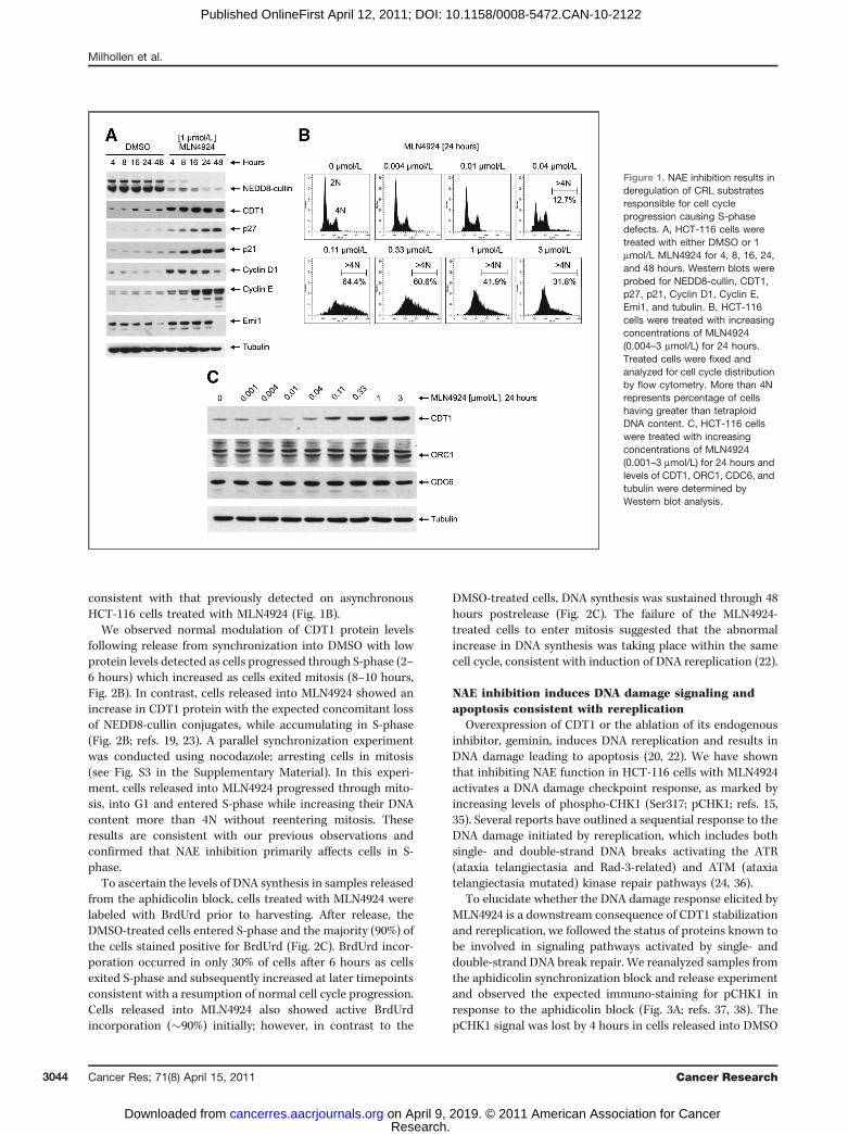
Flow cytometric analysis of HCT-116 cells treated for 24hours with even as low as 0.04 mmol/L MLN4924 revealed, agrowing subpopulation in S-phase as well as a smaller subsethaving greater than tetraploid (>4N) DNA content (Fig. 1B).Increasing MLN4924 concentrations exacerbated this cellcycle phenotype with an increase in cells with more than4N DNA. At higher concentrations the accumulation of cellshaving more than 4N DNA content is diminished and is likelythe result of increased cell death consistent with the appear-ance of cleaved caspase-3 and cleaved PARP as previouslyreported (15), as well as an increase in Annexin V positive cellsfollowing MLN4924 treatment (Supplementary Fig. S1). ABrdUrd-incorporation time-course experiment was con-ducted on asynchronous HCT-116 cells treated withMLN4924 to measure the amount of DNA synthesis in theaccumulating S-phase cells following treatment. Greater than90% of the cells treated with MLN4924 incorporated BrdUrd asearly as 8 hours, compared to only 59% of cells treated withDMSO. The increased BrdUrd incorporation observed with theMLN4924-treated cells was sustained out to 24 hours, suggest-ing persistent DNA synthesis occurred within these cells,leading to increased DNA content (see Fig. S2 in the Supple-mentary Material). Cells accumulating in S-phase, failing toenter mitosis, and collecting DNA with more than 4N contentare consistent with loss of NAE function causing DNA rere-plication (15, 22).
The pre-RC component CDT1 is a CRL1Skp2 and CRL4-DDB1Cdt2 substrate and has been shown to induce DNArereplication in cells when overexpressed (22, 23, 31, 32).We observed a dose-dependent increase in CDT1 levels fol-lowingMLN4924 treatment (Fig. 1C) and the increased steady-state levels of CDT1 protein correlated with the increase incells with more than 4N DNA content (Fig. 1B and C). Weanalyzed additional components within the pre-RC (ORC1 andCDC6) following MLN4924 treatment (33, 34) and found theirprotein levels only modestly affected (Fig. 1C).
NAE inhibition deregulates CDT1 in S-phase leading toincreased DNA synthesis and rereplication
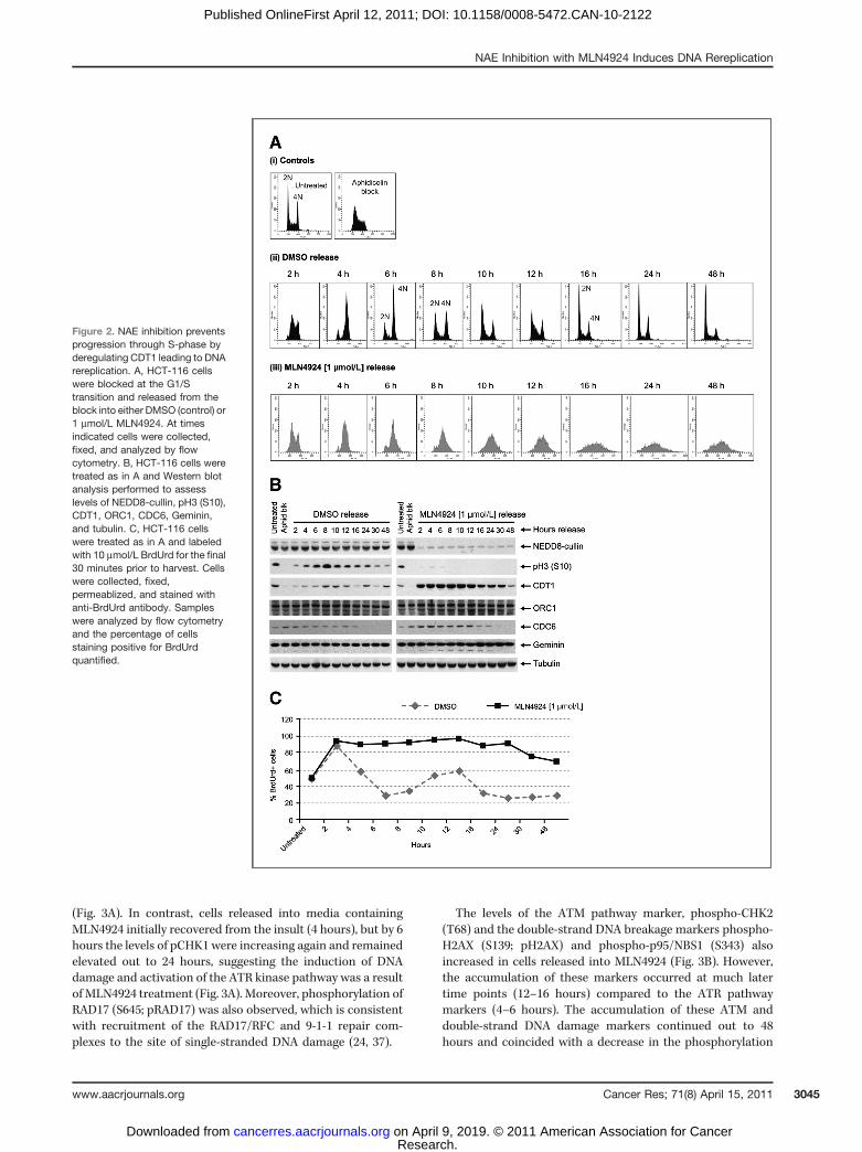
To further examine the effects of NAE inhibition on cellcycle progression we synchronized HCT-116 cells in early S-phase using aphidicolin and subsequently released the cellsinto MLN4924 or vehicle control containing media. Analysis ofcollected cells by flow cytomoetry indicated that control cellsprogressed normally through S-phase and mitosis and by 16hours had lost their synchrony (Fig. 2A). In contrast, cellsreleased into MLN4924 accumulated in S-phase as early as 4hours after release from aphidicolin (Fig. 2A) and continued toincrease DNA content (>4N) over the time course tested(Fig. 2A). Consistent with these cells being blocked in S-phasewas the absence of detectable pH3, demonstrating a failure toenter mitosis (Fig. 2B). The cell cycle profile observed was
NAE Inhibition with MLN4924 Induces DNA Rereplication
www.aacrjournals.org Cancer Res; 71(8) April 15, 2011 3043
Research. on April 9, 2019. © 2011 American Association for Cancercancerres.aacrjournals.org Downloaded from
Published OnlineFirst April 12, 2011; DOI: 10.1158/0008-5472.CAN-10-2122
consistent with that previously detected on asynchronousHCT-116 cells treated with MLN4924 (Fig. 1B).
We observed normal modulation of CDT1 protein levelsfollowing release from synchronization into DMSO with lowprotein levels detected as cells progressed through S-phase (2–6 hours) which increased as cells exited mitosis (8–10 hours,Fig. 2B). In contrast, cells released into MLN4924 showed anincrease in CDT1 protein with the expected concomitant lossof NEDD8-cullin conjugates, while accumulating in S-phase(Fig. 2B; refs. 19, 23). A parallel synchronization experimentwas conducted using nocodazole; arresting cells in mitosis(see Fig. S3 in the Supplementary Material). In this experi-ment, cells released into MLN4924 progressed through mito-sis, into G1 and entered S-phase while increasing their DNAcontent more than 4N without reentering mitosis. Theseresults are consistent with our previous observations andconfirmed that NAE inhibition primarily affects cells in S-phase.
To ascertain the levels of DNA synthesis in samples releasedfrom the aphidicolin block, cells treated with MLN4924 werelabeled with BrdUrd prior to harvesting. After release, theDMSO-treated cells entered S-phase and the majority (90%) ofthe cells stained positive for BrdUrd (Fig. 2C). BrdUrd incor-poration occurred in only 30% of cells after 6 hours as cellsexited S-phase and subsequently increased at later timepointsconsistent with a resumption of normal cell cycle progression.Cells released into MLN4924 also showed active BrdUrdincorporation (�90%) initially; however, in contrast to the
DMSO-treated cells, DNA synthesis was sustained through 48hours postrelease (Fig. 2C). The failure of the MLN4924-treated cells to enter mitosis suggested that the abnormalincrease in DNA synthesis was taking place within the samecell cycle, consistent with induction of DNA rereplication (22).
NAE inhibition induces DNA damage signaling andapoptosis consistent with rereplication
Overexpression of CDT1 or the ablation of its endogenousinhibitor, geminin, induces DNA rereplication and results inDNA damage leading to apoptosis (20, 22). We have shownthat inhibiting NAE function in HCT-116 cells with MLN4924activates a DNA damage checkpoint response, as marked byincreasing levels of phospho-CHK1 (Ser317; pCHK1; refs. 15,35). Several reports have outlined a sequential response to theDNA damage initiated by rereplication, which includes bothsingle- and double-strand DNA breaks activating the ATR(ataxia telangiectasia and Rad-3-related) and ATM (ataxiatelangiectasia mutated) kinase repair pathways (24, 36).
To elucidate whether the DNA damage response elicited byMLN4924 is a downstream consequence of CDT1 stabilizationand rereplication, we followed the status of proteins known tobe involved in signaling pathways activated by single- anddouble-strand DNA break repair. We reanalyzed samples fromthe aphidicolin synchronization block and release experimentand observed the expected immuno-staining for pCHK1 inresponse to the aphidicolin block (Fig. 3A; refs. 37, 38). ThepCHK1 signal was lost by 4 hours in cells released into DMSO
Figure 1. NAE inhibition results inderegulation of CRL substratesresponsible for cell cycleprogression causing S-phasedefects. A, HCT-116 cells weretreated with either DMSO or 1mmol/L MLN4924 for 4, 8, 16, 24,and 48 hours. Western blots wereprobed for NEDD8-cullin, CDT1,p27, p21, Cyclin D1, Cyclin E,Emi1, and tubulin. B, HCT-116cells were treated with increasingconcentrations of MLN4924(0.004–3 mmol/L) for 24 hours.Treated cells were fixed andanalyzed for cell cycle distributionby flow cytometry. More than 4Nrepresents percentage of cellshaving greater than tetraploidDNA content. C, HCT-116 cellswere treated with increasingconcentrations of MLN4924(0.001–3 mmol/L) for 24 hours andlevels of CDT1, ORC1, CDC6, andtubulin were determined byWestern blot analysis.
Milhollen et al.
Cancer Res; 71(8) April 15, 2011 Cancer Research3044
Research. on April 9, 2019. © 2011 American Association for Cancercancerres.aacrjournals.org Downloaded from
Published OnlineFirst April 12, 2011; DOI: 10.1158/0008-5472.CAN-10-2122
(Fig. 3A). In contrast, cells released into media containingMLN4924 initially recovered from the insult (4 hours), but by 6hours the levels of pCHK1 were increasing again and remainedelevated out to 24 hours, suggesting the induction of DNAdamage and activation of the ATR kinase pathway was a resultof MLN4924 treatment (Fig. 3A). Moreover, phosphorylation ofRAD17 (S645; pRAD17) was also observed, which is consistentwith recruitment of the RAD17/RFC and 9-1-1 repair com-plexes to the site of single-stranded DNA damage (24, 37).
The levels of the ATM pathway marker, phospho-CHK2(T68) and the double-strand DNA breakage markers phospho-H2AX (S139; pH2AX) and phospho-p95/NBS1 (S343) alsoincreased in cells released into MLN4924 (Fig. 3B). However,the accumulation of these markers occurred at much latertime points (12–16 hours) compared to the ATR pathwaymarkers (4–6 hours). The accumulation of these ATM anddouble-strand DNA damage markers continued out to 48hours and coincided with a decrease in the phosphorylation
Figure 2. NAE inhibition preventsprogression through S-phase byderegulating CDT1 leading to DNArereplication. A, HCT-116 cellswere blocked at the G1/Stransition and released from theblock into either DMSO (control) or1 mmol/L MLN4924. At timesindicated cells were collected,fixed, and analyzed by flowcytometry. B, HCT-116 cells weretreated as in A and Western blotanalysis performed to assesslevels of NEDD8-cullin, pH3 (S10),CDT1, ORC1, CDC6, Geminin,and tubulin. C, HCT-116 cellswere treated as in A and labeledwith 10 mmol/L BrdUrd for the final30 minutes prior to harvest. Cellswere collected, fixed,permeablized, and stained withanti-BrdUrd antibody. Sampleswere analyzed by flow cytometryand the percentage of cellsstaining positive for BrdUrdquantified.
NAE Inhibition with MLN4924 Induces DNA Rereplication
www.aacrjournals.org Cancer Res; 71(8) April 15, 2011 3045
Research. on April 9, 2019. © 2011 American Association for Cancercancerres.aacrjournals.org Downloaded from
Published OnlineFirst April 12, 2011; DOI: 10.1158/0008-5472.CAN-10-2122

of CHK1 and RAD17, suggesting a transition from single-strand DNA damage to the accumulation of double-strandDNA breaks (Fig. 3A and B). Induction of the apoptoticmarkers cleaved PARP and cleaved caspase-3 in theMLN4924-treated samples appeared to correlate with activa-tion of the double-strand DNA response pathway (Fig. 3C).Immunofluoresence staining of CDT1, pCHK1, pH2AX, andcleaved-caspase-3 confirmed that the sequential nature ofthese events was occurring uniformly across the treatedpopulation (see Fig. S4 in the Supplementary Material).
Knockdown of CDT1 suppresses rereplication in thepresence of MLN4924
To investigate whether the rereplication phenotypeobserved following NAE inhibition by MLN4924 was mediatedby CDT1 stabilization, we knocked down levels of CDT1 inHCT-116 cells using siRNA and monitored the ability of thecells to synthesize DNA in response to MLN4924. Effectiveknockdown of CDT1 was achieved and only modest accumu-lation of CDT1 was observed in these cells following MLN4924treatment, presumably due to incomplete CDT1 proteindepletion [Fig. 4A(i), lanes 3 and 4]. The control transfectedsample treated with MLN4924 had the expected increase inCDT1 protein [Fig. 4A(i), lane 2]. Additional CRL substratesNRF2 and p27 (15) accumulated in both the GL2 and CDT1RNAi-transfected samples when treated with MLN4924 asexpected [Fig. 4a(i), lanes 2 and 4]. BrdUrd incorporationwas significantly reduced in cells lacking CDT1 following 48hours of siRNA treatment in the presence or absence ofMLN4924 compared to control cells (Fig. 4B). In these samples77% of the GL2 RNAi-transfected control cells were synthesiz-ing DNA following treatment with MLN4924, compared toonly 28% of the CDT1 RNAi-transfected cells, suggesting thataccumulation of CDT1 following MLN4924 treatment med-
iates the rereplication phenotype (see Fig. S5 in the Supple-mentary Material for FACS histograms). Moreover, DNAdamage activation (pCHK1) was not observed in the CDT1RNAi-transfected cells following MLN4924 treatment [Fig. 4A(ii), lanes 3 and 4], compared to GL2-transfected cells treatedwith MLN4924 [Fig. 4A(ii), lane 2]. These data suggest thatfollowing NAE inhibition, the induction of rereplication andDNA damage observed correlates to the stabilization of CDT1demonstrating its critical role in the response.
Cullin knockdown stabilizes CDT1 and recapitulates therereplication phenotype
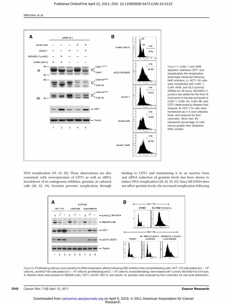
Depending on the cellular context, CRL1Skp2 and/or CRL4-DDB1CDT2 can facilitate the ubiquitination and proteasomaldegradation of CDT1 (32). Silencing of DDB1 and CDT2 bysiRNA has been shown to result in the accumulation of CDT1and lead to rereplication (39). To confirm that inhibition ofcullin 1 (CUL1) and cullin 4 (CUL4a/b) neddylation byMLN4924 results in CDT1 elevation and rereplication wemeasured CDT1 levels and cell cycle distributions followingsiRNA depletion of CUL1 and/or CUL4a/b. Interestingly, CUL1or CUL4a/b depletion alone resulted in little elevation of CDT1at 48 hours even though a modest increase in more than 4NDNA content was observed (Fig. 5). This may be the result of atransient elevation of CDT1 initiating rereplication that per-sisted even when alternative mechanisms of CDT1 turnoverhave reduced CDT1 protein to baseline levels (40) or from asmall subset of the population that was untransfected. Thecombined knockdown of CUL1 and CUL4a/b resulted inaccumulation of CDT1 and the substantial rereplication phe-notype comparable to MLN4924 treatment [Fig. 5A(ii) and B].These data add to the reports of overlapping function of CUL1and CUL4a/b in controlling CDT1 turnover following replica-tion initiation. To further define the role of CDT1 in initiating
Figure 3. DNA rereplicationinduced by NAE inhibition resultsin DNA damage and apoptosis.HCT-116 cells were treated as inFigure 2A and Western blotanalysis performed to detectmarkers of (A) ATR signaling[pCHK1 (S317) and pRAD17(S645)], (B) ATM signaling [pCHK2(T68), pH2AX (S139), and pNBS1(S343)], and (C) apoptosis(cleaved PARP and cleavedcaspase-3).
Milhollen et al.
Cancer Res; 71(8) April 15, 2011 Cancer Research3046
Research. on April 9, 2019. © 2011 American Association for Cancercancerres.aacrjournals.org Downloaded from
Published OnlineFirst April 12, 2011; DOI: 10.1158/0008-5472.CAN-10-2122

these events we depleted CDT1 along with CUL1 and CUL4a/bin cotransfection experiments (see Fig. S6 in the Supplemen-tary Material) and confirmed a reduction in cells having morethan 4N DNA content. A small population of cells with morethan 4N DNA content was still detectable, likely due toincomplete knockdown of CDT1 (as observed in Fig. S6B,lanes 4, 6, and 8); however, we cannot completely exclude thepossibility of other CRL substrates playing a role in thephenotype.
Effects of NAE inhibition on cycling and noncyclingcellsOur results suggest that following NAE inhibition, the
dominant phenotype leading to cell death is the inductionof DNA rereplication and DNA damage as cells transitionthrough S-phase. Therefore nonproliferating cells would fail toundergo DNA rereplication following MLN4924 treatment. Toillustrate this, MCF10A cells were treated with MLN4924 for 24hours under conditions in which the cells were cycling (sub-confluent) or noncycling (confluent, contact inhibited), andcompared to actively cycling HCT-116 cells. Western blotanalysis confirmed reduced NEDD8-cullin levels, indicatingNEDD8 pathway inhibition under all growth conditions(Fig. 6A, lanes 2, 3, 5, 6, 8, and 9); however, accumulation ofCDT1 and induction of DNA damage (pCHK1 activation) wereonly observed in the cycling cells (Fig. 6A, lanes 2, 3, 8, and 9).Moreover, flow cytometry analysis of cycling and noncyclingMCF10A cells treated with 1 mmol/L MLN4924 for 24 hoursshowed that only the cycling cells demonstrated a rereplica-tion cell cycle distribution similar to HCT-116 cells treatedwith MLN4924 (Fig. 6B). This suggests that proliferating cells
are more sensitive to the DNA rereplication phenotype gen-erated following NAE inhibition.
Discussion
In this report we characterize the cellular effects of speci-fically inhibiting NAE using the small molecule MLN4924.Inhibition of NAE results in cell death in human tumor cellsand our data support a model for the primary cellularmechanism of action as induction of DNA rereplication(Fig. 7). Our model suggests loss of NAE function preventsthe proper CRL-dependent regulation of CDT1, thereby lead-ing to the induction of DNA rereplication and subsequentDNA damage. The initial engagement of the ATR repairpathway occurs in response to single-strand DNA breaks,resulting in checkpoint activation designed to arrest cellsfor DNA repair. The persistent rereplication observed even-tually leads to double-strand breaks, as demonstrated byphosphorylation of proteins downstream of the ATM repairpathway. The inability of cells to repair the DNA damageultimately leads to induction of apoptosis and cell death. It isnoteworthy that similar cell cycle affects were observed whenNAE activity was reduced in a temperature-sensitive mutanthamster cell line as well as through knockdown of NAE bysiRNA in HCT-116 cells (15, 41).
NAE inhibition effectively shuts down CRL activity andderegulates the turnover of many proteins within the cell(15). Unexpectedly, our data suggest that CDT1 stabilizationappears to be a critical factor leading to the observed rere-plication outcome (22, 42). The inability to properly degradeCDT1 in S-phase of the cell cycle has been shown to result in
Figure 4. CDT1 RNAi rescues the DNA rereplication phenotype upon NAE inhibition. A, HCT-116 cells were transfected with CDT1 or GL2 (control)siRNAs for 48 hours. MLN4924 (1 mmol/L) was added for the final 16 hours prior to harvest and relevant pathway markers were analyzed by Westernblot [CDT1, NRF2, p27, pCHK1 (S317), and tubulin]. B, HCT-116 cells were transfected with CDT1 or GL2 (control) siRNAs for 48 hours, 1 mmol/LMLN4924 was added during the final 16 hours as indicated, and all samples were pulse labeled with 10 mmol/L BrdUrd for 30 minutes. Cells werecollected, fixed, permeablized, and stained with anti-BrdUrd antibody. Samples were analyzed by flow cytometry and the percentage of cells stainingpositive for BrdUrd quantified.
NAE Inhibition with MLN4924 Induces DNA Rereplication
www.aacrjournals.org Cancer Res; 71(8) April 15, 2011 3047
Research. on April 9, 2019. © 2011 American Association for Cancercancerres.aacrjournals.org Downloaded from
Published OnlineFirst April 12, 2011; DOI: 10.1158/0008-5472.CAN-10-2122
DNA rereplication (19, 23, 43). These observations are alsoconsistent with overexpression of CDT1 as well as siRNAknockdown of its endogenous inhibitor, geminin, in culturedcells (20, 22, 44). Geminin prevents rereplication through
binding to CDT1 and maintaining it in an inactive formand siRNA reduction of geminin levels has been shown toinduce DNA rereplication (20, 23, 44, 45). Since MLN4924 doesnot affect geminin levels, the increased rereplication following
Figure 5. Cullin 1 and 4A/Bdepletion stabilizes CDT1 andrecapitulates the rereplicationphenotype observed followingNAE inhibition. A, HCT-116 cellswere transfected with Cullin 1,Cullin 4A/B, and GL2 (control)siRNAs for 48 hours. MLN4924 (1mmol/L) was added for the final 16hours prior to harvest and levels ofCullin 1, Cullin 4A, Cullin 4B, andCDT1 determined by Western blotanalysis. B, HCT-116 cells weretransfected as in A and collected,fixed, and analyzed by flowcytometry. More than 4Nrepresents percentage of cellshaving greater than tetraploidDNA content.
Figure 6. Proliferating cells are more sensitive to DNA rereplication affects following NAE inhibition than nonproliferating cells. HCT-116 cells plated at 5� 105
cells/mL, andMCF10A cells plated at 5� 105 cells/mL (proliferating) and 2� 106 cells/mL (nonproliferating), were treated with 1 mmol/LMLN4924 for 24 hours.A, Western blots were probed for NEDD8-cullin, CDT1, pCHK1 (S317), and tubulin. B, samples were analyzed by flow cytometry for cell cycle distribution.
Milhollen et al.
Cancer Res; 71(8) April 15, 2011 Cancer Research3048
Research. on April 9, 2019. © 2011 American Association for Cancercancerres.aacrjournals.org Downloaded from
Published OnlineFirst April 12, 2011; DOI: 10.1158/0008-5472.CAN-10-2122

NAE inhibition is likely not due to deregulation of geminin (ref.15; Fig. 2B). Although additional components of the pre-RCcomplex, ORC1 and CDC6, were evaluated, our results showedonly minimal effects from NAE inhibition on their cellularprotein levels.To confirm the critical role of CDT1 in the induction of
rereplication following MLN4924 treatment, we sought torescue the phenotype by knocking down CDT1 levels withsiRNA. Following CDT1 knockdown, cells incorporate lessBrdUrd even in the presence of MLN4924 indicating a sup-pression of DNA synthesis and rereplication, suggesting a keyrole of CDT1 in the phenotype. Noncycling MCF10A cells,when treated with MLN4924, did not undergo rereplicationand failed to stabilize CDT1 compared to cycling MCF10Acells, supporting our findings (see Fig. 6). Based on these data,cells must progress though S-phase in the presence of
MLN4924 to be deleteriously affected, suggesting that prolif-erating cancer cells may be more sensitive to NAE inhibitioncompared to quiescent cells.
Since there are at least 2 CRLs responsible for CDT1degradation (CUL1 and CUL4), we sought to determine theimportance of each in regulating CDT1 levels in HCT-116 cells(32, 42, 43). We demonstrated that only the combined siRNA-mediated knockdown of CUL1 and CUL4a/b was sufficient tostabilize CDT1 and elicit the rereplication phenotype. Knock-down of CUL1 or CUL4a/b alone yielded only a modest changein cell cycle distribution without an apparent stabilization ofCDT1 supporting their overlapping functions in regulating theturnover of CDT1 (19). Cotransfection experiments knockingdown CDT1 along with CUL1 and/or CUL4 reduced thepopulation of cells having more than 4N DNA content, sup-porting a primary role of CDT1 in the observed rereplicationphenotype. However, a small fraction of cells with more than4N DNA content was still evident suggesting that other, yet tobe defined, CRL substrates may also play a role in therereplication response. Interestingly, we observed thatCUL4 appeared to be approximately 90% neddylated com-pared to approximately 10% for CUL1 which suggest a hier-archy of control of CDT1 turnover within the CUL1/CUL4 axis.Thus, the neddylation status of CUL4 and/or CUL1 in differentcell types may play a role in predicting sensitivity or magni-tude of response to MLN4924-induced DNA rereplication andwarrants further investigation.
A major consequence of DNA rereplication is DNA damageand activation of checkpoint signaling (20, 22, 46–48). Previousstudies have reported that the ATR/CHK1 kinase pathway isactivated in response to rereplication and phosphorylation ofRAD17 occurs at sites of damaged DNA following rereplication(24, 37, 39). Our results show that both CHK1 and RAD17 arephosphorylated in response to MLN4924 treatment, suggest-ing that the cells undergo single-strand DNA damage follow-ing rereplication, consistent with accumulation of damagedreplication forks (37, 49). CHK1 and RAD17 phosphorylation issustained for at least 30 hours following induction of rerepli-cation during which time cells continue to synthesize DNA.The fact that cells continue to incorporate BrdUrd suggeststhat MLN4924 may in some way interfere with CRL substrateswhich regulate the intra-S-phase checkpoint, and this maywarrant further investigation (49, 50).
In the absence of NAE function cells no longer enter mitosisand are not capable of properly regulating DNA replication.The induction of rereplication following siRNA knockdown ofgeminin has been shown to activate the ATM/CHK2 kinaseand MRE11-RAD50-NBS1 (MRN) complexes (24, 36). Theactivation of the ATR/CHK1 pathway occurs upstream ofthe ATM/CHK2 pathway and appears to be required for itsactivation (24, 39), and see model in Fig. 7). Consistent withthese observations, we detected the appearance of markers ofdouble-strand DNA damage and ATM repair pathway activa-tion (pCHK2, pH2AX, and pNBS1) following ATR activation.These observations illustrate a switch from the accumulationof single-strand DNA damage to double-strand DNA damageand support a sequential response to DNA rereplicationconsistent with published reports (24). The appearance of
Figure 7. Proposed model for induction of DNA rereplication followingNAE inhibition with MLN4924. NAE inhibition prevents the proper CRL-dependent turnover of the preRC component CDT1, which induces DNArereplication as the cells progress through S-phase. Resulting DNAdamage activates the ATR/CHK1 pathway, and prevents further cell cycleprogression; in turn, persistent rereplication then signals the ATM/CHK2pathway. Failure to repair damaged DNA ultimately leads to activation ofapoptosis and loss of cell viability. Model adapted with permission from J.J. Lin and A. Dutta (24).
NAE Inhibition with MLN4924 Induces DNA Rereplication
www.aacrjournals.org Cancer Res; 71(8) April 15, 2011 3049
Research. on April 9, 2019. © 2011 American Association for Cancercancerres.aacrjournals.org Downloaded from
Published OnlineFirst April 12, 2011; DOI: 10.1158/0008-5472.CAN-10-2122

markers of apoptosis correlates with the markers of double-strand DNA damage, suggesting that cell death is occurringbecause of accumulated double-strand DNA damage (seemodel in Fig. 7). Interestingly, p53 was phosphorylated(Ser15) in response to MLN4924 which may suggest a roleof p53 in the rereplication response. However we observed asimilar cellular phenotype of NAE inhibition in a panel of cellsirrespective of their p53 status (see Fig. S7 in the Supplemen-tary Material; ref. 44). Further work is required to understandif there is a specific genetic background that may make cellsparticularly sensitive to NAE-mediated DNA rereplication andcell death.
In this report we outline an unexpected cellular mechanismof action for MLN4924 (and NAE inhibition) in various humantumor cell lines in which the deregulation of CDT1, a key CRLsubstrate, leads to DNA rereplication. It will be important tofurther define the mechanisms involved in the cellularresponse to rereplication, how rereplication affects cell sensi-tivity, and how this may impact the potential mechanism(s) ofresistance toMLN4924. Based on our data we hypothesize thatcells that have defective DNA damage repair pathways (e.g.BRCA1 mutants, Fanconi Anemia) might be more sensitive toMLN4924 than cells with functional DNA repair pathways (48).In addition, combination of MLN4924 with known DNA dama-ging and/or S-phase-inducing agents, as well as inhibitors of
DNA repair pathways, could be a way to further sensitize cellsto MLN4924 treatment. To this end, a genome wide RNAiscreen using MLN4924 is in process to support rational drugcombinations and, interestingly, it appears thatMLN4924 has aprofile that is distinct from other FDA-approved DNA dama-ging agents (E. Lightcap, manuscript in preparation). Furtherunderstanding of this mechanism and defining the types ofDNA damage and/or damage repair pathways involved in theMLN4924 response will be required as MLN4924 progressesthrough clinical development.
Disclosure of Potential Conflicts of Interest
No potential conflicts of interest were disclosed.
Acknowledgments
We thank A. Dutta and J.J. Lin for insightful discussions of the data andreview of the manuscript. We also thank J. Ecsedy, N. D’Amore, A. Berger, and N.Bence for critical review of the manuscript as well as FireKite, UK, for editing ofthe manuscript.
The costs of publication of this article were defrayed in part by the paymentof page charges. This article must therefore be hereby marked advertisement inaccordance with 18 U.S.C. Section 1734 solely to indicate this fact.
Received June 15, 2010; revised February 7, 2011; accepted February 19, 2011;published online April 12, 2011.
References1. Welchman RL, Gordon C, Mayer RJ. Ubiquitin and ubiquitin-like
proteins as multifunctional signals. Nat Rev Mol Cell Biol 2005;6:599–609.
2. Hershko A. The ubiquitin system for protein degradation and some ofits roles in the control of the cell division cycle. Cell Death Differ2005;12:1191–7.
3. Kerscher O, Felberbaum R, Hochstrasser M. Modification of proteinsby ubiquitin and ubiquitin-like proteins. Annu Rev Cell Dev Biol2006;22:159–80.
4. Yeh ET, Gong L, Kamitani T. Ubiquitin-like proteins: new wines in newbottles. Gene 2000;248:1–14.
5. Herrmann J, Lerman LO, Lerman A. Ubiquitin and ubiquitin-likeproteins in protein regulation. Circ Res 2007;100:1276–91.
6. Hershko A, Ciechanover A. The ubiquitin system. Annu Rev Biochem1998;67:425–79.
7. Gong L, Yeh ET. Identification of the activating and conjugatingenzymes of the NEDD8 conjugation pathway. J Biol Chem 1999;274:12036–42.
8. Huang DT, Ayrault O, Hunt HW, Taherbhoy AM, Duda DM, Scott DC,et al. E2-RING expansion of the NEDD8 cascade confers specificity tocullin modification. Mol Cell 2009;33:483–95.
9. Pan ZQ, Kentsis A, Dias DC, Yamoah K, Wu K. Nedd8 on cullin:building an expressway to protein destruction. Oncogene 2004;23:1985–97.
10. Soucy TA, Smith PG, Rolfe M. Targeting NEDD8-activated cullin-RING ligases for the treatment of cancer. Clin Cancer Res 2009;15:3912–6.
11. Petroski MD, Deshaies RJ. Function and regulation of cullin-RINGubiquitin ligases. Nat Rev Mol Cell Biol 2005;6:9–20.
12. Podust VN, Brownell JE, Gladysheva TB, Luo RS, Wang C, CogginsMB, et al. A Nedd8 conjugation pathway is essential for proteolytictargeting of p27Kip1 by ubiquitination. Proc Natl Acad Sci U S A2000;97:4579–84.
13. Read MA, Brownell JE, Gladysheva TB, Hottelet M, Parent LA,Coggins MB, et al. Nedd8 modification of cul-1 activates SCF(beta
(TrCP))-dependent ubiquitination of IkappaBalpha. Mol Cell Biol2000;20:2326–33.
14. Duda DM, Borg LA, Scott DC, Hunt HW, Hammel M, Schulman BA.Structural insights into NEDD8 activation of cullin-RING ligases:conformational control of conjugation. Cell 2008;134:995–1006.
15. Soucy TA, Smith PG, Milhollen MA, Berger AJ, Gavin JM, Adhikari S,et al. An inhibitor of NEDD8-activating enzyme as a new approach totreat cancer. Nature 2009;458:732–6.
16. Arias EE, Walter JC. Replication-dependent destruction of Cdt1 limitsDNA replication to a single round per cell cycle in Xenopus eggextracts. Genes Dev 2005;19:114–26.
17. Bell SP, Dutta A. DNA replication in eukaryotic cells. Annu RevBiochem 2002;71:333–74.
18. Remus D, Beuron F, Tolun G, Griffith JD, Morris EP, Diffley JF.Concerted loading of Mcm2–7 double hexamers around DNA duringDNA replication origin licensing. Cell 2009;139:719–30.
19. Kim Y, Kipreos ET. Cdt1 degradation to prevent DNA rereplication:conserved and non-conserved pathways. Cell Div 2007;2:18–27.
20. Melixetian M, Ballabeni A, Masiero L, Gasparini P, Zamponi R, BartekJ, et al. Loss of Geminin induces rereplication in the presence offunctional p53. J Cell Biol 2004;165:473–82.
21. Saxena S, Dutta A. Geminin and p53: deterrents to rereplication inhuman cancer cells. Cell Cycle 2003;2:283–6.
22. Vaziri C, Saxena S, Jeon Y, Lee C, Murata K, Machida Y, et al. A p53-dependent checkpoint pathway prevents rereplication. Mol Cell2003;11:997–1008.
23. Fujita M. Cdt1 revisited: complex and tight regulation during the cellcycle and consequences of deregulation in mammalian cells. Cell Div2006;1:22.
24. Lin JJ, Dutta A. ATR pathway is the primary pathway for activating G2/M checkpoint induction after rereplication. J Biol Chem 2007;282:30357–62.
25. Carrano AC, Eytan E, Hershko A, Pagano M. SKP2 is required forubiquitin-mediated degradation of the CDK inhibitor p27. Nat Cell Biol1999;1:193–9.
Milhollen et al.
Cancer Res; 71(8) April 15, 2011 Cancer Research3050
Research. on April 9, 2019. © 2011 American Association for Cancercancerres.aacrjournals.org Downloaded from
Published OnlineFirst April 12, 2011; DOI: 10.1158/0008-5472.CAN-10-2122

26. Pagano M, Tam SW, Theodoras AM, Beer-Romero P, Del Sal G, ChauV, et al. Role of the ubiquitin-proteasome pathway in regulatingabundance of the cyclin-dependent kinase inhibitor p27. Science1995;269:682–5.
27. Wang W, Nacusi L, Sheaff RJ, Liu X. Ubiquitination of p21Cip1/WAF1by SCFSkp2: substrate requirement and ubiquitination site selection.Biochemistry 2005;44:14553–64.
28. Margottin-Goguet F, Hsu JY, Loktev A, Hsieh HM, Reimann JD,Jackson PK. Prophase destruction of Emi1 by the SCF(betaTrCP/Slimb) ubiquitin ligase activates the anaphase promoting complex toallow progression beyond prometaphase. Dev Cell 2003;4:813–26.
29. Lin DI, Barbash O, Kumar KG, Weber JD, Harper JW, Klein-SzantoAJ, et al. Phosphorylation-dependent ubiquitination of cyclin D1 bythe SCF(FBX4-alphaB crystallin) complex. Mol Cell 2006;24:355–66.
30. Ye X, Nalepa G, Welcker M, Kessler BM, Spooner E, Qin J, et al.Recognition of phosphodegron motifs in human cyclin E by the SCF(Fbw7) ubiquitin ligase. J Biol Chem 2004;279:50110–9.
31. Hu J, McCall CM, Ohta T, Xiong Y. Targeted ubiquitination of CDT1 bythe DDB1-CUL4A-ROC1 ligase in response to DNA damage. Nat CellBiol 2004;6:1003–9.
32. Nishitani H, Sugimoto N, Roukos V, Nakanishi Y, Saijo M, Obuse C,et al. Two E3 ubiquitin ligases, SCF-Skp2 and DDB1-Cul4, targethuman Cdt1 for proteolysis. EMBO J 2006;25:1126–36.
33. Kim J, FengH, Kipreos ET. C. elegans CUL-4 prevents rereplication bypromoting the nuclear export of CDC-6 via a CKI-1-dependent path-way. Curr Biol 2007;17:966–72.
34. Mendez J, Zou-Yang XH, Kim SY, Hidaka M, Tansey WP, Stillman B.Human origin recognition complex large subunit is degraded byubiquitin-mediated proteolysis after initiation of DNA replication.Mol Cell 2002;9:481–91.
35. Zhao H, Piwnica-Worms H. ATR-mediated checkpoint pathwaysregulate phosphorylation and activation of human Chk1. Mol CellBiol 2001;21:4129–39.
36. Lee AY, Liu E, Wu X. The Mre11/Rad50/Nbs1 complex plays animportant role in the prevention of DNA rereplication in mammaliancells. J Biol Chem 2007;282:32243–55.
37. Davidson IF, Li A, Blow JJ. Deregulated replication licensing causesDNA fragmentation consistent with head-to-tail fork collision. Mol Cell2006;24:433–43.
38. Lau E, Chiang GG, Abraham RT, Jiang W. Divergent S phase check-point activation arising from prereplicative complex deficiency con-trols cell survival. Mol Biol Cell 2009;20:3953–64.
39. Liu E, Lee AY, Chiba T, Olson E, Sun P, Wu X. The ATR-mediated Sphase checkpoint prevents rereplication in mammalian cells whenlicensing control is disrupted. J Cell Biol 2007;179:643–57.
40. Lin JJ, Milhollen MA, Smith PG, Narayanan U, Dutta A. NEDD8-targeting drug MLN4924 elicits DNA rereplication by stabilizingCDT1 in S phase, triggering checkpoint activation, apoptosis, andsenescence in cancer cells. Cancer Res 2010;70:10310–20.
41. Handeli S, Weintraub H. The ts41 mutation in Chinese hamster cellsleads to successive S phases in the absence of intervening G2,M, andG1. Cell 1992;71:599–611.
42. Sugimoto N, Yoshida K, Tatsumi Y, Yugawa T, Narisawa-Saito M,Waga S, et al. Redundant and differential regulation of multiplelicensing factors ensures prevention of rereplication in normal humancells. J Cell Sci 2009;122:1184–91.
43. Senga T, Sivaprasad U, Zhu W, Park JH, Arias EE, Walter JC, et al.PCNA is a cofactor for Cdt1 degradation by CUL4/DDB1-mediated N-terminal ubiquitination. J Biol Chem 2006;281:6246–52.
44. Zhu W, Chen Y, Dutta A. Rereplication by depletion of geminin is seenregardless of p53 status and activates a G2/M checkpoint. Mol CellBiol 2004;24:7140–50.
45. Nishitani H, Lygerou Z. Control of DNA replication licensing in a cellcycle. Genes Cells 2002;7:523–34.
46. Archambault V, Ikui AE, Drapkin BJ, Cross FR. Disruption of mechan-isms that prevent rereplication triggers a DNA damage response. MolCell Biol 2005;25:6707–21.
47. Green BM, Li JJ. Loss of rereplication control in Saccharomycescerevisiae results in extensive DNA damage. Mol Biol Cell 2005;16:421–32.
48. Zhu W, Dutta A. An ATR- and BRCA1-mediated Fanconi anemiapathway is required for activating the G2/M checkpoint and DNAdamage repair upon rereplication. Mol Cell Biol 2006;26:4601–11.
49. Tercero JA, Longhese MP, Diffley JF. A central role for DNAreplication forks in checkpoint activation and response. Mol Cell2003;11:1323–36.
50. Skaar JR, Pagano M. Control of cell growth by the SCF and APC/Cubiquitin ligases. Curr Opin Cell Biol 2009;21:816–24.
NAE Inhibition with MLN4924 Induces DNA Rereplication
www.aacrjournals.org Cancer Res; 71(8) April 15, 2011 3051
Research. on April 9, 2019. © 2011 American Association for Cancercancerres.aacrjournals.org Downloaded from
Published OnlineFirst April 12, 2011; DOI: 10.1158/0008-5472.CAN-10-2122

2011;71:3042-3051. Published OnlineFirst April 12, 2011.Cancer Res Michael A. Milhollen, Usha Narayanan, Teresa A. Soucy, et al. Consistent with Deregulating CDT1 TurnoverRereplication and Apoptosis in Human Tumor Cells Inhibition of NEDD8-Activating Enzyme Induces
Updated version
10.1158/0008-5472.CAN-10-2122doi:
Access the most recent version of this article at:
Material
Supplementary
http://cancerres.aacrjournals.org/content/suppl/2011/04/08/0008-5472.CAN-10-2122.DC1
Access the most recent supplemental material at:
Cited articles
http://cancerres.aacrjournals.org/content/71/8/3042.full#ref-list-1
This article cites 50 articles, 21 of which you can access for free at:
Citing articles
http://cancerres.aacrjournals.org/content/71/8/3042.full#related-urls
This article has been cited by 23 HighWire-hosted articles. Access the articles at:
E-mail alerts related to this article or journal.Sign up to receive free email-alerts
SubscriptionsReprints and
To order reprints of this article or to subscribe to the journal, contact the AACR Publications
Permissions
Rightslink site. (CCC)Click on "Request Permissions" which will take you to the Copyright Clearance Center's
.http://cancerres.aacrjournals.org/content/71/8/3042To request permission to re-use all or part of this article, use this link
Research. on April 9, 2019. © 2011 American Association for Cancercancerres.aacrjournals.org Downloaded from
Published OnlineFirst April 12, 2011; DOI: 10.1158/0008-5472.CAN-10-2122