inhibition of the transepithelial potential difference and short circuit current in the isolated...
TRANSCRIPT
Camp. Biorhem. Physiol. Vol. 102C. No. 1, pp. 29-32, 1992 Printed in Great Britain
0306-4492/92 $5.00 + 0.00 C 1992 Pergamon Press Ltd
INHIBITION OF THE TRANSEPITHELIAL POTENTIAL DIFFERENCE AND SHORT CIRCUIT CURRENT IN THE
ISOLATED FROG SKIN BY ALLOXAN*
CLAUDIA SOTO, JOSE L. KEYES,? ARELI RAMIREZ, FERNANDO PAZ and CUAUHTEMOC PEREZ
Departamento de Fisiologia y Biofisica, Centro de Investigacibn y de Estudios Avanzados de1 Instituto Polit&cnico National and Departamento de Sistemas Biokjgicos, Universidad Aut6noma Metropolitana-
Xochimilco, Mexico, 07000. Tel.: 5-754 0200 Ext. 5123: Fax: 5-586 2792
(Received 29 October 1991)
Abstract-l. Electrical parameters: short circuit current (KC), transepithelial potential difference (PD) ’ and electrical resistance (R) were measured in isolated frog skin (Ram pipiens) in the presence and in the
absence of alloxan. 2. Alloxan decreased SCC and PD in a concentration-dependent pattern, while R remained unchanged. 3. The effect on SCC and PD was observed after 25 min of exposure to the drug. Maximal average
effect was 20% in SCC and 17.5% in PD. 4. These results suggest that alloxan decreased epithelial sodium transport, through interference with
the activity of the Na+-K+-APTase.
INTRODUCTION
Alloxan has been widely used to induce experimental diabetes mellitus. In 1943, it was reported that alloxan produced selective necrosis of b-pancreatic cells (Dunn, 1943) and this has been proposed as the explanation for its ability to induce experimental diabetes mellitus, however the cellular mechanisms responsible for this effect have not been fully disclosed. In spite of this, it has proven to be an adequate tool since the biochemical alterations present in the treated animals closely resemble those observed in diabetic human beings (Rerup, 1970).
Several propositions have been made to explain alloxan effect. Alloxan depicts high affinity for thiol groups (-SH) (Lazarow and Patterson, 1948, Patterson et al., 1949). It reversibly reacts with them either to form dialuric acid or irreversibly to produce secondary products (Lazarow, 1949). It has been proposed that interaction with thiol groups causes inactivation of some enzyme activities which possess -SH groups (Lenzen and Panten, 1988; Webb, 1966).
Cooperstein and Lazarow (1964) pointed out that alloxan does not penetrate the plasma membrane of cell islets from toad fish and therefore its site of action is probably located at the external leaflet of the plasma membrane. In agreement with this proposition, Watkins et al. (1961) observed that alloxan increased membrane permeability to mannitol in the same experimental model. However, alloxan did not change
*Part of this material was presented in abstracts form at the XIth International Congress of Pharmacology, Amsterdam, The Netherlands. Eur. J. Pharmacol. 183, 282.
Correspondence should be sent to: Dr Jest L. Reyes, Departamento de Fisiologia y Biofisica Centro de Inves- tigacidn y de Estudios Avanzados de1 IPN, Apartado Postal 14-740 MBxico, D.F. 07000.
permeability to sucrose, D-mannitol or L-glucose in rat pancreatic islet (McDaniel et al., 1975).
Alloxan interacts with the mechanisms involved in the membrane transport of ions. Irreversible inhibition of the calcium-dependent ATPase in smooth muscle by alloxan was reported (Kwan, 1988). Bianchi et al.
(1988) reported decrease in the activity of (Na+-K+)- ATPase (EC 3.6.3.1) of sciatic nerve, in alloxan- treated rats and they suggested that the effect was due to changes in the enzyme lipidic microenvironment. Scant information is available on the effect of alloxan on the activity of (Na+-K+)-ATPase in epithelia. The aim of this study was to analyze the effects of alloxan on the electrical parameters of the isolated frog skin which are dependent on the activity of this ATPase, using this model as a bioassay, since inhibition of (Na+-K+)-ATPase by ouabain decreases both SCC and PD in isolated frog skin (Aceves and Erlij, 1971).
MATERIALS AND METHODS
Transepithelial sodium transport was measured in isolated frog skin (Ram pipiens), mounted in Ussing type chambers as previously reported (Ussing and Zehran, 1951; Reyes et al., 1989).BrieAy, skin segments were maintained at robm temuerature (20-22°C). bubbled with O,/CO,(95/5%). He&chambers were filled with Ringer soh&on of the foi- lowing composition (mM): NaCl, 90; NaHCO,, 25: KCI, 5; CaCI,, 1; MgCI,, 2 and glucose 10, adjusted at pH 7.4 and 230 mOsm. Exposed area was 2.1 cm2, Skin was maintained in a period of stabilization for at least 30 min. During this period Ringer solution in both chambers was twice removed and replaced by fresh solution. After this period, either alloxan or vehicle (Ringer solution) were added to the chamber corresponding to the serosal side. Concentrations tested were: 1, 10 and IOOpM. Transepithelial potential difference and short circuit current were recorded each 5 min, for 90 min after the addition of alloxan or vehicle. Alloxan was purchased from Sigma Co. (St. Louis, MO). Other reagents were obtained, analytical grade, from local dealers (Merck).
29

30 CLAUDIA SOTO et al
1.2 r CONTROL
1 .o
“9 IO
0.8
0.6
I
ALLOXAN (IOpM)
1
T a t,, ,I,, , 0 IO 20 30 40 50 60 70 80 PO
min
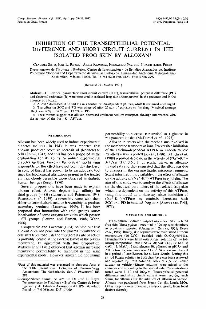
Fig. 1. Time course of the effect of alloxan on short circuit current (SCC) in the isolated frog skin. The ratio of SCC before (IO) and after (It) the addition of either the vehicle (open circles) or alloxan (closed circles) is shown for a
representative experiment.
RESULTS
Time course of the short circuit current (SCC) in the absence of alloxan in a typical experiment is shown in Fig. 1. Results are expressed as the ratio between initial value of SCC (10, before the addition of either vehicle or alloxan) and the value of SCC at any subsequent time, It, (It/IO). A tendency to increase was observed in control skin, reaching a value of approximately 10% at 90 min. Skin exposed to alloxan (10 p M, final concentration) in the serosal chamber showed an opposite tendency to control series. A sustained decrease in SCC was observed after 25 min of exposition to this drug. Decrement reached approximately 30% of the initial value at 90 min.
Similar results were observed for the ratio of the transepithelial potential difference before (Vo) and after (Vt) the addition of either vehicle or alloxan (VtjVo). Alloxan induced a decrement of this ratio (Fig. 2), whereas in control skin Vt/Vo ratio showed a tendency to increase (approximately 15% above the initial value).
The effect of alloxan was assessed at three final concentrations (1, 10 and 100 PM). Decrement in SCC and in transepithelial potential difference ratios was concentration-dependent (Figs 3 and 4, respectively).
F 0 CONTROL
1.2 0 ALLOXAN (1 PM)
t-- 0.8
t f
I I 1 , I ,
CONTROL
1.2 ALLOXAN(lOpM)
0.8
F CONTROL
1.2 ALLOXAN (iOOp M 1
I 1 1 I 1 1 1 , I ,
10 20 30 40 50 60 70 80 90 min
Fig. 3. Effect of increasing concentrations of alloxan on the It/IO ratio in isolated frog skin. Symbols are as in Fig. 1.
Mean + S.E.M. are shown. *P < 0.05.
The magnitude of this effect at 10 and 1OOpM was similar. Control skins showed a smooth tendency to increase its electrical parameters (It/IO 8% and Vt/Vo 17% on average, respectively).
Maximal effect of alloxan was achieved by 90 min. The concentration effect is shown in Fig. 5. Decrement
CONTROL
0.6 i,,
(10 PM)
, , I I IO
I 20 30 40 50 60 70 80 90
min
Fig. 2. Time course of the effect of alloxan on transepithelial potential difference (PD). The ratio of PD before (Vo) and after (Vt) the addition of either vehicle (open circles) or alloxan (closed circles) is shown
for one representative experiment.

Inhibition of the transepithelial potential in isolated frog skin 31
1.4 0 CONTROL * ALLOXANII p MI
1 ‘
1.4
F
CONTROL
Al.LOXAN(lOpMf
1.2
* Vt
Go LO
0.8 i-
In=9
LL /*
‘I-* i\zn=7
1.4
t
CONTROL ALLOXAN WOp MI
t.2
0.8
, I I ‘ ,
K) 20 50 40 50 60 70 80 90 min
Fig. 4. Effect of increasing concentrations of alloxan on the Vt/Vo ratio, in isolated frog skin. Symbols are as in Fig. 1.
Mean f S.E.M. are shown. *P < 0.05.
in electrical parameters was statistically significant at 10 and 100 PM concentrations (P < 0.05).
It is remarkable that electrical resistance was not si~nificantiy changed (Table I), suggesting that the observed effects on short circuit current and on transepithelial potential difference, do not reflect toxic effects of alloxan, but rather a pharmacological action.
DISCUSSION
Short circuit current and transepithelial potential difference in isolated frog skin mounted in Ussing’s type chambers depend on the magnitude of epithelial sodium transport. (Ussing and Zehran, 1951). In turn, this transport is dependent on the activity of the (Na+-K+)-ATPase. In this study the effect of alloxan on the electrical parameters of the isolated frog skin was assessed in order to disclose whether or not this drug affected the transport of sodium. Main finding was that alloxan inhibited sodium transport as estimated from the decrements in the transepithelial potential difference and in the short circuit current. This inhibition depicted a concentration-dependent pattern. Maximal decrease in SCC averaged 20% below the basal values. Maximal decrease in trans-
t.20
F
1.00 - 18) : 1 (71
9 0.80 - \
00) .
IO
OL-lC ’ t 10-S 10-J io-4 iMf
ALLOXAN
Fig. 5. Concentration-dependent effect of ahoxan on It/IO and Vt/Vo ratios in the isolated frog skin. Measurements were made 90 min after the addition of the drug. Mean &
S.E.M. *P -c 0.05 ( ) = number of experiments.
epithelial potential difference was 17.5% on average. In contrast to the effect of alloxan, a smooth increase in SCC and in PD was observed in control skins that were not exposed to this drug.
These findings suggest that alloxan inhibited sodium transport in the frog skin epithelia. This effect has not been reported for this epithelium and probably is due to inhibition of the driving force for this transport that is provided by the (Na+-K+)-ATPase. Our results are in agreement with the tinding that alloxan inhibited the activity of the (Na+-K+)-ATPase in sciatic nerve of diabetic rats (Bianchi et al., 1988). It has been also reported that alloxan inhibited the ATP-driven Caz+ transport in vascular muscle mi~rosomal fractions (Kwan, 1988). This info~ation is suggestive of an inhibitory effect of alloxan on transport ATPases as one of the possible mechanisms to explain the effect of this drug. The consequences of this inhibition of epithelial ionic transport have not been previously assessed.
Lazarow showed that alloxan binds to membrane sulphydryl groups and (Na+-K+)-ATPase posses these groups. Esmann has pointed out that the - SH groups have a critical role on the catalytic cycle of the
Table 1. Time course of the effect of alloxan on the electrical resistance of isolated frog skin
Alloxan Time (min) (PM) n 30 60 90
0 9 I.054 f 0.039* 1 .150 + 0.047 I.065 + 0.127 1 .o 8 I .03s + 0.035 1.010 + 0.060 I .002 + 0.060
10.0 I I.003 f 0.029 0.984 + 0.033 1.068 + 0.039 100.0 IO 1.031 +0.064 1.126+0.057 l.l42+0.086
*Figures are the ratios RJR,, where R, and R, are the transepithelial resistance in the presence and in the absence of alloxan, respect- ively. Mean + S.E.M. are shown.

32 CLAUDIA SOTO et al.
enzyme (15). Therefore we may suggest that the effect observed by us is related to interaction of alloxan with these groups.
It is remarkable the lack of change on electrical resistance in the skins exposed to alloxan, in which decrements in transepithelial potential difference and in short circuit current were present. This finding suggests that the effect of the drug is mainly due to an inhibition of the sodium transport, as a consequence of a pharmacological effect. It might be expected that a toxic effect would decrease the electrical resistance, in addition to the changes in XC and PD.
Acknowledgemenls-Authors are indebted to Elvis Hernindez for secretarial assistance, Alfred0 Padilla for making the illustrations and to Mario Raya for technical help. This work was partially supported by a grant from Consejo National de Ciencia y Tecnologia.
REFERENCES
Aceves J. and Erlij D. (1971) Sodium transport across the isolated epithelium of the frog skin. J. Physiol. 212, 195~210.
Bianchi R.. Marini P.. Merhni S.. Fabris M., Triban Ch., Mussini E. and Fiori M. G. (1988) ATPase activity defects in alloxan-induced diabetic sciatic nerve recovered by ganglioside treatment. Diabetes 37, 1340&1345.
Cooperstein S. J. and Lazarow A. (1964) Distribution of alloxan-C’4 in islet and other tissues of the toadfish. Am. J. Physiol. 207, 423-430.
Dunn D. S., Sheehan H. L. and McLetchin N. G. B. (1943) Necrosis of islets of Langerhans produced experimentally. Lancet 244, 4844481.
Esmann M. (1982) Sulfhydryl groups of (Na+-K+) ATPase from rectal glands of Squalus Acanthias.
Titrations and classification. Biochim. Biophys. Acta 688, 260-270.
Kwan Ch-Y. (1988) The plasma membrane component is the primary site of action of alloxan on the ATP-driven CaZ+ transport in vascular-muscle microsomal fractions. Biochem. J. 254, 293-296.
Lazarow A. and Patterson J. W. (1948) The mechanism of cysteine and glutathione protection against alloxan diabetes. Science 108, 308.
Lazarow A. (1949) Factors controlling the development and progression of diabetes. Physiol. Reo. 28, 48-74.
Lenzen S. and Panten U. (1988) Alloxan: history and mechanism of action. Diabetologia 31, 337.-342.
McDaniel M. L., Anderson S.. Fink J., Roth C. and Lacy P. E. (1975) Effect of alloxan on permeability and hexose transport in rat pancreatic islets. Endocrinology 97, 68-75.
Patterson J. W.. Lazarow A. and Levey J. (1949) Reactions of alloxan and dialuric acid with the sulfhydryl group. J. Biol. Chem. 177, 197-208.
Rerup C. (1970) Drugs producing diabetes through damage of the insulin secreting cells. Pharmacol. Rev. 22, 485-517.
Reyes J. L., Melendez E.. Escalante B. A. and Namorado M. C. (1989) Effect of synthesis inhibitors of thrombox- ane A2 and prostaglandin Ez on the regulation of sodium and water. J. Pharmacol. E-x-p. Ther. 251, 694-699.
Ussing H. H. and Zehran K. (1951) Active transport of sodium as the source of electric current in the short- circuited isolated frog skin. Acta Physiol. Stand. 23, 110-127.
Watkins D., Cooperstein S. J. and Lazarow A. (1961) Studies on the site of action of alloxan: effect of alloxan on the permeability of the toadfish islet cell membrane to mannitol. Biol. BUN. 121, 412-413.
Webb L. (1966) Alloxan. In Enzyme and Melabolic Inhibitors. Vol. III, pp. 367-419. Academic Press. New York.