injections - sonoran health arts institutesonoranhealth.org/resources/otho.pdfgeneral principles...
TRANSCRIPT
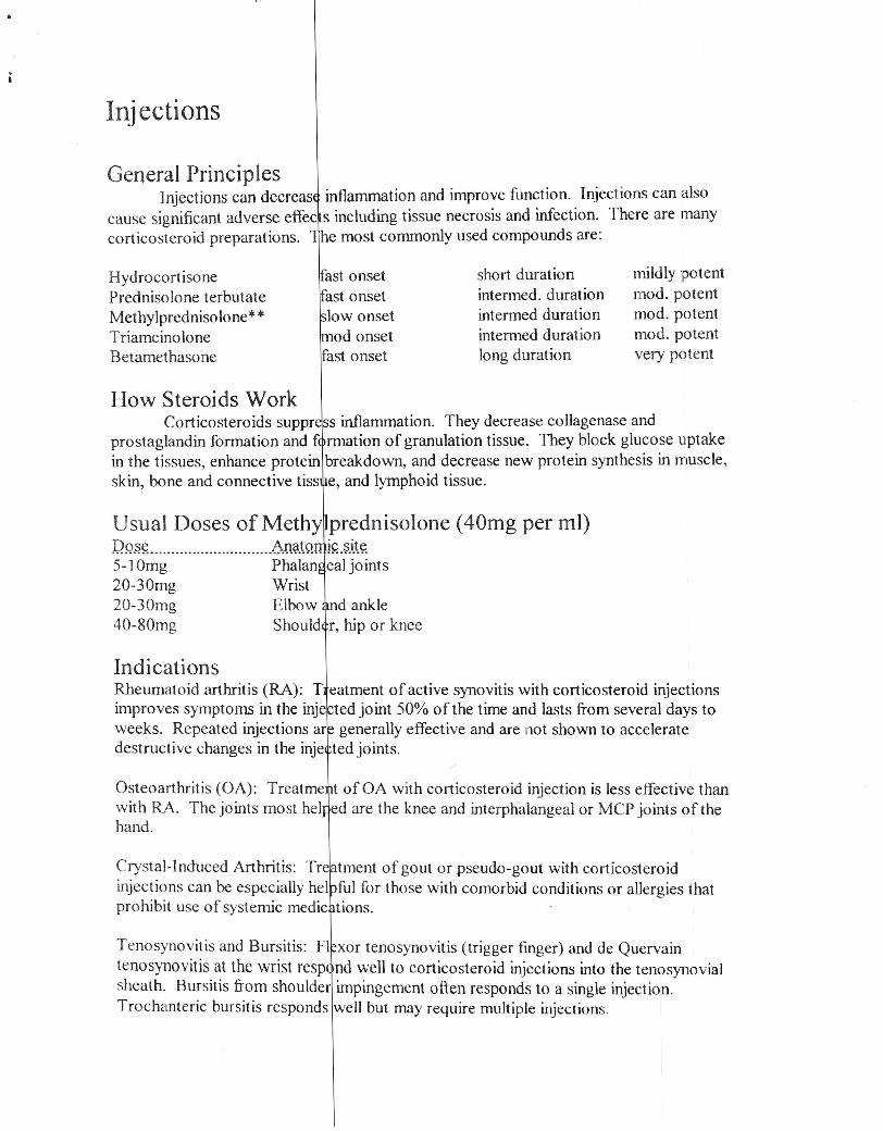
General PrinciplesInjections can decreas~ inflammation and improve function. Injections can also
cause significant adverse effec s including tissue necrosis and infection. There are manycorticosteroid preparations. he most commonly used compounds are:
Injections
HydrocortisonePrednisolone terbutateMethylprednisolone* *TriamcinoloneBetamethasone
fast onsetrast onsetlow onsetod onset
fast onset
short durationintermed. durationintermed durationintermed durationlong duration
mildly potentmod. potentmod. potentmod. potentvery potent
How Steroids WorkCorticosteroids suppr~s inflammation. They decrease collagenase and
prostaglandin formation and fi rmation of granulation tissue. They block glucose uptakein the tissues, enhance protein breakdown, and decrease new protein synthesis in muscle,skin, bone and connective tiss e, and lymphoid tissue.
Usual Doses of Meth1prednisolone (40mg per ml)~~i'cie~g""""""""""""'X:i~~~~~tt~ints20-30mg Wrist20-30mg Elbow ~nd ankle40-80mg Should~r, hip or knee
IndicationsRheumatoid arthritis (RA): Tfatment of active synovitis with corticosteroid injectionsimproves symptoms in the inje ted joint 50% of the time and lasts from several days toweeks. Repeated injections ar generally effective and are not shown to acceleratedestructive changes in the inje ted joints.
Osteoarthritis (OA): Treatme~t of OA with corticosteroid injection is less effective thanwith RA. The joints most helned are the knee and interphalangeal or MCP joints of thehand.
Crystal-Induced Arthritis: Tr~tment of gout or pseuckl-gout with corticosteroidinjections can be especially hel ful for those with comorbid conditions or allergies thatprohibit use of systemic medic tions.
Tenosynovitis and Bursitis: F1xor tenosynovitis (trigger finger) and de Quervaintenosynovitis at the wrist resp nd well to corticosteroid injections into the tenosynovialsheath. Bursitis from shoulder impingement often responds to a single injection.Trochanteric bursitis responds well but may require multiple injections.

Olecranon and prepat~lIar intrabursal injections carry an increased risk forinfection. These areas should only be injected with steroid when the patient's problemhas not resolved with time an there is clearly no evidence of underlying infection (tap).
Diabetic patients are a risk for serious infection and for systemic effects ofabsorbed corticosteroids.
Greene, Walter 8.,2001. Es entials of Musculoskeletal Care. American Academy ofOrthopedic Surgeons. Rose ont, Illinois.
I

Amount of displacementof fracture fragmentsNondispJacedDisplacedAngulatedBayonetedDistracted
Skin IntegrityClosedOpen
Description
agments are in anatomic alignmentagments are no longer in anatomic alignmentagments are malaligned (measure angulation)istal fragment longitudinally overlaps the proximal fragistal fragment is separated from the proximal frag (gap)
Descriptionkin over and near fracture is intactkin over and near the fracture is lacerated OR abraded
Adverse Outcomes of 'racturesAny fracture can result in delayed or malunion. Limb function can be affected by
nearby joint contracture, stiffu ss, limb shortening or malalignment. Osteomyelitis maydevelop if fracture is open. N rve andlor vascular damage may occur with severefractures. Compartment syndr me can evolve with excess swelling. Complex regionalsympathetic dystrophy is rare.
ReferralNever hesitate to call fi r opinion (know how to describe the fracture well prior to
calling). Patients with open, u stable, irreducible, suspected compartment syndrome,nerve, vascular or muscle dam ge need further eva!. Displaced fractures require furtherevaluation. Pediatric patients ith fractures anywhere near a growth plate or around theelbow need further evaluation. Ankle fractures are tricky, request consult.
Unstable fractures incl de both bone, comminuted, oblique, spiral and intra-articular.
PearlsEducate your patients ell on cast care (neuro-vascular compromise) and always
see them if there are concerns. Never be afraid to remove a cast. You can always re-apply it later. Ifconcerned a ut swelling, don't be afraid to bi-valve a cast (saw the castin half, re-apply and wrap wi a e wrap). Don't keep joints immobilized longer thannecessary; this will result in in reased stiffuess. Toddler fractures take 1 week per yearof life to heal. Teens take 4-6 weeks. Adults take 6+ weeks. Ifworried about theretention of the fracture line, bing pt back every week and a few weeks and x-ray. It isnecessary to x-ray unstable fra tures every week.
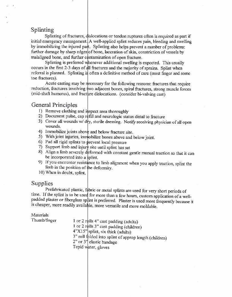
SplintingSplinting of fractures, islocations or tendon ruptures often is required as part if
initial emergency management. A well-applied splint reduces pain, bleeding and swellingby immobilizing the injured pa . Splinting also helps prevent a number of problems:further damage by sharp edges of bone, laceration of skin, constriction of vessels bymalaligned bone, and further c ntamination of open fracture.
Splinting is preferred henever additional swelling is expected. This usuallyoccurs in the first 2-3 days of II fractures and the majority of sprains. Splint whenreferral is planned. Splinting i often a definitive method of care (most finger and sometoe fractures).
Acute casting may be tcessary for the following reasons: fractures that requirereduction, fractures involving wo adjacent bones, spiral fractures, strong muscle forces(mid-shaft humerus), and fract re dislocations. (consider bi-valving cast)
General Principles1) Remove clothing and ~spect area thoroughly2) Document pulse, cap r fill and neurologic status distal to fracture3) Cover aUwounds wi d ,sterile dressing. Notify receiving physician of all open
wounds.4) Immobilize joints abov and below fracture site.5) With joint injuries, . obilize bones above and below joint.6) Pad all rigid splints to revent local pressure7) Support limb and inju site until splint has set8) Align a limb severely d formed with constant gentle manual traction so that it can
be incorporated into a plint.9) If you encounter resist ce to limb alignment when you apply traction, splint the
limb in the position of he deformity.10) When in doubt, splint.
SuppliesPrefabricated plastic, ~briC or metal splints are used for very short periods of
time. If the splint is to be use for more than a few hours, custom application of a well-padded plaster or fiberglass sp int is preferred. Plaster is used more frequently because itis cheaper, more readily availa Ie, more versatile and more moldable.
MaterialsThumb/finger 1 or 2 r lis 4" cast padding (adults)
I or 2 r lis 3" cast padding (children)4"XIS" splint, six thick (adults)3" roll Ided into splint ofapprop length (children)2" or 3' elastic bandage'Tepid ater, gloves
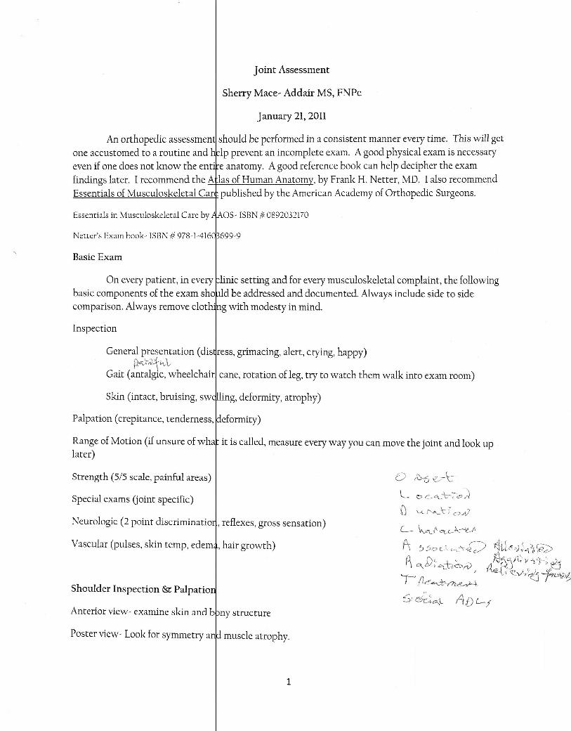
Joint Assessment
Sherry Mace~ Addair MS, FNPc
January 21, 2011
An orthopedic assessmen should be performed in a consistent manner every time. This will getone accustomed to a routine and lp prevent an incomplete exam. A good physical exam is necessaryeven if one does not know the ent' e anatomy. A good reference book can help decipher the examfindings later. I recommend the A las of Human Anatom , by Frank H. Netter, MD. I also recommendEssentials of Musculoskeletal Car published by the American Academy of Orthopedic Surgeons.
Essentials in Musculoskeletal Care by AAOS~ ISBN '# 0892032170
Netter'S Exam b()ok~ ISBN '# 978-1~416
Basic Exam
On every patient, in eve~linic setting and for every musculoskeletal complaint, the followingbasic components of the exam sho ld be addressed and documented. Always include side to sidecomparison. Always remove clot 'ng with modesty in mind.
Inspection
General presentation (disqress, grimacing, alert, crying, happy)p~,>;.lt"Vt
Gait (antalgic, wheelchair) cane, rotation of leg, try to watch them walk into exam room)
Skin (intact, bruising, swtilling, deformity, atrophy)
Palpation (crepitance, tenderness, !deformity)
Range of Motion (if unsure of whaf it is called, measure every way you can move the joint and look uplater)
Strength (5/5 scale, painful areas) o t0t.5 e--IcL 0 c-.oe- t-:-o ~SpeCial exams Uoint specific)
Neurologic (2 point discriminatioq, reflexes, gross sensation)
Vascular (pulses, skin temp, edem*, hair growth)
Shoulder Inspection & Palpatio$;d~t:\L ADL-f
Anterior view~ examine skin and bbny structure
Poster view~ Look for symmetry anJdmuscle atrophy.
1
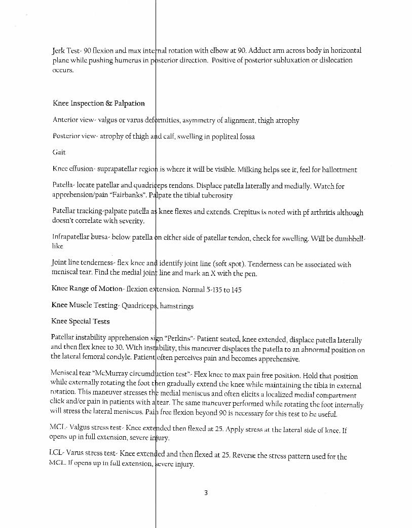
Jerk Test- 90 flexion and max intetnal rotation with elbow at 90. Adduct arm across body in horizontalplane while pushing humerus in Pfsterior direction. Positive of posterior subluxation or dislocationoccurs.
Knee Inspection &: Palpation
Anterior view- valgus or varus defrrmities, asymmetry of alignment, thigh atrophy
Posterior view- atrophy of thigh a$d calf, swelling in popliteal fossa
Gait
Knee effusion' suprapatellar regio is where it will be visible. Milking helps see it, feel for ballottment
Patella' locate patellar and quadri eps tendons. Displace patella laterally and medially. Watch forapprehension/pain "Fairbanks". P lpate the tibial tuberosity
Patellar tracking'palpate patella a knee flexes and extends. Crepitus is noted with pf arthritis althoughdoesn't correlate with severity.
Infrapatellar bursa' below patella tn either side of patellar tendon, check for swelling. Will be dumbbell,like
Joint line tenderness' flex knee an~ identify joint line (soft spot). Tenderness can be associated withmeniscal tear. Find the medial joint line and mark an X with the pen.
Knee Range of Motion' flexion dtension. Normal 5,135 to 145
Knee Muscle Testing- Quadricep~, hamstrings
Knee Special Tests
Patellar instability apprehension s~.n "Perkins", Patient seated, knee extended, displace patella laterallyand then flex knee to 30. With ins bility, this maneuver displaces the patella to an abnormal position onthe lateral femoral condyle. Patient often perceives pain and becomes apprehensive.
Meniscal tear "McMurray circumd ction test"- Flex knee to max pain free position. Hold that positionwhile externally rotating the foot t en gradually extend the knee while maintaining the tibia in externalrotation. This maneuver stresses th medial meniscus and often elicits a localized medial compartmentclick and/or pain in patients with a tear. The same maneuver performed while rotating the foot internallywill stress the lateral meniscus. Pai free flexion beyond 90 is necessary for this test to be useful.
MCl, Valgus stress test- Knee ext~ded then flexed at 25. Apply stress at the lateral side of knee. Ifopens up in full extension, severe i~iury.
lCl, Varus stress test, Knee exten~ed and then flexed at 25. Reverse the stress pattern used for theMeL If opens up in full extension, bevere injury.
3
Faber "Figure of 4 test"- FleXion/alduction/external rotation maneuver to detect hip and SI jointpathology. With patient supine, p ace the affected hip in flexion, abduction and external rotation andthen press the hip back into ext en ion by placing the foot of the opposite tibia. If the maneuver is painful,the n the hip or SI region may be a fected.
Baby- Developmental Dysplasia ofjthe Hip
Ortobni· newborns
Barlow··newborns
Galcazzi- toddlers
e'Nice clip on you tube to show technique ~•
Ottawa Ankle Rules
Ankle sprains graded HIT
I-ll-lU-
StretchingPartial TearingComplete Rupture
T~der to palp; mild swelling; normal functionT· der to palp; mod swelling & ecchymosis, some instability,twt bearPa n; marked ecchymosis/edema; gross instability; no \vt bear
Usually don't need x-rays for gradep I &: II. Per Ottawa rules, x-rays arc necessary for the patient who:
1. ISlnable to bear weight initially after injury or when examined2. H, s tenderness over tl.1eposterior edge .0£ the distal6cI11 or distal tip of
th· medial or lateral malleolus3. H s tenderness over the fifth metatarsal or tarsal navicular
5