instituto de tecnologia química e biológica | universidade
TRANSCRIPT

i
Marija Petković
Dissertation presented to obtain the Ph.D degree
in Biochemistry
Instituto de Tecnologia Química e Biológica | Universidade
Nova de Lisboa
Oeiras, September, 2011
Revealing fungal activity in
the presence of ionic liquids


i
I declare that the work presented in this thesis, except where otherwise stated,
is based on my own research. It was supervised by Doctor Cristina Silva
Pereira and Professor Luís Paulo Rebelo. The work was mainly performed in
Instituto de Tecnologia Química e Biológica, Universidade Nova de Lisboa,
between March 2007 and June 2011. Part of the results was attained during
research visits to The Queen‟s University Ionic Liquid Laboratories, The
Queen‟s University of Belfast, and the Institute of Pharmaceutical Biology,
University of Regensburg.
I am grateful for the financial support provided by Fundação para a
Ciência e Tecnologia (fellowship BD 31451/2006). The work was partially
supported by a grant from Iceland, Liechtenstein and Norway through the
EEA financial mechanism (Project PT015).


iii
Acknowledgements
I wish to thank my supervisors Cristina Silva Pereira and Luís Paulo
Rebelo for making this work possible. Cristina, your creativity and dedication
were inspiring. Thanks to all former and present members of Applied and
Environmental Mycology Laboratory at ITQB for their continuous support.
The last four years would definitely be less thrilling without the
watchful eye of Ken Seddon. Thanks Jamie for all the laughs and cries.
Nimal, thank you for your serenity and advice. Thanks to the QUILL‟s crew
of 2008 for a great time in Belfast.
Professor Heilmann, Birgit, Professor Kunz, Doris and Rosemarie,
thank you all for welcoming me so warmly in “the most northern city of
Italy”.
Marijana, hvala ti što si promenila moj život i nesebično delila svoje
mudrosti i iskustva. Tebi i Marini, hvala za noći ispunjene razgovorom i
vinom u Oeirasu. Hvala svim mojim dragim prijateljima u Srbiji. Marko,
Miša, Jelena i Saša, vreme kao da ne postoji sa vama. Diego and Jojo, thanks
for bringing joy to my days; don‟t let anything change you. Hvala svim
prijateljima iz “Srpski KUD – Tuga”, za smeh i zdravice.
Mama, tata i Marina, hvala na vašoj bezrezervnoj podršci. Biti daleko
od vas je sigurno bio nateži deo ove teze. Sr. Francisco, Sra. Manuela, Teresa
e Pedro, muito obrigada por me acolherem e aceitarem no seio da família que
agora também é minha.
Ricardo, I would need to invent a word to thank you enough.


v
“Our virtues and our failings are inseparable, like force and matter.
When they separate, man is no more.”
Nikola Tesla


vii
Summary
Ionic liquids constitute a vast and heterogeneous group of chemicals,
generally non-volatile and of high solvent quality. They are already used in
industrial processes; future applications depend heavily on conscious design
of ionic liquids. Given especially the global demand for sustainable
chemicals, understanding environmental risks is a priority, necessitating a
multidisciplinary research approach, covering a broad range of disciplines
from biology to chemistry.
Ascomycota fungi are highly suitable model organisms, especially
due to their environmental ubiquity and important role in the biotic decay of
pollutants. This thesis reports the first ever use of Ascomycota fungi to
investigate ionic liquids ecotoxicity and environmental persistence. Fungal
strains of Penicillium and Aspergillus were in general found to be more
tolerant to ionic liquids containing imidazolium, pyridinium, pyrrolidinium,
cholinium or phosphonium cations, than any other microorganism tested to
date (Chapters II, III and IV). The capacity of the strains to tolerate the ionic
liquids tested was apparently correlated to their phylogeny. Ionic liquid
toxicity was evaluated using common parameters, such as growth inhibition
and death. Less frequently evaluated parameters were also analysed,
including monitoring of the integrity of the cellular boundaries of fungal
conidia by microscopy (Chapter IV) and determining the diffusible fungal
metabolome by ESI-MS and LC (Chapter II and V). Overall, these data
significantly contribute to current understanding of structure-activity
relationships in ionic liquids. For example toxicity is apparently a function of
alkyl chain length of both anion and cation (Chapters III and IV, respectively).
A critical review of current understanding of toxicity and environmental
impact of the principal ionic liquid groups made it clear that the common
generalisation of ionic liquids being either “green” or “toxic”

ix
solvents is misleading as neither of these statements is completely true
(Chapter I, section 1.3.1).
Above issues have inspired conscious design of a novel group of ionic
liquids - cholinium alkanoates, which display low toxicity and high
biodegradability (Chapter III). They are amongst the most interesting groups
of ionic liquids so far investigated, and have proven to be a remarkable
breakthrough as efficient solvents for suberin in cork.
Under laboratory conditions the ability of filamentous fungi to
produce secondary metabolites is often repressed. These natural compounds
are of heightened interest, especially given their structural diversity and
potential biological activity. Addition of sub-lethal concentrations of ionic
liquids to the growth media altered the profile of the diffusible fungal
metabolome (i.e. low molecular weight compounds secreted by the fungi).
Three of five tested ionic liquids activated cryptic metabolites with
demonstrable biological effects on HeLa cells and bacteria (Chapter V).
These findings merit more detailed follow-up – the data presented here are
preliminary but encouraging. In summary, ionic liquids potential to activate
cryptic fungal metabolites is highly promising in the discovery of valuable
novel natural products.
The work presented in this thesis constitutes a foundation for
developing novel environmentally sustainable biotechnological processes.


xi
Sumário
Os líquidos iónicos reúnem um numeroso e heterogéneo grupo de substâncias
químicas, geralmente não-voláteis e de excelente desempenho como
solventes. Apesar de serem já utilizados em inúmeros processos industriais,
aplicações futuras dependem particularmente de uma formulação consciente
de líquidos iónicos. Testemunhamos, globalmente, a procura de produtos
químicos sustentáveis, pelo que compreender os riscos ambientais
consequentes, é uma prioridade. É fundamental investigar estas questões de
forma multidisciplinar, abrangendo um vasto leque de disciplinas, da biologia
à química.
Os fungos Ascomycota são excelentes organismos modelo,
especialmente dada a sua ubiquidade ambiental e proeminente capacidade de
degradar inúmeros poluentes. Esta tese reúne o primeiro estudo que recorreu
a fungos Ascomycota para investigar a ecotoxicidade e a persistência
ambiental de líquidos iónicos. Estirpes de Penicillium e Aspergillus
demonstraram ser, de uma forma geral, mais tolerantes aos líquidos iónicos
contendo catiões do tipo imidazólio, piridínio, pirrolidínio, colínio ou
fosfónio, do que qualquer outro microorganismo testado até à data (Capítulos
II, III e IV). É muito provável que a capacidade das estripes fúngicas de
tolerar os líquidos iónicos esteja correlacionada com a sua filogenia. A
toxicidade dos líquidos iónicos foi estimada usando parâmetros comuns, tais
como inibição do crescimento e morte. Os dados foram complementados
estudando parâmetros raramente considerados, como a monitorização, por
microscopia, da integridade dos conídios (Capítulo IV) e a determinação, por
ESI-MS e LC, do metaboloma extracelular (Capítulo II e V). Estes resultados
contribuem, de forma útil, para o estado actual da arte sobre a relação
estrutura-actividade em líquidos iónicos. Por exemplo, a toxicidade é,
aparentemente, função do comprimento da cadeia alquilo do anião e/ou catião
(Capítulos III e IV, respectivamente). A revisão crítica do conhecimento
actual sobre a toxicidade e o impacto ambiental dos principais grupos de


xiii
líquidos iónicos evidenciou que a sua vulgarização como solventes "verdes"
ou "tóxicos" é fraudulenta, sendo que nenhuma destas declarações é
completamente verdadeira (Capítulo I, secção 1.3.1.).
As questões acima descritas inspiraram a formulação consciente de
um novo grupo de líquidos iónicos – os alcanoatos de colínio. Estes exibem
baixa toxicidade e elevada biodegradabilidade (Capítulo III). Constituem
entre os líquidos iónicos até agora estudados um dos grupos mais
interessantes e permitiram um avanço notável: a solubilização eficiente de
suberina em cortiça.
Em condições de laboratório, a capacidade dos fungos filamentosos
de produzirem metabolitos secundários está reprimida. O elevado interesse
nestes compostos naturais justifica-se pela sua diversidade estrutural e
possível actividade biológica. Os líquidos iónicos, presentes em
concentrações sub-letais no meio de crescimento do fungo, alteraram o perfil
do metaboloma extracelular (ou seja, compostos de baixo peso molecular
secretados pelo fungo). Três, entre os cinco líquidos iónicos aqui testados,
activaram metabolitos crípticos com efeitos biológicos patenteáveis em
células HeLa e bactérias (Capítulo V). Estes dados são preliminares, mas
encorajadores e merecem estudos decorrentes. Em resumo, o potencial dos
líquidos iónicos para activar a produção de metabolitos fúngicos crípticos é
promissor na descoberta de produtos naturais de valor acrescentado.
O trabalho apresentado nesta tese é uma base sólida de conhecimento
para o desenvolvimento de processos biotecnológicos ambientalmente
sustentáveis.


xv
Contents
Statement............................................................................................................i
Acknowledgements..........................................................................................iii
Summary.........................................................................................................vii
Sumário...........................................................................................................xi
List of ionic liquids abbreviations...............................................................xix
List of acronyms............................................................................................xxi
Chapter I Introduction
1.1. Biology and ecology of filamentous fungi...........................................5
1.2. Ionic liquids........................................................................................12
1.2.1. Green solvents............................................................................12
1.2.2. Ionic liquids: Properties and applications...................................13
1.2.3. Ionic liquids as solutes................................................................16
1.3. Ionic liquids in biological systems.........................................................17
1.3.1. Ecotoxicity and biodegradability of ionic liquids.......................18
Abstract............................................................................................18
Introduction......................................................................................19
Toxicity of ionic liquids containing aromatic head groups in the
cation................................................................................................22
Imidazolium-based ionic liquids.................................................22
Pyridinium-based ionic liquids...................................................41
Quinolinium-based ionic liquids.................................................46
Toxicity of ionic liquids containing alicyclic head groups in the
cation................................................................................................46
Pyrrolidinium-, piperidinium- and morpholinium-based ionic
liquids..........................................................................................46
Quaternary ammonium ionic liquids...........................................49
Quaternary phosphonium ionic liquids.......................................53

Biodegradability of ionic liquids......................................................55
Molecular toxicity of ionic liquids...................................................57
Modes of toxicity of ionic liquids....................................................60
Quantitative structure-activity relationships (QSAR) for ionic
liquids...............................................................................................61
General considerations.....................................................................63
Acknowledgements.........................................................................65
1.3.2. Whole-cell biocatalysis with ionic liquids..................................66
References.......................................................................................................67
Chapter II Exploring fungal activity in the presence of ionic liquids
Abstract............................................................................................................91
Introduction.....................................................................................................92
Experimental....................................................................................................93
Chemicals...................................................................................................93
Fungal strains..............................................................................................94
Toxicity tests..............................................................................................94
Toxicity data analysis.................................................................................95
Metabolites extraction................................................................................96
ESI-MS analysis.........................................................................................96
Computational interpretation of MS data...................................................97
Results and discussion.....................................................................................97
Toxicological assessment...........................................................................97
Metabolic footprinting..............................................................................101
Conclusions...................................................................................................103
Acknowlegdements.......................................................................................105
References.....................................................................................................106
Chapter III Novel biocompatible cholinium-based ionic liquids -
toxicity and biodegradability
Abstract..........................................................................................................110

xvii
Introduction...................................................................................................111
Experimental..................................................................................................112
Chemicals.................................................................................................112
Ionic liquids..............................................................................................113
Thermal properties analysis......................................................................116
Fungal strains...........................................................................................117
Toxicity tests............................................................................................117
Biodegradability assessment of the ionic liquids.....................................118
Results and discussion...................................................................................119
Conclusions...................................................................................................127
Acknowlegdements.......................................................................................128
References.....................................................................................................129
Chapter IV Unravelling the mechanism of toxicity of
alkyltributylphosphonium chlorides in
Aspergillus nidulans conidia
Abstract..........................................................................................................134
Introduction...................................................................................................135
Experimental..................................................................................................136
Chemicals.................................................................................................136
Ionic liquids..............................................................................................137
Fungal strain.............................................................................................137
Toxicity tests............................................................................................137
Membrane and cell wall integrity assays..................................................138
Scanning electron microscopy..................................................................140
Biodegradability assessment....................................................................140
Results and discussion...................................................................................141
Conclusions...................................................................................................150
Acknowlegdements.......................................................................................151
References.....................................................................................................153

Chapter V Preliminary evaluation of the biological potential of
diffusible fungal metabolites induced by an ionic liquid
Abstract..........................................................................................................161
Introduction...................................................................................................162
Experimental..................................................................................................163
Chemicals.................................................................................................163
Microorganisms........................................................................................164
Fungal cultures.........................................................................................165
Fungal metabolites extraction (fSM extracts)..........................................165
Liquid chromatography analyses of fSM extracts....................................166
Biological activity tests............................................................................167
Results and discussion...................................................................................169
Metabolic footprinting by LC...................................................................169
Biological activity of ionic liquid-induced fungal metabolites................172
Conclusions...................................................................................................178
Acknowlegdements.......................................................................................179
References.....................................................................................................180
Chapter VI Final discussion
Final discussion.............................................................................................185
References.....................................................................................................192

xix
List of ionic liquids abbreviations
cations
[Cnmim]+ 1-alkyl-3-methylimidazolium
[Cnpy] +
1-alkylpyridinium
[Cnmpy]+ 1-alkyl-3-methylpyridinium
[Cnmpy]+ 1-alkyl-4-methylpyridinium
[CnOC1py]+
1-alkoxymethylpyridinium cation
[Cnquin]+ 1-alkylquinolinium
[Cnmpyr] +
1-alkyl-1-methylpyrrolidinium
[Cnmpip]+ 1-alkyl-1-methylpiperidinium
[Cnmmor]+ 4-alkyl-4-methylmorpholinium
[Cnemor]+ 4-alkyl-4-ethylmorpholinium
[Nw x y z]+ generic tetraalkylammonium
[Pw x y z]+ generic tetraalkylphosphonium
[EtNH3][NO3] ethylammonium nitrate
[N1 1 1 2OH]+ cholinium
anions
[BF4]- tetrafluoroborate
[PF6]- hexafluorophosphate
[SbF6]- hexafluoroantimonate
[N(CN)2]- dicyanamide
[C1SO3]- or [CH3SO3]
- methanesulfonate
[C2SO4]- ethylsulfate
[C8SO4]- octylsulfate
[O2CMe]- or [O2CC1]
- ethanoate
[CnH2n+1CO2]- alkanoate
[N(SO2CF3)2]- or [NTf2]
- bis{(trifluoromethyl)sulfonyl}amide
[N(CF3)2]-
bis(trifluoromethyl)amide
[(EtO)2PO2]- or [C2PO2]
- diethylphosphate

[CF3SO3]- or [OTf]
- 1,1,1-trifluoromethanesulfonate
[SCN]- thiocyanate
[lac]- lactate

xxi
List of acronyms
APIs active pharmaceutical ingredients
ATPases a class of enzymes that catalyze the decomposition of
adenosine triphosphate (ATP) into adenosine diphosphate
(ADP)
ATR-FTIR attenuated total reflectance fourier transform infrared
spectroscopy
BAC benzalkonium chloride
BASIL Biphasic Acid Scavenging utilizing Ionic Liquids
BATIL Biodegradability and Toxicity of Ionic Liquids meeting
DSC differential scanning calorimetry
EC50 effective concentration which causes 50 % of the maximal
response
ESI-MS electrospray ionisation mass spectrometry
fSM fungal secondary metabolites
HCA hierarchical cluster analysis
HF hydrogen fluoride
ISO International Organization for Standardization
ISO 11348 Water quality - Determination of the inhibitory effect of water
samples on the light emission of Vibrio fischeri (Luminescent
bacteria test)
Kow 1-octanol/water partition coefficient
LC liquid chromatography
MBC minimal bactericidal concentration
MBEC minimum biofilm eradication concentrations
MEA malt extract agar
MIC minimal inhibitory concentration
m.p. melting point
NADPH reduced form of nicotinamide adenine dinucleotide phosphate

NMR nuclear magnetic resonance
OECD Organisation for Economic Co-operation and Development
QSAR quantitative structure-activity relationships
REACH Registration, Evaluation, Authorisation and Restriction of
CHemical substances
scCO2 supercritical carbon dioxide
SDS sodium dodecylsulphate
TGA thermal gravimetric analysis
VOCs volatile organic solvents

Chapter I
Introduction

Chapter I
2

Chapter I
3
Chapter I Introduction
1.1. Biology and ecology of filamentous fungi
1.2. Ionic liquids
1.2.1. Green solvents
1.2.2. Ionic liquids: Properties and applications
1.2.3. Ionic liquids as solutes
1.3. Ionic liquids in biological systems
1.3.1. Ecotoxicity and biodegradability of ionic liquids
1.3.2. Whole-cell biocatalysis with ionic liquids

Chapter I
4

Chapter I
5
1.1. Biology and ecology of filamentous fungi
The following lines focus on general features of filamentous fungi. They do
not provide a comprehensive review of the field. It contains, however,
information essential for critical analysis of the ecological and
biotechnological importance of filamentous fungi. Both concepts are
significant for understanding of the chapters enclosed in the thesis.
Fungi are eukaryotic, heterotrophic, organisms. They were initially thought to
be related to plants, in particular due to the presence of vacuoles. Knowledge
of their protein-coding sequences,1 and specific characteristics, e.g. chitin
being a constituent of the cell wall and glycogen acting as a storage polymer,
altered this view. In the early 1990‟s, the link between fungi and animals was
finally accepted. Fungi were recognised by Whittaker in 1969 as one of the
five kingdoms in a life-form classification system.2 This concept was
preserved in the six3 or seven
4 kingdoms classification systems proposed
afterwards. It includes an enormous number of fungal species, recently
estimated to total 1.5 million, with only 5 % described to date.5 They are
divided into four phyla, namely Chytridiomycota, Zygomycota,
Basidiomycota and Ascomycota. Ascomycota is the largest phylum and
comprises more than 60000 species,6 including Aspergillus and Penicillium
genera which are of particular interest here (Figure 1). Knowledge on their
complex physiology is being the subject of on-going research, including a
continuously growing number of fully sequenced genomes of different species
(more than ten at the moment of writing).7-9

Chapter I
6
Figure 1. The seven kingdom life classification system according to Patterson and
Sogin.4 The four phyla in the fungal kingdom are listed, as are Penicillium and
Aspergillus genera within Ascomycota.
Vegetative cells of filamentous fungi, hyphae, are 3-10 μm wide
tubular filaments showing apical growth (Figure 2). They are interconnected
and form branching networks, mycelium. It assembles differentiated hyphae,
which are able to coordinate distinct roles in nutrient acquisition or
development of reproductive structures.
Figure 2. Microscopic image of 48 h culture of Aspergillus nidulans
(63× magnification). The image was captured in Applied and Environmental
Mycology Laboratory, ITQB, by Diego O. Hartmann.
Eubacteria
Archaebacteria
Protozoa
Chromista
Plantae
Fungi
Animalia
Chytridiomycota
Zygomycota
Basidiomycota
Ascomycota
Penicillium
Aspergillus
Pro
kar
yo
taE
uk
ary
ota

Chapter I
7
Cells are separated by perforated septa which ensure that intracellular material
can freely stream within the hyphae. When grown on solid substrate
filamentous fungi form radially symmetrical colonies which grow at a
constant rate (approximately 0.5 mm h-1
at 37 °C for A. nidulans10
).
Submerged in liquid culture they appear as dispersed filaments or mycelial
masses. In response to different physical (e.g. agitation, rheology, light) and
chemical factors (e.g. dissolved oxygen, carbon dioxide), hyphae can change
morphology and physiology.11
The hyphal wall constitutes ~20 % of the cellular biomass and it is a
remarkable element of the vegetative fungal cell.12
It defines and maintains
cell shape, and is composed of several cross-linked structural elements. The
hyphal wall is extremely dynamic, at the level of both its
composition/structure and function. The cell wall is a central trafficking
element, controlling interactions with the environment, including cellular
processes of uptake, secretion, recognition and communication. It comprises
an inner layer of rigid fibrous polysaccharides and an outer layer of gel-like
polymers (Figure 3).
Figure 3. Schematic representation of the fungal cell wall and its major components:
glucan, chitin and glycoproteins (adapted from Selitrennikoff13
).
In Ascomycota fungi, the outer layer is essentially composed of glucans,
mainly β-(1-3) and β-(1-6)-linked glucose units and chitin. The inner layer is

Chapter I
8
a mechanically resistant polymer made of β(1-4)-linked N-acetylglucosamine
units. Glycoproteins, containing mannose, galactose and xylose, and β-(1-3)-
glucan are assembled as the embedding matrix. Some glycoproteins are
covalently bound to the plasma membrane and/or the cell wall by
glycosylphosphatidylinositol anchors.14
Hydrophobins found to coat aerial
hyphae, are hydrophobic proteins that reduce surface tension at the water-air
interface. These proteins may also play a significant role in fungal
pathogenesis, symbiosis and protection from environmental stress, e.g.
desiccation.15
The fungal cell wall may further contain pigments such as
melanins, which increase its mechanical strength, resistance to lytic enzymes
and afford radiation protection.16
They might also act as virulence factors of
plant pathogens.
The hyphal tip is generally considered as the most active and dynamic
area of the hyphae, controlling nutrient uptake, secretion,17
hyphal extension
and cell wall synthesis.18
There are two prevailing opinions regarding
mechanisms involved in cell wall biosynthesis: steady-state growth and cell
wall remodelling theories. Both assume turgor pressure as the driving force
of the biosynthesis, and that components of the cell wall are transported in
vesicles to the apex. In the steady-state growth theory, transported
components are cross-linked and converted from a plastic to a rigid
structure.19
The cell wall remodelling theory suggests a balance between cell
wall synthesis and lysis partially plasticises the apex of the hyphae (Figure
4).14,20
Our understanding of the dynamics of cell wall biosynthesis and
assembly is still incomplete.

Chapter I
9
Figure 4. Schematic drawing of the cell wall remodelling theory, showing apical
hyphal growth (a) and branching (b). Microvesicles (empty, solid and starred circles)
are being transported to these two sites, and may contain cell wall biosynthetic
enzymes and other materials necessary. They are associated with cytoskeleton -
microfilaments (beaded lines) and microtubules (parallel lines). Apical and branch
domes are depicted as plastic cell wall regions that become progressively rigid. Stars
at the branching and apical site represent lytic enzymes. Model adapted from
Gooday.18
Filamentous fungi use mycelial growth to colonise large areas,
ramifying throughout the substrate. Aided by high turgor pressure they can
also penetrate hard surfaces efficiently.21
Owing to broad extracellular
enzyme diversity, they can utilise a range of substrates, including recalcitrant
polymers. Accordingly to the “bulk-flow theory” the degrading extracellular
enzymes are exported from the cell together with apical cell wall
components.19
Generally, uptake of nutrients occurs through the plasma
membrane, involving proton-motive force or specific transport proteins, such
as permeases or carriers.
Ascomycota fungal life cycle can follow two distinct routes,
producing either conidia (asexual stage) or ascospores (sexual stage). Figure
5 illustrates, as an example, the life cycle of A. nidulans. Morphological
features of spore-bearing structures have traditionally been used for
classification/identification purposes. The experimental work presented in
this thesis is concerned with Ascomycota fungi in their asexual stage.
Conidia of Aspergillus and Penicillium spp. are rounded, pigmented,
hydrophobic, non-motile cells (see micrograph of A. nidulans conidia, Figure
bb a

Chapter I
10
5A in chapter IV).22
They are produced in vast numbers and actively
discharged or readily dispersed by raindrops, air currents or through contact
with insects. The conidia are produced by fertile conidiogenous cells on
specialised hyphae (conidiophores). During their maturation the composition
of the cell wall is altered to form a four-layer wall, important for protection
and facilitating dormancy of the conidium.23
Conidiation is regulated by
environmental conditions, e.g. generally enhanced by hyphal exposure to air
or nutrient limitation.5 Light is also an important factor, especially because
the circadian rhythm regulates conidiation. Depending on environmental
conditions, deposited conidia might germinate or remain dormant. To
germinate, it first absorbs water and swells, a germ tube is formed and, at the
same time, the metabolism is accelerated, i.e. respiration rate, enzyme
activation and biosynthesis increase.5
Figure 5. Schematic representation of the life cycle of Ascomycota fungi. As an
example, Aspergillus nidulans asexual and sexual stages are shown: (1) and
(5) vegetative mycelia; (2) conidiophores; (3) conidia; (4) germinating conidia;
(6) ascocarp; (7) ascus; (8) ascospores; (9) germinating ascospore.
Fungi occupy critical ecological niches. For example they ensure
degradation of lignocellulosic material,24
generally through lignin peroxidise,
manganese peroxidises and laccase,24
and by endo- and exoglucanase
4
3
2
1 5
6
789
ASEXUAL CYCLE
SEXUAL CYCLE

Chapter I
11
activity.25
Filamentous fungi are increasingly attracting interest as
bioremediation agents, especially due to their catabolic potential and high
surface area.26
They are ubiquitous in all environments and able to survive
even under extreme conditions. For example they can often be isolated from
environments with high heavy metals,27
dyes,28
polycyclic aromatic
hydrocarbons29
or chlorinated phenols concentrations.30
Their presence in
Dead Sea31
or at polar locations32
is illustrative. Penicillium and Aspergillus
spp. isolated from marine environments can be regarded as xerophiles, since
they were able to survive at water activity below 0.85.33
In these conditions,
strains accumulate „compatible solutes‟, e.g. glycerol and erythritol, to
prevent loss of water.34
Others, e.g. Penicillium fellutanum, accumulate
glycine betaine and choline-O-sulfate.35
These compounds, as well as choline
chloride, when added to growth media (2 or 10 mM), act as osmoprotectors.
Under low water activity conditions, the plasma membrane alters its fluidity;
this is thought to be related with alterations in the sterol/phospholipids ratio
and fatty acid unsaturation.36
The above illustrates that filamentous fungi are environmentally
ubiquitous, have complex physiology, high biological robustness and
potentially useful catabolism. In the following chapters of this thesis,
Penicillium and Aspergillus strains high capacity to grow in medium
supplemented with an ionic liquid will be demonstrated. The high relevance
of this feature to biotechnological applications will be analysed and discussed.

Chapter I
12
1.2. Ionic liquids
1.2.1. Green solvents
The term “green chemistry” has evolved significantly since being proposed
by Anastas in 1998 “…the utilisation of a set of principles that reduces or
eliminates the use or generation of hazardous substances in the design,
manufacture and application of chemical products.”37
Behind this idea lies
the awareness that many man-made chemicals are hazardous and adversely
affect the environment. In recent decades, the scientific and industrial
communities have become more engaged in green chemistry, especially
evident by the foundation of the scientific journal Green Chemistry in 1999.
Preventing pollution from occurring in the first place is clearly preferable to
polluting and subsequently treating, and now recognised as crucial for both
social and economic growth. At the same time, industrial processes now face
many economical, technical and regulatory barriers,38
being challenged, as
never before, to achieve environmental and economic sustainability.
Green chemistry advances are permitting the replacement of
petrochemical feedstocks by carbohydrate-based ones, the use of novel
solvents and synthetic pathways, e.g. highly selective catalysis and/or
biocatalysis. Solvents are of particular interest due to their widespread use in
cleaning, coating, synthesis, separation and other applications. They control
heat and mass transfer in most processes and influence reaction rates and
selectivity. Annually, 4 × 10⁶ tons of solvents are used in Europe
39 and as
much again in the U.S..38
Many conventional organic solvents are petroleum-
based, volatile, flammable, corrosive, toxic and environmentally persistent.
They contribute heavily to increase the Environmental Factor of a process,
which, as proposed by Sheldon, expresses the amount of waste produced over
the amount of product generated.40
Alternative solvent formulations, their
reduction and reuse or even development of solvent-free processes, should be
a global priority.

Chapter I
13
Amongst alternative solvents, water, carbon dioxide, fluorous
compounds and ionic liquids are the most widely studied.41
Other potentially
applicable groups are the alcohols, viz. methanol, ethanol and glycerol; and
the esters, e.g. ethyl lactate. Fluorous compounds, despite some advantageous
properties, carry a high risk of environmental persistence.41
In contrast, water
and carbon dioxide are naturally abundant, inexpensive and non-toxic. Used
as liquid or supercritical (sc) fluids, both are regarded as highly promising and
efficient yet environmentally friendly solvents.42
For example scCO2 was
successfully used in synthesis43
and extraction.44-46
In the last decades, ionic
liquids were often proposed as “revolutionary”47
and as “green solvents of the
future”48
(for further details see section 1.2.2). Numerous questions were, and
are still being raised regarding ecotoxicity and environmental persistence (see
section 1.3.1.).
1.2.2. Ionic liquids: Properties and applications
The history of liquid salts started in the early 20th century, when the physical
properties of ethylammonium nitrate ([EtNH3][NO3]; m.p. 13-14 °C) were
first reported. However, only since the first reports of dialkylimidazolium
ionic liquids, especially chemically stable variants thereof, has academia
eagerly revisited the field.49,50
Ionic liquids are salts, therefore composed of ions, which are liquid
below the conventional temperature of 100 °C. This definition, as pointed out
by MacFarlane and Seddon,51
contains their only two truly generic properties.
The temperature threshold is, however, subjective and has no physical
meaning.
“There are not many common characteristics of ionic liquids but lots of
exceptions.”, Hiroyuki Ohno52

Chapter I
14
In the literature it is frequently stated that ionic liquids are green
solvents, non-volatile, chemically and thermally stable, non-flammable,
recyclable etc. However, none of these definitions can be used
indiscriminately to every ionic liquid. Generally, their non-volatility is the
main advantage over conventional volatile organic solvents (VOCs), yet some
were recently shown to be distilled avoiding thermal degradation at 200-
300 °C and low pressure (0.05 mbar).53
Their non-flammability,54
chemical55
and thermal stability,56
and recyclability57
strongly depend on operational
conditions. Ionic liquids should not be classified as green solvents when
produced from petroleum feedstock, employing significant amounts of
solvents and energy, and/or when they themselves are highly toxic and
environmentally persistent (see section 1.3.1.). This means the whole
“synthesis tree” needs to be considered when evaluating a solvents‟
greenness.42
Microwave irradiation in a solvent-free route58
and neutralisation
are greener alternatives of synthesis than alkylation and metathesis.59
Novel
synthesis, purification and analytical methods are continuously being
developed, guaranteeing advances towards safer ionic liquids of reproducible
quality.60
The indisputable potential of ionic liquids lies in their enormous
chemical diversity regarding both cation and anion. Consequently, their
properties, both physical (melting point, density and viscosity) and chemical
(polarity, hydrogen bonding ability) are highly tuneable. There are an
estimated 1018
possible low-melting point salts61
made of bulky, asymmetrical
ions with delocalised charges and weak intermolecular forces. Their vast
chemical diversity has been reviewed by Imperato et al..62
It is to a certain
extent disappointing that, to date, the majority of studies focus on
dialkylimidazolium cations combined with fluorinated anions. We are,
however, witnessing growing interest in novel formulations, e.g. cations
derived from chiral amino acids63
or fructose,64
and anions built from
dicarboxilic acids.65

Chapter I
15
Ionic liquids excellent solvent quality for both polar and non-polar
solutes can be explained by their ordered nanostructure which contains both
high- and low- charge-density domains.66,67
Hydrogen-bond donor and/or
acceptor capacity and charge delocalisation also contribute to their quality as
solvents.68
They are commonly considered as “designer solvents” able to
control thermodynamics, kinetics and reaction outcomes.69
Understanding
some structure-properties relationships is important for selection of an
appropriate ionic liquid for a given application. Low-melting point salts can
be obtained by combining bulky ions of low symmetry with long alkyl
substituents.70
These features generally reduce density70
and possibly also
conductivity due to reduced mobility of large ions and formation of ion pairs
or aggregates.71
Elongation of the alkyl substituent leads to higher
hydrophobicity, viscosity,55
and thermal stability.72
Viscosity of ionic liquids
is highly dependent on H-bonding and van der Waals interactions and
generally is higher than that of other solvents.71
This is a critical property in
chemical engineering, but the system‟s viscosity can be significantly reduced
by adding co-solvents to the ionic liquid.70
Ionic liquids have already found application in many chemical
reactions, both as solvents73,74
and catalysts.75
They show great potential in
electrochemistry for batteries76
and fuel cells.77
Despite some existing
industrial processes (ca. 10 as of 2008), applications of ionic liquids in the
biological sciences are still limited. This will be discussed in the following
chapters. Their biotechnological relevance is highlighted by their utility for
extracting added-value natural compounds, e.g. erythromycin78
or for
optimising two-phase biotransformation processes.78,79

Chapter I
16
1.2.3. Ionic liquids as solutes
In the previous section properties and applications of neat ionic liquids were
discussed. From an environmental perspective (as is the topic of this thesis)
they need to be also considered as solutes. Once an ionic liquid accidently
reaches an environmental compartment both, cation and anion, will interact
with a dynamic and complex matrix. This includes numerous compounds,
such as minerals, humic substances, other pollutants etc., under variable
conditions, e.g. pH, temperature and ionic strength. There are a range of
physicochemical processes which a pollutant might undergo in the
environment, such as oxidation, hydrolysis, photochemical reactions and
complex formation.80
One cannot accurately predict the type of interactions,
if any, taking place between the ionic liquid and the molecular or ionic species
in their vicinity. Under certain conditions, it is likely that ion pairs (between
each other or with divergent ions) or aggregates might be formed. Ion pairs
are held together by long-range, non-directional electrostatic forces. They
may appear as solvent-separated, solvent-shared or contact ion pairs, having
two, one or no solvent shells, respectively.81
Ion pair formation depends
greatly on the concentration and nature of both solute and solvent. In ionic
liquids ecotoxicity this might be critical since formation of ion pairs has,
under some conditions, been suggested to increase toxicity,82,83
whilst in
others to decrease bioavailability.84
The critical aggregate/micelle
concentrations in aqueous solution have been determined for some ionic
liquids.85,86
These concentrations are however much higher than expected to
find in any environmental sphere. Further studies are necessary on ionic
liquids behaviour in complex mixtures.

Chapter I
17
1.3. Ionic liquids and biological systems
Section 1.3.1. is the reprint of a published review article (176 references),
complemented with recent literature findings (10 references):
Ionic liquids: a pathway to environmental acceptability
Marija Petkovic, Kenneth R. Seddon, Luís Paulo N. Rebelo, and Cristina Silva
Pereira, Chemical Society Reviews, 2011, 40, 1383–1403.

Chapter I
18
1.3.1. Ecotoxicity and biodegradability of ionic liquids
Ionic liquids: A pathway to environmental acceptability
Marija Petkovic,a Kenneth R. Seddon,
a,b Luís Paulo N. Rebelo,
a and
Cristina Silva Pereiraa,c,*
aInstituto de Tecnologia Química e Biológica, Universidade Nova de Lisboa,
Av. da República, 2780-157, Oeiras, Portugal
bThe Queen‟s University Ionic Liquid Laboratories, QUILL, The Queen‟s
University of Belfast, Belfast, BT9 5AG, UK
cInstituto de Biologia Experimental e Tecnológica (IBET), Apartado 12,
2781-901, Oeiras, Portugal
Abstract
Ionic liquids were initially proposed as replacements for conventional organic
solvents; however, their chemistry has developed remarkably and offers
unexpected opportunities in numerous fields, ranging from electrochemistry
to biology. As a consequence of ionic liquids advancing towards potential
and actual applications, a comprehensive determination of their
environmental, health and safety impact is now required. This critical review
aims to present an overview of the current understanding of the toxicity and
environmental impact of the principal ionic liquid groups, and highlights
some emerging concerns. Each cation type is considered separately,
examining the significance of the biological data, and identifying the most
critical questions, some yet unresolved. The need for more, and more
detailed, studies is highlighted.

Chapter I
19
Introduction
Ionic liquids are increasingly attracting interest in both the academic and the
industrial fora, as demonstrated in the continually growing number of
publications and patents (currently >10,000 and >2,000, respectively).
Though their history effectively started in 1914 when the physical properties
of ethylammonium nitrate ([EtNH3][NO3]; m.p. 13-14 °C) were first
reported,87
the accepted use of the term “ionic liquid” is quite recent, and as a
major scientific discipline they have flourished only in the past decade.
Ionic liquids are salts, completely composed of ions, and generally are
liquid below 100 °C.61
Their negligible vapour pressure, conventional non-
flammability, and outstanding solvation potential are the basis for them often
being classified as “green” solvents.88,89
Their potential is further emphasised
by the fact that their physical and chemical properties may be finely tuned by
varying both the cation and the anion. This dual nature – as well as that due
to the existence of two (high and low) electrically charged nanodomains66
is, relative to conventional molecular organic solvents, a remarkable
advantage. Their generic (but not universal) properties are enabling rapid
advances in numerous applications, with some extant processes at an
industrial scale, e.g. BASF (BASIL,90
aluminium plating, cellulose
dissolution),74
Institut Français du Pétrole (Difasol),91
Degussa (paint
additives),74
Linde (hydraulic ionic liquid compressor),92
Pionics (batteries),93
and G24i (solar cells).94
Ionic liquids are also providing unexpected
opportunities at the interface of chemistry with the life sciences, e.g. acting as
solvents in enzymatic95
and whole-cell biocatalysis,96,97
and as protein
stabilisation agents.98-100
In addition, their potential use as active
pharmaceutical ingredients, though still rather exploratory, further highlights
their potential in biochemical studies.101-104
Globally, there is a growing awareness of the environmental impact
of man-made chemicals, which consequently results in more severe legislation
and restrictions. The conscious design of chemicals and the use of structure-

Chapter I
20
activity relationships are essential tools to deliver safer chemicals with
enhanced technical performance. These are embedded in the Twelve
Principles of Green Chemistry,105
which endeavour to prevent hazard
generation, and challenge and encourage the development of creative
solutions to improve old or to create novel processes.106
Examples are spread
across a broad diversity of fields, from catalysis and alternative solvents to
renewable feedstocks.38
The potential of ionic liquids to conceptually fulfil
the requirements of environmental sustainability is remarkable. However,
they are still fairly innovative (neoteric) solvents comprising a very
heterogeneous group of fluids that cannot, a priori, be considered benign,
especially because precise knowledge is still nebulous. Their lack of vapour
pressure is, relative to traditional volatile molecular solvents, a significant
advantage, yet true “greenness” should incorporate a sustainable synthesis,107
low toxicity, and limited environmental persistence (Green Chemistry
Principles 2, 3 and 10, respectively).105
These aspects are yet to be
comprehensively considered for the majority of ionic liquids.
The understanding of ionic liquids (their core chemistry, syntheses
and purification methods) has advanced significantly over the past decade,
and is currently set on solid ground, opening doors to the design of
biocompatible ionic liquids,62
incorporating (inter alia) amino acids,108
carboxylic acids,109
non-nutritive sweeteners,110
or glucose.111
Our current
understanding of the issues of purity, and the influence of impurities on both
physicochemical and toxicological studies, now allows the design of precise
and reproducible syntheses, and the collection of viable and accurate
data.60,112,113
Up to now, a broad range of testing models - bacteria, fungi,
crustacean, algae, plants, mammalian cell lines and animals (representing the
five Kingdoms in the classification of living organisms)2,114
- has been used to
evaluate the ecotoxicity of ionic liquids. The organisms within each Kingdom
carry a set of general, yet specific, characteristics (e.g. multi or unicellular,
carrying or not a cell wall), and in the presence of ionic liquids will show very

Chapter I
21
distinct behaviours. The toxicity tests are often carried out under dissimilar
conditions, e.g. incubation periods and end points, therefore providing
uncorrelated information on lethal or sub-lethal effects.
The environmental fate of ionic liquids is a complex situation which
crosses numerous unknown abiotic and biotic factors. A better structure-
based understanding of this is critical, and only recently have their major
abiotic mechanisms been analysed, e.g. their sorption in soils.115,116
Jastorff
and co-workers have proposed a multidimensional risk analysis, correlating
five distinct indicators, namely release, spatiotemporal range,
bioaccumulation, biological activity, and uncertainty, which can be used for
predicting the environmental impact of chemicals, e.g. antifouling biocides117
and ionic liquids.118
Though a multidimensional analysis is important for the
risk assessment of any chemical, for most ionic liquids the proposed
indicators are yet to be comprehensively addressed. With the expanding
number of studies on the toxicity and biodegradability of ionic liquids, certain
trends are becoming apparent and, though data interpretation and comparison
should be made very cautiously, their predictive value is unquestionable.
Nonetheless, there are yet major questions to be resolved, such as their modes
of toxicity, biodegradation pathways, and behaviour concerning biosorption.
Some recent initiatives, such as the BATIL (Biodegradability and
Toxicity of Ionic Liquids) meetings (Berlin, 2007 and Frankfurt, 2009)119
and
the online available UFT / Merck Ionic Liquids Biological Effects
Database,120
are valuable contributions to the exchange of multidisciplinary
data, promoting a more articulate research strategy. The present review
provides a critical outlook on the current understanding of the toxicity and
environmental impact of the most commonly encountered ionic liquids, and
highlights some emerging concerns. It examines the most relevant toxicity
data, structured by the head group of the cations. Each bioassay has been
explained so as to make it accessible to the reader who is not so familiar with
the different tested organisms, the observed behaviours, and ultimately, their
significance in the context of ecotoxicity. Though these subjects have been

Chapter I
22
already reviewed,121-123
we aim here to achieve overall a higher degree of
systematisation of the data, and obviously higher clarity, especially for
chemists. The major correlations between the chemical structure of the ions
and their observed toxicities have been highlighted, despite the fact that,
mostly due to their commercial availability, the great majority of the extant
studies have focussed on the imidazolium family. It should be made clear,
from the outset, that the common generalisations that ionic liquids are either
“green” or “toxic” solvents should be avoided: both extremes are totally
misleading.
Toxicity of ionic liquids containing aromatic head groups in the cation
Imidazolium-based ionic liquids
The imidazolium-based ionic liquids are the most commonly investigated
group, for synthesis, in physical chemistry, and for environmental studies:
they were one of the first to find application on an industrial scale.74
In the context of the toxicity of ionic liquids, there are numerous
studies that may assist their advanced design, in order to deliver either
biocompatible and/or biodegradable materials, or novel biocides. A
pioneering study was performed more than ten years ago by Davis and
co-workers, where for the first time imidazolium ionic liquids based on a
biologically active molecule, namely miconazole (C18H14Cl4N2O) (Figure 1a)
were synthesised and characterised.124
Some years later, Pernak and co-
workers developed new cationic surfactants: 1-alkoxymethyl-
(3-nicotionylaminomethyl)benzimidazolium chlorides (m.p. 110 to 155 °C)
(Figure 1b), and observed that their antimicrobial properties, defined by
minimal inhibitory and bactericidal concentrations (MIC and MBC,
distinguishing between growth inhibition and death, respectively) against
microbial strains relevant for human health, increased with the length of the
alkoxy chain (between 2 to 12 carbon atoms: 1.4 mM > MIC > 0.034 mM,

Chapter I
23
respectively).125
These data constituted one of the first systematic studies on
the ecotoxicity of ionic liquids.
(a)
(b)
Figure 1. (a) The chemical structure of miconazole (C18H14Cl4N2O), a biologically
active molecule used as a base for the synthesis of imidazolium ionic liquids by Davis
et al.124
; (b) the chemical structure of 1-alkoxymethyl-
(3-nicotionylaminomethyl)benzimidazolium chlorides, cationic surfactants developed
by Pernak and co workers.125
The same methodology was subsequently applied to investigate
several novel imidazolium ionic liquids, comprising compounds with slight
modifications in the substituted chain of the cation and different anions (Table
1), namely 1-alkyl-3-methylimidazolium chlorides and bromides; 1-alkyl-3-
hydroxyethyl-2-methylimidazolium chlorides;126
1-alkoxymethyl-3-
methylimidazolium chlorides, tetrafluoroborates ([BF4]-) and
hexafluorophosphates ([PF6]-);
127 1,3-dialkoxymethylimidazolium
chlorides;128
and 1-alkyl- and 1-alkoxymethylimidazolium lactates (DL and
L).129
The length of the alkyl or the alkoxy side chains varied between one
and sixteen carbon atoms, as depicted in Table 1. These studies demonstrated
high data consistency and a clear trend towards a stronger toxic effect with the
increase in length of the side chain. This effect was constrained, however,
since further elongation of the side chains, in position R1 (>10 - 12)
127 or
symmetrical chains in positions R1 and R
3 (>7 - 9)
128 resulted in lower
antimicrobial activities, probably due to steric effects which may limit
interaction with the cell surface. In some cases, the growth media have shown

Chapter I
24
eye-gauged turbidity, suggesting that the ionic liquid solubility limit was
reached.127
Table 1. Structural formulations of imidazolium ionic liquids screened in the
antimicrobial activity tests.
Overall, it becomes apparent that the effect of the tested anions was
secondary to the effect of the cations on the observed toxicities (this was most
evident for the less toxic cations), yet their broad diversity (often chemically
unrelated) does not yet allow a conclusive analysis of their effect. In Figure 2,
the MIC values of several 1-alkoxymethyl-3-methylimidazolium cations
combined with Cl-, [BF4]
- and [PF6]
- for three different microbial species are
presented.127 The L-lactate salts were more toxic than DL-lactate salts,
129 thus
agreeing with previous observations.130
These data have inspired some
innovative applications, viz. 1-alkoxymethyl-3-methylimidazolium
tetrafluoroborate, as a formalin substitute in embalming and tissue
preservation,131
and as a wood preservative.132
R1 R
2 R
3 Anion ref
CnH2n+1, n = 8, 10, 12, 14, 16 H CH3 Br- or Cl
-
126
CnH2n+1, n = 14, 16 CH3 C2H4OH Cl-
126
CnH2n+1, n = 1-12 H H DL- or L- lactate 129
CH2OCnH2n+1, n = 4-12 H H DL- or L- lactate 129
CH2OCnH2n+1, n = 3-12, 14, 16 H CH3 Cl-, [BF4]
-, or [PF6]
-
127
CH2OCnH2n+1, n = 3-12 H R3 = R
1 Cl
-
128

Chapter I
25
Figure 2. The influence of the length of
the alkoxy side chain and of the anion on
the antimicrobial activity of
1-alkoxymethyl-3-methylimidazolium
ionic liquids {where R = CH2OCnH2n+1, n
= 6 - 12, and Cl- (●), [BF4]
- (■) and [PF6]
-
(▲) are the anions} towards
(a) Staphylococcus aureus (Gram-
positive bacteria), (b) Escherichia coli
(Gram-negative bacteria), and
(c) Candida albicans (yeast).
For each species, minimal inhibitory and
bactericidal/fungicidal concentrations
(MIC and MBC/MFC, respectively) were
obtained from the study by Pernak et
al.127
It was underlined that toxicity is correlated with the lipophilicity of
the cation, suggesting that interaction with the surface of the microbial cells
plays a major role. This assumption was validated by the observation that
Gram-positive bacterial strains (e.g. Staphylococcus aureus) were more
susceptible than Gram-negative strains (e.g. Escherichia coli). The
classification of Gram-positive and Gram-negative bacteria is based on the
chemical and physical properties of their cell walls (defined by an empirical
staining method). Gram-positive bacteria have thicker and more hydrophobic
0
2
4
6
8
10
5 6 7 8 9 10 11 12 13 14
MB
C /
mM
n (R = CH2OCnH2n+1)
0
2
4
6
8
10
MIC
/ m
M
0
2
4
6
8
10
5 6 7 8 9 10 11 12 13 14
MB
C /
mM
n (R = CH2OCnH2n+1)
0
2
4
6
8
10
MIC
/ m
M
0
2
4
6
8
10
5 6 7 8 9 10 11 12 13 14
MF
C /
mM
n (R = CH2OCnH2n+1)
0
2
4
6
8
10
MIC
/ m
M(b) (a)
(c)

Chapter I
26
cell walls,133
and a much higher peptidoglycan content (~90 %); the cell walls
of Gram-negative bacteria are chemically more complex and own an
additional outer membrane mostly composed of lipopolysaccharides.134
The
latter is often associated with the higher resistance of Gram-negative bacteria
to biocides.135
Moreover, the methicillin-resistant S. aureus (MRSA) strains
showed tolerance similar to Gram-negative bacteria,127
probably due to their
generally thickened cell walls and chemically altered peptidoglycan.135
Docherty et al. noticed that while assessing the antimicrobial activity of
[Cnmim]Br, which was correlated with the number of colony forming units
(throughout 8 h, 2 h intervals), both the most and the least resistant strains
were Gram-positive.136
Based on this, the authors opposed the current
opinion, suggesting that the structure of the bacterial cell wall was not
determinant for the observed behaviour. The characteristics of the microbial
strains could partially explain their atypical behaviour (e.g. antibiotic
resistance); however, this information was not included, and their conclusions
were probably confuted by the use of a single concentration and a shorter
exposure time compared with those commonly used to determine MICs (e.g.
24 h127
).
As initially proposed by Gathergood et al.,137
incorporation of an
oxygen atom in the side chains of the imidazolium cation reduced
significantly toxicity and enhanced primary biodegradability.138
In a recent
study, potent antimicrobial agents were obtained by introducing selenium and
aryl moiety as the side chain.139
In addition, incorporation of a methyl group
in the 1-position,126,138
or a 2-hydroxyethyl group in 3-position of the ring,126
has not consistently affected toxicity. This emphasises the complexity of
comparing independent studies. Furthermore, amongst the tested anions, Br-,
[BF4]-, [PF6]
-, [N(CN)2]
-, [NTf2]
- and [C8SO4]
-, the last two
were contributing
more to the overall toxicity, but in most cases the inhibitory end points were
not reached (MICs > 70 mM).138
More recently, the Agar Diffusion Test using a model Gram-positive
bacteria (Clostridium butyricum) was suggested as a high throughput

Chapter I
27
screening method for the biocompatibility of several [Cnmim]+ ionic
liquids.140
Briefly, a disc soaked with the testing substance is placed on a
lawn of a microbial culture, and after incubation the diameter of the inhibition
zone is measured. However, no correlation between the inhibition zone
diameters and the EC50 values (effective concentration scale based on a 50 %
response) could be defined, suggesting that the former cannot be used
quantitatively.140
Agar Diffusion Tests assume that the chemical diffuses
freely, and does not aggregate or interact with the solid nutrient medium.141
Bearing in mind the high viscosities and densities of many ionic liquids, these
assumptions are somewhat questionable, as highlighted by the authors.142
While the selection of a single microbial species to determine endpoints can
be extremely useful, generalisation and extrapolation of results should be
strongly avoided. Generally, studies on biotransformations in ionic liquids are
often based on a single species and the bioassay is adjusted to the purpose of
the study. Though from an ecotoxicological perspective, they lack a
quantitative significance, some might present valuable additional data, namely
by defining lethal endpoints.143,144
Some of these studies focussed on water-
immiscible ionic liquids, yet (in our opinion) an optimal methodology for
these is yet to be presented; the current approaches lead to contradictory
observations, e.g. the cellular membrane of E. coli was, after exposure to
[C4mim][NTf2] (20 % v/v), undamaged97,145,146
or severely disrupted.96
Microorganisms may form a biofilm where they are enclosed in a
protective extracellular polymeric matrix, which usually confers higher
resistance to antimicrobial agents. Though this is different from cell
immobilisation, the latter was observed to increase Saccharomyces cerevisiae
tolerance to [Cnmim][PF6], yet it has not altered the toxicity mode of action,
since the longest alkyl chains (n = 4, 5, 6 or 8) were the most toxic.147
Carson
et al. evaluated for the first time the antimicrobial and antibiofilm activities of
[Cnmim]Cl (n = 6, 8, 10, 12 or 14) testing e.g. clinical MRSA strains and
biofilm forming S. epidermidis strains.148
The ionic liquids with alkyl chains
of length 12 or 14 were proposed as surface biocides, reporting the lowest

Chapter I
28
Minimum Biofilm Eradication Concentrations (MBEC), e.g. varying for
[C14mim]Cl from 124 to 1984 µM for S. aureus and Proteus mirabilis
biofilms, respectively: longer or shorter chains were less effective. Their
potential is undeniable, especially regarding the increasing number of multi-
antibiotic-resistant clinical strains, yet some critical questions, facing either
regulatory demands (e.g. environmental persistence) or practical aspects (e.g.
antibiofilm activity against mixed communities) need to be addressed. Based
on current knowledge, especially our recent discovery of biocompatible ionic
liquids able to dissolve complex biopolymers,149
their use in combination with
antibiotics should be encouraged.
Though some of the aforementioned ecotoxicological studies on ionic
liquids have already included some microorganisms belonging to the
Kingdom Fungi (unicellular yeasts), filamentous fungi were studied for the
first time by our group.150
Fungi are ubiquitous in all environmental
compartments and are critical soil colonisers, playing a major role in the
biotic decay of pollutants, especially by virtue of their high diversity of
species, broad enzymatic capacities (playing a central role in the carbon
cycle), extensive hyphae (i.e. long branching filamentous cells, collectively
called a mycelium) reach, and high surface-to-cell ratio.5 Ascomycota fungal
strains were able to tolerate very high concentrations of ionic liquids (0.05 M)
with a range of cations: the imidazolium ones were the most toxic, followed
by the groups of pyridinium, pyrrolidinium, and piperidinium ionic liquids;
cholinium salts were the most benign. The anion effect was less significant
and, as often reported, less predictable.150
Molecular 1-methylimidazole leads
to complete inhibition of growth in all the tested fungal strains, thus
exhibiting a more toxic effect than the imidazolium ionic liquids (e.g.
[C4mim]Cl inhibited only four of the ten tested fungal strains). This
contradicts previous observations (two different studies: cell lines and
V. fischeri) where the toxicity of the free base was lower than that of
[C4mim]Cl.136,151
The reasons for this discrepancy are not clear: deviations
may arise from the use of distant model organisms, different cultivation

Chapter I
29
media, testing concentrations (higher in fungi by one order of magnitude) and
alternate sources of 1-methylimidazole. One major breakthrough in the
fungal study150
was the suggestion of a high degree of correlation between the
phylogenetic origin of the strains and their response to the ionic liquid
environment, which may allow rationalisation of future toxicological
assessments.150
It also became obvious that sub-lethal concentrations of these
ionic liquids have ubiquitously caused metabolic alterations (i.e.
metabolomics) and that the [C2mim]+ cation, whilst being toxic and non-
biodegradable, was the most effective.
Following the recommendations of regulatory agencies
(e.g. Organisation for Economic Co-operation and Development, OECD), the
cytotoxicity of novel chemicals is commonly analysed by measuring enzyme
activities which may be correlated with cell proliferation and viability (period
of exposure 24, 48 or 72 h).152
Ranke et al. were the first to propose the use
of rat cell lines, namely leukæmia IPC-81 and/or C6 glioma, to evaluate the
cytotoxicity of ionic liquids.83,153
Cellular sorption (i.e. adsorption to the
membrane surface and uptake into the cell) was reported to be dose-dependent
and amongst the tested ionic liquids, [Cnmim][BF4] (n = 4, 6, or 8), the
longest alkyl chain showed, as expected, the highest affinity and
cytotoxicity.153
The use of gradient centrifugation of the lysated cells proved
unsuitable to monitor the ionic liquid distribution in the membrane, nuclei and
cytoplasm. Generally, the cytotoxicity of [C4mim]X was much higher than
their corresponding Na+ or Li
+ salts, indicating a major contribution from the
cation.82
However, as summarised in Table 2, the physical and chemical
characteristics of the anion greatly influenced its intrinsic cytotoxicity, and
very lipophilic and/or unstable anions (e.g. some fluorinated ones), were
reported to play a major role in the cytotoxicity of the ionic liquids. The
higher cytotoxicity of [CF3SO3]-
(i.e. [OTf]-), relative to [CH3SO3]
-,
emphasised the major role of the anion lipophilicity; the higher cytotoxicity of
[SbF6]-, relative to [BF4]
- or [PF6]
-, could be due to its higher vulnerability to
hydrolysis (i.e. low chemical stability), forming HF. This was partially

Chapter I
30
substantiated in a later study where, after nine days, the hydrolysis rates of
some ionic liquids containing [PF6]-, [BF4]
- and [SbF6]
- anions were reported
to be null, moderate and extremely high, respectively.154
Table 2. Influence of the anion on the cytotoxicity of [C4mim]X (IPC-81 cell line).
Values, given as EC50, were adapted from the UFT/Merck database156
(except for the
methylpoly(oxy-1,2-ethanediyl)sulfates82
) and log10(Kow) of the anions were predicted
using algorithms available on the ChemSpider website.157
anion structure name EC50
(µM) log10(Kow)
Cl- chloride 3850 0.00
Br- bromide 2670 0.00
I- iodide 3030 0.00
[Co(CO)4]- tetracarbonylcobaltate(-1) 277 -
[SCN]- thiocyanate 2610 0.58
[N(CN)2]- dicyanamide 1420 -0.67
[HSO4]- hydrogensulfate 1940 -1.03
[C1SO4]- methylsulfate 1630 -0.595±0.4
[C8SO4]- octylsulfate 1680 3.27
[H3CO(CH2)2O(CH2)2OSO3]- 2-(2-methoxyethoxy)ethylsulfate 1440 -0.80
[H3C(OCH2CH2)nOSO3]- methylpoly(oxy-1,2-ethanediyl)sulfate 1100 -
[H3C(C6H4)SO3]- 4-methylbenzenesulfonate 1950 0.93
[CH3SO3]- methanesulfonate 3250 -1.89
[OTf]- (i.e. [CF3SO3]
-) trifluoromethanesulfonate 1050 -0.37
[BF4]- tetrafluoroborate 1030 -
[PF6]- hexafluorophosphate 1250 -
[SbF6]- hexafluoroantimonate 180 -
[N(CF3)2]- bis(trifluoromethyl)amide 154 3.37
[NTf2]- (i.e. [N(SO2CF3)2]
-) bis{(trifluoromethyl)sulfonyl}amide 481 1.49
[(C2F5)3PF3]- tris(pentafluoroethyl)trifluorophosphate 23.7 -
Despite the apparent high chemical stability of Na[PF6], it was
suggested that the formation of ions pairs might explain its higher
cytotoxicity, two and ten times more than [C4mim][PF6] in IPC-81,83
and

Chapter I
31
HeLa cells,155
respectively. These observations clearly indicate that
cytotoxicity of the ionic liquids may be influenced by side-reactions, strongly
suggesting the need for integration of complementary chemical analyses.
Stolte et al. demonstrated that the model of concentration addition,
which assumes that single substances of a mixture display a similar mode of
toxic action and at the same target sites, could reasonably estimate the EC50
values of ionic liquids.82
[C4mim][NTf2] constituted an exception, exhibiting
three times higher cytotoxicity than estimated from the EC50 values of the
cations and the anions corresponding salts.
This deviant behaviour was suggested to involve the formation of ion
pairs in the aqueous media of cation and anion moiety;82
but no direct
observation for ion pairing in water exists. The significant contribution of
[NTf2]- to the cytotoxicity of several imidazolium ionic liquids was reinforced
in other studies, e.g. in IPC-81 cells158
and MCF7 human breast cancer
cells.159
In the latter study, the authors have used saturated solutions of the
hydrophobic [Cnmim][NTf2] (n = 3 or 6), thus avoiding the addition to the
aqueous media of an organic solvent, e.g. dimethyl sulfoxide,83
that despite
being used below its toxicity threshold may lead to slight over-estimation of
cytotoxicity.159
The introduction of a terminal hydroxyl or nitrile group, or
ether functions in the substituted chain of the imidazolium cation, decreased
the ionic liquid cytotoxicity, independently of the anion ([NTf2]- or halide).
158
The effect of ether functions has been previously reported in bacteria,137
yet
Stolte et al. demonstrated that its effect was strongly dependent on its position
in the side chain.158
Despite the fact that generally elongation of the alkyl chain in the
imidazolium ring leads to a regular increase of cytotoxicity, some exceptions
have been reported, e.g. while increasing the length of the alkyl chain in
position R1 or R
3.155
This cannot be attributed to the cell line (HeLa tumour
cells), since in a later study with the same cells the effect of the substituted
alkyl, allyl and benzyl chains exhibited the expected trends of cytotoxicity,
with [NTf2]- as the most toxic anion.
160 Ranke and co-authors collated their

Chapter I
32
systematic data on ionic liquids cytotoxicity (IPC-81 cells)121
which, when
taken with a complementary study,161
established that the cytotoxicity of the
halides is strongly correlated with the lipophilicity of the imidazolium cation,
a further characteristic of a mode of toxicity which involves disruption of the
cell membrane. In addition, [C2mim][BF4] was observed to increase the
production of reactive oxygen species and the intracellular calcium
concentration, while reducing the mitochondrial membrane potential,
suggesting that its cytotoxicity involves membrane damage, correlated to
cellular apoptosis (i.e. programmed cell death) and, to a less extent, necrosis
(i.e. cell death due to injury or trauma).160
The high consistency of these data
is further highlighted by comparing the EC50 values after 48 h reported by
Garcia-Lorenzo et al.162
and Ranke et al.,161
even though different cell lines
were used (CaCo-2 and IPC-81, respectively). The most different source of
data (human colon carcinoma cell lines, HT-29 and CaCo-2)163,164
exhibited
much higher EC50 values, most probably due to the very short exposure time
imposed to the cell lines (4 h instead of the commonly used 24-72 h). Despite
that, the typical toxicity trends were generally maintained, underlining the
significant role of the exposure time in the assessment of dose response
relationships.
In some of the aforementioned studies, several ionic liquids were
observed to induce a sub-lethal stimulus for short exposure times,155,158,165
a
phenomenon (known as the hormetic effect), that is well described for toxic
compounds,166
though, up to now, its rationale remains unclear. In the studies
by Frade et al., some ionic liquids, e.g. 1-benzyl-3-methylimidazolium
bis{(trifluoromethyl)sulfonyl}amide, have, at the selected temporal end point
(4 h), significantly increased CaCo-2 cell viability (> 40 %) and were
classified as “unsafe”.164
The ranges of doses tested (~0.5 to 17 mM) have
only covered the stimulatory effect of the „U‟-shaped curves, though it is
likely that this effect was hormesis: solely based on these data, the
classification must be considered speculative.

Chapter I
33
Recently, some imidazolium ionic liquids were suggested to display a
potential as future anticancer drugs, since they caused high cytostatic
(i.e. inhibition of cell proliferation) and low cytotoxic (i.e. cell death) effects
in several tumour cell lines, including MCF7 and HT-29.167
These values
were very heterogeneous, reinforcing the need of further standardisation while
evaluating the cytotoxicity of ionic liquids.
It is unquestionable that, due to their general properties, the vast
majority of ionic liquids do not present any risk of atmospheric
contamination; however, considering their industrial exploitation,74
they may
present ecotoxicological risks to both aquatic and soil environments, e.g. due
to accidental discharges. Consequently, it becomes critical to screen the
ecotoxicity of ionic liquids on selected aquatic or terrestrial model organisms.
One fast screening method, commonly used, is the standard ecotoxicological
bioassay (ISO 11348),168
which correlates the reduction of luminescence in
cultures of Vibrio fischeri (Gram-negative bacteria) with cellular toxicity
defining e.g. EC50 values. The EC50 data for [Cnmim]+ (n = 3 to 10) ionic
liquids (exposure time of 30 min) reinforced that their toxicity, higher than
that of selected conventional organic solvents (e.g. methanol or propanone),
was controlled by the cation lipophilicity (i.e. alkyl chain length), yet
incorporating a minor contribution from the anion (Cl-, Br
-, [BF4]
- and [PF6]
-
).83
The indisputable correlation between the toxicity of [Cnmim]+ ionic
liquids and the alkyl chain length was reinforced afterwards in a study that
also highlighted the consistency of the V. fischeri bioassay,169
as judged by
comparison of the EC50 values reported in different studies, even despite the
use of divergent exposure periods.83,136,170
Daphnia magna, an aquatic
crustacean, is also commonly used in ecotoxicity tests (OECD chronic assays)
and recognised as an adequate model organism because of its rapid
reproduction rate and sensitivity to changing environmental conditions.171
The crustacean‟s high susceptibility to [C4mim]+ ionic liquids was
demonstrated by a dose-response analysis (lethal effect, LC50), with the cation
and the anions playing either a major (i.e. the corresponding simple salts of

Chapter I
34
the anions showed lower toxicities) or a secondary role in toxicity,
respectively.172
Amongst the tested anions, namely Cl-, [BF4]
-, Br
- and [PF6]
-,
the last two were the most and the least toxic, respectively, and at sub-lethal
concentrations, [C4mim]+
combined with either Br- or [BF4]
- caused the most
dramatic chronic effects, decreasing the number of first-brood neonates, total
number of neonates, and average brood size.171
A comparison between the
acute effects of some imidazolium ionic liquids on D. magna (LC50 after 24 or
48 h) and V. fischeri (EC50 after 30 or 15 min) reinforced the higher
susceptibility of the crustacean, by at least one order of magnitude,
independent of the anions (Cl-, Br
-, [BF4]
-, [PF6]
-, or [N(CN)2]
-).
170,173 In
addition, in both organisms, the alkyl chain length in the cation played a
deciding role in toxicity, and the halides were the least toxic anions towards
V. fischeri, yet in D. magna, the bromide was, as reported before,172
the most
toxic. The EC50 values of different studies were in good agreement; however,
in the V. fischeri bioassay, different exposure periods lead to profound
discrepancies, as noticed by comparison of EC50 values after 15,173
and
30 minutes.83,170
Interestingly, in D. magna, the activity of key antioxidant
enzymes (superoxide dismutase, catalase, glutathione peroxide and S-
transferase) increased at ionic liquid concentrations close to the EC50 values,
suggesting that oxidative stress plays an important role in their toxicity
mechanism.174
Still in the context of ecotoxicological risks of ionic liquids in aquatic
environments, algae appear as a large and diverse group of eukaryotic aquatic
photosynthetic organisms classified in different phyla (groups), e.g. green
algae and diatoms. The blue-green algae have been misplaced, but based on
their characteristics, they have been re-grouped with other prokaryotes in the
kingdom Monera, and renamed cyanobacteria.175
These groups display very
particular characteristics, some which will be discussed in correlation with
their susceptibilities to ionic liquids. The acute effect of several imidazolium
ionic liquids to green algae has been systematically investigated, focussing on
the effect of the side chain length and substitution, and testing several anions,

Chapter I
35
namely on Oocystis submarina,176
Pseudokirchneriella subcapitata,154,165,177-
179 Scenedesmus vacuolatus,
180 Scenedesmus quadricauda and
Chlamydomonas reinhardtii,181
and Scenedesmus obliquus and Chlorella
ellipsoidea.182
A direct comparison between the EC50 values of several
imidazolium halides on S. vacuolatus,180
S. quadricauda,181
P. subcapitata165
(formerly known as S. capricornutum) and C. reinhardtii181
is depicted in
Table 3. The high data heterogeneity was probably due to the use of
dissimilar methods, e.g. measures of cell density by electrical conductance,
fluorometry, or optical density. This partially justifies the significant
difference between the EC50 values defined by dioxygen evolution and growth
measurements, after short (2 h)178
or long (24-96 h)165,180
exposure times,
respectively.
Table 3. Comparison of toxicological data of three independent studies (different
methods and incubation times) using green algae. The inhibitory effect of [Cnmim]Br
(n = 4, 6 and 8) is given as log10(EC50) (EC50 in µM).
Algae Selenastrum
capricornutuma
Scenedesmus
vacuolatusb
Scenedesmus
quadricaudac
Chlamydomonas
reinhardtiic
incubation
time / h 48 72 96 24 96 96
log10(EC50)
[C4mim]Br 3.46 3.36 3.02 2.25 1.34 3.69
[C6mim]Br 2.57 2.54 2.46 0.08 -0.5 3.02
[C8mim]Br 1.65 1.63 1.57 -2.67 -1.74 1.17 a Values obtained from Cho et al,165 b Values obtained from Stolte et al,180 c Values obtained
from Kulacki et al.181
Amongst these variables, the exposure time played a major role in the dose-
response behaviour, e.g. [C4mim]Br effect towards P. subcapitata165
(Table 3)
or [Cnmim]Br (n = 4, 6, 8, 10 or 12) towards S. obliquus.182
Their toxic effect
is also affected by environmental conditions, e.g. [Cnmim]Br (n = 4, 6, 8, 10
or 12) toxicity towards C. ellipsoidea was affected by the incubation
temperature.182
Nevertheless, the determining effect of the cation lipophilicity

Chapter I
36
to the overall toxicity was, in all these studies, apparent, since the elongation
of the side chain increased considerably the ionic liquids toxicities.
The imidazolium ionic liquids, especially those carrying long side-
chains, may interact and disrupt the biological membranes, as demonstrated
by the increased membrane/water partition coefficients,180
strongly
corroborating that their mode of toxic action is baseline toxicity.183-185
In the
very lipophilic compounds, e.g. alkyl chains carrying 14 or 16 carbons atoms,
a cut-off effect has been reported, on algae and also on bacteria,127,177,180
which may be related to their slow uptake due to steric effects. In addition,
decreasing the cation lipophilicity by introducing hydroxyl, ether, nitrile, or
chlorine in the side chains,180,186,187
or by substitution of a methyl group with
hydrogen in the 1-position of the ring,186
has consistently reduced the ionic
liquids toxicity to the green algae and diatoms. By combining [C4mim]+ with
numerous anions (including some rarely studied), their individual contribution
was systematically investigated as depicted in Table 4.154,188
Table 4. Comparison of the anion influence on the overall toxicity of [C4mim]X
toward four different species of green algae. All values are given as EC50 (in µM).
X-
Scenedesmus
vacuolatusa
(72 h)
Selenastrum
capricornutumb
(96 h)
Chlorella
vulgarisc
(72 h)
Oocystis
submarinac
(72 h)
EC50 / μM
Cl- 140 2884 1026 2224
Br- - 2137 - -
[BF4]- 130 2512 425 708
[PF6]- - 1318 - -
[SbF6]- - 135 - -
[OTf]- ([CF3SO3]
-) - 2188 1417 1690
[N(CF3)2]- 840 - - -
[NTf2]-
([N(SO2CF3)2]-)
50 - - -
[C8SO4]- 60 2239 - -
a Values obtained from Matzke, et al.188 b Values obtained from Cho et al.154 c Values
obtained from Latała et al.189

Chapter I
37
As reported before in cell lines,83
low doses of some ionic liquids
were observed to cause a stimulatory effect on algae growth (the hormetic
effect).165,180,181
Kulacki and co-workers suggested that the cell wall structure
of S. quadricauda and C. reinhardti, mostly composed of cellulose and
glycoprotein, respectively, could possibly explain the higher resistance of the
latter (EC50 values higher by several orders of magnitude) to [Cnmim]Br (n =
4, 6 or 8) (Table 3).181
This hypothesis was not supported while comparing
wild-type (having a cell wall) and mutant (lacking a cell wall) strains of
C. reinhardti, which have demonstrated similar susceptibility to [Cnmim]Br (n
= 4 or 8) and [C4py]Br. Even so, the significant role of the cell wall in the
algae susceptibility to tetraalkylammonium ionic liquids was also evident,
with the mutant strain presenting values of EC50 which are lower by two
orders of magnitude.190
The cell wall composition might also explain the
higher susceptibility of diatoms (one of the most common types of
phytoplankton), relative to green algae, e.g. Cyclotella meneghiniana,176,189
Bacillaria paxillifer191
and Skeletonema marinoi,189
thought to be related to
their silica coating,192
and their more negatively charged cell walls. This
observation was further supported by comparison of susceptibility of different
diatoms, namely Skeletonema marinoi and Phaeodactylum tricornutum.187
The former has a typical silica cell wall and was more susceptible than the
latter, which does not require silica for its growth. Fourier transform infrared
spectroscopy (FTIR) was used to access specific alterations in cell wall
components, using a method adapted from Cornmell (discussed in more detail
in “Quaternary ammonium ionic liquids” section).193
When exposed to
[C4mim]Cl, S. marinoi and P. tricornutum showed cell wall alterations in the
silica/protein or in protein/lipid spectral features, respectively. In addition,
the obtained EC50 values of [C4mim]Cl for these two diatoms were one order
of magnitude higher than previously observed by Latała et al.;189,191
however,
numerous differences in experimental methods could be noted.
The media composition was found to strongly influence the cultures‟
susceptibility to some imidazolium halides, e.g. nutrient availability181
or

Chapter I
38
media salinity,84,176
reinforcing the importance of testing under realistic
environmental conditions. Concerning the influence of salinity, some
discrepancy was noticed between studies, probably derived from different
species and methods used. Latała et al. associated loss of toxicity in an high
saline regime, with ion pairing or complexation of the imidazolium cations.84
On the contrary, in a recent study by Samorì et al., salinity was not altering
their toxicity towards pre-adapted diatom culture.187
Additionally, ecotoxicity tests on monocotyledonous and
dicotyledonous plants are often recommended in regulation of hazard
assessments, such as lesser duckweed (Lemna minor) and wheat (Triticum
aestivum), and cress (Lepidium sativum), respectively. Addition of
[Cnmim][BF4] (n = 4 or 8)151,194
or [Cnmim]Cl (n = 2, 4 or 8)195
affected the
growth of these plants, and as expected, the imidazolium cations with the
longest alkyl chains displayed the highest toxicities (doses tested varied
between 10-100 mg l-1
and 10-1000 mg kg-1
in aqueous and soil media,
respectively). Apparently [C2mim]Cl was similarly194
or more toxic195
than
[C4mim]Cl, thought to be related to its weaker sorption to soils. Addition of
organic matter or smectite (with high cation exchange capacity) reduced the
bioavailability of the ionic liquids, since lower toxicities (and in some cases
even hormesis) were observed.194
Amongst the tested anions, [NTf2]- was
shown to be the most toxic to wheat, independent of the soil composition.196
Exposure of wheat seedlings to increasing concentrations of [C4mim][BF4]
caused major alterations in several growth parameters, perceived by an
increased soluble proteins content and peroxidase activities, and a decrease of
amylase activities in seeds and chlorophyll content (also reported in
cress195
).197
Some studies have ascertained that different organisms exhibit fairly
diverse susceptibilities to ionic liquids, yet the high consistency of the toxicity
trends defined suggest a similar mode of toxic action. Stolte et al.
demonstrated a very consistent response of V. fischeri (bacteria) and
S. vacuolatus (green algae), both for the cation (following several structural

Chapter I
39
alterations, alkyl side chain length, and substitution) and the anion, even
though the former is generally more resistant. The water plant L. minor was
more susceptible to [C4mim]Cl than V. fischeri; however, [C4mim][NTf2],
which was significantly more toxic to the luminescent bacteria and green
algae, at the doses tested have stimulated plant growth (hormetic effect).180
Daphnia magna (crustacean) was reported to be more susceptible to the tested
ionic liquids than P. subcapitata (green algae),177
and both were much more
susceptible than Danio rerio (fish).186
Matzke et al. reported a rather
thorough study on the toxicity of some imidazolium ionic liquids, focussing
on very different trophic levels (i.e. position that an organism occupies in the
food chain), including marine bacteria, green algae, plants, and a soil
invertebrate.188
The contribution of the cation to the overall toxicity was
consistent in all organisms, with longer alkyl side chains exhibiting higher
toxicities, while the anion effect varied in different species. Nevertheless, an
overview of the anion effects suggest that Cl- and [BF4]
- contributed in a
similar way to the overall toxicity; [C8SO4]- was more toxic to marine
bacterium and algae, and the fluorine-based anions: [NTf2]- (i.e.
[N(SO2CF3)2]-) and [N(CF3)2]
- led, despite the differences in the organisms, to
higher toxicities. The significantly higher toxicity of [C14mim][NTf2]
compared with both [C14mim]Cl and Li[NTf2] (for an inferred mixture
toxicity),188
imply that the toxicity mode of some ionic liquids cannot be
solely explained by the model of concentration addition (as previously
observed for [C4mim][NTf2]82
), and may reinforce that ion-pairing and/or
clustering, or some other synergistic effect, may be playing a major role.
Though not very frequently, other organisms have also been used to
better understand the toxicity of ionic liquids, yet, despite the fact their insight
relies predominantly on the type of bioassay used, a rapid helicopter view is
proposed in this review. The dose-response curves of several [Cnmim]+ (n =
4, 8 or 14) ionic liquids on Caenorhabditis elegans (nematode),198
on Physa
acuta (freshwater snail),199
and on Dreissena polymorpha (zebra mussel)200
reinforced the determining role of the alkyl chain length in toxicity. The snail

Chapter I
40
feeding rates were significantly reduced when exposed to sub-lethal
concentrations of the ionic liquids, e.g. close to the LC50 for [C4mim]Br and
[C6mim]Br (1 and 0.22 mM, respectively).199
This should emphasise that
though mortality data are valuable, they project a one-sided aspect that often
underestimates the severe effects caused by sub-lethal concentrations of the
toxic, e.g. as those observed for fungi,150
further highlighting the importance
of measuring different endpoints in toxicity assessment.
Exposure of rats and/or mice to [C4mim]Cl led to slight irritation of
the skin and the eye, and an acute oral LD50 of 550 mg kg-1
of body weight.201
Moreover, after intravenous or oral administration of sub-lethal
concentrations, the ionic liquid was readily absorbed and ultimately
eliminated in urine, and hence no direct evidence supports its metabolism.202
In addition, under a sub-lethal dose (225 mg kg-1
d-1
), [C4mim]Cl and
[C10mim]Cl have apparently caused, in mice, maternal mortality and despite
an unaltered number of viable fœtuses, their average weight was lower and
the number of total malformations increased (although not statistically
significant, it might suggest a teratogenic potential).203-205
Interestingly,
decreasing the length of the alkyl side chain, from 4 to 2 carbon atoms
reduced substantially the toxicity (fœtal weight was conserved). In addition,
the developmental toxicity of some imidazolium ionic liquids depends on the
concentration (dose) and is stage-sensitive, e.g. [C8mim]Br exhibits increased
embryonic mortality and caused morphological malformations in the frog
Rana nigromaculata206
and in the goldfish Carassius auratus.207
Toxic compounds can have a deleterious action on the genetic
material of the cells, affecting their integrity (genotoxicity) and increasing the
frequency of mutation above the natural background level (mutagenicity). No
genotoxicity (Sister Chromatid Exchange test) was detected at [C4mim][BF4]
doses up to 20 µM, while [C10mim][BF4] showed a dose dependent trend at a
much lower dose (10 µM).151
In addition [Cnmim]Br (n = 4, 6 or 8) {and also
[Cnmpy]Br (n = 4, 6 or 8) and [Nn n n n]Br (n = 1, 2, 4 or 6)} cannot be
classified as mutagenic (Ames Test evaluates the ability of defective mutants

Chapter I
41
of Salmonella typhimurium to reverse back the mutation).208
Obviously any
extrapolation of these initial results to higher organisms is questionable.
Importantly, the model of mixture toxicity (i.e. the individual toxics
will exhibit similar or different modes of action, leading to a concentration
addition or an independent effect, respectively) has again underestimated the
toxic effect of different ionic liquid mixtures ([C4mim][BF4], [C8mim][BF4]
and [C14mim][NTf2]) on green algae (S. vacuolatus) and wheat (T. aestivum),
further suggesting that interactions between the compounds or the
environment have occurred.209
The anion [NTf2]- may initiate the observed
divergence, since as previously reported,82,188
it significantly contributes to the
ionic liquid toxicity. Recently, Zhang et al. have observed that various four
and two-component mixtures of ionic liquids showed mainly concentration
addition effect on V. quinghaiensis.210
Certain compositions were resulting in
both synergistic and antagonistic toxic effects. Interestingly, cadmium209
and
pesticides211
acted either antagonistically or synergistically on the ionic liquid
toxicity, respectively. These findings imply that to better understand ionic
liquids ecotoxicological risks, complex scenarios of mixture toxicity and pre-
pollution need to be accounted for.
Pyridinium-based ionic liquids
Ionic liquids containing a pyridinium head group constitute a rather
important and well-studied group, as underlined recently by Madaan et al. in a
review on their synthesis, accenting the limitless pool of structural variations,
properties and applications, especially as surfactants and antimicrobial
agents.212
Their usability as biocides was initially considered by Pernak et al.
in studies focussing on the antimicrobial properties of some very uncommon
and structurally interesting salts (m.p. ranging from 65 to 175 °C), as depicted
in Figure 3, namely 1-alkoxymethyl-3-carbamoylpyridinium, 1-alkoxymethyl-
3-(1-benzimidazolmethylamino) pyridinium, 1-alkoxymethyl-3-[1-
(benzotriazol-1-yl)methylamino] pyridinium and 1,3-bis[3-(1-
alkoxymethyl)pyridinyl]-1,3-diazapropane dichloride.125,213

Chapter I
42
(a)
(b)
(c)
(d)
Figure 3. Chemical structures of some interesting pyridinium-based cations
investigated by Pernak et al.: (a) 1-alkoxymethyl-3-carbamoylpyridinium,
(b) 1,3-bis[3-(1-alkoxymethyl)pyridinyl]-1,3-diazapropane dichloride,
(c) 1-alkoxymethyl-3-[1-(benzotriazol-1-yl)methylamino]pyridinium and
(d) 1-alkoxymethyl-3-(1-benzimidazolmethylamino)pyridinium.125,213
The latter group, with alkoxy chain lengths of 9 to 12 carbon atoms and a
melting point ranging from 68-73 °C, showed high biocidal potential and was
generally more efficient against Gram-positive bacteria than Gram-
negative.125
As was found for the imidazolium ionic liquids, the length of the
alkoxymethyl substituent in the pyridinium cation played a major role in
toxicity.
In a subsequent study, these authors investigated 1-alkoxymethyl-
3-hydroxypyridinium and 1-alkoxymethyl-3-dimethylaminopyridinium
chlorides (Table 5), which reported null/weak and strong biocidal effects,
respectively.214
In both groups, the alkoxy chain lengths (3 to 18 carbon
atoms) played a major role, following the typical trend of increased toxicity
with higher lipophilicity, yet in the latter group this effect was not unlimited
and a threshold was detected above fourteen carbons atoms. Incorporating
different substituents in position R3 in the pyridine ring (1-dodecyloxymethyl-

Chapter I
43
3-R-pyridinium chloride), strongly influenced the toxicity, e.g. hydroxyl and
bromide lead to the lowest and highest antimicrobial activity,
respectively.214,215
Interestingly, the size of the anion also played a role in
toxicity;213
however no explanation was given.
Table 5. Structural variations of pyridinium salts, 1-alkoxymethyl-
3-hydroxypyridinium and 1-alkoxymethyl-3-(dimethylamino)pyridinium chlorides
screened in antimicrobial activity tests by Pernak et al.214
R1 = CnH2n+1 R
2
n = 3-12, 14, 16, or 18 OH
n = 3-12, 14, 16, or 18 N(CH3)2
The ecotoxicity of [Cnmpy]Br has been analysed using several
organisms, namely the luminescent bacteria V. fischeri,136
the freshwater snail
P. acuta,199
the zebra mussel Dreissena polymorpha,200
and the green algae
P. subcapitata,178
focussing on the effect of the alkyl side chain on the
pyridinium cation (n = 4, 6 or 8). As for the imidazolium-based salts, toxicity
was observed to increase with the length of the chain, and a correlation
between toxicity and log10(Kow) (where Kow is the 1-octanol/water partition
coefficient) of the cation was proposed.136
Docherty et al. have noted that
methylation of the pyridinium ring (R3/R
5) increased toxicity, possibly due to
the more hydrophobic character of the cation.136
The toxicity of
methylpyridine was slightly higher than [C4mpy]Br, suggesting that even
though the alkyl chain is contributing to toxicity, the ionic liquid may have a
greener character than its synthetic precursors.136
The cytotoxicity of pyridinium ionic liquids to several cell lines was
analysed mostly focussing on the effect of alkyl160
or alkoxymethyl216
side

Chapter I
44
chains, or methylation159
of the pyridinium ring, and, though less
significantly, most of these studies have also covered the anion effect. As
expected, increasing the length of the side chain, both alkyl and alkoxymethyl,
increased toxicity, e.g. [Cnpy]+ (n = 4, 6, or 8) (HeLa)
160 and [CnOC1py]
+ (1-
alkoxymethylpyridinium cation) (n = 3 to 11) (IPC-81).216
The incorporation
of methyl groups in the pyridinium ring alters their toxicity,215,216
but as
previously observed in the imidazolium ionic liquids, the data do not allow a
conclusive discussion. For example, Ranke et al. reported that an additional
methyl group in positions (R2 or R
3) or R
4 resulted in reduced and increased
toxicity (IPC-81 cells), respectively,161
whereas Kumar et al. observed that
methylation in any position lead to a minor reduction of toxicity (MCF7
cells).159
In agreement with previous observations, the effect of the anion
becomes more pronounced when combined with less toxic cations, e.g. Br-
and [NTf2]-, exhibited apparently the same effect when combined with
[C8mpy]+,159
but the latter was significantly more toxic with [Cnpy]+ (n = 2 or
4).160
Decisively, the selection of benign anions such as saccharinate or
acesulfamate (chemical structures presented in Figure 4) may constitute an
advance towards a more conscious design of pyridinium ionic liquids.215,216
(a)
(b)
Figure 4. Chemical structures of “benign” anions used in ionic liquid chemistry:
(a) saccharinate (1,1-dioxo-1,2-benzothiazol-3-one) and (b) acesulfamate (6-methyl-
2,2-dioxo-1,2,3-oxathiazin-4-olate).215,216
Their cytotoxicity was much lower than that of chlorides with the same
alkoxymethyl chain length (n = 3), yet for longer chains (n ≥ 7) their effect

Chapter I
45
was less obvious. Interestingly, the anions were demonstrated to play a minor
effect on the ionic liquid molecular toxicity (inhibition of acetylcholinesterase
activity).216
Comparison of the toxicities displayed by the pyridinium and the
imidazolium cations carrying same-length alkyl side chains does not allow a
consistent conclusion, e.g. [C4mpy]Br was, relative to [C4mim]Br, observed
to be more toxic against V. fischeri,136
while less toxic to P. acuta.199
There
are numerous examples substantiating these discrepancies within several
model organisms, e.g. in C. vulgaris and O. submarina,189
P. subcapitata,178,217
and D. polymorpha.200
[C4py]Cl was, relative to
[C4mim]Cl, less toxic (D. magna),177
but exchanging Cl- by [NTf2]
-186 inverted
the toxicity ranking of the two groups. The anion has also been observed to
influence toxicity even in the very resistant Penicillium strains (fungi), e.g. 50
mM of [C4py]Cl, [C2py][O2CMe] and [C2py][lactate] inhibited growth in
30 %, 20 % and 0 % of the tested strains, respectively,150
while in bacteria
[C4py]+
combined with Cl-, Br
- or [N(CN)2]
- exhibited toxicities between 2.6
to 2.0 mM.136
Couling et al. proposed a theoretical model for toxicity
prediction which correlates several molecular descriptors with the EC50 values
of several ionic liquids (V. fischeri and D. magna bioassays).173
Despite the
fact that lower toxicity of pyridinium salts was proposed, the deviant
behaviours observed experimentally exposed some of the model weaknesses,
yet to be resolved. Nevertheless, it highlighted the importance of considering
multiple molecular descriptors while predicting toxicity, such as the number
of nitrogen or electronegative atoms in the cation.
The effect of the head group on the toxicity/cytotoxicity of ionic
liquids has been well addressed by Stolte and co-workers, since comparison of
seven different cationic cores against V. fischeri (bacteria), L. minor (plant)
and S. vacuolatus (algae) clearly demonstrated the higher toxicity of the
aromatic cations (containing imidazolium and pyridinium rings), relative to
the alicyclic (containing morpholinium, piperidinium, or pyrrolidinium) and
quaternary ammonium cations.180
Their toxicity could be reasonably

Chapter I
46
explained by the lipophilicity of the cations (measured by chromatography,
log10(Kow) values between 0.18 to 0.68). Irrespective of the head group, these
ionic liquids (carrying a butyl side chain) showed relatively low cytotoxicity
to IPC-81 cells, with the exception of 1-alkyl-4-(dimethylamino)pyridinium
salts. The latter exhibited much higher cytotoxicity than that predicted by its
lipophilicity, probably suggesting a specific mode of toxic action in this
cells.158
Quinolinium-based ionic liquids
Quinolinium ionic liquids are still rarely studied (Figure 5); even so
their cytotoxicity potential (IPC-81) and molecular toxicity (inhibition of
acetylcholinesterase) was observed to be extremely high, and to increase with
the elongation of the substituted alkyl chain.121
Figure 5. The structure of the 1-alkylquinolinium cation.
For example [Cnquin]+ was more toxic than [Cnmim]
+ (n ≥ 4) by at least one
order of magnitude. Therefore, it is not surprising that they exhibited lower
MBEC values, demonstrating higher potential than [Cnmim]Cl,148
to eradicate
bacterial biofilms.218
Toxicity of ionic liquids containing alicyclic head groups in the cation
Pyrrolidinium-, piperidinium- and morpholinium-based ionic liquids
The ecotoxicity of ionic liquids with nitrogen-containing alicyclic
cations, namely pyrrolidinium, piperidinium and morpholinium (Figure 6) is
yet to be comprehensively investigated. Pretti and co-workers focussed for

Chapter I
47
the first time on their aquatic toxicity, testing, inter alia, several cations and
anions, and reported that e.g. exposure to 0.24 µM of [C4mpyr][NTf2] did not
cause, after 96 h, lethal effects in the zebrafish (D. rerio).219
More recently,
their cytotoxicity in IPC-81158
and MCF7159
cell lines was investigated,
focussing on the effect of the substituted chains on the cation, and of the
anions. That extensive set of data can be found in the review by Ranke et
al.,121
and in the UFT / Merck Ionic Liquids Biological Effects Database.156
(a)
(b)
(c)
Figure 6. Structures of common alicyclic cations used to form ionic liquids:
(a) 1-butyl-1-methylpyrrolidinium ([C4mpyr]+), (b) 1-methyl-1-octylpiperidinium
([C8mpip]+), and (c) 4-ethyl-4-methylmorpholinium ([C2mmor]
+) and 4-butyl-
4-ethylmorpholinium ([C4emor]+).
The data suggested that increasing the number of carbon atoms in the alicyclic
ring generally increases toxicity, e.g. the piperidinium cation was more toxic
than the pyrrolidinium cation (six and five member rings, respectively).159
Moreover, these non-aromatic head groups were generally less toxic than their

Chapter I
48
aromatic analogues.158
This is highlighted by comparing the toxicities of
ionic liquids with aromatic pyridinium and non-aromatic piperidinium rings,
e.g. [C8mpy]Br was 23-fold more toxic than [C8mpip]Br, though the most
toxic anions may lead to an exception, e.g. [C8mpy][NTf2] was slightly less
toxic than [C8mpip][NTf2].159
As reported before, the length of the alkyl chains played a major role,
as cytotoxicity increased with lipophilicity, e.g. [Cnmpyr]+ (n = 3, 4, 6 or
8)159,161
and [Cnmpip]+ (n = 3, 4, or 8).
159 Also as expected, incorporation of
two hexyl chains instead of one on the pyrrolidinium ring increased
cytotoxicity significantly,161
and the incorporation of an oxygen atom in the
alkyl side chain reduced toxicity.158
The anion effect on the ionic liquids‟
cytotoxicity, even accounting for their lipophilicity and chemical stability,
was unpredictable, as observed while ranking their contribution, e.g. in
[C4mpyr]X, Cl- < Br
- < [NTf2]
- < [BF4]
-,121
and in [C8mpip]X, [N(CN)2]-
< [BF4]- < [OTf]
- < Br
- < [NTf2]
-.159
To date, the ecotoxicity of these alicyclic rings has seldom been
studied; nevertheless, their lower toxicity, relative to the corresponding
aromatic rings, was generally evident in the bioassays with V. fischeri,
S. vacuolatus and L. minor.180
Further evidence for this can be highlighted,
e.g. [C4mpyr]+ combined with different anions ([N(CN)2]
- and lactate)
140,150
and [C4mpip][O2CMe]150
were less toxic than the corresponding imidazolium
ionic liquids. However, other organisms, namely the green alga
P. subcapitata, showed quite similar susceptibilities with [C4mpyr]Br and
[C4mim]Br.217
The morpholinium ionic liquids, due to the incorporation of a
oxygen atom in the ring, were the least toxic, e.g. [C2mmor]Br and
[C4emor]Br were apparently nontoxic to V. fischeri220
or P. subcapitata and
D. magna.186
The significant effect of the [NTf2]- anion was again observed,
since substituting the halide in [C4mmor]X by [NTf2]- increased, up to one
hundred times, its toxicity against V. fischeri and S. vacuolatus.180
Due to the apparent low toxicity of ionic liquids with nitrogen-
containing alicyclic cations, there are no doubts that they will be in the heart

Chapter I
49
of a more conscious design, and will certainly be harnessed more in the near
future.
Quaternary ammonium ionic liquids
Quaternary ammonium salts (often referred to just as quats) are well
known and widely used chemicals in numerous applications, as disinfectants,
surfactants, antistatic agents, catalysts, etc.221
Their properties depend on the
chain length and functional groups, and on the anion.222
Pernak and co-
authors were the firsts to consider their antimicrobial activity, focussing on
[N1 1(CH2OCnH2n+1)(CH2R)]Cl (R = CH2OH, CH2CH2OH, CH(CH3)OH,
CH2OC(O)C9H19, or CH2OC(O)Ph)223
and some very uncommon chiral ionic
liquids, namely trialkyl[(1R,2S,5R)-(-)-menthoxymethyl]ammonium
chlorides,224
as depicted in Table 6.
Their antimicrobial activity was, as expected, governed by the length
of the alkoxy and alkyl side chain (R1), and was generally higher against
Gram-positive bacteria, with Pseudomonas aeruginosa (Gram-negative) as
the most resistant one (able to tolerate benzalkonium chloride (BAC) up to
23 µM).223
Usually, the most toxic ionic liquids carried the longest
alkoxymethyl chains, yet this effect was not monotonic and a threshold (as
previously discussed for the imidazolium127
and pyridinium214
ionic liquids)
was detected above 10, 14 or 16 carbon atoms, depending on the functional
group. The alcohol derivatives [N1 1(CH2OCnH2n+1)(CH2R)]Cl (R = CH2OH,
CH2CH2OH, or CH(CH3)OH) were slightly less toxic than those of the esters
[N1 1(CH2OCnH2n+1)(CH2R)]Cl (R = CH2OC(O)C9H19, or CH2OC(O)Ph).223
A high biocompatibility was observed for the [N2 4(C2OH)2]+
{butylethyldi(hydroxyethyl)ammonium} and [N1 1 2 4]+
(butylethyldimethylammonium) cations, combined with [C2SO4]- and
[N(CN)2]- anions, against C. sporogenes, either by determining EC50 or
inhibition zone diameters.140
The antimicrobial activity of several [N4 4 4 4]+
carboxylates was screened solely by using the Agar diffusion test, however

Chapter I
50
(mostly due to the method weaknesses), the uneven data cannot be compared
with other studies.225
Table 6. Structural variations of substituted tetraalkylammonium ionic liquids
screened in antimicrobial activity tests.
cation structure R1 R
2 R
3 anion ref
CnH2n+1
n = 3-18
HOCH2
HOC2H4
HO(CH3)CH
C9H19COOCH2
PhCOOCH2
- Cl-
223
CnH2n+1
n = 2-12
CH2Ph
CH3
C2H5
CH3
C2H5 Cl
-
224
- - -
Cl-
[NO3]-
[NO2]-
[BF4]-
[NTf2]-
226
DL- or L-
lactate 227
acesulfamate
saccharinate
(see Figure
4)
215
mixture of
R = CnH2n+1
n = 8-18
(mostly
n = 12)
- -
Cl-
[NO3]-
[NO2]-
[BF4]-
226
DL- or L-
lactate 227
acesulfamate
saccharinate 215
As previously reported for the pyridinium ionic liquids,215,216
the
selection of the benign anion acesulfamate (Figure 4b) resulted in potent
antimicrobial activity, e.g. alkoxymethyl(2-hydroxyethyl)
dimethylammonium acesulfamates carrying long alkoxymethyl chains (but

Chapter I
51
not too long).228
The effect of other anions on the antimicrobial activity of
several [N1 1 10 10]+ or benzalkonium (predominantly [N1 1 12(CH2C6H5)]
+, but
also contains C14 and C16 homologues) salts, further highlighted their potential
use as disinfectants and wood preservatives, especially of nitrites and
nitrates,226
lactates (DL-lactate and L-lactate),227
acesulfamates, and
saccharinates.215
On the other hand, some of these ionic liquids should be
handled carefully. For example, didecyldimethylammonium saccharinate
may be regarded as harmful; it displayed EC50 values < 10 μM in six human
cancer cell lines,229
a high potential for skin irritation,215
acute oral toxicity in
rats at 300 to 2000 mg kg-1
of body weight215,226
and lowest-observed-adverse-
effect-level (LOAEL) at 10 mg kg-1
of body weight.229
The toxicity of ammonium ionic liquids against V. fischeri
(bacteria);173
D. magna (crustacean) and P. subcapitata (algae),177,186
D. rerio
(fish),186
and IPC-81 cells (leukæmia cells)161
has been also considered.
Previously described toxicity trends were generally demonstrated by the data,
and hence the highest toxicity was observed for those carrying the longest
alkyl chains, namely of [N1 8 8 8][NTf2] and EcoEng 500 (Peg-5 cocomonium
methosulfate; see Figure 7),177
and of AmmoEng 100 and 130 (Figure 7).186
The tetraalkylammonium ionic liquids have shown low potential as anti-
tumour drugs, since longer alkyl chains lead simultaneously to higher
cytostatic and cytotoxic activity towards several human tumour cell lines
([N1 8 8 8]+ cation), while shorter ones (up to 6 carbon atoms) were observed to
be rather inactive.159
The cytotoxicity of [N1 1 2 4]+ ionic liquids (IPC-81 cells) reinforced
the lower toxicity of halides when compared to [NTf2]-.158
Despite the lower
cytotoxicity of [N1 1 2 4]Cl121,158
and its “safeness” towards bacteria
(V. fischeri) and algae (S. vacuolatus), it was highly toxic toward the
duckweed (L. minor).180
In addition, the higher antimicrobial activity of
[N1 8 8 8]Cl against bacteria, relative to [N1 8 8 8][NTf2], was attributed to its
faster absorption by the cells, which rapidly caused cell death and lysis.193

Chapter I
52
(a)
(b)
(c)
Figure 7. Structures of (a) EcoEng 500 (Peg-5 cocomonium methosulfate),
(b) AmmoEng 130 and (c) AmmoEng 100.
The accumulation of the cation in the cells was detected after 3 h of exposure
to [N1 8 8 8]Cl by FTIR; however, the analysis was strongly influenced by the
degree of cell lysis, and the bioaccumulated and the bioadsorbed fractions
were equally accounted for. In addition, the ability to disrupt liposomes
(model for a cell membrane) and toxicity/cytotoxicity of some potassium and
lithium N,N,N-trialkylammoniododecaborates could be well correlated with
the anion lipophilicity (alkyl chains carrying 1 to 6 carbon atoms).230
In the search for ammonium ionic liquids of higher biocompatibility
(and potential biodegradability), some of the most interesting groups are those
containing the 2-hydroxyethyltrimethylammonium cation (henceforward
described as cholinium). The selection of a benign cation, such as cholinium
(cholinium chloride is known as choline, and is part of the vitamin B
complex), combined with benign anions constituted a major breakthrough in
the conscious design of ionic liquids.65,228,231-236
The low toxicity of several
cholinium ionic liquids has been widely demonstrated, e.g. saccharinate and
acesulfamate (D. magna)237
; dimethylphosphate (C. sporogenes)143
; several
m
n
n
m

Chapter I
53
phosphate-based anions (mouse macrophage cells)238
; lactates (DL-lactate)150
and alkanoates (Penicillium sp.).109
The last of these were analysed by our
group for the first time, and included a range of linear alkanoate anions
([CnH2n+1CO2]-, n = 1 to 9), as well as two structural isomers (for n = 3 or
4).109
From the data, it became apparent that the toxicity increased with the
chain length of the anion and that the branched isomers were of lower toxicity
than the corresponding linear ones with the same number of carbon atoms.
Additionally, the high biodegradability potential of cholinium alkanoates109
and the tremendous solvent ability of some of them, either towards suberin149
or stearic acid,239,240
has been proven, reinforcing their major utility and
interest.
The lower toxicity of quaternary ammonium ionic liquids was
predicted by computational modelling of the toxicity of ionic liquids, using
quantitative structure-activity relationships (QSAR) defined by the data
retrieved in two bioassays (V. fischeri and D. magna), which have correlated
low toxicity with the lack of a nitrogen-containing ring and, in the case of
cholinium, with the presence of the oxygen atom.173
This is supported by the
extant data, e.g. in P. acuta199
and in HeLa cells.160
Quaternary phosphonium ionic liquids
The ecotoxicity of phosphonium ionic liquids, despite their high
interest, is still seldom investigated. Some halides, e.g. [P4 4 4 4]+
(tetrabutylphosphonium cation), exhibited similar levels of acute toxicity to
the freshwater snail P. acuta as those of the imidazolium salts carrying the
same chain length.199
The antimicrobial properties of several [P6 6 6 n]+ (n = 2
to 16, even numbers) halides (Figure 8) demonstrated that their toxicity was
very selective (e.g. MIC of [P6 6 6 6]Cl was 2.5 µM in E. coli and 152 µM in
Candida albicans).241
The authors suggested that the structure of the cation
plays a significant role in toxicity since the antimicrobial activity of the
phosphonium chlorides was observed to decrease for the longest alkyl chains
(carrying 8 to 14 carbon atoms). Interestingly, exchange of the halide by
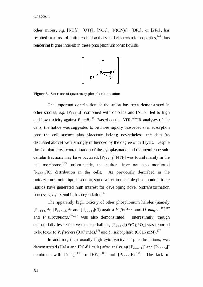
Chapter I
54
other anions, e.g. [NTf2]-, [OTf]
-, [NO3]
-, [N(CN)2]
-, [BF4]
-, or [PF6]
-, has
resulted in a loss of antimicrobial activity and electrostatic properties,241
thus
rendering higher interest in these phosphonium ionic liquids.
Figure 8. Structure of quaternary phosphonium cation.
The important contribution of the anion has been demonstrated in
other studies, e.g. [P6 6 6 14]+
combined with chloride and [NTf2]-
led to high
and low toxicity against E. coli.193
Based on the ATR-FTIR analyses of the
cells, the halide was suggested to be more rapidly biosorbed (i.e. adsorption
onto the cell surface plus bioaccumulation); nevertheless, the data (as
discussed above) were strongly influenced by the degree of cell lysis. Despite
the fact that cross-contamination of the cytoplasmatic and the membrane sub-
cellular fractions may have occurred, [P6 6 6 14][NTf2] was found mainly in the
cell membrane;193
unfortunately, the authors have not also monitored
[P6 6 6 14]Cl distribution in the cells. As previously described in the
imidazolium ionic liquids section, some water-immiscible phosphonium ionic
liquids have generated high interest for developing novel biotransformation
processes, e.g. xenobiotics-degradation.79
The apparently high toxicity of other phosphonium halides (namely
[P4 4 4 4]Br, [P6 6 6 14]Br and [P6 6 6 14]Cl) against V. fischeri and D. magna,173,177
and P. subcapitata,177,217
was also demonstrated. Interestingly, though
substantially less effective than the halides, [P2 4 4 4][(EtO)2PO2] was reported
to be toxic to V. fischeri (0.07 mM),173
and P. subcapitata (0.016 mM).177
In addition, their usually high cytotoxicity, despite the anions, was
demonstrated (HeLa and IPC-81 cells) after analysing [P4 4 4 14]+ and [P6 6 6 14]
+
combined with [NTf2]-160
or [BF4]-,161
and [P4 4 4 4]Br.161
The lack of

Chapter I
55
systematisation in these studies does not permit a conclusive rationalisation.
Nonetheless, [P6 6 6 14]+ combined with different anions, namely [PF6]
- and
[NTf2]-, displayed high and low cytotoxic activities against a broad diversity
of human cancer cell lines, respectively.159
The same cation combined with
[BF4]- exhibited very inconsistent behaviour, thought to be related to its low
solubility in water (and therefore lower membrane permeability potential).
Biodegradability of ionic liquids
The biodegradability of ionic liquids plays an important role in evaluating
their environmental impact. This subject has been recently discussed in an
excellent review by Coleman and Gathergood;242
hence only selected key
relevant facts will be discussed here. Most biodegradability assays are
defined under static laboratory conditions and, despite their importance, are
usually unrealistic, failing to reproduce the numerous abiotic and biotic
processes occurring in the environment. The biodegradability potential of a
chemical is often discussed accordingly to the OECD guidelines, meaning that
its rapid and complete mineralisation indicates that the chemical is readily
biodegradable, while its weak or extensive molecular cleavage suggests
primary or ultimate biodegradability potential, respectively.
The conscious design of chemicals and the rules of thumb for
delivering biodegradable chemicals, including ionic liquids, were excellently
reviewed by Boethling et al.243
Generalisations should be made cautiously,
yet it is generally accepted that several molecular features strongly enhance
biodegradability (not necessarily resulting in intermediate chemicals of lower
toxicity), such as the presence of esters, amides, hydroxyl, aldehyde,
carboxylic acid groups, or linear alkyl chains. Some examples can be found
in the available literature on ionic liquids, e.g. the incorporation of an ester
group in the side chain of several imidazolium170,244
or pyridinium245
ionic
liquids has significantly improved the primary biodegradability of the cation
(i.e. weak molecular cleavage). Those carrying alkyl chains of four carbon
atoms have been observed to be poorly biodegradable,169,170,246,247
even when

Chapter I
56
unsaturated groups (allyl or vinyl) or a sulfonate group were incorporated.248
Pham et al. proposed a degradation pathway for [C4mpy]Br through
hydroxylation of the side chain, even though that the intermediate chemical
product (3-methylpyridine) could not be conclusively identified.249
Nevertheless, pyridinium ionic liquids have, relative to the corresponding
imidazolium ones, higher biodegradability potential.250
For example,
[Cnmim]Br and [Cnmpy]Br (n = 6 or 8) were both degraded by an activated
sludge microbial community, with the longer alkyl chains reaching, after over
40 days, partial and total mineralisation, respectively; however, after 25 days
only [C8mpy]Br was shown to be fully mineralised (i.e. readily
biodegradable).247
More recently, the degradation pathway of [Cnmpy]Br (n
= 4, 6 or 8) was reported to involve unsaturation of the alkyl side chain and
hydroxylation of the aromatic ring,251
thus opposing that previously suggested
by Pham et al.249
Taken together, these studies suggested that there are
different possible degradation pathways that need to be considered, which are
ultimately defined by the metabolic capacities of the microbial community
and stand for a “regional” impact of the data.
Additionally, some phosphonium ionic liquids, namely
tricyclohexylphosphine- and trihexylphosphine-derived cations with various
ester side chains, and combined with different anions, were observed to be
very resistant to microbial attack.252
On the other hand, the cholinium cation
was reported to be readily biodegradable,243
and more recently, our group
demonstrated the high biodegradability potential of several cholinium
alkanoates.109
The complexity in designing more reliable bioassays for determining
the biodegradability potential of ionic liquids is partially due to critical
knowledge gaps on their potential uses and disposal methodologies, their
contamination levels and fate, and/or bioaccumulation factors in the
environment. Ionic liquids sorption onto sediments and soil will have a major
influence on their transport, reactivity (e.g. oxidation, hydrolysis and
photolysis), bioavailability and ultimately, biodegradability. Even if still

Chapter I
57
poorly investigated, there is an increasing interest in the behaviour of ionic
liquids in soils; however, up to now, most studies have focussed on the
imidazolium family, further demonstrating their high persistence and
ecotoxicological risk.253,254
Their sorption and desorption in soil were
observed to be ruled by ionic interactions;115,255,256
nonetheless, their
lipophilicity, the length of the side chains, and Coulombic interactions may
greatly influence their mobility.116
The first biodegradability assessment in
soil was conducted using several imidazolium-based ionic liquids, monitoring
solely CO2 evolution, which is not sufficiently informative to judge toxicity
and biodegradability.257
Nevertheless, mostly based on the high capacity of
several environmental fungal strains (which are commonly found in soil) to
tolerate high concentrations of ionic liquids,109,150,258,259
the role of filamentous
fungi in their biodegradation in soil is expected to be high.
The prevailing opinion is that the cation and anion will undergo
completely distinct fate mechanisms; however ion pairs possibly formed
between them and/or with other ions ubiquitously present in that environment
may be involved. The faster biodegradability of certain anions relative to the
cations has been demonstrated, e.g. alkylsulfates,137,170
saccharinate and
acesulfamates,216
and alkanoates,109
further suggesting that their degradation
followed distinctive pathways. This also stresses the importance of
considering both the cation and the anion for successful conscious design of
ionic liquids.
Molecular toxicity of ionic liquids
Some general considerations on molecular toxicity of ionic liquids
will now be analysed. While molecular toxicity falls under the umbrella of
toxicity, a direct extrapolation of any toxic behaviour based on in vitro
analyses of single molecules/reactions may lead to an erroneous conclusion.
Chemicals (or their biotransformation products) can display many modes of
molecular toxicity (i.e. molecular mechanisms whereby chemicals cause
toxicity) through interaction with specific biologic macromolecules, such as

Chapter I
58
proteins and DNA.260
Encouraged by this, Jastorff and co-workers have
included the concept of molecular toxicity as a standard component of their
ecotoxicity test battery, selecting the acetylcholinesterase assay.188,202,261,262
Preference was given to this enzyme (already mentioned in the sections for
pyridinium216
and quinolinium121
ionic liquids) because it plays an essential
role in the nervous system of almost all higher organisms, and the enzyme-
active site is highly conserved amongst organisms. By the extensive set of
IC50 data based on this assay (effective concentration scale based on a 50 %
inhibition response) presented by Ranke et al., it became apparent that the
chain length of the cation strongly influenced the activity of the enzyme,
probably due to the binding to the lipophilic active site; however, the effects
of additional methyl groups in the ring and of the anions were unclear.121
Even so, the fluorinated anions displayed consistently higher molecular
toxicity,121
probably due to their higher chemical instability. In addition, the
introduction of polar hydroxyl or ether functions in the side chains induces
reduced toxicity, relative to the alkyl chain.262
The introduction of an
additional hydroxyl group in position R3 in the pyridinium ring may have
prevented the interaction of the quaternary nitrogen with the active site of the
enzyme,216
since a reduction, by two orders of magnitude, was observed, e.g.
while comparing 1-alkoxymethyl-3-hydroxypyridinium216
and 1-alkyl-3-
methylpyridinium ionic liquids.261
The head groups in the cation played a
deciding role, mostly due to their ability to bind to the active site of the
enzyme or its gorge.121,262
Essentially, the large aromatic systems of 1-alkyl-
4-(dimethylamino)pyridinium and quinolinium, and the non-aromatic
morpholinium and tetrabutylammonium cations, were responsible for leading
to strong and weak inhibitions, respectively.262
In addition, the sterically
bulky structure and positive charged moiety shielded by the long alkyl chains
of the tetraalkylphosphonium and tetraalkylammonium ionic liquids have
probably reduced the interaction with the active centre of the enzyme, leading
to weak inhibition, yet the benzyl aromatic residue in the
benzyldecyldimethylammonium cation rationalises the deviant behaviour of

Chapter I
59
high toxicity. In two recent studies, imidazolium-based ionic liquids were
observed to inhibit acetylcholinesterase activity in living systems, i.e. rat
pheochromocytoma PC12 cells263
and earthworm.264
There are only a few studies on the molecular toxicity of ionic liquids
focussing on, instead of the acetylcholinesterase, other systems such as the
adenosine monophosphate (AMP) deaminase,265
or the cytochrome P450
assay.266,267
The activity of AMP deaminase in eukaryotes constitutes a
primary step in the regulation of intracellular adenine nucleotide pools,268
and
has therefore been proposed as a model enzyme for assessment of chemical
risk.269
Likewise, cytochrome P450, a family of monooxygenases ubiquitously
present in organisms, uses a wide spectrum of substrates, and are known to
play an important role in xenobiotic metabolism, catalysing hydroxylation of
C-H bonds.270
The activity of AMP deaminase was inhibited by several
[C4mim]+ ionic liquids and, amongst the tested anions, the chloride and 4-
tosylate exhibited slightly lower molecular toxicity than [BF4]- and [PF6]
-,
with IC50 values of 10 and 5 µM, respectively.265
Likewise, inhibition of
cytochrome P450 BM-3 activity in the presence of [Cnmim]Cl (n = 4, 6 or 8)
was stronger for the longer alkyl side chains.266
This agrees with that
previously observed while using the acetylcholinesterase assay,121
though
higher resistance of cytochrome P450 was noticed with IC50 values higher by at
least one order of magnitude. Surprisingly, the head group of the cation, and
especially its aromaticity, apparently has played a minor role in the ionic
liquid molecular toxicity against cytochrome P450, since imidazolium,
pyridinium and pyrrolidinium chlorides exhibited IC50 values of 148, 195 and
175 mM, respectively.266
In addition, the activity of the purified human
enzyme cytochrome P450 3A4 was significantly inhibited in the presence of
[Cnmim][BF4] (n = 1 or 4) and [C4py][BF4].267
On the other hand,
imidazolium ionic liquids apparently cannot be used as substrates by
cytochrome P450 BM-3, since the enzyme could not hydrolyse any of the
tested [Cnmim]Cl (monitored by NADPH consumption).266

Chapter I
60
Modes of toxicity of ionic liquids
The toxic effect of any chemical is an expression of the disorder of
particular metabolic pathways, and their potential modes of action include
disruption of membranes, mostly affecting membrane permeability and
transport proteins, enzyme inhibition, and DNA damage. A better knowledge
of chemical toxicity, e.g. in biocidal and drug design, is essential to identify
their modes of action, and ultimately to further advance towards their
conscious design.
The hydrophobic interaction between chemicals and biological
membranes results in non-specific toxicity, called baseline toxicity or
narcosis, which is mainly governed by solubility and partitioning;185
and,
though argued, there are possibly both polar and non-polar narcosis
mechanisms.184
However, chemicals can also interact with biological systems
through hydrogen or covalent bonding and charge transfer, with higher
(relative to baseline toxicity) effectiveness. Baseline toxicity has been
mentioned often in this review, by suggestion if not implicitly, as the basis for
the observed correlation between the ionic liquid lipophilicity (at the level of
both the cation and the anion) and toxicity. It becomes important to select
suitable descriptors for the lipophilicity of ionic liquids, such as the Kow and
the membrane/water partition coefficients.180
Even though the significance of
the Kow coefficients is still under debate, they are often used, mostly because
they can be retrieved by direct measurement, by reversed phase liquid
chromatography, or predicted by computational methods.161
Jastorff and co-
workers observed a good correlation between the cations‟ lipophilicity
(defined chromatographically) and the toxicity of the corresponding ionic
liquid (focussing on hydrophilic and chemically stable anions, such as the
halides).158,180
Likewise, the high correlation observed by us between the
antimicrobial activity of several cholinium alkanoates and the calculated
anion hydrophobicity log10(Kow),109
may be explained by the cation
hydrophilicity and benign nature. Significantly, even when a good correlation
between lipophilicity and toxicity was observed, this effect, probably due to

Chapter I
61
limited solubility and/or steric limitations, was not universal. There are
several examples where very lipophilic cations127-129,214,228
or anions,109
commonly carrying side chains longer than 10-12 carbon atoms, exhibited
lower toxicity than that predicted. In the latest study, the cholinium
dodecanoate exhibits lower toxicity than expected, probably due to its limited
solubility. In addition, in a very recent study focussing on imidazolium ionic
liquids, a strong relationship between antimicrobial efficacy, structure of the
cation and surface activity of the aqueous solutions containing [Cnmim]+
(n = 8 to 18) was reported.271
Liposomes (phospholipid vesicles) constitute a simple and attractive
methodology to analyse interactions of ionic liquids with cellular
membrane.230
Both [C8mim]+ and [NTf2]
- were demonstrated to cause severe
disruptions in a supported phospholipid bilayer, with [NTf2]-
leading to the
most extensive loss of lipids.272
The higher toxicity of the latter was
tentatively explained using molecular simulations, predicting that [NTf2]- was
readily incorporated into the cholesterol bilayer, strongly limiting the
adsorption of the cation and, consequently, the insertion of the butyl chain of
[C4mim]+ into the bilayer.
273
As an attempt to rationalise the toxicity mode of ionic liquids, some
processes have been proposed to play a key role. Nevertheless, one should
bear in mind the high complexity of living organisms and the numerous
physical and biochemical processes occurring simultaneously. The important
observations on the critical alterations detected in fungal metabolism after
exposure to sub-lethal concentrations of some hydrophilic ionic liquids150
may
suggest a specific mode of toxicity.
Quantitative structure-activity relationships (QSAR) for ionic liquids
Noteworthy efforts are increasingly being reported in order to define
computational methods which can be used to reasonably predict ionic liquid
toxicity. There is no doubt that computer modelling will, as more ecotoxicity
data becomes available, develop further. Couling et al. applied QSAR

Chapter I
62
modelling to their experimental data on the aquatic toxicity of twenty-five
ionic liquids (containing imidazolium, pyridinium, tetraalkylammonium, and
tetraalkylphosphonium cations) (correlation with R2 = 0.78-0.88).
173 Based on
the derived descriptors, calculated at a low semi-empirical computational
level, certain generalised predictions could be raised: toxicity is mainly
governed by the cation, with the aromatics displaying higher toxicity than
non-aromatic ones, and increasing with longer substituted alkyl chains.
Additionally, in the aromatic ring, an increased number of nitrogen atoms
leads to higher toxicity, while its methylation decreases it. A different
approach was used by Luis et al., since they designed an algorithm based on
group contribution methods to estimate the aquatic toxicity of forty-three
imidazolium, pyridinium and pyrrolidinium ionic liquids (correlation with R2
= 0.92).274
Based on this modelling, some new generalisations were
suggested, such as a toxic ranking of the head groups (pyrrolidinium <
imidazolium < pyridinium), while the anion effect remained less predictable,
though some (bromide, dicyanamide and ethylsulfate) were considered highly
toxic. Garcia-Lorenzo et al. built a QSAR model, according to the
Topological Sub-Structural Molecular Design (TOSS-MODE) approach,
which uses graph-based molecular descriptors (based on spectral moments) to
predict the cytotoxicity of fifteen imidazolium-derived ionic liquids in CaCo-
2 cells (correlation with R2 = 0.98). Different mathematical models were
proposed by Torrecilla et al.,275
using empirical formulae (elemental
composition) and molecular weights of 153 ionic liquids (ammonium,
imidazolium, morpholinium, phosphonium, piperidinium, pyridinium,
pyrrolidinium and quinolinium salts) available in the open literature121
to
estimate their cytotoxicity (IPC-81 cells) and molecular toxicity
(acetylcholinesterase) by neural network (NN) models (correlation with R2 =
0.98 and 0.97, respectively). Recently, COSMO-RS (Conductor like
Screening Model for Realistic Solvents) molecular descriptors, which related
cytotoxicity to the polar charge distribution of the cations and the anions,
were used (non-linear neural network) (96 formulations in IPC-81 cells,121

Chapter I
63
correlation with R2 = 0.98): this leads to the highly significant conclusion that
the cytotoxicity of ionic liquids cannot be systematically estimated by a
summation of the independent contributions of the intrinsic toxicity of the
cation and anion.275,276
More recently, Alvarez-Guerra et al. applied PLS-DA
(Partial Least Squares-Discriminant Analysis) model on the set of EC50 values
for 131 ionic liquids in V. fischeri.277
Toxicity of ionic liquids was classified
as positive (high) or negative (low) relatively to toluene or chloroform, while
the contribution of each ion to this classification was calculated.
General considerations
In the light of the studies referred to above, it is clear that the
numerous formulations of ionic liquids available provide a great pool of, and
impetus for, many commercial applications, but not without significant
toxicological and environmental concerns. The vast majority of toxicological
studies on ionic liquids, available up to the present date, have focussed on
imidazolium ionic liquids. In addition, frequently, the ionic liquids “selected”
for study tackled under a common assay were randomly chosen. Despite the
scientific weight of these studies, the lack of systematisation (e.g. monitoring
the effect of defined structural alterations in a specific head group) means that
it is impossible at the moment to achieve a holistic analysis, which weakens
conclusions and devalues the predictive algorithms under development.
Furthermore, the selected bioassay, despite its relevance, appears to
be sometimes subjective. Ecotoxicity examination should be based on a
realistic analysis of the risk of exposure, accounting predicatively for which
environmental compartments are most likely to be involved, yet it is
necessarily regionally-oriented. Legislation demands and standardised tests
should be kept as a priority, engaging models of different complexity.
However, one may doubt their suitability, since testing a vast number of
organisms and the diversity of all available formulations is costly and
irrational. Indeed, testing the most sensitive species and determining the
chemical lethal concentration, will provide useful information, but this may

Chapter I
64
lack a real ecological meaning, as sub-lethal concentrations significantly
affect the ecology of the exposed niche (e.g. species interactions and food web
balance). In addition, the environmental persistence of any chemical should
be taken as one of the most critical ecotoxicological parameters (advancing
their conscious design), and putatively their mitigation will involve the
tolerant species able to metabolise it. The most frequently used toxicity tests
establish median effective concentrations (inhibitory or lethal), which are
useful parameters for comparison of different chemicals, but it is obvious that
further standardisation should be pursued. The conditions selected, e.g. media
composition, temperature, light/dark cycle, and more importantly the
exposure time, have generated a considerable volume of data on ionic liquid
toxicity that cannot be directly compared. Furthermore, straightforward
comparisons were not easy because of the use of diverse end-points and units.
The European Community regulation on chemicals and their safe use
- REACH (Registration, Evaluation, Authorisation and Restriction of
CHemical substances)278
- aims to increase the awareness of the industry on
hazards and risk management. REACH registration, in force since 2007, is
mandatory for any chemical produced in the quantity over one tonne per year.
Although it is being criticised for its ever-increasing cost and the number of
animals employed in testing,279
it undoubtedly provides a meaningful, and
necessary, framework to raise human and environmental safety. Currently,
only the ionic liquids which have already found application on industrial scale
are undergoing REACH registration, such as [C2mim]X (X = Cl-, [C2SO4]
-,
[C1SO3]-, [O2CMe]
- and [NTf2]
-) and [C4mim]Cl,
274 but there are no doubts
that this number will continuously increase. They have received the
classification of substances, i.e. as salts, which assumes that in the water/soil
matrix, the cation and the anion will behave as independent chemical entities,
displaying distinct toxicity and biodegradability potential. But, as noted
above, it appears (irrespective of speculation as to the cause) that the
cytotoxicity of ionic liquids cannot be systematically estimated by a
summation of the independent effects of the cation and anion.275,276

Chapter I
65
Moreover, it should also be noted that, to date, mixtures (binary or ternary) of
ionic liquids have been rarely investigated.209
In conclusion, one should recognise that the ionic liquid scientific
community needs to increase the public awareness of the immense diversity
of possible formulations and properties included in such a classification. For
example, despite the significance of the data, especially as the first study
reporting aquatic toxicity,219
the attention attracted by the headline “Warning
shot for green chemistry” in Nature,280
or “Ionic liquids toxic to fish” in
Dalton Transactions,281
was almost certainly counterproductive and
misleading, distorting as it did the excellent data reported by the original
authors, where thirteen out of fifteen tested ionic liquids were shown to have
LC50 values above 100 mg l-1
after 96 h of exposure. In contrast, unwarranted
claims that ionic liquids, as a class of materials, are “green” (and many papers
erroneously claim this in their introductory sentences) are just as damaging to
the field as claims that ionic liquids are “toxic”. With well over one million
simple ionic liquids,282
arguing from the specific to the generic is both
misguided and intellectually dishonest. It should be recognised that sweeping
generalisations do not favour our community, and it is the community itself
that needs to protect ionic liquids indubitable broad utility, obviously by
continuously following our duty to attain higher safety by their conscious
design.
Acknowledgements
M. P. is grateful to Fundação para a Ciência e a Tecnologia (FC&T) for the
fellowship SFRH/BD/31451/2006. The work was partially supported by a
grant from Iceland, Liechtenstein and Norway through the EEA financial
mechanism (Project PT015).

Chapter I
66
1.3.2. Whole-cell biocatalysis with ionic liquids
Biocatalysis is an elegant strategy for the highly selective and efficient
production of fine chemicals. Ionic liquids‟ application in biocatalysis has
been studied widely, and is excellently reviewed by van Rantwijk and
Sheldon.95
Most of these studies exploited ionic liquids containing [BF4]- and
[PF6]- anions, although their hydrolysis
55 was previously shown to drastically
affect chemical and biochemical reactions. The majority of studies to date
have considered enzymatic reactions, with some notable exceptions on
whole-cell biocatalysis. So far analysis of ionic liquid biocompatibility is
concerned exclusively with the damage caused to the plasma membrane of
the whole-cell biocatalysts.283
Single phase or biphasic systems can be
considered. Whilst in single phase cells are grown in contact with water-
miscible ionic liquids,284
in biphasic systems they grow in one of the phases
whilst the cell free layer contains water-immiscible ionic liquids.
Representative examples of biphasic systems have permitted asymmetric
reduction of 4-chloroacetophenone by Lactobacillus kefir146
or Escherichia
coli,96
and phenol degradation by xenobiotic-degrading bacteria.79
In these
systems, partition of the ionic liquid phase prevents accumulation of a toxic
product,97
or substrate,79,96,146
in the aqueous growth medium phase. Low
amounts of the ionic liquid will also be solubilised in the aqueous phase,
hence biocompatibility of the biocatalyst must be ensured.
The most recent advances on ionic liquids conscious design
(e.g. Chapter III) strongly indicate a growing interest in their application in
novel biotechnological processes for industrial, pharmacological and
environmental usage.

Chapter I
67
References
1. Baldauf, S. L. and Palmer, J. D., Animals and fungi are each other's
closest relatives: Congruent evidence from multiple proteins. Proc.
Natl. Acad. Sci. USA, 1993, 90, 11558-11562.
2. Whittaker, R. H., New concepts of kingdoms of organisms. Science,
1969, 163, 150-160.
3. Cavalier-Smith, T., Only six kingdoms of life. Proc. R. Soc. London,
Ser. B, 2004, 271, 1251-1262.
4. Patterson, D. J. and Sogin, M. L., Eukaryote origins and protistan
diversity, Hartman, H. and Matsumo, K., Eds. The Origin and
Evolution of the Cell. Conference on the Origin and Evolution of
Prokaryotic and Eukaryotic Cells, New Jersey, 1993, World Scientific
Pub Co. Inc., 13-46.
5. Carlile, M., Watkinson, S. and Gooday, G., The fungi. 2nd ed. Vol. 1.
2001, Amsterdam, Elsevier Academic Press
6. Crous, P. W., Fungal Biodiversity (CBS Laboratory Manual Series).
1st ed, Crous, P. W., Verkley, G. J. M., Groenewald, J. Z., and
Samson, R. A., Eds. 2009, Utrecht, CBS-KNAW Fungal Biodiversity
Centre
7. Galagan, J. E., et al., The genome sequence of the filamentous fungus
Neurospora crassa. Nature, 2003, 422, 859-868.
8. Nierman, W. C., et al., Genomic sequence of the pathogenic and
allergenic filamentous fungus Aspergillus fumigatus. Nature, 2005,
438, 1151-1156.
9. Galagan, J. E., et al., Sequencing of Aspergillus nidulans and
comparative analysis with A. fumigatus and A. oryzae. Nature, 2005,
438, 1105-1115.
10. Adams, T. H., Wieser, J. K. and Yu, J. H., Asexual sporulation in
Aspergillus nidulans. Microbiol. Mol. Biol. Rev., 1998, 62, 35-+.
11. Papagianni, M., Fungal morphology and metabolite production in
submerged mycelial processes. Biotechnol. Adv., 2004, 22, 189-259.
12. de Groot, P. W. J., Brandt, B. W., Horiuchi, H., Ram, A. F. J., de
Koster, C. G. and Klis, F. M., Comprehensive genomic analysis of
cell wall genes in Aspergillus nidulans. Fungal Genet. Biol., 2009, 46,
S72-S81.
13. Selitrennikoff, C. P., Antifungal proteins. Appl. Environ. Microbiol.,
2001, 67, 2883-2894.
14. Bowman, S. M. and Free, S. J., The structure and synthesis of the
fungal cell wall. Bioessays, 2006, 28, 799-808.
15. Wosten, H. A. B., Hydrophobins: Multipurpose proteins. Annu. Rev.
Microbiol., 2001, 55, 625-646.
16. Jacobson, E. S., Pathogenic roles for fungal melanins. Clin.
Microbiol. Rev., 2000, 13, 708-+.

Chapter I
68
17. Bourett, T. M., James, S. W. and Howard, R. J., The Endomembrane
system of the fungal cell, in The Mycota VIII, Biology of the Fungal
Cell, Howard, R. J. and Gow, N. A. R., Eds. 2007, Springer-Verlag:
Berlin Heidelberg. p. 1-47.
18. Gooday, G. W., The dynamics of hyphal growth. Mycol. Res., 1995,
99, 385-394.
19. Wessels, J. G. H., Wall growth, protein excretion and morphogenesis
in fungi. New Phytol., 1993, 123, 397-413.
20. Fontaine, T., Hartland, R. P., Beauvais, A., Diaquin, M. and Latge, J.
P., Purification and characterization of an endo-1,3-beta-glucanase
from Aspergillus fumigatus. Eur. J. Biochem., 1997, 243, 315-321.
21. Money, N. P., On the origin and functions of hyphal walls and turgor
pressure. Mycol. Res., 1999, 103, 1360-1360.
22. Ebbole, D. J., The conidium, in Cellular and Molecular Biology of
Filamentous Fungi, Borkovich, K. A. and Ebbole, D. J., Eds. 2010,
ASM Press: Washington D.C.
23. Ni, M., Gao, N., Kwon, N.-J., Shin, K.-S. and Yu, J.-H., Regulation of
Aspergillus conidiation, in Cellular and Molecular Biology of
Filamentous Fungi, Borkovich, K. A. and Ebbole, D. J., Eds. 2010,
ASM Press: Washington D.C.
24. Shary, S., Ralph, S. A. and Hammel, K. E., New insights into the
ligninolytic capability of a wood decay ascomycete. Appl. Environ.
Microbiol., 2007, 73, 6691-6694.
25. Karlsson, J., Momcilovic, D., Wittgren, B., Schülein, M., Tjerneld, F.
and Brinkmalm, G., Enzymatic degradation of carboxymethyl
cellulose hydrolyzed by the endoglucanases Cel5A, Cel7B, and
Cel45A from Humicola insolens and Cel7B, Cel12A and Cel45Acore
from Trichoderma reesei. Biopolymers, 2002, 63, 32-40.
26. Pinedo-Rivilla, C., Aleu, J. and Collado, I. G., Pollutants
Biodegradation by Fungi. Curr. Org. Chem., 2009, 13, 1194-1214.
27. Kapoor, A. and Viraraghavan, T., Fungal biosorption - An alternative
treatment option for heavy metal bearing wastewaters: A review.
Bioresour. Technol., 1995, 53, 195-206.
28. Jarosz-Wilkołazka, A., Kochmańska-Rdest, J., Malarcżyk, E.,
Wardas, W. and Leonowicz, A., Fungi and their ability to decolourize
azo and anthraquinonic dyes. Enzyme Microb. Technol., 2002, 30,
566-572.
29. Potin, O., Rafin, C. and Veignie, E., Bioremediation of an aged
polycyclic aromatic hydrocarbons (PAHs)-contaminated soil by
filamentous fungi isolated from the soil. Int. Biodeterior. Biodegrad.,
2004, 54, 45-52.
30. Carvalho, M. B., Martins, I., Leitão, M. C., Garcia, H., Rodrigues, C.,
San Romão, M. V., McLellan, I., Hursthouse, A. and Silva Pereira,
C., Screening pentachlorophenol degradation ability by environmental
fungal strains belonging to the phyla Ascomycota and Zygomycota. J.
Ind. Microbiol. Biotechnol., 2009, 36, 1249-1256.

Chapter I
69
31. Kis-Papo, T., Oren, A., Wasser, S. P. and Nevo, E., Survival of
filamentous fungi in hypersaline dead sea water. Microb. Ecol., 2003,
45, 183-190.
32. Kernaghan, G. and Harper, K. A., Community structure of
ectomycorrhizal fungi across an alpine/subalpine ecotone. Ecography,
2001, 24, 181-188.
33. Raghukumar, C., Marine fungal biotechnology: an ecological
perspective. Fungal Divers., 2008, 31, 19-35.
34. Pascual, S., Melgarejo, P. and Magan, N., Accumulation of
compatible solutes in Penicillium frequentans grown at reduced water
activity and biocontrol of Monilinia laxa. Biocontrol Sci. Technol.,
2000, 10, 71-80.
35. Park, Y. I. and Gander, J. E., Choline derivatives involved in
osmotolerance of Penicillium fellutanum. Appl. Environ. Microbiol.,
1998, 64, 273-278.
36. Turk, M., Montiel, V., Žigon, D., Plemenitaš, A. and Ramos, J.,
Plasma membrane composition of Debaryomyces hansenii adapts to
changes in pH and external salinity. Microbiology, 2007, 153, 3586-
3592.
37. Anastas, P. T. and Warner, J. C., Green chemistry: Theory and
practice. 1998, New York, Oxford University Press
38. Warner, J. C., Cannon, A. S. and Dye, K. M., Green chemistry.
Environ. Impact Assess. Rev., 2004, 24, 775-799.
39. Whim, B. P. and Johnson, P. G., eds. Directory of Solvents. 1996,
Blackie Academic & Professional: London.
40. Sheldon, R. A., Atom efficiency and catalysis in organic synthesis.
Pure Appl. Chem., 2000, 72, 1233-1246.
41. Horváth, I. T., Solvents from nature. Green Chem., 2008, 10, 1024-
1028.
42. Jessop, P. G., Searching for green solvents. Green Chem., 2011.
43. Tokuda, H., Hayamizu, K., Ishii, K., Abu Bin Hasan Susan, M. and
Watanabe, M., Physicochemical properties and structures of room
temperature ionic liquids. 1. Variation of anionic species. J. Phys.
Chem. B, 2004, 108, 16593-16600.
44. Krieger, N., Bhatnagar, T., Baratti, J. C., Baron, A. M., de Lima, V.
M. and Mitchell, D., Non-aqueous biocatalysis in heterogeneous
solvent systems. Food Technol. Biotechnol., 2004, 42, 279-286.
45. McHugh, M. and Krukonis, V., Supercritical fluid extraction. 2nd
ed.
1994, Stoneham, Butterworth-Heinemann
46. Nunes da Ponte, M. L., Da Silva Lopes, J. A., Najdanovic-Visak, V.,
Manic, M., Cardoso Mesquita, A. C., Moreira da Silva, R. P. and
Sollarri Allegro, I. M., World Pat., 093273, 2010.
47. Ludwig, R., Ionic liquids - Revolutionary potential for chemistry?
ChemPhysChem, 2006, 7, 1415-1416.
48. Earle, M. and Seddon, K. R., Ionic liquids. Green solvents for the
future. Pure Appl. Chem., 2000, 72, 1391-1398.

Chapter I
70
49. Wilkes, J. S., Levisky, J. A., Wilson, R. A. and Hussey, C. L.,
Dialkylimidazolium chloroaluminate melts - a new class of room-
temperature ionic liquids for electrochemistry, spectrocopy, and
synthesis. Inorg. Chem., 1982, 21, 1263-1264.
50. Wilkes, J. S. and Zaworotko, M. J., Air and water stable 1-ethyl-3-
methylimidazolium based ionic liquids. J. Chem. Soc., Chem.
Commun., 1992, 965-967.
51. MacFarlane, D. R. and Seddon, K. R., Ionic liquids - Progress on the
fundamental issues. Aust. J. Chem., 2007, 60, 3-5.
52. Ohno, H., Functional design of ionic liquids. Bull. Chem. Soc. Jpn.,
2006, 79, 1665-1680.
53. Earle, M. J., Esperança, J. M. S. S., Gilea, M. A., Lopes, J. N. C.,
Rebelo, L. P. N., Magee, J. W., Seddon, K. R. and Widegren, J. A.,
The distillation and volatility of ionic liquids. Nature, 2006, 439, 831-
834.
54. Smiglak, M., Reichert, W. M., Holbrey, J. D., Wilkes, J. S., Sun, L.
Y., Thrasher, J. S., Kirichenko, K., Singh, S., Katritzky, A. R. and
Rogers, R. D., Combustible ionic liquids by design: is laboratory
safety another ionic liquid myth? Chem. Commun., 2006 (24), 2554-
2556.
55. Huddleston, J. G., Visser, A. E., Reichert, W. M., Willauer, H. D.,
Broker, G. A. and Rogers, R. D., Characterization and comparison of
hydrophilic and hydrophobic room temperature ionic liquids
incorporating the imidazolium cation. Green Chem., 2001, 3, 156-
164.
56. Kroon, M. C., Buijs, W., Peters, C. J. and Witkamp, G. J., Quantum
chemical aided prediction of the thermal decomposition mechanisms
and temperatures of ionic liquids. Thermochim. Acta, 2007, 465, 40-
47.
57. Wu, B., Liu, W., Zhang, Y. and Wang, H., Do we understand the
recyclability of ionic liquids? Chem.–Eur. J., 2009, 15, 1804-1810.
58. Varma, R. S. and Namboodiri, V. V., An expeditious solvent-free
route to ionic liquids using microwaves. Chem. Commun., 2001 (7),
643-644.
59. Hirao, M., Sugimoto, H. and Ohno, H., Preparation of novel room-
temperature molten salts by neutralization of amines. J. Electrochem.
Soc., 2000, 147, 4168-4172.
60. Stark, A., Behrend, P., Braun, O., Müller, A., Ranke, J., Ondruschka,
B. and Jastorff, B., Purity specification methods for ionic liquids.
Green Chem., 2008, 10, 1152-1161.
61. Stark, A. and Seddon, K. R., Ionic Liquids, in Kirk-Othmer
Encyclopaedia of Chemical Technology, Seidel, A., Ed. 2007, John
Wiley & Sons, Inc.: New Jersey. p. 836-920.
62. Imperato, G., König, B. and Chiappe, C., Ionic green solvents from
renewable resources. Eur. J. Org. Chem., 2007, 1049-1058.

Chapter I
71
63. Bao, W. L., Wang, Z. M. and Li, Y. X., Synthesis of chiral ionic
liquids from natural amino acids. J. Org. Chem., 2003, 68, 591-593.
64. Handy, S. T., Okello, M. and Dickenson, G., Solvents from
biorenewable sources: Ionic liquids based on fructose. Org. Lett.,
2003, 5, 2513-2515.
65. Fukaya, Y., Iizuka, Y., Sekikawa, K. and Ohno, H., Bio ionic liquids:
room temperature ionic liquids composed wholly of biomaterials.
Green Chem., 2007, 9, 1155-1157.
66. Rebelo, L. P. N., Lopes, J. N. C., Esperança, J. M. S. S., Guedes, H. J.
R., Łachwa, J., Najdanovic-Visak, V. and Visak, Z. P., Accounting
for the unique, doubly dual nature of ionic liquids from a molecular
thermodynamic, and modeling standpoint. Accounts Chem. Res.,
2007, 40, 1114-1121.
67. Canongia Lopes, J. N. A. and Padua, A. A. H., Nanostructural
organization in ionic liquids. J. Phys. Chem. B, 2006, 110, 3330-5.
68. Welton, T., Room-temperature ionic liquids. Solvents for synthesis
and catalysis. Chem. Rev., 1999, 99, 2071-2083.
69. Earle, M. J., Katdare, S. P. and Seddon, K. R., Paradigm confirmed:
The first use of ionic liquids to dramatically influence the outcome of
chemical reactions. Org. Lett., 2004, 6, 707-710.
70. Wasserscheid, P. and Keim, W., Ionic liquids - New "solutions" for
transition metal catalysis. Angew. Chem.-Int. Edit., 2000, 39, 3772-
3789.
71. Endres, F. and El Abedin, S. Z., Air and water stable ionic liquids in
physical chemistry. Phys. Chem. Chem. Phys., 2006, 8, 2101-2116.
72. Kosmulski, M., Gustafsson, J. and Rosenholm, J. B., Thermal
stability of low temperature ionic liquids revisited. Thermochim. Acta,
2004, 412, 47-53.
73. Holbrey, J. D. and Seddon, K. R., Ionic Liquids. Clean Prod.
Process., 1999, 1, 223-236.
74. Plechkova, N. V. and Seddon, K. R., Applications of ionic liquids in
the chemical industry. Chem. Soc. Rev., 2008, 37, 123-150.
75. Cole, A. C., Jensen, J. L., Ntai, I., Tran, K. L. T., Weaver, K. J.,
Forbes, D. C. and Davis, J. H., Novel brønsted acidic ionic liquids
and their use as dual solvent-catalysts. J. Am. Chem. Soc., 2002, 124,
5962-5963.
76. Sakaebe, H., Matsumoto, H. and Tatsumi, K., Application of room
temperature ionic liquids to Li batteries. Electrochim. Acta, 2007, 53,
1048-1054.
77. Armand, M., Endres, F., MacFarlane, D. R., Ohno, H. and Scrosati,
B., Ionic-liquid materials for the electrochemical challenges of the
future. Nat. Mater., 2009, 8, 621-629.
78. Cull, S. G., Holbrey, J. D., Vargas-Mora, V., Seddon, K. R. and Lye,
G. J., Room-temperature ionic liquids as replacements for organic
solvents in multiphase bioprocess operations. Biotechnol. Bioeng.,
2000, 69, 227-233.

Chapter I
72
79. Baumann, M. D., Daugulis, A. J. and Jessop, P. G., Phosphonium
ionic liquids for degradation of phenol in a two-phase partitioning
bioreactor. Appl. Microbiol. Biotechnol., 2005, 67, 131-137.
80. Ibanez, J. G., Hernandez-Esparza, M., Doria-Serrano, C., Fregoso-
Infante, A. and Singh, M. M., Environmental chemistry:
fundamentals. 1st ed. 2007, New York, Springer
81. Marcus, Y. and Hefter, G., Ion pairing. Chem. Rev., 2006, 106, 4585-
4621.
82. Stolte, S., Arning, J., Bottin-Weber, U., Matzke, M., Stock, F., Thiele,
K., Uerdingen, M., Welz-Biermann, U., Jastorff, B. and Ranke, J.,
Anion effects on the cytotoxicity of ionic liquids. Green Chem., 2006,
8, 621-629.
83. Ranke, J., Mölter, K., Stock, F., Bottin-Weber, U., Poczobutt, J.,
Hoffmann, J., Ondruschka, B., Filser, J. and Jastorff, B., Biological
effects of imidazolium ionic liquids with varying chain lengths in
acute Vibrio fischeri and WST-1 cell viability assays. Ecotox.
Environ. Safe., 2004, 58, 396-404.
84. Latała, A., Nędzi, M. and Stepnowski, P., Toxicity of imidazolium
ionic liquids towards algae. Influence of salinity variations. Green
Chem., 2010, 12, 60-64.
85. Bowers, J., Butts, C. P., Martin, P. J., Vergara-Gutierrez, M. C. and
Heenan, R. K., Aggregation behavior of aqueous solutions of ionic
liquids. Langmuir, 2004, 20, 2191-2198.
86. Blesic, M., Marques, M. H., Plechkova, N. V., Seddon, K. R., Rebelo,
L. P. N. and Lopes, A., Self-aggregation of ionic liquids: micelle
formation in aqueous solution. Green Chem., 2007, 9, 481-490.
87. Walden, P., Ueber die Molekulargrösse und elektrische Leitfähigkeit
einiger geschmolzenen salze. Bull. Acad. Impér. Sci. St. Pétersbourg,
1914, 8, 405-422.
88. Deetlefs, M. and Seddon, K. R., Ionic liquids: fact and fiction. Chim.
Oggi-Chem. Today, 2006, 24, 16-23.
89. Plechkova, N. V. and Seddon, K. R., Ionic liquids: "designer"
solvents for green chemistry, in Methods and Reagents for Green
Chemistry: An Introduction, Tundo, P., Perosa, A., and Zecchini, F.,
Eds. 2007, Wiley: New York. p. 105-130.
90. Maase, M. and Massonne, K., Biphasic acid scavenging utilizing
ionic liquids: The first commercial process with ionic liquids, in Ionic
Liquids IIIB: Fundamentals, Progress, Challenges, and Opportunities
- Transformations and Processes, Rogers, R. D. and Seddon, K. R.,
Eds. 2005, American Chemical Society: Washington D.C. p. 126-132.
91. Chauvin, Y., Olefin metathesis: The early days (Nobel lecture).
Angew. Chem.-Int. Edit., 2006, 45, 3740-3747.
92. Adler, R., Reports on Science and Technology, 2006, Linde
Technology, Wiesbaden.
93. Teramoto, D., Yokoyama, R., Kagawa, H., Sada, T. and Ogata, N., A
novel ionic liquid-polymer electrolyte for the advanced lithium-ion

Chapter I
73
battery, in Molten Salts and Ionic Liquids: Never the Twain?, Gaune
Escard, M. and Seddon, K. R., Eds. 2010, Wiley: New York. p. 367-
388.
94. G24 Innovations. www.g24i.com
95. van Rantwijk, F. and Sheldon, R. A., Biocatalysis in ionic liquids.
Chem. Rev., 2007, 107, 2757-2785.
96. Bräutigam, S., Bringer-Meyer, S. and Weuster-Botz, D., Asymmetric
whole cell biotransformations in biphasic ionic liquid/water-systems
by use of recombinant Escherichia coli with intracellular cofactor
regeneration. Tetrahedron: Asymmetry, 2007, 18, 1883-1887.
97. Pfruender, H., Jones, R. and Weuster-Botz, D., Water immiscible
ionic liquids as solvents for whole cell biocatalysis. J. Biotechnol.,
2006, 124, 182-190.
98. Fujita, K., MacFarlane, D. R. and Forsyth, M., Protein solubilising
and stabilising ionic liquids. Chem. Commun., 2005 (38), 4804-4806.
99. Byrne, N., Wang, L. M., Belieres, J. P. and Angell, C. A., Reversible
folding-unfolding, aggregation protection, and multi-year
stabilization, in high concentration protein solutions, using ionic
liquids. Chem. Commun., 2007 (26), 2714-2716.
100. Vrikkis, R. M., Fraser, K. J., Fujita, K., MacFarlane, D. R. and Elliott,
G. D., Biocompatible ionic liquids: A new approach for stabilizing
proteins in liquid formulation. J. Biomech. Eng.-Trans. ASME, 2009,
131, 074514-074518.
101. Hough, W. L., Smiglak, M., Rodríguez, H., Swatloski, R. P., Spear, S.
K., Daly, D. T., Pernak, J., Grisel, J. E., Carliss, R. D., Soutullo, M.
D., Davis, J. H. and Rogers, R. D., The third evolution of ionic
liquids: active pharmaceutical ingredients. New J. Chem., 2007, 31,
1429-1436.
102. Hough, W. L. and Rogers, R. D., Ionic liquids then and now: From
solvents to materials to active pharmaceutical ingredients. Bull.
Chem. Soc. Jpn., 2007, 80, 2262-2269.
103. Bica, K., Rijksen, C., Nieuwenhuyzen, M. and Rogers, R. D., In
search of pure liquid salt forms of aspirin: ionic liquid approaches
with acetylsalicylic acid and salicylic acid. Phys. Chem. Chem. Phys.,
2010, 12, 2011 - 2017.
104. Stoimenovski, J., MacFarlane, D. R., Bica, K. and Rogers, R. D.,
Crystalline vs. Ionic Liquid Forms of Active Pharmaceutical
Ingredients: A Position Paper. Pharm. Res., 2010, 27, 521-526.
105. Anastas, P. T. and Kirchhoff, M. M., Origins, current status, and
future challenges of green chemistry. Accounts Chem. Res., 2002, 35,
686-694.
106. Poliakoff, M., Fitzpatrick, J. M., Farren, T. R. and Anastas, P. T.,
Green chemistry: Science and politics of change. Science, 2002, 297,
807-810.

Chapter I
74
107. Deetlefs, M. and Seddon, K. R., Assessing the greeness of some
typical laboratory ionic liquids preparations. Green Chem., 2010, 12,
17-30.
108. Fukumoto, K., Yoshizawa, M. and Ohno, H., Room temperature ionic
liquids from 20 natural amino acids. J. Am. Chem. Soc., 2005, 127,
2398-2399.
109. Petkovic, M., Ferguson, J. L., Gunaratne, H. Q. N., Ferreira, R.,
Leitão, M. C., Seddon, K. R., Rebelo, L. P. N. and Silva Pereira, C.,
Novel biocompatible cholinium-based ionic liquids - toxicity and
biodegradability. Green Chem., 2010, 12, 643-649.
110. Carter, E. B., Culver, S. L., Fox, P. A., Goode, R. D., Ntai, I., Tickell,
M. D., Traylor, R. K., Hoffman, N. W. and Davis, J. H., Sweet
success: ionic liquids derived from non-nutritive sweeteners. Chem.
Commun., 2004 (6), 630-631.
111. Poletti, L., Chiappe, C., Lay, L., Pieraccini, D., Polito, L. and Russo,
G., Glucose-derived ionic liquids: exploring low-cost sources for
novel chiral solvents. Green Chem., 2007, 9, 337-341.
112. Seddon, K. R., Stark, A. and Torres, M.-J., Influence of chloride,
water, and organic solvents on the physical properties of ionic liquids.
Pure Appl. Chem., 2000, 72, 2275-2287.
113. Marsh, K. N., Brennecke, J. F., Chirico, R. D., Frenkel, M., Heintz,
A., Magee, J. W., Peters, C. J., Rebelo, L. P. N. and Seddon, K. R.,
Thermodynamic and thermophysical properties of the reference ionic
liquid: 1-hexyl-3-methylimidazoilum
bis[(trifluoromethyl)sulfonyl]amide (including mixtures), Part 1.
Experimental methods and results (IUPAC Technical Report). Pure
Appl. Chem., 2009, 81, 781-790.
114. Margulis, L. and Schwartz, K. V., Five kingdoms: an illustrated guide
to the phyla of life on earth. 3rd ed, Freeman, W. H., Ed. 1998, New
York, Elsevier
115. Matzke, M., Thiele, K., Müller, A. and Filser, J., Sorption and
desorption of imidazolium based ionic liquids in different soil types.
Chemosphere, 2009, 74, 568-574.
116. Mrozik, W., Jungnickel, C., Ciborowski, T., Pitner, W. R., Kumirska,
J., Kaczyński, Z. and Stepnowski, P., Predicting mobility of
alkylimidazolium ionic liquids in soils. J. Soils Sediments, 2009, 9,
237-245.
117. Ranke, J. and Jastorff, B., Multidimensional risk analysis of
antifouling biocides. Environ. Sci. Pollut. Res., 2000, 7, 105-114.
118. Jastorff, B., Störmann, R., Ranke, J., Mölter, K., Stock, F.,
Oberheitmann, B., Hoffmann, W., Hoffmann, J., Nuchter, M.,
Ondruschka, B. and Filser, J., How hazardous are ionic liquids?
Structure-activity relationships and biological testing as important
elements for sustainability evaluation. Green Chem., 2003, 5, 136-
142.

Chapter I
75
119. Dechema, "BATIL (Biodegradability and Toxicity of Ionic Liquids),
Berlin, 6th-9
th May, 2007", http://events.dechema.de/batil2.html; G.
Adamová, M. J. Earle, J. L. Ferguson, M. A. Gìlea and K. R. Seddon,
“Report on the 2nd
international conference on Biodegradability And
Toxicity of Ionic Liquids (BATIL2)”, March 2010,
http://www.rsc.org/Publishing/Journals/gc/News/2010/BATIL2_2009
.asp.
120. The UFT / Merck Ionic Liquids Biological Effects Database.
http://www.il-eco.uft.uni-bremen.de
121. Ranke, J., Stolte, S., Stormann, R., Arning, J. and Jastorff, B., Design
of sustainable chemical products - The example of ionic liquids.
Chem. Rev., 2007, 107, 2183-2206.
122. Zhao, D. B., Liao, Y. C. and Zhang, Z. D., Toxicity of ionic liquids.
Clean-Soil Air Water, 2007, 35, 42-48.
123. Pham, T. P. T., Cho, C. W. and Yun, Y. S., Environmental fate and
toxicity of ionic liquids: a review. Water Research 2010, 43, 516-521.
124. Davis, J. H., Forrester, K. J. and Merrigan, T., Novel organic ionic
liquids (OILs) incorporating cations derived from the antifungal drug
miconazole. Tetrahedron Lett., 1998, 39, 8955-8958.
125. Pernak, J., Rogoża, J. and Mirska, I., Synthesis and antimicrobial
activities of new pyridinium and benzimidazolium chlorides. Eur. J.
Med. Chem., 2001, 36, 313-320.
126. Demberelnyamba, D., Kim, K. S., Choi, S. J., Park, S. Y., Lee, H.,
Kim, C. J. and Yoo, I. D., Synthesis and antimicrobial properties of
imidazolium and pyrrolidinonium salts. Bioorg. Med. Chem., 2004,
12, 853-857.
127. Pernak, J., Sobaszkiewicz, K. and Mirska, I., Anti-microbial activities
of ionic liquids. Green Chem., 2003, 5, 52-56.
128. Pernak, J., Sobaszkiewicz, K. and Foksowicz-Flaczyk, J., Ionic
liquids with symmetrical dialkoxymethyl-substituted imidazolium
cations. Chem.–Eur. J., 2004, 10, 3479-3485.
129. Pernak, J., Goc, I. and Mirska, I., Anti-microbial activities of protic
ionic liquids with lactate anion. Green Chem., 2004, 6, 323-329.
130. Leitch, E. C. M. and Stewart, C. S., Escherichia coli O157 and non-
O157 isolates are more susceptible to L-lactate than to D-lactate.
Appl. Environ. Microbiol., 2002, 68, 4676-4678.
131. Majewski, P., Pernak, A., Grzymisławski, M., Iwanik, K. and Pernak,
J., Ionic liquids in embalming and tissue preservation. Can traditional
formalin-fixation be replaced safely? Acta Histochem., 2003, 105,
135-142.
132. Pernak, J., Zabielska-Matejuk, J., Kropacz, A. and Foksowicz-
Flaczyk, J., Ionic liquids in wood preservation. Holzforschung, 2004,
58, 286-291.
133. Maeda, T., Manabe, Y., Yamamoto, M., Yoshida, M., Okazaki, K.,
Nagamune, H. and Kourai, H., Synthesis and antimicrobial
characteristics of novel biocides, 4,4'-(1,6-

Chapter I
76
hexamethylenedioxydicarbonyl)bis(1-alkylpyridinium iodide)s.
Chem. Pharm. Bull., 1999, 47, 1020-1023.
134. Salton, M. R. J. and Pavlik, J. G., Studies of the bacterial cell wall VI.
Wall composition and sensitivity to lysozyme. Biochim. Biophys.
Acta, 1960, 39, 398-407.
135. Russell, A. D., Similarities and differences in the responses of
microorganisms to biocides. J. Antimicrob. Chemother., 2003, 52,
750-763.
136. Docherty, K. M. and Kulpa, C. F., Toxicity and antimicrobial activity
of imidazolium and pyridinium ionic liquids. Green Chem., 2005, 7,
185-189.
137. Gathergood, N., Scammells, P. J. and Garcia, M. T., Biodegradable
ionic liquids - Part III. The first readily biodegradable ionic liquids.
Green Chem., 2006, 8, 156-160.
138. Morrissey, S., Pegot, B., Coleman, D., Garcia, M. T., Ferguson, D.,
Quilty, B. and Gathergood, N., Biodegradable, non-bactericidal
oxygen-functionalised imidazolium esters: A step towards 'greener'
ionic liquids. Green Chem., 2009, 11, 475-483.
139. Alberto, E. E., Rossato, L. L., Alves, S. H., Alves, D. and Braga, A.
L., Imidazolium ionic liquids containing selenium: synthesis and
antimicrobial activity. Org. Biomol. Chem., 2011, 9, 1001-1003.
140. Rebros, M., Gunaratne, H. Q. N., Ferguson, J. L., Seddon, K. R. and
Stephens, G., A high throughput screen to test the biocompatibility of
water-miscible ionic liquids. Green Chem., 2009, 11, 402-408.
141. Bonev, B., Hooper, J. and Parisot, J., Principles of assessing bacterial
susceptibility to antibiotics using the agar diffusion method. J.
Antimicrob. Chemother., 2008, 61, 1295-1301.
142. Wood, N. and Stephens, G., Accelerating the discovery of
biocompatible ionic liquids. Phys. Chem. Chem. Phys., 2010, 12,
1670-1674.
143. Dipeolu, O., Green, E. and Stephens, G., Effects of water-miscible
ionic liquids on cell growth and nitro reduction using Clostridium
sporogenes. Green Chem., 2009, 11, 397-401.
144. Ganske, F. and Bornscheuer, U. T., Growth of Escherichia coli,
Pichia pastoris and Bacillus cereus in the presence of the ionic
liquids [BMIM][BF4] and [BMIM][PF6] and organic solvents.
Biotechnol. Lett., 2006, 28, 465-469.
145. Matsumoto, M., Mochiduki, K. and Kondo, K., Toxicity of ionic
liquids and organic solvents to lactic acid-producing bacteria. J.
Biosci. Bioeng., 2004, 98, 344-347.
146. Pfruender, H., Amidjojo, M., Kragl, U. and Weuster-Botz, D.,
Efficient whole-cell biotransformation in a biphasic ionic liquid/water
system. Angew. Chem.-Int. Edit., 2004, 43, 4529-4531.
147. Yang, Z. H., Zeng, R., Wang, Y., Li, X. K., Lv, Z. S., Lai, B., Yang,
S. Q. and Liao, J. G., Tolerance of immobilized yeast cells in

Chapter I
77
imidazolium-based ionic liquids. Food Technol. Biotechnol., 2009,
47, 62-66.
148. Carson, L., Chau, P. K. W., Earle, M. J., Gilea, M. A., Gilmore, B. F.,
Gorman, S. P., McCann, M. T. and Seddon, K. R., Antibiofilm
activities of 1-alkyl-3-methylimidazolium chloride ionic liquids.
Green Chem., 2009, 11, 492-497.
149. Garcia, H., Ferreira, R., Petkovic, M., Ferguson, J. L., Leitão, M. C.,
Gunaratne, H. Q. N., Seddon, K. R., Rebelo, L. P. N. and Silva
Pereira, C., Dissolution of cork biopolymers by biocompatible ionic
liquids. Green Chem., 2010, 12, 367-369.
150. Petkovic, M., Ferguson, J. L., Bohn, A., Trindade, J., Martins, I.,
Carvalho, M. B., Leitão, M. C., Rodrigues, C., Garcia, H., Ferreira,
R., Seddon, K. R., Rebelo, L. P. N. and Silva Pereira, C., Exploring
fungal activity in the presence of ionic liquids. Green Chem., 2009,
11, 889-894.
151. Jastorff, B., Mölter, K., Behrend, P., Bottin-Weber, U., Filser, J.,
Heimers, A., Ondruschka, B., Ranke, J., Schaefer, M., Schröder, H.,
Stark, A., Stepnowski, P., Stock, F., Störmann, R., Stolte, S., Welz-
Biermann, U., Ziegert, S. and Thöming, J., Progress in evaluation of
risk potential of ionic liquids-basis for an eco-design of sustainable
products. Green Chem., 2005, 7, 362-372.
152. Berridge, M. V., Tan, A. S., McCoy, K. D. and Wang, R., The
biochemical and cellular basis of cell proliferation assays that use
tetrazolium salts. Biochemica, 1996, 4, 14-19.
153. Ranke, J., Cox, M., Müller, A., Schmidt, C. and Beyersmann, D.,
Sorption, cellular distribution, and cytotoxicity of imidazolium ionic
liquids in mammalian cells – influence of lipophilicity. Toxicol.
Environ. Chem., 2006, 88 273-285.
154. Cho, C. W., Pham, T. P. T., Jeon, Y. C. and Yun, Y. S., Influence of
anions on the toxic effects of ionic liquids to a phytoplankton
Selenastrum capricornutum. Green Chem., 2008, 10, 67-72.
155. Stepnowski, P., Składanowski, A. C., Ludwiczak, A. and Łaczyńska,
E., Evaluating the cytotoxicity of ionic liquids using human cell line
HeLa. Hum. Exp. Toxicol., 2004, 23, 513-517.
156. Byrne, N., Wang, L. M., Belieres, J. P. and Angell, C. A., Reversible
folding-unfolding, aggregation protection, and multi-year
stabilization, in high concentration protein solutions, using ionic
liquids. Chem. Commun., 2007, 2714-2716.
157. Chemistry Development Inc, ACD/Log P DB, 2009, Toronto,
Advanced Chemistry Development Inc (ACD).
158. Stolte, S., Arning, J., Bottin-Weber, U., Müller, A., Pitner, W. R.,
Welz-Biermann, U., Jastorff, B. and Ranke, J., Effects of different
head groups and functionalised side chains on the cytotoxicity of
ionic liquids. Green Chem., 2007, 9, 760-767.
159. Kumar, R. A., Papaïconomou, N., Lee, J. M., Salminen, J., Clark, D.
S. and Prausnitz, J. M., In vitro cytotoxicities of ionic liquids: Effect

Chapter I
78
of cation rings, functional groups, and anions. Environ. Toxicol.,
2009, 24, 388-395.
160. Wang, X. F., Ohlin, C. A., Lu, Q. H., Fei, Z. F., Hu, J. and Dyson, P.
J., Cytotoxicity of ionic liquids and precursor compounds towards
human cell line HeLa. Green Chem., 2007, 9, 1191-1197.
161. Ranke, J., Müller, A., Bottin-Weber, U., Stock, F., Stolte, S., Arning,
J., Störmann, R. and Jastorff, B., Lipophilicity parameters for ionic
liquid cations and their correlation to in vitro cytotoxicity. Ecotox.
Environ. Safe., 2007, 67, 430-438.
162. García-Lorenzo, A., Tojo, E., Tojo, J., Teijeira, M., Rodríguez-
Berrocal, F. J., González, M. P. and Martínez-Zorzano, V. S.,
Cytotoxicity of selected imidazolium-derived ionic liquids in the
human CaCo-2 cell line. Sub-structural toxicological interpretation
through a QSAR study. Green Chem., 2008, 10, 508-516.
163. Frade, R. F. M., Matias, A., Branco, L. C., Afonso, C. A. M. and
Duarte, C. M. M., Effect of ionic liquids on human colon carcinoma
HT-29 and CaCo-2 cell lines. Green Chem., 2007, 9, 873-877.
164. Frade, R. F. M., Rosatella, A. A., Marques, C. S., Branco, L. C.,
Kulkarni, P. S., Mateus, N. M. M., Afonso, C. A. M. and Duarte, C.
M. M., Toxicological evaluation on human colon carcinoma cell line
(CaCo-2) of ionic liquids based on imidazolium, guanidinium,
ammonium, phosphonium, pyridinium and pyrrolidinium cations.
Green Chem., 2009, 11, 1660-1665.
165. Cho, C. W., Pham, T. P. T., Jeon, Y. C., Vijayaraghavan, K., Choe,
W. S. and Yun, Y. S., Toxicity of imidazolium salt with anion
bromide to a phytoplankton Selenastrum capricornutum: Effect of
alkyl-chain length. Chemosphere, 2007, 69, 1003-1007.
166. Calabrese, E. J., Overcompensation stimulation: A mechanism for
hormetic effects. Crit. Rev. Toxicol., 2001, 31, 425-470.
167. Malhotra, S. V. and Kumar, V., A profile of the in vitro anti-tumor
activity of imidazolium-based ionic liquids. Bioorg. Med. Chem. Lett.,
2010, 20, 581-585.
168. ISO 11348 Water quality - Determination of the inhibitory effect of
water samples on the light emission of Vibrio fischeri (Luminescent
bacteria test), 2007.
169. Romero, A., Santos, A., Tojo, J. and Rodríguez, A., Toxicity and
biodegradability of imidazolium ionic liquids. J. Hazard. Mater.,
2008, 151, 268-273.
170. Garcia, M. T., Gathergood, N. and Scammells, P. J., Biodegradable
ionic liquids - Part II. Effect of the anion and toxicology. Green
Chem., 2005, 7, 9-14.
171. Luo, Y. R., Li, X. Y., Chen, X. X., Zhang, B. J., Sun, Z. J. and Wang,
J. J., The developmental toxicity of 1-methyl-3-octylimidazolium
bromide on Daphnia magna. Environ. Toxicol., 2008, 23, 736-744.

Chapter I
79
172. Bernot, R. J., Brueseke, M. A., Evans-White, M. A. and Lamberti, G.
A., Acute and chronic toxicity of imidazolium-based ionic liquids on
Daphnia magna. Environ. Toxicol. Chem., 2005, 24, 87-92.
173. Couling, D. J., Bernot, R. J., Docherty, K. M., Dixon, J. K. and
Maginn, E. J., Assessing the factors responsible for ionic liquid
toxicity to aquatic organisms via quantitative structure-property
relationship modeling. Green Chem., 2006, 8, 82-90.
174. Yu, M., Wang, S. H., Luo, Y. R., Han, Y. W., Li, X. Y., Zhang, B. J.
and Wang, J. J., Effects of the 1-alkyl-3-methylimidazolium bromide
ionic liquids on the antioxidant defense system of Daphnia magna.
Ecotox. Environ. Safe., 2009, 72, 1798-1804.
175. Allaby, M., The Concise Oxford Dictionary of Botany. 1st ed. 1992,
New York, Oxford University Press
176. Latała, A., Stepnowski, P., Nędzi, M. and Mrozik, W., Marine
toxicity assessment of imidazolium ionic liquids: Acute effects on the
Baltic algae Oocystis submarina and Cyclotella meneghiniana. Aquat.
Toxicol., 2005, 73, 91-98.
177. Wells, A. S. and Coombe, V. T., On the freshwater ecotoxicity and
biodegradation properties of some common ionic liquids. Org.
Process Res. Dev., 2006, 10, 794-798.
178. Pham, T. P. T., Cho, C. W., Min, J. and Yun, Y. S., Alkyl-chain
length effects of imidazolium and pyridinium ionic liquids on
photosynthetic response of Pseudokirchneriella subcapitata. J.
Biosci. Bioeng., 2008, 105, 425-428.
179. Ventura, S. P. M., Gonçalves, A. M. M., Gonçalves, F. and Coutinho,
J. A. P., Assessing the toxicity on [C3mim][Tf2N] to aquatic
organisms of different trophic levels. Aquat. Toxicol., 2009, 96, 290-
297.
180. Stolte, S., Matzke, M., Arning, J., Böschen, A., Pitner, W. R., Welz-
Biermann, U., Jastorff, B. and Ranke, J., Effects of different head
groups and functionalised side chains on the aquatic toxicity of ionic
liquids. Green Chem., 2007, 9, 1170-1179.
181. Kulacki, K. J. and Lamberti, G. A., Toxicity of imidazolium ionic
liquids to freshwater algae. Green Chem., 2008, 10, 104-110.
182. Ma, J. M., Cai, L. L., Zhang, B. J., Hu, L. W., Li, X. Y. and Wang, J.
J., Acute toxicity and effects of 1-alkyl-3-methylimidazolium
bromide ionic liquids on green algae. Ecotox. Environ. Safe., 2010,
73, 1465-1469.
183. Roberts, D. W. and Costello, J., QSAR and mechanism of action for
aquatic toxicity of cationic surfactants. QSAR Comb. Sci., 2003, 22,
220-225.
184. Roberts, D. W. and Costello, J. F., Mechanisms of action for general
and polar narcosis: A difference in dimension. QSAR Comb. Sci.,
2003, 22, 226-233.

Chapter I
80
185. Mackay, D., Arnot, J. A., Petkova, E. P., Wallace, K. B., Call, D. J.,
Brooke, L. T. and Veith, G. D., The physicochemical basis of QSARs
for baseline toxicity. SAR QSAR Environ. Res., 2009, 20, 393 - 414.
186. Pretti, C., Chiappe, C., Baldetti, I., Brunini, S., Monni, G. and Intorre,
L., Acute toxicity of ionic liquids for three freshwater organisms:
Pseudokirchneriella subcapitata, Daphnia magna and Danio rerio.
Ecotox. Environ. Safe., 2009, 72, 1170-1176.
187. Samorì, C., Sciutto, G., Pezzolesi, L., Galletti, P., Guerrini, F.,
Mazzeo, R., Pistocchi, R., Prati, S. and Tagliavini, E., Effects of
Imidazolium Ionic Liquids on Growth, Photosynthetic Efficiency, and
Cellular Components of the Diatoms Skeletonema marinoi and
Phaeodactylum tricornutum. Chem. Res. Toxicol., 2011, 24, 392-401.
188. Matzke, M., Stolte, S., Thiele, K., Juffernholz, T., Arning, J., Ranke,
J., Welz-Biermann, U. and Jastorff, B., The influence of anion species
on the toxicity of 1-alkyl-3-methylimidazolium ionic liquids observed
in an (eco)toxicological test battery. Green Chem., 2007, 9, 1198-
1207.
189. Latała, A., Nędzi, M. and Stepnowski, P., Toxicity of imidazolium
and pyridinium based ionic liquids towards algae. Chlorella vulgaris,
Oocystis submarina (green algae) and Cyclotella meneghiniana,
Skeletonema marinoi (diatoms). Green Chem., 2009, 11, 580-588.
190. Sena, D. W., Kulacki, K. J., Chaloner, D. T. and Lamberti, G. A., The
role of the cell wall in the toxicity of ionic liquids to the alga
Chlamydomonas reinhardtii. Green Chem., 2010, 12, 1066-1071.
191. Latała, A., Nędzi, M. and Stepnowski, P., Toxicity of imidazolium
and pyridinium based ionic liquids towards algae. Bacillaria paxillifer
(a microphytobenthic diatom) and Geitlerinema amphibium (a
microphytobenthic blue green alga). Green Chem., 2009, 11, 1371-
1376.
192. Brunner, E., Gröger, C., Lutz, K., Richthammer, P., Spinde, K. and
Sumper, M., Analytical studies of silica biomineralization: towards an
understanding of silica processing by diatoms. Appl. Microbiol.
Biotechnol., 2009, 84, 607-616.
193. Cornmell, R. J., Winder, C. L., Tiddy, G. J. T., Goodacre, R. and
Stephens, G., Accumulation of ionic liquids in Escherichia coli cells.
Green Chem., 2008, 10, 836-841.
194. Matzke, M., Stolte, S., Arning, U., Uebers, U. and Filser, J.,
Imidazolium based ionic liquids in soils: effects of the side chain
length on wheat (Triticum aestivum) and cress (Lepidium sativum) as
affected by different clays and organic matter. Green Chem., 2008,
10, 584-591.
195. Studzińska, S. and Buszewski, B., Study of toxicity of imidazolium
ionic liquids to watercress (Lepidium sativum L.). Anal. Bioanal.
Chem., 2009, 393, 983-990.
196. Matzke, M., Stolte, S., Arning, J., Uebers, U. and Filser, J., Ionic
liquids in soils: effects of different anion species of imidazolium

Chapter I
81
based ionic liquids on wheat (Triticum aestivum) as affected by
different clay minerals and clay concentrations. Ecotoxicology, 2009,
18, 197-203.
197. Wang, L. S., Wang, L., Wang, L., Wang, G., Li, Z. H. and Wang, J.
J., Effect of 1-butyl-3-methylimidazolium tetrafluoroborate on the
wheat (Triticum aestivum L.) seedlings. Environ. Toxicol., 2009, 24,
296-303.
198. Swatloski, R. P., Holbrey, J. D., Memon, S. B., Caldwell, G. A.,
Caldwell, K. A. and Rogers, R. D., Using Caenorhabditis elegans to
probe toxicity of 1-alkyl-3-methylimidazolium chloride based ionic
liquids. Chem. Commun., 2004, 668-669.
199. Bernot, R. J., Kennedy, E. E. and Lamberti, G. A., Effects of ionic
liquids on the survival, movement, and feeding behavior of the
freshwater snail, Physa acuta. Environ. Toxicol. Chem., 2005, 24,
1759-1765.
200. Costello, D. M., Brown, L. M. and Lamberti, G. A., Acute toxic
effects of ionic liquids on zebra mussel (Dreissena polymorpha)
survival and feeding. Green Chem., 2009, 11, 548-553.
201. Landry, T. D., Brooks, K., Poche, D. and Woolhiser, M., Acute
toxicity profile of 1-butyl-3-methylimidazolium chloride. Bull.
Environ. Contam. Toxicol., 2005, 74, 559-565.
202. Sipes, I. G., Knudsen, G. A. and Kuester, R. K., The effects of dose
and route on the toxicokinetics and disposition of 1-butyl-3-
methylimidazolium chloride in male F-344 rats and female B6C3F1
mice. Drug Metab. Dispos., 2008, 36, 284-293.
203. Bailey, M. M., Townsend, M. B., Jernigan, P. L., Sturdivant, J.,
Hough-Troutman, W. L., Rasco, J. F., Swatloski, R. P., Rogers, R. D.
and Hood, R. D., Developmental toxicity assessment of the ionic
liquid 1-butyl-3-methylimidazolium chloride in CD-1 mice. Green
Chem., 2008, 10, 1213-1217.
204. Lovich, A. N., Lockhard, J. E., White, R. L., Bailey, M. M., Rasco, J.
F., Henson, M. B., Jernigan, P. L., Sturdivant, J., Swatloski, R. P.,
Rogers, R. D. and Hood, R. D., A comparison of the effects of
prenatal exposure of CD-1 mice to three imidazolium-based ionic
liquids. Birth Defects Res., Part A, 2009, 85, 431-431.
205. Bailey, M. M., Jernigan, P. L., Henson, M. B., Sturdivant, J., Rasco,
J. F., Lovich, A. N., Lockhard, J. E., Hough, W. L., Di Bona, K. R.,
Beaird, J., Sherrill, J., Swatloski, R. P., Rogers, R. D. and Hood, R.
D., A comparison of the effects of prenatal exposure of CD-1 mice to
three imidazolium-based ionic liquids. Birth Defects Res. Part B-Dev.
Reprod. Toxicol., 2010, 89, 233-238.
206. Li, X. Y., Zhou, J., Yu, M., Wang, J. J. and Pei, Y. C., Toxic effects
of 1-methyl-3-octylimidazolium bromide on the early embryonic
development of the frog Rana nigromaculata. Ecotox. Environ. Safe.,
2009, 72, 552-556.

Chapter I
82
207. Wang, S., Huang, P., Li, X., Wang, C., Zhang, W. and Wang, J.,
Embryonic and developmental toxicity of the ionic liquid 1-methyl-3-
octylimidazolium bromide on goldfish. Environ. Toxicol., 2009, 25,
243-250
208. Docherty, K. M., Hebbeler, S. Z. and Kulpa, C. F., An assessment of
ionic liquid mutagenicity using the Ames Test. Green Chem., 2006, 8,
560-567.
209. Matzke, M., Stolte, S., Böschen, A. and Filser, J., Mixture effects and
predictability of combination effects of imidazolium based ionic
liquids as well as imidazolium based ionic liquids and cadmium on
terrestrial plants (Triticum aestivum) and limnic green algae
(Scenedesmus vacuolatus). Green Chem., 2008, 10, 784-792.
210. Zhang, J., Liu, S. S., Dou, R. N. and Liu, H. L., Evaluation on the
toxicity of ionic liquid mixture with antagonism and synergism to
Vibrio qinghaiensis sp.-Q67. Chemosphere, 2011, 82, 1024-1029.
211. Zhang, J., Liu, S. S. and Liu, H. L., Effect of ionic liquid on the
toxicity of pesticide to Vibrio qinghaiensis sp.-Q67. J. Hazard.
Mater., 2009, 170, 920-927.
212. Madaan, P. and Tyagi, V. K., Quaternary pyridinium salts: A review.
J. Oleo Sci., 2008, 57, 197-215.
213. Pernak, J., Kalewska, J., Ksycińska, H. and Cybulski, J., Synthesis
and anti-microbial activities of some pyridinium salts with
alkoxymethyl hydrophobic group. Eur. J. Med. Chem., 2001, 36, 899-
907.
214. Pernak, J. and Branicka, M., The properties of 1-alkoxymethyl-3-
hydroxypyridinium and 1-alkoxymethyl-3-dimethylaminopyridinium
chlorides. J. Surfactants Deterg., 2003, 6, 119-123.
215. Hough-Troutman, W. L., Smiglak, M., Griffin, S., Reichert, W. M.,
Mirska, I., Jodynis-Liebert, J., Adamska, T., Nawrot, J., Stasiewicz,
M., Rogers, R. D. and Pernak, J., Ionic liquids with dual biological
function: sweet and anti-microbial, hydrophobic quaternary
ammonium-based salts. New J. Chem., 2009, 33, 26-33.
216. Stasiewicz, M., Mulkiewicz, E., Tomczak-Wandzel, R., Kumirska, J.,
Siedlecka, E. M., Gołębiowski, M., Gajdus, J., Czerwicka, M. and
Stepnowski, P., Assessing toxicity and biodegradation of novel,
environmentally benign ionic liquids (1-alkoxymethyl-3-
hydroxypyridinium chloride, saccharinate and acesulfamates) on
cellular and molecular level. Ecotox. Environ. Safe., 2008, 71, 157-
165.
217. Cho, C. W., Jeon, Y. C., Pham, T. P. T., Vijayaraghavan, K. and Yun,
Y. S., The ecotoxicity of ionic liquids and traditional organic solvents
on microalga Selenastrum capricornutum. Ecotox. Environ. Safe.,
2008, 71, 166-171.
218. Busetti, A., Crawford, D. E., Earle, M. J., Gilea, M. A., Gilmore, B.
F., Gorman, S. P., Laverty, G., Lowry, A. F., McLaughlin, M. and
Seddon, K. R., Antimicrobial and antibiofilm activities of 1-

Chapter I
83
alkylquinolinium bromide ionic liquids. Green Chem., 2010, 12, 420-
425.
219. Pretti, C., Chiappe, C., Pieraccini, D., Gregori, M., Abramo, F.,
Monni, G. and Intorre, L., Acute toxicity of ionic liquids to the
zebrafish (Danio rerio). Green Chem., 2006, 8, 238-240.
220. Pretti, C., Renzi, M., Focardi, S. E., Giovani, A., Monni, G., Melai,
B., Rajamani, S. and Chiappe, C., Acute toxicity and biodegradability
of N-alkyl-N-methylmorpholinium and N-alkyl-DABCO based ionic
liquids. Ecotox. Environ. Safe., 2011, 74, 748-753.
221. Jones, R. A., Quaternary ammonium salts: their use in phase-transfer
catalysis. 2nd ed. 2001, New York, Academic Press
222. Ying, G. G., Fate, behavior and effects of surfactants and their
degradation products in the environment. Environ. Int., 2006, 32, 417-
431.
223. Pernak, J. and Chwała, P., Synthesis and anti-microbial activities of
choline-like quaternary ammonium chlorides. Eur. J. Med. Chem.,
2003, 38, 1035-1042.
224. Pernak, J. and Feder-Kubis, J., Synthesis and properties of chiral
ammonium-based ionic liquids. Chem.–Eur. J., 2005, 11, 4441-4449.
225. Saadeh, S. M., Yasseen, Z., Sharif, F. A. and Abu Shawish, H. M.,
New room temperature ionic liquids with interesting ecotoxicological
and antimicrobial properties. Ecotox. Environ. Safe., 2009, 72, 1805-
1809.
226. Pernak, J., Smiglak, M., Griffin, S. T., Hough, W. L., Wilson, T. B.,
Pernak, A., Zabielska-Matejuk, J., Fojutowski, A., Kita, K. and
Rogers, R. D., Long alkyl chain quaternary ammonium-based ionic
liquids and potential applications. Green Chem., 2006, 8, 798-806.
227. Cybulski, J., Wiśniewska, A., Kulig-Adamiak, A., Lewicka, L.,
Cieniecka-Rosłonkiewicz, A., Kita, K., Fojutowski, A., Nawrot, J.,
Materna, K. and Pernak, J., Long-alkyl-chain quaternary ammonium
lactate based ionic liquids. Chem.–Eur. J., 2008, 14, 9305-9311.
228. Pernak, J., Syguda, A., Mirska, I., Pernak, A., Nawrot, J., Prądzyńska,
A., Griffin, S. T. and Rogers, R. D., Choline-derivative-based ionic
liquids. Chem.–Eur. J., 2007, 13, 6817-6827.
229. Jodynis-Liebert, J., Nowicki, M., Murias, M., Adamska, T.,
Ewertowska, M., Kujawska, M., Piotrowska, H., Konwerska, A.,
Ostalska-Nowicka, D. and Pernak, J., Cytotoxicity, acute and
subchronic toxicity of ionic liquid, didecyldimethylammonium
saccharinate, in rats. Regul. Toxicol. Pharmacol., 2010, 57, 266-273.
230. Schaffran, T., Justus, E., Elfert, M., Chen, T. and Gabel, D., Toxicity
of N,N,N-trialkylammoniododecaborates as new anions of ionic
liquids in cellular, liposomal and enzymatic test systems. Green
Chem., 2009, 11, 1458-1464.
231. Renshaw, R. R., Preparation of choline and some of its salts. Organic
and Biological, 1909, 128-130.
232. Abbott, A. P. and Davies, D. L., World Pat., 056700, 2000.

Chapter I
84
233. Abbott, A. P., Capper, G., Davies, D. L., Rasheed, R. K. and
Tambyrajah, V., Novel ambient temperature ionic liquids for zinc and
zinc alloy electrodeposition. Trans. Inst. Met. Finish., 2001, 79, 204-
206.
234. Abbott, A. P., Capper, G., Davies, D. L., Rasheed, R. H. and
Tambyrajah, V., Quaternary ammonium zinc- or tin-containing ionic
liquids: water insensitive, recyclable catalysts for Diels-Alder
reactions. Green Chem., 2002, 4, 24-26.
235. Abbott, A. P., Capper, G., Davies, D. L., Munro, H., Rasheed, R. K.
and Tambyrajah, V., Electrochemical studies of ambient temperature
ionic liquids based on choline chloride. ACS Symp. Ser., 2003, 856,
439-452.
236. Abbott, A. P., Davies, D. L. and Jenkins, P., Reactions in ionic liquids
based on choline chloride. Spec. Chem., 2004, 24, 36-37.
237. Nockemann, P., Thijs, B., Driesen, K., Janssen, C. R., Van Hecke, K.,
Van Meervelt, L., Kossmann, S., Kirchner, B. and Binnemans, K.,
Choline saccharinate and choline acesulfamate: Ionic liquids with low
toxicities. J. Phys. Chem. B, 2007, 111, 5254-5263.
238. Weaver, K. D., Kim, H. J., Sun, J. Z., MacFarlane, D. R. and Elliott,
G. D., Cyto-toxicity and biocompatibility of a family of choline
phosphate ionic liquids designed for pharmaceutical applications.
Green Chem., 2010, 12, 507-513.
239. Klein, R., Touraud, D. and Kunz, W., Choline carboxylate
surfactants: biocompatible and highly soluble in water. Green Chem.,
2008, 10, 433-435.
240. Klein, R., Kellermeier, M., Drechsler, M., Touraud, D. and Kunz, W.,
Solubilisation of stearic acid by the organic base choline hydroxide.
Colloid Surf. A-Physicochem. Eng. Asp., 2009, 338, 129-134.
241. Cieniecka-Rosłonkiewicz, A., Pernak, J., Kubis-Feder, J., Ramani, A.,
Robertson, A. J. and Seddon, K. R., Synthesis, anti-microbial
activities and anti-electrostatic properties of phosphonium-based ionic
liquids. Green Chem., 2005, 7, 855-862.
242. Coleman, D. and Gathergood, N., Biodegradation studies of ionic
liquids. Chem. Soc. Rev., 2010, 39, 600-637.
243. Boethling, R. S., Sommer, E. and DiFiore, D., Designing small
molecules for biodegradability. Chem. Rev., 2007, 107, 2207-2227.
244. Gathergood, N., Garcia, M. T. and Scammells, P. J., Biodegradable
ionic liquids: Part I. Concept, preliminary targets and evaluation.
Green Chem., 2004, 6, 166-175.
245. Harjani, J. R., Singer, R. D., Garcia, M. T. and Scammells, P. J.,
Biodegradable pyridinium ionic liquids: design, synthesis and
evaluation. Green Chem., 2009, 11, 83-90.
246. Stolte, S., Abdulkarim, S., Arning, J., Blomeyer-Nienstedt, A. K.,
Bottin-Weber, U., Matzke, M., Ranke, J., Jastorff, B. and Thoming,
J., Primary biodegradation of ionic liquid cations, identification of
degradation products of 1-methyl-3-octylimidazolium chloride and

Chapter I
85
electrochemical wastewater treatment of poorly biodegradable
compounds. Green Chem., 2008, 10, 214-224.
247. Docherty, K. M., Dixon, J. K. and Kulpa, C. F., Biodegradability of
imidazolium and pyridinium ionic liquids by an activated sludge
microbial community. Biodegradation, 2007, 18, 481-493.
248. Harjani, J. R., Farrell, J., Garcia, M. T., Singer, R. D. and Scammells,
P. J., Further investigation of the biodegradability of imidazolium
ionic liquids. Green Chem., 2009, 11, 821-829.
249. Pham, T. P. T., Cho, C. W., Jeon, C. O., Chung, Y. J., Lee, M. W. and
Yun, Y. S., Identification of Metabolites Involved in the
Biodegradation of the Ionic Liquid 1-Butyl-3-methylpyridinium
Bromide by Activated Sludge Microorganisms. Environ. Sci.
Technol., 2009, 43, 516-521.
250. Grabińska-Sota, E. and Kalka, J., An assessment of the toxicity of
pyridinium chlorides and their biodegradation intermediates. Environ.
Int., 2003, 28, 687-690.
251. Docherty, K. M., Joyce, M. V., Kulacki, K. J. and Kulpa, C. F.,
Microbial biodegradation and metabolite toxicity of three pyridinium-
based cation ionic liquids. Green Chem., 2010, 12, 701-712.
252. Atefi, F., Garcia, M. T., Singer, R. D. and Scammells, P. J.,
Phosphonium ionic liquids: design, synthesis and evaluation of
biodegradability. Green Chem., 2009, 11, 1595-1604.
253. Markiewicz, M., Hupka, J., Joskowska, M. and Jungnickel, C. H.,
Potential application of ionic liquids in aluminium production -
economical and ecological assessment. Physicochem. Probl. Mineral
Pro., 2009, 43, 73-84.
254. Studzińska, S., Kowalkowski, T. and Buszewski, B., Study of ionic
liquid cations transport in soil. J. Hazard. Mater., 2009, 168, 1542-
1547.
255. Stepnowski, P., Preliminary assessment of the sorption of some alkyl
imidazolium cations as used in ionic liquids to soils and sediments.
Aust. J. Chem., 2005, 58, 170-173.
256. Stepnowski, P., Mrozik, W. and Nichthauser, J., Adsorption of
alkylimidazolium and alkylpyridinium ionic liquids onto natural soils.
Environ. Sci. Technol., 2007, 41, 511-516.
257. Modelli, A., Sali, A., Galletti, P. and Samori, C., Biodegradation of
oxygenated and non-oxygenated imidazolium-based ionic liquids in
soil. Chemosphere, 2008, 73, 1322-1327.
258. Deive, F. J., Rodríguez, A., Varela, A., Rodrigues, C., Leitão, M. C.,
Houbraken, J. A. M. P., Pereiro, A. B., Longo, M. A., Sanromán, M.
A., Samson, R. A., Rebelo, L. P. N. and Silva Pereira, C., Impact of
ionic liquids on extreme microbial biotypes from soil. Green Chem.,
2011, 13, 687-696.
259. Singer, S. W., Reddy, A. P., Gladden, J. M., Guo, H., Hazen, T. C.,
Simmons, B. A. and VanderGheynst, J. S., Enrichment, isolation and

Chapter I
86
characterization of fungi tolerant to 1-ethyl-3-methylimidazolium
acetate. J. Appl. Microbiol., 2011, 110, 1023-1031.
260. Walker, C. H., Biochemical biomarkers in ecotoxicology - some
recent developments. Sci. Total Environ., 1995, 171, 189-195.
261. Stock, F., Hoffmann, J., Ranke, J., Stormann, R., Ondruschka, B. and
Jastorff, B., Effects of ionic liquids on the acetylcholinesterase - a
structure-activity relationship consideration. Green Chem., 2004, 6,
286-290.
262. Arning, J., Stolte, S., Böschen, A., Stock, F., Pitner, W. R., Welz-
Biermann, U., Jastorff, B. and Ranke, J., Qualitative and quantitative
structure activity relationships for the inhibitory effects of cationic
head groups, functionalised side chains and anions of ionic liquids on
acetylcholinesterase. Green Chem., 2008, 10, 47-58.
263. Samorì, C., Pasteris, A., Galletti, P. and Tagliavini, E., Acute toxicity
of oxygenated and nonoxygenated imidazolium-based ionic liquids to
Daphnia magna and Vibrio fischeri. Environ. Toxicol. Chem., 2007,
26, 2379-2382.
264. Luo, Y. R., Wang, S. H., Yun, M. X., Li, X. Y., Wang, J. J. and Sun,
Z. J., The toxic effects of ionic liquids on the activities of
acetylcholinesterase and cellulase in earthworms. Chemosphere,
2009, 77, 313-318.
265. Składanowski, A. C., Stepnowski, P., Kleszczyński, K. and
Dmochowska, B., A potential molecular method for risk assessment
of synthetic nitro- and polycyclic musks, imidazolium ionic liquids
and N-glucopyranosyl ammonium salts. Environ. Toxicol.
Pharmacol., 2005, 19, 291-296.
266. Tee, K. L., Roccatano, D., Stolte, S., Arning, J., Bernd, J. and
Schwaneberg, U., Ionic liquid effects on the activity of
monooxygenase P450BM-3. Green Chem., 2008, 10, 117-123.
267. Chefson, A. and Auclair, K., CYP3A4 activity in the presence of
organic cosolvents, ionic liquids, or water-immiscible organic
solvents. ChemBioChem, 2007, 8, 1189-1197.
268. Merkler, D. J., Wali, A. S., Taylor, J. and Schramm, V. L., Amp
deaminase from yeast - role in Amp degradation, large-scale
purification, and properties of the native and proteolyzed enzyme. J.
Biol. Chem., 1989, 264, 21422-21430.
269. Sabina, R. L., Paul, A. L., Ferl, R. J., Laber, B. and Lindell, S. D.,
Adenine nucleotide pool perturbation is a metabolic trigger for AMP
deaminase inhibitor-based herbicide toxicity. Plant Physiol., 2007,
143, 1752-1760.
270. Guengerich, F. P., Common and uncommon cytochrome P450
reactions related to metabolism and chemical toxicity. Chem. Res.
Toxicol., 2001, 14, 611-650.
271. Łuczak, J., Jungnickel, C., Łącka, I., Stolte, S. and Hupka, J.,
Antimicrobial and surface activity of 1-alkyl-3-methylimidazolium
derivatives. Green Chem., 2010, 12, 593-601.

Chapter I
87
272. Evans, K. O., Supported phospholipid bilayer interaction with
components found in typical room-temperature ionic liquids - a
QCM-D and AFM study. Int. J. Mol. Sci., 2008, 9, 498-511.
273. Cromie, S. R. T., Del Popolo, M. G. and Ballone, P., Interaction of
Room Temperature Ionic Liquid Solutions with a Cholesterol Bilayer.
J. Phys. Chem. B, 2009, 113, 11642-11648.
274. Luis, P., Ortiz, I., Aldaco, R. and Irabien, A., A novel group
contribution method in the development of a QSAR for predicting the
toxicity (Vibrio fischeri EC50) of ionic liquids. Ecotox. Environ. Safe.,
2007, 67, 423-429.
275. Torrecilla, J. S., Palomar, J., Lemus, J. and Rodriguez, F., A
quantum-chemical-based guide to analyze/quantify the cytotoxicity of
ionic liquids. Green Chem., 2010, 12, 123-134.
276. Palomar, J., Torrecilla, J. S., Lemus, J., Ferro, V. R. and Rodriguez,
F., A COSMO-RS based guide to analyze/quantify the polarity of
ionic liquids and their mixtures with organic cosolvents. Phys. Chem.
Chem. Phys., 2010, 12, 1991-2000.
277. Alvarez-Guerra, M. and Irabien, A., Design of ionic liquids: an
ecotoxicity (Vibrio fischeri) discrimination approach. Green Chem.,
2011, 13, 1507-1516.
278. Matsumoto, M., Mochiduki, K., Fukunishi, K. and Kondo, K.,
Extraction of organic acids using imidazolium-based ionic liquids and
their toxicity to Lactobacillus rhamnosus. Sep. Purif. Technol., 2004,
40, 97-101.
279. Hartung, T. and Rovida, C., Chemical regulators have overreached.
Nature, 2009, 460, 1080-1081.
280. Peplow, M., Warning shot for green chemistry. Nature news, 2005,
DOI:10.1038/news051031-8.
281. Crombie, J., Ionic liquids toxic to fish. Dalton Trans., 2005, 91.
282. Seddon, K. R., in The International George Papatheodorou
Symposium: Proceedings, Boghosian, S., Dracopoulos, V.,
Kontoyannis, C. G., and Voyiatzis, G. A., Eds. 1999, Institute of
Chemical Engineering and High Temperature Chemical Processes:
Patras. p. 131-135.
283. Weuster-Botz, D., Process intensification of whole-cell biocatalysis
with ionic liquids. Chem Rec, 2007, 7, 334-340.
284. Hussain, W., Pollard, D. J. and Lye, G. J., The bioreduction of a beta-
tetralone to its corresponding alcohol by the yeast Trichosporon
capitatum MY1890 and bacterium Rhodococcus erythropolis
MA7213 in a range of ionic liquids. Biocatal. Biotransform., 2007,
25, 443-452.

Chapter I
88

Chapter II
Exploring fungal activity in the presence of ionic liquids
This chapter is reprint of the published refereed article:
Exploring fungal activity in the presence of ionic liquids
Marija Petkovic, Jamie L. Ferguson, Andreas Bohn, Joana Trindade,
Isabel Martins, Mariana B. Carvalho, Maria C. Leitão, Cátia Rodrigues,
Helga Garcia, Rui Ferreira, Kenneth R. Seddon, Luís Paulo N. Rebelo and
Cristina Silva Pereira, Green Chemistry, 2009, 11, 889-894.

Chapter II
90

Chapter II
91
Exploring fungal activity in the presence of ionic liquids
Marija Petkovic,a Jamie L. Ferguson,
b Andreas Bohn,
a Joana Trindade,
a
Isabel Martins,a Mariana B. Carvalho,
a Maria C. Leitão,
a Cátia Rodrigues,
a
Helga Garcia,a Rui Ferreira,
a Kenneth R. Seddon,
a,b Luís Paulo N. Rebelo
a and
Cristina Silva Pereiraa,c,*
aInstituto de Tecnologia Química e Biológica, Universidade Nova de Lisboa,
Av. da República, 2780-157, Oeiras, Portugal
bThe Queen‟s University Ionic Liquid Laboratories, QUILL, The Queen‟s
University of Belfast, Belfast, BT9 5AG, UK
cInstituto de Biologia Experimental e Tecnológica (IBET), Apartado 12,
2781-901, Oeiras, Portugal
Abstract
In this work, the toxicological assessment towards filamentous fungi
(Penicillium sp.) as model eukaryotic organisms of sixteen ionic liquids
(containing an imidazolium, pyridinium, or cholinium cation) is presented.
Amongst these fungi are members which show much higher tolerance towards
ionic liquids than any other microorganism so far studied. Furthermore,
guided by the paradigm that the choice of an ionic liquid as catalyst can alter
the outcome of a given chemical reaction, the ability of ionic liquids to alter
the metabolic profile in fungi was studied. The metabolic footprint, as
investigated by electrospray ionisation mass spectrometry, revealed that
fungal cultures respond to specific ionic liquids by changing their cell
biochemistry, resulting in an altered pattern of secondary metabolites.

Chapter II
92
Introduction
In the last two decades, ionic liquids attracted a lot of scientific and
commercial interest, as demonstrated in numerous publications and patents.
They are, by definition, salts that are liquid at, or near, room temperature,
completely composed of ions.1,2
Their negligible vapour pressure and usual
non-flammability are the basis for them sometimes being classified as “green”
solvents.3 In addition, ionic liquids can be, by design, chemically and
thermally stable, recyclable, and with tuneable physical and chemical
properties. Integration with their outstanding solvation ability opens doors for
numerous industrial applications as replacements for conventional organic
solvents.1 Further to remarkable developments in their core chemical
properties and applications, ionic liquids are now providing unexpected
opportunities at the interface of chemistry with the life sciences, e.g. acting as
solvents in enzymatic4 and whole-cell
5 biocatalysis. In order to move ionic
liquids beyond being an academic curiosity, their environmental, health, and
safety impacts must be fully determined.6 Although this field is still in its
infancy, significant efforts are being made to obtain ecotoxicological data and
design ionic liquids composed of naturally-derived materials for reduced
toxicity and increased biodegradability.7 Reports that ionic liquids can be
used as active pharmaceutical ingredients (APIs) serve to further emphasise
their potential for in vitro biochemical studies.8,9
Filamentous fungi are significant members of the Mycota kingdom,
widespread in nature, especially in soil.10
They play an important role in the
carbon cycle on Earth, food spoilage, and as pathogens. On the other hand,
due to the enormous diversity of species and their rich enzymatic systems,
resulting in a broad range of secondary metabolites, they are widely used in
biotechnological processes for production of chemicals, pharmaceuticals, food
ingredients, and enzymes.11
Many secondary metabolites of filamentous fungi
have found application as therapeutic and/or bioactive compounds (e.g.
antibiotics, cholesterol-lowering agents, anti-tumour agents, and

Chapter II
93
immunosuppressors).12,13
Their ability to adapt to extreme environmental
conditions and to degrade some of the most recalcitrant materials (such as
lignin and aromatic xenobiotics) makes them a very attractive model for
screening the toxicity of chemicals with an unknown risk factor. In this work,
for the first time, filamentous fungi were used as model eukaryotic organisms
to assess the possible impact of ionic liquids whenever they are released into
the environment.
Metabolic profiling using mass spectrometry (MS) became one of the
most important techniques in fields ranging from diagnostics to functional
genomics,14
from taxonomy of filamentous fungi,15
to screening of new
compounds for biological activity. Here, metabolic footprinting is focussed
on qualitative scanning of metabolites secreted by the cells. Components of
the substrate that were transformed by the organism are also a part of the
footprint.14
The cell chemodiversity strongly depends on inherited phylogenetic
information (genomics), as well as on environmental conditions. Here, a
relationship is proposed between the phylogenetic origin of fungal strains,
their specific response to the presence of ionic liquids, and the modification of
the metabolic profile caused by it.
Experimental
Chemicals
All ionic liquids used in this study (see Figure 1) were prepared by QUILL
(The Queen´s University Ionic Liquids Laboratories, Belfast, UK), except for
cholinium chloride (Sigma, Germany), 1-ethyl-3-methylimidazolium
thiocyanate, [C2mim][SCN] (Fluka, Switzerland) and 1-ethyl-
3-methylimidazolium ethyl sulfate, [C2mim][C2SO4] (Solvent Innovation,
Germany). D(+)-glucose, K2HPO4, ZnSO4·7H2O, CuSO4·5H2O,
FeSO4·7H2O, KCl, glycerol (≥ 99.5 %), Tween 20 and ethyl ethanoate were

Chapter II
94
purchased from Sigma Aldrich (Germany); CsCl, MgSO4·7H2O, NaNO3,
1-methylimidazole, and pyridine from Fluka (Switzerland); NaCl from
Panreac (Spain), and ethanenitrile from Merck (Germany).
Ionic liquids prepared at QUILL were characterised by a combination
of 1H NMR,
13C NMR, electrospray ionisation mass spectrometry, halide
content, CHN elemental analysis, and water content. Typical halide content
was < 0.1 %, and the maximum amount in one sample was 1.2 %.
Fungal strains
The following fungal strains were used, all belonging to the Instituto de
Biologia Experimental e Tecnológica (IBET) culture collection: Penicillium
brevicompactum Dierckx (IBETPeA), P. olsonii Bainier and Sartory (DSM
16515), P. janczewskii K.M. Zalessky (IBETF5), P. glandicola (Oudem)
Seifert and Samson (IBETPeB), P. corylophilum Dierckx (IBETF6),
P. glabrum (Wehmer) Westling (DSM 16516), P. restrictum J.C. Gilman and
E.V. Abbot (IBETF4), P. adametzii Zaleski (IBETF2), P. variabile Sopp
(IBETD2) and P. diversum Raper and Fennell (IBETPeE). They had
previously been isolated from cork samples purchased from several
Portuguese cork industries.16,17
Fungi were grown on MEA (malt extract agar) medium for one week
at 27 °C. Fungal conidia were harvested using 0.1 % v/v Tween 20
(polyoxyethylene (20) sorbitan monolaurate) and filtered through glass wool.
The suspension obtained was centrifuged (18500 g, 4 °C) and washed three
times with 0.85 wt.% NaCl. The fungal conidia pellet was resuspended in
10 % v/v glycerol and stored at -80 °C.
Toxicity tests
The toxicity of ionic liquids towards filamentous fungi was determined by
measuring the culture medium absorbance at 600 nm.
The minimal culture medium containing D(+)-glucose (1.0 g l-1
) and
K2HPO4 (1.0 g l-1
) was dissolved in distilled water, pH of the solution was

Chapter II
95
adjusted to 7.0 using phosphoric acid, sterilised in an autoclave (20 min;
121 °C), and supplemented with the mixture of essential salts, previously
sterilised by filtration: NaNO3 (3.0 g l-1
), ZnSO4·7H2O, (0.01 g l-1
),
CuSO4·5H2O (0.005 g l-1
), MgSO4·7H2O (0.5 g l-1
), FeSO4·7H2O (0.01 g l-1
),
and KCl (0.5 g l-1
). Ionic liquids were added in order to obtain a final
concentration of 50 mM (corresponding to 7-20 g l-1
, depending on the
molecular weight of the ionic liquid).
Each liquid medium (2 ml) was inoculated with a suspension of
fungal conidia in order to obtain the final concentration of 105 conidia per ml,
and divided into eight wells (0.25 ml each) of a 96 well microtitre plate.
Cultures were incubated in the dark at 27 °C.
The control samples (inoculated) and the blanks (non-inoculated)
were produced and incubated under the same conditions: negative control (an
ionic liquid free medium), osmotic stress controls (media supplemented with
either aqueous NaCl (50 mM) or aqueous CsCl (50 mM): high charge density
and low polarisability, or low charge density and high polarisability,
respectively), organic function controls (media with aqueous
1-methylimidazole (50 mM) or aqueous pyridine (50 mM)), and blank
samples (media with addition of the selected substances).
Fungal growth (or lack thereof) was followed daily by measuring the
absorbance (600 nm) of the medium. Increase of absorbance was taken as
indication of growth. Conidia formation (gauged by eye) and/or the
absorbance approaching a constant value indicated that the culture entered a
stationary growth phase. The culture supernatant liquid was separated from
the mycelium by centrifuging (4 °C) and filtration (0.2 µm nylon membrane),
and stored at -20 °C awaiting further analysis.
Toxicity data analysis
The toxicity data were analysed using scripts written in the R language,
version 2.7.1 (for revision see reference18,19
). The relation between any two
toxicity profiles was quantified by the Jaccard distance, which measures

Chapter II
96
dissimilarity between sample sets consisting of binary data. The obtained
distance matrix was used to assess the existence of groups of fungal strains
with similar toxicity profiles by means of hierarchical cluster analysis (HCA).
Results are presented based on Ward‟s method for cluster linkage, which is
designed to minimise the variance of distances within each cluster.19
The
robustness of the resulting dendrogram was validated using several other
linkage methods as well. Given the binary nature of the data (growth or its
inhibition), the distance measured between strains is equivalent to the number
of different cases between two fungal strains.
Metabolites extraction
Fungal secondary metabolites (fSM, ionic liquid free) were extracted from the
fungal cultures. The mycelium-free cultures (1 ml) were lyophilised (freeze-
dried) in order to remove water, and ethyl ethanoate (1 ml) was added to the
residue. Ethyl ethanoate was selected, due to its common use for extraction of
fSM and to its limited miscibility with the ionic liquids studied. The upper
layer (ethyl ethanoate) contains the fSM. This extraction was repeated once
more and the combined ethyl ethanoate extracts were evaporated in a vacuum
concentrator. The residue was dissolved in ethanenitrile (0.4 ml) and
ultrasonicated (10 min). The same procedure was applied on blank samples
previously prepared (see Toxicity Tests).
ESI-MS analysis
The electrospray mass spectra were recorded on a Bruker Esquire 3000 plus
ion trap mass spectrometer in the positive and negative polarity modes
(Bruker Daltonics, Billerica, MA). The samples (fSM in ethanenitrile) were
injected at a rate of 0.1 ml h−1
into the electrospray ionisation (ESI) probe.
The capillary temperature was set to 250 °C. All spectra were acquired using
the Esquire Control Programme (Bruker Daltonics, Billerica, MA) and then
converted to ASCII file format for computational interpretation.

Chapter II
97
Computational interpretation of MS data
Mass spectrometry data were analysed with the same software as the toxicity
data. After binning the intensities of mass spectra to integral m/z values,
spectra pre-processing, peak detection and alignment of the spectra were
performed using the msProcess package.20
Peak intensities were normalised
by their total, and values below 0.001 were set to zero. Peaks of the sample
spectra were compared with the spectra from corresponding solvents and
blanks, and coinciding peaks were eliminated from the peak matrix. To
eliminate ionic liquid cations or anions with z=1, only m/z values between 150
and 1100 were considered in the analysis. Ionic liquid solubility in ethyl
ethanoate (see Metabolites Extraction) is very limited but its nature may lead
to an intense m/z peak even at a negligible concentration. The resulting binary
matrices, peaks present or absent, were further analysed by HCA in an
analogous manner to the toxicity data.
Results and discussion
Toxicological assessment
In this work, an examination of the toxicity of ionic liquids to filamentous
fungi, belonging to the Penicillium genus, is presented. The selection of
sixteen ionic liquids as test compounds was made by combining seven
different cations and six different anions (see Figure 1).
This approach enabled us to investigate inter alia the toxic effect of
the head group in the cation. Focussing on chloride, ethanoate, and DL-lactate
anions (herein depicted as lactate) enabled the toxic effect of the anion to be
studied.

Chapter II
98
(1, [C2mim]+)
(2, [C4mim]+)
(3, [C2py]+)
(4, [C4py]+)
(5, [N1 1 1 2OH]+)
(6, [C4mpyr]+)
(7, [C4mpip]+)
(a, chloride)
(b, [C2SO4]
-)
(c, [SCN]-)
(d, [O2CMe]-)
(e, [lac]-)
(f, [NTf2]
-)
Figure 1. Chemical structures of all ionic liquids used (cations and anions, numbers
and letters, respectively). (1a) 1-ethyl-3-methylimidazolium chloride ([C2mim]Cl);
(1b) 1-ethyl-3-methylimidazolium ethyl sulfate [C2mim][C2SO4]); (1c) 1-ethyl-3-
methylimidazolium thiocyanate ([C2mim][SCN]); (1d) 1-ethyl-3-methylimidazolium
ethanoate ([C2mim][O2CC1]); (1e) 1-ethyl-3-methylimidazolium DL-lactate
([C2mim][lac]); (2a) 1-butyl-3-methylimidazolium chloride ([C4mim]Cl); (3a) 1-
butylpyridinium chloride ([C4py]Cl); (4d) 1-ethylpyridinium ethanoate
([C2py][O2CC1]); (4e) 1-ethylpyridinium DL-lactate ([C2py][lac]); (5a) cholinium
chloride ([N1 1 1 2OH]Cl); (5d) cholinium ethanoate ([N1 1 1 2OH][O2CC1]); (5e)
cholinium DL-lactate ([N1 1 1 2OH][lac]); (5f) cholinium
bis((trifluoromethyl)sulfonyl)amide ([N1 1 1 2OH][NTf2]); (6a) 1-butyl-1-
methylpyrrolidinium chloride ([C4mpyr]Cl); (6e) 1-butyl-1-methylpyrrolidinium DL-
lactate ([C4mpyr][lac]); (7d) 1-butyl-1-methylpiperidinium ethanoate
([C4mpip][O2CC1]).
In order to prove that the ionic liquids are not causing a toxic effect
solely due to osmotic stress, the effects of sodium chloride and caesium
chloride were tested. The latter loosely approximates the low-charge density
and high polarisability of ionic liquids. Its addition to the growth medium has
inhibited fungal growth (except for the case of P. adametzii), causing a
stronger growth inhibitory effect than all the tested ionic liquids. These data
were not used in the hierarchical clusters presented (see Figures 2 and 3), but
their inclusion does not alter the cluster profile. 1-Methylimidazole and

Chapter II
99
pyridine, as commonly used building blocks in ionic liquid chemistry, were
also tested to assess whether their unsaturated nature had a deciding role to
play in the antifungal activity of the ionic liquids.
The hierarchical cluster analysis (HCA) of the inhibitory effect of
twenty selected compounds at 50 mM upon the growth of ten Penicillium
strains is illustrated in Figure 2. The ionic liquid growth inhibitory effect was
simplified to a binary matrix of fungal growth inhibition or no inhibition. It
became evident that the chemical nature of the cationic head group influenced
the overall toxicity of the ionic liquid, which has been demonstrated in
numerous other studies.21-25
The imidazolium-based ionic liquids have the highest toxicity
rankings of those studied, with an average of three toxic cases. Comparison
of [C2mim]Cl and [C4mim]Cl showed the expected tendency24
of increased
toxicity with increasing alkyl side chain length. The [N1 1 1 2OH]+ cation
elicited the lowest toxic effect, and choline chloride, [N1 1 1 2OH]Cl, failed to
inhibit growth in any of the fungal strains. With the exception of [C2py][lac],
which showed no toxicity, the remaining ionic liquids (containing pyridinium,
pyrrolidinium, or piperidinium cations) lay in between these extremes.
The control compounds, 1-methylimidazole and pyridine, inhibited
growth in 100% and 60% of the cases, respectively, proving to be
significantly more toxic to fungi than any of their derived ionic liquids
(studied herein).
Within the two groups of ionic liquids containing [C2mim]+ or
[N1 1 1 2OH]+ combined with Cl
-, [O2CMe]
- or [lac]
-, the [O2CMe]
- anion was
the most toxic. None of the other ionic liquids tested showed any significant
correlation with the nature of the selected anion. This suggests that a more
extensive study would be of great value. Here, it is noted that, while the
cation has the more predictable effect, the anion also contributes toward the
antifungal activity (cf. with rat leukaemia cell line).23

Chapter II
100
Figure 2. Hierarchical cluster analysis of the growth behaviour of Penicillium strains
in the ionic liquid containing media and controls (see Experimental section for
details). Black fields show cases of growth inhibition; row labels indicate the tested
ionic liquids (ordered by common cation group) and control substances; the number
in brackets corresponds to the toxicity ranking (from 0 to 10).
The HCA of the toxicity profiles (Figure 2) revealed four groups of
fungal strains with similar growth behaviours. The group {A, B} showed the
highest ionic liquid susceptibility. The group {C, D} showed intermediate
susceptibility, and the remaining groups {E, F, G} and {H, I, J} low
susceptibility. It was interesting to compare and correlate these data with the
clusters in a phylogenetic study on Penicillium strains by Wang and Zhuang,
which they based on calmodulin gene partial sequences (about 600
nucleotides).26
Their study distinguished eleven clusters and covered all the
[C2mim]Cl (2)
[C2mim][C2SO4] (2)
[C2mim][SCN] (4)
[C2mim][O2CC1] (4)
[C2mim][lac] (2)
[C4mim]Cl (4)
mim (10)
[C4py]Cl (3)
[C2py][O2CC1] (2)
[C2py][lac] (0)
pyridine (6)
[C4mpyr]Cl (2)
[C4mpip][O2CC1] (2)
[C4mpyr][lac] (2)
[N1 1 1 2OH]Cl (0)
[N1 1 1 2OH][O2CC1] (2)
[N1 1 1 2OH][lac] (1)
[N1 1 1 2OH][NTf2] (1)
NaCl (0)
negative control (0)

Chapter II
101
strains featured in our work, except {A} and {I}, and a high degree of
correlation between the phylogenetic background of the strains and their
response to the ionic liquid environment was observed (see Table 1).
Table 1. Comparison of the results of this work with those of Wang and Zhuang26
.
Toxicity study (this work) Phylogenetic study26
{B} {XI}
{C, D} {VIII}
{E, F, G}, {H, J} {I, II}
This detected correlation between the genetic proximity of the strains
and their susceptibility to different environmental conditions can be used in
the rationalisation of toxicological studies, and in the prediction of the
behaviour of different strains.
Metabolic footprinting
In order to assess the ability of ionic liquids to alter the metabolism of fungi,
fungal culture extracts (fSM extracts, ionic liquid free) were analysed by ESI-
MS, the spectra being interpreted qualitatively according to the presence or
absence of peaks.
The selection of samples (combinations of fungal strains and ionic
liquids) was made based on the analysis of the toxicological data presented
above. The strains {B}-{H} were used, including one or more strains from
each cluster. The ionic liquids, [C2mim]Cl, [C2mim][lac], [C4py]Cl,
[C2py][lac], [N1 1 1 2OH]Cl and [N1 1 1 2OH][lac], were selected according to
Figure 2, comprising all cationic groups which have shown distinct effects,
imidazolium and cholinium, as the most and the least toxic, respectively, and
pyridinium, with intermediate toxic effect. In addition, negative and sodium
chloride controls were included.

Chapter II
102
After growth, extraction of fungal metabolites, and collection of their
±ESI-MS spectra, the HCA of these “metabolic footprints” for all ionic
liquids tested per strain showed no discernable pattern (data not shown).
However, the metabolic footprints induced by [C2mim]Cl and [C2mim][lac]
were clustered together for all fungal strains, and were disassociated from the
metabolic footprints induced by the other ionic liquids (data not shown). This
observation is confirmed in the integrated assessment of the joint peak lists,
uniting all individual peak matrices (depicted in Figure 3), and this result was
robust against variations of the clustering algorithm.
Figure 3. Hierarchical cluster analysis of the joint peak lists, uniting all individual
peak matrices of positive and negative mode, detected by ESI-MS after fungal growth
in the ionic liquid containing media and controls (see Experimental section for
details). Black fields indicate the presence of a peak at a given m/z value.
[C2m
im] ][la
c]
[C2m
im]C
l
[C2py][la
c]
[C4py]C
l
[N1
11
2O
H]C
l
[N1
11
2O
H][la
c]
neg
ative c
ontr
ol
NaC
l

Chapter II
103
The remaining metabolic footprints produced a single cluster divided
into two subclusters: those induced by [C4py]Cl and [C2py][lac], and those
induced by [N1 1 1 2OH]Cl, [N1 1 1 2OH][lac] and controls. In the latter, the
metabolic footprints induced by the cholinium-based ionic liquids showed
weak correlation with controls and between themselves.
Distinct groups are also revealed when one merely accounts for the
number of the peaks in the metabolic profiles. The correlation between the
number of detectable mass species in the extracts and the toxicity of the
corresponding ionic liquids is apparent. [C2mim]Cl and [C2mim][lac] induced
the highest number of peaks (≥38), significantly different from the controls
(≤15). Interestingly, despite the fact that [C2py][lac] and [N1 1 1 2OH]Cl showed
no inhibition to fungal growth, both have still induced significant metabolic
alterations.
In order to provide a more complete picture of the specific events in
the metabolic network (strains and ionic liquid dependency), quantitative
analysis will be attempted in the future.
Conclusions
This is the first time that filamentous fungi have been used in a toxicological
study on ionic liquids. The most significant observation is the very high
tolerance of Penicillium strains towards the ionic liquids. Complete inhibition
of growth was noted in only 20 % of ionic liquids/strains cases. The
concentration of ionic liquids that was tested in this study (50 mM) is much
higher, by several orders of magnitude, than in any other testing system
reported so far.21,25
In addition, we have observed that fungi can tolerate, in some cases,
even higher ionic liquid concentrations, e.g. P. olsonii can grow in the
presence of 0.375 M of [C2mim]Cl (data not shown).
Testing a vast number of ionic liquids and organisms is rather time-
consuming and costly, but the cluster strategy presented here, along with the
concept of QSAR (quantitative structure-activity relationship), which refers to

Chapter II
104
estimation of the hazard potential of ionic liquids based on structure,
simplifies their risk evaluation.
These data show that the ionic liquid induced effects on fungal
metabolism cannot be simply explained by the ionic liquid toxicity.
Moreover, the metabolic footprints induced by the ionic liquids do not
correlate with those induced by common salts, such as sodium chloride. Even
caesium chloride, in the specific case of P. adametzii, clustered closer to the
controls than to the ionic liquids. This is supported by preliminary data on the
fungal secretome (bidimensional electrophoresis analysis) induced by the
different ionic liquids (unpublished data). Results suggest that some ionic
liquids, e.g. [N1 1 1 2OH]Cl and [C4mpyrr]Cl, have induced the expression of a
distinct set of fungal extracellular proteins, which could be grouped into ionic
liquid responsive, ionic liquid specific or non-specific, and inorganic salt
responsive proteins. In addition, these metabolic alterations cannot be, in the
case of all ionic liquids, a result of their co- or direct metabolism by the fungi.
That would certainly not be the case of imidazolium cation, which is resistant
to microbial degradation.27,28
The behaviour of fungi, and any other organisms, is highly linked to
different omes (genome, transcriptome, proteome and metabolome) and
influenced by environmental conditions. Transcriptional profiling and
proteomic analysis are becoming routine techniques and, joined with
metabolite analysis, could build a platform applicable in many areas. Going
beyond ecotoxicological studies to describe the effect of toxics on living cells,
it could be useful in the discovery of novel natural compounds and in the
development of efficient and environmentally friendly bioprocesses.
The test systems described in this work are highly promising,
especially considering that filamentous fungi have the ability to produce
biologically active secondary metabolites.12,13
Further studies are necessary to
prove whether or not ionic liquids have the ability to induce a tailored
metabolic alteration. Should this be observed, it would be a significant

Chapter II
105
breakthrough in whole-cell biocatalysis, creating a new concept: an ionic
liquid controlled bio-tool for designing targeted end products.
Acknowledgments
M. P. has done most of the experimental work and data interpretation (except
computational analysis). The spectrometry analyses were done in
collaboration with experts from MS unit at ITQB. She has also prepared the
draft of the manuscript.
M. P. is grateful to Fundação para a Ciência e a Tecnologia (FC&T) for the
fellowship SFRH/BD/31451/2006. The work was partially supported by a
grant from Iceland, Liechtenstein and Norway through the EEA financial
mechanism (Project PT015) and by FC&T (Project REDE/1504/REM/2005).
The authors wish to acknowledge Mª Carmo Basílio and Dr. Vitória San
Romão for providing the fungal strains used in this study and Dr. Ana V.
Coelho and Mª Conceição Almeida for providing data from the Mass
Spectrometry Laboratory at ITQB-UNL, Oeiras, Portugal. J.F. and K.R.S.
wish to thank the QUILL Industrial Advisory Board and the EPSRC (Porfolio
Partnership Scheme, grant number EP/D029538/1) for their continued
support.

Chapter II
106
References
1. Plechkova, N. V. and Seddon, K. R., Applications of ionic liquids in
the chemical industry. Chem. Soc. Rev., 2008, 37, 123-150.
2. Stark, A. and Seddon, K. R., Ionic Liquids, in Kirk-Othmer
Encyclopaedia of Chemical Technology, Seidel, A., Ed. 2007, John
Wiley & Sons, Inc.: New Jersey. p. 836-920.
3. Deetlefs, M. and Seddon, K. R., Ionic liquids: fact and fiction. Chim.
Oggi-Chem. Today, 2006, 24, 16-23.
4. van Rantwijk, F. and Sheldon, R. A., Biocatalysis in ionic liquids.
Chem. Rev., 2007, 107, 2757-2785.
5. Pfruender, H., Jones, R. and Weuster-Botz, D., Water immiscible
ionic liquids as solvents for whole cell biocatalysis. J. Biotechnol.,
2006, 124, 182-190.
6. Ranke, J., Stolte, S., Stormann, R., Arning, J. and Jastorff, B., Design
of sustainable chemical products - The example of ionic liquids.
Chem. Rev., 2007, 107, 2183-2206.
7. Fukaya, Y., Iizuka, Y., Sekikawa, K. and Ohno, H., Bio ionic liquids:
room temperature ionic liquids composed wholly of biomaterials.
Green Chem., 2007, 9, 1155-1157.
8. Hough, W. L., Smiglak, M., Rodríguez, H., Swatloski, R. P., Spear, S.
K., Daly, D. T., Pernak, J., Grisel, J. E., Carliss, R. D., Soutullo, M.
D., Davis, J. H. and Rogers, R. D., The third evolution of ionic
liquids: active pharmaceutical ingredients. New J. Chem., 2007, 31,
1429-1436.
9. Rogers, R. D. and Voth, G. A., Guest editorial - Ionic liquids.
Accounts Chem. Res., 2007, 40, 1077-1078.
10. Pitt, J. I. and Hocking, A. D., Fungi and Food Spoilage. 1997,
London, Blackie Academic and Professional, Chapman Hall.
11. Larsen, T. O., Smedsgaard, J., Nielsen, K. F., Hansen, M. E. and
Frisvad, J. C., Phenotypic taxonomy and metabolite profiling in
microbial drug discovery. Nat. Prod. Rep., 2005, 22, 672-695.
12. Newman, D. J. and Cragg, G. M., Natural products as sources of new
drugs over the last 25 years. J. Nat. Prod., 2007, 70, 461-477.
13. Newman, D. J., Cragg, G. M. and Snader, K. M., Natural products as
sources of new drugs over the period 1981-2002. J. Nat. Prod., 2003,
66, 1022-1037.
14. Villas-Boas, S. G., Mas, S., Akesson, M., Smedsgaard, J. and Nielsen,
J., Mass spectrometry in metabolome analysis. Mass Spectrom. Rev.,
2005, 24, 613-646.
15. Frisvad, J. C., Smedsgaard, J., Larsen, T. O. and Samson, R. A.,
Mycotoxins, drugs and other extrolites produced by species in
Penicillium subgenus Penicillium. Stud. Mycol., 2004, 201-241.
16. Oliveira, A. C., Peres, C. M., Pires, J. M. C., Silva Pereira, C.,
Vitorino, S., Marques, J. J. F., Crespo, M. T. B. and San Romão, M.

Chapter II
107
V., Cork stoppers industry: defining appropriate mould colonization.
Microbiol. Res., 2003, 158, 117-124.
17. Soares, G. A. M., Basílio, M. C., R., T. and San Romão, M. V.,
Biological Resource Centres and the Use of Microbes, Smith, D. and
Lima, N., Eds. 2003, Micoteca da Universidade do Minho: Braga. p.
161-170.
18. R: A language and environment for statistical computing, 2005,
Vienna, R Foundation for Statistical Computing.
19. Tan, P. N., Steinbach, M. and Kumar, V., Introduction to Data
Mining. 2005, Addison-Wesley
20. msProcess. R package, 2008, Palo Alto, California, Insightful
Corporation.
21. Pernak, J., Sobaszkiewicz, K. and Mirska, I., Anti-microbial activities
of ionic liquids. Green Chem., 2003, 5, 52-56.
22. Cieniecka-Rosłonkiewicz, A., Pernak, J., Kubis-Feder, J., Ramani, A.,
Robertson, A. J. and Seddon, K. R., Synthesis, anti-microbial
activities and anti-electrostatic properties of phosphonium-based ionic
liquids. Green Chem., 2005, 7, 855-862.
23. Stolte, S., Arning, J., Bottin-Weber, U., Matzke, M., Stock, F., Thiele,
K., Uerdingen, M., Welz-Biermann, U., Jastorff, B. and Ranke, J.,
Anion effects on the cytotoxicity of ionic liquids. Green Chem., 2006,
8, 621-629.
24. Stolte, S., Arning, J., Bottin-Weber, U., Müller, A., Pitner, W. R.,
Welz-Biermann, U., Jastorff, B. and Ranke, J., Effects of different
head groups and functionalised side chains on the cytotoxicity of
ionic liquids. Green Chem., 2007, 9, 760-767.
25. García-Lorenzo, A., Tojo, E., Tojo, J., Teijeira, M., Rodríguez-
Berrocal, F. J., González, M. P. and Martínez-Zorzano, V. S.,
Cytotoxicity of selected imidazolium-derived ionic liquids in the
human CaCo-2 cell line. Sub-structural toxicological interpretation
through a QSAR study. Green Chem., 2008, 10, 508-516.
26. Wang, L. and Zhuang, W. Y., Phylogenetic analyses of penicillia
based on partial calmodulin gene sequences. Biosystems, 2007, 88,
113-126.
27. Docherty, K. M., Dixon, J. K. and Kulpa, C. F., Biodegradability of
imidazolium and pyridinium ionic liquids by an activated sludge
microbial community. Biodegradation, 2007, 18, 481-493.
28. Romero, A., Santos, A., Tojo, J. and Rodríguez, A., Toxicity and
biodegradability of imidazolium ionic liquids. J. Hazard. Mater.,
2008, 151, 268-273.

Chapter II
108

Chapter III
Novel biocompatible cholinium-based ionic liquids - toxicity and
biodegradability
The chapter III is reprint of the published refereed article:
Novel biocompatible cholinium-based ionic liquids - toxicity and
biodegradability
Marija Petkovic, Jamie L. Ferguson, H. Q. Nimal Gunaratne, Rui Ferreira,
Maria C. Leitão, Kenneth R. Seddon, Luís Paulo N. Rebelo and Cristina Silva
Pereira, Green Chemistry, 2010, 12, 643-649.

Chapter III
110
Novel biocompatible cholinium-based ionic liquids - toxicity and
biodegradability
Marija Petkovic,a Jamie L. Ferguson,
b H. Q. Nimal Gunaratne,
b Rui Ferreira,
a
Maria C. Leitão,a Kenneth R. Seddon,
a,b Luís Paulo N. Rebelo
a and Cristina
Silva Pereiraa,c,*
aInstituto de Tecnologia Química e Biológica, Universidade Nova de Lisboa,
Av. da República, 2780-157, Oeiras, Portugal
bThe Queen‟s University Ionic Liquid Laboratories, QUILL, The Queen‟s
University of Belfast, Belfast, BT9 5AG, UK
cInstituto de Biologia Experimental e Tecnológica (IBET), Apartado 12,
2781-901, Oeiras, Portugal
Abstract
The synthesis, characterisation and toxicological assessment of a new group
of environmentally friendly ionic liquids are presented. Focussing on the
toxic effect of the anion, the ionic liquids were designed by combining the
benign cholinium cation, [N1 1 1 2OH]+, with a range of linear alkanoate anions
([CnH2n+1CO2]-, n = 1 - 9), as well as two structural isomers (n = 3 or 4). The
toxicity of these ionic liquids was evaluated using filamentous fungi as model
eukaryotic organisms. Surprisingly, most of the tested strains showed active
growth in media containing extremely high ionic liquid concentrations, up to
molar ranges in some cases. The biodegradability of these ionic liquids was
assessed, and new biotechnological applications for them are proposed, e.g. as
solvents for biopolymers. This study leads to the better understanding of the
anion influence on the ionic liquid toxicity, but its core is the recognition that
conscious design of ionic liquids can be used to deliver truly biocompatible
salts without adversely affecting one of the most striking of their properties –
their outstanding solvent ability.

Chapter III
111
Introduction
The chemistry of ionic liquids has developed dramatically during the last
decade. Their generic, yet not universal, properties, such as negligible vapour
pressure, non-flammability, chemical and thermal stability, and outstanding
solvation ability, enabled rapid advance in numerous applications.1 The
potential of ionic liquids is further emphasised because their physical and
chemical properties may be finely tuned by varying the cation and the anion.2
Their dual nature is, relative to conventional molecular organic solvents, a
clear advantage.
In order to improve old, or to create novel, ionic liquid based
processes, aiming at cost-efficiency and sustainability, an interdisciplinary
approach is essential. So-called risk-conscious design and “thinking in terms
of structure-activity relationships” should be applied before implementing any
new materials on a large scale. Thus, a better, structure-based, understanding
of the environmental fate of ionic liquids is critical.3 Unfortunately, this is a
complex equation which crosses numerous unknown abiotic and biotic
factors. The enormous diversity of ionic liquids is also a major concern from
the ecotoxicological point of view, since testing such a vast number of ionic
liquids and organisms is inconceivable.
In the expanding studies on the toxicity and biodegradability of ionic
liquids, they were not a priori accepted as environmentally benign.4 It is
widely accepted that the head group of the cation has a deciding role in
toxicity,5,6
that longer side chains have a more severe impact on living cells,7
and that incorporation of an ester group significantly increases their
biodegradability.8 The anion plays an essential role in the ionic liquid
physicochemical properties,2 and it is known to contribute to the overall
toxicity,9 but its effect is usually neglected. The predictive value of certain
trends, both for the cation and the anion, is unquestionable; however,
generalisations should be taken cautiously, given the current limited
understanding of the modes of toxicity of ionic liquids, their biodegradation

Chapter III
112
pathways, and their behaviour concerning biosorption, bioaccumulation, etc.
This complexity can be simplified if ionic liquids are synthesised from
carefully selected naturally-derived materials, which are safer, carrying
reduced toxicity and enhanced biodegradability.10
Although a wide range of cholinium ionic liquids have been known
for some time,11-17
the alkanoates have been largely ignored. We present here
a new group of cholinium ionic liquids. Cholinium, a quaternary ammonium
cation, [N1 1 1 2OH]+, which is an essential micronutrient,
18 was chosen as the
benign cation and combined with alkanoates of systematically elongated
chains. In order to better understand the anion influence on ionic liquid
toxicity, the toxicities of the obtained ionic liquids were evaluated using
filamentous fungi – previously demonstrated to be an excellent test system.19
Experimental section
Chemicals
The materials used in the ionic liquid syntheses, including source and grade,
were as follows: cholinium hydrogencarbonate (Aldrich, 75 wt.% in H2O),
ethanoic acid (Aldrich, 99-100 % puriss), propanoic acid (Aldrich, 99.5 %),
butanoic acid (Lancaster, 99+ %), 2-methylpropanoic acid (Aldrich, 99 %),
pentanoic acid (Aldrich, 99 %), 2,2-dimethylpropanoic acid (Aldrich, 99 %),
hexanoic acid (Alfa Aesar, 99 %), octanoic acid (Sigma, 99 %) and decanoic
acid (Aldrich, 96 %). Sodium salts of the corresponding carboxylic acids
were prepared via addition of the acid to sodium methoxide (Sigma Aldrich
25 wt.% in methanol), vacuum-filtration, and washing the precipitated salt
with cold methanol, followed by drying under high vacuum (80 °C, >14 h).
Other chemicals used in the study were: cholinium chloride (choline;
Sigma, ≥98 %), sodium dodecyl sulfate (SDS, Amersham Biosciences,
>99 %), benzalkonium chloride (BAC, predominantly
[C12H25N(CH3)2(CH2C6H5)]Cl, but also contains C14 and C16 homologues,

Chapter III
113
Aldrich), ethanol (Panreac, absolute PA) and propanone (Fisher Scientific,
analytical grade). D(+)-glucose, K2HPO4, ZnSO4·7H2O, CuSO4·5H2O,
FeSO4·7H2O, KCl, glycerol (≥ 99.5 %), Tween 20 and were purchased from
Sigma Aldrich (Germany). MgSO4·7H2O and NaNO3 were purchased from
Fluka (Switzerland).
Ionic liquids
The ionic liquids were prepared by dropwise addition of the corresponding
acid (1:1) to aqueous cholinium hydrogencarbonate, stirring at ambient
temperature and pressure. Water was then removed under reduced pressure,
first using a rotary evaporator (e.g. 70 °C, 30 min), and then stir-heating in
vacuo (65-70 °C, 24 h, ca. 0.01 mbar).
The ionic liquids were characterised by 1H and
13C NMR (Bruker
Avance III spectrometer, 400 MHz) spectroscopy at 25 °C. Their purity was
confirmed by electrospray ionisation mass spectrometry (ESI-MS) (Waters
LCT Premier fitted with electrospray). The spectroscopic analyses are
provided below. The water contents, determined by Karl-Fischer titration,
were below 0.1 wt.%. The obtained salts fulfilled the requirements of the
present study.
Cholinium ethanoate: 1H NMR (D2O, 400 MHz): δ/ppm = 1.86 (s, 3H,
CH3COO); 3.15 (s, 9H, N(CH3)3); 3.46 (t, J = 4.8 Hz, 2H, CH2OH); 4.01 (m,
2H, NCH2). 13
C NMR (D2O, 100 MHz): δ/ppm = 23.591 (CH3COO); 54.404
(t, N(CH3)3); 56.038 (CH2OH); 67.839 (t, NCH2); 181.714 (COO). MS ES+
m/z (% rel. Intensity): 104 M+ (100). Calc. 104.1075, found 104.1078.
Cholinium propanoate: 1H NMR (D2O, 400 MHz): δ/ppm = 0.88 (t,
J = 7.7 Hz, 3H, CH3CH2); 1.99 (q, J = 7.5 Hz, 2H, CH2COO); 3.02 (s, 9H,
N(CH3)3); 3.34 (t, J = 5.0 Hz, 2H, CH2OH); 3.87 (m, 2H, NCH2). 13
C NMR
(D2O, 100 MHz): δ/ppm = 10.21(CH3CH2); 30.65 (CH2COO); 53.77 (t,
N(CH3)3); 55.47 (CH2OH); 57.33 (t, NCH2); 184.21 (COO). MS ES+ m/z (%
rel. Intensity): 104 M+ (100). Calc. for C5H14NO 104.1075, found 104.1079.

Chapter III
114
MS ES- m/z (% rel. Intensity): 73 M- (100). Calc. for C3H5O2 73.0710, found
73.0290.
Cholinium butanoate: 1H NMR (D2O, 400 MHz): δ/ppm = 0.76 (t, J = 7.5 Hz,
3H, CH3CH2); 1.42 (m, 2H, CH3CH2); 2.02 (t, J = 7.1 Hz, 2H, CH2COO);
3.07 (s, 9H, N(CH3)3); 3.38 (t, J = 4.7 Hz, 2H, CH2OH); 3.92 (m, 2H, NCH2).
13C NMR (D2O, 100 MHz): δ/ppm = 13.26 (CH3CH2); 19.34 (CH3CH2);
39.62 (CH2COO); 53.81 (t, N(CH3)3); 55.52 (CH2OH); 67.6 (t, NCH2);
183.79 (COO). MS ES+ m/z (% rel. Intensity): 104 M+ (100). Calc. for
C5H14NO 104.1075, found 104.1076. MS ES- m/z (% rel. Intensity): 87 M
-
(100). Calc. for C4H7O2 87.0446, found 87.0443.
Cholinium pentanoate: 1H NMR (D2O, 400 MHz): δ/ppm = 0.86 (t,
J = 6.0 Hz, 3H, CH3CH2); 1.27 (m, 2H, CH3CH2); 1.50 (m, 2H,
CH3CH2CH2); 2.15 (t, J = 6.0 Hz, 2H, CH2COO); 3.17 (s, 9H, N(CH3)3); 3.49
(t, J = 6.0 Hz, 2H, CH2OH); 4.02 (m, 2H, NCH2). 13
C NMR (D2O, 100
MHz): δ/ppm = 13.73 (CH3CH2); 22.55 (CH3CH2); 28.74 (CH3CH2CH2);
37.66 (CH2COO); 54.26 (t, N(CH3)3); 55.98 (CH2OH); 67.77 (t, NCH2);
184.24 (COO). MS ES+ m/z (% rel. Intensity): 104 M+ (100). Calc. for
C5H14NO 104.1075, found 104.1079. MS ES- m/z (% rel. Intensity): 115 M-
(100). Calc. for C5H9O2 101.0603, found 101.0622.
Cholinium hexanoate: 1H NMR (D2O, 400 MHz): δ/ppm = 0.76 (t, J = 7.2 Hz,
3H, CH3CH2); 1.17 (m, 4H, CH3(CH2)2); 1.43 (m, 2H, CH2CH2COO); 2.04 (t,
J = 7.6 Hz, 2H, CH2COO); 3.08 (s, 9H, N(CH3)3); 3.40 (t, J = 5.0 Hz, 2H,
CH2OH); 3.94 (m, 2H, NCH2). 13
C NMR (D2O, 100 MHz): δ/ppm = 13.30
(CH3CH2); 21.77 (CH3CH2); 25.58 (CH3CH2CH2); 31.01 (CH2CH2COO);
37.63 (CH2COO); 53.82 (t, N(CH3)3); 55.53 (CH2OH); 67.37 (t,
NCH2);184.00 (COO). MS ES+ m/z (% rel. Intensity): 104 M+ (100). Calc.
for C5H14NO 104.1075, found 104.1077. MS ES- m/z (% rel. Intensity): 115
M- (100). Calc. for C6H11O2 115.0759, found 115.0758.
Cholinium octanoate: 1H NMR (D2O, 400 MHz): δ/ppm = 0.76 (t, J = 7.0 Hz,
3H, CH3CH2); 1.18 (m, 8H, CH3(CH2)4); 1.43 (m, 2H, CH2CH2COO); 2.05 (t,
J = 7.7 Hz, 2H, CH2COO); 3.09 (s, 9H, N(CH3)3); 3.40 (t, J = 4.9 Hz, 2H,

Chapter III
115
CH2OH); 3.94 (m, 2H, NCH2). 13
C NMR (D2O, 100 MHz): δ/ppm = 13.55
(CH3CH2); 22.12 (CH3CH2); 26.02 (CH3CH2CH2); 28.45 (CH3(CH2)2CH2);
28.91 (CH3(CH2)3CH2); 31.22 (CH2CH2COO); 37.74 (CH2COO); 53.85 (t,
N(CH3)3); 55.54 (CH2OH); 67.40 (t, NCH2); 183.65 (COO). MS ES+ m/z (%
rel. Intensity): 104 M+ (100). Calc. for C5H14NO 104.1075, found 104.1077.
MS ES- m/z (% rel. Intensity): 143 M- (100). Calc. for C8H15O2 143.1072,
found 143.1060.
Cholinium decanoate: 1H NMR (D2O, 400 MHz): δ/ppm = 0.78 (t, J = 7.0 Hz,
3H, CH3CH2); 1.19 (m, 12H, CH3(CH2)6); 1.44 (m, 2H, CH2CH2COO); 2.05
(t, J = 7.5 Hz, 2H, CH2COO); 3.10 (s, 9H, N(CH3)3); 3.41 (t, J = 4.9 Hz, 2H,
CH2OH); 3.95 (m, 2H, NCH2). 13
C NMR (D2O, 100 MHz): δ/ppm = 13.73
(CH3CH2); 22.45 (CH3CH2); 26.25 (CH3CH2CH2); 29.16 (CH3(CH2)2CH2);
29.29 (CH3(CH2)3CH2); 29.43 (CH3(CH2)4CH2); 29.44 (CH3(CH2)5CH2);
31.74 (CH2CH2COO); 37.85 (CH2COO); 53.86 (t, N(CH3)3); 55.56 (CH2OH);
67.44 (t, NCH2); 183.03 (COO). MS ES+ m/z (% rel. Intensity): 104 M+
(100). Calc. for C5H14NO 104.1075, found 104.1075. MS ES- m/z (% rel.
Intensity): 171 M- (100). Calc. for C10H19O2 171.1385, found 171.1384.
Cholinium 2-methylpropanoate: 1H NMR (D2O, 400 MHz): δ/ppm = 0.9 (d, J
= 7.0 Hz, 6H, (CH3)2CH); 2.35 (m, H, CH(CH3)2); 3.17 (s, 9H, N(CH3)3);
3.48 (t, J = 3.0 Hz, 2H, CH2OH); 4.03 (m, 2H, NCH2). 13
C NMR (D2O, 100
MHz): δ/ppm = 19.38 ((CH3)2CH); 35.89 (CHCOO); 53.81 (t, N(CH3)3);
55.53 (CH2OH); 57.36 (t, NCH2); 187.58 (COO). MS ES+ m/z (% rel.
Intensity): 104 M+ (100). Calc. for C5H14NO 104.1075, found 104.1078. MS
ES- m/z (% rel. Intensity): 87 M- (100). Calc. for C4H7O2 87.0446, found
87.0448.
Cholinium 2,2-dimethylpropanoate: 1H NMR (D2O, 400 MHz): δ/ppm = 1.00
(s, 9H, C(CH3)3); 3.10 (s, 9H, N(CH3)3); 3.41 (t, J = 5.0 Hz, 2H, CH2OH);
3.96 (m, 2H, NCH2). 13
C NMR (D2O, 100 MHz): δ/ppm=27.47 (C(CH3)3);
39.76 (C(CH3)3); 53.82 (t, N(CH3)3); 55.54 (CH2OH); 67.37 (t, NCH2);
188.83 (COO). MS ES+ m/z (% rel. Intensity): 104 M+ (100). Calc. for

Chapter III
116
C5H14NO 104.1075, found 104.1079. MS ES- m/z (% rel. Intensity): 101 M-
(100). Calc. for C5H9O2 101.0603, found 101.0603.
Thermal properties analysis
The thermal stabilities and decomposition temperatures of the ionic liquids
were measured using a thermal gravimetric analyser (TGA Q5000 V3.10
Build 258). All samples were recorded in aluminium pans under a dinitrogen
atmosphere. Samples were heated up to 300 °C at a heating rate of 5 °C min-1
until complete thermal degradation was achieved. Universal Analysis,
version 4.4A software, was used to determine the onset (Tonset) and the
decomposition (Tdec) temperatures, as the temperatures at which the baseline
slope changes during the heating, and at which 50 % of weight loss was
observed, respectively. Additionally, their melting (Tm) and glass transition
temperatures (Tg) were determined by differential scanning calorimetry (DSC)
(DSC Q2000 V24.2 Build 107). Heating and cooling cycles were conducted
at the rate of 5 °C min-1
. Results are presented in Table 1.
Table 1. The thermal properties of the ionic liquids: onset (Tonset), decomposition
(Tdec), melting (Tm) and glass transition (Tg) temperatures.
Ionic liquid Tonset*/ °C Tdec*/ °C Tm/ °C Tg/ °C
cholinium ethanoate 169 210 80
cholinium propanoate 113 172 n.d. -74
cholinium butanoate 97 166 45
cholinium 2-methylpropanoate 110 172 35
cholinium pentanoate 165 203 31
cholinium 2,2-dimethylpropanoate 112 177 57
cholinium hexanoate 106 169 52
cholinium octanoate 107 166 26
cholinium decanoate 116 168 50
*Tonset and Tdec defined as the temperatures at which the baseline slope changes during the
heating, and at 50 % weight loss, respectively. Please note these are from scanning TGA,
and do not represent isothermal stabilities.

Chapter III
117
Fungal strains
The following fungal strains were used, all belonging to the Instituto de
Biologia Experimental e Tecnológica (IBET) culture collection: Penicillium
brevicompactum Dierckx (IBETPeA), P. glandicola (Oudem) Seifert and
Samson (IBETPeB), P. corylophilum Dierckx (IBETF6) and P. diversum
Raper and Fennell (IBETPeE). They had previously been isolated from cork
samples purchased from several Portuguese cork industries.20,21
Fungi were grown on MEA (malt extract agar) medium for one week
at 27 °C. Fungal conidia were harvested using 0.1 % v/v Tween 20
(polyoxyethylene (20) sorbitan monolaurate) and filtered through glass wool.
The suspension obtained was centrifuged (18500 g, 4 °C) and washed three
times with 0.85 wt.% NaCl. The fungal conidia pellet was resuspended in
10 % v/v glycerol and stored at -80 °C.
Toxicity tests
The toxicity of ionic liquids to the fungal strains was evaluated by
determining their minimal inhibitory concentrations (MIC) and minimal
fungicidal concentrations (MFC), distinguishing between growth inhibition
and death, respectively.
The minimal culture medium containing D(+)-glucose (1.0 g l-1
) and
K2HPO4 (1.0 g l-1
) was dissolved in distilled water, pH of the solution was
adjusted to 7.0 using phosphoric acid, sterilised in an autoclave (20 min;
121 °C), and supplemented with the mixture of essential salts, previously
sterilised by filtration: NaNO3 (3.0 g l-1
), ZnSO4·7H2O, (0.01 g l-1
),
CuSO4·5H2O (0.005 g l-1
), MgSO4·7H2O (0.5 g l-1
), FeSO4·7H2O (0.01 g l-1
),
and KCl (0.5 g l-1
). The testing compounds (ionic liquids and control
compounds) were added to obtain final concentrations from 2.5 mM up to
2 M (distributed stepwise from 0.5 mM to 0.1 M) in growth media.
Each liquid medium (1 ml) was inoculated with a suspension of
fungal conidia in order to obtain the final concentration of 105 conidia per
cm3, and divided into four wells (0.25 ml each) of a 96-well microtitre plate.

Chapter III
118
Cultures were incubated in the dark, at 27 °C, for 14 days. Fungal growth (or
lack thereof) was followed by measuring the absorbance (600 nm) of the
medium and, when necessary, at the end of incubation gauging by eye the
formation of mycelium (turbidity) and/or conidia. The lowest concentration
that inhibited the formation of mycelium was taken as the MIC.
Additionally, all the samples where no active growth was detected
were used as inocula and spread, with a ~1 µl loop, onto malt extract agar
medium (Oxoid, UK). The plates were incubated in the dark, at 27 °C, for
7 days. The lowest concentration of the test compound which results in
unviable conidia was taken as MFC. MIC and MFC values should not be
interpreted as absolute ones, but rather as an indication of the inhibitory and
the fungicidal upper concentrations limits.
The control samples were incubated under the same conditions:
negative control (ionic liquid free medium), cationic surfactant control
(BAC), anionic surfactant control (SDS), anion effect control (sodium salts),
common organic solvents (ethanol and propanone) and blank samples (non-
inoculated). Cholinium chloride (choline) was also tested to measure the
cation effect.
Biodegradability assessment of the ionic liquids
Penicillium corylophilum was chosen for studies on ionic liquid
biodegradability. Concentrations of tested compounds used in this assay were
below the previously determined MICs. Fungal cultures of 20 ml were
inoculated as described above, and incubated in the dark, at 27 °C, under
agitation (90 rpm), for 28 days. The aliquot of 1 cm3 was taken from the
cultures, filtered (0.2 µm), freeze-dried to remove water, dissolved in 0.7 ml
of deuteriated water, and analysed by 1H NMR spectroscopy. Ionic liquid
biodegradability in the static fungal cultures (toxicity tests) was analysed by
1H NMR spectroscopy, and in some cases the anion biodegradability
confirmed by liquid chromatography.22

Chapter III
119
Results and Discussion
In the present work, a new group of ionic liquids, composed by the benign
cholinium cation18
and alkanoate anions, was investigated (Figure 1). The
physical and thermal properties of cholinium ethanoate and propanoate have
been reported previously,23
but these salts are seldom studied. The linear
chains in the alkanoate anions were systematically elongated, from ethanoate
to decanoate, and two branched isomers, 2-methylpropanoate and
2,2-dimethylpropanoate, were also analysed. This constitutes, pace cholinium
ethanoate,19
the very first systematic study on their toxicity.
Figure 1. Chemical route for obtaining the cholinium alkanoate ionic liquids,
[N1 1 1 2OH][CnH2n+1CO2] (n = 1-9) and chemical structures of the anions used:
(a) ethanoate, (b) propanoate, (c) butanoate, (d) pentanoate, (e) hexanoate, (f)
octanoate, (g) decanoate, (h) 2-methylpropanoate, (i) 2,2-dimethylpropanoate.
Previously, it was demonstrated that several phylogenetically closely
related fungal strains, all belonging to the Penicillium genus, were
distinctively grouped according to their growth susceptibility in media
containing ionic liquids.19
Importantly, a high degree of correlation between
the strains genetic proximity and their response to the ionic liquid
environment was also inferred.
H2O
=
a
b
c
d
e
f
g
h
i
-H2O
=
a
b
c
d
e
f
g
h
i
-

Chapter III
120
One representative fungal strain from each susceptibility group was
therefore selected for this work. They were, set by order of tolerance:
P. brevicompactum, P. glandicola, P. corylophilum and P. diversum. These
fungal strains were able to grow in media containing relatively high
concentrations of cholinium alkanoates (Table 2), and yet showed very
distinct behaviours, e.g. MIC values varied from 2.5 mM to 1.5 M (Figure 2).
The fungi tolerance ranking defined here is consistent with that previously
reported,19
further highlighting the robustness and the predictive value of the
testing system, indifferent to the chemical nature of the salt.
The selection of tested compounds enabled us to investigate the specific
influence of the anion on the overall toxicity of the ionic liquid. It becomes
apparent that the anion toxicity is defined by the length of the linear chain and
can be ranked as follows:
ethanoate < propanoate < butanoate < pentanoate < hexanoate < octanoate < d
ecanoate
Figure 2. Minimal inhibitory concentrations (MIC) of the cholinium alkanoates
([N1 1 1 2OH][CnH2n+1CO2], n = 1-9 linear chain length): comparison of MIC values of
cholinium alkanoates in four fungal strains, Penicillium brevicompactum,
P. glandicola, P. corylophilum and P. diversum. MIC values were plotted in
logarithmic scale.
0,00
0,01
0,10
1,00
10,00
1 2 3 4 5 6 7 8 9
MIC
/ M
n in anion
P. brevicompactum P. glandicola P. corylophilum P. diversum

Chapter III
121
Table 2. Minimal inhibitory and fungicidal concentrations (MIC and MFC,
respectively) of cholinium alkanoates (mM) in four fungal strains: Penicillium
brevicompactum (P. b.), P. glandicola (P. g.), P. corylophilum (P. c.) and P. diversum
(P. d.). Results obtained for control compounds (sodium salts of corresponding
carboxylic acids, cholinium chloride, SDS, BAC, ethanol and propanone) are also
presented.
Fungal strains
Test compounds
P. b. P. g. P. c. P. d.
MIC MFC MIC MFC MIC MFC MIC MFC
cholinium ethanoate >1500 >2000 800 900 450 500 450 500
cholinium propanoate 600 600 500 700 700 700 250 375
cholinium butanoate 500 1000 500 750 150 150 75 75
cholinium 2-
methylpropanoate 500 1500 600 750 250 250 100 200
cholinium pentanoate n.d. n.d. 400 600 300 300 25 50
cholinium 2,2-
dimethylpropanoate 750 1500 400 700 750 1500 75 150
cholinium hexanoate 400 500 200 200 62.5 70 10 25
cholinium octanoate 30 30 10 20 10 30 2.5 5
cholinium decanoate 10 10 5 7.5 2.5 5 1 2.5
sodium ethanoate 1250 1500 750 750 400 500 200 500
sodium propanoate 1000 1250 600 750 300 400 75 150
sodium butanoate 250 >1000 400 600 400 500 50 75
sodium 2-
methylpropanoate 750 >1000 400 600 400 400 75 150
sodium pentanoate n.d. n.d. n.d. n.d. 100 100 30 50
sodium 2,2-
dimethylpropanoate 500 1000 250 400 250 250 50 75
sodium hexanoate 100 150 50 50 50 50 10 25
sodium octanoate 20 25 7.5 20 20 30 2.5 5
sodium decanoate 15 20 1 5 10 15 5 5
cholinium chloride >2000 >2000 1250 >2000 1250 >2000 800 >2000
SDS <1 <1 <0.1 <0.1 <0.1 125 <0.1 <0.1
BAC <2.5 <2.5 <0.25 <0.25 2.5 5 <1 <1
ethanol 1000 1000 n.d. n.d. 2000 2000 500 1000
propanone >4000 >4000 n.d. n.d. >4000 >4000 >4000 >4000
n.d. not determined. MIC and MFC concentrations (M) were reproducible in the four replicates. Data
consistency was further confirmed re-testing, randomly, ~20 % of the fungal cultures. The concentrations
ranged from 2.5 mM to 2 M and were distributed stepwise increasing from 0.5 mM to 0.1 M.

Chapter III
122
A similar effect was reported for the cation, where high correlation between
toxicity and the side chain in the cation has been observed.24
The
dodecanoate anion was apparently slightly less toxic than decanoate (data not
shown), but was not considered in the present study because it has induced the
formation of a precipitate.
MIC and MFC values followed the same trend (Table 2). Fungal
ionic liquid susceptibility versus the length of the linear chain in the anion is
depicted in Figure 3 for P. brevicompactum and P. diversum (the most and
least tolerant fungal strains, respectively).
Figure 3. Minimal inhibitory and fungicidal concentrations (MIC and MFC,
respectively) of the cholinium alkanoates [N1 1 1 2OH][CnH2n+1CO2], n = 1-9 linear
chain length): comparison of MIC and MFC values of cholinium alkanoates for the
least (a) and the most (b) tolerant fungal strains, P. diversum and P. brevicompactum,
respectively. MIC and MFC values were plotted in logarithmic scale.
0,001
0,010
0,100
1,000
1 2 3 4 5 6 7 8 9
ion
ic l
iqu
id c
on
cen
trati
on
/ M
n in anion
MIC MFC
0,01
0,10
1,00
10,00
1 2 3 4 5 6 7 8 9
ion
ic liq
uid
con
cen
trati
on
/ M
n in anion
MIC MFC
(b)
(a)

Chapter III
123
In more than 40 % of the cases, the reported MIC and MFC values were very
similar, suggesting that growth inhibition and the conidia viability are
somehow correlated events.
2-Methylpropanoate and 2,2-dimethylpropanoate are branched
isomers of butanoate and pentanoate, respectively. They exhibited, relative to
the linear isomers, lower inhibitory and fungicidal effects, especially evident
in the latter isomer (Table 2). Although MIC and MFC values in the non-
linear isomers were in good agreement for the same strains, no trend could be
defined. Likewise, no correlation can be observed between their toxicities
and that of propanoate (equal linear chain length).
The toxicity of cholinium alkanoates, as a function of the anion, may
be partially understood by QSAR analyses using different molecular
descriptors, such as the 1-octanol/water partition coefficient, log10(Kow).25
In
studies with ionic liquids which are not hydrophobic, log10(Kow) can be
measured from thermodynamic data26
or indirectly determined by
chromatographic methods if the anion is a constant (the anion governs
miscibility with the HPLC mobile phase).27
Most studies have focussed on
the cation, leaving the effect of the anion neglected. Nevertheless, a
correlation between toxicity and lipophilicity of the cation or the anion has
been observed elsewhere.28
In the present study, the log10(Kow) values of the
isolated anions were predicted using algorithms29
available on the
ChemSpider website, www.chemspider.com. These values established a
relative scale of lipophilicity for the anions studied, increasing linearly, from -
0.285 ± 0.184 to 3.965 ± 0.185, with the increase of the number of carbon
atoms in the linear anions. The branched isomers constitute an outlier group,
reporting, relative to their linear isomers, slightly lower log10(Kow) values.
The lipophilic trend shown by this set of data describes perfectly the
observation that toxicity increases with the elongation of the linear chain in
the anion. In addition, branching, which generally reduces lipophilicity,30
resulted in lower toxicity. This interpretation thus constitutes a reasonable
view of the ionic liquids‟ toxicity, but experimental standardised methods, and

Chapter III
124
probably other molecular descriptors, are necessary for better understanding
the specificity of their toxicity mechanisms.
The cationic and anionic surfactant controls, SDS and BAC, and the
common organic solvents, ethanol and propanone, were either extremely more
toxic or less toxic, respectively (Table 2). The sodium salts used as controls
showed the same trend, but were slightly more toxic than the ionic liquids,
reiterating the biocompatible nature of cholinium in this test system (Table 2).
Fungal susceptibility to the ionic liquids and the salt controls versus the length
of the linear chain in the anion is depicted in Figure 4 for P. glandicola. In
comparison, cholinium chloride, which measured individually the cation
contribution, showed the lowest ionic liquid toxicity. This reinforces the
statement that ionic liquid toxicity is a very complex equation that cannot be
explained exclusively by the properties of the individual counter ions.
Figure 4. Comparison of the toxic effects (minimal inhibitory concentration, MIC) of
the tested cholinium alkanoates [N1 1 1 2OH][CnH2n+1CO2], n = 1-9 linear chain length)
and the sodium alkanoates in Penicillium glandicola. MIC values were plotted in
logarithmic scale.
The biodegradation of ionic liquids in P. corylophilum cultures, after
four weeks of incubation, was monitored by NMR spectroscopy (Table 3).
The peaks attributed to the cholinium cation were conserved in the spectral
analyses, thus suggesting that it was only partially degraded (Figure 5).
0,0010
0,0100
0,1000
1,0000
1 2 3 4 5 6 7 8 9
MIC
/ M
n in anion
cholinium alkanoates sodium alkanoates

Chapter III
125
Under aerobic conditions, choline has been previously reported to undergo
almost complete biodegradation (93 %).31
The anions demonstrated distinct
biodegradability potentials, which were confirmed by liquid chromatography
(data not shown).
Table 3. Biodegradation assessment of ionic liquids using fungal strain Penicillium
corylophilum. Concentrations of tested compounds used in this assay were below the
previously determined minimal inhibitory concentrations (MIC) of ionic liquids.
Very efficient degradation was observed in the longer linear chain
anions, butanoate, pentanoate, hexanoate and octanoate, shown by the
disappearance of the anion peaks in the spectra. In contrast, the short linear
chain anions (ethanoate and propanoate) were not fully degraded, most
probably due to their high concentration in media (0.375 and 0.5 M). This
was supported by the chromatographic analyses of P. corylophilum static
cultures (toxicity tests). The anion biodegradability was observed to be highly
concentration dependent, e.g. ethanoate, 0.125 and 0.25 M, was degraded
after two weeks up to 52 % and 16 %, respectively. The branched chain
anions, 2,2-dimethylpropanoate and 2-methylpropanoate, both less toxic than
their corresponding linear isomers, were more resistant to fungal attack,
ionic liquid MIC / M tested
conc / M
anion degradation
(1H NMR spectral analyses)
cholinium ethanoate 0.45 0.375 not detected
cholinium propanoate 0.7 0.5 not detected
cholinium butanoate 0.15 0.1 complete degradation
cholinium 2-
methylpropanoate 0.25 0.1 partial degradation
cholinium pentanoate 0.3 0.1 complete degradation
cholinium
2,2-dimethylpropanoate 0.75 0.5 not detected
cholinium hexanoate 0.0625 0.05 complete degradation
cholinium octanoate 0.01 0.01 complete degradation

Chapter III
126
leading to null or partial degradation. Chain branching is generally
considered to increase resistance to aerobic biodegradation.31
9.0
0
2.0
1
1.8
5
1.8
8
1.8
9
2.8
2
choline butanoate initial
cholinium butanoate initial
ppm (t1)
1.001.502.002.503.003.504.00
2.0
0
1.9
6
9.0
0
0.2
80
.59
0.3
5
final after 28 days
cholinium butanoate initial
ppm (t1)
1.001.502.002.503.003.504.00
1.9
4
2.0
7
9.0
01
.05
0.3
3
2.6
5
0.4
1
final after 28 days
1.8
7
2.0
0
9.0
0
0.9
3
5.9
0
cholinium 2-methylpropanoate initial
(a)
(b)

Chapter III
127
Figure 5. Biodegradability assessment using Penicillium corylophilum. 1H NMR
spectra at the beginning and the end of the incubation period (28 days) for
(a) cholinium butanoate (0.1 M), (b) cholinium 2-methylpropanoate (0.1 M), and (c)
cholinium hexanoate (0.05 M).
The distinctive biodegradability potential of isomers is well proven
while comparing the biodegradation levels of butanoate and
2-methylpropanoate in media containing the same ionic liquid concentration
(0.1 M) (Figure 5 a, b). In the static cultures (toxicity tests), only partial
biodegradation of the ionic liquids was detected (data not shown). This
suggests that the cultivation conditions (e.g. agitation, dioxygen availability
and incubation time) play a critical role, influencing biodegradability.
Conclusions
The benign cholinium cation, combined with a range of alkanoate anions,
allowed the synthesis of a novel set of highly promising biocompatible ionic
liquids, [N1 1 1 2OH][CnH2n+1CO2] (n = 1 - 9). Their toxicity evaluation, using
filamentous fungi as model eukaryotic organisms, demonstrated that fungi can
actively grow in media containing, in some cases, concentrations up to molar
range. The cholinium alkanoates were less toxic than their corresponding
ppm (t1)
0.501.001.502.002.503.003.504.00
1.9
7
1.9
8
9.0
0
0.1
80
.41
0.2
7
final after 28 days9
.00
2.0
2
1.8
9
1.8
7
1.8
7
3.7
6
2.7
6
cholinium hexanoate initial
(c)

Chapter III
128
sodium salts. It becomes apparent that the anion toxicity is defined by its
lipophilicity, as a consequence of the length of the linear chain and its
branching. The cation was not completely mineralised by the fungi, and the
isomers demonstrated also distinct biodegradability potentials, with branched
isomers being more resistant to P. corylophilum attack. The methodology
used for toxicity assessment has proven to be a rather robust one.
Nevertheless, advanced studies on the ionic liquids‟ interaction with the
conidial coats and other cellular boundary structures are necessary to further
predict their environmental risks. Cholinium alkanoates are not only
environmentally benign and biodegradable, but also extraordinarily good
solvents for some very recalcitrant plant biocomposites, in some cases
reaching dissolution yields of 60 wt.%.22
There are no doubts that this family
of ionic liquids, which promises high value for future biotechnological
applications, will be harnessed further in future.
Acknowledgments
M. P. has performed most of the experiments, including biological assays and
organic synthesis. Some of the chemical characterisation tests were done in
collaboration with QUILL staff members. She analysed the data and prepared
the draft of the manuscript.
M. P. is grateful to Fundação para a Ciência e a Tecnologia (FC&T) for the
fellowship SFRH/BD/31451/2006. The work was partially supported by a
grant from Iceland, Liechtenstein and Norway through the EEA financial
mechanism (Project PT015) and by FCT (Projects: REDE/1504/REM/2005
and PTDC/QUI/71331/2006). The authors thank Mª Carmo Basílio and
Dr. Vitória San Romão for providing the fungal strains used in this study, and
Dr. Helena Matias for providing data from the NMR facility - Centro de
Ressonância Magnética António Xavier at ITQB-UNL, Oeiras, Portugal.
J.L.F. and K.R.S. thank the QUILL Industrial Advisory Board, and the
EPSRC (Portfolio Partnership Scheme, grant number EP/D029538/1), for
their continuing support.

Chapter III
129
References
1. Plechkova, N. V. and Seddon, K. R., Applications of ionic liquids in
the chemical industry. Chem. Soc. Rev., 2008, 37, 123-150.
2. Stark, A. and Seddon, K. R., Ionic Liquids, in Kirk-Othmer
Encyclopaedia of Chemical Technology, Seidel, A., Ed. 2007, John
Wiley & Sons, Inc.: New Jersey. p. 836-920.
3. Ranke, J., Stolte, S., Stormann, R., Arning, J. and Jastorff, B., Design
of sustainable chemical products - The example of ionic liquids.
Chem. Rev., 2007, 107, 2183-2206.
4. Zhao, D. B., Liao, Y. C. and Zhang, Z. D., Toxicity of ionic liquids.
Clean-Soil Air Water, 2007, 35, 42-48.
5. Cieniecka-Rosłonkiewicz, A., Pernak, J., Kubis-Feder, J., Ramani, A.,
Robertson, A. J. and Seddon, K. R., Synthesis, anti-microbial
activities and anti-electrostatic properties of phosphonium-based ionic
liquids. Green Chem., 2005, 7, 855-862.
6. García-Lorenzo, A., Tojo, E., Tojo, J., Teijeira, M., Rodríguez-
Berrocal, F. J., González, M. P. and Martínez-Zorzano, V. S.,
Cytotoxicity of selected imidazolium-derived ionic liquids in the
human CaCo-2 cell line. Sub-structural toxicological interpretation
through a QSAR study. Green Chem., 2008, 10, 508-516.
7. Pernak, J., Sobaszkiewicz, K. and Mirska, I., Anti-microbial activities
of ionic liquids. Green Chem., 2003, 5, 52-56.
8. Gathergood, N., Garcia, M. T. and Scammells, P. J., Biodegradable
ionic liquids: Part I. Concept, preliminary targets and evaluation.
Green Chem., 2004, 6, 166-175.
9. Stolte, S., Arning, J., Bottin-Weber, U., Matzke, M., Stock, F., Thiele,
K., Uerdingen, M., Welz-Biermann, U., Jastorff, B. and Ranke, J.,
Anion effects on the cytotoxicity of ionic liquids. Green Chem., 2006,
8, 621-629.
10. Ohno, H. and Fukumoto, K., Amino acid ionic liquids. Accounts
Chem. Res., 2007, 40, 1122-1129.
11. Renshaw, R. R., Preparation of choline and some of its salts. Organic
and Biological, 1909, 128-130.
12. Abbott, A. P. and Davies, D. L., World Pat., 056700, 2000.
13. Abbott, A. P., Capper, G., Davies, D. L., Rasheed, R. K. and
Tambyrajah, V., Novel ambient temperature ionic liquids for zinc and
zinc alloy electrodeposition. Trans. Inst. Met. Finish., 2001, 79, 204-
206.
14. Abbott, A. P., Davies, D. L., Capper, G., Rasheed, R. K., Tambyrajah,
V., Abbott, P., Davies, L. and Rasheed, K., World Pat., 26701, 2002.
15. Abbott, A. P., Capper, G., Davies, D. L., Munro, H., Rasheed, R. K.
and Tambyrajah, V., Electrochemical studies of ambient temperature
ionic liquids based on choline chloride. ACS Symp. Ser., 2003, 856,
439-452.

Chapter III
130
16. Abbott, A. P., Davies, D. L. and Jenkins, P., Reactions in ionic liquids
based on choline chloride. Spec. Chem., 2004, 24, 36-37.
17. Pernak, J., Syguda, A., Mirska, I., Pernak, A., Nawrot, J., Prądzyńska,
A., Griffin, S. T. and Rogers, R. D., Choline-derivative-based ionic
liquids. Chem.–Eur. J., 2007, 13, 6817-6827.
18. Blusztajn, J. K., Developmental neuroscience - Choline, a vital amine.
Science, 1998, 281, 794-795.
19. Petkovic, M., Ferguson, J. L., Bohn, A., Trindade, J., Martins, I.,
Carvalho, M. B., Leitão, M. C., Rodrigues, C., Garcia, H., Ferreira,
R., Seddon, K. R., Rebelo, L. P. N. and Silva Pereira, C., Exploring
fungal activity in the presence of ionic liquids. Green Chem., 2009,
11, 889-894.
20. Oliveira, A. C., Peres, C. M., Pires, J. M. C., Silva Pereira, C.,
Vitorino, S., Marques, J. J. F., Crespo, M. T. B. and San Romão, M.
V., Cork stoppers industry: defining appropriate mould colonization.
Microbiol. Res., 2003, 158, 117-124.
21. Soares, G. A. M., Basílio, M. C., R., T. and San Romão, M. V.,
Biological Resource Centres and the Use of Microbes, Smith, D. and
Lima, N., Eds. 2003, Micoteca da Universidade do Minho: Braga. p.
161-170.
22. Garcia, H., Ferreira, R., Petkovic, M., Ferguson, J. L., Leitão, M. C.,
Gunaratne, H. Q. N., Seddon, K. R., Rebelo, L. P. N. and Silva
Pereira, C., Dissolution of cork biopolymers by biocompatible ionic
liquids. Green Chem., 2010, 12, 367-369.
23. Fukaya, Y., Iizuka, Y., Sekikawa, K. and Ohno, H., Bio ionic liquids:
room temperature ionic liquids composed wholly of biomaterials.
Green Chem., 2007, 9, 1155-1157.
24. Carson, L., Chau, P. K. W., Earle, M. J., Gilea, M. A., Gilmore, B. F.,
Gorman, S. P., McCann, M. T. and Seddon, K. R., Antibiofilm
activities of 1-alkyl-3-methylimidazolium chloride ionic liquids.
Green Chem., 2009, 11, 492-497.
25. Leo, A., Hansch, C. and Elkins, D., Partition coefficients and their
uses. Chem. Rev., 1971, 71, 525-+.
26. Domańska, U., Bogel-Łukasik, E. and Bogel-Łukasik, R., 1-
octanol/water partition coefficients of 1-alkyl-3-methylimidazolium
chloride. Chem.–Eur. J., 2003, 9, 3033-3041.
27. Stepnowski, P. and Storoniak, P., Lipophilicity and metabolic route
prediction of imidazolium ionic liquids. Environ. Sci. Pollut. Res.,
2005, 12, 199-204.
28. Ranke, J., Müller, A., Bottin-Weber, U., Stock, F., Stolte, S., Arning,
J., Störmann, R. and Jastorff, B., Lipophilicity parameters for ionic
liquid cations and their correlation to in vitro cytotoxicity. Ecotox.
Environ. Safe., 2007, 67, 430-438.
29. Chemistry Development Inc, ACD/Log P DB, 2009, Toronto,
Advanced Chemistry Development Inc (ACD).

Chapter III
131
30. Silverman, R. B., The Organic Chemistry of Drug Design and Drug
Action. 2nd ed. 2004, London, UK, Elsevier
31. Boethling, R. S., Sommer, E. and DiFiore, D., Designing small
molecules for biodegradability. Chem. Rev., 2007, 107, 2207-2227.


Chapter IV
133
Chapter IV
Unravelling the mechanism of toxicity of alkyltributylphosphonium
chlorides in Aspergillus nidulans conidia
This chapter is the reprint of the manuscript:
Unravelling the mechanism of toxicity of alkyltributylphosphonium chlorides
in Aspergillus nidulans conidia
Marija Petkovic, Diego O. Hartmann, Gabriela Adamová, Kenneth R.
Seddon, Luís Paulo N. Rebelo and Cristina Silva Pereira, New Journal of
Chemistry, DOI: 10.1039/c1nj20470j.

Chapter IV
134
Unravelling the mechanism of toxicity of alkyltributylphosphonium
chlorides in Aspergillus nidulans conidia
Marija Petkovic,†,a
Diego O. Hartmann,†,a
Gabriela Adamová,b Kenneth R.
Seddon,a,b
Luís Paulo N. Rebeloa and Cristina Silva Pereira
a,b,c,*
aInstituto de Tecnologia Química e Biológica, Universidade Nova de Lisboa,
Av. da República, 2780-157, Oeiras, Portugal
bThe Queen‟s University Ionic Liquid Laboratories, QUILL, The Queen‟s
University of Belfast, Belfast BT9 5AG, UK
cInstituto de Biologia Experimental e Tecnológica (IBET), Apartado 12,
2781-901, Oeiras, Portugal
†equally contributing authors
Abstract
The mechanism of toxicity of alkyltributylphosphonium chlorides [P4 4 4 n]Cl
(n = 1, 3-8, 10, 12 or 14) in conidia of the filamentous fungus Aspergillus
nidulans is reported. Systematic elongation of one of the alkyl substituents
resulted generally in higher toxicity, as defined by their inhibitory and lethal
effects. In this study, fluorescence microscopy is proposed as a direct method
for assessing the impact of ionic liquids on the plasma membrane integrity.
Data was complemented by microscopic evaluation of the conidia cell wall
and morphology. The higher toxicity of phosphonium ionic liquids carrying
long alkyl substituents is most likely due to their strong interaction with the
conidia cellular boundaries.

Chapter IV
135
Introduction
The potential of ionic liquids has been thoroughly explored over the last two
decades. From estimated millions of possible formulations,1 several hundred
ionic liquids are already well known and characterised.2 Ionic liquids are
generally defined as salts that are liquid below 100 °C,1 and some are
regarded as green solvents due to their excellent solvation capacity, negligible
vapour pressure, and bulk non-flammability.3 Their inherent potential comes
also from their structural diversity and tuneable physical and chemical
properties. However, ionic liquids comprise a very heterogeneous group of
fluids that are not intrinsically green: some are toxic and non-biodegradable,
as seen in recent reviews on their biodegradability and environmental
impact.4,5
A considerable amount of data is now available on the chemistry1 and
physical properties2 of ionic liquids, and numerous applications have been
already proposed.6 Imidazolium ionic liquids are still at the heart of most of
these studies, but the focus of interest is moving towards other cationic
groups. These include quaternary phosphonium ionic liquids, some of which
are currently produced in tonne quantities, particularly by Cytec Industries
Inc.7 They are generally considered thermally and chemically more stable
(the latter due to the absence of an acidic proton) than the quaternary
ammonium or imidazolium salts.8,9
Their properties, including biological
activity, have inspired numerous patents for a broad range of applications, e.g.
intermediates in the chemical synthesis of terpenes,10
antistatic agents,11
biocides, either alone12
or combined with other compounds,13,14
plant growth
regulators15
and anti-cancer agents.16
Amongst the quaternary phosphonium ionic liquids, the
tetraalkylphosphonium ones are, to present, the most studied. They have been
already investigated for diverse applications, e.g. solvents in separation
processes17,18
and phenol bioremediation,19
media for chemical reactions,20,21
lubricants,22
and antimicrobial23
and anti-cancer agents.24
In the search for

Chapter IV
136
benign counterions, tetraalkylphosphonium cations were combined with
various amino acids25,26
or acesulfamate.27
Up to now, there are only few studies regarding the toxicity of
phosphonium ionic liquids. Their apparent high toxicity was observed in
various aquatic organisms, i.e. Vibrio fischeri and Daphnia magna,28,29
and
Pseudokirchneriella subcapitata.29,30
To the best of our knowledge, the only
systematic study on tetraalkylphosphonium ionic liquids was performed with
[P6 6 6 n]Cl (n = 6 to 16).31
Toxicity against several bacterial and yeast strains
was shown to generally increase with the elongation of the alkyl substituent.
This toxicity trend has also been observed for different ionic liquids
cations,32,33
and correlates well with the cation lipophilicity.34
In this work, we present a toxicity and biodegradability assessment of
[P4 4 4 n]Cl, where n = 1, 3-8, 10, 12 or 14, towards Aspergillus nidulans. Their
inhibitory and lethal effects against fungal conidia were, as expected,
determined by the length of the alkyl substituent in the cation. To better
understand their mechanism of toxicity, we have also observed their effect on
the integrity of conidia boundaries and morphology. Fluorescence
microscopy has been used previously to illustrate the effect of a single
imidazolium-based ionic liquid on eukaryotic cells (HeLa cells).35
However,
this is the first systematic study where the interaction of ionic liquids with
cellular boundaries is being accounted for with fluorescence microscopy.
This technique allowed a direct measure of their effect, strongly suggesting
that narcosis36-38
is the basis of their toxic action.
Experimental
Chemicals
All compounds used in the preparation of minimal media and in plasma
membrane and cell wall integrity assays, with the exception of NaCl (Panreac,
99.5%), were purchased from Sigma Aldrich: D(+)-glucose, K2HPO4,
ZnSO4·7H2O, CuSO4·5H2O, FeSO4·7H2O, MgSO4·7H2O, NaNO3 and KCl;

Chapter IV
137
and dimethylsulfoxide (dmso; 99.5%), propidium iodide (≥ 94%, stock
solution 20 mM in dmso), Calcofluor White M2R (Fluorescent Brightener 28,
stock solution 5 mM in water) and glycerol (≥ 99.5%), respectively.
Deuteriated propanone used in the biodegradability assessment was purchased
from EURISO-TOP (France).
Ionic liquids
All ionic liquids used in this study were prepared by QUILL (Queen´s
University Ionic Liquids Laboratory, Belfast, UK), except for [P4 4 4 4]Cl and
[P4 4 4 14]Cl which were supplied by Cytec Industries, Canada. The
comprehensive study on synthesis and physicochemical properties of these
ionic liquids will be published elsewhere.39
[P4 4 4 n]Cl, where n = 3, 5-8, 10 or
12, were prepared by nucleophilic (SN2) addition of tributylphosphine to the
respective 1-chloroalkane. [P4 4 4 1]Cl was synthesised by a neutralisation
reaction of methanolic tributylmethylphosphonium methylcarbonate with
concentrated aqueous hydrochloric acid. Ionic liquids were characterised by
1H,
13C and
31P NMR spectroscopy, mass spectrometry, CHN elemental
analysis, and halide and water content analyses.
Fungal strain
Aspergillus nidulans strain FGSC A4 was cultivated on DG18 agar, for one
week at 27 °C. Fungal conidia were harvested using 0.1 % v/v Tween 20
(polyoxyethylene (20) sorbitan monolaurate) and filtered through glass wool.
The suspension obtained was centrifuged (18500 g, 4 °C) and washed three
times with 0.85 wt.% NaCl. The fungal conidia pellet was resuspended in
10 % v/v glycerol and stored at -80 °C.
Toxicity tests
The toxicity of ionic liquids to A. nidulans was evaluated by determining their
minimal inhibitory (MIC) and fungicidal concentrations (MFC),
distinguishing between growth inhibition and death, respectively.

Chapter IV
138
The minimal culture medium containing glucose (1.0 g l-1
) and
K2[HPO4] (1.0 g l-1
) was dissolved in distilled water, sterilised in an autoclave
(20 min; 121 °C), and supplemented with the mixture of essential salts,
previously sterilised by filtration: NaNO3 (3.0 g l-1
), ZnSO4·7H2O, (0.01 g l-1
),
CuSO4·5H2 O(0.005 g l-1
), MgSO4·7H2O (0.5 g l-1
), FeSO4·7H2O (0.01 g l-1
)
and KCl (0.5 g l-1
). The ionic liquids were added to the minimal culture
media with final concentrations from 10 μM to 95 mM (distributed stepwise
from 1.25 μM to 2.5 mM). Each liquid medium (1 cm3) was inoculated with a
suspension of fungal conidia, in order to obtain the final concentration of 105
conidia per cm3, and divided into four wells (0.25 cm
3 each) of a 96-well
microtitre plate. Cultures were incubated in the dark, at 27 °C, for seven
days. Fungal growth (or lack thereof) was evaluated at the end of incubation,
gauging by eye the formation of mycelium (turbidity) and/or conidia. The
lowest concentration that inhibited growth was taken as the MIC.
Additionally, all the samples where no active growth was detected were used
as inocula and spread, with a 1 µl loop, onto malt extract agar medium
(Oxoid). The plates were incubated in the dark, at 27 °C, for seven days. The
lowest concentration of the testing compound which resulted in unviable
conidia was taken as the MFC. MIC and MFC values should not be
interpreted as absolute ones, but rather as an indication of the inhibitory and
the fungicidal upper concentration limits.
Membrane and cell wall integrity assays
Four ionic liquids were chosen for the plasma membrane and cell wall
integrity assays, namely [P4 4 4 n]Cl, where n = 1, 4, 8 or 12. The selected
testing concentrations (0.01, 0.1, 1, 10 and 100 mM) range below and above
the previously obtained MIC and MFC values in order to facilitate the
comparison of ionic liquids effects. Sodium chloride solutions (0.05, 0.5, 1
and 2 M) were included as osmotic stress control (membrane integrity assays).
Aspergillus nidulans was cultivated on DG18 agar, at 27 °C, in the
dark, for 5-6 days prior to harvest. Conidia were harvested with a saline

Chapter IV
139
solution (0.85% w/v NaCl) and used immediately. A suspension of 106
conidia per cm3 of the testing ionic liquid solution was incubated for one hour
at 27 °C under agitation (90 rpm). The incubation time for cell wall integrity
assay was prolonged to two and four hours, since within the first hour no
significant alterations were detected for [P4 4 4 1]Cl and [P4 4 4 4]Cl. In both
assays, conidia suspension was centrifuged (15 000 rpm, 4 °C) and washed
three times with a saline solution in order to remove the ionic liquid.
For the membrane integrity assay, conidia were incubated afterwards
with propidium iodide (PI) (λex = 538 nm, λem = 617 nm, red), at the final
concentration of 20 μM, for 15 min, at 27 °C in the dark and under agitation.
Residual dye was removed by centrifugation (15 000 rpm, 4 °C) and washing
(3×) and conidia were resuspended in 100 μl of saline solution with 10% v/v
glycerol. Slides were mounted with 10 μl of the obtained suspension, in
triplicates (technical replicates). Conidia were observed with an
Axio Imager.M1 fluorescence microscope (Zeiss) using a 15 AlexaFluor 546
filter set (PI-stained conidia) and differential interference contrast (DIC, total
number of conidia). The objective was an EC Plan-Neofluar with 40×
magnification and images were captured with an ORCA-ER digital camera
(Hamamatsu). Three fields of view from each slide were chosen randomly.
Counting of conidia was carried out manually in JMicroVision v1.27. The
percentage of membrane-damaged cells was obtained as (number of PI-
stained conidia / total number of conidia) × 100. The experiment was
repeated three times. When membrane damage was higher than 50%, the
assay was repeated with shorter exposure periods (15, 30 and 45 minutes).
In the cell wall integrity assay, conidia were stained with Calcofluor
White (CFW) (λex = 346 nm, λem = 460 nm, blue), at the final concentration of
25 µM, incubated for 30 min at 27 °C, in the dark, under agitation and further
processed as described above. In this assay, a 49 DAPI filter set (CFW-
staining) and DIC were used with a Plan-Apochromat 63× oil immersion
objective. Three fields of view, chosen randomly from each slide, were

Chapter IV
140
imaged. The experiment was repeated twice. This assay provided only
qualitative analysis of the alterations of the cell wall.
The water activity (aw) of some testing solutions was determined with
a portable water activity indicator (HydroPalm AW1) following the
manufacturer‟s instructions at 27ºC.
Scanning Electron Microscopy
Freshly harvested conidia of A. nidulans (as described in the previous
subsection) were incubated with a 100 mM solution of [P4 4 4 n]Cl, where
n = 1, 8 or 12, at a final concentration of 106 conidia per cm
3. After two hours
at 27 °C under agitation, conidia were centrifuged and washed (4×) with
saline solution in order to remove the ionic liquid. Water was removed from
the samples by lyophilisation and coated with a 15 nm layer of Au/Pd using a
sputter coater (ex-Polaron E5100). Electron micrographs were recorded using
an analytical field emission gun-scanning electron microscope (FEG-SEM:
JEOL JSM7001F with Oxford light elements EDS detector) operated at 2 kV.
The selected micrographs, from the biological triplicates, are regarded as
representative ones of the overall sample (5000×) and individual conidia
(15000×).
Biodegradability assessment
Concentrations of ionic liquids used in the biodegradability assay were
approximately one half of the previously determined MIC values. Fungal
cultures of 20 cm3 were inoculated as described in the subsection Toxicity
tests, and incubated in the dark at 27 °C under agitation (90 rpm) for 21 days.
The culture extracts were filtered (glass fibre prefilters) and lyophilised to
remove water. Solid remains were dissolved in 1 cm3 of deuteriated
propanone, filtered and analysed by 1H and
31P NMR spectroscopy.

Chapter IV
141
Results and discussion
In the present work, the toxicological assessment of a series of quaternary
phosphonium ionic liquids, namely [P4 4 4 n]Cl, where n = 1, 3-8, 10, 12, or 14
(Figure 1), was performed. While the [P6 6 6 n]+ series of ionic liquids,
31 and
some based on the [P4 4 4 n]+ cation,
40 have already been investigated for their
ecotoxicity, this is the first systematic study on [P4 4 4 n]Cl.
Figure 1. The structure of the alkyltributylphosphonium cation, [P4 4 4 n]+, where
n = 1, 3 - 8, 10, 12 or 14.
Filamentous fungi of the Penicillium genus show high tolerance and
biodegradation ability towards different groups of ionic liquids.41,42
Aspergillus nidulans (whose genome is fully sequenced), due to its high
phylogenetic correlation with Penicillium spp.,43
was therefore selected for
this study.
The toxic effect of the [P4 4 4 n]Cl (n = 1, 3-8, 10, 12 or 14) was
defined solely by the cation structure, and for n ≥ 4 increased, almost
exponentially, with the elongation of the alkyl chain substituent (Table 1 and
Figure 2A). MIC and MFC values were distributed over a broad range (Table
1), e.g. MICs of [P4 4 4 4]Cl and [P4 4 4 14]Cl were 37.6 and 0.011 mM,
respectively.
The toxicity of [P4 4 4 n]Cl (n = 4-8, 10, 12 or 14) as a function of the
cation might be explainable, despite its limitations, by the 1-octanol/water
partition coefficient, log10(k0). For [P4 4 4 4]+ and [P4 4 4 14]
+, log10(ko) values
were 2.5 and 6.9, respectively.34
They suggest a relative scale of
hydrophobicity for [P4 4 4 n]+ when n ≥ 4, which is expected to increase linearly
with the increase of the number of carbon atoms in the alkyl substituent.

Chapter IV
142
Table 1. Numerical values of minimal inhibitory and fungicidal concentrations (MIC
and MFC, respectively) of the alkyltributylphosphonium chlorides, [P4 4 4 n]Cl, defined
for Aspergillus nidulans.
n MIC / mM MFC / mM
1 32.5 92.5
3 77.6 87.6
4 37.6 85.23
5 7.53 17.56
6 4.52 7.03
7 1.91 2.41
8 0.8 0.9
10 0.24 0.24
12 0.021 0.021
14 0.011 0.011
While considering the MFC values, [P4 4 4 n]Cl, where n = 1, 3 or 4,
reported similar lethal effects. Their inhibitory effects showed that [P4 4 4 3]Cl
was an exception, since the MIC value was higher than expected, when
compared with [P4 4 4 1]Cl and [P4 4 4 4]Cl. No clear explanation exists for this
observation. It might be hypothesised that the longer alkyl substituents will
lead to toxicity, probably by interaction with the cellular boundaries.
Therefore, for [P4 4 4 n]Cl, where n = 1, 3 or 4, the effect will be reasonably
similar, i.e. controlled by the three butyl substituents.
The toxicity trend observed herein has been reported before in
numerous studies focussing on other common groups of ionic liquids, such as
the imidazolium and ammonium derivatives.32,33
This trend, as
aforementioned, is usually correlated with increased lipophilicity of the ionic
liquids.34
Through interaction with biological membranes, chemicals may
cause a loss of membrane integrity, leakage of intracellular material and,
ultimately, cell death. Apart from studies on artificial models of the cell
membrane (i.e. supported phospholipid bilayers44
or liposomes45
), to the best

Chapter IV
143
of our knowledge, hitherto no direct evidence of this effect has been reported
in the ionic liquid literature. Infrared spectroscopy has been used as an
attempt to directly detect the fraction of ionic liquid bound to the bacterial
cells.46
When applied to whole cells the bioaccumulated and the bioadsorbed
fractions are, however, equally accounted for. Interestingly, using a similar
method it was possible to detect compositional alterations in the cell wall of
diatoms after exposure to [C4mim]Cl.47
In this study, we propose a rapid method to detect the effects of ionic
liquids in cellular boundaries, instead of determining their fate in loco.
Alterations of the plasma membrane integrity are commonly identified by
microscopy or detection of intracellular components leakage.48
While
considering the former, fluorescence microscopy is a reliable technique for
observation of cellular structures of filamentous fungi.49
Propidium iodide (PI) and Calcofluor White (CFW) fluorescent
probes were successfully used in fungi for a rapid assessment of viability and
membrane integrity50
and cell wall detection,51,52
respectively. PI can only
enter membrane-damaged cells where it binds to nucleic acids (detected as red
fluorescence).
Aspergillus nidulans conidia were exposed to distinct concentrations
of [P4 4 4 n]Cl (n = 1, 4, 8 or 12) for one hour and stained with PI. The
concentrations selected were 0.01, 0.1, 1, 10 and 100 mM, covering values
below and above the MFC for all tested ionic liquids. For each testing
condition a percentage of cells with membrane damage was determined. The
saline solution control (0.85% w/v NaCl) showed approximately 5% of
conidia with membrane damage, considered as a basal level of injured cells or
a consequence of the harvesting procedure. The water activity (aw) of the
ionic liquid solutions was equal to that of distilled water (aw = 1). In addition,
after exposing conidia to sodium chloride solutions up to 2 M (aw ≥ 0.937),
the level of membrane damage was similar to that observed in the saline
solution control. Both observations, suggest that membrane damage observed
after exposure to ionic liquids was not due to osmotic stress.

Chapter IV
144
Figure 2. (A) Minimal inhibitory and fungicidal concentrations (MIC and MFC,
respectively) of the alkyltributylphosphonium chlorides, [P4 4 4 n]Cl, where n = 1, 3 - 8,
10, 12 or 14, defined for Aspergillus nidulans. MIC and MFC values are plotted on a
logarithmic scale. (B) Percentage of membrane-damaged conidia after one hour of
incubation with alkyltributylphosphonium chlorides, [P4 4 4 n]Cl (n = 1, 4, 8 or 12),
obtained as (number of propidium iodide-stained conidia / total number of
conidia) × 100.
0,01
0,1
1
10
100
1000
1 2 3 4 5 6 7 8 9 10 11 12 13 14
ion
ic liq
uid
con
cen
trati
on
/ m
M
n in cation
MIC MFC
0
10
20
30
40
50
60
70
80
90
100
0 0.01 0.1 1 10 100
% m
emb
ran
e-d
am
aged
con
idia
ionic liquid concentration / mM
P4441Cl P4444Cl P4448Cl P44412Cl[P4 4 4 1]Cl [P4 4 4 4]Cl [P4 4 4 8]Cl [P4 4 4 12]Cl
(a)
(b)

Chapter IV
145
Percentages of membrane-damaged conidia after exposure to
concentrations below MFC values of [P4 4 4 1]Cl and [P4 4 4 4]Cl were, with
some exceptions, reasonably similar to the control (Table 2, Figure 2B).
Obviously, the increase in concentration, still below the MFC, was followed
by a slight increase in the number of membrane-damaged cells. The same
trend was observed for [P4 4 4 8]Cl and [P4 4 4 12]Cl (~ 4 to 14%).
Table 2. Percentage of membrane-damaged conidia after one hour of incubation with
alkyltributylphosphonium chlorides, [P4 4 4 n]Cl (n = 1, 4, 8 or 12), obtained as
(number of propidium iodide-stained conidia / total number of conidia) × 100.
Percentage of membrane-damaged conidia in the saline solution control was 5.11 %.
Emboldened values were obtained with concentrations above the MFC. The standard
deviation of biological triplicates is shown in parenthesis.
n Ionic liquid concentration / mM
0.01 0.1 1 10 100
1 4.9 (0.7) 5.7 (1.0) 8.7 (2.9) 14.0(4.5) 19.7 (3.4)
4 4.3 (0.4) 5.0 (0.4) 8.2 (1.5) 9.8 (4.2) 32.7 (4.3)
8 7.3 (3.2) 11.1 (1.0) 54.8 (0.9) 81.6 (3.0) 93.9 (1.5)
12 11.6 (2.4) 86.2 (4.2) 90.8 (4.6) 93.6 (2.3) 95.3 (0.2)
While testing ionic liquids concentrations above the MFC value (i.e.
leading to cell death), the [P4 4 4 n]Cl with shorter (n = 1 or 4) and longer (n = 8
or 12) alkyl substituents induced moderate and severe membrane damage,
respectively. [P4 4 4 8]Cl and [P4 4 4 12]Cl at the lowest ionic liquid
concentrations causing death, show membrane damage in 55% and 86% of the
conidia, respectively. The values for [P4 4 4 1]Cl and [P4 4 4 4]Cl, however, did
not exceed 20% and 33%, respectively. This is illustrated in Figure 3, where
the total number of conidia in each field of view obtained at the DIC (A - E)
can be compared with the number of conidia stained by PI (A´ - E´) in the
control or in 100 mM solution of the ionic liquid. An extremely high degree
of plasma membrane damage could be observed for [P4 4 4 n]Cl, where n = 8 or
12.

Chapter IV
146
Figure 3. Membrane integrity assay. Conidia treated with 100 mM of
alkyltributylphosphonium chlorides, [P4 4 4 n]Cl (n = 1, 4, 8 or 12), during one hour of
incubation, and stained with propidium iodide, PI. Left column (A - E) shows totality
of conidia in the differential interference contrast (DIC); right column shows PI-
stained conidia (A´ - E´). Observed fluorescence indicates membrane-damaged
conidia in: A, A´ saline solution control; B, B´ n = 1; C, C´ n = 4; D, D´ n = 8 and E,
E´ n = 12. Scale bar (E‟): 20 µm.
Disruption of membrane permeability, apparently ruling the toxicity
of the alkyltributylphosphonium chlorides carrying the longer alkyl
substituents (n > 4), is not the main mode of toxicity for the shorter ones.
High percentages of membrane damage within a short exposure time suggest
that toxicity is probably due to loss of plasma membrane integrity, as reported

Chapter IV
147
before for other compounds.50
Permeabilisation of the plasma membrane
might also be associated with the late stage of apoptosis (programmed cell
death), however, even after two hours, apoptotic cells of A. nidulans show
intact plasma membranes.53
In our study, when the percentage of membrane-
damaged conidia was higher than 50% ([P4 4 4 n]Cl, where n = 8 or 12) the
assay was repeated with shorter exposure periods (15, 30 and 45 minutes).
The percentages of membrane-damaged conidia were similar, regardless of
the exposure time (data not shown). The data makes apparent that the
observed membrane damage is a direct effect of the ionic liquids. Severe
membrane disruption also explains the similarity between the observed MIC
and MFC values, indicating that inhibition of fungal growth is accomplished
by cell death.
In order to better understand the interactions of these ionic liquids
with the conidia boundaries, the cell wall was also investigated. It is a well-
organised complex of glucans, mannoproteins and chitin (constituting ~20%
of cellular biomass),54
crucial for maintaining cell morphology and acting as a
barrier against mechanical and environmental stress. CFW is a fluorescent
probe that binds to chitin and glucans of the cell wall. When stained with
CFW, conidia with intact cell walls show a homogenous distribution of
fluorescence, when observed under the microscope (see Figure 4A). If the
continuity of the conidia cell wall is disrupted, heterogeneous binding of CFW
and irregular distribution of the fluorescence is expected. This fluorescent
dye was successfully used to detect cell wall alterations provoked by
chemical52
or environmental stress.55
Aspergillus nidulans conidia were
treated with [P4 4 4 n]Cl (n = 1, 4, 8 or 12) for one hour, at the concentrations
used in the previously described membrane damage assessment.

Chapter IV
148
Figure 4. Cell wall damage assay. Conidia treated with 100 mM of
alkyltributylphosphonium chlorides, [P4 4 4 n]Cl (n = 1 or 12), during one, two or four
hours of incubation and stained with Calcofluor White M2R (CFW). A: saline
solution control; B: no obvious alteration of the cell wall in the presence of [P4 4 4 1]Cl
(even distribution of fluorescence); C: significant cell wall damage in the presence of
[P4 4 4 12]Cl. Scale bar (C, 4 h): 10 µm.
[P4 4 4 n]Cl with concentrations above the MFC, where n = 8 or 12, were
leading to alterations in the fluorescence distribution on the conidia cell wall
classified as moderate and strong, respectively. However, in the case of n = 1
or 4, no alterations were observed. The influence of exposure time, namely
one, two or four hours, is exemplified for [P4 4 4 n]Cl (n = 1 or 12) in Figures
4B and 4C. After two and four hours, concentrations above the MFC of all
tested ionic liquids caused a greater effect in the fluorescence distribution
compared with one hour exposure, with the exception of [P4 4 4 1]Cl.

Chapter IV
149
Excluding the latter, concentrations above the MFC values appeared to cause
cell wall damage. This suggests that, along with membrane damage, cell wall
damage plays an important role in the toxicity of [P4 4 4 n]Cl. Sena et al. also
suggested this, based on the observation that algae mutants lacking cell walls
showed, relatively to the wild type, higher and similar susceptibility to
tetrabutylammonium bromide and 1-butyl-3-methylimidazolium bromide,
respectively.56
Fungal cell wall damage is also expected to cause alterations in the overall
morphology of conidia. Some of these changes were noted in the DIC of cells
treated with ionic liquids for two hours or more (data not shown) and
confirmed by FEG-SEM. The A. nidulans conidia from control samples
showed a typical spherical morphology, with a convoluted surface.57
Amongst the selected ionic liquids for this analysis, only [P4 4 4 1]Cl treatment
presented conidia with morphology similar to the control. [P4 4 4 8]Cl and
[P4 4 4 12]Cl caused major alterations resulting in an irregular shape (Figure 5).
The conidia of the control sample and treated with [P4 4 4 1]Cl had a
characteristic flat-ball shape (Figures 5A and 5B) which was not observed in
the case of [P4 4 4 8]Cl and [P4 4 4 12]Cl (Figures 5C and 5D). Conidia with
intact membranes collapse during dehydration under high-vacuum conditions
(sample preparation), due to a loss of turgor pressure.58
Membrane damage,
on the contrary, circumvents this effect.
Quaternary phosphonium ionic liquids were observed to be very
resistant to microbial attack, e.g. tricyclohexylphosphine- and
trihexylphosphine-derived cations.59
The capacity of A. nidulans to
biodegrade aerobically [P4 4 4 n]Cl (n = 1, 3-8, 10, 12 or 14) was also assessed.
After 21 days, the extracellular extract was collected and analysed by 31
P and
1H NMR spectroscopy. Under the testing conditions, no alterations of the
spectral peaks of the cations could be observed (data not shown), strongly
suggesting null or very weak degradation. Recently, rapid mineralisation of
[P4 4 4 4]+ and [P4 4 4 6]
+ by Sphingomonas paucimobilis bacterium was inferred
by an indirect impedance test.60
These data, despite the lack of direct proof of

Chapter IV
150
cation depletion from the cultivation media, should stimulate further
exploration of the potential of phosphonium ionic liquids.
Figure 5. Scanning Electron Microscopy (SEM) analysis of conidia treated with
100 mM of alkyltributylphosphonium chlorides, [P4 4 4 n]Cl (n = 1, 8 or 12), during
two hours of incubation. Micrographs A - D present conidia at 5000× magnification,
and A´ - D´ show individualised conidium at 15000×. A, A´: saline solution control;
B, B´: n = 1; C, C´: n = 8; D, D´: n = 12. Scale bars (D, D´): 1 µm.
Conclusions
Toxicity of alkyltributylphosphonium chlorides [P4 4 4 n]Cl (n = 4-8,
10, 12 or 14) towards A. nidulans conidia was observed to increase with the
systematic elongation of one alkyl substituent. Analyses of conidia plasma

Chapter IV
151
membrane damage and cell wall integrity clearly suggest that the toxicity of
[P4 4 4 n]Cl, where n ≥ 4, is being ruled by direct interaction with both cellular
structures. This behaviour may also explain the high similarity of their MIC
and MFC values. For the shorter alkyl substituents (n = 1 or 4), despite
provoking membrane damage in ~ 20% of the conidia, inhibition of growth
and death are more separated events. [P4 4 4 1]Cl, even after four hours of
exposure, had no perceived effects on the cell wall, suggesting that its ability
to protect the membrane from damage depends on the ionic liquid structure.
The high potential of fluorescence microscopy to screen membrane
damage provoked by other groups of ionic liquids has been demonstrated
here. The selected probe is adequate for a broad range of organisms, such as
bacteria61
and mammalian cells.62
Screening membrane damage at lethal
concentrations of ionic liquids will rapidly inform if narcosis is ruling their
toxicity. This robust toxicity screening method should stimulate ionic liquids
research towards their conscious design.
Acknowledgements
M. P. has performed toxicity and biodegradability assessment. Data analysis
and the preparation of the manuscript draft were done in collaboration with
Diego O. Hartmann.
M. P. and D. O. H. are grateful to Fundação para a Ciência e a Tecnologia
(FC&T) for the fellowships SFRH/BD/31451/2006 and
SFRH/BD/66396/2009, respectively. The work was partially supported by a
grant from Iceland, Liechtenstein and Norway through the EEA financial
mechanism (Project PT015). G. A. and K. R. S. wish to thank the QUILL
Industrial Advisory Board and the EPSRC (Portfolio Partnership Scheme,
grant number EP/D029538/1) for their continuing support, and Cytec
Industries, Canada, for support of G.A., and the supply of some ionic liquids.
The fluorescence microscope used in this study belongs to the Cell Imaging
Unit (UIC) at the Gulbenkian Science Institute, Oeiras, Portugal. The authors
are thankful to Eng. Isabel Nogueira from Instituto Superior Técnico, Lisbon,
Portugal, for the acquisition of SEM micrographs and Eng. Maria Cristina
Leitão from ITQB for technical support. The NMR experiments were
performed at CERMAX, the ITQB magnetic resonance centre. The NMR

Chapter IV
152
spectrometers are part of The National NMR Network
(REDE/1517/RMN/2005), supported by "Programa Operacional Ciência e
Inovação (POCI) 2010” and FCT.
Chapter IV
153
References
1. Stark, A. and Seddon, K. R., Ionic Liquids, in Kirk-Othmer
Encyclopaedia of Chemical Technology, Seidel, A., Ed. 2007, John
Wiley & Sons, Inc.: New Jersey. p. 836-920.
2. Dong, Q., Muzny, C. D., Kazakov, A., Diky, V., Magee, J. W.,
Widegren, J. A., Chirico, R. D., Marsh, K. N. and Frenkel, M.,
ILThermo: A free-access web database for thermodynamic properties
of ionic liquids. J. Chem. Eng. Data, 2007, 52, 1151-1159.
3. Deetlefs, M. and Seddon, K. R., Ionic liquids: fact and fiction. Chim.
Oggi-Chem. Today, 2006, 24, 16-23.
4. Coleman, D. and Gathergood, N., Biodegradation studies of ionic
liquids. Chem. Soc. Rev., 2010, 39, 600-637.
5. Petkovic, M., Seddon, K. R., Rebelo, L. P. N. and Silva Pereira, C.,
Ionic liquids: a pathway to environmental acceptability. Chem. Soc.
Rev., 2011, 40, 1383-1403.
6. Plechkova, N. V. and Seddon, K. R., Applications of ionic liquids in
the chemical industry. Chem. Soc. Rev., 2008, 37, 123-150.
7. Bradaric, C. J., Downard, A., Kennedy, C., Robertson, A. J. and
Zhou, Y. H., Industrial preparation of phosphonium ionic liquids.
Green Chem., 2003, 5, 143-152.
8. Fraser, K. J. and MacFarlane, D. R., Phosphonium-Based Ionic
Liquids: An Overview. Aust. J. Chem., 2009, 62, 309-321.
9. Del Sesto, R. E., Corley, C., Robertson, A. and Wilkes, J. S.,
Tetraalkylphosphonium-based ionic liquids. J. Organomet. Chem.,
2005, 690, 2536-2542.
10. Szarvas, L., Massonne, K., Laas, H., Exner, K. M., Glas, D. and
Exner, K., World Pat., 058924, 2005.
11. Shiomi, Y., World Pat., 119631, 2007.
12. Grade, R., U.S. Pat., 4,874,526, 1989.
13. Donofrio, D. K. and Whitekettle, W. K., U.S. Pat., 5,010,066, 1991.
14. Lenoir, P. M., Yang, J. and Yin, B., World Pat., 015088, 2009.
15. Newallis, P. E. and Poje, A. J., U.S. Pat., 3,972,928, 1976.
16. Zarling, D. A., Basu, H. S., Kalyanaraman, B., Joseph, J., Basu, H.
and Zarling, D., World Pat., 109740, 2008.
17. Domańska, U. and Paduszyński, K., Gas-liquid chromatography
measurements of activity coefficients at infinite dilution of various
organic solutes and water in tri-iso-butylmethylphosphonium tosylate
ionic liquid. J. Chem. Thermodyn., 2010, 42, 707-711.
18. Liu, Y. H., Zhu, L. L., Sun, X. Q. and Chen, J., Toward Greener
Separations of Rare Earths: Bifunctional Ionic Liquid Extractants in
Biodiesel. AIChE J., 2010, 56, 2338-2346.
19. Baumann, M. D., Daugulis, A. J. and Jessop, P. G., Phosphonium
ionic liquids for degradation of phenol in a two-phase partitioning
bioreactor. Appl. Microbiol. Biotechnol., 2005, 67, 131-137.

Chapter IV
154
20. Karodia, N., Guise, S., Newlands, C. and Andersen, J. A., Clean
catalysis with ionic solvents - phosphonium tosylates for
hydroformylation. Chem. Commun., 1998, 2341-2342.
21. McNulty, J., Capretta, A., Wilson, J., Dyck, J., Adjabeng, G. and
Robertson, A., Suzuki cross-coupling reactions of aryl halides in
phosphonium salt ionic liquid under mild conditions. Chem.
Commun., 2002, 1986-1987.
22. Minami, I., Inada, T., Sasaki, R. and Nanao, H., Tribo-Chemistry of
Phosphonium-Derived Ionic Liquids. Tribol. Lett., 2010, 40, 225-235.
23. Kanazawa, A., Ikeda, T. and Endo, T., Synthesis and Antimicrobial
Activity of Dimethyl-Substituted and Trimethyl-Substituted
Phosphonium Salts with Alkyl Chains of Various Lengths.
Antimicrob. Agents Chemother., 1994, 38, 945-952.
24. Kumar, V. and Malhotra, S. V., Study on the potential anti-cancer
activity of phosphonium and ammonium-based ionic liquids. Bioorg.
Med. Chem. Lett., 2009, 19, 4643-4646.
25. Fukumoto, K. and Ohno, H., Design and synthesis of hydrophobic
and chiral anions from amino acids as precursor for functional ionic
liquids. Chem. Commun., 2006, 3081-3083.
26. Kagimoto, J., Fukumoto, K. and Ohno, H., Effect of
tetrabutylphosphonium cation on the physico-chemical properties of
amino-acid ionic liquids. Chem. Commun., 2006, 2254-2256.
27. Pernak, J., Stefaniak, F. and Węglewski, J., Phosphonium
acesulfamate based ionic liquids. Eur. J. Org. Chem., 2005, 650-652.
28. Couling, D. J., Bernot, R. J., Docherty, K. M., Dixon, J. K. and
Maginn, E. J., Assessing the factors responsible for ionic liquid
toxicity to aquatic organisms via quantitative structure-property
relationship modeling. Green Chem., 2006, 8, 82-90.
29. Wells, A. S. and Coombe, V. T., On the freshwater ecotoxicity and
biodegradation properties of some common ionic liquids. Org.
Process Res. Dev., 2006, 10, 794-798.
30. Cho, C. W., Jeon, Y. C., Pham, T. P. T., Vijayaraghavan, K. and Yun,
Y. S., The ecotoxicity of ionic liquids and traditional organic solvents
on microalga Selenastrum capricornutum. Ecotox. Environ. Safe.,
2008, 71, 166-171.
31. Cieniecka-Rosłonkiewicz, A., Pernak, J., Kubis-Feder, J., Ramani, A.,
Robertson, A. J. and Seddon, K. R., Synthesis, anti-microbial
activities and anti-electrostatic properties of phosphonium-based ionic
liquids. Green Chem., 2005, 7, 855-862.
32. Carson, L., Chau, P. K. W., Earle, M. J., Gilea, M. A., Gilmore, B. F.,
Gorman, S. P., McCann, M. T. and Seddon, K. R., Antibiofilm
activities of 1-alkyl-3-methylimidazolium chloride ionic liquids.
Green Chem., 2009, 11, 492-497.
33. Pernak, J., Smiglak, M., Griffin, S. T., Hough, W. L., Wilson, T. B.,
Pernak, A., Zabielska-Matejuk, J., Fojutowski, A., Kita, K. and

Chapter IV
155
Rogers, R. D., Long alkyl chain quaternary ammonium-based ionic
liquids and potential applications. Green Chem., 2006, 8, 798-806.
34. Ranke, J., Müller, A., Bottin-Weber, U., Stock, F., Stolte, S., Arning,
J., Störmann, R. and Jastorff, B., Lipophilicity parameters for ionic
liquid cations and their correlation to in vitro cytotoxicity. Ecotox.
Environ. Safe., 2007, 67, 430-438.
35. Wang, X. F., Ohlin, C. A., Lu, Q. H., Fei, Z. F., Hu, J. and Dyson, P.
J., Cytotoxicity of ionic liquids and precursor compounds towards
human cell line HeLa. Green Chem., 2007, 9, 1191-1197.
36. Roberts, D. W. and Costello, J. F., Mechanisms of action for general
and polar narcosis: A difference in dimension. QSAR Comb. Sci.,
2003, 22, 226-233.
37. McGrath, J. A., Parkerton, T. F. and Di Toro, D. M., Application of
the narcosis target lipid model to algal toxicity and deriving
predicted-no-effect concentrations. Environ. Toxicol. Chem., 2004,
23, 2503-2517.
38. Könemann, H., Quantitative structure-activity relationships in fish
toxicity studies Part 1: Relationship for 50 industrial pollutants.
Toxicology, 1981, 19, 209-221.
39. Adamová, G., Gardas, R. L., Nieuwenhuyzen, M., Puga, A. V.,
Rebelo, L. P. N., Robertson, A. J. and Seddon, K. R.,
Alkyltributylphosphonium chloride ionic liquids: synthesis,
physicochemical properties and crystal structure. Dalton Trans.,
2011, submitted for publication.
40. Bernot, R. J., Kennedy, E. E. and Lamberti, G. A., Effects of ionic
liquids on the survival, movement, and feeding behavior of the
freshwater snail, Physa acuta. Environ. Toxicol. Chem., 2005, 24,
1759-1765.
41. Petkovic, M., Ferguson, J. L., Bohn, A., Trindade, J., Martins, I.,
Carvalho, M. B., Leitão, M. C., Rodrigues, C., Garcia, H., Ferreira,
R., Seddon, K. R., Rebelo, L. P. N. and Silva Pereira, C., Exploring
fungal activity in the presence of ionic liquids. Green Chem., 2009,
11, 889-894.
42. Petkovic, M., Ferguson, J. L., Gunaratne, H. Q. N., Ferreira, R.,
Leitão, M. C., Seddon, K. R., Rebelo, L. P. N. and Silva Pereira, C.,
Novel biocompatible cholinium-based ionic liquids - toxicity and
biodegradability. Green Chem., 2010, 12, 643-649.
43. Seifert, K. A. and Lévesque, C. A., Phylogeny and molecular
diagnosis of mycotoxigenic fungi. Eur. J. Plant Pathol., 2004, 110,
449-471.
44. Evans, K. O., Supported phospholipid bilayer interaction with
components found in typical room-temperature ionic liquids - a
QCM-D and AFM study. Int. J. Mol. Sci., 2008, 9, 498-511.
45. Schaffran, T., Justus, E., Elfert, M., Chen, T. and Gabel, D., Toxicity
of N,N,N-trialkylammoniododecaborates as new anions of ionic

Chapter IV
156
liquids in cellular, liposomal and enzymatic test systems. Green
Chem., 2009, 11, 1458-1464.
46. Cornmell, R. J., Winder, C. L., Tiddy, G. J. T., Goodacre, R. and
Stephens, G., Accumulation of ionic liquids in Escherichia coli cells.
Green Chem., 2008, 10, 836-841.
47. Samorì, C., Sciutto, G., Pezzolesi, L., Galletti, P., Guerrini, F.,
Mazzeo, R., Pistocchi, R., Prati, S. and Tagliavini, E., Effects of
Imidazolium Ionic Liquids on Growth, Photosynthetic Efficiency, and
Cellular Components of the Diatoms Skeletonema marinoi and
Phaeodactylum tricornutum. Chem. Res. Toxicol., 2011, 24, 392-401.
48. Liu, J., Zong, Y. Y., Qin, G. Z., Li, B. Q. and Tian, S. P., Plasma
membrane damage contributes to antifungal activity of silicon against
Penicillium digitatum. Curr. Microbiol., 2010, 61, 274-279.
49. Brul, S., Nussbaum, J. and Dielbandhoesing, S. K., Fluorescent
probes for wall porosity and membrane integrity in filamentous fungi.
J. Microbiol. Methods, 1997, 28, 169-178.
50. Palma-Guerrero, J., Huang, I. C., Jansson, H. B., Salinas, J., Lopez-
Llorca, L. V. and Read, N. D., Chitosan permeabilizes the plasma
membrane and kills cells of Neurospora crassa in an energy
dependent manner. Fungal Genet. Biol., 2009, 46, 585-594.
51. Hill, T. W., Loprete, D. M., Momany, M., Ha, Y., Harsch, L. M.,
Livesay, J. A., Mirchandani, A., Murdock, J. J., Vaughan, M. J. and
Watt, M. B., Isolation of cell wall mutants in Aspergillus nidulans by
screening for hypersensitivity to Calcofluor White. Mycologia, 2006,
98, 399-409.
52. Li, X., Yu, H.-Y., Lin, Y.-F., Teng, H.-M., Du, L. and Ma, G.-G.,
Morphological changes of Fusarium oxysporum induced by CF66I,
an antifungal compound from Burkholderia cepacia. Biotechnol.
Lett., 2010, 32, 1487-1495.
53. Semighini, C. P., Hornby, J. M., Dumitru, R., Nickerson, K. W. and
Harris, S. D., Farnesol-induced apoptosis in Aspergillus nidulans
reveals a possible mechanism for antagonistic interactions between
fungi. Mol. Microbiol., 2006, 59, 753-764.
54. de Groot, P. W. J., Brandt, B. W., Horiuchi, H., Ram, A. F. J., de
Koster, C. G. and Klis, F. M., Comprehensive genomic analysis of
cell wall genes in Aspergillus nidulans. Fungal Genet. Biol., 2009, 46,
S72-S81.
55. Rangel, D. E. N., Alston, D. G. and Roberts, D. W., Effects of
physical and nutritional stress conditions during mycelial growth on
conidial germination speed, adhesion to host cuticle, and virulence of
Metarhizium anisopliae, an entomopathogenic fungus. Mycol. Res.,
2008, 112, 1355-1361.
56. Sena, D. W., Kulacki, K. J., Chaloner, D. T. and Lamberti, G. A., The
role of the cell wall in the toxicity of ionic liquids to the alga
Chlamydomonas reinhardtii. Green Chem., 2010, 12, 1066-1071.

Chapter IV
157
57. Ma, H., Snook, L. A., Tian, C., Kaminskyj, S. G. W. and Dahms, T.
E. S., Fungal surface remodelling visualized by atomic force
microscopy. Mycol. Res., 2006, 110, 879-886.
58. Kaminskyj, S. G. W. and Dahms, T. E. S., High spatial resolution
surface imaging and analysis of fungal cells using SEM and AFM.
Micron, 2008, 39, 349-361.
59. Atefi, F., Garcia, M. T., Singer, R. D. and Scammells, P. J.,
Phosphonium ionic liquids: design, synthesis and evaluation of
biodegradability. Green Chem., 2009, 11, 1595-1604.
60. Abrusci, C., Palomar, J., Pablos, J. L., Rodriguez, F. and Catalina, F.,
Efficient biodegradation of common ionic liquids by Sphingomonas
paucimobilis bacterium. Green Chem., 2011, 13, 709-717.
61. Kim, Y. M., Farrah, S. and Baney, R. H., Membrane damage of
bacteria by silanols treatment. Electron. J. Biotechnol., 2007, 10, 252-
259.
62. Ibey, B. L., Roth, C. C., Pakhomov, A. G., Bernhard, J. A., Wilmink,
G. J. and Pakhomova, O. N., Dose-dependent thresholds of 10-ns
electric pulse induced plasma membrane disruption and cytotoxicity
in multiple cell lines. Plos One, 2011, 6, e15642.

Chapter IV
158

Chapter V
Preliminary evaluation of the biological potential of diffusible
fungal metabolites induced by an ionic liquid
The study reported here forms part of ongoing work, and includes
unpublished data (manuscript in preparation).

Chapter V
160

Chapter V
161
Preliminary evaluation of the biological potential of diffusible fungal
metabolites induced by an ionic liquid
Marija Petkovic,a Maria Cristina Leitão,
a Birgit Kraus,
b Jörg Heilmann,
b Luís
Paulo N. Rebelo,a Cristina Silva Pereira
a,c,*
aInstituto de Tecnologia Química e Biológica, Universidade Nova de Lisboa,
Av. da República, 2780-157, Oeiras, Portugal
bInstitute of Pharmaceutical Biology, University of Regensburg,
Universitätsstr. 31, 93053 Regensburg, Germany
cInstituto de Biologia Experimental e Tecnológica (IBET), Apartado 12,
2781-901, Oeiras, Portugal
Abstract
The ability of filamentous fungi to tolerate ionic liquids is remarkably high
compared to that seen in other microorganisms. When exposed to sub-lethal
concentrations of ionic liquids, they respond by altering their cellular
biochemistry. This leads to an altered pattern of diffusible secondary
metabolites. Here we present a preliminary evaluation of the biological
activity of the diffusible fungal metabolome by testing on HeLa cells and
bacteria. Further studies are necessary to profile the identity of divergent
metabolites, and their pharmacological potential. Ionic liquids constitute a
virtually unlimited source of stimuli for activating secondary metabolite
biosynthetic pathways, raising the question whether they can be used for
tailored alterations. This constitutes a fascinating advance toward biological
applications for ionic liquids, and may pave the way to important
biotechnological breakthroughs.

Chapter V
162
Introduction
Filamentous fungi fully deserve being referred to as “creative”
microorganisms1 or even “metabolic artists”
2 due to their remarkable potential
to produce a variety of metabolites. Fungal secondary metabolites (fSM) are
known and have been used as pharmaceuticals for decades. The best-known
are the antibiotics (e.g. penicillin3), immunosuppressive agents (e.g.
cyclosporin A4 and mycophenolic acid
5) and cholesterol-lowering agents (e.g.
lovastatin6) produced by fungi belonging to the Ascomycota phylum.
According to Pelãez et al. more than half the fSM described between 1993
and 2001 (1500 compounds) demonstrated antibacterial, anti-fungal or anti-
tumour activity.7 They constitute a largely unexplored source of valuable
natural products and/or chemical functionalities that can be used in drug
design.8
Fungal secondary metabolites are chemically diverse and complex
low molecular weight molecules, generally not essential for growth of the
producer fungi.9 The biosynthesis of fSM strongly depends on inherited
phylogenetic information (genomics), morphological stage, and
environmental conditions.10
Many compounds possess biological activity and
are thought to be associated with intra- and inter-species communication,
defence or inhibition of competitors.11
Major disadvantages in the finding of novel natural compounds, so
far, has been associated with the need to accurately reproduce conditions
which support growth or metabolic capacities found in nature.12
Identification
of numerous secondary metabolite biosynthetic and regulatory genes has
opened new ways to circumvent these difficulties (Brakhage et al.13
and
references herein). Numerous silent gene clusters, associated with „„cryptic”
or „„orphan” metabolites, can be activated, using cultivation-dependent
approaches and/or molecular techniques.13
Bode et al. suggested the
'OSMAC (One Strain-Many Compounds) approach where numerous variables
can be tested to diversify the production of fSM, such as media composition,

Chapter V
163
pH value, temperature, oxygen supply, light, incubation time, enzyme
inhibitors and co-cultivation.2 Recently the latter has been attracting
significant attention.14,15
Williams et al. identified novel fSM after exposing
growing fungal cultures to chemical inhibitors of histone deacetylation and
DNA methylation.16
These reactions are associated with chromatin
remodelling and are thought to control the expression of some fSM gene
clusters.13
In this study, ionic liquids, salts containing various chemical
functions, are used as a novel class of chemical stimuli to induce cryptic fSM
production. Penicillium brevicompactum diffusible fSM produced during
growth in media supplemented with selected ionic liquids were qualitatively
profiled by liquid chromatography (LC). We further tested their cytotoxicity
to human epithelial cervix carcinoma cell line HeLa and antibacterial activity
against Gram-positive Bacillus subtilis and Gram-negative Escherichia coli.
The potential for use of ionic liquids to induce production of novel fSM with
pharmacological activity is discussed.
Experimental
Chemicals
All ionic liquids used in this study were prepared by QUILL (The Queen‟s
University Ionic Liquid Laboratories, Belfast, UK), except for cholinium
chloride (purchased from Sigma, Germany). For ionic liquid structures see
Figure 1. Ionic liquids were characterised using a combination of 1H NMR,
13C NMR, electrospray ionisation mass spectrometry, halide content, CHN
elemental and water content analysis.
D(+)glucose, K2HPO4, ZnSO4·7H2O, CuSO4·5H2O, FeSO4·7H2O,
KCl, glycerol (≥ 99.5 %), Tween 20 and ethyl ethanoate were purchased from
Sigma Aldrich (Germany), MgSO4·7H2O NaNO3, and mycophenolic acid
(MPA) from Fluka (Switzerland), NaCl from Panreac (Spain), ethanenitrile
from Merck (Germany) and chloroform from Fischer Scientific Inc (UK).

Chapter V
164
Cell culture reagents were purchased from Biochrom (Germany), Lysogeny
Broth (LB) medium (formulation: 10 g l-1
tryptone, 10 g l-1
, NaCl, 5 g l-1
yeast
extract) from NZYTech Lda. (Portugal).
[N1 1 1 2OH]+
[C4mim]+
[C4mpy]+
[C4mpyr]+
chloride
[NTf2]-
Figure 1. Chemical structures of the cations and anions used in this study:
cholinium chloride ([N1 1 1 2OH]Cl); cholinium bis((trifluoromethyl)sulfonyl)amide
([N1 1 1 2OH][NTf2]); 1-butyl-3-methylimidazolium chloride ([C4mim]Cl); 1-butyl-3-
methylpyridinium chloride ([C4mpy]Cl); 1-butyl-1-methylpyrrolidinium chloride
([C4mpyr]Cl).
Microorganisms
Fungi. Penicillium brevicompactum strain Dierckx (IBETPeA) was used in
this study. It had previously been isolated from several separate commercially
obtained Portuguese cork samples.17,18
The strain belongs to the Instituto de
Biologia Experimental e Tecnológica (IBET) culture collection.
Fungi were grown on MEA (malt extract agar) medium for one week
at 27 °C. Fungal spores were harvested using 0.1 % v/v Tween 20
(polyoxyethylene (20) sorbitan monolaurate) and filtered through glass wool.
The suspension obtained was centrifuged (18500 g, 4 °C) and washed three

Chapter V
165
times with 0.85 % w/v NaCl. The fungal spore pellet was resuspended in
10 % v/v glycerol and stored at -80 °C.
Bacteria. Bacillus subtilis (trpC2 metC3; wild type) belongs to the Microbial
Development Laboratory (ITQB). Escherichia coli (One Shot®) TOP10 was
purchased from Invitrogen. Bacterial cultures were grown in LB media for
24 h at 37 °C under agitation (120 rpm).
Fungal cultures
The minimal culture medium containing glucose (1.0 g l-1
) and K2HPO4
(1.0 g l-1
) was dissolved in distilled water, pH adjusted to 7.0 using
phosphoric acid, sterilised in an autoclave (20 min; 121 °C), and
supplemented with the mixture of essential salts previously sterilised by
filtration: NaNO3 (3.0 g l-1
), ZnSO4·7H2O, (0.01 g l-1
), CuSO4·5H2O
(0.005 g l-1
), MgSO4·7H2O (0.5 g l-1
), FeSO4·7H2O (0.01 g l-1
), and KCl
(0.5 g l-1
). Ionic liquids were added to a final concentration of 50 mM
(corresponding to 6-20 g l-1
, depending on the molecular weight of the ionic
liquid).
Fungal cultures, inoculated with a suspension of P. brevicompactum
spores (105 spores per ml), were cultivated in 100 ml
Erlenmeyer flasks,
containing 50 ml growth medium supplemented with 50 mM of an ionic
liquid. Cultures were incubated for a fortnight in the dark, at 27 °C, under
agitation (90 rpm). Mycelia were separated from the medium by filtration
through a glass fibre filter. Corresponding negative controls (fungi grown
without addition of ionic liquid) and blanks (non-inoculated media containing
ionic liquid) were prepared and treated in the same manner. All cultures were
prepared in triplicate.
Fungal metabolites extraction (fSM extracts)
Metabolite containing culture media (filtrates) were dried under vacuum
(lyophilised) and extracted twice using 10 ml ethyl ethanoate. In the case of
cultures grown in media supplemented with [N1 1 1 2OH][NTf2],

Chapter V
166
trichloromethane was instead used for extraction. Solvents were chosen
because of their limited miscibility with the ionic liquids tested. Solvents
were removed from the culture extracts under a gentle nitrogen stream, and
dried pellets dissolved (dissolution aided by ultrasonication for 10 minutes) in
200 μl methanol (for fSM qualitative profiling) or 25 μl of ethanol (for
biological activity testing). Throughout the text these samples are referred to
as fSM extracts. The same procedure was used for control samples (blanks
and negative controls).
Liquid chromatography analyses of fSM extracts
Chromatographic profile of diffusible fungal metabolites. Diffusible fungal
metabolites in fSM extracts were chromatographically separated using a
Waters (Waters Corporation, Milford, USA) Acquity chromatographer with
Photodiode Array (PDA) detector, cooling auto-sampler, and column oven. A
Symmetry® C18 column (250 × 4.6 mm), packed with end-capped particles
(5 μm, pore size 100 Å) (Waters Corporation) was used at 26 °C. Data were
acquired using Empower 2 software, 2006 (Waters Corporation). All solvents
were of the highest analytical grade, and water obtained from a Milli-Q
system (Millipore). Injections of the fSM extracts in methanol were made
using a 10 μl loop operated in full-loop mode. The mobile phase, at a flow
rate of 0.9 ml min-1
, consisted of a solution of 0.1 % trifluoroacetic acid
(solvent A) and ethanenitrile (solvent B), set to: a linear gradient of 99.5 % to
0 % of solvent A in 30 min, maintaining 100 % of solvent B for 10 min, 2 min
to return to the initial conditions, and additional 10 min to re-equilibrate the
column. Detection wavelengths were 200, 245, 254 and 280 nm. MPA was
detected at 200 nm, at retention time 18.84 min. The external standard
method was used for quantifying MPA in samples, within the calibration
range 20-325 μg ml-1
.
Chromatographic quantification of residual ionic liquid in the fSM extracts.
Contamination of the fSM extract with ionic liquid, even at low level, might
influence biological activity. Residual levels of each ionic liquid were

Chapter V
167
determined by quantifying the amount of the cation. Measured ionic liquid
concentrations were used to normalise each fSM extract and corresponding
blanks.
Waters Corporation equipment and software used are described
above. All solvents were analytical grade, and water was obtained from a
Milli-Q system (Millipore). For ionic liquid analysis a Synergi Polar-RP
column (150 × 4.6 mm) was used, packed with polar end-capped particles
(4 μm, pore size 80 Å) (Phenomenex, Torrance, USA) and set at 26 °C.
Injections of fSM extracts were made using a 10 μl loop operated in full-loop
mode. Chromatographic separations were operated isocratically and the
external standard method was used for cation quantification. Conditions used
for the chromatographic separations are listed in Table 1.
Table 1. Chromatographic conditions used for quantifying residual levels of ionic
liquid cation in culture extracts.
cation WL
(nm) mobile phase
flow rate
(ml min-1
) RT (min)
[N1 1 1 2OH]+
215
2% (v/v) methanol and 98%
(v/v) of an aqueous solution of
0.1% (v/v) HFB acid
adjusted to pH 6
1.0 4.37
[C4mim]+
218
2% (v/v) acetonitrile and 98%
(v/v) of an aqueous solution of
5 mM phosphate buffer,
adjusted to pH 3
1.2
7.30
[C4mpy]+ 11.86
[C4mpyr]+
6.23
WL – detection wavelength, RT – retention time, HFB acid - heptafluorobutyric acid
Biological activity tests
Cytotoxicity test. The cytotoxicity of fSM extracts and ionic liquids was
determined using the MTT assay,19
monitoring reduction of 3-(4,5-
dimethylthiazol-2-yl)-2,5-diphenyl tetrazolium bromide, a yellow salt, to a
blue product, by mitochondrial dehydrogenases. HeLa cells (human epithelial

Chapter V
168
cervix carcinoma cells provided by the American Type Culture Collection,
ATCC) with a passage number between 25 and 35 were used. Cells were
cultured in Earle‟s minimum essential medium containing 0.85 g l-1
NaHCO3,
supplemented with foetal calf serum (10 % v/v), L-glutamine (2 mM) and
non-essential amino acids (1 % v/v). Ethanolic test solutions, namely extracts
of fSM and blanks, contained 40-88 mg ml-1
and 30-100 mg ml-1
of dry
material, respectively. These were diluted 20 fold in culture media prior to
application, to ensure that ethanol concentration did not affect cell viability
(≤ 0.5 % v/v).
96-well micro-plates were used for MTT assays, using the two fold
dilution method. Equal volumes of cell suspension and test solution in culture
media were added to each well, to obtain final volume of 150 μl with
1.125×10⁴ cells. The plates were incubated for 68 h at 37 °C in a 5 % CO2
atmosphere. A solution of MTT was then added to each well to reach a final
concentration of 3.6 mg ml-1
, and then incubated as before for further 4 h.
Afterwards, the supernatant media was removed and substituted by a 10 % v/v
SDS solution, followed by overnight incubation in the dark at room
temperature. At the end of the experiment, MTT reduction was monitored
with a micro-plate reader scoring absorbance at 560 nm. [N1 1 1 2OH][NTf2]
itself forms a bright yellow precipitate with MTT; therefore the method was
modified for testing relevant samples. Briefly, cells were incubated with the
testing solutions for 72 h, supernatant removed, cells washed with phosphate
buffer solution, and fresh culture media and MTT then added.
IC50 values, i.e. the concentration of test substance that lowers MTT
reduction by 50% compared to an untreated control, were calculated from the
concentration–response curves. Each experiment was repeated at least three
times (five times in the case of ionic liquids). The average IC50 reported
showed standard deviations below 15 %, except for the modified method
(~30 %).
Antibacterial test. Antibacterial activity of the fSM extracts and
corresponding blanks, was measured monitoring growth of Bacillus subtilis

Chapter V
169
and Escherichia coli in LB media. Assays were done in 96-well micro-plates
using the two fold dilution method. Ethanolic fSM extracts and blanks were
diluted 20 fold in LB media to ensure the ethanol concentration did not affect
bacterial viability (≤ 1% v/v).
The final concentration of bacterial cells in each well was 106 cfu per
ml (colony forming units per ml). Micro-plates were incubated at 37 °C
under agitation (120 rpm), and growth followed by measuring absorbance
(600 nm) hourly during the first 10 h and at the end of 24 h. Assays were
repeated three times.
Results and discussion
Metabolic footprinting by LC
Supplementation of growth media with ionic liquids was previously observed
to alter the ESI-MS profile of fSM extracts (see Figure 3 in Chapter II).
Metabolic footprints were not correlated with those induced by common salts,
such as sodium or caesium chloride. Amongst previously tested fungal
strains, P. brevicompactum consistently showed the highest tolerance toward
ionic liquids (see Chapter II and III), and is a well-known producer of
important secondary metabolites, such as mycophenolic acid and
adenophostins.5,20
This fungal strain was selected here for a preliminary study
of the fSM induced by specific ionic liquids. The selection of ionic liquids
was mainly focused on different head groups, whilst keeping the substituted
alkyl chain and anion constant. Growth medium was supplemented with
50 mM of each ionic liquid, obtained through combination of the following
cations: [C4mim]+, [C4mpy]
+, [C4mpyr]
+ and [N1 1 1 2OH]
+ with chloride anion.
Additionally, [N1 1 1 2OH]+ was also combined with [NTf2]
- anion.
Significant differences were detected in the LC profiles of fSM
extracts from cultures grown in media supplemented with the various ionic
liquids (Figure 2). Taking into account all chromatographic peaks in each LC
profile, the highest diversity was detected in cultures grown in ionic liquid-

Chapter V
170
free media (negative control), or media supplemented with the least toxic
ionic liquids, namely [N1 1 1 2OH]Cl and [C4mpyr]Cl. This might be a direct
result of the larger amount of fungal biomass formed in these cultures.
Figure 2. Chromatographic profiles of Penicillium brevicompactum culture extracts
after growth in media supplemented with ionic liquids (from the top: [N1 1 1 2OH]Cl,
[C4mim]Cl, [C4mpy]Cl and [C4mpyr]Cl) and negative control (dashed line) at
detection wavelength 200 nm. Mycophenolic acid was eluted at 18.84 min.
The finding that the number of detectable mass species (ESI-MS) was
higher in cultures grown in an ionic liquid containing media than in the
controls (as reported in Chapter II) differs from findings of the present study.
There are, however, numerous differences between the two studies, especially
considering sensitivity/resolution of analytical methods used. In the study
reported in this chapter the aim was to increase production scale, implement
straightforward analyses, and ultimately, obtain a simpler purification system.
Despite the higher sophistication of MS techniques, liquid chromatographic
methods are state of the art as regards metabolomic screens for taxonomic
purposes.21
Taking into account only the chromatographic peaks absent in the LC
profile of the negative control (culture grown in ionic liquid free media), the

Chapter V
171
ability of each ionic liquid to induce novel diffusible metabolites was apparent
(Table 2). This is also illustrated by the chromatographic peak matrix at the
detection wavelength 200 nm, presented in Figure 3.
Table 2. Number of differentially detected chromatographic peaks in Penicillium
brevicompactum culture extracts after growth in media supplemented with ionic
liquids, relative to the negative control. As an example four detection wavelengths
were selected.
fSM extracts detection wavelength (nm)
200 245 254 280
[N1 1 1 2OH]Cl 7 21 13 15
[N1 1 1 2OH][NTf2] 7 6 7 10
[C4mim]Cl 5 4 3 4
[C4mpy]Cl 4 4 6 3
[C4mpyr]Cl 10 13 11 11
The number of chromatographic peaks, specific for the
negative control, was: 10, 17, 25 and 21 detected at 200,
245, 254 and 280 nm, respectively.
The metabolic alterations observed cannot be solely attributed to
biodegradation of the ionic liquid by the fungi. Penicillium brevicompactum
grown at a micro-culture scale failed to degrade [C4mim]+ and [C4mpy]
+
cations (data not shown).
The data strongly suggest that the addition of sub-lethal
concentrations of ionic liquids in growth media activate the biosynthesis of
some new metabolites. Although a more in depth analysis of their chemical
identity needs to be carried out, evaluation of the biological activity of crude
extracts was taken as a starting point for identifying products with
pharmacological potential.

Chapter V
172
Figure 3. Differential chromatographic peak matrix of Penicillium brevicompactum
cultures after growth in media supplemented with ionic liquids, relative to the
negative control (i.e. excluding peaks detected also in the negative control) at the
detection wavelength 200 nm.
Biological activity of ionic liquid-induced fungal metabolites
HeLa cells are a widely used in cytotoxicity testing (e.g. ionic
liquids22,23
or natural products24
). Here, the well-established MTT assay was
used to evaluate cell viability19
after a 72 hour exposure to compounds under
evaluation. These included pure ionic liquids and fSM extracts from
P. brevicompactum cultures grown in the presence of an ionic liquid, as well
as corresponding blanks. IC50 values (average of five or three independent
experiments) are summarised in the Table 3.
RT
(min) [N1 1 1 2OH]Cl [N1 1 1 2OH][NTf2] [C4mim]Cl [C4mβpy]Cl [C4mpyr]Cl control
5.17
5.63
5.85
7.28 7.56
7.90
8.30 8.45
9.70
10.30 10.42
10.90
11.92 12.35
12.74 13.77
15.92
17.02
17.24
17.93
18.36 19.48
19.83
22.90 23.71
24.05
24.22
24.93
25.48
25.70 28.13
31.12
31.31 35.81
36.40
37.12

Chapter V
173
Table 3. Inhibitory concentrations (IC50) of fungal metabolite extracts, after growth
of P. brevicompactum in media supplemented with the ionic liquids, ionic liquids
themselves and blanks, in HeLa cells after 72 h of incubation. Standard deviation was
below 15%, except in [N1 1 1 2OH][NTf2] experiments. Mycophenolic acid (MPA)
weight percentage is also indicated (estimated by LC).
IC50 (μg ml-1
) % (w/w)
Ionic liquid compound blank
sample
fSM
extract MPA
[N1 1 1 2OH]Cl >23732 n.d. 31.52 0.35
[N1 1 1 2OH][NTf2] 1403 n.d. 6.69 0.12
[C4mim]Cl 128 125 75.33 0.18
[C4mpy]Cl 130 110 174.67 0.03
[C4mpyr]Cl 377 n.d. 37.73 1.86
fungal growth
medium - n.d. 10.47 0.49
The cytotoxicity of pure ionic liquids followed the general trend
described in literature,25
reinforcing the robustness of the testing model. The
most toxic cations were those containing aromatic rings, namely [C4mim]+
and [C4mpy]+, followed by the non-aromatic [C4mpyr]
+ and ultimately, the
[N1 1 1 2OH]+. [N1 1 1 2OH]Cl, known to be an essential nutrient for humans,
26
showed no significant toxicity, i.e. the reduction of viability to 61.3 % was
observed with the highest tested concentration (>24 mg ml-1
). With this
exception, all ionic liquids tested can be classified as moderately cytotoxic
according to the classification proposed by Stolte et al.27
The observed effect
of [N1 1 1 2OH][NTf2] needs reassessment by a method other than MTT assay as
despite use of a modified bioassay, very high standard deviations were
observed (see Experimental section). In previous studies using HeLa cells,
the IC50 value for [N1 1 1 2OH]Cl was lower (approximately 10 mg ml-1
),22
whilst
for [C4mim]Cl it was one order of magnitude higher (approximately
2 mg ml-1
)23
than the values reported here. Reasons for this discrepancy are

Chapter V
174
not clear; different media composition, concentrations of cells, and incubation
times were used and ultimately different enzyme assays were explored. This
may contribute to the discrepancy with previous reports.
The most potent biological activity was observed for the fSM extracts
collected from P. brevicompactum cultures grown in negative control media
and in media supplemented with [N1 1 1 2OH][NTf2], followed by [N1 1 1 2OH]Cl
and [C4mpyr]Cl (Table 3). Corresponding blanks, i.e. extracted non-
inoculated media containing ionic liquid, revealed no cytotoxicity. Effects
observed for these three fSM extracts can, therefore, be clearly attributed to
the natural compounds. Examples of dose-response curves are depicted in
Figure 4 for [C4mpyr]Cl, its blank and the induced fungal metabolites.
Figure 4. Dose-response curves for (a) [C4mpyr]Cl, and (b) [C4mpyr]Cl blank and
fungal metabolites produced in its presence.
The observed IC50 values for [C4mim]Cl and [C4mpy]Cl were 128 and
130 μg ml-1 ,
respectively (Table 3). Estimates based on the residual weight
percentage of these ionic liquids in their corresponding fSM extracts (Table 4)
rule out an ionic liquid contribution to the measured effects. The low
percentage of ionic liquid in these samples was insufficient to provoke a
measurable effect on cell viability.
0
20
40
60
80
100
0,1 1,0 10,0 100,0 1000,0
via
bil
ity
(%
)
concentration (mM)
0
20
40
60
80
100
0 1 10 100 1000
via
bil
ity
(%
)
concentration (μg ml-1)
fSM extract blank
(a) (b)

Chapter V
175
Table 4. Quantification of the residual amount of ionic liquid in the fSM extracts and
the corresponding blanks.
residual ionic liquid content (%)
ionic liquid blank fSM extract
[N1 1 1 2OH]Cl n.d. 0.09
[N1 1 1 2OH][NTf2] 13.78 3.35
[C4mim]Cl 12.58 5.92
[C4mpy]Cl 21.90 7.83
[C4mpyr]Cl - n.d.
Likewise, estimating cytotoxicity of [C4mim]Cl and [C4mpy]Cl
blank samples based on the ionic liquid content, suggests that unidentified
compound(s) other than ionic liquids were responsible for the cytotoxicity
observed. The chance that these ionic liquids were transformed into toxic
compounds by the HeLa cells themselves is very remote.28
The concentration
of ionic liquids in the blank samples was always higher than in the
corresponding fSM extracts. This might result from particular partition
phenomena. Therefore, the cytotoxicity of blank samples should be evaluated
individually.
IC50 values of the P. brevicompactum fSM extracts produced by
growth in the presence of ionic liquids were > 30 μg ml-1
, with the exception
of [N1 1 1 2OH][NTf2]. Regarding the cytotoxic activity of crude extracts of
natural products, there are some commonly accepted threshold values, e.g.
plant extracts are considered to be active when IC50 ≤ 20 μg ml-1
.29
Despite
their utility, such thresholds do not account for the total diversity of
compounds in crude extracts. In the present study, compounds in the extracts
are highly diverse, making it impossible to directly compare their cytotoxicity.
The most biologically potent compounds might be present in the crude extract
at low or even trace amounts only.

Chapter V
176
The antibacterial activity of P. brevicompactum fSM extracts, as well
as their corresponding blanks, was assessed using Gram-negative Escherichia
coli and Gram-positive Bacillus subtilis (Figure 4, a and b). Under test
conditions, none of these samples showed rapid bactericidal effects; however,
in some cases obvious growth inhibition, i.e. bacteriostatic effect, was noted.
The fSM extracts collected from cultures grown in media supplemented with
[N1 1 1 2OH]Cl and [C4mpyr]Cl caused a 45 and 60 % growth reduction in
B. subtilis , respectively. These extracts also affected E. coli cultures, both
reducing growth by approximately 30 %. Growth reduction in E. coli cultures
was estimated in blank samples (~10 %) with a similar [C4mpyr]Cl
concentration to that estimated in the fSM extracts. As discussed above,
blank samples might contain unidentified antibacterial compounds other than
ionic liquids. [N1 1 1 2OH]Cl blank sample had no detectable ionic liquid
concentration (Table 4), yet displayed some growth inhibitory activity (Figure
4 a), further reinforcing this possibility.
The differences in the cell wall structures of E. coli and B. subtilis
might contribute to their distinct susceptibilities. Gram-positive bacteria have
thicker and more hydrophobic cell walls,30
and a much higher peptidoglycan
content (~90 %); the cell walls of Gram-negative bacteria are chemically
more complex and possess an additional outer membrane composed
principally of lipopolysaccharides.31
The latter is often associated with the
higher resistance of Gram-negative bacteria to biocides.32
(a)

Chapter V
177
Figure 5. Growth rates of (a) Escherichia coli and (b) Bacillus subtilis after 24 h
incubation with fSM extracts and corresponding blanks. Growth rate values (%) were
calculated by comparison with the control sample.
Mycophenolic acid is a natural compound known to be cytotoxic33
and have limited antibacterial activity.34
This compound is known to be
produced by P. brevicompactum and was detected in all fSM extracts tested
(Table 3). The levels of MPA detected suggest that other cytotoxic
compounds are present in fSM extracts. This could explain why, for example
fSM extracts from cultures grown in media with [N1 1 1 2OH]Cl are similarly
cytotoxic as those derived from [C4mpyr]Cl supplemented media. Using the
same cell line the EC50 value for MPA was 0.256 µg ml-1
(48 h exposure).
This serves as a preliminary indication that the MPA abundance in fSM does
not correlate with the samples EC50 values. Another hypothesis is that some
0
10
20
30
40
50
60
70
80
90
100
[N1 1 1 2OH]Cl [N1 1 1 2OH][NTf2] [C4mim]Cl [C4mPy]Cl [C4mpyrr]Cl fungal growth
medium
gro
wth
ra
te o
f E
sch
eri
ch
ia c
oli
(%
)
blanks fSM extracts
[N1 1 1 2OH]Cl [N1 1 1 2OH][NTf2] [C4mim]Cl [C4mβpy]Cl [C4mpyr]Cl negative control
0
20
40
60
80
100
120
[N1 1 1 2OH]Cl [N1 1 1 2OH][NTf2] [C4mim]Cl [C4mPy]Cl [C4mpyrr]Cl fungal growth
medium
gro
wth
ra
te o
f B
acil
lus
sub
tili
s (%
)
blanks fSM extracts
[N1 1 1 2OH]Cl [N1 1 1 2OH][NTf2] [C4mim]Cl [C4mβpy]Cl [C4mpyr]Cl negative control
(b)

Chapter V
178
compounds in the fSM mixture might, antagonistically or synergistically, alter
the cytotoxicity of a particular compound. At the level of compound
diversity, here inferred in a preliminary fashion from the chromatographic
peaks detected e.g. at 200 nm, only one peak (different from MPA) was found
to be exclusively present in fSM from control media, and media with
[N1 1 1 2OH]Cl or [C4mpyr]Cl. This might suggest that the observed biological
activities could be, at least in part, be accounted for by similar compounds in
divergent samples.
fSM extracts from cultures grown in media with [N1 1 1 2OH]Cl or
[C4mpyr]Cl or [N1 1 1 2OH][NTf2], showed the highest cytotoxicity and, with the
exception of [N1 1 1 2OH][NTf2], also the highest antibacterial activity. Any
extrapolation between such distinctive bioassays is purely speculative. Three
of five tested ionic liquids activate the production of new metabolites with
demonstrable biological effects. These findings merit a more detailed follow-
up.
Conclusions
It was observed that addition of sub-lethal concentrations of ionic liquids to
the growth media apparently activates biosynthesis of new metabolites,
relative to the minimal culture medium. In other Ascomycota fungi, ionic
liquids increase expression levels of core genes in some fSM biosynthetic
clusters (unpublished data). This might be, at least in part, related to the mode
of toxicity of ionic liquids tested, a hypothesis that needs further investigation.
Data on biological activity of fSM extracts suggested that some compounds,
other than MPA, in these mixtures were active towards HeLa cells and
bacteria (more evident in Gram-positive ones). Similar extracts have caused
read-outs suggestive of their anti-cancer activity (work performed in
cooperation with Thelial Technologies S.A., unpublished data), reinforcing
ionic liquids' potential in discovery of novel pharmaceutically valuable natural
compounds.

Chapter V
179
Ionic liquids constitute a virtually unlimited source of stimuli for
induction of secondary metabolite production. Currently, our major challenge
is to identify induced fungal metabolites, and to perform advanced studies on
their bioactivity.
Acknowledgements
M. P. has performed all the experimental work (except chromatographic
analysis), data analysis and prepared the draft of the manuscript.
M. P. is grateful to Fundação para a Ciência e a Tecnologia (FC&T) for the
fellowship SFRH/BD/31451/2006. The work was partially supported by a
grant from Iceland, Liechtenstein and Norway through the EEA financial
mechanism (Project PT015). The authors are grateful to Prof. Werner Kunz
and Miss Doris Rengstl (Regensburg University) for their contribution to this
study. Bacillus subtilis and Escherichia coli cultures were kindly supplied by
Dr. Adriano O. Henriques (Microbial Development Laboratory, ITQB) and
Dr. Luís Jaime Mota (Infection Biology Laboratory, ITQB), respectively.

Chapter V
180
References
1. Dreyfuss, M. M. and Chapela, I. H., Potential of fungi in the
discovery of novel, low-molecular weight pharmaceuticals, in The
Discovery of Natural Products with Therapeutic Potential, Gullo, V.
P., Ed. 1994, Butterworth-Heinemann: Boston. p. 49-80.
2. Bode, H. B., Bethe, B., Hofs, R. and Zeeck, A., Big effects from
small changes: Possible ways to explore nature's chemical diversity.
ChemBioChem, 2002, 3, 619-627.
3. Ligon, B. L., Penicillin: its discovery and early development. Semin.
Pediatr. Infect. Dis., 2004, 15, 52-57.
4. Ruegger, A., Kuhn, M., Lichti, H., Loosli, H. R., Huguenin, R.,
Quiquerez, C. and Wartburg, A. V., Cyclosporin A, a peptide
metabolite from Trichoderma polysporum (Link ex Pers) Rifai, with a
remarkable immunosuppressive activity. Helv. Chim. Acta, 1976, 59,
1075-1092.
5. Bentley, R., Mycophenolic acid: A one hundred year odyssey from
antibiotic to immunosuppressant. Chem. Rev., 2000, 100, 3801-3825.
6. Albersschonberg, G., Joshua, H., Lopez, M. B., Hensens, O. D.,
Springer, J. P., Chen, J., Ostrove, S., Hoffman, C. H., Alberts, A. W.
and Patchett, A. A., Dihydromevinolin, a potent hypocholesterolemic
metabolite produced by Aspergillus terreus. J. Antibiot., 1981, 34,
507-512.
7. Pelãez, F., Biological activities of fungal metabolites, in Handbook of
Industrial Mycology, An, Z., Ed. 2005, Marcel Dekker: New York. p.
49-92.
8. Clardy, J. and Walsh, C., Lessons from natural molecules. Nature,
2004, 432, 829-837.
9. Vining, L. C., Functions of secondary metabolites. Annu. Rev.
Microbiol., 1990, 44, 395-427.
10. Keller, N. P., Turner, G. and Bennett, J. W., Fungal secondary
metabolism - From biochemistry to genomics. Nat. Rev. Microbiol.,
2005, 3, 937-947.
11. Leeder, A. C., Palma-Guerrero, J. and Glass, N. L., The social
network: deciphering fungal language. Nat. Rev. Microbiol., 2011, 9,
440-451.
12. Knight, V., Sanglier, J. J., DiTullio, D., Braccili, S., Bonner, P.,
Waters, J., Hughes, D. and Zhang, L., Diversifying microbial natural
products for drug discovery. Appl. Microbiol. Biotechnol., 2003, 62,
446-458.
13. Brakhage, A. A. and Schroeckh, V., Fungal secondary metabolites -
Strategies to activate silent gene clusters. Fungal Genet. Biol., 2011,
48, 15-22.
14. Rasmussen, T. B., Skindersoe, M. E., Bjarnsholt, T., Phipps, R. K.,
Christensen, K. B., Jensen, P. O., Andersen, J. B., Koch, B., Larsen,

Chapter V
181
T. O., Hentzer, M., Eberl, L., Hoiby, N. and Givskov, M., Identity and
effects of quorum-sensing inhibitors produced by Penicillium species.
Microbiology-(UK), 2005, 151, 1325-1340.
15. Schroeckh, V., Scherlach, K., Nutzmann, H. W., Shelest, E., Schmidt-
Heck, W., Schuemann, J., Martin, K., Hertweck, C. and Brakhage, A.
A., Intimate bacterial-fungal interaction triggers biosynthesis of
archetypal polyketides in Aspergillus nidulans. Proc. Natl. Acad. Sci.
USA, 2009, 106, 14558-14563.
16. Williams, R. B., Henrikson, J. C., Hoover, A. R., Lee, A. E. and
Cichewicz, R. H., Epigenetic remodeling of the fungal secondary
metabolome. Org. Biomol. Chem., 2008, 6, 1895-1897.
17. Oliveira, A. C., Peres, C. M., Pires, J. M. C., Silva Pereira, C.,
Vitorino, S., Marques, J. J. F., Crespo, M. T. B. and San Romão, M.
V., Cork stoppers industry: defining appropriate mould colonization.
Microbiol. Res., 2003, 158, 117-124.
18. Soares, G. A. M., Basílio, M. C., R., T. and San Romão, M. V.,
Biological Resource Centres and the Use of Microbes, Smith, D. and
Lima, N., Eds. 2003, Micoteca da Universidade do Minho: Braga. p.
161-170.
19. Mosmann, T., Rapid colorimetric assay for cellular growth and
survival - application to proliferation and cyto-toxicity assays. J.
Immunol. Methods, 1983, 65, 55-63.
20. Takahashi, M., Tanzawa, K. and Takahashi, S., Adenophosyins,
newly discovered metabolites of Penicillium brevicompactum, act as
potent agonists of the inositol 1,4,5-triphosphate receptor. J. Biol.
Chem., 1994, 269, 369-372.
21. Frisvad, J. C., Andersen, B. and Thrane, U., The use of secondary
metabolite profiling in chemotaxonomy of filamentous fungi. Mycol.
Res., 2008, 112, 231-240.
22. Wang, X. F., Ohlin, C. A., Lu, Q. H., Fei, Z. F., Hu, J. and Dyson, P.
J., Cytotoxicity of ionic liquids and precursor compounds towards
human cell line HeLa. Green Chem., 2007, 9, 1191-1197.
23. Stepnowski, P., Składanowski, A. C., Ludwiczak, A. and Łaczyńska,
E., Evaluating the cytotoxicity of ionic liquids using human cell line
HeLa. Hum. Exp. Toxicol., 2004, 23, 513-517.
24. Vogel, S., Ohmayer, S., Brunner, G. and Heilmann, J., Natural and
non-natural prenylated chalcones: Synthesis, cytotoxicity and anti-
oxidative activity. Bioorg. Med. Chem., 2008, 16, 4286-4293.
25. Petkovic, M., Seddon, K. R., Rebelo, L. P. N. and Silva Pereira, C.,
Ionic liquids: a pathway to environmental acceptability. Chem. Soc.
Rev., 2011, 40, 1383-1403.
26. Blusztajn, J. K., Developmental neuroscience - Choline, a vital amine.
Science, 1998, 281, 794-795.
27. Stolte, S., Arning, J., Bottin-Weber, U., Müller, A., Pitner, W. R.,
Welz-Biermann, U., Jastorff, B. and Ranke, J., Effects of different

Chapter V
182
head groups and functionalised side chains on the cytotoxicity of
ionic liquids. Green Chem., 2007, 9, 760-767.
28. Wang, K., Shindoh, H., Inoue, T. and Horii, I., Advantages of in vitro
cytotoxicity testing by using primary rat hepatocytes in comparison
with established cell lines. J. Toxicol. Sci., 2002, 27, 229-37.
29. Geran, R. I., Greenberg, N. H., McDonald, M. M., Schumacher, A.
M. and Abbott, B. J., Protocols for screening chemical agents and
natural products against animal tumor and other biological systems.
Cancer Chemother. Rep., Part 3, 1972, 3, 17-19.
30. Maeda, T., Manabe, Y., Yamamoto, M., Yoshida, M., Okazaki, K.,
Nagamune, H. and Kourai, H., Synthesis and antimicrobial
characteristics of novel biocides, 4,4'-(1,6-
hexamethylenedioxydicarbonyl)bis(1-alkylpyridinium iodide)s.
Chem. Pharm. Bull., 1999, 47, 1020-1023.
31. Salton, M. R. J. and Pavlik, J. G., Studies of the bacterial cell wall VI.
Wall composition and sensitivity to lysozyme. Biochim. Biophys.
Acta, 1960, 39, 398-407.
32. Russell, A. D., Similarities and differences in the responses of
microorganisms to biocides. J. Antimicrob. Chemother., 2003, 52,
750-763.
33. Franklin, T. J., Jacobs, V., Jones, G., Ple, P. and Bruneau, P.,
Glucuronidation associated with intrinsic resistance to mycophenolic
acid in human colorectal carcinoma cells. Cancer Res., 1996, 56, 984-
987.
34. Abraham, E. P., The effect of mycophenolic acid on the growth of
Staphylococcus aureus in heart broth. Biochem. J., 1945, 39, 398-408.

Chapter VI
Final discussion

Chapter VI
184

Chapter VI
185
Final discussion
The following discussion focuses essentially on data presented in the previous
research chapters and on future perspectives. The importance of the studies
herein discussed is reinforced through a brief analysis of succeeding studies.
I would like to thank the members of Applied and Environmental Mycology
Laboratory, namely Diego O. Hartmann, Paula Cristina Alves and Isabel
Martins for sharing their unpublished data.
There are no doubts that ionic liquids are promising alternative solvents, often
improving process efficiency and sustainability. Concomitantly with the
growing interest in their applications, numerous questions regarding their
environmental impact are being analysed. Progress on understanding ionic
liquids ecotoxicity, despite existing knowledge gaps, is already substantial
and provides initial guidelines for their conscious design. We anticipate a
rising interest of industry in environmentally sustainable ionic liquids. The
work presented in this thesis contributed to the aforementioned and is
certainly paving the way for novel applications.
Filamentous fungi are ubiquitous, easily adaptable microorganisms,
which play important ecological roles.1,2
They were used in ecotoxicological
studies on ionic liquids for the first time by our group. Ascomycota fungal
strains are, in general, able to tolerate very high concentrations of ionic
liquids, e.g. Penicillium olsonii can grow in a minimal growth medium
supplemented with 375 mM of [C2mim]Cl (Chapter II) and Aspergillus
nidulans tolerates concentrations above 1 M (unpublished data). The
supremacy of fungal over bacterial strains isolated from soils with high saline
or hydrocarbon level, with regard to their ability to grow in molar solutions of
ionic liquids, was also reported by our group.3
Amongst ten tested Penicillium strains there were four groups of
different tolerance levels, regardless of the chemical nature of the ionic liquid.
This distinction appeared to correlate well with the phylogenetic proximity of

Chapter VI
186
the fungal strains (Chapter II), and allowed us to select representative ones for
further evaluations (Chapter III). The predictive value of this correlation can
be enhanced by providing more inputs and including phylogenetically distant
fungal strains. This approach can be used in rationalisation of toxicological
studies.
The risk analysis of each ionic liquid needs to be considered in the
context of its application, which directly influences possible exposure routes.
Predicting their fate in the environment is complex and implicates high
uncertainty. In any environmental compartment, numerous abiotic and biotic
processes and interactions with neighbouring molecular or ionic species will
influence the ionic liquid fate. Physicochemical properties of ionic liquids
will greatly determine their mobility, partition and transport within and
between environmental compartments. Furthermore, ionic liquids can
undergo various physical, chemical or biological transformations, which
might result in products of higher toxicity or persistence. Systematic studies
on abiotic degradation of ionic liquids are, so far, scarce. Some ionic liquids
might be more recalcitrant and act as persistent and/or bioaccumulative
toxicants, which ultimately will depend on their bioavailability. It is also
possible that some toxic ionic liquids enter the food chain through multiple
routes.
The most probable sources of environmental contamination with ionic
liquids are accidental spills and waste disposal. Those of high hydrophobicity
will strongly interact with the soil, due to sorption affinity and reduced
mobility. Their initial concentrations in the polluted soil might therefore be
very high, especially in case of an accidental spill. In this scenario,
filamentous fungi showing high catabolic potential and resistance will play a
deciding role in the mitigation of the ionic liquid. Predicting tolerance level
of fungi through analysis of their phylogenetic classification, as discussed
above, can be used to select possible candidates for bioremediation of ionic
liquids.

Chapter VI
187
Biodegradability is certainly one of the most critical parameters for
the evaluation of ionic liquids environmental impact. It is expected that the
cation and the anion will undergo different metabolic routes, as observed in
our studies on their biodegradability. Namely, complete degradation of
alkanoate anions, [CnH2n+1CO2]- where n = 3, 4, 5 or 7, was observed, while
cholinium cation was only partially degraded by P. corylophilum
(Chapter III). Introducing chemical functions such as esters, carboxylic
groups or linear alkyl chains, generally enhance primary biodegradability.
The long linear alkyl chains certainly contribute to the high toxicity of
[P4 4 4 n]+ cation towards A. nidulans (Chapter IV). They impaired plasma
membrane damage in conidia and despite vegetative growth no
biodegradation was observed under any of the tested conditions. For
imidazolium-based ionic liquids, the most studied and currently the only
group that is under REACH evaluation, no evidence of aromatic ring
biotransformation was observed in our studies. Recently, an indirect
impedance method was used to measure CO2 produced during Sphingomonas
paucimobilis growth in media supplemented with ionic liquids. For example,
39 % of biodegradation for [C4mim]Br was reported; however, a direct proof
of the cation degradation was not provided, which in my opinion weakens the
authors‟ conclusions. Finding optimal conditions for biodegradation of ionic
liquids should be of high importance in future. This must include various
cultivation conditions, e.g. light, pH, nutrients, and different inocula, such as
pre-adapted and mixed cultures ones. Community approaches are key to
understanding ionic liquids biodegradation in the environment. In addition,
the catabolic potential of uncommon strains should be further explored,
including those from extreme environments or polluted sites.3
The toxicological data presented in previous chapters, indisputably
recognises filamentous fungi as relevant model organisms. We started with a
rapid evaluation of a broad set of ionic liquids by scoring the effect of a single
concentration (Chapter II). Certain toxicity trends became immediately
apparent, and in the subsequent studies we focussed on relevant families of

Chapter VI
188
ionic liquids. By changing systematically the structure of either the anion
(Chapter III) or the cation (Chapter IV) and determining minimal inhibitory
and fungicidal concentrations (MIC and MFC, respectively), specific
structure-activity trends were defined. Some aspects of the specific mode of
toxicity were herein investigated for the first time, namely the alterations of
fungal diffusible metabolome in the presence of sub-lethal concentrations of
ionic liquids.
From the enormous pool of possible cation/anion combinations, we
have tested ca. 40 different ionic liquids. The integrated analysis of the data
reveals the following ranking for the influence of cation head groups on
observed toxicity:
cholinium < piperidinium ≈ pyrrolidinium < pyridinium
≤ imidazolium < quaternary phosphonium
Cholinium elicited the lowest toxicity, regardless of the choice of anion, while
the strongest inhibitory and fungicidal effect was observed for [P4 4 4 n]Cl.
Quaternary phosphonium ionic liquids are produced at industrial scale and yet
scarcely considered in ecotoxicological studies. These initial findings
accentuate that further risk analysis, including exposure and possible
biodegradation routes, is necessary. Aromatic imidazolium and pyridinium-
based ionic liquids were more toxic than those containing an alicyclic
pyrrolidinium or piperidinium ring. This general trend was observed also in
cytotoxicity assessment using human epithelial cervix carcinoma HeLa cells
(Chapter V) and, with some exceptions, in numerous studies with different
testing models (Chapter I, section 1.3.1. with more than 150 references). This
information becomes critical to understand ionic liquid interactions with
organisms located at different trophic levels.
The elongation of the alkyl chain in the cation severely increased the
ionic liquids toxicity, the most evident in the systematic study on [P4 4 4 n]Cl,
where n = 1, 3 - 8, 10, 12 or 14 (Chapter IV). Minimal inhibitory

Chapter VI
189
concentrations of [P4 4 4 1]Cl and [P4 4 4 14]Cl were 32.5 and 0.011 mM,
respectively, for A. nidulans.
The anion contribution to the overall toxicity of ionic liquids was
often considered limited or less predictable, mainly due to the comparison of
very different chemical structures. With combination of the benign cholinium
cation with a range of alkanoates we made a step forward in conscious design
of ionic liquids (Chapter III). Toxicity of [N1 1 1 2OH][CnH2n+1CO2], where n =
1 - 9, although low, was defined by the length of the linear chain in the anion.
For example, active growth of the most susceptible fungal strain P. diversum
was inhibited in medium containing 450 mM cholinium ethanoate or 1 mM
cholinium decanoate. Non-linear alkanoates, 2,2-dimethylpropanoate and 2-
methylpropanoate, showed lower inhibitory and fungicidal effects relative to
the linear isomers. Our conclusions further support the need of considering
both cation and anion in optimisation of ionic liquids formulations.
The potential modes of toxicity of chemicals in general include
disruption of membranes, mostly affecting membrane permeability and
transport proteins, enzyme inhibition, and DNA damage. Elongation of the
alkyl chain in cation or anion leads to increased lipophilicity and, therefore,
increased toxicity. This effect, considered as non-specific or baseline
toxicity,4 is widely documented for ionic liquids and other compounds. The
extent of damage of the plasma membrane of A. nidulans conidia increased
with the lipophilicity of [P4 4 4 n]+ cation, where n > 4, as directly assessed by
fluorescence microscopy. This also explains the high similarity of the
observed MIC and MFC values; i.e. severe membrane damage resulted in cell
death. Predicted lipophilicity parameters were used to explain the toxicity
trend for linear and non-linear alkanoate anions (Chapter III). On the
contrary, most recent findings obtained in our group suggest that plasma
membrane damage is unlikely the basis of cholinium alkanoates toxicity
(unpublished data). It seems that other toxicity mechanisms might therefore
be involved. For example, membrane damage was observed in only 9.7 % of
A. nidulans conidia in 100 mM cholinium octanoate solution

Chapter VI
190
(MFC = 22.75 mM). A possible explanation is repulsion between an overall
negatively charged cell surface and the carboxylic group, which prevents an
interaction with the linear chain of the alkanoate anions.
The aforementioned ecotoxicological data paved the way for some
promising biotechnological applications. The demonstration that
[N1 1 1 2OH][CnH2n+1CO2], n = 5, 7 or 9, are remarkable solvents, was one of the
most important achievements of the multidisciplinary approach undertaken in
our team.5 These ionic liquids, biodegradable and of low toxicity, can
selectively dissolve suberin in cork. New efficient environmentally
sustainable processes for selective extraction of plant biopolyesters, aiming at
developing new biomaterials, will be explored. The excellent solvation ability
of these benign ionic liquids suggests that they might also be used to augment
bioremediation/biodegradation efficiency. Consciously designed ionic liquids
of good solvent quality, integrated with catabolic potential and unique
metabolic response of fungi to ionic liquids, should inspire innovative
biorefinery concepts.
Ionic liquids carrying short alkyl chains in the cation were observed
to have a specific mode of toxic action by interfering with metabolic pathways
in fungi. Comparative profiling of fungal metabolites revealed that sub-lethal
concentrations of ionic liquids alter the cell biochemistry (Chapter II and V).
The observed responses were significantly different from that caused by
inorganic salts, such as sodium or caesium chloride, ruling out osmotic stress
effect as a key factor. In addition, they cannot be a result of co-metabolism of
ionic liquids, at least in the case of imidazolium and pyridinium-based ones
which were not biodegraded under the testing conditions. In a subsequent
study, it was observed that ionic liquids provoked major alterations in
transcript levels of genes related with growth and development processes in
A. nidulans (unpublished data). Major changes in the fungal proteome were
also observed, with ~ 10 % of the mycelial polypeptides being differentially
expressed (unpublished data). These observations reveal the complexity of
the ionic liquids mode of toxicity in fungi at a molecular level. Future studies

Chapter VI
191
will include time-course analysis of expression level of selected genes. The
target events to be followed are asexual and sexual reproduction and
biosynthesis of membranes and cell wall. These will provide more detailed
information on specific response of fungi to ionic liquids.
Only a limited fraction of fungal secondary metabolites are currently
known, and progress is hampered because traditional cultivation methods fail
to reveal their diversity. Discovery that biosynthesis of new fungal
metabolites can be activated simply by adding sub-lethal concentrations of
ionic liquids to growth media is, in my opinion, the most enthusiastic for the
future investigations. Transcriptomics data show that ionic liquids provoked
in A. nidulans major alterations in the expression level of genes coding for
secondary metabolite biosynthetic enzymes (unpublished data). Some of the
up-regulated genes are thought to be involved in yet uncharacterised
biosynthetic pathways. In the study herein presented, three of five tested ionic
liquids activated biosynthesis of metabolites with demonstrable cytotoxic and
moderate antibacterial effects (Chapter V). This data is highly promising
especially considering the vast number of possible ionic liquids formulations.
A more in depth analysis of this effect is necessary to validate whether ionic
liquids, in general, can be used to specifically induce or enhance the
production of fungal metabolites with pharmacological value. Data on
biological activity of P. brevicompactum culture extracts after growth in
media supplemented with ionic liquids suggested that bioactive compounds,
other than mycophenolic acid, were present. Although of a preliminary
nature, this study provides a proof of concept. At present, our major
challenge is to identify these novel metabolites, and to perform advanced
studies on their bioactivity.
If the specific mode of toxicity of ionic liquids can be advantageously
exploited in the discovery of natural compounds, it would constitute a
remarkable advance for ionic liquids in biological sciences.

Chapter VI
192
References
1. Shary, S., Ralph, S. A. and Hammel, K. E., New insights into the
ligninolytic capability of a wood decay ascomycete. Appl. Environ.
Microbiol., 2007, 73, 6691-6694.
2. Pinedo-Rivilla, C., Aleu, J. and Collado, I. G., Pollutants
Biodegradation by Fungi. Curr. Org. Chem., 2009, 13, 1194-1214.
3. Deive, F. J., Rodríguez, A., Varela, A., Rodrigues, C., Leitão, M. C.,
Houbraken, J. A. M. P., Pereiro, A. B., Longo, M. A., Sanromán, M.
A., Samson, R. A., Rebelo, L. P. N. and Silva Pereira, C., Impact of
ionic liquids on extreme microbial biotypes from soil. Green Chem.,
2011, 13, 687-696.
4. Roberts, D. W. and Costello, J. F., Mechanisms of action for general
and polar narcosis: A difference in dimension. QSAR Comb. Sci.,
2003, 22, 226-233.
5. Garcia, H., Ferreira, R., Petkovic, M., Ferguson, J. L., Leitão, M. C.,
Gunaratne, H. Q. N., Seddon, K. R., Rebelo, L. P. N. and Silva
Pereira, C., Dissolution of cork biopolymers by biocompatible ionic
liquids. Green Chem., 2010, 12, 367-369.