integron gene cassettes: a repository of novel protein
TRANSCRIPT
Integron Gene Cassettes: A Repository of Novel ProteinFolds with Distinct Interaction SitesVisaahini Sureshan1, Chandrika N. Deshpande1¤a, Yan Boucher2, Jeremy E. Koenig3¤b, Midwest Center
for Structural Genomics4, H. W. Stokes5, Stephen J. Harrop6, Paul M. G. Curmi6,7, Bridget C. Mabbutt1*
1 Department of Chemistry and Biomolecular Sciences, Macquarie University, Sydney, New South Wales, Australia, 2 Department of Biological Sciences, University of
Alberta, Edmonton, Alberta, Canada, 3 Department of Biochemistry and Molecular Biology, Dalhousie University, Halifax, Nova Scotia, Canada, 4 University of Toronto,
Toronto, Ontario, Canada, 5 ithree institute, University of Technology, Sydney, New South Wales, Australia, 6 School of Physics, University of New South Wales, New South
Wales, Australia, 7 Centre for Applied Medical Research, St Vincent’s Hospital, Sydney, New South Wales, Australia
Abstract
Mobile gene cassettes captured within integron arrays encompass a vast and diverse pool of genetic novelty. In most cases,functional annotation of gene cassettes directly recovered by cassette-PCR is obscured by their characteristically highsequence novelty. This inhibits identification of those specific functions or biological features that might constitutepreferential factors for lateral gene transfer via the integron system. A structural genomics approach incorporating x-raycrystallography has been utilised on a selection of cassettes to investigate evolutionary relationships hidden at thesequence level. Gene cassettes were accessed from marine sediments (pristine and contaminated sites), as well as a range ofVibrio spp. We present six crystal structures, a remarkably high proportion of our survey of soluble proteins, which werefound to possess novel folds. These entirely new structures are diverse, encompassing all-a, a+b and a/b fold classes, andmany contain clear binding pocket features for small molecule substrates. The new structures emphasise the largerepertoire of protein families encoded within the integron cassette metagenome and which remain to be characterised.Oligomeric association is a notable recurring property common to these new integron-derived proteins. In some cases, theprotein–protein contact sites utilised in homomeric assembly could instead form suitable contact points for heterogeneousregulator/activator proteins or domains. Such functional features are ideal for a flexible molecular componentry needed toensure responsive and adaptive bacterial functions.
Citation: Sureshan V, Deshpande CN, Boucher Y, Koenig JE, Genomics MCfS, et al. (2013) Integron Gene Cassettes: A Repository of Novel Protein Folds withDistinct Interaction Sites. PLoS ONE 8(1): e52934. doi:10.1371/journal.pone.0052934
Editor: Anthony George, University of Technology Sydney, Australia
Received September 16, 2012; Accepted November 26, 2012; Published January 18, 2013
Copyright: � 2013 Sureshan et al. This is an open-access article distributed under the terms of the Creative Commons Attribution License, which permitsunrestricted use, distribution, and reproduction in any medium, provided the original author and source are credited.
Funding: This work was supported by the following sources: National Health and Medical Research Council (Australia) grant 488502, National Institutes of Healthgrants GM074942 and GM094568, and the US Department of Energy, Office of Biological and Environmental Research (contract DE-AC02-06CH11357). The fundershad no role in study design, data collection and analysis, decision to publish, or preparation of the manuscript.
Competing Interests: The authors have declared that no competing interests exist.
* E-mail: [email protected]
¤a Current address: Centenary Institute, Newtown, New South Wales, Australia¤b Current address: Department of Applied Human Nutrition, Mount St. Vincent University, Halifax, Nova Scotia, Canada
Introduction
Lateral gene transfer (LGT) allows bacteria to acquire new
genetic material and respond to fluid environmental pressures,
with a degree of evolutionary change not possible through gradual
mutation alone [1,2]. In recent years, integrons have emerged as
key players in microbial LGT and are one of the most efficient
genetic elements for the capture and expression of foreign genes
[3,4]. Initially discovered in the context of the spread of multi-
drug-resistance in human pathogens, integrons are unique in their
ability to combine genes from diverse sources in a linear array
suitable for co-expression. The defining feature of integrons is a
site-specific recombination system that allows genes that are part
of mobilizable elements called gene cassettes to be inserted, excised
and rearranged [5,6].
The integrons predominantly responsible for the spread of
antibiotic resistance genes are very similar in DNA sequence. The
best example of this is the class 1 integron which essentially
represents a single element that has become mobilized by
incorporation into other mobile elements such as transposons
and plasmids [7,8]. Class 1 integrons have been responsible for the
rapid spread of more than one hundred known resistance genes
through a diverse range of pathogens [9]. The class 1 integron,
however, is just one example of a very broad group of elements
that are phylogenetically diverse [10]. Unlike the class 1, most
other integron classes are located in the bacterial chromosome,
and have gene cassette arrays which characteristically differ to the
class 1 integron arrays. The arrays can be very large, in the case of
Vibrios comprising hundreds of cassettes [10,11]. The genes
within the cassettes are remarkably diverse, with only a very tiny
fraction encoding identifiable resistance genes [12,13], and ,80%
shown to carry ORFs with either no known homology or
homologous to ORFs of unknown function [10].
The potential impact of the integron in shaping bacterial
evolution is largely dependent on the extent to which the mobilised
gene cassettes replicate functions already resident within their host
genome or contribute additional functions which, while not
encoding essential proteins, may provide adaptive traits to the
host under certain environmental conditions [14,15]. Most
analyses of the cassette gene pool to date have been focused on
PLOS ONE | www.plosone.org 1 January 2013 | Volume 8 | Issue 1 | e52934

sequence-based annotation. Given the high degree of novelty,
these approaches are unable to enlighten as to whether the
recovered genes encode additional representatives of known
protein families, or in fact comprise an additional substantial
reservoir of unique functions that heightens the value of an
integron array in exploring new ecological niches.
One route to discerning between these two options is through
the elucidation of the three-dimensional structure of the encoded
protein products, which remains strongly conserved, unlike amino
acid sequence [16]. If novel cassettes primarily encode sequence-
divergent variants of known proteins, this can be verified through
shared fold and geometry of active site; however if the
overwhelming novelty of the integron is derived from the presence
of many new protein families that we have not seen before, then
they are likely to possess novel folds as well.
We have chosen to examine protein structures encoded by the
cassette metagenome to discern the degree to which the novel gene
sequences truly represent proteins of new fold and function. We
have focused on integron gene cassettes recovered by cassette-
PCR [17] from uncultured bacteria in environmental samples, as
well as from strain isolates of Vibrio cholerae and the related Vibrio
metecus (formerly paracholerae). Here, we describe six cassette-
encoded proteins within our final group of 19 crystal structures,
each found to display a novel fold, and indicating the cassette
metagenome to be remarkably rich in new protein families. This
group of structures directly accessed from integron arrays provides
additional diversity to those genetic elements known to have
undergone successful integron-mediated lateral transfer. Their
molecular features and organisations contribute new currency to
recent discussions assessing the degree to which biochemical
function and/or protein network capacity determines transferabil-
ity of genes [18–20].
Materials and Methods
Ethics statementPermits were not required for sampling. Small sediment samples
(0.5–1 L) were taken on public land, without causing any
disruption in the environment.
Gene cassette sourceGene cassettes from V. cholerae, V. metecus (formerly V. paracholerae)
and environmental sites in the Halifax Harbour (Nova Scotia)
vicinity are delineated by the prefixes ‘Vch’, ‘Vpc’ and ‘Hfx’,
respectively. Hfx_cass1, Hfx_cass2 and Hfx_cass5 were
isolated from sediments of a salt marsh and two distinct raw
sewage effluent outfalls, respectively, as described previously [13].
Strains of V. cholerae and V. metecus were isolated from a brackish
coastal pond (Oyster Pond, Falmouth, MA, USA), as follows.
Several water samples (1 ml) were spread directly on thiosulfate
citrate bile salts (TCBS) agar (selective for V. cholerae family [21])
and incubated overnight at 37uC. Isolated colonies of a yellow
colour (sucrose positive [22]) were picked and re-streaked on
tryptic soy broth media. After another overnight incubation,
isolated colonies were picked and re-streaked on TCBS media and
again incubated overnight. This procedure was repeated twice to
ensure pure cultured isolates, on which cassette-PCR [17] was
performed to isolate integron gene cassettes, including Vch_cass3and Vpc_cass2. The cassette Vch_cass14 was sourced by
cassette-PCR [17] from the Argentinean ‘Arg3’ O139 strain of V.
cholerae within a previously described library [23].
Recombinant protein productionOpen-reading frames (ORFs) were cloned with N-terminal
histidine tags into p15TV-L vectors (designed by J. Guthrie,
University Health Network, Toronto), and expressed in BL21-
CodonPlus(DE3)-RIPL E. coli (Stratagene, La Jolla). Selenomethi-
onine (SeMet)-derivatised protein was prepared via growth in M9-
SeMet media (Medicilon, Shanghai) at 37uC and induction with
1 mM IPTG at 20uC for 20 h. Harvested cells were resuspended
in buffer A (300 mM NaCl, 5% v/v glycerol, 1 mM tris(2-
carboxyethyl)phosphine, 50 mM 4-(2-hydroxyethyl)-1-piperazine
ethanesulfonic acid (HEPES) buffer (pH 7.5)) supplemented with
5 mM imidazole, 1 mM benzamidine and 0.5 mM phenyl methyl
sulfonyl fluoride, and lysed by sonication. Soluble His-tagged
proteins were purified from the supernatant by Ni-nitriloacetic
acid affinity chromatography (Qiagen, Mississauga) at 4uC, eluting
with batch-wise applications of buffer A containing increasing
concentrations of imidazole (5, 30, 250 mM). After addition of
ethylenediamine tetraacetic acid (EDTA) to 1 mM, the purified
fraction was equilibrated into buffer A (lacking the glycerol
component) at 4uC.
Vch_cass14 was cloned with an N-terminal histidine tag into
pET15b vectors (Merck, Kilsyth) and expressed and purified in
BL21(DE3) Rosetta E. coli (Merck, Kilsyth), as described [24].
SeMet-derivatised protein was produced in 1 l cultures via auto-
induction using PASM-5052 media [25]. Purified protein was
dialysed into 50 mM Tris buffer pH 9.0, following screening of a
range of solubilisation buffers [24].
X-ray crystallographyProtein samples were concentrated (.10 mg/ml) and placed at
room-temperature in sitting-drop format with 1:1 and 2:1 ratios of
protein and precipitant in total volumes of 0.4–2.0 ml (Intelliplates,
Art Robbins; Flexible Microtest Plate, Becton Dickinson) against
in-house sparse-matrix crystallisation screens [26]. Vch_cass14was additionally trialled with a full range of commercial screens
(Qiagen). Diffraction-quality crystals were grown in either
hanging- or sitting-drop formats under the following conditions:
Hfx_cass1 - 25% PEG3350, 0.2 M ammonium sulphate, bis-Tris
buffer, pH 6.5; Hfx_cass2 - 20% PEG3350, 0.2 M tri-lithium
citrate; Hfx_cass5 – 29% PEG3350, 0.1 M HEPES buffer
pH 7.5; Vch_cass3 - 0.1 M sodium acetate, 2 M sodium
formate, pH 4.6; Vch_cass14 – 20% PEG3350, 0.2 M lithium
acetate; Vpc_cass2 – 20% PEG3350, 0.2 M di-sodium tartrate,
treated with chymotrypsin [26].
All diffraction data were collected at 100 K at the Advanced
Photon Source (Argonne National Laboratory, Illinois). At beam-
line 19-ID, collection utilised an ADSC QUANTUM 315 CCD
detector and 0.979 A X-rays (Hfx_cass1, Hfx_cass5,Vch_cass3 and Vpc_cass2); at beamline 19-BM, a SBC-3
CCD detector and 0.979 A X-rays were used (Hfx_cass2); and at
beamline 23-ID-B, a MARMosaic 300 CCD detector and 1.033 A
X-rays were utilised (Vch_cass14). Data were processed using
MOSFLM [27], SCALA [28], HKL3000 [29], SCALEPACK
[30] and CCP4 software [31]. The PHENIX suite [32] was used
to solve phases from Se-derivatised methionines within each
protein chain and for automated building and refinement. Manual
model-building of protein chains, water molecules and bound
components was performed with Coot [33]. Topology and
parameter files for sulphate and acetate ions in the Hfx_cass1and Vch_cass14 models were obtained from the HIC-Up
database [34]. Electron density of the linear molecule observed
within the cavity of Vch_cass14 did not resemble any of the
crystallisation components, and has been left unmodelled. Model
Novel Protein Folds Encoded within Gene Cassettes
PLOS ONE | www.plosone.org 2 January 2013 | Volume 8 | Issue 1 | e52934

geometry was assessed with PROCHECK [35] and MOLPROB-
ITY [36].
Statistics for solution and refinement of the structures are
presented in Table 1. Structures in this work display 1.80–2.30 A
resolution, with crystallographic and free R-factors ranging
between 0.173–0.195 and 0.224–0.241, respectively. All six
structures have been deposited in the Protein Data Bank (PDB)
under the following accession codes: Hfx_cass1, 3FUY;
Hfx_cass2, 3FXH; Hfx_cass5, 3IF4; Vch_cass3, 3FY6;
Vch_cass14, 3IMO; and Vpc_cass2, 3JRT.
Structures were analysed for homologues using facilities within
DALI [37], as well as the SSM-server [38] and FATCAT [39].
Sequence homology searches were performed using Blast [40] and
TBlastN [40] against the non-redundant and environmental non-
redundant databases. Multiple sequence alignments were done
with CLUSTALW 2 [41]. Analysis of subunit interactions, surface
clefts, and detection of matches to active site, ligand-binding and
DNA-binding templates (August 2011) utilised PISA (version 1.18)
[42], IsoCleft Finder [43] and ProFunc [44] tools. ProtParam [45]
formulae were utilised to calculate Mr values. Molecular images
were generated with Pymol [46].
Size-exclusion chromatographyOligomeric states were determined for some recombinant
protein products by size exclusion chromatography performed at
0.5 ml/min on a Superdex 200 column (106300 mm, GE
Healthcare) pre-equilibrated with 50 mM HEPES buffer
(pH 7.5, with 300 mM NaCl. For Vch_cass14, the running
buffer was 50 mM Tris (pH 9.0). Elution volumes were calibrated
with size standards (13.7–440 kDa) and blue dextran (GE
Healthcare).
Results
The gene cassettes described in this study arise both from
chromosomal integron arrays of multiple Vibrio strains (V. cholerae
and V. metecus), as well as metagenomic DNA extracted from
environmental sites of varying anthropogenic disturbance [13].
Crystal structures have been solved for Hfx_cass1 (pristine salt
marsh), Hfx_cass2 (sewage outfall A), Hfx_cass5 (sewage outfall
B), Vch_cass3 (V. cholerae, Oyster Pond isolate), Vch_cass14 (V.
cholerae, Arg3 strain) and Vpc_cass2 (V. metecus, Oyster Pond
isolate). Amino acid sequences of these structural targets are
depicted in Figure 1. The cassettes sampled directly from the
environment had no sequence homologues (Hfx_cass1 and
Hfx_cass2), or none outside of the cassette metagenome
(Hfx_cass5). The remaining three Vibrio-associated cassettes
encoded ORFs displaying some sequence identity (,40–60%) to
hypothetical proteins of no annotated function within various
gram-negative bacterial genomes. The three-dimensional struc-
tures and topologies of the six proteins described here, encom-
passing all-a, a/b and a+b fold families, do not directly match
previously known structures and reveal new folds not present in
current structural databases.
All-alpha fold structuresHfx_cass2. The crystal structure of Hfx_cass2 (PDB 3FXH)
depicts a homodimer incorporating a compact all-a fold of six
helical segments. N- and C-terminal helices of each chain lie anti-
parallel to one another across a hydrophobic interface (shown in
Figure 2), creating a core central bundle of helices (a1, a6, a19 and
a69). Hydrophobic side chains from helix 1 (Ile14, Leu91) and
helix 6 (Leu97, Ile100, Leu104 and Leu107) of both chains bury
,1800 A2 surface area to stabilise the dimer.
The externally exposed face of each protomer is, by contrast,
markedly acidic. It displays a pair of short helices (a2 and a3)
angled at ,60u, below which helix a4 extends fully. A prominent
17-residue loop connecting helices a3 and a4 also contributes at
this site, positioning the Asp60 side chain opposite Glu47 (helix 3)
across a hydrophobic triangular-shaped crevice (see Figure 2C).
Residues 60–66 of the loop appear to be most flexible, possibly
modulating access for any interacting ligand at this position.
Running almost perpendicular to the cavity, helix 2 side chains
(Asp30, Glu34, Glu37, Glu39) and Glu74 (from helix 4) create an
exposed acidic stripe, extending 19 A. The protein encoded within
this single gene cassette thus presents a prominent binding groove,
potentially gated by acidic residues.
No structural or sequence-based homologues are currently
identifiable for this novel variant of helical fold. Recombinant
Hfx_cass2 preparations are found to organise as stable dimers in
solution.
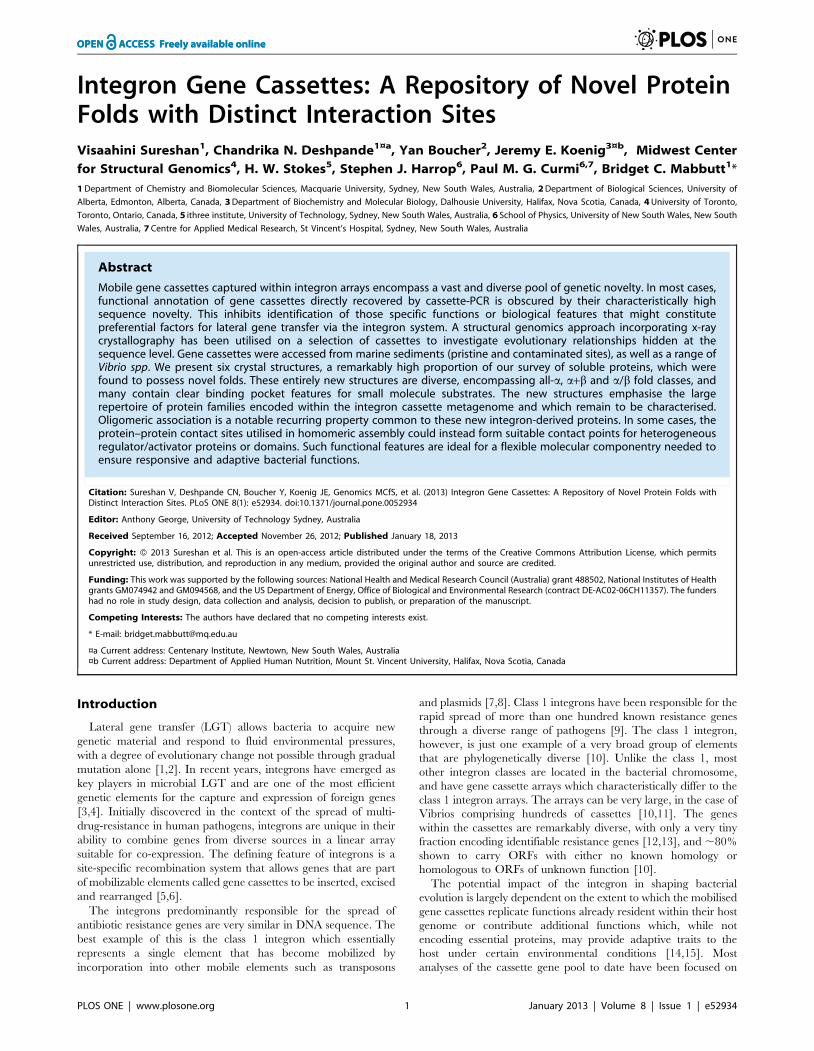
Vpc_cass2. The structure of Vpc_cass2 (PDB 3JRT) is
based on a four-helix bundle, in this case interrupted by two
helical extensions, helices a9 (residues 118–131) and a0 (135–142)
wrapping about the bundle at midpoint. The resulting helix order
is 1-2-3-a9-a0-4, as depicted in Figure 3. Helix 4 is disrupted by an
exposed and markedly flexible loop (Cys143-Leu156) which
incorporates a b-turn (145–148).
In the crystal, the Vpc_cass2 bundle is organised into a
relatively globular dimer through intermolecular interactions
engaging ,25% of residues of each chain. Clusters of hydrophobic
side chains on the surfaces of helix 1, helix 2 and the loop
connecting helices a9and a0 (Val107, Ala108, Val1100) mediate
the dimeric interface. These same helix and loop components also
contribute to hydrogen bonding and salt bridge stabilisation of the
dimer. A spread of basic side groups is a distinctive feature of the
exposed surface of the dimer, incorporating Arg62, Lys100,
Lys101 and Arg103 sidechains from both chains (Figure 3).
The identification of structural relatives for Vpc_cass2 is
somewhat obscured by its relatively simple helical form, but
diverse tools indicate a relationship to the KNTase_C (kanamycin
nucleotidyltransferase C-terminal domain) clan (CL0291) of
proteins [47]. The fold homology is most readily seen for the 4-
helix families specifically annotated as NTase_sub_bind (PF08780:
PDB 1JOG, rmsd 2.9 A; PDB 1WTY, rmsd 3.2 A) and DUF86
(PF01934: PDB 1YLM, rmsd 3.3 A). The nucleotidyltransferases
of this clan organize as two component systems (independent
domains or gene pairs encoding a hetero-oligomeric complex): an
a/b domain for nucleotide binding and a separate domain (often
helical) providing for a wide range of substrate types. It is the
defined helical substrate-binding domains to which Vpc_cass2 is
related.
A comparison of these closest structural relatives with
Vpc_cass2 convincingly shows our new structure to possess
distinct features, most obviously (i) a unique loop disrupting helix
4, (ii) elongation of helices a9 and a0, and (iii) the absence of
additional helix between helices 2 and 3. Although there is no
relationship at the sequence level, the majority of structures
defined across this clan consistently show dimeric organisation
mediated largely through hydrophobic residues of helix 2.
Significantly, the packing geometry of these various dimeric
structures are markedly different. Often the pair of helix bundles
are angled, so creating a deep ‘‘V’’-shaped interdomain cleft
embellished with distinct basic patches, perhaps suitable for
nucleic acid binding. However, in the case of Vpc_cass2, the
alignment angle between chains is considerably different, resulting
in a compact and relatively flat surface (panel B, Figure 3).
Novel Protein Folds Encoded within Gene Cassettes
PLOS ONE | www.plosone.org 3 January 2013 | Volume 8 | Issue 1 | e52934

Ta
ble
1.
Cry
stal
log
rap
hic
dat
aco
llect
ion
and
refi
ne
me
nt
stat
isti
csfo
rst
ruct
ure
de
term
inat
ion
.
Da
tase
tS
pa
ceg
rou
pa
.s.u
aU
nit
cell
(A)
Un
itce
ll( 6
)R
eso
luti
on
(A)
To
tal
Re
fl.
Un
iqu
eR
efl
.,
I/s
.
Co
mp
lete
-ne
ss(%
)M
ult
i-p
lici
tyR
me
rge
b
Hfx
_ca
ss1
(ou
ter
she
ll)P
32
3a
=7
1.6
b=
71
.6c
=9
2.7
a=
90b
=9
0c
=1
20
2.0
0(2
.11
–2
.00
)2
02
52
8(2
93
80
)3
59
48
(52
87
)7
.4(0
.9)
10
0.0
(10
0.0
)5
.6(5
.6)
0.0
82
(0.5
60
)
Hfx
_ca
ss2
(ou
ter
she
ll)C
21
a=
40
.7b
=6
6.8
c=
48
.9a
=9
0b
=1
13
.5c
=9
01
.84
(1.8
7–
1.8
4)
70
00
4(1
78
9)
10
18
7(2
52
)8
.8(3
.0)
97
.3(9
5.8
)6
.9(7
.1)
0.0
87
(0.6
69
)
Hfx
_ca
ss5
(ou
ter
she
ll)P
21
4a
=6
1.3
b=
44
.5c
=8
2.5
a=
90b
=1
09
.8c
=9
02
.18
(2.2
4–
2.1
8)
15
14
62
(30
52
)2
16
18
(76
3)
10
.3(2
.6)
97
.6(6
8.6
)7
.0(4
.0)
0.0
93
(0.5
31
)
Vch
_ca
ss3
(ou
ter
she
ll)P
212
12
14
a=
50
.3b
=9
7.4
c=
11
5.4
a=
90b
=9
0c
=9
02
.10
(2.2
1–
2.1
0)
12
94
02
(18
69
8)
33
58
4(4
81
5)
11
.4(1
.8)
99
.3(9
9.2
)3
.9(3
.9)
0.0
88
(0.4
03
)
Vch
_ca
ss1
4(o
ute
rsh
ell)
C2
4a
=1
19
.9b
=6
4.7
c=
81
.98
a=
90b
=1
31
.3c
=9
01
.80
(1.9
0–
1.8
0)
16
41
16
(23
62
3)
43
70
2(6
36
5)
10
.6(1
.1)
99
.9(1
00
.0)
3.8
(3.7
)0
.06
6(0
.62
9)
Vp
c_ca
ss2
(ou
ter
she
ll)P
64
1a
=8
5.5
b=
85
.5c
=4
6.7
a=
90b
=9
0c
=1
20
2.3
0(2
.42
–2
.30
)1
23
96
0(1
85
36
)8
80
1(1
28
1)
15
.6(1
.6)
99
.9(1
00
.0)
14
.1(1
4.5
)0
.07
2(0
.47
3)
Re
fin
em
en
tst
ati
stic
sH
fx_
cass
1H
fx_
cass
2H
fx_
cass
5V
ch_
cass
3V
ch_
cass
14
Vp
c_ca
ss2
Wat
er
mo
lecu
les
16
39
61
32
32
32
09
31
Rc
ryst
/Rfr
ee
0.1
99
/0.2
34
0.1
91
/0.2
41
0.1
83
/0.2
33
0.1
73
/0.2
24
0.1
95
/0.2
30
0.1
93
/0.2
34
Re
fle
ctio
ns
inR
cry
st/R
fre
e3
59
48
/67
04
99
42
/10
00
21
12
7/1
95
13
35
84
/20
00
41
94
8/4
29
48
78
2/8
71
Re
solu
tio
nra
ng
e(A
)9
2.8
5-2
.00
50
.00
-1.8
45
0.0
0-2
.18
74
.43
-2.1
04
5.0
8-1
.80
42
.76
-2.3
0
Ove
rall
iso
tro
pic
B-f
acto
r(A
2)
38
.83
9.7
34
.42
8.5
30
.66
6.9
r.m
.s.d
.b
on
dle
ng
ths
(A)
0.0
07
0.0
07
0.0
05
0.0
06
0.0
07
0.0
08
r.m
.s.d
.b
on
dan
gle
s(u
)0
.98
90
.92
40
.87
60
.94
00
.94
40
.96
8
Ram
ach
and
ran
plo
tc:
Mo
stfa
vou
red
reg
ion
(%)
97
.09
8.2
96
.69
9.4
95
.89
5.8
Ad
dit
ion
ally
allo
we
d(%
)3
.01
.82
.90
.63
.73
.6
aC
hai
ns
pe
ras
ymm
etr
icu
nit
(a.s
.u).
bgg
i|Ih
i2
I hi|/gg
iI h,
wh
ere
I his
the
me
anin
ten
sity
of
refl
ect
ion
h.
cFr
om
Mo
lPro
bit
y[3
6].
do
i:10
.13
71
/jo
urn
al.p
on
e.0
05
29
34
.t0
01
Novel Protein Folds Encoded within Gene Cassettes
PLOS ONE | www.plosone.org 4 January 2013 | Volume 8 | Issue 1 | e52934

Two close sequence homologues of Vpc_cass2 can be seen
within the genomes of Shewanella baltica and Moritella spp.,
displaying ,50% amino acid identity (and which retain 62–70%
sequence homology). Spatial mapping of the invariant amino acids
onto the Vpc_cass2 fold reveals strong preservation of the
hydrophobic residues forming the dimer interface, suggesting
retention of the dimeric structure. An additional cluster of
conserved residues projects across the interface in the vicinity of
the carboxyl end of helix 2, incorporating Lys63 and Glu66 side
chains grouped with His1099 (Figure 3C). This preserved feature
likely contributes to the biochemistry of a substrate site for this
protein family. The basic surface residues of the Vpc_cass2dimer, however, appear to be unique to just this member of the
sequence group.
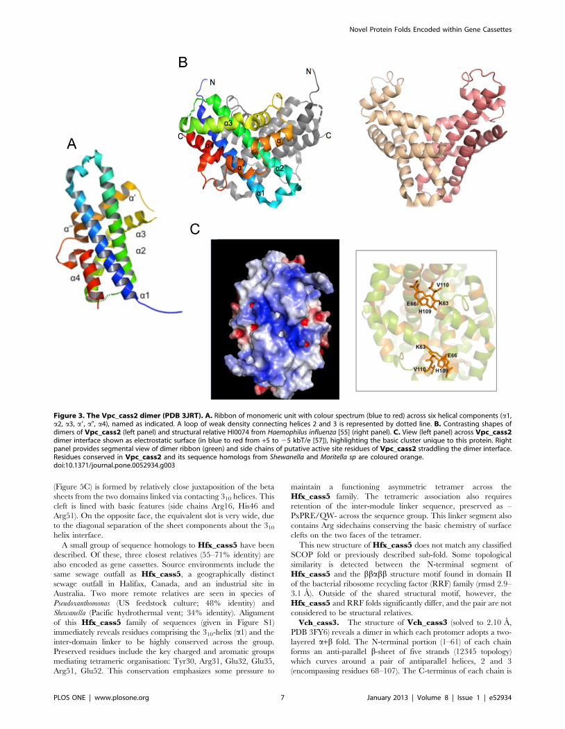
Alpha/beta fold structureHfx_cass1. Hfx_cass1 (PDB 3FUY) is a trimer of distinctive
flattened shape (75 A625 A), in which each protomer adopts a
three-layered a/b-fold. Each subunit contains a mixed six-
stranded central sheet underlying two extended a-helices and
flanked on the alternate face by a 310-helix (Figure 4). Weaving
outwards from the centre of the trimer, strands b1 and b2 of each
sheet form a simple meander, followed by two inverted b-a-b
motifs. Whilst the b-a-b motif incorporating strands b5/b6 utilises
conventional topology, the first motif (connecting b3/b4) involves
a rare left-hand cross-over (observed only in ,1.5% of super-
secondary structures [48]). The novel crossover loop is relatively
long, incorporating a G/P-rich segment of eight residues (G30-
D37).
Within the flattened structure of Hfx_cass1, only a small
proportion (,11%) of residues engage in interactions across
subunit interfaces. The trimer is primarily stabilised by hydro-
phobic contacts from b1 strand residues at the centre to
neighbouring loop features (b1-b2, b29-b39, b3-a4 and b69-a39).
A salt bridge engages two adjacent loops (His82/Asp1449) and
hydrogen bonds occur between residues on the inner b-strands
and nearby loops and the C-terminal 310-helix (Ser129-Asp151-
Gly339).
As a result of a b-bulge between b4 and the adjacent b5 strand
(at residues 74, 75 and 89), strands b3 and b4 are splayed apart,
interacting at their carboxyl ends only. The b-bulge secondary
feature has long been associated with active sites of proteins [49].
In the case of Hfx_cass1, the two splayed strands create a narrow
polar cavity, bound above and below by helices 1 and 3, and
occupied by water molecules in all three subunits (Figure 4).
Surrounded by pronounced acidic clusters, largely from side
Figure 1. Structural elements and topology of novel gene cassette proteins. Sequences of gene cassette ORFs aligned with secondarystructural features observed within crystal structures (arrows, b-strands; blocks, a-helices; dashed lines, undefined flexible regions). Sequences do notinclude additional affinity tags used for recombinant production. Schematic diagrams show structures of monomer forms for proteins A. Hfx_cass2,B. Vpc_cass2, C. Hfx_cass1, D. Hfx_cass5, E. Vch_cass3 and F. Vch_cass14.doi:10.1371/journal.pone.0052934.g001
Novel Protein Folds Encoded within Gene Cassettes
PLOS ONE | www.plosone.org 5 January 2013 | Volume 8 | Issue 1 | e52934

chains on loop features, this region has the appearance of a
functional binding site. Acidic side chains contributing to the open
cavities adjacent to each cleft include Asp79 and Asp80 (b49-b59
loop) as well as Glu139, Asp144, Asp149 (b6-a3 loop).
We note that in the packing of our Hfx_cass1 crystal, these
proposed binding sites engage surface side chains from protomers
of neighbouring trimers. A search against a database of cognate
binding sites [43], identified some features at this location common
to enzymes utilising nucleotide-based cofactors (e.g. adenosine
and/or nicotinamide moieties). However, Hfx_cass1 displays
none of the known sequence motifs for binding these cofactors.
While no direct sequence or structural homologues of
Hfx_cass1 have yet been reported, some sub-fold similarity is
detected to the zinc transporter CzrB (PDB 3BYP [50]) from
Thermus thermophilus. The cytosolic zinc-binding domain of CzrB,
an integral membrane transporter, aligns (2.8 A rmsd) with C-
terminal residues (38–148) of Hfx_cass1. The CzrB fold
incorporates a helix followed by a b strand and an inverted b-a-
b motif, hence overlapping a portion of the b-a-b repeat motifs of
Hfx_cass1. In CzrB, the domain presents a cluster of zinc-binding
residues for metal chelation and controls a dimerisation event
critical to function [50]. However, these active site residues are not
replicated in the equivalent strands (b3-b6) of Hfx_cass1, to
which there appears to be no functional relationship.
Alpha + beta fold structuresHfx_cass5. The 2.18 A structure of Hfx_cass5 (PDB 3IF4)
reveals a symmetrical domain-swapped dimer of compact a+bdomains. As shown in Figure 5, this structure forms half of a
structurally asymmetric tetramer. One face of each domain
contains a five-stranded b-sheet, predominantly antiparallel in
nature (strand order: 691243). Overlaying this sheet, creating an
alternate face to the domain, are two helices (a29 and a39) and a
parallel b-ribbon formed by strand b59 and the N-terminal
segment of b1. An extended loop and short 310-helix (a1: Tyr28-
Ala33) between strands b2 and b3 connect the two domain faces.
Residues 1–46 of each chain contribute strands b1-b4 and helix a1
of one domain; a Pro-containing segment with slightly elevated B-
factors creates the inter-domain linker; residues 54–98 form the
alpha helices and intervening strands (b59 and b69) within the
second domain.
At the centre of the crystallised tetramer, the 310 helices at the
edges of two opposing subunits come into contact via well-ordered
stacking of protruding polar and charged side chains (Tyr28,
Tyr30, Arg31, Glu35; see Fig. 5B). This contact means that the 310
helices from the remaining two domains are separated further out
along the tetrameric interface.
The flattened nature of the tetramer and the asymmetrical
interactions of its component dimers results in two large faces with
markedly different surface features. On one face, a narrow slot
Figure 2. Helical packing in the Hfx_cass2 dimer (PDB 3FXH). A. Ribbon depiction with colour spectrum from N-terminus (blue) to C-terminus(red) for each chain. B. Dimer interface engages hydrophobic residues from Chain A (tan) and Chain B (green). Acidic groups located either side of asmall hydrophobic pocket on the external face are indicated (red). C. Electrostatic surface potential of the dimer surface. Key acidic features (Glu47,Asp60 and helix 2 side chains) are labelled.doi:10.1371/journal.pone.0052934.g002
Novel Protein Folds Encoded within Gene Cassettes
PLOS ONE | www.plosone.org 6 January 2013 | Volume 8 | Issue 1 | e52934
(Figure 5C) is formed by relatively close juxtaposition of the beta
sheets from the two domains linked via contacting 310 helices. This
cleft is lined with basic features (side chains Arg16, His46 and
Arg51). On the opposite face, the equivalent slot is very wide, due
to the diagonal separation of the sheet components about the 310
helix interface.
A small group of sequence homologs to Hfx_cass5 have been
described. Of these, three closest relatives (55–71% identity) are
also encoded as gene cassettes. Source environments include the
same sewage outfall as Hfx_cass5, a geographically distinct
sewage outfall in Halifax, Canada, and an industrial site in
Australia. Two more remote relatives are seen in species of
Pseudoxanthomonas (US feedstock culture; 48% identity) and
Shewanella (Pacific hydrothermal vent; 34% identity). Alignment
of this Hfx_cass5 family of sequences (given in Figure S1)
immediately reveals residues comprising the 310-helix (a1) and the
inter-domain linker to be highly conserved across the group.
Preserved residues include the key charged and aromatic groups
mediating tetrameric organisation: Tyr30, Arg31, Glu32, Glu35,
Arg51, Glu52. This conservation emphasizes some pressure to
maintain a functioning asymmetric tetramer across the
Hfx_cass5 family. The tetrameric association also requires
retention of the inter-module linker sequence, preserved as –
PxPRE/QW- across the sequence group. This linker segment also
contains Arg sidechains conserving the basic chemistry of surface
clefts on the two faces of the tetramer.
This new structure of Hfx_cass5 does not match any classified
SCOP fold or previously described sub-fold. Some topological
similarity is detected between the N-terminal segment of
Hfx_cass5 and the bbabb structure motif found in domain II
of the bacterial ribosome recycling factor (RRF) family (rmsd 2.9–
3.1 A). Outside of the shared structural motif, however, the
Hfx_cass5 and RRF folds significantly differ, and the pair are not
considered to be structural relatives.
Vch_cass3. The structure of Vch_cass3 (solved to 2.10 A,
PDB 3FY6) reveals a dimer in which each protomer adopts a two-
layered a+b fold. The N-terminal portion (1–61) of each chain
forms an anti-parallel b-sheet of five strands (12345 topology)
which curves around a pair of antiparallel helices, 2 and 3
(encompassing residues 68–107). The C-terminus of each chain is
Figure 3. The Vpc_cass2 dimer (PDB 3JRT). A. Ribbon of monomeric unit with colour spectrum (blue to red) across six helical components (a1,a2, a3, a9, a0, a4), named as indicated. A loop of weak density connecting helices 2 and 3 is represented by dotted line. B. Contrasting shapes ofdimers of Vpc_cass2 (left panel) and structural relative HI0074 from Haemophilus influenza [55] (right panel). C. View (left panel) across Vpc_cass2dimer interface shown as electrostatic surface (in blue to red from +5 to 25 kbT/e [57]), highlighting the basic cluster unique to this protein. Rightpanel provides segmental view of dimer ribbon (green) and side chains of putative active site residues of Vpc_cass2 straddling the dimer interface.Residues conserved in Vpc_cass2 and its sequence homologs from Shewanella and Moritella sp are coloured orange.doi:10.1371/journal.pone.0052934.g003
Novel Protein Folds Encoded within Gene Cassettes
PLOS ONE | www.plosone.org 7 January 2013 | Volume 8 | Issue 1 | e52934
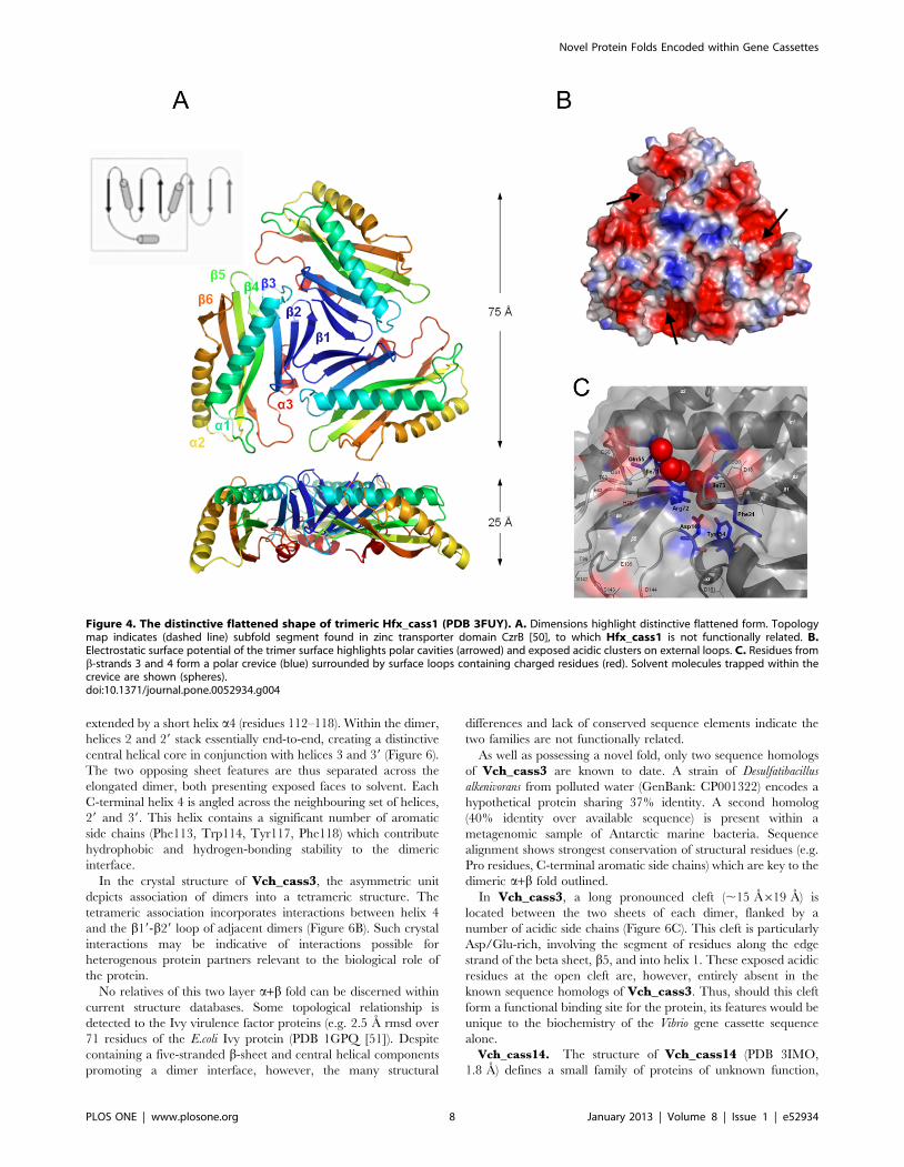
extended by a short helix a4 (residues 112–118). Within the dimer,
helices 2 and 29 stack essentially end-to-end, creating a distinctive
central helical core in conjunction with helices 3 and 39 (Figure 6).
The two opposing sheet features are thus separated across the
elongated dimer, both presenting exposed faces to solvent. Each
C-terminal helix 4 is angled across the neighbouring set of helices,
29 and 39. This helix contains a significant number of aromatic
side chains (Phe113, Trp114, Tyr117, Phe118) which contribute
hydrophobic and hydrogen-bonding stability to the dimeric
interface.
In the crystal structure of Vch_cass3, the asymmetric unit
depicts association of dimers into a tetrameric structure. The
tetrameric association incorporates interactions between helix 4
and the b19-b29 loop of adjacent dimers (Figure 6B). Such crystal
interactions may be indicative of interactions possible for
heterogenous protein partners relevant to the biological role of
the protein.
No relatives of this two layer a+b fold can be discerned within
current structure databases. Some topological relationship is
detected to the Ivy virulence factor proteins (e.g. 2.5 A rmsd over
71 residues of the E.coli Ivy protein (PDB 1GPQ [51]). Despite
containing a five-stranded b-sheet and central helical components
promoting a dimer interface, however, the many structural
differences and lack of conserved sequence elements indicate the
two families are not functionally related.
As well as possessing a novel fold, only two sequence homologs
of Vch_cass3 are known to date. A strain of Desulfatibacillus
alkenivorans from polluted water (GenBank: CP001322) encodes a
hypothetical protein sharing 37% identity. A second homolog
(40% identity over available sequence) is present within a
metagenomic sample of Antarctic marine bacteria. Sequence
alignment shows strongest conservation of structural residues (e.g.
Pro residues, C-terminal aromatic side chains) which are key to the
dimeric a+b fold outlined.
In Vch_cass3, a long pronounced cleft (,15 A619 A) is
located between the two sheets of each dimer, flanked by a
number of acidic side chains (Figure 6C). This cleft is particularly
Asp/Glu-rich, involving the segment of residues along the edge
strand of the beta sheet, b5, and into helix 1. These exposed acidic
residues at the open cleft are, however, entirely absent in the
known sequence homologs of Vch_cass3. Thus, should this cleft
form a functional binding site for the protein, its features would be
unique to the biochemistry of the Vibrio gene cassette sequence
alone.
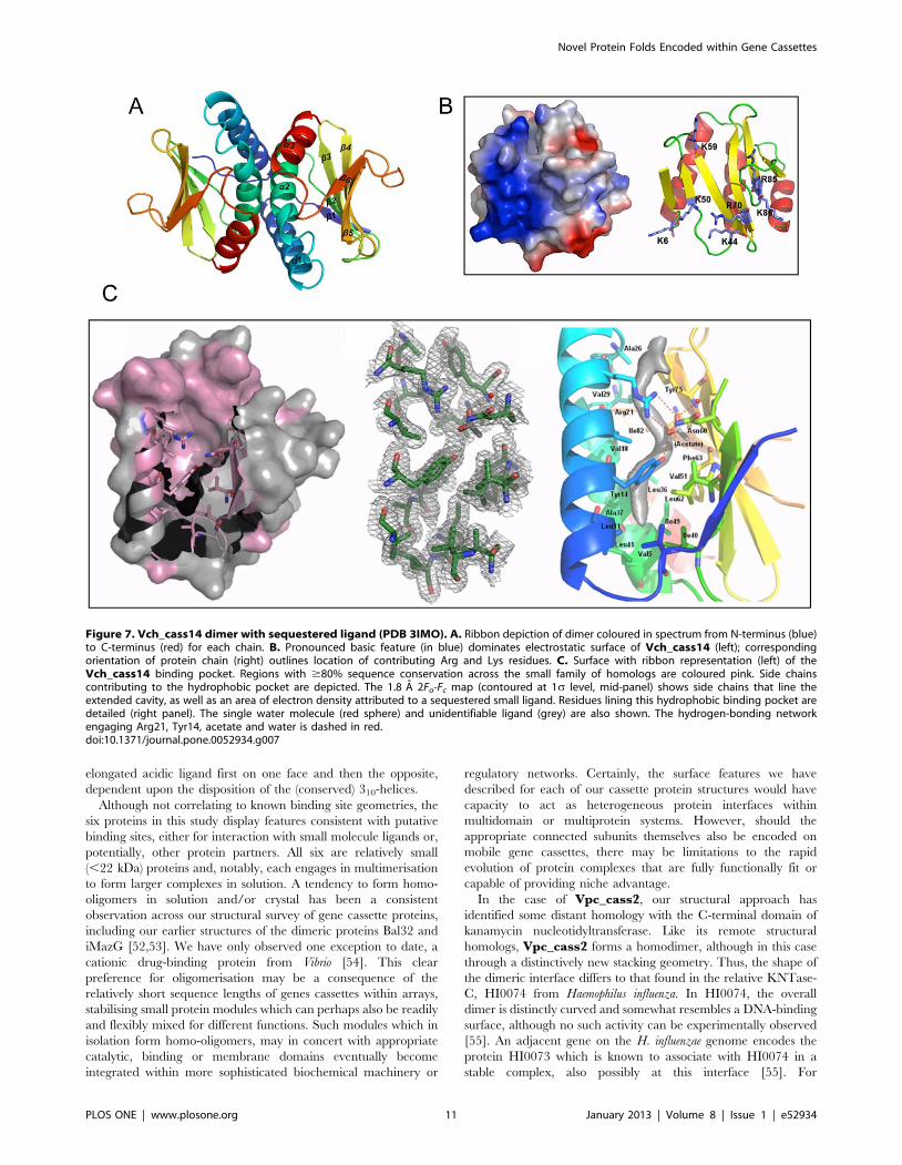
Vch_cass14. The structure of Vch_cass14 (PDB 3IMO,
1.8 A) defines a small family of proteins of unknown function,
Figure 4. The distinctive flattened shape of trimeric Hfx_cass1 (PDB 3FUY). A. Dimensions highlight distinctive flattened form. Topologymap indicates (dashed line) subfold segment found in zinc transporter domain CzrB [50], to which Hfx_cass1 is not functionally related. B.Electrostatic surface potential of the trimer surface highlights polar cavities (arrowed) and exposed acidic clusters on external loops. C. Residues fromb-strands 3 and 4 form a polar crevice (blue) surrounded by surface loops containing charged residues (red). Solvent molecules trapped within thecrevice are shown (spheres).doi:10.1371/journal.pone.0052934.g004
Novel Protein Folds Encoded within Gene Cassettes
PLOS ONE | www.plosone.org 8 January 2013 | Volume 8 | Issue 1 | e52934

other members of which occur in the genomes of soil-and water-
dwelling bacteria (e.g. Sorangium cellulosum, sce0458; Rhodopseudomo-
nas palustris, RPE_5052). Vch_cass14 forms a dimer (an
organisation confirmed in solution by size exclusion) in which
each subunit adopts a two-layer a+b sandwich-type fold, as shown
in Figure 7. Each chain forms a single anti-parallel sheet of six
strands overlaid by a second face of three helices at a 45u angle.
The topology order is relatively novel for this fold class: b1-a1-a2-
b2-b3-b4-b5-b6-a3. The two protomers of the dimer interact
orthogonally via their helix faces, with each central helix a2
making extensive hydrophobic contact across to all three helices of
the paired module. A notable feature of the Vch_cass14 dimer is
the highly positively-charged surface displayed across each
exposed b-sheet.
Internal to each monomer lies a particularly deep binding
pocket formed by helices a1 and a2 and residues from the central
four strands of the b-sheet (Figure 7C). The pocket is extensively
lined with hydrophobic side chains (Phe73; Val5, Val18, Val29
and Val51; Leu11, Leu36 and Leu62; Ile15, Ile40, Ile49 and Ile82;
Ala22, Ala26, Ala33 and Ala37; Tyr75) and could accommodate a
ligand up to ,15 A in length. In the crystal form we have isolated,
electron density consistent with a linear organic molecule is
observed in this site, as shown in Figure 7. At the entrance to the
pocket, a distinct cluster of polar residues (Arg21, Asn60 and
Tyr14) is observed, engaged in this structure in a hydrogen
bonding network with acetate and water molecules.
Across the known sequence relatives of Vch_cass14, similarity
is relatively strong (46–62% identity), with sequences relating to
helix a1, helix a2 and the connecting loop particularly well
conserved (Figure S2). Amongst the three closest homologs
(Vch_cass14, sce0458 and RPE_4052), all 20 of the internal
pocket residues are invariant or conservatively substituted. The
deep hydrophobic pocket observed in our Vch_cass14 structure
is thus likely retained as a binding site across this family of
proteins. Mapping of other conserved residues onto the
Vch_cass14 structure additionally reveals a high degree of
conservation for most hydrophobic side chains of the three helices,
i.e. residues participating in dimer interactions. This most certainly
points to a dimeric form being most relevant to biological function
for this protein family.
Figure 5. Hfx_cass5 contains domain-swapped dimers (PDB 3IF4). A. Ribbon depiction of two domain-swapped chains, each coloured fromN-terminus (blue) to C-terminus (red). B. Tetrameric organisation depicted in ribbon form (left panel), showing engagement between two dimers(green and blue), each comprising two chains. Side chain stacking of key contacting residues (Tyr28, Tyr30, Arg31 and Glu35) is depicted.Corresponding view of electrostatic surface is also shown (right), with deep cleft centrally positioned. C. Rotated view of electrostatic surface,positioned to emphasise narrow dimensions of this basic slot along one tetramer surface.doi:10.1371/journal.pone.0052934.g005
Novel Protein Folds Encoded within Gene Cassettes
PLOS ONE | www.plosone.org 9 January 2013 | Volume 8 | Issue 1 | e52934
Discussion
The six new proteins whose structures are presented in this work
were selected from integron gene cassette sequences on the basis of
having no sequence similarity to any protein of known three-
dimensional structure. The crystal structures we have since defined
for these six proteins verify that the originating gene cassettes code
for folded proteins which possess entirely new topologies. This
attests to the notion that the integron/gene cassette metagenome is
a source of a remarkable degree of biological diversity at the
protein level, and that the genes encoded within it express
functionally active proteins [52].
Although the structures of this set of integron/gene cassette
proteins are novel, four of the six (Vpc_cass2, Hfx_cass5,
Vch_cass4 and Vch_cass14) share some similarity to small
sequence groupings of undefined structure and function within
gene databases. Thus, the information gained from the new
structures featured here impacts beyond the specific structural
targets to begin to delineate entirely new protein families and their
associated members. In some examples, the identified sequence
relatives of our set of integron-derived proteins are also themselves
localised within gene cassettes.
For the majority, the structures described here provide features
consistent with binding functions for the new proteins. The
structure of Vch_cass14 contains a deep cleft in which is
sequestered an organic molecule incorporating an extended
aliphatic chain plus acetate group. This is suggestive of a function
consistent with an enzyme for catalysis, small substrate-sequester-
ing protein or a transport protein. The structures of Hfx_cass1and Hfx_cass2 each have surface clefts reminiscent of enzyme
active sites. At present, the arrangements of the side chains in these
clefts do not show exact template match to any previously defined
active site chemistry, thus limiting any direct functional inference.
The Hfx_cass5 family described here presents one of the most
intriguing of the new protein structures. The structure is a
tetramer built from two domain-swapped dimers. The tetramer is
asymmetric, with one subunit from each domain-swapped dimer
forming a central contact with its equivalent subunit from the
other dimer. This contact is mediated by a 310 helix whose
sequence is strongly conserved in related proteins. On one face of
the Hfx_cass5 tetramer, the two closest domains of separate
dimer groupings form a narrow slot that is lined with basic side
chains. This creates the appearance of a binding site for an
extended, acidic ligand. Because of the asymmetry of chain
packing, the equivalent (far wider) slot on the opposite face of the
oligomer lacks such coherent binding-site features. It is tempting to
speculate this protein could act as a bistable switch, binding an
Figure 6. The dimer of Vch_cass3 (PDB 3FY6). A. The two chains of the Vch_cass3 dimer are illustrated, including stabilising hydrophobicresidues from helices 2, 3 and 4. B View depicting Vch_cass3 dimers interacting via helix 4 and b1-b2 loop regions. Located in a single asymmetricunit are dimer 1 (yellow, green chains) and dimer 2 (pink, cyan chains). C. The proposed functional site formed by the dimer (blue and green chains) isdistinct from any conserved regions, and consists of acidic residues (red) belonging to b5, and helices 1 and 2.doi:10.1371/journal.pone.0052934.g006
Novel Protein Folds Encoded within Gene Cassettes
PLOS ONE | www.plosone.org 10 January 2013 | Volume 8 | Issue 1 | e52934
elongated acidic ligand first on one face and then the opposite,
dependent upon the disposition of the (conserved) 310-helices.
Although not correlating to known binding site geometries, the
six proteins in this study display features consistent with putative
binding sites, either for interaction with small molecule ligands or,
potentially, other protein partners. All six are relatively small
(,22 kDa) proteins and, notably, each engages in multimerisation
to form larger complexes in solution. A tendency to form homo-
oligomers in solution and/or crystal has been a consistent
observation across our structural survey of gene cassette proteins,
including our earlier structures of the dimeric proteins Bal32 and
iMazG [52,53]. We have only observed one exception to date, a
cationic drug-binding protein from Vibrio [54]. This clear
preference for oligomerisation may be a consequence of the
relatively short sequence lengths of genes cassettes within arrays,
stabilising small protein modules which can perhaps also be readily
and flexibly mixed for different functions. Such modules which in
isolation form homo-oligomers, may in concert with appropriate
catalytic, binding or membrane domains eventually become
integrated within more sophisticated biochemical machinery or
regulatory networks. Certainly, the surface features we have
described for each of our cassette protein structures would have
capacity to act as heterogeneous protein interfaces within
multidomain or multiprotein systems. However, should the
appropriate connected subunits themselves also be encoded on
mobile gene cassettes, there may be limitations to the rapid
evolution of protein complexes that are fully functionally fit or
capable of providing niche advantage.
In the case of Vpc_cass2, our structural approach has
identified some distant homology with the C-terminal domain of
kanamycin nucleotidyltransferase. Like its remote structural
homologs, Vpc_cass2 forms a homodimer, although in this case
through a distinctively new stacking geometry. Thus, the shape of
the dimeric interface differs to that found in the relative KNTase-
C, HI0074 from Haemophilus influenza. In HI0074, the overall
dimer is distinctly curved and somewhat resembles a DNA-binding
surface, although no such activity can be experimentally observed
[55]. An adjacent gene on the H. influenzae genome encodes the
protein HI0073 which is known to associate with HI0074 in a
stable complex, also possibly at this interface [55]. For
Figure 7. Vch_cass14 dimer with sequestered ligand (PDB 3IMO). A. Ribbon depiction of dimer coloured in spectrum from N-terminus (blue)to C-terminus (red) for each chain. B. Pronounced basic feature (in blue) dominates electrostatic surface of Vch_cass14 (left); correspondingorientation of protein chain (right) outlines location of contributing Arg and Lys residues. C. Surface with ribbon representation (left) of theVch_cass14 binding pocket. Regions with $80% sequence conservation across the small family of homologs are coloured pink. Side chainscontributing to the hydrophobic pocket are depicted. The 1.8 A 2Fo-Fc map (contoured at 1s level, mid-panel) shows side chains that line theextended cavity, as well as an area of electron density attributed to a sequestered small ligand. Residues lining this hydrophobic binding pocket aredetailed (right panel). The single water molecule (red sphere) and unidentifiable ligand (grey) are also shown. The hydrogen-bonding networkengaging Arg21, Tyr14, acetate and water is dashed in red.doi:10.1371/journal.pone.0052934.g007
Novel Protein Folds Encoded within Gene Cassettes
PLOS ONE | www.plosone.org 11 January 2013 | Volume 8 | Issue 1 | e52934
Vpc_cass2, the exposed surfaces of the dimer are relatively flat,
with a distinct conserved cluster of charged residues straddled
across the interface, so more suggestive of a protein (rather than
any DNA) interaction site.
At the genetic level, genes for bacterial NTase substrate-binding
proteins are often found adjacent to separate nucleotidyltransfer-
ase genes, i.e. those encoding the corresponding catalytic subunit.
In the case of KNTase, the two relevant elements are actually
encoded within a single gene, so containing both the helical
substrate-binding domain and the distinct a/b nucleotide-binding
domain positioned about a long binding-site cleft [56]. Lehmann
et al. have documented substrate-binding/nucleotide-binding
module pairs to be quite prevalent in bacterial genomes,
particularly from harsh conditions and pathogens [55]. Thus,
the mobile gene cassette element Vpc_cass2 could serve as an
example of one half of a bipartite system, with the capacity to
become established with a nucleotidyltransferase second domain
to provide a functional enzyme offering selective advantage.
Our structural study has shown that the highly novel (,80%
unknown) gene cassette metagenome is not merely a repository of
sequence divergent variants of known proteins, but in fact
mobilises a repertoire of genes belonging to new, currently
uncharacterised protein families. The significant structural spread
we observe across these six representatives of cassette-proteins
suggests each to either perform a biological function hitherto
undescribed in other bacterial proteins, or to achieve a known
function by a new mechanism, and thus apt to be under different
regulatory control. In either scenario, it is probable that the
phenotypes provided by cassette proteins expand the functional
repertoire of the recipient organism, just as might also be provided
by other types of mobilised features within, for instance, genomic
islands. This hypothesis is strengthened by the persistence of
sequence homologs in the cassette metagenome across widely
varying geographical locations experiencing similar environmental
stressors. The high degree of novelty that we consistently observe
across sequences and structures of integron/gene cassette proteins
attests to the fact that this pool of genes remains relatively
uncharacterised by normal sequencing efforts. Thus, to fully
characterise and understand the global proteome, it remains
essential to continue to independently target the metagenomic
element.
Supporting Information
Figure S1 Sequence alignment of Hfx_cass5 and relatedcassette-proteins. Secondary structure elements are from
Hfx_cass5, as determined in this work. Invariant (white
characters, black shading) and chemically equivalent (black
characters, grey shading) residues across $80% of the family are
shown. Red dots delineate exposed residues engaged in tetrameric
interaction. Sequences are: (Hfx_cass5) cassette protein sourced
from a raw sewage effluent outfall in the North West Arm, Halifax,
Canada; (B0BGV4) cassette protein sourced from the same sewage
outfall; (B0BK21) cassette protein sourced from a geographically
distinct raw sewage effluent outfall in Halifax Harbour, Halifax,
Canada; (Bal50) cassette protein sourced from soil contaminated
with industrial waste, at an electricity power station in Balmain,
Sydney, Australia. (Pxanth) Pseudoxanthomonas suwonensis 11-1 from
compost-feedstock enrichment culture, bioreactor, USA [58];
(Shew2602) Shewanella loihica PV-4 from deep sea hydrothermal
vent near Hawaii, Pacific Ocean [59].
(DOCX)
Figure S2 Sequence alignment of Vch_cass14 and itsrelated sequences. Secondary structure elements are from
Vch_cass14, as determined in this work. White characters with
black shading are indicative of identical residues across the three
sequences. Black residues on grey shading indicate chemically
equivalent residues shared by all sequences. Red dots and blue
triangles delineate residues forming pocket surface and dimer
interface, respectively. Sequences are: (Vch_cass14) cassette
isolated from the chromosomal integron of V. cholera; (sce0458)
hypothetical protein encoded by S. cellulosum; (BosA53) hypothet-
ical protein encoded by R. palustris.
(DOCX)
Acknowledgments
We thank Aled Edwards and Andrej Joachimiak for supporting this
collaboration with the Midwest Center for Structural Genomics (MCSG).
The complete list of authors from the MCSG is: R. Di Leo, O. Kagan, S.
Gebert, V. Egorova, E. Evdokimova, X. Xu, M. Kudrytska, H. Cui, J.
Osipiuk, C. Chang, K. Tan, M.Cuff, A. Edwards, A. Joachimiak and A.
Savchenko.
The authors also thank Maurizio Labbate (University of Technology
Sydney), Ian Paulsen and Robert Willows (Macquarie University) for useful
discussions, and David Langley for some additional synchrotron data
acquisition (Australian Synchrotron, Melbourne). VS acknowledges receipt
of an APA (Australian Federal Government) scholarship. CND acknowl-
edges receipt of a MQRES (Macquarie University) and EIPRS (Australian
Federal Government) scholarship.
Author Contributions
Conceived and designed the experiments: MCSG HWS PMGC BCM.
Performed the experiments: VS CND YB JEK MCSG SJH. Analyzed the
data: VS CND HWS SJH PMGC BCM. Contributed reagents/materials/
analysis tools: YB JEK MCSG PMGC BCM. Wrote the paper: VS CND
YB HWS PMGC BCM.
References
1. Marri PR, Hao W, Golding GB (2007) The role of laterally transferred genes in
adaptive evolution. BMC Evol Biol. 7 Suppl 1: S8.
2. Eisen JA (2000) Horizontal gene transfer among microbial genomes: new
insights from complete genome analysis. Curr Opin Genet Dev 10: 606–11.
3. Stokes HW, Hall RM (1989) A novel family of potentially mobile DNA elements
encoding site-specific gene-integration functions: integrons. Mol Microbiol 3:
1669–83.
4. Labbate M, Case RJ, Stokes HW (2009) The integron/gene cassette system: an
active player in bacterial adaptation. Methods Mol Biol 532:103–25.
5. Joss MJ, Koenig JE, Labbate M, Polz MF, Gillings MR (2009) ACID:
annotation of cassette and integron data. BMC Bioinformatics 10: 118.
6. Hall RM, Collis CM (1995) Mobile gene cassettes and integrons: capture and
spread of genes by site-specific recombination. Mol Microbiol 15: 593–600.
7. Brown HJ, Stokes HW, Hall RM (1996) The integrons In0, In2, and In5 are
defective transposon derivatives. J Bacteriol 178: 4429–37.
8. Gillings M, Boucher Y, Labbate M, Holmes A, Krishnan S, et al. (2008) The
evolution of class 1 integrons and the rise of antibiotic resistance. J Bacteriol 190:
5095–100.
9. Partridge SR, Tsafnat G, Coiera E, Iredell JR (2009) Gene cassettes and cassette
arrays in mobile resistance integrons. FEMS Microbiol Rev 33: 757–84.
10. Boucher Y, Labbate M, Koenig JE, Stokes HW (2007) Integrons: mobilizable
platforms that promote genetic diversity in bacteria. Trends Microbiol 15: 301–
9.
11. Mazel D, Dychinco B, Webb VA, Davies J (1998) A distinctive class of integron
in the Vibrio cholerae genome. Science 280: 605–8.
12. Holmes AJ, Gillings MR, Nield BS, Mabbutt BC, Nevalainen KM, et al. (2003)
The gene cassette metagenome is a basic resource for bacterial genome
evolution. Environ Microbiol 5: 383–94.
13. Koenig JE, Boucher Y, Charlebois RL, Nesbø C, Zhaxybayeva O, et al. (2008)
Integron-associated gene cassettes in Halifax Harbour: assessment of a mobile
gene pool in marine sediments. Environ Microbiol 10: 1024–38.
14. Mazel D (2006) Integrons: agents of bacterial evolution. Nat Rev Microbiol 4:
608–20.
15. Michael CA, Gillings MR, Holmes AJ, Hughes L, Andrew NR, et al. (2004)
Mobile gene cassettes: a fundamental resource for bacterial evolution. Am Nat,
164: 1–12.
Novel Protein Folds Encoded within Gene Cassettes
PLOS ONE | www.plosone.org 12 January 2013 | Volume 8 | Issue 1 | e52934
16. Laskowski RA, Watson JD, Thornton JM (2003) From protein structure to
biochemical function? J Struct Funct Genomics 4: 167–77.17. Stokes HW, Holmes AJ, Nield BS, Holley MP, Nevalainen KM, et al. (2001)
Gene cassette PCR: sequence-independent recovery of entire genes from
environmental DNA. Appl Environ Microbiol 67: 5240–6.18. Jain R, Rivera MC, Lake JA (1999) Horizontal gene transfer among genomes:
the complexity hypothesis. Proc Natl Acad Sci U S A 96: 3801–6.19. Cohen O, Gophna U, Pupko T (2011), The complexity hypothesis revisited:
connectivity rather than function constitutes a barrier to horizontal gene
transfer. Mol Biol Evol. 28: 1481–9.20. Wiedenbeck J, Cohan FM (2011) Origins of bacterial diversity through
horizontal genetic transfer and adaptation to new ecological niches. FEMSMicrobiol Rev 35: 957–76.
21. Kobayashi T, Enomoto S, Sakazaki R, Kuwahara S (1963) [A new selective isolation
medium for the Vibrio group; on a modified Nakanishi’s Medium (Tcbs Agar Medium)].
Nihon Saikingaku Zasshi 18: 387–92.
22. Donovan TJ, van Netten P (1995) Culture media for the isolation andenumeration of pathogenic Vibrio species in foods and environmental samples.
Int J Food Microbiol 26: 77–91.23. Labbate M, Boucher Y, Joss MJ, Michael CA, Gillings MR, et al. (2007), Use of
chromosomal integron arrays as a phylogenetic typing system for Vibrio cholerae
pandemic strains. Microbiology 153:1488–98.24. Robinson A, Guilfoyle AP, Sureshan V, Howell M, Harrop SJ, et al. (2008)
Structural genomics of the bacterial mobile metagenome: an overview. MethodsMol Biol 426: 589–95.
25. Studier FW (2005) Protein production by auto-induction in high density shakingcultures. Protein Expr Purif 41: 207–34.
26. Dong A, Xu X, Edwards AM, Midwest Center for Structural Genomics,
Structural Genomics Consortium, et al. (2007) In situ proteolysis for proteincrystallization and structure determination. Nat Methods 4: 1019–21.
27. Leslie AGW (1992) Recent changes to the MOSFLM package for processingfilm and image plate data. Joint CCP4 + ESF-EAMCB Newsletter on Protein
Crystallography, 26.
28. Evans P (2006) Scaling and assessment of data quality. Acta Crystallogr D BiolCrystallogr 62: 72–82.
29. Minor W, Cymborowski M, Otwinowski Z, Chruszcz M (2006) HKL-3000: theintegration of data reduction and structure solution–from diffraction images to
an initial model in minutes. Acta Crystallogr D Biol Crystallogr 62: 859–66.30. Borek D, Minor W, Otwinowski Z (2003) Measurement errors and their
consequences in protein crystallography. Acta Crystallogr D Biol Crystallogr, 59:
2031–8.31. The CCP4 suite (1994) Programs for protein crystallography. Acta
Crystallogr D Biol Crystallogr 50: 760–3.32. Zwart PH, Afonine PV, Grosse-Kunstleve RW, Hung LW, Ioerger TR, et al.
(2008) Automated structure solution with the PHENIX suite. Methods Mol Biol.
426: 419–35.33. Emsley P, Cowtan K (2004) Coot: model-building tools for molecular graphics.
Acta Crystallogr D Biol Crystallogr, 60: 2126–32.34. Kleywegt GJ (2007) Crystallographic refinement of ligand complexes. Acta
Crystallogr D Biol Crystallogr 63: 94–100.35. Laskowski RA, MacArthur MW, Moss DS, Thornton JM (1993) PROCHECK:
a program to check the stereochemical quality of protein structures. J App Cryst
26: 283–291.36. Davis IW, Leaver-Fay A, Chen VB, Block JN, Kapral GJ, et al. (2007).
MolProbity: all-atom contacts and structure validation for proteins and nucleicacids. Nucleic Acids Res 35: W375–83.
37. Holm L, Kaariainen S, Rosenstrom P, Schenkel A (2008) Searching protein
structure databases with DaliLite v.3. Bioinformatics 24: 2780–1.38. Krissinel E, Henrick K (2004) Secondary-structure matching (SSM), a new tool
for fast protein structure alignment in three dimensions. Acta Crystallogr D BiolCrystallogr 60: 2256–68.
39. Ye Y, Godzik A (2004) FATCAT: a web server for flexible structure comparison
and structure similarity searching. Nucleic Acids Res 32: W582–5.
40. Altschul SF, Madden TL, Schaffer AA, Zhang J, Zhang Z, et al. (1997) Gapped
BLAST and PSI-BLAST: a new generation of protein database search
programs. Nucleic Acids Res 25: 3389–402.
41. Larkin MA, Blackshields G, Brown NP, Chenna R, McGettigan PA, et al. (2007)
Clustal W and Clustal X version 2.0. Bioinformatics 23: 2947–8.
42. Krissinel E, Henrick K (2007) Inference of macromolecular assemblies from
crystalline state. J Mol Biol 372: 774–97.
43. Najmanovich R, Kurbatova N, Thornton J (2008) Detection of 3D atomic
similarities and their use in the discrimination of small molecule protein-binding
sites. Bioinformatics 24: i105–11.
44. Laskowski RA, Watson JD, Thornton JM (2005) ProFunc: a server for predicting
protein function from 3D structure. Nucleic Acids Res 33: W89–93.
45. Gasteiger E, Hoogland C, Gattiker A, Duvaud S, Wilkins MR, et al. (2005)
Protein Identification and Analysis Tools on the ExPASy Server. In: Walker JM
editor. The Proteomics Protocols Handbook, Humana Press. pp. 571–607.
46. DeLano WL (2002) The PyMOL Molecular Graphics System. Available:
http://www.pymol.org/pymol. Accessed 2012 Dec 21.
47. Punta M, Coggill PC, Eberhardt RY, Mistry J, Tate J, et al. (2012) The Pfam
protein families database. Nucleic Acids Res 40: D290–301.
48. Cole BJ, Bystroff C (2009) Alpha helical crossovers favor right-handed
supersecondary structures by kinetic trapping: the phone cord effect in protein
folding. Protein Sci 18: 1602–8.
49. Richardson JS (1981) The anatomy and taxonomy of protein structure. Adv
Protein Chem 34: 167–339.
50. Cherezov V, Hofer N, Szebenyi DM, Kolaj O, Wall JG, et al. (2008) Insights
into the mode of action of a putative zinc transporter CzrB in Thermus
thermophilus. Structure 16: 1378–88.
51. Abergel C, Monchois V, Byrne D, Chenivesse S, Lembo F, et al. (2007)
Structure and evolution of the Ivy protein family, unexpected lysozyme
inhibitors in Gram-negative bacteria. Proc Natl Acad Sci U S A 104: 6394–9.
52. Robinson A, Wu PS, Harrop SJ, Schaeffer PM, Dosztanyi Z, et al. (2005)
Integron-associated mobile gene cassettes code for folded proteins: the structure
of Bal32a, a new member of the adaptable alpha+beta barrel family. J Mol Biol,
346: 1229–41.
53. Robinson A, Guilfoyle AP, Harrop SJ, Boucher Y, Stokes HW, et al. (2007) A
putative house-cleaning enzyme encoded within an integron array: 1.8 A crystal
structure defines a new MazG subtype. Mol Microbiol 66: 610–21.
54. Deshpande CN, Harrop SJ, Boucher Y, Hassan KA, Di Leo R, et al. (2011)
Crystal structure of an integron gene cassette-associated protein from Vibrio
cholerae identifies a cationic drug-binding module. PLOS One 6: e16934.
55. Lehmann C, Lim K, Chalamasetty VR, Krajewski W, Melamud E, et al. (2003)
The HI0073/HI0074 protein pair from Haemophilus influenzae is a member of a
new nucleotidyltransferase family: structure, sequence analyses, and solution
studies. Proteins 50: 249–60.
56. Pedersen LC, Benning MM, Holden HM (1995) Structural investigation of the
antibiotic and ATP-binding sites in kanamycin nucleotidyltransferase. Biochem-
istry 34: 13305–11.
57. Baker NA, Sept D, Joseph S, Holst MJ, McCammon JA (2001) Electrostatics of
nanosystems: application to microtubules and the ribosome. Proc Natl Acad Sci
USA. 98: 10037–41.
58. Weon HY, Kim BY, Kim JS, Lee SY, Cho YH, et al. (2006). Pseudoxanthomonas
suwonensis sp. nov., isolated from cotton waste composts. Int J Syst Evol Microbiol
56: 659–62.
59. Gao H, Obraztova A, Stewart N, Popa R, Fredrickson JK, et al. (2006)
Shewanella loihica sp. nov., isolated from iron-rich microbial mats in the Pacific
Ocean. Int J Syst Evol Microbiol 56: 1911–6.
Novel Protein Folds Encoded within Gene Cassettes
PLOS ONE | www.plosone.org 13 January 2013 | Volume 8 | Issue 1 | e52934