integumentary system. functions of the integumentary system · protection · thermoregulation ·...
TRANSCRIPT

Integumentary System
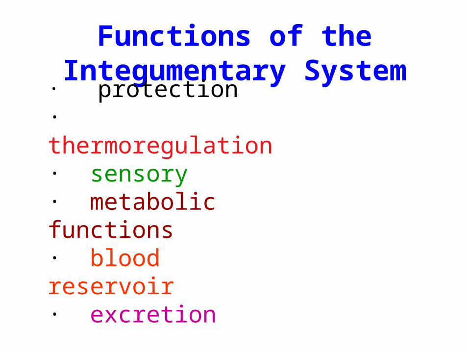
Functions of the Integumentary System
· protection· thermoregulation· sensory· metabolic functions· blood reservoir· excretion

Anatomy of the Skin
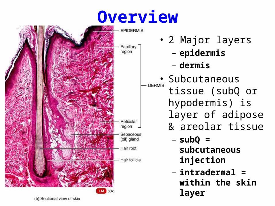
Overview• 2 Major layers
– epidermis
– dermis
• Subcutaneous tissue (subQ or hypodermis) is layer of adipose & areolar tissue– subQ = subcutaneous
injection
– intradermal = within the skin layer

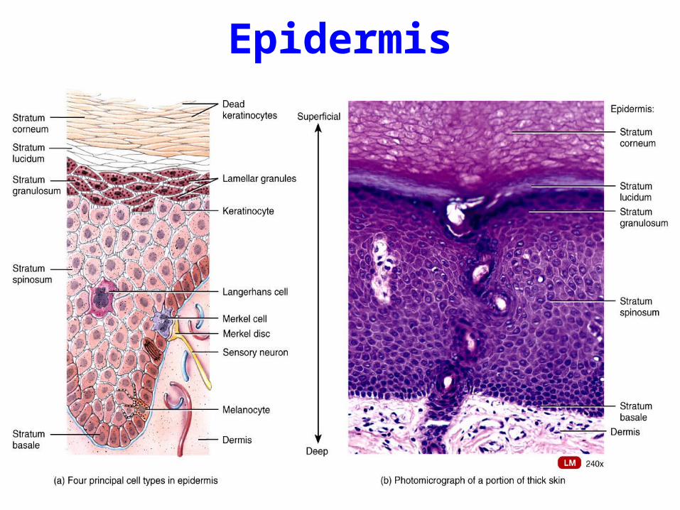
Epidermis• Stratified squamous
epithelium• Contains no blood
vessels• 4 types of cells• 5 distinct strata (layers)
of cells– stratum corneum
– stratum lucidum
– stratum granulosum
– stratum spinosum
– stratum basale
Epidermis

Cell types of the Epidermis• Keratinocytes: 90%
– produce keratin
• Melanocytes: 8%– produces melanin – melanin transferred to other
cells with long cell processes
• Langerhans cells– from bone marrow– provide immunity
(macrophages)
• Merkel cells– in deepest layer– form touch receptor with
sensory neuron

Layers of the Epidermis
• Stratum corneum• Stratum lucidum• Stratum granulosum• Stratum spinosum• Stratum basale

Epidermis

Dermis
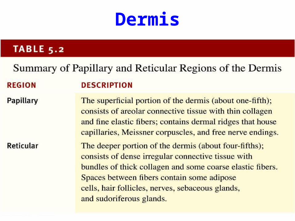
Dermis
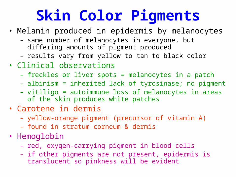
Skin Color Pigments• Melanin produced in epidermis by melanocytes
– same number of melanocytes in everyone, but differing amounts of pigment produced
– results vary from yellow to tan to black color
• Clinical observations– freckles or liver spots = melanocytes in a patch– albinism = inherited lack of tyrosinase; no pigment– vitiligo = autoimmune loss of melanocytes in areas of the skin produces
white patches
• Carotene in dermis– yellow-orange pigment (precursor of vitamin A)– found in stratum corneum & dermis
• Hemoglobin– red, oxygen-carrying pigment in blood cells– if other pigments are not present, epidermis is translucent so pinkness will
be evident

Skin Color as Diagnostic Clue• Jaundice
– yellowish color to skin and whites of eyes
– buildup of yellow bilirubin in blood from liver disease
• Cyanotic– bluish color to nail beds and skin
– hemoglobin depleted of oxygen looks purple-blue
• Erythema– redness of skin due to enlargement of capillaries in dermis
– during inflammation, infection, allergy or burns
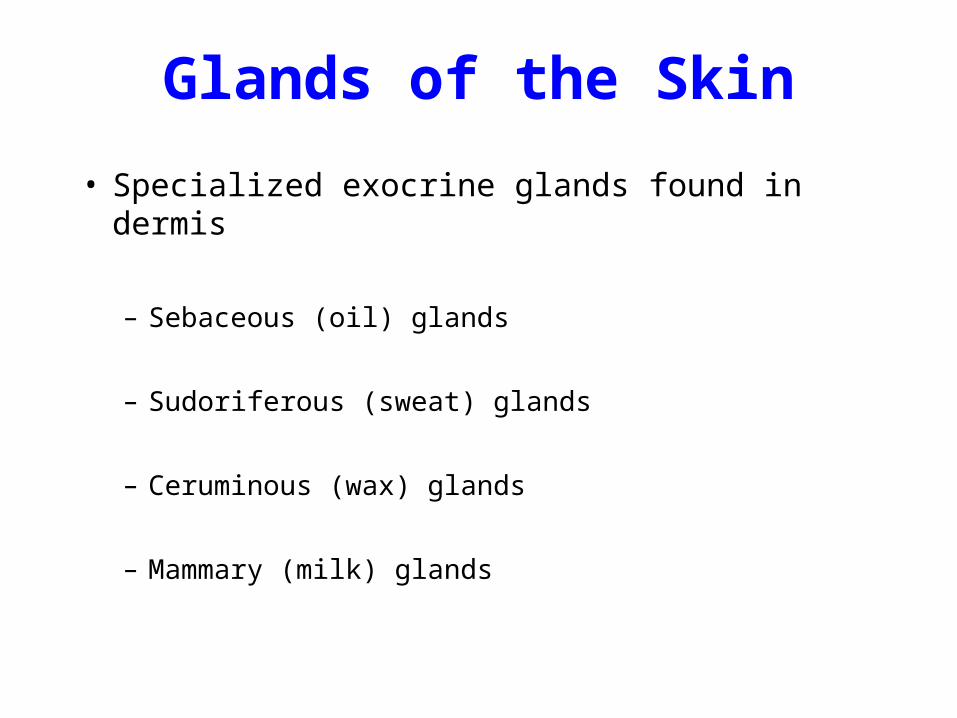
Glands of the Skin
• Specialized exocrine glands found in dermis
– Sebaceous (oil) glands
– Sudoriferous (sweat) glands
– Ceruminous (wax) glands
– Mammary (milk) glands
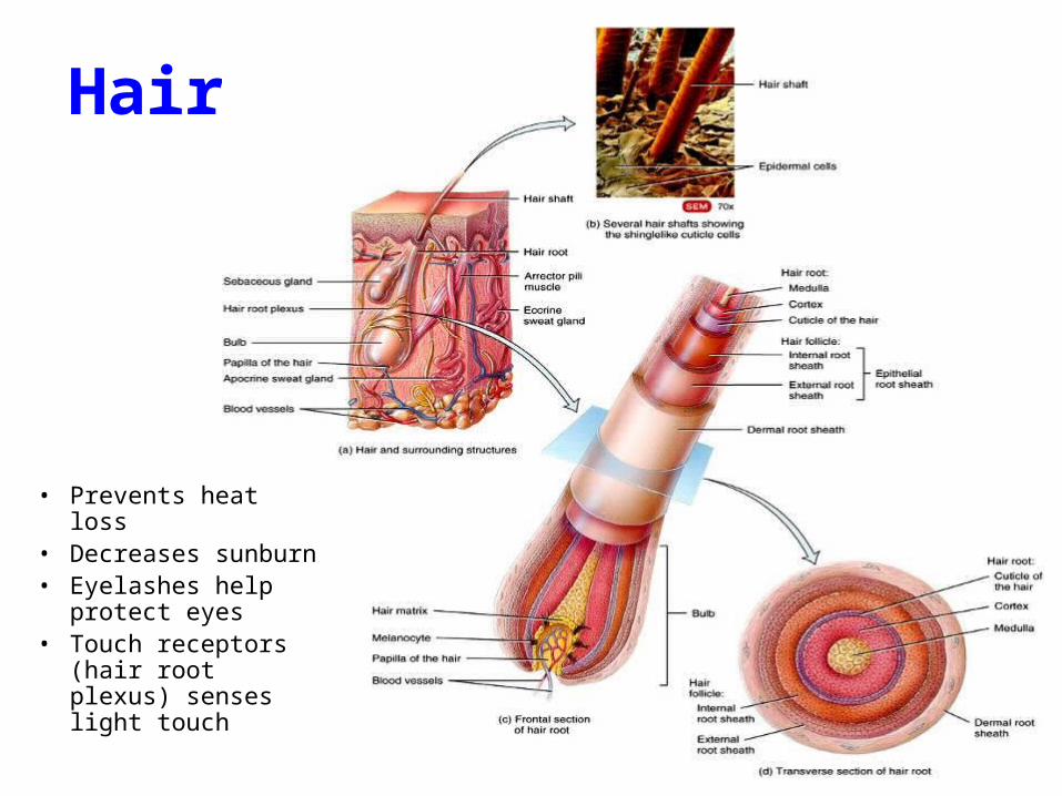
Hair
• Prevents heat loss• Decreases sunburn• Eyelashes help
protect eyes• Touch receptors
(hair root plexus) senses light touch

Nails

Burns
Rule of
Nines
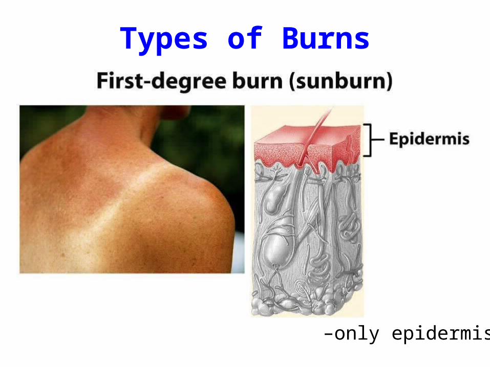
Types of Burns
–only epidermis

Types of Burns
• destroys entire epidermis & part of dermis • fluid-filled blisters separate epidermis & dermis• epidermal derivatives are not damaged• heals without grafting in 3 to 4 weeks & may scar
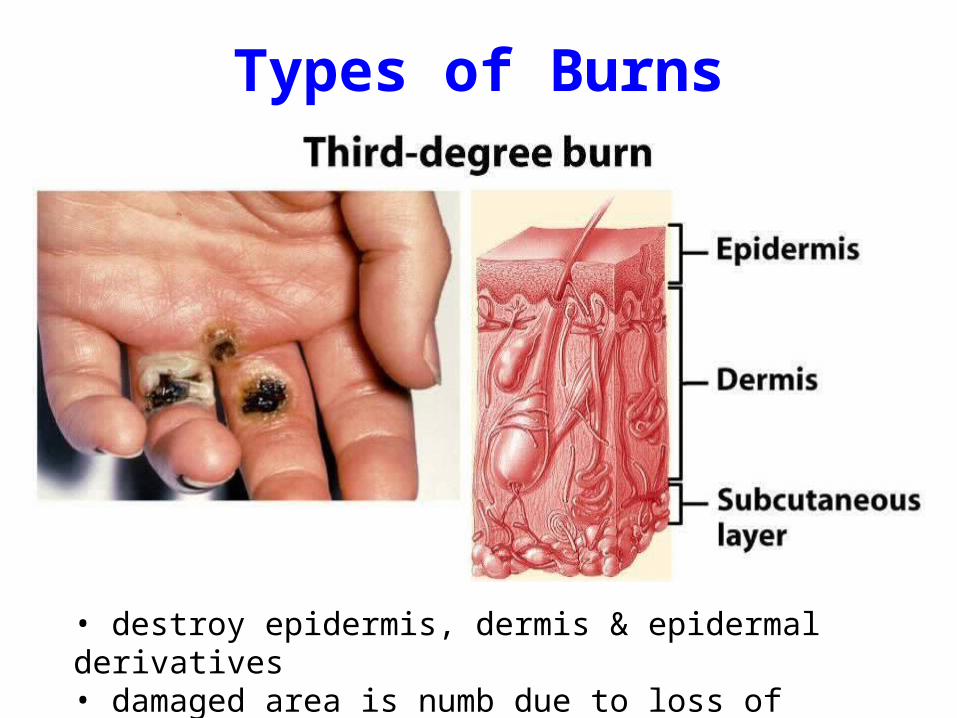
Types of Burns
• destroy epidermis, dermis & epidermal derivatives• damaged area is numb due to loss of sensory nerves

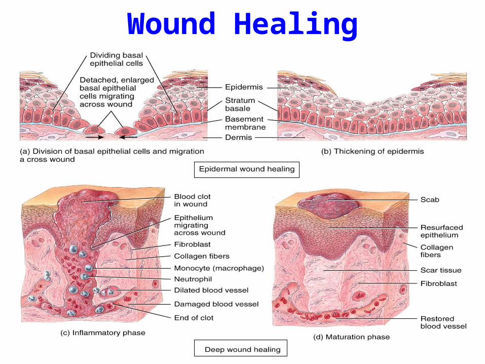
Wound Healing Two kinds of wound-healing processes can occur,
depending on the depth of the injury.
– Epidermal wound healing occurs following superficial
wounds that affect only the epidermis.
• Return to normal function is the rule.
– Deep wound healing occurs when an injury extends to the
dermis and subcutaneous layer.
• Loss of some function and development of scar tissue is the rule.
Wound Healing