interaction of dinuclear ruthenium(ii) supramolecular cylinders with dna: sequence-specific binding,...
TRANSCRIPT

DOI: 10.1002/chem.200801364
Interaction of Dinuclear Ruthenium(II) Supramolecular Cylinders withDNA: Sequence-Specific Binding, Unwinding, and Photocleavage
Jaroslav Malina,[a] Michael J. Hannon,[b] and Viktor Brabec*[a]
Introduction
Since DNA encodes and regulates most aspects of life itrepresents a very important potential drug target. Thedesign of agents that can bind and react in a sequence-spe-cific manner with DNA is of a great importance in probingbiological processes and in developing therapeutic drugs. Inthis field, ruthenium complexes have attracted particular in-terest.[1–5] In accordance with their chirality and photophysi-cal and photoredox properties, they offer many possibilitiesfor use in biochemical applications such as chiral or lumines-cent DNA probes, chemical photonucleases, and DNA pho-toreagents. A number of ruthenium compounds have at-tracted interest as new metal-based antitumor drugs, andtwo of them—NAMI-A and KP1019—have reached clinical
trials. Most of the studied complexes can undergo coordina-tive binding to biomolecules, and a few are coordinativelysaturated yet display some anticancer activity mediated bynoncovalent biomolecule binding.
Recently, metallosupramolecular chemistry has been usedto design a new class of synthetic agents, namely, tetracat-ionic supramolecular cylinders, that bind strongly and non-covalently in the major groove of DNA and cause remark-able intramolecular coiling of DNA.[6–8] In these cylinders,for example, [FeII
2L3]4+ (Figure 1 a and b), three bis-pyridyl-ACHTUNGTRENNUNGimine ligand strands are wrapped in a helical fashion about
two metal centers, and face–edge p–p and metal–ligand in-teractions contribute to the structural rigidity of the unit.
Efforts to combine the striking DNA-binding features ofmetallosupramolecular cylinders with the photoactive prop-erties of ruthenium recently came to fruition, and a new dir-uthenium triple-stranded cylinder based on the design of theprevious iron cylinder [Fe2L3]
4+ was prepared.[9,10] X-raycrystallography demonstrated that the structure of [Ru2L3]
4+
(Figure 1 a and c) is analogous to that of the correspondingiron(II) cylinder. Fluorescence, CD, and LD studies[10]
showed that [Ru2L3]4+ binds and coils DNA. In addition,
the high stability of this compound, due to the inert ruthe-nium(II) centers, makes this type of agent particularly suita-ble for use in biological studies and perhaps as chemothera-peutics. It exhibits cytotoxic activity against human breast
Abstract: Metallosupramolecularchemistry was used to design a newclass of synthetic agents, namely, tetra-cationic supramolecular cylinders, thatbind strongly and noncovalently in themajor groove of DNA. To gain addi-tional information on interactions ofthe cylinders with DNA we exploredDNA unwinding and sequence-specificbinding properties, as well as DNAphotonuclease activity of ruthenium(II)metallosupramolecular cylinder[Ru2L3]
4+ , where L is a bis-pyridyl-
ACHTUNGTRENNUNGimine ligand. We found that [Ru2L3]4+
unwinds negatively supercoiled plasmidDNA and exhibits binding preferenceto regular alternating purine–pyrimi-dine sequences in a similar way to the[Fe2L3]
4+ analogue. Photocleavagestudies showed that, unlike [Fe2L3]
4+ ,
[Ru2L3]4+ induces single-strand breaks
on irradiation by visible and UVA lightand cleaves DNA mainly at guanineresidues contained preferentially inregularly alternating purine–pyrimidinenucleotides. As [Ru2L3]
4+ binds andcleaves DNA in a sequence-dependentmanner, it may provide a useful toolfor basic and applied biology, such asfor controlled manipulation of thegenome.
Keywords: DNA cleavage · DNAunwinding · helical structures ·ruthenium · supramolecular chemis-try
[a] Dr. J. Malina, Prof. Dr. V. BrabecInstitute of BiophysicsAcademy of Sciences of the Czech Republic, v.v.i.Kralovopolska 135, 61265 Brno (Czech Republic)Fax: (+420) 541-240-499E-mail : [email protected]
[b] Prof. Dr. M. J. HannonSchool of Chemistry, University of BirminghamEdgbaston, Birmingham B15 2TT (UK)
Supporting information for this article is available on the WWWunder http://dx.doi.org/10.1002/chem.200801364.
� 2008 Wiley-VCH Verlag GmbH & Co. KGaA, Weinheim Chem. Eur. J. 2008, 14, 10408 – 1041410408
cancer cells HBL-100 and T47D,[10] which classifies it as amember of a new and promising group of noncovalentDNA-binding anticancer metallodrugs. A key goal of devel-oping ruthenium cylinders was to harness the potential pho-tochemical properties of ruthenium diimine centers withinthe cylinders. Herein we show that these agents have DNAphotocleavage properties and that this can be used for DNAfootprinting. Together with DNA unwinding assays we usethis cylinder footprinting and competitive deoxyribonucle-ACHTUNGTRENNUNGase I (DNase I) footprinting studies to probe DNA recogni-tion and specificity of these cylinders. The results reveal that[Ru2L3]
4+ unwinds negatively supercoiled plasmid DNA andpreferentially binds to regular alternating purine–pyrimidinesequences in a similar way to [Fe2L3]
4+ .[11] Photocleavagestudies show that, unlike [Fe2L3]
4+ , [Ru2L3]4+ induces single-
strand breaks on irradiation by visible and UVA light andcleaves DNA mainly at guanine residues by generating sin-glet oxygen.
Results
Ethidium bromide displacement : The binding strength of[Ru2L3]
4+ to calf thymus (ct) DNA was quantified by meansof the competition between cylinders and ethidium bromide(EtBr), as in our previous work.[11] Displacement of EtBrfrom all studied DNAs was accompanied by a decrease inthe fluorescence intensity measured at 595 nm. The appar-ent binding constant Kapp for [Ru2L3]
4+ binding to ct DNA,
calculated as described in the Experimental Section, was5.8 � 107
m�1. Thus, this competitive binding study shows that
[Ru2L3]4+ has a binding affinity for ct DNA similar to that
of [Fe2L3]4+ .[11]
Unwinding of negatively supercoiled DNA : Local unwind-ing is a pronounced conformational alteration induced inDNA by [Fe2L3]
4+ .[11] Hence, it was of interest to examineinduction of DNA unwinding by its ruthenium counterpartand compare it with DNA unwinding by [Fe2L3]
4+ . Figure 2
shows an electrophoresis gel in which increasing amounts of[Ru2L3]
4+ have been bound to a mixture of relaxed and neg-atively supercoiled pUC19 DNA. The unwinding angle isgiven by F=�18 s/rb(c), where s is the superhelical densityand rb(c) the number of cylinders bound per nucleotide forwhich the supercoiled and relaxed forms co-migrate.
The number of cylinders bound per nucleotide was takento be equal to the mixing ratios, under the assumption thatall cylinders present in the sample are completely bound toDNA. This assumption is substantiated by the large appar-ent binding constant (5.8 �107
m�1) determined for binding
of [Ru2L3]4+ to DNA with a random nucleotide sequence
(vide infra; strong association is characterized by Kapp>
106m�1).[12] The high value of Kapp and the low ratio of cylin-
ders to DNA molecules in the experiment implies that atequilibrium almost no free cylinders will be present. Underthe present experimental conditions, s was calculated to be�0.058 on the basis of the data of cisplatin, for which rb(c)was determined in this study and F= 138 was assumed. Byusing this approach a DNA unwinding angle of 13�28 wasdetermined for [Ru2L3]
4+ . This is less than that induced byiron(II) cylinders (27�38,[11]) which is surprising given theirvery similar sizes and identical charges. This may reflectsubtle effects arising from differences in the extent of polari-zation of the protons on the exterior of these cylinders.
Photocleavage of DNA
Plasmid DNA photocleavage : On irradiation with UVA(365 nm, irradiation at intraligand spectroscopic bands ofthe cylinder; Figure 3 a) or Vis light (lmax�580 nm, irradia-tion at the metal-to-ligand charge-transfer (MLCT) bands ofthe cylinder; Figure 3 b), [Ru2L3]
4+ induces cleavage of plas-mid DNA from the supercoiled form (sc) to the nicked form(oc). The amount of nicked DNA increases with irradiation
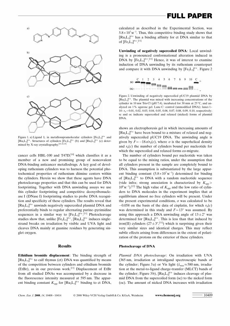
Figure 1. a) Ligand L in metallosupramolecular cylinders [Fe2L3]4+ and
[Ru2L3]4+ . Structures of cylinders [Fe2L3]
4+ (b) and [Ru2L3]4+ (c) deter-
mined by X-ray crystallography.[10, 24, 25]
Figure 2. Unwinding of negatively supercoiled pUC19 plasmid DNA by[Ru2L3]
4+ . The plasmid was mixed with increasing concentrations of thecylinder in 10 mm Tris·Cl (pH 7.4), incubated for 30 min at 25 8C, and an-alyzed on 1% agarose gel. Lane C: control (unmodified DNA); lanes 1–10: rb =0.01, 0.02, 0.03, 0.04, 0.05, 0.06, 0.07, 0.08, 0.09, 0.10, respectively;sc and oc indicate supercoiled and relaxed (nicked) forms of plasmidDNA.
Chem. Eur. J. 2008, 14, 10408 – 10414 � 2008 Wiley-VCH Verlag GmbH & Co. KGaA, Weinheim www.chemeurj.org 10409
FULL PAPER

time and cylinder concentration (Figure 4). The absence ofthe bands corresponding to the linear form of DNA on thegel indicates that cleavage involves only one strand of DNAat these low cylinder concentrations.
Mechanism of photocleavage : Ruthenium complexes caninduce single-strand DNA breaks on irradiation by differentphotochemical processes such as singlet-oxygen production,hydroxyl-radical formation, and electron transfer. To deter-mine which process is responsible for photoactivated cleav-age of plasmid DNA, photocleavage of pUC19 in the pres-
ence of [Ru2L3]4+ and different inhibitors was examined
(Figure 5). Since plasmid cleavage is not inhibited in thepresence of hydroxyl radical (OHC) scavengers such as man-nitol (Figure 5 a, lane 3)[13] and DMSO (Figure 5 a, lane 4)[14]
even at high concentrations, the hydroxyl radical is unlikelyto be responsible for cleavage. In the presence of superoxidedismutase (SOD), an effective quencher of the superoxideanion radical (O2C
�), cleavage was enhanced (Figure 5 a,lane 5). Strong enhancement of photonuclease activity bySOD was also observed for [RuACHTUNGTRENNUNG(bpz)3]
2+ (bpz= 2,2’-bipyra-zyl),[15] and this effect was partly attributed to increased pro-duction of singlet oxygen (1O2) and an electron-transfer pro-cess.[16] To test the possibility that photoinduced cleavage in-volves formation of singlet oxygen, cleavage was carried outin the presence of sodium azide and D2O. While sodiumazide is one of the most effective singlet-oxygen quench-ers,[17] 1O2 would be expected to induce more strand scissionin D2O than in H2O due to its longer lifetime in the formersolvent.[18] As shown in Figure 5 a (lanes 6 and 7), cleavageof pUC19 by [Ru2L3]
4+ was inhibited in the presence ofsodium azide and enhanced in D2O, that is, 1O2 is likely tobe responsible for the cleavage reaction.
Stability of [Ru2L3]4+ under irradiation by UVA and Vis
light was tested under the same experimental conditions,and no decomposition of the cylinder was observed.
Photocleavage of a DNA restriction fragment : The gels inFigure 6 a and b show the photocleavage activity of[Ru2L3]
4+ on irradiation by UVA and Vis light, respectively.The main targets of the photocleavage activity on irradiationby both UVA and Vis light are identical and are identifiedas guanine residues, which are known to be preferentially at-tacked by singlet oxygen at neutral pH. This is in agreement
Figure 3. Agarose gels showing photocleavage of pUC19 by [Ru2L3]4+ on
irradiation by UVA (a) and Vis (b) light. Lane 1: DNA in the absence ofmetal complex incubated in the dark for 80 (a) or 120 min (b); lanes 2and 3: DNA incubated in the dark for 80 (a) or 120 min (b) in the pres-ence of [Ru2L3]
4+ at 100:1 and 50:1 ratio, respectively; lane 4: DNA inthe absence of the cylinder irradiated for 80 min by UVA (a) or for120 min by Vis (b); lanes 5–7: DNA in the presence of [Ru2L3]
4+ at 100:1ratio irradiated for 20, 40, and 80 min by UVA (a) or for 30, 60, and120 min by Vis (b), respectively; lanes 8–10: DNA in the presence of[Ru2L3]
4+ at 50:1 ratio irradiated for 20, 40, and 80 min by UVA (a) orfor 30, 60 and 120 min by Vis (b), respectively; sc and oc indicate super-coiled and relaxed (nicked) forms of plasmid DNA.
Figure 4. Time dependence of photocleavage activity of [Ru2L3]4+ on ir-
radiation by UVA (a) and Vis (b) light. & and & represent percentage ofnicked DNA in the reaction mixtures containing [Ru2L3]
4+ at 100:1 and50:1 ratios, respectively.
Figure 5. a) Photoactivated cleavage of 200 mm pUC19 in the presence of2 mm [Ru2L3]
4+ and different inhibitors after irradiation by UVA light for100 min in 10 mL of 10 mm Tris (pH 7.4). Lane 1: pUC19 in the absenceof [Ru2L3]
4+ , no inhibitor; lane 2: pUC19 and [Ru2L3]4+ , no inhibitor;
lane 3: in the presence of mannitol (100 mm); lane 4: in the presence ofDMSO (200 mm); lane 5: in the presence of superoxide dismutase(1000 UmL�1); lane 6: in the presence of sodium azide (10 mm); lane 7:pUC19 and [Ru2L3]
4+ in D2O (>90% D2O). b) Bar-graph representationof the effect of inhibitors on the Vis- (light bars) and UVA-activated(dark bars) cleavage activity of [Ru2L3]
4+ .
www.chemeurj.org � 2008 Wiley-VCH Verlag GmbH & Co. KGaA, Weinheim Chem. Eur. J. 2008, 14, 10408 – 1041410410
V. Brabec et al.

with a photocleavage mechanism involving production ofsinglet oxygen, as suggested by previous results, possibly incombination with an electron-transfer pathway. The sites ofphotocleavage on irradiation by UVA light are summarizedin Figure 7 and compared with preferential binding sites(vide infra). The sites of photocleavage upon irradiation byVis light are identical (see Figure 6 a and b).
DNase I footprinting : The 158 base-pair (bp) restrictionfragment of pSP73 plasmid identical to that used in the pho-
tocleavage experiment (Figures 6 and 7) was also employedfor DNase I footprinting. [Ru2L3]
4+ was mixed with the158 bp restriction fragment at base:cylinder ratios of 6:1,8:1, 10:1, and 20:1, and then partial cleavage by DNase Iwas performed. The autoradiogram of the DNA cleavage-in-hibition patterns is shown in Figure 8. The extent ofDNase I cleavage varies along the fragment sequence. Inter-estingly, cutting is strongly reduced in several sequenceseven in the absence of the cylinder (Figure 8, lane 5). In thepresence of the cylinder several footprints in the gel demon-strate that [Ru2L3]
4+ is capable of recognizing specific DNAsequences. To obtain further information on the binding spe-cificity of [Ru2L3]
4+ , intensities from the gel lanes containingDNA and Ru cylinder at base:cylinder ratios of 8:1 and 10:1were measured by densitometry. The resulting differentialcleavage plots are shown in Figure 9 a. Negative values indi-cate sites of drug protection against DNase I cleavage, andpositive values indicate regions of drug-induced enhance-ment of cleavage. A stretch of DNA of about 70 bp withinthe 158 bp restriction fragment was sufficiently well resolvedthat cleavage could be quantified. There are four main short
Figure 6. Autoradiograms of 13 % polyacrylamide denaturing gels show-ing photocleavage of 158 bp fragment by [Ru2L3]
4+ on irradiation byUVA (a) and Vis (b) light. Lanes 1, 2: DNA in the absence of [Ru2L3]
4+
irradiated for 4 and 2 h, respectively; lanes 3, 4: DNA in the presence of[Ru2L3]
4+ irradiated for 4 and 2 h, respectively; lanes G+A and G corre-spond to Maxam–Gilbert G+A and G ladders. The nucleotide sequenceof the fragment and the peak areas corresponding to each band areshown on the right side of the gel; gray line, Maxam–Gilbert G ladder,black line, DNA with [Ru2L3]
4+ irradiated for 4 h.
Figure 7. Part of the sequence of 158-mer HindIII/NdeI fragment of theplasmid pSP73 showing preferential binding (shown as light bars) andphotocleavage (indicated by arrows) sites of [Ru2L3]
4+ . The binding siteswere obtained by shifting the sites of inhibited DNase I cleavage by 2 bpin the 3’ direction.
Figure 8. Autoradiogram of DNase I footprint of 3’-end-labeled bottomstrand of 158-mer HindIII/NdeI restriction fragment of plasmid pSP73 inthe presence of different concentrations of [Ru2L3]
4+ . Lanes 1–4: DNAmixed with [Ru2L3]
4+ at 6:1, 8:1, 10:1 and 20:1 (base:cylinder) ratios, re-spectively; lane 5: DNA in the absence of cylinder; lanes G+A and Gcorrespond to Maxam–Gilbert G+A and G ladders. The nucleotide se-quence of the fragment is shown on the right side of the gel, and num-bers refer to the sequence shown in the corresponding differential cleav-age plots in Figure 9.
Chem. Eur. J. 2008, 14, 10408 – 10414 � 2008 Wiley-VCH Verlag GmbH & Co. KGaA, Weinheim www.chemeurj.org 10411
FULL PAPERInteraction of Dinuclear RuII Supramolecular Cylinders with DNA

nucleotide sequences protected by [Ru2L3]4+ , around posi-
tions 30, 52, 73, and 89. These sequences consist of regularlyalternating purine–pyrimidine nucleotides. Differentialcleavage plots of [Ru2L3]
4+ and the M enantiomer of[Fe2L3]
4+ (M- ACHTUNGTRENNUNG[Fe2L3]4+) at base:cylinder ratios of 10:1 and
20:1 are compared in Figure 9 b. The Ru and Fe cylindersexhibit very similar patterns of protection and enhancement,but the extent of protection by [Ru2L3]
4+ is somewhatweaker.
To identify the sites of cylinder binding from the sites ofinhibited DNase I cleavage, a 3’ shift of about 2–3 base pairsmust be considered because of the bias introduced by thenuclease on DNA cleavage.[19,20] The resulting binding sitesof [Ru2L3]
4+ are summarized in Figure 7 and compared withphotocleavage sites. Notably, the strongest photocleavagesites are situated at the preferential DNA binding sites or intheir close proximity.
Conclusion
Metal-based compounds that bind to DNA in a differentway to conventional cisplatin and its analogues, particularlythose that bind by noncovalent interactions, have considera-ble potential as anticancer agents with a new spectrum ofactivity. Modification of DNA secondary structure by bind-ing of molecules of biological significance is an importantaspect of recognition by DNA processing proteins in thecell.
The results of the present and earlier[10] work demonstratethat several features of target DNA binding mode of the
ruthenium(II) metallosupramolecular cylinder and its ironcounterpart are similar, which is not unexpected since bothcylinders have essentially the same size and charge. A some-what surprising result was that [Ru2L3]
4+ unwinds DNA no-ticeably less than [Fe2L3]
4+ . At present, we have no conclu-sive explanation for this observation. It cannot be excludedthat the difference in DNA unwinding is a consequence ofvery subtle differences in the structures of the two cylinders,consistent with the working hypothesis that the size andshape of the cylinder are crucial for recognition of thepurine–pyrimidine tracts to which they preferentially bind(Figure 7 and ref. [11]). Interestingly, [Ru2L3]
4+ unwinds theDNA duplex by 138, that is, to an extent similar to DNA un-winding by cisplatin,[21] which is also a low molecular massagent but has a nonintercalative DNA binding mode.
An intriguing feature of DNA binding of [Ru2L3]4+ is its
highly effective DNA-photocleavage ability, a feature notaccessible with [Fe2L3]
4+ . Mechanistic studies reveal thatsinglet oxygen (1O2) may play an important role in photo-cleavage. In addition, [Ru2L3]
4+ exhibits sequence selectivityin DNA photocleavage with a preference for regularly alter-nating purine–pyrimidine nucleotides. Agents showing pho-toinduced cleavage of DNA have significant advantage overtheir “chemical nuclease” analogues in that other chemicalreagents, such as a reducing agent and/or H2O2, is not re-quired for their activity. Besides, compounds cleaving DNAon photoactivation usually show localized effects in thera-peutic applications and are much less toxic in the absence oflight, so that they are particularly useful in photodynamictherapy as specific photoreagents.
The results described here may help to further understandthe selectivity and efficiency of DNA recognition and cleav-age by dinuclear ruthenium(II) metallosupramolecular cylin-ders, as well as in developing new useful DNA probes andeffective metal-based nucleases. The design of moleculesthat bind and cleave a selected DNA sequence provides anintriguing opportunity for basic and applied biology. For ex-ample, such molecules offer new prospects for controlledmanipulation of the genome. In addition, long-wavelengthDNA cleavage activity makes these ruthenium(II) metallo-supramolecular cylinders potential candidates for furtherdesign of molecules suitable for photodynamic therapy ap-plications and as alternatives to the agents already used inclinical photodynamic therapy.
Experimental Section
Starting materials: The synthesis of cylinder [Ru2L3]4+ (Figure 1) has
been described previously.[10] A stock solution of the cylinder was pre-pared by dissolving the solid PF6 salt in a small volume of DMSO (lessthan 10 % of the final volume) followed by dilution with water to thefinal concentration of 1 mm. The concentration was checked by UV/Visabsorbance spectroscopy by using the extinction coefficient e485 nm =
16870 m�1 cm�1. Ct DNA (42 % G +C, mean molecular mass ca. 2� 107)
was prepared and characterized as described previously.[22] PlasmidspUC19 (2686 bp) and pSP73 (2464 bp) were isolated according to stan-dard procedures. NdeI and HindIII restriction endonucleases were pur-
Figure 9. a) Differential cleavage plots for [Ru2L3]4+-induced differences
in susceptibility to DNase I digestion on the bottom strand of 158-merHindIII/NdeI fragment of the plasmid pSP73 at 8:1 (full squares) and10:1 (open squares) base:cylinder ratio. b) Comparison of differentialcleavage plots for [Ru2L3]
4+- (open squares) and M-[Fe2L3]4+-induced
(full squares) differences in susceptibility to DNase I digestion on thebottom strand of 158-mer HindIII/NdeI fragment at 10:1 and 20:1 base:cylinder ratio, respectively (data for M-[Fe2L3]
4+ obtained under other-wise identical experimental conditions were taken from ref. [11]). Verticalscales are in units of ln(fc)�ln(f0), where fc is the fractional cleavage atany bond in the presence of cylinder, and f0 the fractional cleavage of thesame bond in the control, given similar extents of overall digestion. Posi-tive values indicate enhancement, and negative values inhibition.
www.chemeurj.org � 2008 Wiley-VCH Verlag GmbH & Co. KGaA, Weinheim Chem. Eur. J. 2008, 14, 10408 – 1041410412
V. Brabec et al.

chased from New England Biolabs (Beverly, MA). [a-32P]-dATP was ob-tained from MP Biomedicals, LLC (Irvine, CA). The Klenow fragmentfrom DNA polymerase I (exonuclease minus, mutated to remove the 3’-5’ proofreading domain), KF�, was purchased from Takara (Japan). Ac-rylamide and bis-acrylamide were obtained from Merck KGaA (Darm-stadt, Germany), and agarose from FMC BioProducts (Rockland, ME,USA). The Wizard SV and PCR Clean-Up System used to extract andpurify the 158 bp DNA fragment (vide infra) was purchased from Prome-ga. EtBr and deuterium oxide (D2O) were purchased from MerckKGaA, and DNase I was obtained from Roche (Mannheim, Germany).Superoxide dismutase, mannitol, sodium azide, and DMSO were ob-tained from Sigma-Aldrich (Prague). The 158 bp DNA fragment was pre-pared by digesting supercoiled pSP73 plasmid with NdeI restriction endo-nuclease and 3’-end-labeled by treatment with KF� and [a-32P]-dATP.After radioactive labeling, the DNA cleaved with NdeI was digested withHindIII. The cleavage resulted in 158 and 2306 bp fragments. The 158 bpfragment was purified by electrophoresis on 1% agarose gel and isolatedfrom the gel by Promega Wizard SV Gel cleanup system.
Competition assays : The competition assays were all undertaken withfixed DNA and competitor (EtBr) concentrations and variable helicateconcentration. The DNA-EtBr complexes were excited at 546 nm andthe fluorescence was measured at 595 nm. Aliquots of a 1 mm stock solu-tion of the cylinders were added to the solution of EtBr and DNA(10 mm Tris pH 7.4, 1 mm ethylenediaminetetraacetic acid (EDTA),1.3 mm EtBr, and 3.9 mm DNA), and the fluorescence was measured aftereach addition until it was reduced to 50%. In general the experimentswere designed so that the weaker binder was displaced by the strongerone. The apparent binding constants Kapp for both enantiomers were cal-culated from KEB[EB] =Kapp ACHTUNGTRENNUNG[drug], where [EB] is the concentration ofEtBr (1.3 mm), [drug] is the concentration of cylinders at 50% reductionof fluorescence, and KEB is known (KEB = 1�107
m�1 for ct DNA).[23]
Unwinding of negatively supercoiled DNA: Unwinding of closed circularsupercoiled pUC19 plasmid DNA was assayed by an agarose gel mobilityshift assay.[21] The unwinding angle F induced per cylinder bound toDNA was calculated by determination of the cylinder:base ratio at whichcomplete transformation of the supercoiled to the relaxed form of theplasmid was attained. An aliquot of the sample was subjected to electro-phoresis on 1% native agarose gel running at 25 8C in the dark with Tris-acetate/EDTA (TAE) buffer and the voltage set at 25 V. The gels werethen stained with EtBr and photographed with a transilluminator.
Photocleavage experiments
Instrumentation : The light source used in DNA photocleavage experi-ments was a Photoreactor LZC-ICH2 from Luzchem (Canada) fittedwith UVA lamps (4.3 mW cm�2, lmax 365 nm) or with Vis lamps (coolwhite fluorescent tubes, 400–700 nm with a maximum around 580 nm,2.8 mW cm�2). The temperature in the light chamber during irradiationwas approximately 37 8C.
Photocleavage of plasmid pUC19 : Photocleavage reactions were carriedout in 10 mL volumes contained in 0.65 mL eppendorf tubes. Reactionmixtures containing plasmid DNA pUC19 and [Ru2L3]
4+ at 100:1 and50:1 (DNA base:cylinder) ratios in 10 mm Tris·HCl (pH 7.4) were irradi-ated by UVA light for 20, 40, and 80 min or by Vis light for 30, 60, and90 min. Higher mixing ratios than 50:1 were not used because the cylin-der unwinds the supercoiled form of the plasmid and reduces its mobilityin the gel, which complicates analysis of the results. All samples werethen mixed with loading buffer and loaded onto a 1 % agarose gel run-ning at 25 8C in the dark with TAE buffer and the voltage set at 25 V.The gels were then stained with EtBr, followed by photography withtransilluminator.
Photocleavage of 158 bp DNA fragment : Photocleavage reactions werecarried out in 5 mL volumes contained in 0.65 mL eppendorf tubes. Thereaction mixtures were prepared with 150 mm ct DNA (0.048 mg/ mL,150 mm related to the phosphorus content) containing 3’-end-labeled re-striction fragment and 7.5 mm ruthenium cylinder in 10 mm Tris·HCl(pH 7.4). The samples were irradiated by UVA or Vis light for 2 or 4 hand then analyzed on 13% polyacrylamide (PAA) gel under denaturingconditions.
Stability of [Ru2L3]4+ on irradiation: Stability of [Ru2L3]
4+ on irradiationby UVA and Vis light was tested in the following way. [Ru2L3]
4+ was di-luted in 10 mm Tris·HCl, pH 7.4 to a concentration of 1 � 10�5
m and irra-diated for 120 min. Absorbance at 485 nm, which is a maximum of theMLCT band indicative of stability of the cylinder, was measured andcompared.
DNase I footprinting: One subclass of footprinting agents that has beendeveloped for determining the sequence-specific binding of small mole-cules to DNA comprises enzymes such as DNase I.[20] DNase I is an en-donuclease that specifically cleaves the O3’�P bond of the phosphodiest-er backbone of the double-helical DNA substrate. A solution (9 mL) con-taining 1.11 � TKMC buffer (10 mm Tris pH 7.9, 10 mm KCl, 10 mm
MgCl2, and 5 mm CaCl2), 3’-end-labeled restriction fragment, 4.5� 10�4m
ct DNA (144 mg/ mL, 4.5� 10�4m related to the phosphorus content), and
cylinder was incubated for 15 min at 25 8C. Cleavage was initiated by ad-dition of 1 mL of 50 mg DNase I per milliliter and allowed to continue for30 s at room temperature before quenching with 2.5 mL of DNase I stopsolution (3 m NH4OAc and 0.25 m EDTA). Optimal enzyme dilutionswere established in preliminary calibration experiments. The sample wasthen precipitated with ethanol, lyophilized, and resuspended in a form-ACHTUNGTRENNUNGamide loading buffer. DNA cleavage products were resolved by PAA gelelectrophoresis under denaturing conditions (13 %/8 m urea PAA gel).The autoradiograms were visualized and quantified by using the bio-imaging analyzer. Assignment of the cleavage to a particular base wasmade so that it corresponds to the cleavage of the phosphodiester bondon the 5’ side of that base.
Other physical methods : Absorption spectra were measured with aVarian Cary 4000 UV/Vis spectrophotometer equipped with a thermo-electrically controlled cell holder and quartz cells with a path length of1 cm. The PAA gels were visualized by using a BAS 2500 Fujifilm bio-imaging analyzer, and the radioactivities associated with bands werequantitated with the AIDA image analyzer software (Raytest, Germany).
Acknowledgements
This research was supported by the Academy of Sciences of the CzechRepublic (Grants B400040601, IAA400040803, 1QS500040581,KAN200200651, AV0Z50040507, AV0Z50040702), the Ministry of Edu-cation of the Czech Republic (MSMT LC06030, ME08017), the GrantAgency of the Ministry of Health of the CR (NR8562-4/2005) and con-ducted in the context of COST D39 (WGs D39/002/07 and D39/004/06).We thank Gabriel Pascu for cylinder synthesis.
[1] M. J. Clarke, Coord. Chem. Rev. 2002, 232, 69 –93.[2] M. Galanski, V. B. Arion, M. A. Jakupec, B. K. Keppler, Curr.
Pharm. Des. 2003, 9, 2078 – 2089.[3] E. Alessio, G. Mestroni, A. Bergamo, G. Sava, Curr. Topics Med.
Chem. 2004, 4, 1525 – 1535.[4] V. Brabec, O. Novakova, Drug Resist. Updates 2006, 9, 111 –122.[5] C. A. Vock, W. H. Ang, C. Scolaro, A. D. Phillips, L. Lagopoulos, L.
Juillerat-Jeanneret, G. Sava, R. Scopelliti, P. J. Dyson, J. Med. Chem.2007, 50, 2166 –2175.
[6] M. J. Hannon, Chem. Soc. Rev. 2007, 36, 280 –295.[7] M. J. Hannon, L. J. Childs, Supramol. Chem. 2004, 16, 7 –22.[8] M. J. Hannon, V. Moreno, M. J. Prieto, E. Moldrheim, E. Sletten, I.
Meistermann, C. J. Isaac, K. J. Sanders, A. Rodger, Angew. Chem.2001, 113, 903 –908; Angew. Chem. Int. Ed. 2001, 40, 879 –884.
[9] A. C. G. Hotze, B. M. Kariuki, M. J. Hannon, Angew. Chem. 2006,118, 4957 –4960; Angew. Chem. Int. Ed. 2006, 45, 4839 – 4842.
[10] G. I. Pascu, A. C. G. Hotze, C. Sanchez-Cano, B. M. Kariuki, M. J.Hannon, Angew. Chem. 2007, 119, 4452 –4456; Angew. Chem. Int.Ed. 2007, 46, 4374 –4378.
[11] J. Malina, M. J. Hannon, V. Brabec, Nucleic Acids Res. 2008, 36,3630 – 3638.
Chem. Eur. J. 2008, 14, 10408 – 10414 � 2008 Wiley-VCH Verlag GmbH & Co. KGaA, Weinheim www.chemeurj.org 10413
FULL PAPERInteraction of Dinuclear RuII Supramolecular Cylinders with DNA

[12] S. Kemp, N. J. Wheate, D. P. Buck, M. Nikac, J. G. Collins, J. R. Al-drich-Wright, J. Inorg. Biochem. 2007, 101, 1049 –1058.
[13] C. C. Cheng, S. E. Rokita, C. J. Burrows, Angew. Chem. 1993, 105,290 – 292; Angew. Chem. Int. Ed. Engl. 1993, 32, 277 – 278.
[14] S. A. Lesko, R. J. Lorentzen, P. O. P. Tso, Biochemistry 1980, 19,3023 – 3028.
[15] E. Gicquel, N. Paillous, P. Vicendo, Chem. Commun. 1998, 997 – 998.[16] E. Gicquel, N. Paillous, P. Vicendo, Photochem. Photobiol. 2000, 72,
583 – 589.[17] J. R. Kanofsky, Photochem. Photobiol. 1991, 53, 93 –99.[18] A. U. Khan, J. Phys. Chem. 1976, 80, 2219 – 2228.[19] J. C. Dabrowiak, J. Goodisman in Quantitative Footprinting Analysis
of Drug-DNA Interactions (Ed.: N. R. Kallenbach), Academic Press,New York, 1989, pp. 143 – 174.
[20] K. R. Fox, M. J. Waring in High-resolution Footprinting Studies ofDrug-DNA Complexes Using Chemical and Enzymatic Probes,
(Eds.: J. B. Chaires, M. J. Waring), Academic Press Inc, San Diego/CA, 2001, pp. 412 –430.
[21] M. V. Keck, S. J. Lippard, J. Am. Chem. Soc. 1992, 114, 3386 –3390.[22] V. Brabec, E. Palecek, Biophys. Chem. 1976, 4, 76 –92.[23] M. D. Wyatt, B. J. Garbiras, M. K. Haskell, M. Lee, R. L. Souhami,
J. A. Hartley, Anti-Cancer Drug Des. 1994, 9, 511 – 529.[24] M. J. Hannon, C. L. Painting, A. Jackson, J. Hamblin, W. Errington,
Chem. Commun. 1997, 1807 – 1808.[25] J. M. C. A. Kerckhoffs, J. C. Peberdy, I. Meistermann, L. J. Childs,
C. J. Isaac, C. R. Pearmund, V. Reudegger, S. Khalid, N. W. Alcock,M. J. Hannon, A. Rodger, Dalton Trans. 2007, 734 –742.
Received: July 5, 2008Published online: October 8, 2008
www.chemeurj.org � 2008 Wiley-VCH Verlag GmbH & Co. KGaA, Weinheim Chem. Eur. J. 2008, 14, 10408 – 1041410414
V. Brabec et al.