international journal of medical laboratory …ijml.ssu.ac.ir/article-1-180-en.pdf · international...
TRANSCRIPT

International Journal of Medical Laboratory 2017;4(2): 123-134.
Original Article
*Corresponding Author: Department of Biochemistry, Faculty of Medicine, Iran University of Medical
Sciences, Tehran, Iran. Tel: +989352461622, Fax: +982188622742, Email: [email protected]
Bioinformatic Analysis of L-Asparaginase II from Citrobacter
Freundii 1101, Erwinia Chrysanthemi DSM 4610, E.coli
BL21 and Klebsiella Pneumoniae ATCC 10031
Khosrow Aghaiypour1Ph.D., Elham Bahreini2*Ph.D., Shiva Jafari1M.Sc.
1Gene-Bank Department, Razi Vaccine and Serum Research Institute (RVSRI), Karaj, Iran. 2Department of Biochemistry, Faculty of Medicine, Iran University of Medical Sciences, Tehran, Iran.
A B S T R A C T
Article history Received 2 Apr 2017
Accepted 14 May 2017
Available online 25 Jun 2017
Key words Asparaginase
Bioinformatic
Citrobacter freundii
E.coli
Erwinia chrysanthemi
Klebsiella pneumoniae
Background and Aims: L-Asparaginase II is a cornerstone of treatment
protocols for acute lymphoblastic leukemia. Only asparaginase II obtained from
E.coli K12 and Erwinia chrysanthemi have been used in human as therapeutic
drug. The therapeutic effects of asparaginase II from E.coli K12 and Erwinia
chrysanthemi are accompanied by side effects. It is desirable to search for other
asparaginase II sources with novel properties that could be therapeutic and
produce an enzyme with less adverse effects.
Materials and Methods: Previously, we performed the in vitro studies,
including cloning, sequencing and expression of L-asparaginase II genes (ansB)
from Citrobacter freundii 1101, Erwinia chrysanthemi DSM 4610, E.coli BL21
and Klebsiella pneumoniae ATCC 10031. In this article, the obtained results
were compared bioinformatically. The nucleotide and amino acid sequence
alignments were carried out by ClustalW2. Protein localization and signal
peptides were predicted by PSORT and SIG-Pred softwares, respectively.
Percentages of hydrophobic and hydrophilic residues were calculated by
Genscript software. The physicochemical parameters were computed using
Expasy’s ProtParam prediction server. The secondary and 3D structures were
predicted by SOPMA and the online server Phyre2, respectively. The
antigenicity of the asparaginase IIs was predicted using Semi-empirical method.
Results: E.coli BL21 and Citrobacter freundii 1101 had the most similarity in
physicochemical parameters and antigenicity with E.coli K12. Also, Erwinia
chrysanthemi DSM 4610 had the most similarity in physicochemical parameters
and antigenicity with Erwinia chrysanthemi.
Conclusions: In spite of these similarities with drug types, the potentiality of
other low-similar asparaginase IIs should also be determined and compared with
drug types.
Dow
nloa
ded
from
ijm
l.ssu
.ac.
ir at
17:
12 IR
DT
on
Thu
rsda
y A
ugus
t 30t
h 20
18

Kh. aghaiypour et al.
International Journal of Medical Laboratory 2017;4(2): 123-134. 124
Introduction
Significant increase in the amount of data
available on the internet and in public databases
combined with the increasing processing speed
of workstations has created new opportunities for
researchers to make scientific discoveries. The
vast of biological data that has thus become
accessible to the biological research community
has considerably changed biologists’ way of
doing science. Recently, “In silico“ methods and
their utility are widely practical in protein and
genome sequence analysis [1]. Computational
software provides researchers to understand
physicochemical and structural properties of
protein [2]. A large number of online tools and
servers are available from different sources for
making prediction regarding the identification
and structure of proteins. The various parameters
like sequence length, number of amino acids
and the physicochemical properties of a protein
such as molecular weight (MW), atomic
composition, extinction coefficient (EC),
isoelectric point (PI), grand average of
hydropathicity (GRAVY), aliphatic index (AI),
instability index, etc. could be computed by
various computational tools for the prediction
and characterization of protein structure [3, 4].
The amino acid sequence provides most of
the information required for determining and
characterizing the molecule’s function, physical
and chemical properties.
L-asparaginase/L-glutaminase is a generic
denomination for enzymes that catalyze the
transformation of L-asparagine or L-glutamine
into their respective acids and ammonia [5, 6].
These enzymes can be specific for asparagine,
with negligible activity against glutamine, and
thus termed asparaginases (EC 3.5.1.1), or
can catalyze both asparagine and glutamine
conversion, in which case they receive the
denomination of glutaminase-asparaginases
(EC 3.5.1.38) [7]. Based on the sequence
homology analysis, as well as on biochemical
and crystallographic data, available asparaginase
sequences can be divided into three families.
The first family corresponds to the bacterial-
type asparaginases, the second to plant-type
asparaginases and the third to enzymes similar to
Rhizobium etli asparaginase [8].
Bacterial-type L-asparaginases can be further
classified into two subtypes: type I and type II.
Type I L-asparaginase was found to be expressed
constitutively, whereas type II is induced by
anaerobiosis. Only the type II Lasparaginases
presents tumor inhibitory activity and, for this
reason, have been extensively studied [7].
Bacterial L-asparaginases II have been used
as therapeutic agents in treatment of acute
childhood lymphoblastic leukemia. Its
antileukemic effect is believed to be resulted
from the depletion of circulating asparagine,
which is not essential for normal cells, but
essential for most malignant lymphoblastic
cells [7, 9, 10]. Tumor-inhibitory asparaginases
have also been isolated from a number of
bacterial sources (such as Proteus vulgaris,
Corynebacterium glutamicum, Pseudomonas
putida, Serratia marcescens and others), but
only the enzymes from E.coli strain K12 and
Erwinia chrysanthemi strain (previously known
as Erwinia carotovora) have been and are being
Dow
nloa
ded
from
ijm
l.ssu
.ac.
ir at
17:
12 IR
DT
on
Thu
rsda
y A
ugus
t 30t
h 20
18
BIOINFORMATIC ANALYSIS OF L-ASPARAGINASE II
125 International Journal of Medical Laboratory 2017;4(2): 123-134.
frequently used in cancer therapy; because
serious side effects like neurotoxicity, hepatitis
and other dysfunctions due to intrinsic
glutaminase activity restrict their clinical
applications except for E.coli strain K12 and
Erwinia chrysanthemi as they possess strong
preference to asparagine over glutamine and
show less severe immune related side effects
[11-15]. Using a strategy based on the
polymerase chain reaction, ansB gene of
Citrobacter freundii strain 1101 [16], Erwinia
chrysanthemi strain DSM 4610, E.coli strain
BL21 and Klebsiella pneumoniae strain ATCC
10031 were cloned, sequenced and recorded in
GenBank by Aghaiypour et al. in our previous
study. Most of the available researches on
asparaginase II are focused on identification,
purification and application of the protein. In
this article, it was used some bioinformatics
software and servers to characterize asparaginase
II from these bacteria species and compared
with two therapeutic asparaginase IIs from
E.coli strain K12 and Erwinia chrysanthemi
[17-19]. This bioinformatics and in silico study
could be a rapid method to analyse, compare,
predict and estimate before starting in vivo study
and drug design.
Materilas and Methods
Sequences ansB genes and protein
construction
The complete nucleotide and related amino acid
sequences of ansB genes from Citrobacter
freundii 1101, Erwinia chrysanthemi DSM 4610,
E.coli BL21, Klebsiella pneumoniae ATCC
10031, E.coli K12 and Erwinia chrysanthemi
were retrieved from GeneBank using their
accession numbers of EU624347, JF972567,
FJ643626, FJ189504, P00805 and P06608,
respectively. These protein sequences were
retrieved in FASTA format and used for further
analysis. The sequence alignment for the
mentioned genes was performed by ClustalW2, a
multiple sequence alignment program. The
alignment results were compared with E.coli
K12 and Erwinia chrysanthemi.
Computational tools and servers
The amino acid composition of the obtained
sequences were analyzed using the bioinformatic
tools. Protein localization was predicted by
PSORT software. SIG-Pred online software used
for predicting signal peptides and possible
cleavage positions. Percentages of hydrophobic
and hydrophilic residues were calculated from the
primary structure analysis by Genscript software.
The physicochemical parameters, theoretical PI,
MW, total number of positive (+R) and negative
(-R) residues, EC, half-life, instability index and
GRAVY were computed using Expasy’s
ProtParam prediction server. GRAVY value for
a peptide or protein is calculated as the sum of
hydropathy values of all the amino acids, divided
by the number of residues in the sequence. The
self optimized prediction method with alignment
(SOPMA) method was used for the secondary
structure prediction. The modelled 3D structure
was generated using the online server Phyre2.
Results
Sequence alignments of ansB-nucleotide and
asparaginase II-amino acid are given in table 1
and table 2, respectively. The scores describe the
percent of similarity in sequences. Table 3 shows
the amino acid composition (in%) and table 4
Dow
nloa
ded
from
ijm
l.ssu
.ac.
ir at
17:
12 IR
DT
on
Thu
rsda
y A
ugus
t 30t
h 20
18
Kh. aghaiypour et al.
International Journal of Medical Laboratory 2017;4(2): 123-134. 126
determines the percent of the hydrophilic and
hydrophobic residue contents in the asparaginase
II proteins using ProtParam and Genscript
softwares, respectively. Parameters computed
using Expasy’s ProtParam tool were represented
in table 5. ProtParam tool computes different
physicochemical parameters depending on the
queries submitted to the databases. The formula
(n.atoms), MW, absorption 0.1% (or 1g/l) 280
nm, theoretical PI, number of +R (positive
residues: Arg+Lys) and -R (negative residues:
Asp+Glu), EC, half-life, instability index,
GRAVY and AI were depicted in this table.
Table 6 shows signal peptide sequences
predicted by SIG-Pred software. The results of
secondary structure of asparaginase II proteins
predicted by SOPMA are represented in table 7.
The 3D structure of asparaginase IIsʹ monomer
was predicted by the online server Phyre2 was
illustrated in figure 1. The antigenicity of the
asparaginase IIs from was predicted using semi-
empirical method. The obtained antigenicity
scores were 1.0327 for Erwinia chrysanthemi,
1.0270 for E.coli K12, 1.0279 for Citrobacter
freundii 1101, 1.0380 for Klebsiella pneumoniae
ATCC 10031, 1.0314 for Erwinia chrysanthemi
DSM 4610 and 1.0253 for E.coli BL21,
respectively. The possible localizations of all
asparaginase IIs are given in table 8. As
predicted by PSORT server the studied
asparaginase IIs are periplasmic protein.
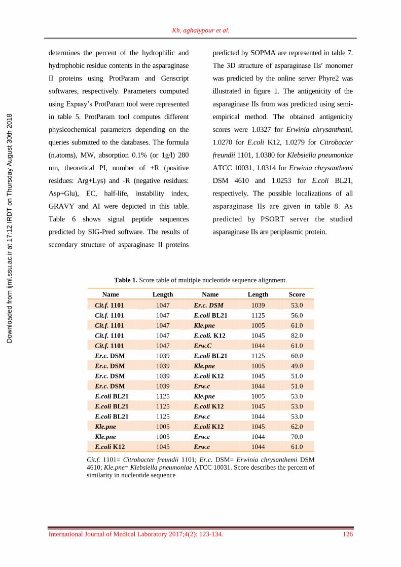
Table 1. Score table of multiple nucleotide sequence alignment.
Name Length Name Length Score
Cit.f. 1101 1047 Er.c. DSM 1039 53.0
Cit.f. 1101 1047 E.coli BL21 1125 56.0
Cit.f. 1101 1047 Kle.pne 1005 61.0
Cit.f. 1101 1047 E.coli. K12 1045 82.0
Cit.f. 1101 1047 Erw.C 1044 61.0
Er.c. DSM 1039 E.coli BL21 1125 60.0
Er.c. DSM 1039 Kle.pne 1005 49.0
Er.c. DSM 1039 E.coli K12 1045 51.0
Er.c. DSM 1039 Erw.c 1044 51.0
E.coli BL21 1125 Kle.pne 1005 53.0
E.coli BL21 1125 E.coli K12 1045 53.0
E.coli BL21 1125 Erw.c 1044 53.0
Kle.pne 1005 E.coli K12 1045 62.0
Kle.pne 1005 Erw.c 1044 70.0
E.coli K12 1045 Erw.c 1044 61.0
Cit.f. 1101= Citrobacter freundii 1101; Er.c. DSM= Erwinia chrysanthemi DSM
4610; Kle.pne= Klebsiella pneumoniae ATCC 10031. Score describes the percent of
similarity in nucleotide sequence
Dow
nloa
ded
from
ijm
l.ssu
.ac.
ir at
17:
12 IR
DT
on
Thu
rsda
y A
ugus
t 30t
h 20
18

BIOINFORMATIC ANALYSIS OF L-ASPARAGINASE II
127 International Journal of Medical Laboratory 2017;4(2): 123-134.
Table 2. Score table of multiple protein sequence alignment
Name Length Name Length Score
Cit.f. 1101 348 Erw.c.DSM 348 43.0
Cit.f. 1101 348 E.coli BL21 348 93.0
Cit.f. 1101 348 Kle. pne 334 45.0
Cit.f. 1101 348 E.coli K12 348 92.0
Cit.f. 1101 348 Erw.c 348 46.0
Erw.c.DSM 348 E.coli BL21 348 43.0
Erw.c.DSM 348 Kle. pne 334 69.0
Erw.c.DSM 348 E.coli K12 348 43.0
Erw.c.DSM 348 Erw.c 348 92.0
E.coli BL21 348 Kle. pne 334 46.0
E.coli BL21 348 E.coli K12 348 98.0
E.coli BL21 348 Erw.c 348 45.0
Kle. pne 334 E.coli K12 348 45.0
Kle. pne 334 Erw.c 348 70.0
E.coli K12 348 Erw.c 348 45.0
Cit.f.1101= Citrobacter freundii 1101; Erw.c. DSM= Erwinia chrysanthemi DSM
4610; Kle.pne= Klebsiella pneumoniae ATCC 10031. Score describes the percent of
similarity in amino acid sequence.
Table 3. Amino acid composition (in %) of the asparaginase IIs using ProtParam software
Amino acid
composition
E.coli
K12 Erw.c. Cit.f. 1101
Erw.c.
DSM 4610
E.coli
BL21
Kle. pne.
ATCC 10031
Ala (A) 38(10.9%) 33(9.5%) 39(11.2%) 34(9.8%) 39(11.2%) 40 (12.0%)
Arg (R) 8(2.3%) 19(5.5%) 9(2.6%) 20(5.7%) 8(2.3%) 16(4.8%)
Asn (N) 23(6.6%) 14(4.0%) 20(5.7%) 16(4.6%) 23(6.6%) 11(3.3%)
Asp (D) 27(7.8%) 21(6.0%) 24(6.9%) 21(6.0%) 28(8.0%) 20(6.0%)
Cys (C) 2(0.6%) 0(0.0%) 2(0.6%) 0(0.0%) 2(0.6%) 0(0.0%)
Gln (Q) 13(3.7%) 8(2.3%) 10(2.9%) 8(2.3%) 13(3.7%) 14(4.2%)
Glu (E) 7(2.0%) 15(4.3%) 11(3.2%) 13(3.7%) 7(2.0%) 12(3.6%)
Gly (G) 30(8.6%) 32(9.2%) 31(8.9%) 33(9.5%) 30(8.6%) 28(8.4%)
His (H) 3(0.9%) 6(1.7%) 4(1.1%) 7(2.0%) 3(0.9%) 7(2.1%)
Ile (I) 13(3.7%) 20(5.7%) 12(3.4%) 19(5.5%) 13(3.7%) 22(6.6%)
Leu (L) 26(7.5%) 31(8.9%) 26(7.5%) 33(9.5%) 26(7.5%) 25(7.5%)
Lys (K) 24(6.9%) 18(5.2%) 24(6.9%) 16(4.6%) 24(6.9%) 14(4.2%)
Met (M) 8(2.3%) 10(2.9%) 9(2.6%) 11(3.2%) 8(2.3%) 8(2.4%)
Phe (F) 11(3.2%) 11(3.2%) 11(3.2%) 11(3.2%) 11(3.2%) 8(2.4%)
Pro (P) 13(3.7%) 12(3.4%) 13(3.7%) 13(3.7%) 13(3.7%) 16(4.8%)
Ser (S) 17(4.9%) 20(5.7%) 16(4.6%) 19(5.5%) 16(4.6%) 20(6.0%)
Thr (T) 35(10.1%) 26(7.5%) 36(10.3%) 25(7.2%) 35(10.1%) 28(8.4%)
Trp (W) 1(0.3%) 1(0.3%) 1(0.3%) 1(0.3%) 1(0.3%) 0(0.0%)
Tyr (Y) 12(3.4%) 13(3.7%) 12(3.4%) 14(4.0%) 12(3.4%) 10(3.0%)
Val (V) 37(10.6%) 38(10.9%) 38(10.9%) 34(9.8%) 36(10.3%) 35(10.5%)
Cit.f.1101= Citrobacter freundii 1101; Erw.c.= Erwinia chrysanthemi; Kle.pne= Klebsiella pneumoniae ATCC
10031.
Dow
nloa
ded
from
ijm
l.ssu
.ac.
ir at
17:
12 IR
DT
on
Thu
rsda
y A
ugus
t 30t
h 20
18

Kh. aghaiypour et al.
International Journal of Medical Laboratory 2017;4(2): 123-134. 128
Table 4. Hydrophilic and hydrophobic residue contents in asparaginase IIs using Genscript software
Bacteria Percentage of
hydrophilic residue
Percentage
hydrophobic of residue Other
Net hydrophobic
content
Cit.f.1101 21% 43% 36% High
Erw.c. DSM 22% 45% 33% High
E.coli BL21 20% 42% 38% High
Kle. pne 21% 46% 33% High
E.coli K12 20% 42% 38% High
Erw.c 23% 45% 32% High
Cit.f.1101= Citrobacter freundii 1101; Erw.c. DSM= Erwinia chrysanthemi DSM 4610;
Kle.pne= Klebsiella pneumoniae ATCC 10031.
Table 5. The computed parameters using Expasy’s ProtParam tool
Bacteria Formula(n.atoms) MW
Abs. 0.1%
(280 nm)
(1cm)
Theo. pI -R +R EC
M-1cm-1 II GRAVY AI
Cit.f.1101 C1626H2602N438O513S11
(5190) 147.39 0.635 6.12 35 33 23380 23.16 -0.076 85.46
Erw.c. DSM C1674H2681N463O499S11
(5328)
150.52 0.701 8.56 34 36 26360 17.58 0.009 96.38
E.coli BL21 C1622H2591N439O518S10
(5180)
147.40 0.634 5.68 35 32 23380 18.33 -0.128 84.91
Kle. pne C1563H2531N435O482S8
(5019) 141.54 0.421 6.39 32 30 14900 29.12 0.050 97.25
E.coli K12 C1623H2595N439O517S10
(5184) 147.40 0.634 5.96 34 32 23380 18.27 -0.128 85.46
Erw.c C1673H2692N458O502S10
(5335) 150.3 0.662 7.84 36 37 24870 17.20 0.042 98.30
Cit.f.1101= Citrobacter freundii 1101; Erw.c. DSM= Erwinia chrysanthemi DSM 4610; Kle.pne= Klebsiella
pneumoniae ATCC 10031; MW= Molecular weight; Abs= Absorption; pI= Theoretical isoelctric point; +R=
positive residues: Arg+Lys; -R= negative residues: Asp+Glu; EC= Extinction coefficient; II= Instability index;
GRAVY= Grand average of hydropathicity; AI= Aliphatic index
Table 6. Signal peptide sequence predicted by SIG-Pred software
Bactria Number of aa Signal peptide sequence Active mature enzyme (one sub U)
Cit.f.1101 19 or 22 MEFFKRTALAALVMGFSGA|ALA 329 or 326 aa
Erw.c. DSM 21 MERWFKSLFVMVLFFVFTANA 327 aa
E.coli BL21 19 or 22 MEFFKKTALAALVMGFSGA|ALA 329 or 326 aa
Kle. pne 29 MSSLAFSETRLPHIVILATGGTIAGSAAS 305 aa
E.coli K12 19 and 22 MEFFKKTALAALVMGFSGA|ALA 329 or 326 aa
Erw.c 21 and 22 MERWFKSLFVLVLFFVFTA|SAA 327 or 328 aa
Cit.f.1101= Citrobacter freundii 1101; Erw.c. DSM= Erwinia chrysanthemi DSM 4610; Kle.pne= Klebsiella
pneumoniae ATCC 10031; aa= Amino acid
Dow
nloa
ded
from
ijm
l.ssu
.ac.
ir at
17:
12 IR
DT
on
Thu
rsda
y A
ugus
t 30t
h 20
18
BIOINFORMATIC ANALYSIS OF L-ASPARAGINASE II
129 International Journal of Medical Laboratory 2017;4(2): 123-134.
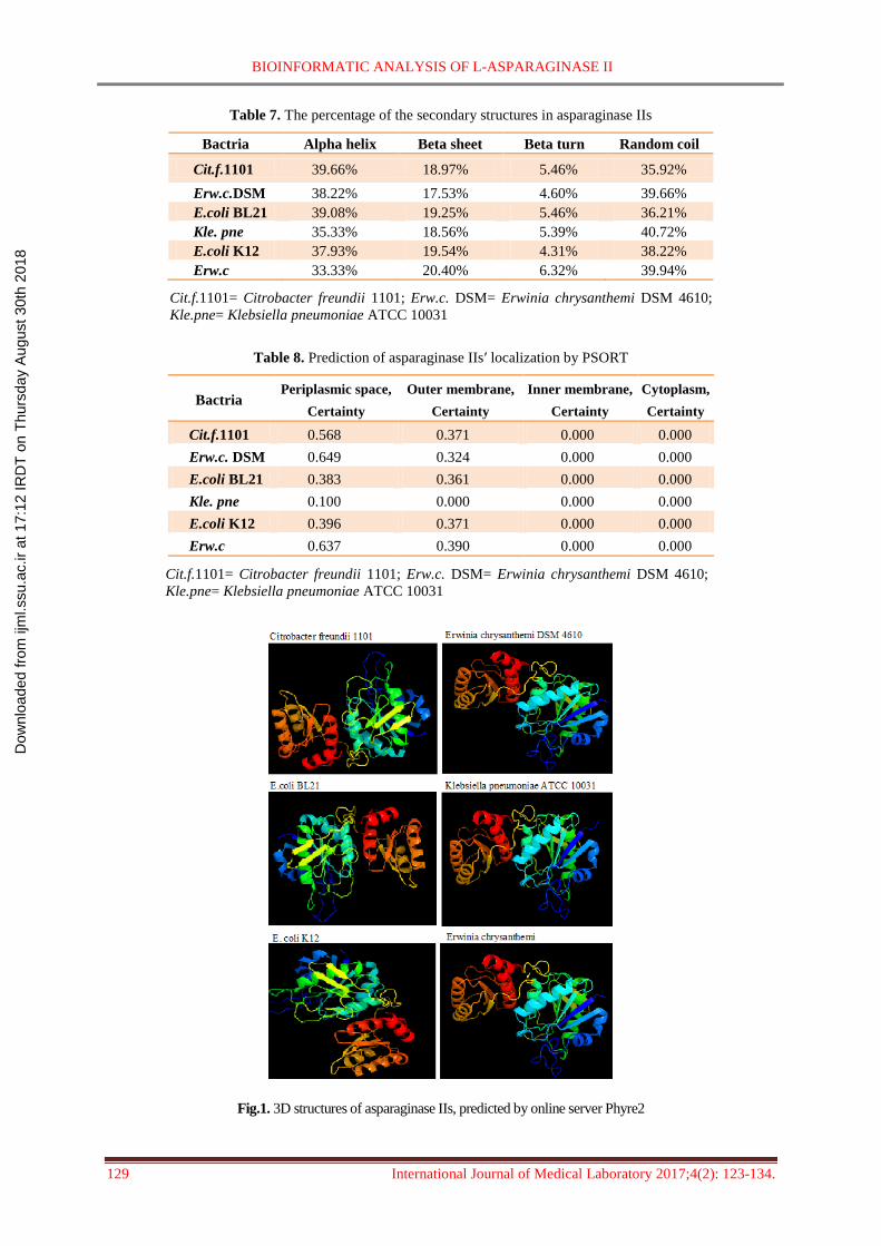
Table 7. The percentage of the secondary structures in asparaginase IIs
Bactria Alpha helix Beta sheet Beta turn Random coil
Cit.f.1101 39.66% 18.97% 5.46% 35.92%
Erw.c.DSM 38.22% 17.53% 4.60% 39.66%
E.coli BL21 39.08% 19.25% 5.46% 36.21%
Kle. pne 35.33% 18.56% 5.39% 40.72%
E.coli K12 37.93% 19.54% 4.31% 38.22%
Erw.c 33.33% 20.40% 6.32% 39.94%
Cit.f.1101= Citrobacter freundii 1101; Erw.c. DSM= Erwinia chrysanthemi DSM 4610;
Kle.pne= Klebsiella pneumoniae ATCC 10031
Table 8. Prediction of asparaginase IIsʹ localization by PSORT
Cytoplasm,
Certainty
Inner membrane,
Certainty
Outer membrane,
Certainty
Periplasmic space,
Certainty Bactria
0.000 0.000 0.371 0.568 Cit.f.1101
0.000 0.000 0.324 0.649 Erw.c. DSM
0.000 0.000 0.361 0.383 E.coli BL21
0.000 0.000 0.000 0.100 Kle. pne
0.000 0.000 0.371 0.396 E.coli K12
0.000 0.000 0.390 0.637 Erw.c
Cit.f.1101= Citrobacter freundii 1101; Erw.c. DSM= Erwinia chrysanthemi DSM 4610;
Kle.pne= Klebsiella pneumoniae ATCC 10031
Fig.1. 3D structures of asparaginase IIs, predicted by online server Phyre2
Dow
nloa
ded
from
ijm
l.ssu
.ac.
ir at
17:
12 IR
DT
on
Thu
rsda
y A
ugus
t 30t
h 20
18
Kh. aghaiypour et al.
International Journal of Medical Laboratory 2017;4(2): 123-134. 130
Discussion
Asparaginase II from various sources has been
studying for a long time in different fields such
as structure and its potency as an antitumor
agent. For example, Swain et al. designed the
crystal structure of L-asparginase [20]; Guo et
al. compared the antitumor activity and the
effect of recombinant enzyme both in vitro and
vivo [21]. Mohamed et al. isolated E.coli MG27
from the River Nile, amplified and cloned asnB
and then characterized it by DNA sequencing
and bioinformatics software [22]. In addition to
such experimental studies that are high cost,
time-consuming and may be impossible to
apply for various organisms at the same time,
bioinformatics provide an inexpensive and
rapid approach to study biomolecules from
various organisms without any vital changes in
protein structure and activity. In this in silico
study, we analyzed and compared asparaginase
II from four bacteria that were cloned,
sequenced and expressed in our lab and
recorded in GenBank, previously.
Despite the high differences in the alignment
scores of nucleotide sequences, the alignment
scores of amino acid sequences were not very
different among the mentioned bacteria (Tables
1 and 2). Because of variation in amino acid
codons, assessment of amino acid sequence
could be more reliable than nucleotide
sequences. Citrobacter freundii 1101 and E.coli
BL21 has the highest score alignment with
E.coli K12 in amino acid sequences. Erwinia
chrysanthemi DSM 4610 hass the highest score
alignment with Erwinia chrysanthemi in amino
acid sequences. The amino acid sequence
provides most of the information required for
determining and characterizing the molecule’s
function, physical and chemical properties.
It may be seemed from the results of the
primary analysis (Tables 3 and 4) that all
asparaginase II proteins are hydrophobic in
primary nature due to the presence of high non-
polar residues, but below GRAVY analysis
show other results. The low presence of
cysteine residues or its absence in asparaginase
II (<0.6%) indicates the lack of disulphide
bridges (“SS” bonds).
The parameters in table 5 were computed
using Expasy’s ProtParam tool. The estimated
molecular weights are in the range of
141.54- 150.52 KD with the lowest MW for
Klebsiella pneumoniae ATCC 10031 and the
highest MW for Erwinia chrysanthemi DSM
4610, respectively. The isoelctric point is the
pH at which the surface protein is covered with
charge, but the net charge of protein is zero.
The pI calculation is based on the peptide
sequence alone without considering the effect
of modifications. It is calculated using pKa
values of amino acids,s which depend on their
side chain [23]. The pI of a protein greater than
seven (pI>7) indicates that they are basic and
lower than seven (pI<7) indicates that they are
acidic. Asparaginase II from E.coli BL21 with
the lowest computed pI=5.68 is acidic and
asparaginase II from Erwinia chrysanthemi
DSM 4610 with the highest computed pI=8.56
is basic. The computed pI has useful application
such as developing buffer system for separation
by isoelectric focusing method and purification
Dow
nloa
ded
from
ijm
l.ssu
.ac.
ir at
17:
12 IR
DT
on
Thu
rsda
y A
ugus
t 30t
h 20
18

BIOINFORMATIC ANALYSIS OF L-ASPARAGINASE II
131 International Journal of Medical Laboratory 2017;4(2): 123-134.
preparation by chromatography technique [24].
The EC illustrates how much light a protein
absorbs relative to their composition at a certain
wavelength. ProtParam computes EC for a
range of (276 nm, 278 nm, 219 nm, 280 nm and
282 nm) wave length, 280 nm is favorable
because proteins absorb strongly there with
respect to the concentration of Cys, Trp and
Tyr, while other substances commonly in
protein solution do not [25]. EC of asparaginase
IIs at 280 nm were ranging from 14900 M-1cm-1
to 26360M-1cm-1 with the lowest range for
Klebsiella pneumoniae ATCC 10031 and the
highest range for Erwinia chrysanthemi DSM
4610. The computed protein EC helps in the
quantitative study of protein-protein and
protein-ligand interactions in solution. The
instability index provides an estimate of the
stability of a protein in a test tube. This method
assigns a weight value of instability. A protein
whose instability index is smaller than 40 is
predicted as stable, a value above 40 predicts that
the protein may be unstable [26]. The instability
index for all mentioned asparaginase IIs is
smaller than 40.
GRAVY that is defined at one specific position
in a sequence is the mean value of the
hydrophobicity of the amino acids within
a window, usually 19 residues long, around
each position [27]. Hydrophobic-hydrophilic
interactions have a strong impact on the three
dimensional structure a protein will adopt.
Because structure, not amino acid sequence
order, carries out certain functions it is important
to understand how these forces affect the protein
folding process. GRAVY is the average value of
the hydropathy index at each position. The
hydropathy values range from -2 to +2 for most
proteins, with the positively rated proteins being
more hydrophobic [28]. GRAVY scores of
some asparaginase IIs are negative and others
approximately around the zero in the range
of -0.128 to 0.050. Then, among the study
asparaginase IIs, ones from E.coli strains of K12,
BL21 and Citrobacter freundii 1101 with the
negative scores are more hydrophilic and
asparaginase IIs from Erwinia chrysanthemi,
Erwinia chrysanthemi DSM 4610 and Klebsiella
pneumoniae ATCC 10031 with the positive
score are more hydrophobic molecules. AI
described as the relative volume occupied by
aliphatic side chains of the amino acids such as
alanine, valine, isoleucine and leucine, is
regarded as a positive factor for the increase of
thermal stability of globular proteins. The AI of
proteins from thermophilic bacteria was found to
be significantly higher than that of ordinary
proteins and hence, it can serve as a measure of
thermostability of proteins [29, 30]. High
aIvalues of the analyzed asparaginase IIs showed
an increase in the thermostability of the proteins.
The half-life is a prediction of the time it takes
for half of the amount of protein in a cell to
disappear after its synthesis in the cell. The
ProtParam biocomputed half-life of all six
asparaginase II is 30 hours in vitro and more than
10 hours in vivo.
Signal peptides occur in bacterial periplasmic
and secretory proteins. The primary analysis by
SIG-Pred software suggested a signal peptide at
the N terminal and the subsequent fragment as
active unit in the mature enzyme (Table 6).
Secondary structure features as predicted using
SOPMA indicated whether a given amino acid
Dow
nloa
ded
from
ijm
l.ssu
.ac.
ir at
17:
12 IR
DT
on
Thu
rsda
y A
ugus
t 30t
h 20
18

Kh. aghaiypour et al.
International Journal of Medical Laboratory 2017;4(2): 123-134. 132
lied in a helix, strand or coil and also the
percentage of amino acids located in these
structures. The results revealed that alpha helix
and random coils were dominated among
secondary structure elements followed by
beta sheet and beta turns for all sequences. All
known types of asparaginase II are active as
homotetramers [31, 32]. The 3D structure of
asparaginase IIsʹ monomer was predicted by
the online server Phyre2 (Fig. 1). In the case of
3D structure, hydrophilic domains tend to be on
exterior surface, while hydrophobic domains
avoids external environment and forms internal
core of the protein. The closest interactions
between two pairs of subunits lead to the
formation of two intimate dimmers within which
the four non-allosteric catalytic centers are
created. Such formation of tetramers, for reasons
that are not completely clear, appear to be
essential for the catalytic ability of asparaginase
II [33, 34]. PSORT server is the most precise
bacterial localization prediction tool available
[35]. The localization of all asparaginase IIs
were predicted by PSORT server as periplasmic
protein (Table 8).
Antigenicity is a local property of the protein
sequences and that protein sequence properties
of composition, secondary structure, solvent
accessibility and evolutionary conservation are
the determinants of antigenicity and specificity
in immune response [30]. The predicted
antigenicity scores of all asparaginase IIs were
comparable with each other and with therapeutic
asparaginase IIs from Erwinia chrysanthemi and
E.coli K12. Although these similarities
suggest that asparaginase IIs from E.coli
BL21, Cit.f.1101 and Erwinia chrysanthemi
DSM 4610 would be the choice for in vivo
study, the potentiality of other non-similar
asparaginase IIs with drug types should also
be determined and compared; maybe others
being more potential than drug types.
Conclusions
We compared bioinformatics data from six
asparaginase IIs by online predictor softwares.
The aim of the present study was to get how much
non-therapeutic asparaginase IIs that we had
studied, sequenced and cloned in our previous
researches would be similar to the therapeutic
asparaginase IIs from E.coli K12 and Erwinia
chrysanthemi. E.coli BL21 and Citrobacter
freundii 1101 had the most similarity in amino
acid sequence (98% and 92%, respectively),
GRAVY, MW, AI and antigenicity with E.coli
K12. Also, Erwinia chrysanthemi DSM 4610 had
the most similarity in amino acid sequence (92%),
GRAVY, MW, AI and antigenicity with Erwinia
chrysanthemi. Although such in silico analysis
provides valuable information, more study is
needed to reduce the drug’s toxicity and improve
its potency without any vital changes in protein
structure and activity by means of bioinformatics
because it is possible to reduce the costs of study
and produce new variants of drug.
Conflict of Interest The authors declare no conflict of interest.
Acknowledgments The basic of our study was supported by Razi Vaccine
and Serum Research Institute (Iran). Thanks to the
members of the biotechnology department of Razi
Vaccine and Serum Research Institute for their favors
in improving this study.
Dow
nloa
ded
from
ijm
l.ssu
.ac.
ir at
17:
12 IR
DT
on
Thu
rsda
y A
ugus
t 30t
h 20
18

BIOINFORMATIC ANALYSIS OF L-ASPARAGINASE II
133 International Journal of Medical Laboratory 2017;4(2): 123-134.
References
[1]. Ekins S, Mestres J, Testa B. In silico
pharmacology for drug discovery: methods for
virtual ligand screening and profiling. Br J
Pharmacol. 2007; 152(1): 9-20.
[2]. Gentleman RC, Carey VJ, Bates DM, Bolstad B,
Dettling M, Dudoit S, et al. Bioconductor: open
software development for computational biology
and bioinformatics. Genome Biol. 2004; 5(10): R80.
[3]. Katoh K, Misawa K, Kuma KI, Miyata T.
MAFFT: a novel method for rapid multiple
sequence alignment based on fast Fourier
transform. Nucleic Acids Res. 2002; 30(14):
3059-3066.
[4]. Gasteiger E, Hoogland C, Gattiker A, Duvaud
SE, Wilkins MR, Appel RD, et al. Protein
identification and analysis tools on the ExPASy
server. Humana Press; 2005.
[5]. Pasut G, Sergi M, Veronese FM. Anti-cancer
PEG-enzymes: 30 years old, but still a current
approach. Adv Drug Delivery Rev. 2008; 60(1):
69-78.
[6]. Narta UK, Kanwar SS, Azmi W .
Pharmacological and clinical evaluation of
L-asparaginase in the treatment of leukemia.
Critic Rev Oncol/Hematol. 2007; 61(3): 208-21.
[7]. Sanches M, Krauchenco S, Polikarpov I.
Structure, substrate complexation and reaction
mechanism of bacterial asparaginases. Curr
Chem Biol. 2007; 1(1): 75-86.
[8]. Borek10 D, Jaskólski M. Sequence analysis of
enzymes with asparaginase activity. Acta
Biochim Pol. 2001; 48: 893-902.
[9]. Cedar H, Schwartz JH. Localization of the
two L-asparaginases in anaerobically grown
Escherichia coli. J Biologic Chem. 1967;
242(16): 3753-755.
[10]. Graham ML. Pegaspargase: a review of
clinical studies. Adv Drug Delivery Rev. 2003;
55(10): 1293-302.
[11]. Ramya LN, Doble M, Rekha VP, Pulicherla
KK. L-Asparaginase as potent anti-leukemic
agent and its significance of having reduced
glutaminase side activity for better treatment of
acute lymphoblastic leukaemia. Appl Biochem
Biotechnol. 2012; 167(8): 2144-159.
[12]. Kumar DS, Sobha K. L-Asparaginase from
microbes: a comprehensive review. Adv Biores.
2012; 3(12): 137-57.
[13]. El-Bessoumy AA, Sarhan M, Mansour J.
Production, isolation, and purification of L-
asparaginase from Pseudomonas aeruginosa
50071 using solid-state fermentation. BMB
Reports. 2004; 37(4): 387-93.
[14]. Hüser A, Klöppner U, Röhm KH. Cloning,
sequence analysis, and expression of ansB from
Pseudomonas fluorescens, encoding periplasmic
glutaminase/asparaginase. FEMS Microbiol
Lett. 1999; 178(2): 327-35.
[15]. Heinemann B, Howard AJ. Production of
tumor-inhibitory L-asparaginase by submerged
growth of Serratia marcescens. Appl Microbiol.
1969; 18(4): 550-54.
[16]. Bahreini E, sabaghi A, Aghaeipour Kh.
Cloning, sequencing and expression of L-
Asparaginase II gene from Citrobacter freundii
1101. International Journal of Biology Research
2017; 2(2): 7-12
[17]. Choi JH, Lee SY. Secretory and extracellular
production of recombinant proteins using
Escherichia coli. Appl Microbiol Biotech. 2004;
64(5): 625-35.
[18]. Kotzia GA, Labrou NE. L-Asparaginase from
Erwinia chrysanthemi 3937: cloning, expression
and characterization. Journal of biotechnology
2007; 127(4): 657-69.
[19]. Harms E, Wehner A, Jennings MP, Pugh
KJ, Beacham IR, Rohm KH. Construction of
expression systems for Escherichia coli
asparaginase II and two-step purification of the
recombinant enzyme from periplasmic extracts.
Protein Expr Purifi. 1991; 2(2-3): 144-50.
[20]. Swain AL, Jaskolski M, Housset D, Rao JK,
Wlodawer A. Crystal structure of Escherichia
coli L-asparaginase, an enzyme used in cancer
therapy. Proc Natl Acad Sci USA 1993; 90(4):
1474-478.
[21]. Guo QL,Wu MS, Chen Z. Comparison of
antitumor effect of recombinant L-asparaginase
with wild type one in vitro and in vivo. Acta
Pharmacol Sin. 2002; 23(10): 946-51.
[22]. Mohamed ZK, Elnagdy SM, Seufi AE, Gamal
M. Cloning and molecular analysis of
Lasparaginase II gene (ansB). J BioSci
Biotechnol. 2015; 4(3): 291-302.
[23]. Pace CN, Grimsley GR, Scholtz JM. Protein
ionizable groups: pK values and their
contribution to protein stability and solubility. J
Biologic Chem. 2009; 284(20): 13285-3289.
[24]. Gasteiger E, Gattiker A, Hoogland C,
Ivanyi I, Appel RD, Bairoch A. ExPASy: the
proteomics server for in-depth protein
knowledge and analysis. Nucleic Acids Res.
2003; 31(13): 3784-788.
[25]. Schmid FX. Biological Macromolecules: UV-
visible Spectrophotometry. Encyc Life Sci (eLS)
2001; 3: 240-43.
[26]. Guruprasad K, Reddy BB, Pandit MW.
Correlation between stability of a protein and its
dipeptide composition: a novel approach for
predicting in vivo stability of a protein from its
primary sequence. Protein Eng. 1990; 4(2): 155-61.
Dow
nloa
ded
from
ijm
l.ssu
.ac.
ir at
17:
12 IR
DT
on
Thu
rsda
y A
ugus
t 30t
h 20
18
Kh. aghaiypour et al.
International Journal of Medical Laboratory 2017;4(2): 123-134. 134
[27]. Kyte J, Doolittle RF. A simple method for
displaying the hydropathic character of a
protein. J Mol Biol. 1982; 157(1): 105-32.
[28]. Dehouck Y, Gilis D, Rooman M. Database-
derived potentials dependent on protein size for
In silico folding and design. Biophys J. 2004;
87(1): 171-81.
[29]. Atsushi IK. Thermostability and aliphatic
index of globular proteins. J Biochem. 1980;
88(6): 1895-898.
[30]. Ferron F, Longhi S, Canard B, Karlin D. A
practical overview of protein disorder prediction
methods. Proteins 2006; 65(1): 1-4.
[31]. Greenquist AC, Wriston JC. Chemical
evidence for identical subunits in L-asparaginase
from Escherichia coli B. Arch Biochem
Biophys. 1972; 152(1): 280-86.
[32]. Swain AL, Jaskólski M, Housset D, Rao JK,
Wlodawer A. Crystal structure of Escherichia
coli L-asparaginase, an enzyme used in cancer
therapy. Proceed Nati Acad Sci. 1993; 90(4):
1474-478.
[33]. Mezentsev YV, Molnar AA, Sokolov NN,
Lisitsina VB, Shatskaya MA, Ivanov AS, et al.
Specificity of molecular recognition in
oligomerization of bacterial L-asparaginases.
Biochemistry (Moscow) Supplement Series B:
Biomed Chem. 2011; 5(2): 124.
[34]. Michalska K, Jaskolski M. Structural aspects
of L-asparaginases, their friends and relations.
ACTA Biochemical Polonica-English Edition
2006; 53(4): 627.
[35]. Gardy JL, Laird MR, Chen F, Rey S, Walsh CJ,
Ester M, et al. PSORTb v. 2.0: expanded prediction
of bacterial protein subcellular localization and
insights gained from comparative proteome analysis.
Bioinformatics 2005; 21(5): 617-23.
Dow
nloa
ded
from
ijm
l.ssu
.ac.
ir at
17:
12 IR
DT
on
Thu
rsda
y A
ugus
t 30t
h 20
18