international journal of surgery case...
TRANSCRIPT

Pa
VDa
b
a
ARAA
KPDDSCT
1
demp
cc
ao
Pf
h2(
CASE REPORT – OPEN ACCESSInternational Journal of Surgery Case Reports 5 (2014) 547–550
Contents lists available at ScienceDirect
International Journal of Surgery Case Reports
journa l h omepage: www.caserepor ts .com
erforated duodenal diverticulum: Surgical treatmentnd literature review
itor Costa Simõesa,∗, Bruno Santosa, Sara Magalhãesb, Gil Fariaa,onzília Sousa Silvaa, José Davidea
Department of Surgery, Centro Hospitalar do Porto, Porto, PortugalDepartment of Radiology, Centro Hospitalar do Porto, Porto, Portugal
r t i c l e i n f o
rticle history:eceived 18 May 2014ccepted 15 June 2014vailable online 19 June 2014
eywords:erforationuodenumiverticulumurgeryomplicationreatment
a b s t r a c t
INTRODUCTION: Duodenum is the second most frequent location for a diverticulum in the digestive tract.Complications are rare and perforation was only reported in less than 200 cases.PRESENTATION OF CASE: A 79-year-old female was admitted to Emergency Department with abdominalpain and vomiting for the last 24 h. A CT scan was performed and moderated extra-luminal air was iden-tified. During surgery a fourth portion perforated duodenal diverticulum was diagnosed and duodenalresection was performed.DISCUSSION: First reported in 1710, the incidence of duodenal diverticula can be as high as 22%. Nev-ertheless complications are extremely rare and include haemorrhage, inflammation, compression ofsurrounding organs, neoplastic progression, cholestasis and perforation.
As perforations are often retroperitoneal, symptoms are nonspecific and rarely include peritonealirritation, making clinical diagnose a challenge.
CT scan will usually present extra-luminal retroperitoneal air and mesenteric fat stranding, providing
clues for the diagnosis.Although non-operative treatment has been reported in selected patients, standard treatment issurgery and alternatives are diverse including diverticulectomy or duodenopancreatectomy.CONCLUSION: Perforated diverticula of the fourth portion of the duodenum are extremely rare and currentevidence still supports surgery as the primary treatment modality.
© 2014 The Authors. Published by Elsevier Ltd. on behalf of Surgical Associates Ltd. This is an openaccess article under the CC BY-NC-ND license (http://creativecommons.org/licenses/by-nc-nd/3.0/).
. Introduction
First reported by Chomel in 1710, the incidence of duodenaliverticula (DD) can be as high as 22% and complications can bestimated at 0.03% per year.1 Duodenum is the second most com-on site for diverticula in the alimentary tract being the second
ortion the most frequent location.2–4
Perforation is a rare complication of DD, only reported in 162ases,5 but also the most serious one,2 representing a diagnostichallenge,6 and a difficult surgical problem.
Few cases of perforated third and fourth portions of the DD
re reported in literature and so their diagnosis, management andutcomes are based on those reports.∗ Corresponding author at: Department of Surgery, Centro Hospital do Porto, Largorof. Abel Salazar, 4099-001 Porto, Portugal. Tel.: +351 934140762;ax: +351 222053218.
E-mail address: [email protected] (V. Costa Simões).
ttp://dx.doi.org/10.1016/j.ijscr.2014.06.008210-2612/© 2014 The Authors. Published by Elsevier Ltd. on behalf of Surgical Ahttp://creativecommons.org/licenses/by-nc-nd/3.0/).
We present a rare case of perforated diverticulum fromthe fourth part of the duodenum and its successful surgicalmanagement.
2. Presentation of case
A 79 years old female patient with dementia, hypertension,mitral insufficiency and paroxysmal atrial fibrillation, is admittedto the Emergency Department with abdominal pain and vomitingfor the last 24 h.
On arrival her vital signs showed auricular temperature of 36 ◦C,heart rate of 73/min and blood pressure of 125/65 mm Hg. Physi-cal examination elicited pain on palpation of the four quadrantswithout signs of peritoneal irritation. Blood tests showed 28,040white blood cells/�L with 88% neutrophils in the differential count,
haemoglobin value of 13.1 g/dL, platelets count of 259,000/�L, C-reactive protein of 100.23 mg/L, creatinine of 0.94 mg/dL, urea of42 mg/dL, lactate dehydrogenase of 368 U/L, amylase of 107 U/L,with normal liver tests, lipase level and arterial blood gases.ssociates Ltd. This is an open access article under the CC BY-NC-ND license
CASE REPORT – OPEN ACCESS548 V. Costa Simões et al. / International Journal of Surgery Case Reports 5 (2014) 547–550
Fa
cl
tmlbcsTc
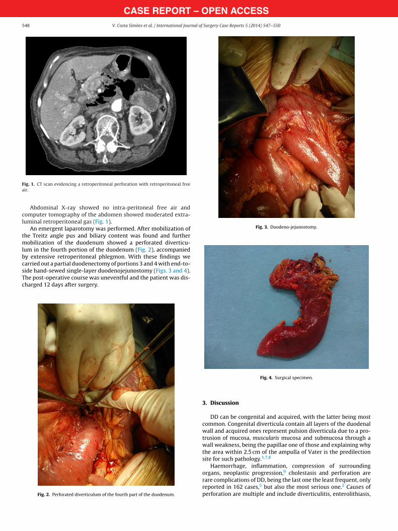
Fig. 3. Duodeno-jejunostomy.
ig. 1. CT scan evidencing a retroperitoneal perforation with retroperitoneal freeir.
Abdominal X-ray showed no intra-peritoneal free air andomputer tomography of the abdomen showed moderated extra-uminal retroperitoneal gas (Fig. 1).
An emergent laparotomy was performed. After mobilization ofhe Treitz angle pus and biliary content was found and further
obilization of the duodenum showed a perforated diverticu-um in the fourth portion of the duodenum (Fig. 2), accompaniedy extensive retroperitoneal phlegmon. With these findings wearried out a partial duodenectomy of portions 3 and 4 with end-to-
ide hand-sewed single-layer duodenojejunostomy (Figs. 3 and 4).he post-operative course was uneventful and the patient was dis-harged 12 days after surgery.Fig. 2. Perforated diverticulum of the fourth part of the duodenum.
Fig. 4. Surgical specimen.
3. Discussion
DD can be congenital and acquired, with the latter being mostcommon. Congenital diverticula contain all layers of the duodenalwall and acquired ones represent pulsion diverticula due to a pro-trusion of mucosa, muscularis mucosa and submucosa through awall weakness, being the papillae one of those and explaining whythe area within 2.5 cm of the ampulla of Vater is the predilectionsite for such pathology.1,7,8
Haemorrhage, inflammation, compression of surrounding9
organs, neoplastic progression, cholestasis and perforation arerare complications of DD, being the last one the least frequent, onlyreported in 162 cases,5 but also the most serious one.2 Causes ofperforation are multiple and include diverticulitis, enterolithiasis,

uEf
uaicvt
macaowia
ia
npTatdim
icpaidt
R
CASE REPORT – OPEN ACCESSV. Costa Simões et al. / International Journal of Surgery Case Reports 5 (2014) 547–550 549
lceration, foreign body, trauma, iatrogenic perforation during anRCP and most frequently ischaemia due to distention related toood retention inside the diverticulum.5,10
As most perforations are retroperitoneal,10 symptoms aresually nonspecific including right upper abdominal pain associ-ted with nauseas and vomiting,6 and rarely include peritonealrritation.3 Blood samples are also unspecific and elevated pan-reatic tests might be present due to the inflammation in theicinity of the diverticulum.1 Retroperitoneal contamination leadso retroperitoneal abscess formation and sepsis.
Conventional radiological examination will show no abnor-alities in half the patients with duodenal perforation.11 Upper
bdominal series can diagnose the DD and if extraversion ofontrast is observed confirms its perforation. Ultrasound studiesre rarely informative. Abdominal CT, usually requested with-ut any hint of suspicion for complicated duodenal pathology,1
ill provide diagnosis. Its findings include mesenteric fat strand-ng, thickened bowel wall and extra-luminal retroperitonealir.12,13
Proper diagnosis and improvements in both antibiotics andntensive care explain the mortality reduction seen in last decadeslthough it remains high, up to 13%.11
In selected patients that are only mildly affected and hado evidence of impending sepsis, non-operative treatment forerforated DD is safe and a practical alternative to surgery.6,7
reatment includes nasogastric suction, bowel rest, intravenousntibiotic therapy, parenteral nutrition, endoscopic cleaning ofhe infected pouch and combined endoscopic and percutaneousrainage of retroperitoneal abscess.14,15 Close clinical observation
s mandatory and surgical intervention is indicated if conservativeanagement fails.Standard treatment for perforated DD is surgical
ntervention.6,11 Diverticulectomy with single or double-layerlosure is the most frequent reported alternative if inflammationermits.15 Some cases of simple intra-abdominal drainage werelso successful.2,16 When substantial duodenal or retroperitonealnflammation is present, more complex procedures like duo-enal diversion, pyloric exclusion, gastro-enteric anastomosis,ube duodenostomy, segmental duodenal resection or even
pylorus preserving Whipple might be adequate.11,15 Laparoscopicapproach have also been described with good results.6
Surgical morbidity includes duodenal leak or fistula, abscess,iatrogenic injury to the common bile duct, acute pancreatitis andpersistent sepsis. Identification of the papilla during surgery mightbe performed by inserting a catheter through cholecystostomy orcholedochotomy.15
4. Conclusion
Third and fourth parts DD perforation is extremely rare,its diagnosis is challenging and surgical intervention is rec-ommended. Non-operative management should be reserved forselected patients.
Conflict of interest
The authors declare that there is no conflict of interest in under-taking this article.
Funding
None.
Ethical approval
Written informed consent was obtained from the patient forpublication of this case report, collecting data and accompanyingimages. A copy of the written consent is available for review by theEditor-in-Chief of this journal on request.
Author contributions
Vitor Costa Simões: Surgery, writing.Bruno Santos: Surgery, writing.Sara Magalhães: Radiologic evaluation.Gil Faria: Surgery, revision.Donzília Sousa Silva: Surgery, revision.José Davide: Surgery, revision.
Key learning points
• Perfurated diverticula of the fourth portion of the duodenum are rare.• Surgery is still the primary treatment modality.
eferences
1. de Perrot T, Poletti PA, Becker CD, Platon A. The complicated duodenal diver-ticulum: retrospective analysis of 11 cases. Clin Imaging 2012;36(July–August(4)):287–94.
2. Ming TY, Feng HK, Cherng YJ, Chuan CD, Pai LT, Chi LY. Clinical challenge: diver-ticulitis of third and fourth portion of the duodenum with perforation. Rev EspEnferm Dig 2012;104(March (3)):156–7.
3. Favre-Rizzo J, Lopez-Tomassetti-Fernandez E, Ceballos-Esparragon J, Santana-Cabrera L, Hernandez-Hernandez JR. Duodenal diverticulum perforated byforeign body. Rev Esp Enferm Dig 2013;105(July (6)):368–9.
6. Rossetti A, Christian BN, Pascal B, Stephane D, Philippe M. Perforated duodenaldiverticulum, a rare complication of a common pathology: a seven-patient caseseries. World J Gastrointest Surg 2013;5(March (3)):47–50.
7. Thorson CM, Paz Ruiz PS, Roeder RA, Sleeman D, Casillas VJ. The perforatedduodenal diverticulum. Arch Surg 2012;147(January (1)):81–8.
8. Oukachbi N, Brouzes S. Management of complicated duodenal diverticula. J ViscSurg 2013;150(June (3)):173–9.
9. Furukawa M, Izumi S, Tsukuda K, Tokumo M, Sakurai J, Mano S. Duodenal carci-noma from a duodenal diverticulum mimicking pancreatic carcinoma. Acta MedOkayama 2012;66(5):423–7.
10. Duarte B, Nagy KK, Cintron J. Perforated duodenal diverticulum. Br J Surg
4. Yeo CJ. Shackelford’s surgery of the alimentary tract. 7th ed. Philadelphia, PA:Elsevier/Saunders; 2013.5. Volchok J, Massimi T, Wilkins S, Curletti E. Duodenal diverticulum: case report
of a perforated extraluminal diverticulum containing ectopic pancreatic tissue.Arch Surg 2009;144(February (2)):188–90.
1
1992;79(September (9)):877–81.1. Schnueriger B, Vorburger SA, Banz VM, Schoepfer AM, Candinas D. Diagnosisand management of the symptomatic duodenal diverticulum: a case seriesand a short review of the literature. J Gastrointest Surg 2008;12(September(9)):1571–6.
– O5 nal of
1
1
1
1
1
OTpc
CASE REPORT50 V. Costa Simões et al. / International Jour
2. Ames JT, Federle MP, Pealer KM. Perforated duodenal diverticulum: clinical andimaging findings in eight patients. Abdom Imaging 2009;34(April (2)):135–9.
3. Pearl MS, Hill MC, Zeman RK. CT findings in duodenal diverticulitis. AJR Am J
Roentgenol 2006;187(October (4)):W392–5.4. Coulier B. Duodenal diverticulitis. JBR-BTR 2008;91(November–December(6)):271.
5. Martinez-Cecilia D, Arjona-Sanchez A, Gomez-Alvarez M, Torres-Tordera E,Luque-Molina A, Valenti-Azcarate V, et al. Conservative management of
pen Accesshis article is published Open Access at sciencedirect.com. It is distribermits unrestricted non commercial use, distribution, and reproductredited.
PEN ACCESS Surgery Case Reports 5 (2014) 547–550
perforated duodenal diverticulum: a case report and review of the literature.World J Gastroenterol 2008;14(March (12)):1949–51.
6. Chen CF, Wu DC, Chen CW, Hsieh JS, Chen CY, Wang JY. Successful manage-ment of perforated duodenal diverticulitis with intra-abdominal drainage andfeeding jejunostomy: a case report and literature review. Kaohsiung J Med Sci
2008;24(August (8)):425–9.uted under the IJSCR Supplemental terms and conditions, whichion in any medium, provided the original authors and source are