intracranial pathology of the visual pathway - eclass.uoa.gr · and conclusive differentiation of...
TRANSCRIPT
European Journal of Radiology 49 (2004) 143–178
Intracranial pathology of the visual pathway
W. Müller-Forell∗
Institute of Neuroradiology, Medical School University of Mainz, Langenbeckstrasse 1, D-55101 Mainz, Germany
Received 4 September 2003; received in revised form 8 September 2003; accepted 9 September 2003
Abstract
Intracranial pathologies involving the visual pathway are manifold. Aligning to anatomy, the most frequent and/or most important extrinsicand intrinsic intracranial lesions are presented. Clinical symptoms and imaging characteristics of lesions of the sellar region are demonstrated indifferent imaging modalities. The extrinsic lesions mainly consist of pituitary adenomas, meningeomas, craniopharyngeomas and chordomas.In (asymptomatic and symptomatic) aneurysms, different neurological symptoms depend on the location of aneurysms of the circle of Willis.Intrinsic tumors as astrocytoma of any grade, ependymoma and primary CNS-lymphoma require the main pathology in the course of thevisual pathway. Vascular and demyelinating diseases complete this overview of intracranial lesions.© 2003 Elsevier Ireland Ltd. All rights reserved.
Keywords: MRI; Intrinsic; Extrinsic brain lesions; Ischemia; Metastasis; Demyelinating disorders
1. Introduction
In intracranial lesions, ophthalmologic symptoms dependon the site of the lesion affecting visual pathway struc-tures. The most important symptom is loss of visual acu-ity, whether acute or slowly progressing. Especially the lat-ter with only subtle visual deficits are often underestimatedand attributed to aging or stress problems and imaging isperformed only when decompensation of the visual systemhas become apparent, due to space occupying intracraniallesions, which sometimes have already destroyed the sur-rounding neural substance.
The variety of intracranial lesions affecting any part ofthe visual pathway is presented according to the anatomiccourse and do not assume completeness.
The multitude of intracranial lesions is dominated by pri-mary brain tumors, whether they are intrinsic or extrin-sic. The main group of intrinsic brain tumors (originatingfrom cells covered by pia) is that of glioma, with threemajor types: astrocytoma, oligodendroglioma and ependy-moma and so-called mixed gliomas that contain two ormore different cell types in varying proportion[1]. Ap-plication of intravenous (i.v.) contrast material enables the
∗ Tel.: +49-6131-17-7139; fax:+49-6131-17-6643.E-mail address: [email protected]
(P.D.W. Müller-Forell).
definition of absent or apparent blood-brain-barrier (BBB)disruption.
Treatment and overall prognosis of the histologically fre-quently benign extrinsic brain tumors (arising from cellsoutside the pia) are based upon the correct diagnosis. In-tracranial metastasis and secondary brain tumors are definedas tumors, involving the CNS and originate from, but arediscontinuous with, primary systemic neoplasms.
Imaging of the intracranial portion of the visual pathwayis the domain of MR, as it enables a focused examinationof the entire visual pathway along its course from the inneraperture of the optic canal to the occipital pole. Its use ismandatory for extrinsic tumors, as the superior contrast res-olution and multiplanar imaging capacity enable the iden-tification of anatomic markers as cardinal features of anextraaxial lesion. These include cerebrospinal fluid (CSF)clefts, recognized as crescentic bands, pial blood vesselsand/or the dura. The use of i.v. contrast agents enables thedemonstration of compartmentalization of extrinsic lesions,since a large number of these tumors show an extensive con-trast enhancement (meningeoma, metastasis), while othersshow none (epidermoid and dermoid tumors)[2].
Metastasis account for up to 30% of all intracranialtumors, the most frequent primary malignancies includelung and breast carcinoma (Fig. 1) [3], hypernephroma,melanoma and neuroblastoma[1]. All areas of the brainmay be affected, with preference of the corticomedullaryjunction as the starting point[1].
0720-048X/$ – see front matter © 2003 Elsevier Ireland Ltd. All rights reserved.doi:10.1016/j.ejrad.2003.09.003

144 W. Müller-Forell / European Journal of Radiology 49 (2004) 143–178
Fig. 1. MR of a 36-year-old female with acute vision loss, predominantly of the right eye, and history of breast carcinoma. Diagnosis: intra- andsuprasellar metastasis. (a) Axial T2w view at the level of the anterior commissure show edema along both optic tracts. (b) Midsagittal T1w native view,demonstrating the impression of the floor of the third ventricle, but no identification of the chiasm. (c) Coronal T1w contrast enhanced view, where thechiasm is identified. Note the additional infiltration of the pituitary gland. ((c) With permission of Müller-Forell, 2002.)
2. Sellar region
As already mentioned, in slowly progressing unilateralvisual impairment lesions of the chiasmal region are fre-quently seen at a stage, when decompensation of the visualsystem has become apparent. In patients suffering from dou-ble vision or acute ptosis, the subjective disabling symptomslead to both an earlier identification of homonymous defectsof the visual field and corresponding earlier indication forimaging. Most common causes for bitemporal hemianopiaare midline tumors, which cause inferior compression of thechiasm at the crossing site of the nasal fibers.
The chiasm represents the centre of the sellar region, themost complex area of the endocranium, consisting of vari-ous tissues, as parenchymal structures, vessels and cranial
nerves. As a consequence, a number of more than 30 dif-ferent pathologic processes, primarily extrinsic lesions mayoccur, involving and affecting structures of the sellar andjuxtasellar region[4]. These mainly benign tumors involvethe brain parenchyma secondarily and are often cured com-pletely without recurrences even if they have reached a con-siderable size. The sellar region is less frequently involvedby intrinsic tumors, which often show recurrences, even forbenign tumors. Visual disturbances, caused by chiasmal oroptic nerve compression, is the most frequent clinical symp-tom, although these are as variable as the locations of thediseases of the sellar region, ranging from endocrinologicaldisorders (pituitary, hypothalamic insufficiency) and com-plex focal episodes (temporal lobe), to cranial nerve deficits(cavernous sinus).

W. Müller-Forell / European Journal of Radiology 49 (2004) 143–178 145
2.1. Neoplasms and tumor-like lesions
2.1.1. Extrinsic lesions
2.1.1.1. Pituitary adenoma. Pituitary adenoma, whichrepresent 10–15% of all intracranial tumors, frequentlymanifest in adults. They are benign, slow growing tu-
Fig. 2. A 35-year-old male presenting with acute vision loss combined with reduced consciousness and severe headache. Diagnosis: adenoma apoplecticum.(a) Axial native CT, where a suprasellar mass with hyperdense rim is seen. Note the different intensities of a fluid level, representing intratumoralhemorrhage. (b) Corresponding axial T2w image with better delineation of two tumor-cysts. (c) Sagittal T1w view showing the entire tumor extension.Note the dislocation of the chiasm (arrow). (d) Coronal T1w view, demonstrating the inferior impression and cranial dislocation of the chiasm (smallarrows), the hazy hyperintensities represent parts of the intratumoral hemorrhage. (e) Corresponding T1w contrast enhanced view.
mors, composed of cells of the adenohypophysis, normallywrapped up by a pseudocapsule, which enables good de-marcation from adjacent structures. In contrast to the groupof microadenomas (diameter<10 mm), patients presentingwith the characteristic clinical symptom of bitemporal hemi-anopia, due to inferior compression of the chiasm, harbourendocrinological mainly inactive macroadenoma (diameter
146 W. Müller-Forell / European Journal of Radiology 49 (2004) 143–178
Fig. 2. (Continued ).
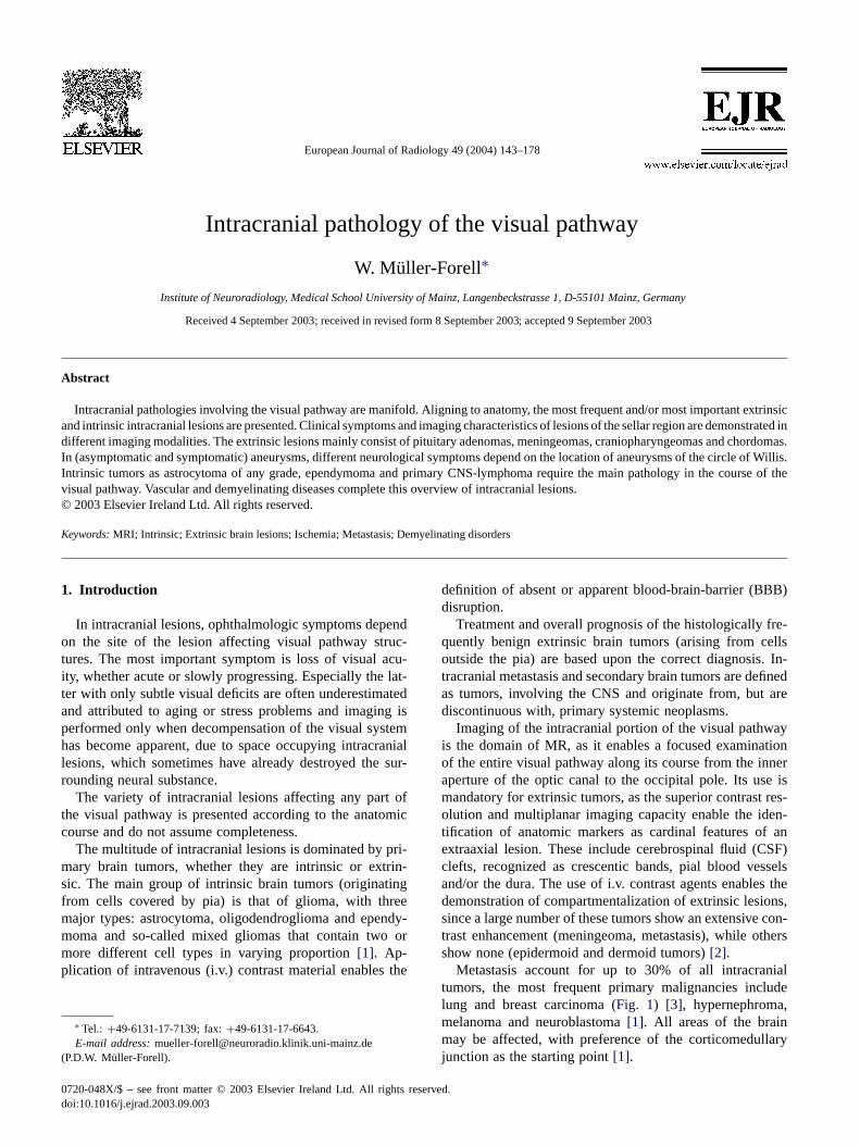
>10 mm). Acute clinical symptoms of headache, nauseaand vomiting are suspicious of an adenoma apoplecticum(Fig. 2). This acute insufficiency of the adenohypophysis isdue to a sudden infarction or hemorrhage in the course oftumor growth, but not as frequent as presumed[5].
MR as the method of choice generally shows lengthen-ing of both T1 and T2 relaxation times. The high anatomicresolution and definition of adjacent tissue, i.e. intracranialoptic nerves, chiasm and cavernous sinus allow an accurateand conclusive differentiation of the tumor and deformed,compressed and flattened visual structures, especially in na-tive, non contrast enhanced T1w images (Fig. 3). Due to thelack of a solid barrier between the pituitary gland and thecavernous sinus the more reliable indicator for cavernoussinus invasion is the demonstration of a carotid artery en-casement or a tumor extension lateral of the cavernous si-nus than an even careful analysis of pre- and postcontrastimages[5,6]. On T1w non contrast enhanced images, mostadenoma show a homogeneous, isointense signal with sig-nificant enhancement after contrast application (Figs. 3 and4). Regressive changes, seen in medium-sized adenomas in-clude cysts and hemorrhages, consequently requiring addi-tional T2w and T2*w sequences (Fig. 2).
Differential diagnosis of pituitary adenoma includesmeningeoma of the sellar region (Figs. 4 and 7), craniopha-ryngeoma (Figs. 9 and 10), optic glioma (Fig. 16), germ celltumors, chordomas (Fig. 13) and even metastasis (Fig. 1).
A pituitary abscess, mostly caused by gram-positive cocci,is highly uncommon, and clinically presentation is domi-nated by headache and visual loss. It is known to occur inthe presence of sellar mass or by contiguous spread from aninfected sphenoid or cavernous sinus[5]. Post contrast imag-
ing demonstrates a rim enhancement around a hypointensecentre, representing the necrosis, and additional infiltrationof the adjacent sinus and intracranial structures (Fig. 5).
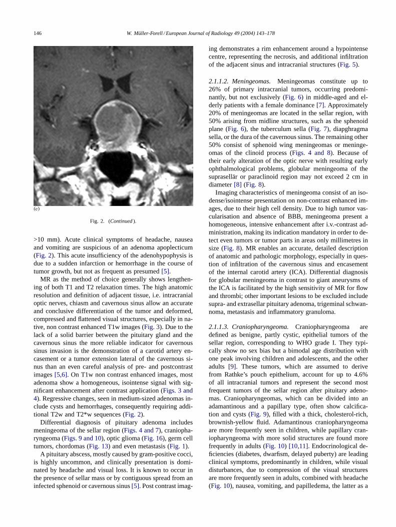
2.1.1.2. Meningeomas. Meningeomas constitute up to26% of primary intracranial tumors, occurring predomi-nantly, but not exclusively (Fig. 6) in middle-aged and el-derly patients with a female dominance[7]. Approximately20% of meningeomas are located in the sellar region, with50% arising from midline structures, such as the sphenoidplane (Fig. 6), the tuberculum sella (Fig. 7), diapghragmasella, or the dura of the cavernous sinus. The remaining other50% consist of sphenoid wing meningeomas or meninge-omas of the clinoid process (Figs. 4 and 8). Because oftheir early alteration of the optic nerve with resulting earlyophthalmological problems, globular meningeoma of thesuprasellär or paraclinoid region may not exceed 2 cm indiameter[8] (Fig. 8).
Imaging characteristics of meningeoma consist of an iso-dense/isointense presentation on non-contrast enhanced im-ages, due to their high cell density. Due to high tumor vas-cularisation and absence of BBB, meningeoma present ahomogeneous, intensive enhancement after i.v.-contrast ad-ministration, making its indication mandatory in order to de-tect even tumors or tumor parts in areas only millimetres insize (Fig. 8). MR enables an accurate, detailed descriptionof anatomic and pathologic morphology, especially in ques-tion of infiltration of the cavernous sinus and encasementof the internal carotid artery (ICA). Differential diagnosisfor globular meningeoma in contrast to giant aneurysms ofthe ICA is facilitated by the high sensitivity of MR for flowand thrombi; other important lesions to be excluded includesupra- and extrasellar pituitary adenoma, trigeminal schwan-noma, metastasis and inflammatory granuloma.
2.1.1.3. Craniopharyngeoma. Craniopharyngeoma aredefined as benigne, partly cystic, epithelial tumors of thesellar region, corresponding to WHO grade I. They typi-cally show no sex bias but a bimodal age distribution withone peak involving children and adolescents, and the otheradults [9]. These tumors, which are assumed to derivefrom Rathke’s pouch epithelium, account for up to 4.6%of all intracranial tumors and represent the second mostfrequent tumors of the sellar region after pituitary adeno-mas. Craniopharyngeomas, which can be divided into anadamantinous and a papillary type, often show calcifica-tion and cysts (Fig. 9), filled with a thick, cholesterol-rich,brownish-yellow fluid. Adamantinous craniopharyngeomaare more frequently seen in children, while papillary cran-iopharyngeoma with more solid structures are found morefrequently in adults (Fig. 10) [10,11]. Endocrinological de-ficiencies (diabetes, dwarfism, delayed puberty) are leadingclinical symptoms, predominantly in children, while visualdisturbances, due to compression of the visual structuresare more frequently seen in adults, combined with headache(Fig. 10), nausea, vomiting, and papilledema, the latter as a

W. Müller-Forell / European Journal of Radiology 49 (2004) 143–178 147
Fig. 3. A 39-year-old male with acute visual deficit (blurred vision) and high prolactin level. Diagnosis: pituitary adenoma (prolactinoma). (a) MidsagittalT1w native view, demonstrating the apical dislocation of the chiasm. The arrow indicates the chiasmatic recess of the still acute angled third ventricle.(b) Coronal T1w native view. Note the dislocation of the pituitary stalk to the left (arrow). (c) Corresponding contrast enhanced view, where the smallresidual adenohypophysis can be delineated (arrows) with bright contrast enhancement.
result of increased intracranial pressure, due to obstructionof the interventricular foramen in quite large lesions[5,11].CT is still justified for differential diagnosis in view of thecharacteristic calcification of parts of the tumor, but MR ismandatory for preoperative planning. MR pattern show agreat variety: adamantinous craniopharyngeoma primarilyshows a combination of T1w hypointense and T2w highlyhyperintense signals, whereas in papillary craniopharyn-
geoma hyperintense signal on T1w and hypointensities onT2w images may dominate[11]. Both tumor types gener-ally show a hyperintense signal of the solid tumor partswith a prominent enhancement of the tumor and the cystwall [5]. Differential diagnosis include cystic pituitary ade-nomas, gliomas of the chiasm or hypothalamus, Rathke’spouch cyst, epidermoid, dermoid, germinoma of the thirdventricle and suprasellar meningeomas[8,12].

148 W. Müller-Forell / European Journal of Radiology 49 (2004) 143–178
Fig. 4. MR (T1w) of a 73-year-old male presenting with slowly progressing consecutive bilateral blindness of 95%. Diagnosis: pituitary adenoma andadditional left sphenoid wing meningeoma. (a) Axial native view presenting isointense signal of both, the median and paramedian space occupyinglesions. (b) Corresponding contrast enhanced view, where homogeneous, but different signal enhancement is seen: the adenoma present only with slight,the meningeoma with intense enhancement, leading to diagnosis. (c) Paramedian sagittal native view showing the cranio-caudal extension of the pituitaryadenoma, with impression and dislocating of the chiasm (arrow). (d) Coronal contrast enhanced view demonstrating the different tumors by someinterposed, extrinsic compressed frontal brain parenchyma. ((a, b) With permission of Müller-Forell, 2002.)
2.1.1.4. Aneurysms. The rupture of an intracranialaneurysm, mainly located at the circle of Willis, is the mostcommon atraumatic cause of subarachnoid hemorrhage(SAH), presenting with a sudden onset of severe headache,varying loss of consciousness, seizure, vomiting and focalneurological deficits, requiring immediate, either neurosur-
gical or interventional neuroradiological therapy, especiallyin view of a high incidence of often life-threatening rebleed-ing. The underlying pathological process of intracranialaneurysms has been recognized as an endothelial dysfunc-tion [13]. Clinical symptoms with visual deficiencies ofsymptomatic unruptured intracranial aneurysms of the in-

W. Müller-Forell / European Journal of Radiology 49 (2004) 143–178 149
Fig. 5. MR of a 47-year-old female who presented in pituitary coma; after substitution, she complained of headache, variable visual deficits and clinicalconstellation of meningism and fever. Diagnosis: pituitary abscess. (a) Sagittal T1w contrast enhanced view with ring shaped enhancement of the pituitaryand sphenoid sinus. (b) Coronal T1w contrast enhanced view demonstrates complete necrosis of the pituitary gland.
fraclinoid or supraclinoid internal carotid artery (ICA), theanterior cerebral artery (ACA), or the basilar artery dependon the site of compression of visual pathway structures.(Figs. 11–13). Neither the extent nor the clinical symptoma-tology of slowly progressing visual field deficits caused byaneurysm may be readily distinguished from those causedby tumors. Orbital pain, periorbital or facial pain is the mostcommon symptom of cavernous carotid aneurysms, fol-lowed by different types of cranial nerve palsies. Isolated NIII palsy may lead to diagnosis of an aneurysm of the pos-terior communicating artery, while an aneurysm of the ICAis suspected in the presence of a combination of NVI or NIV palsy and a sensory deficit of the trigeminal nerve[14].
In acute subarachnoid hemorrhage CT is the method ofchoice, as it demonstrates not only the extent of the bleed-ing itself, but also the volume and distribution of the acutehemorrhage and its complication as acute CSF disturbance.In unruptured aneurysm CT might demonstrate secondarychanges as bony erosion of the sphenoid, or calcification ofthe wall, as these aneurysms are generally larger than rup-tured aneurysms. MR is the method of choice in visualizingthe vicinity of unruptured aneurysm and their relation to theoptic nerve, chiasm or optic tract, due to its multiplanar ca-pability in combination with its high sensitivity and spatialresolution, enabling the detection of flowing blood. Combi-nation of MR images with 3D-TOF (time-of-flight) or 3Dphase contrast MRA, enables a detection rate of aneurysms>3 mm in diameter of≈ 100%[15]. Intra-arterial DSA ofthe cerebral vessels still remains the gold standard in thedetection or exclusion of intracranial aneurysms as it repre-sents not only the most sensitive method, but enables precise
planning and performance of interventional therapy with coilocclusion, preferably using 3D-DSA[16,17].
2.1.2. MiscellaneousGerm cell tumors of the CNS constitute a unique class of
rare tumors of different malignant character, affecting chil-dren and young adults, but represent only 0.5% of all in-tracranial neoplasms. About 80% impinge on the midlinewith preference to the pineal gland, but intra- or suprasellarlocations may be involved (Fig. 14), simultaneously or se-quentially, if the tumor is multifocal[18]. Hormonal distur-bances, due to hypothalamic infiltration may be additional tovisual pathway disturbances, as Parinaud syndrome in caseof involvement of the quadrigeminal plate, or visual lossand/or cranial nerve palsies in case of suprasellar growthwith impingement of the chiasm. On imaging germ cell tu-mors appear as a solid, homogeneous mass, rarely includ-ing cysts, a feature that is in contrast to craniopharyngeoma.In noncontrast enhanced images, they demonstrate an evenunspecific character on methods, CT and MR, but show amarked, mainly homogeneous enhancement after adminis-tration of i.v. contrast material[4,5].
Chordomas are extremely rare, slow growing, osteode-structive, histologically benign, midline tumors, participat-ing in ophthalmologic disorders only in case of extensiontowards the chiasm (Fig. 15). Differential diagnosis of theselesions, presenting on imaging with intermediate signal onboth T1w and T2w images with moderate contrast enhance-ment, should include chondromas, chondrosarcomas, metas-tasis, as well as hormone-inactive pituitary adenomas or evenskull base meningeomas[19].

150 W. Müller-Forell / European Journal of Radiology 49 (2004) 143–178
Fig. 6. MR of a 13-year-old boy with quick vision loss on both eyes (1/40 and 1/20). Diagnosis: meningeoma of the sphenoid plane. (a) Axial T2wview, demonstrating a mass in the anterior cranial fossa bilaterally, with lateralization to the right, and with inhomogeneous signal, apparently includinga cystic tumor part (arrow). (b) Corresponding T1w native view, where the extrinsic nature of the lesion is seen, as it impresses both frontal lobes. Notethe posterior dislocation of the chiasm and both optic tracts (stars). (c) In the T1w native midsagittal view the dislocation of the chiasm is apparent(arrow). (d) Corresponding contrast enhanced view.
W. Müller-Forell / European Journal of Radiology 49 (2004) 143–178 151
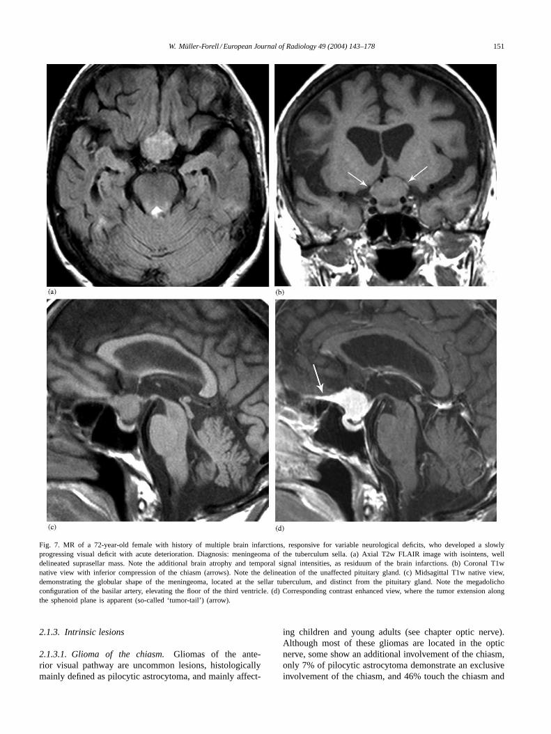
Fig. 7. MR of a 72-year-old female with history of multiple brain infarctions, responsive for variable neurological deficits, who developed a slowlyprogressing visual deficit with acute deterioration. Diagnosis: meningeoma of the tuberculum sella. (a) Axial T2w FLAIR image with isointens, welldelineated suprasellar mass. Note the additional brain atrophy and temporal signal intensities, as residuum of the brain infarctions. (b) Coronal T1wnative view with inferior compression of the chiasm (arrows). Note the delineation of the unaffected pituitary gland. (c) Midsagittal T1w native view,demonstrating the globular shape of the meningeoma, located at the sellar tuberculum, and distinct from the pituitary gland. Note the megadolichoconfiguration of the basilar artery, elevating the floor of the third ventricle. (d) Corresponding contrast enhanced view, where the tumor extension alongthe sphenoid plane is apparent (so-called ‘tumor-tail’) (arrow).
2.1.3. Intrinsic lesions
2.1.3.1. Glioma of the chiasm. Gliomas of the ante-rior visual pathway are uncommon lesions, histologicallymainly defined as pilocytic astrocytoma, and mainly affect-
ing children and young adults (see chapter optic nerve).Although most of these gliomas are located in the opticnerve, some show an additional involvement of the chiasm,only 7% of pilocytic astrocytoma demonstrate an exclusiveinvolvement of the chiasm, and 46% touch the chiasm and

152 W. Müller-Forell / European Journal of Radiology 49 (2004) 143–178
Fig. 8. T1w MR of a 33-year-old female with known amaurosis of the left eye persisting for two years. Acute presentation with complain of double visionfor the previous 2 weeks. Diagnosis: sphenoid wing meningeoma with secondary infiltration of the optic nerve sheath and cavernous sinus. (a) Axialnative view with isointense small mass in the area of the left clinoid process, no definite identification of the left optic nerve. Note the slight wideningof the left ophthalmic artery (white arrow), compared to the right. (b) Corresponding contrast enhanced (FS) view. Although the intracranial tumor partis small, an extent growth en plaque around the left clinoid process into the ipsilateral and towards the contralateral optic canal with involvement of theleft optic sheath is apparent. (With permission of Müller-Forell, 2002.)
hypothalamus (Fig. 16). In cases of an additional or primar-ily hypothalamic involvement the mortality rate increases toover 50%, since no specific therapy alters the final outcome[20].
Only 10% of gliomas of the chiasm occur in patientsolder than 20 years[21], representing a clinical entity dis-tinct from the benign gliomas of childhood, due to the factthat they are not associated with NF1 but include malignantastrocytoma or glioblastoma (Fig. 17) and uniformly show afatal course of usually<1 year[20,22]. Visual impairmentof these patients depends on the size and location of themass.
2.1.3.2. Astrocytoma. Astrocytoma represent the most fre-quent entity of primary brain tumors with up to 60% of allintracranial neoplasms. They arise from differentiated, neo-plastically transformed astrocytes, comprise a wide rangeof age and gender distribution, growth potential, extent ofinvasiveness, morphological features, tendency for progres-sion and clinical course[1,23]. Astrocytomas include pilo-cytic and subependymal giant cell (both WHO I), diffuse lowgrade astrocytoma (WHO II), anaplastic astrocytoma (WHOIII) and glioblastoma (WHO IV). These different entities re-flect the type and sequence of genetic alterations acquiredduring the process of transformation, where the progressionfrom low grade to anapalastic astrocytoma and glioblastomais associated with the cumulative acquisition of multiple ge-
netic alterations[23]. All astrocytoma are characterized bymore or less localized or diffuse infiltration of the adjacentor distant brain parenchyma and may have an inherent ten-dency for malignant progression, with glioblastoma as themost malignant phenotypic endpoint[23,24].
Pilocytic astrocytoma are more circumscribed intrinsic le-sions, which typically present in the fist two decades, withpreference to the optic nerve and chiasm and/or hypotha-lamus (Fig. 18), sometimes associated with clinical diag-nosis of NF1[25]. Diffuse astrocytoma (WHO II), charac-terized by diffuse infiltration of the adjacent parenchymawith a tendency to malignant progression to anaplastic as-trocytoma and, ultimately glioblastoma mainly affect adults.Glioblastoma, representing the most frequent malignant in-tracranial tumor, account for≈ 60% of all astrocytoma. Thehistopathological features are characterized by cellular poly-morphism, nuclear atypia, brisk mitotic activity, thrombosis,microvascular proliferation, and necrosis[24]. Imaging fea-tures of astrocytoma are as variable as the histopathologicalones: low-grade astrocytoma present as mildly hypointenseon T1w and hyperintense on T2w images, mainly withoutany contrast enhancement. BBB disruption may be a factorin tumor progression, and can be assumed in glioblastoma,where the simultaneous delineation of tumor necrotic cysts,sometimes associated with hemorrhages and solid, enhanc-ing tumors parts, combined with reactive brain edema re-flects histopathology[24].

W. Müller-Forell / European Journal of Radiology 49 (2004) 143–178 153
Fig. 9. A 3-year-old boy with recurrent headache, leading to imaging, but no neuro-ophthalmologic deficit. Diagnosis: craniopharyngeoma. (a) AxialCTwith a cystic mass with calcified capsule in a widened sella. (b) Corresponding T2w view, confirming the cystic nature of the mass. (c) Midsagittal T1wnative view, presenting the entire cranio-caudal extension of the mass, which demonstrated homogeneous contrast enhancement (not shown).
2.1.3.3. Cavernoma. Calcification can be seen in cav-ernoma, the only true venous malformation of the CNS,which consists of widened endothelial-lined sinusoidalvascular spaces without any interposed neural tissue.Cavernoma can be found anywhere in the cerebral orspinal tissues, sometimes multifocal. Intralesional hem-orrhages are consistently found at pathology, but onlyfew patients present with clinical symptoms of an acute
bleeding episode, which is more frequent in cavernoma<10 mm in size. CT may identify intralesional calcifi-cation, but specific diagnosis is to be made with MR.Due to the high susceptibility of hemosiderin, especiallyT2*-weighted sequences demonstrate intralesional or oc-cult hemorrhages/calcification (Fig. 19), not only adjacentto the lesion itself, but also in other regions of the CNS[8,26,27].

154 W. Müller-Forell / European Journal of Radiology 49 (2004) 143–178
Fig. 10. A 63-year-old female presenting with unspecific visual deficits and beginning right optic nerve atrophy. Diagnosis: craniopharyngeoma. (a)AxialCT, where only slight perisellar calcification were seen. (b) Coronal T2w view, demonstrating a cystic infraoptic lesion at the right. The arrows indicatethe lateral parts of the impressed chiasm, the small star the unaffected pituitary gland. (c) Midsagittal T1w native view that shows the only slight inferiorimpression of the chiasm. Note the impression of the floor of the third ventricle due to megadolicho-basilar artery. (d) Corresponding contrast enhancedview, where the intrasellar expansion of the tumor is apparent. The star marks the unaffected pituitary gland.
3. Optic tract and area
Patients suffering from lesions of the optic tract and visualarea present with homonymous hemianopia to the contralat-eral visual field, but may demonstrate different extent, de-pending on the kind and location of the underlying pathology(see also chapters of anatomy and neuroophthalmology).
3.1. Neoplasms
3.1.1. EpendymomaEpendymoma (Fig. 20) are slowly growing intrinsic tu-
mors (WHO grade II), arising from ependymal layers of theventricles with variable morphologic features and includesubependymoma, myxopapillary ependymoma (both WHO

W. Müller-Forell / European Journal of Radiology 49 (2004) 143–178 155
Fig. 11. A 48-year-old woman with acute loss of vision in the left eye, accompanied by severe headache. Diagnosis: acute subarachnoid hemorrhage(SAH) caused by an ophthalmic aneurysm of the left ICA. (a) Axial CT with acute subarachnoid hemorrhage of the basal cisterns and a circulartarget-shaped formation in the left subcallosal area. (b) 3D-CT-angiography (left is right and vice verse) showing a lobulated aneurysm of the left ICA.(c) DSA of the left ICA in an oblique view to the right, revealing an upwardly directed, slightly lobulated aneurysm of the C5-part of the left ICA.(With permission of Müller-Forell, 2002.)
I), and anaplastic ependymoma (WHO III) (Fig. 21). Theyaccount for up to 9% of all neuroepithelial tumors at anysite of the ventricular system, preferentially affecting chil-dren and young adults, most commonly in the infratento-rial compartment (fourth ventricle). In cases of involvementof the supratentorial parenchyma (Fig. 21), again mainly in
children and young adults, they originate from embryonicependymal remnants[28]. On imaging these quite distinctlycircumscribed lesions present with heterogeneous signal in-tensities, low on T2w and slightly hyperintense on T1wimaging, reflecting partly cystic, partly calcified, necroticand hemorrhagic areas of the mass, while solid tumor parts
156 W. Müller-Forell / European Journal of Radiology 49 (2004) 143–178
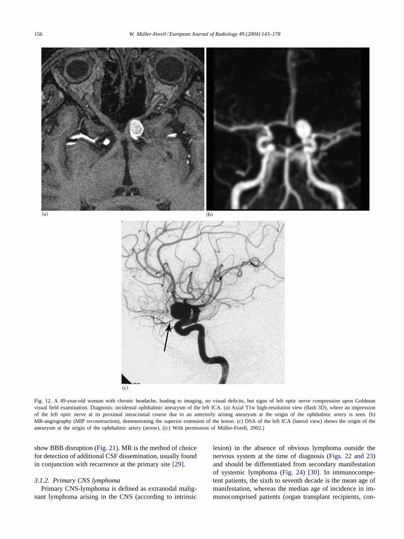
Fig. 12. A 49-year-old woman with chronic headache, leading to imaging, no visual deficits, but signs of left optic nerve compression upon Goldmanvisual field examination. Diagnosis: incidental ophthalmic aneurysm of the left ICA. (a) Axial T1w high-resolution view (flash 3D), where an impressionof the left optic nerve at its proximal intracranial course due to an anteriorly arising aneurysm at the origin of the ophthalmic artery is seen. (b)MR-angiography (MIP reconstruction), demonstrating the superior extension of the lesion. (c) DSA of the left ICA (lateral view) shows the origin of theaneurysm at the origin of the ophthalmic artery (arrow). ((c) With permission of Müller-Forell, 2002.)
show BBB disruption (Fig. 21). MR is the method of choicefor detection of additional CSF dissemination, usually foundin conjunction with recurrence at the primary site[29].
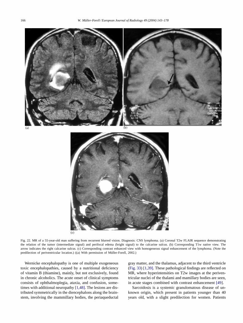
3.1.2. Primary CNS lymphomaPrimary CNS-lymphoma is defined as extranodal malig-
nant lymphoma arising in the CNS (according to intrinsic
lesion) in the absence of obvious lymphoma outside thenervous system at the time of diagnosis (Figs. 22 and 23)and should be differentiated from secondary manifestationof systemic lymphoma (Fig. 24) [30]. In immunocompe-tent patients, the sixth to seventh decade is the mean age ofmanifestation, whereas the median age of incidence in im-munocomprised patients (organ transplant recipients, con-
W. Müller-Forell / European Journal of Radiology 49 (2004) 143–178 157
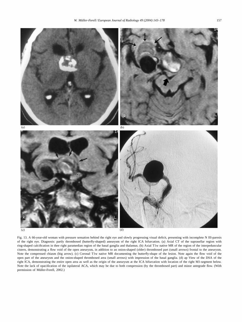
Fig. 13. A 66-year-old woman with pressure sensation behind the right eye and slowly progressing visual deficit, presenting with incomplete N III-paresisof the right eye. Diagnosis: partly thrombosed (butterfly-shaped) aneurysm of the right ICA bifurcation. (a) Axial CT of the suprasellar region withring-shaped calcification in thee right paramedian region of the basal ganglia and thalamus. (b) Axial T1w native MR of the region of the interpeduncularcistern, demonstrating a flow void of the open aneurysm, in addition to an onion-shaped (older) thrombosed part (small arrows) frontal to the aneurysm.Note the compressed chiasm (big arrow). (c) Coronal T1w native MR documenting the butterfly-shape of the lesion. Note again the flow void of theopen part of the aneurysm and the onion-shaped thrombosed area (small arrows) with impression of the basal ganglia. (d) ap View of the DSA of theright ICA, demonstrating the entire open area as well as the origin of the aneurysm at the ICA bifurcation with location of the right M1-segment below.Note the lack of opacification of the ispilateral ACA, which may be due to both compression (by the thrombosed part) and minor antegrade flow. (Withpermission of Müller-Forell, 2002.)

158 W. Müller-Forell / European Journal of Radiology 49 (2004) 143–178
Fig. 14. MR of an 11-year-old girl with universal infantilism but no visual symptoms. Diagnosis: pituitary and hypothalamic germinoma. (a) MidsagittalT1w native view with enlargement of the optic chiasm and pituitary stalk. (b) Corresponding contrast enhanced view with inhomogeneous signalenhancement of the chiasm, hypothalamus, and pituitary gland. (c) Axial T1w, contrast enhanced view with tumor enhancement of the chiasm andthickened pituitary stalk). (With permission of Müller-Forell, 2002.)
genitally immunodeficient or HIV-patients) ranges from 10to 39 years[30]. Although the most common location issupratentorial, involvement of the occipital lobe is seen inonly 3%. As an exquisite sensitivity to steroids exists, thesedrugs should be withheld until tissue is obtained for diag-nosis by stereotactic biopsy[30,31].
On imaging, classic findings show multiple, sometimessolitary masses involving deep gray matter structures, the
corpus callosum, and periventricular regions with moder-ate perifocal edema (Fig. 22). Slightly hyperdense on CTand isointense on all MR sequences lymphoma presentwith diffuse to homogeneous contrast enhancement. Incase of enhancement along perivascular leptomeningealspaces (Fig. 23), CNS lymphoma should be put on topof the differential diagnosis, together with sarcoidosis andtoxoplasmosis.

W. Müller-Forell / European Journal of Radiology 49 (2004) 143–178 159
Fig. 15. MR of a 61-year-old woman with slowly progressing visual deficit (only shadow vision) of the right eye. Diagnosis: chordoma. (a) MidsagittalT1w native view, demonstrating the entire tumor growth with complete destruction of the clivus and hard plate, as well as compression of the brainstem,and slight impression of the chiasm. The different signal intensities are due to small intratumoral hemorrhages. (b) Axial T2w view, where the bilateraldestruction of the optic canal is demonstrated. The arrow indicates the dislocated left Gasserian ganglion. (With permission of Müller-Forell, 2002.)
3.1.3. MiscellaneousMeningeomas may arise at any region of the skull, be-
cause of their slow growth neurologic symptoms may ariselate in respect to tumor size (Fig. 25).
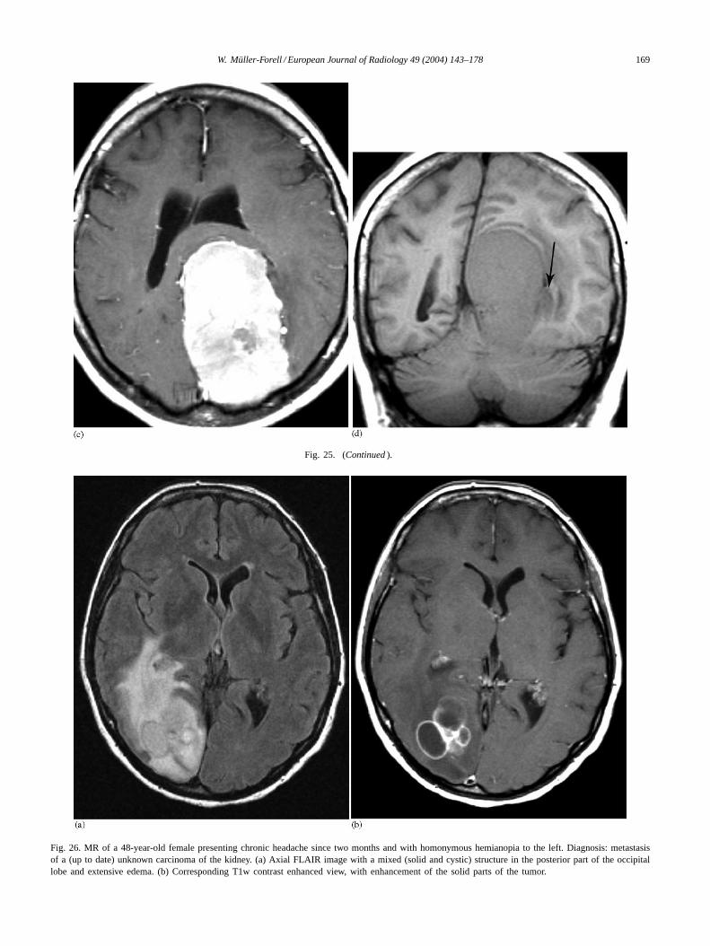
It is the nature of metastasis to mimic many of the primaryCNS tumors, and especially in case of up to now unknownprimary tumor, when neurologic symptoms indicate imag-ing, only histology may lead to the right diagnosis (Fig. 26).
3.2. Nonneoplastic lesions
3.2.1. Vascular lesionsUnderlying pathology of acute functional disorders of the
brain, clinically presenting as stroke, includes a heteroge-neous group of cerebrovascular disorders. The four majorgroups are cerebral infarction (80%) caused by arterial ves-sel occlusion or as a result of inflammatory vascular disease,spontaneous subarachnoid (5%) or intracerebral hemorrhage(15%) and venous occlusion[32].
The main arterial supply of the optic tract and radia-tion arises from the territory of the posterior cerebral artery(PCA), which gives rise to the medial and posterior choroidalarteries, which are in hemodynamic balance with the ante-rior choroidal arteries, arising from the ICA and applyingthe optic tract. Although considerable variation exists, thePCA mainly supplies the inferior temporal and the occipi-tal lobe[33]. In acute cerebral ischemia and infarction theblood flow is significantly diminished, but the damage tothe affected brain depends on vulnerability of the brain, col-
lateral supply, degree and duration of the cerebral ischemia[12].
Imaging is able to reflect the alterations of cell membranefunction and loss of cytoskeletal integrity with subsequentcell death in the course of cerebral infarction[32]. In partic-ular MR studies, including T2w, diffusion weighted (DWI)and MRA (MR-angiography) sequences identify the local-ization of the occlusion and also the area of brain damageearlier and more sensitively than with CT[34]. The acute ac-cumulation of intra- and extracellular edema induces a pro-longation of both T1 and T2, resulting in a high signal onT2w images, not always apparent in early scan, where, onthe other hand indirect signs (also known on CT) of subtleswelling of the affected gyri and compression of the adja-cent sulci should be noted. The most sensitive sequence usedin early infarction is diffusion weighted imaging (DWI), in-cluding ADC-maps (Fig. 27), as the early cytotoxic edemarestricts the water diffusion (restricted Brown’s molecularmovement), resulting in high signal intensity of infractedbrain compared to unaffected normal brain (Fig. 28).
Especially in younger patients, where degenerative arte-rial lesions are unlikely vasculitis should be taken into dif-ferential diagnosis consideration. Vasculitis is defined by itspathological findings of inflammation and necrosis of theblood vessel wall, with involvement of virtually any size ortype of organ system[35]. In general CNS involvement israther uncommon, and isolated CNS vasculitis rather rare[36], but classification of CNS vasculitis vary from pri-mary (e.g. periarteritis nodosa, giant cell arteritis, Wegener’s

160 W. Müller-Forell / European Journal of Radiology 49 (2004) 143–178
Fig. 16. MR of a 9-year-old boy with pubertas praecox. A bilateral atrophy of the optic nerve and visual deficit were unknown, but found on ophthalmologicexamination. Diagnosis: optic glioma with hypothalamic involvement. (a) Axial T2w view, demonstrating hypothalamic and temporomesial infiltration(including the optic tract) of the right side, but another signal enhancement of the left optic tract (arrow). (b) Corresponding T1w native view withenlargement of the right part of the optic chiasm and both optic tracts, with preference to the right. (c) Midsagittal T1w native view, demonstratinga mass without any delineation of the chiasm. The arrow indicates the pituitary stalk. (d) Coronal T1w contrast enhanced view, demonstrating tumorinvasion of both lateral geniculate nuclei (arrow), again with preference to the right.
granulomatosis) or secondary in collagen vascular diseases(e.g. systemic lupus erythematosus, rheumatoid arteritis),from infectious (e.g. bacterial, fugal) or non-infectious (e.g.immune-cell-mediated)[12,37]. Clinical symptoms are vari-able and unspecific, while MR findings may (but not always)show various (although not specific) lesions. As the patho-
logic process of perivascular inflammation may affect notonly capillaries, but also arterioles and arteries simultane-ous finding of microinfarctions, territorial and hemodynamicinfarctions may occur[36]. Confirmation with cerebral an-giography, demonstrating multiple arterial vessel irregulari-ties [12] (Fig. 29), should be aspired.

W. Müller-Forell / European Journal of Radiology 49 (2004) 143–178 161
Fig. 17. MR (T1w, contrast enhanced) of a 70-year-old female with progressive visual deficit to loss within 2 weeks of the right eye and loss of vision(o.1) of the left eye. Diagnosis: glioblastoma of the chiasm (a) Axial view with of a solid tumor with some necrosis of the chiasm, demonstrating alateralization to the right. (b) Coronal view. (c) Midsagittal view, showing beginning invasion of the floor of the third ventricle. (With permissionofMüller-Forell, 2002.)
Sturge Weber syndrome (synonym: encephalotrigeminalangiomatosis), a sporadically occurring phacomatosis, ischaracterized by a ‘port wine’ stain face (naevus flammeus)in the trigeminal distribution, leptomeningeal venous an-giomatosis (caused by faulty development of the venousdrainage), clinically presenting with seizures, dementia,hemiplegia, hemianopia, buphthalmus and glaucoma. In-tracranial involvement is regularly ispilateral of the nevus
flammeus of the face, affecting the occipital lobe preferen-tially [38]. Although CT can identify (specific) dystrophicsubcortical calcification in areas of cortical atrophy, MR ismore sensitive than CT in identifying secondary changesof the affected areas, with cortical atrophy, (compensatory)ventricular and choroid plexus enlargement, and calvarialhemiatrophy (Fig. 30). Superficial gyriform contrast en-hancement is caused by slowly flowing blood within the

162 W. Müller-Forell / European Journal of Radiology 49 (2004) 143–178
Fig. 18. T1w MR of a 9-year-old boy with known NF1, presented with left N VI paresis and right sided papilledema. Diagnosis: pilocytic astrocytomaof the hypothalamus. (a) Axial view visualising the partly cystic, partly solid tumor in the left hypothalamic region. Note the small capsule, delineatingthe cystic tumor from the dislocated, impressed third ventricle (arrow). (b) Corresponding contrast enhanced view with bright signal enhancement of thesolid tumor part. (c) The midsagittal view shows best the retro-chiasmatic extension of the tumor with caudal impression of the third ventricle. Thearrow indicates the chiasm. (With permission of Müller-Forell, 2002.)
persistent plexus of the subarachnoid space and/or BBBloss within cerebral cortex from cerebral ischemia[38].
3.2.2. White matter lesions (MS)Multiple sclerosis is the most common demyelinating dis-
order of the CNS. Two main theories concerning the aeti-ology of this disease are discussed, a genetic and environ-mental, one might be influenced by the other, so that theexpression of a susceptibility gene (or genes) depends onenvironmental factors[39]. Most often the first and onlyclinical symptom in younger patients aged from 20 to 40years, consists of impaired vision, presenting as retro-bulbarneuritis (RBN), followed or combined with fluctuating pe-riods of sensomotoric or gait disturbances[40]. The clinical
course of MS can be divided into a relapsing-remitting and achronic progressive form. MR imaging got a new quality indiagnosis, as new guidelines enable the physician to definethe diagnosis for MS, possible MS or nor MS[41]. Theseguidelines include the evidence of dissemination in time andspace of lesions typical for MS, objectively determined byclinical and imaging signs. They should require evidence ofat least three of the following findings:
1) one gadolinium enhancing lesion or nine T2-hyperin-tense lesions,
2) at least one infratentorial lesions3) at least one juxtacortical lesion4) at least three periventricular lesions.

W. Müller-Forell / European Journal of Radiology 49 (2004) 143–178 163
Fig. 19. MR of a 40-year-old female with unspecific symptoms, but no visual deficit. Diagnosis: incidental detected cavernoma of the right chiasm andoptic tract. (a) Coronal T1w native view with enlargement of the right chiasm. (b) Coronal T2w image, demonstrating target-like signals at the proximalright optic tract. (c) Axial T2*w image with characteristic hemosiderin susceptibility artefact (arrow).
Additional findings of CSF abnormalities (oligoclonalbands), lymphocytic pleocytosis, and abnormal visualevoked potentials (VEP) provide supplement information toclinical findings, typical for MS.
MR is the imaging tool of choice in suspected demyelinat-ing disorders[8,38–40]. Although the sensitivity in detect-ing MR lesions is very high, the correlation of neurologicalsymptoms and localization of imaging findings is rare andonly probable in some cases (Fig. 31). The imaging protocolshould include axial and sagittal PD/T2w and FLAIR se-quences, where the demyelinated areas demonstrate a highsignal [42]. T1w native and contrast enhanced sequencesdemonstrate acute or recurrent inflammatory lesions, due toBBB disruption[43]. The sagittal view is best in order toshow the characteristic so-called Dawson’s finger, presentingas periventricular, pericallosal, ovoid lesions, due to perivas-cular inflammation in the course of medullary veins[43].
BBB disruption of acute or recurrent inflammatory lesionsis apparent in contrast enhanced T1w sequences[43–45].
Acute disseminated encephalomyelitis (ADEM) is char-acterized by an acute monophasic disorder, in contrary toMS. Predominantly occurring following a viral infection,ADEM may affect patients at any age, but with a preferencefor children and young adults[46,47]. Consequently, the si-multaneous occurrence of a variety of polytopic neurolog-ical symptoms such as, e.g. hemi- to tetraplegia or ataxia,combined with optic neuritis and bladder dysfunction maylead to correct diagnosis. The bilateral but slightly asym-metric lesions typically affect both, the white and gray mat-ter, with severe destruction of the latter[39]. The imagingmethod of choice again is MR, as it best shows the mainlysubcortical, confluent foci on T2w images (Fig. 32) and asimilar contrast enhancement of all lesions, if BBB disrup-tion is apparent[46,47].

164 W. Müller-Forell / European Journal of Radiology 49 (2004) 143–178
Fig. 20. MR of a 28-year-old woman with chronic headache and slight, unspecific visual disturbances. Diagnosis: intraventricular ependymoma. (a) AxialT2w image with space occupying tumor in the right temporal lobe, only slight perifocal edema. (b) Corresponding T1w contrast enhanced view withinhomogeneous signal enhancement of the tumor. Note the anteriorly displaced temporal horn (arrow) and the distinct delineation of the process, duetomainly intraventricular location. (c) Coronal T1w contrast enhanced view, note the displacement of the temporal horn (arrow). (With permission of DrLorenz, Department of Radiology of Katholisches Klinikum Mainz.)

W. Müller-Forell / European Journal of Radiology 49 (2004) 143–178 165
Fig. 21. MR of a 5-year-old boy suffering from headache for 10 days, presenting because of an acute N VI-paresis of the right eye and homonymoushemianopia to the left. Diagnosis: anaplastic ependymoma. (a) Axial T2w view, showing a large mass with apparently cystic configuration (due totumor necrosis) in the right occipital lobe with compression of the posterior horn of the right ventricle and marked perifocal edema. Note the left opticradiation. (b) Corresponding T1w native view, confirming the suspicion because of the hypointense signal character of the well delineated mass. (c)Paramedian sagittal T2w view. Note the dislocation of the parieto-occipital sulcus (small white arrows) and a widened cerebello-medullary cisternas anincidental finding. (d) Coronal T1w contrast enhanced image, demonstrating the impression of the ispilateral calcarine sulcus (black arrow), comparedto the contralateral (white arrow).
166 W. Müller-Forell / European Journal of Radiology 49 (2004) 143–178
Fig. 22. MR of a 55-year-old man suffering from recurrent blurred vision. Diagnosis: CNS lymphoma. (a) Coronal T2w FLAIR sequence demonstratingthe relation of the tumor (intermediate signal) and perifocal edema (bright signal) to the calcarine sulcus. (b) Corresponding T1w native view. Thearrow indicates the right calcarine sulcus. (c) Corresponding contrast enhanced view with homogeneous signal enhancement of the lymphoma. (Note thepredilection of periventricular location.) ((a) With permission of Müller-Forell, 2002.)
Wernicke encephalopathy is one of multiple exogeneoustoxic encephalopathies, caused by a nutritional deficiencyof vitamin B (thiamine), mainly, but not exclusively, foundin chronic alcoholics. The acute onset of clinical symptomsconsists of ophthalmoplegia, ataxia, and confusion, some-times with additional neuropathy[1,48]. The lesions are dis-tributed symmetrically in the diencephalons along the brain-stem, involving the mammillary bodies, the periaqueductal
gray matter, and the thalamus, adjacent to the third ventricle(Fig. 33) [1,39]. These pathological findings are reflected onMR, where hyperintensities on T2w images at the periven-tricular nuclei of the thalami and mamillary bodies are seen,in acute stages combined with contrast enhancement[49].
Sarcoidosis is a systemic granulomatous disease of un-known origin, which present in patients younger than 40years old, with a slight predilection for women. Patients

W. Müller-Forell / European Journal of Radiology 49 (2004) 143–178 167
Fig. 23. T1w contrast enhanced MR of a 58-year-old man with acute ptosis of the right eye, deafness, and deficits of several cranial nerves. Diagnosis:primary CNS lymphoma. (a) Axial view at the level of the internal auditory canal, demonstrating tumor involvement of both N VII/N VIII nervecomplexes and a wide meningeal tumor of the right middle cranial fossa with infiltration of the cavernous sinus (arrows). (b) Axial view at the level ofthe chiasm shows significant signal enhancement of the leptomeningeal tumor-coating of both oculomotor nerves in the interpeduncular cistern. (Withpermission of Müller-Forell, 2002.)
Fig. 24. MR of a 9-year-old boy with known ALL, presenting with acute N VI-paresis and progressive loss of consciousness. Diagnosis: extensiveintracranial metastasis of extracranial ALL. (a) Axial FLAIR image with mesencephalic and left thalamic infiltration and additional subcortical edema ofthe left calcarine region. (b) Corresponding T1w contrast enhanced view with only little signs of BBB-disruption. (c) The coronal T1w contrast enhancedview some minutes later demonstrates an intense enhancement in the left occipital lesion, and an additional in the ipsilateral parietal lobe.

168 W. Müller-Forell / European Journal of Radiology 49 (2004) 143–178
Fig. 24. (Continued ).
Fig. 25. MR of a 65-year-old female presenting with seizure and homonymous hemianopia to the right. Diagnosis: left occipital meningeoma. (a) Axialflair image with large mass in the left occipital region. Note the slightly inhomogeneous signal with central hypointensity (corresponding calcification) andsome perifocal edema. (b) Corresponding T1w (IR) view, where the extrinsic character of the tumor is apparent, with compression of the parahippocampalgyrus (arrow) and splenium of the corpus callosum. (c) Corresponding contrast enhanced image. Note the hypointensity of the calcified portion. (d)Coronal T1w (IR) view; the calcarine sulcus and visual cortex (arrow) is extremely dislocated.
W. Müller-Forell / European Journal of Radiology 49 (2004) 143–178 169
Fig. 25. (Continued ).
Fig. 26. MR of a 48-year-old female presenting chronic headache since two months and with homonymous hemianopia to the left. Diagnosis: metastasisof a (up to date) unknown carcinoma of the kidney. (a) Axial FLAIR image with a mixed (solid and cystic) structure in the posterior part of the occipitallobe and extensive edema. (b) Corresponding T1w contrast enhanced view, with enhancement of the solid parts of the tumor.

170 W. Müller-Forell / European Journal of Radiology 49 (2004) 143–178
Fig. 27. MR of a 57-year-old woman presenting with acute homonymous hemianopia to the left. Diagnosis: acute infarction of the right PCA (posteriorcerebral artery). (a) Axial T1w native view with diffuse swelling of the right occipital gyri adjacent to calacarine sulcus, and with additional subcorticalhemorrhagic transformation. (b) Corresponding DWI with bright signal. (c) Corresponding ADC map, demonstrating the acute infarction by hypointensityof the affected area.
W. Müller-Forell / European Journal of Radiology 49 (2004) 143–178 171
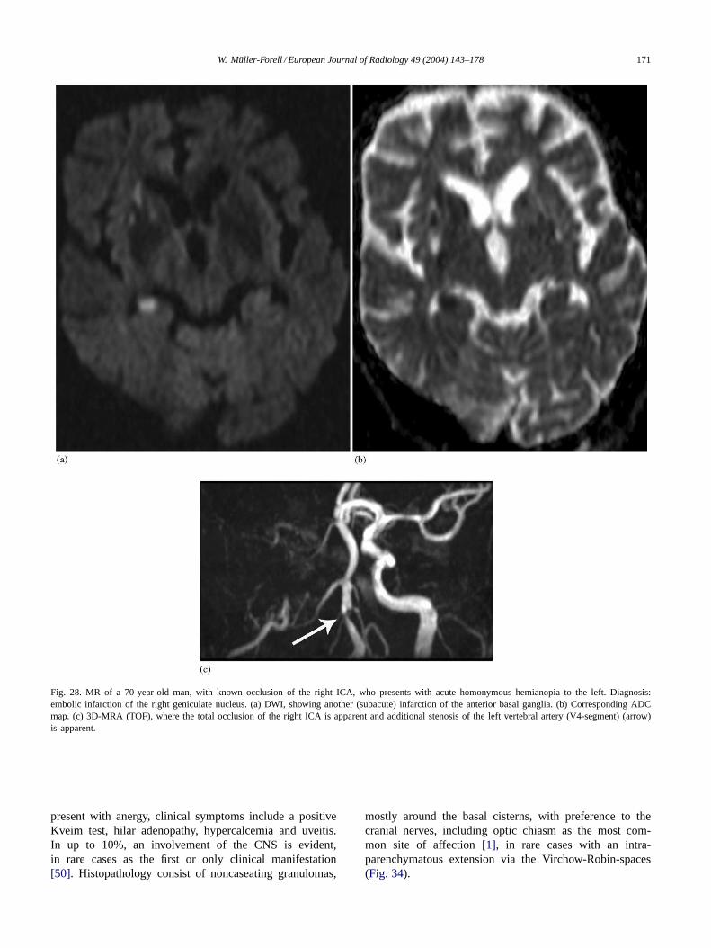
Fig. 28. MR of a 70-year-old man, with known occlusion of the right ICA, who presents with acute homonymous hemianopia to the left. Diagnosis:embolic infarction of the right geniculate nucleus. (a) DWI, showing another (subacute) infarction of the anterior basal ganglia. (b) Corresponding ADCmap. (c) 3D-MRA (TOF), where the total occlusion of the right ICA is apparent and additional stenosis of the left vertebral artery (V4-segment) (arrow)is apparent.
present with anergy, clinical symptoms include a positiveKveim test, hilar adenopathy, hypercalcemia and uveitis.In up to 10%, an involvement of the CNS is evident,in rare cases as the first or only clinical manifestation[50]. Histopathology consist of noncaseating granulomas,
mostly around the basal cisterns, with preference to thecranial nerves, including optic chiasm as the most com-mon site of affection[1], in rare cases with an intra-parenchymatous extension via the Virchow-Robin-spaces(Fig. 34).

172 W. Müller-Forell / European Journal of Radiology 49 (2004) 143–178
Fig. 29. A 33-year-old woman with acute homonymous hemianopia to the left. Diagnosis: infarction of PCA associated with cerebral vasculitis. (a) MR:axial T2w FLAIR image with an area of high signal intensity in the calcarine region. (b) Intra-arterial DSA: early lateral-view of the left VA (vertebralartery) with clearly apparent irregularity with narrowing (arrowhead) and fusiform widening of the PCA in the entire distribution area. (c) Arterial phaseof the left ICA in lateral view, confirming the diagnosis by segmental irregularity (arrowheads) of the distal branches of the MCA (middle cerebralartery). (With permission of Müller-Forell, 2002.)

W. Müller-Forell / European Journal of Radiology 49 (2004) 143–178 173
Fig. 30. MR of an 11-year-old girl with unexplained unconsciousness and known Sturge-Weber syndrome. (a) Axial T1w native view without anyevident abnormality. (b) Corresponding contrast enhanced view, demonstrating leptomeningeal enhancement of the left temporo-occipital region as wellas thickening of and asymmetric enhancement of the ispilateral choroid plexus. (c) Coronal T1w contrast enhanced view. Note the slight thickening andenhancement at the tentorium. (With permission of Müller-Forell, 2002.)
174 W. Müller-Forell / European Journal of Radiology 49 (2004) 143–178
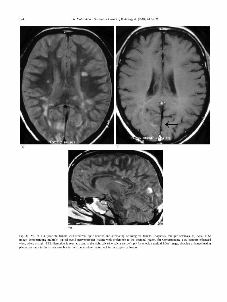
Fig. 31. MR of a 30-year-old female with recurrent optic neuritis and alternating neurological deficits. Diagnosis: multiple sclerosis. (a) Axial PDwimage, demonstrating multiple, typical ovoid periventricular lesions with preference to the occipital region. (b) Corresponding T1w contrast enhancedview, where a slight BBB disruption is seen adjacent to the right calcarine sulcus (arrow). (c) Paramedian sagittal PDW image, showing a demyelinatingplaque not only in the striate area but in the frontal white matter and in the corpus callosum.

W. Müller-Forell / European Journal of Radiology 49 (2004) 143–178 175
Fig. 32. MR of a 6-year-old boy with acute (not longer than 12 h) bilateral vision loss, and acute, diffuse disseminating pain in all parts of the body.Diagnosis: ADEM (acute disseminating encephalomyelitis). (a) Axial T2w view, demonstrating inflammatory affection of both optic tracts towards thelateral geniculate nucleus (right»left), right thalamus and basal ganglia. (b) Coronal T2w view of the posterior orbit with bright signal enhancement ofthe left optic nerve.
Fig. 33. T2w MR of a 47-year-old woman alcoholic presenting with bilateral internuclear ophthalmoplegia (INO) and ataxia. Diagnosis: Wernickeencephalopathy. (a) Axial view of the cerebral peduncle showing the characteristic periaqueductal demyelination. (b) Axial view of the region of thethird ventricle demonstrates the periventricular involvement (thalami). (With permission of Müller-Forell, 2002.)

176 W. Müller-Forell / European Journal of Radiology 49 (2004) 143–178
Fig. 34. MR of a 31-year-old female presenting with seizure, hypakusis, ptosis and internuclear ophthalmoplegia (INO) of the right eye. Diagnosis:sarcoidosis: (a) Axial T2w FLIR image with high signal of the chiasm and adjacent frontal as well as bitemporal subcortical white matter. (b)Corresponding T1w native view with irregular signals in the basal cistern. (c) Corresponding contrast enhanced view demonstrating ubiquitous nodularand confluent signal enhancement in all basal cisterns with interpeduncular dominance. (d) Midsagittal T1w contrast enhanced view, showing that theentire subarachnoid space is affected.
W. Müller-Forell / European Journal of Radiology 49 (2004) 143–178 177
References
[1] Okazaki H. Fundamentals of neuropathology. Morphologic basics ofneurologic disorders. New York: Igaku-Shoin, 1989.
[2] Goldberg HI, Lavi E, Atlas SW. Tumors. In: Atlas SW, editor. Mag-netic resonance imaging of the brain and spine. 2nd ed. Philadelphia:Lippincott-Raven; 1996, p. 423–87.
[3] Müller-Forell W. Imaging of orbital and visual pathway pathology.Springer, 2002.
[4] Osborn A, Rauschning W. Brain tumors and tumorlike masses: clas-sification and differential diagnosis. In: Osborn A, editor. Diagnosticneuroradiology. St. Louis: Mosby; 1994, p. 401–528.
[5] Kucharczyk W, Montanera W, Becker LE. The sella turcica andparasellar region. In: Atlas SW, editor. Magnetic resonance imag-ing of the brain and spine. Philadelphia: Lippincott-Raven; 1996,p. 871–930.
[6] Dietemann JL, Kehrli P, Maillot C, Dinz R, Reis M, Neugroschl C,Vinclair L. Is there a dural wall between the cavernous sinus andthe pituitary fossa? Anatomical and MRI findings. Neuroradiology1998;40:627–30.
[7] Louis DN, Scheithauer BW, Budka H, van Deimling A, KepesJJ.Meningiomas. In: Kleihues P, Cavanee WK, editors. Pathologyand genetics. Tumors of the nervous system. International Agencyfor Research on Cancer, Lyon; 2000, p. 176–84.
[8] Sartor K. MR imaging of the skull and brain. A correlative atlas.Springer, 1992.
[9] Crotty TB, Scheithauer BW, Young Jr, WF, David DH, Shaw EG,Miller GM, Burger PC. Papillary craniopharyngeoma: a clinical studyof 48 cases. J Neurosurg 1995;83:206–14.
[10] Kleihues P, Burger PC, Scheithauer BW.. Histological typing oftumors of the central nervous system. In: World Health OrganizationInternational Histological Classification of Tumors. 2nd ed. Springer,1993.
[11] Sartoretti-Schefer S, Wichmann W, Aguzzi A, Valavanis A. MR dif-ferentiation of adamantinous and squamous-capillary cranipopharyn-geoma. AJNR Am J Neuroradiol 1997;18:77–87.
[12] Osborn A.Stroke. In: Osborn A, editor. Diagnostic neuroradiology.St. Louis: Mosby; 1994a, p. 330–98.
[13] Stehbens WE. Cerebrovascular disease. In: Stehbens WE, Lie JT,editors. Pathology of the cerebral blood vessels. Vascular pathology.London: Chapman & Hall; 1995, p. 437–88.
[14] Kupersmith MJ.Aneurysms involving the motor and sensory visualpathway. In: Kupersmith MJ, editor. Neurovascular neuroophthal-mology. Springer; 1993, p. 239–99.
[15] Wilcock D, Jaspan T, Holland I, Cherryman G, Worthington B. Com-parison of magnetic resonance angiography with conventional an-giography in the detection of intracranial aneurysms in patients pre-senting with subarachnoid hemorrhage. Clin Radiol 1996;51:330–4.
[16] Byrne JV, Guglielmi G. Endovascular treatment of intracranialaneurysms. Springer, 1998.
[17] Sugahara T, Korogi Y, Nagashima K, Hamatake S, Honda S, Taka-hashi M. Comparison of 2D and 3D digital subtraction angiographyin evaluation of intracranial aneurysms. AJNR Am J Neuroradiol2002;23:1545–52.
[18] Rosenblum MK, Matsutani M, Van Meir EG. CNS germ cell tumors.In: Kleihues P, Cavanee WK, editors. Pathology and genetics. Tumorsof the nervous system. Lyon: International Agency for Research onCancer; 2000, p. 208–14.
[19] Meyers SP, Hirsch WL, Curtin HD, Barnes L, Sekhar LN, Sen C.Chordomas of the skull base: MR features. AJNR Am J Neuroradiol1992;13:1627–36.
[20] Dutton JJ. Gliomas of the anterior visual pathway. Surv Ophthalmol1994;38:427–52.
[21] Wulc AE, Bergin DJ, Barnes D, Scaravilli F, Wright JE, McDon-ald WI. Orbital optic nerve glioma in adult life. Arch Ophthalmol1989;107:1013–6.
[22] Hollander MD, FitzPatrick M, O′Connor SG, Flanders AE, TartaglinoLM. Optic gliomas. Radiol Clin North Am 1999;37:59–71.
[23] Cavanee WK, Furnari FB, Nagane M, Huang HJS, Newcomb EW,Bigner DD, Weller M, Berens ME, Plate KH, Israel MA, NobleMD, Kleihues P.Diffusely infiltrating astrocytomas. In: Kleihues P,Cavanee WK, editors. Pathology and genetics of tumors of the centralnervous system. Lyon: IARC Press; 2000, p. 10–21.
[24] Kleihues P, Burger PC, Collins VP, Newcomb EW, Ohgaki H, Cava-nee WK. Glioblastoma. In: Kleihues P, Cavanee WK, editors. Pathol-ogy and genetics. Tumors of the nervous system. Lyon: InternationalAgency for Research on Cancer; 2000, p. 29–39.
[25] Aoki S, Barkovich AJ, Nishimura K, Kjos BO, Macida T, Cogen P,Norman D. Neurofibromatosis types 1 and 2: cranial MR findings.Radiology 1989;172:527–34.
[26] Steiger HJ, Markwalder TM, Reulen HJ. Clinicopathological rela-tions of cerebral cavernous angiomas. Observation in eleven cases.Neurosurgery 1987;21:879–84.
[27] Kupersmith MJ, Kalish H, Epstein F, Yu G, Berenstein A, Woo H,Jafar J, Mandel G, De Lara F. Natural history of brainstem cavernousmalformations. Neurosurgery 2001;48:47–53.
[28] Wiestler OD, Schiffer D, Coons SW, Prayson RA, Rosenblum MK.Ependymoma. In: Kleihues P, Cavanee WK, editors. Pathology andgenetics. Tumors of the nervous system. Lyon: International Agencyfor Research on Cancer; 2000, p. 72–6.
[29] Burger PC, Scheithauer BW. Tumors of neuroglia and choroid plexusepithelium. In: Tumors of the central nervous system. WashingtonDC: Armed Forces Institute of Pathology; 1994, p. 25–161.
[30] Paulus W, Jellinger K, Morgello S, Deckert-Schlüter M. Malignantlymphomas. In: Kleihues P, Cavanee WK, editors. Pathology andgenetics. Tumors of the nervous system. Lyon: International Agencyfor Research on Cancer; 2000, p. 198–203.
[31] Nasir S, DeAngelis LM. Update on the management of primary CNSlymphoma. Oncology (Huntingt) 2000;14:228–34.
[32] Hankey GJ, Warlow CP. The role of imaging in the management ofcerebral and ocular ischemia. Neuroradiology 1991;33:381–90.
[33] Van der Zwan A, Hillen B, Tulleken CA, Dujovny M, Dragovic L.Variability of the territory of the major cerebral arteries. J Neurosurg1992;77:927–40.
[34] Janssen O, Brückmann H, Ischämische H. In: Sartor K, editor. Neu-roradiologie, 2nd ed. Stuttgart: Thieme; 2001, p. 140–58.
[35] Fauci AS, Haynes B, Katz P. NIH conference. The spectrum of vas-culitis: clinical, pathologic, immunologic, and therapeutic consider-ations. Ann Intern Med 1978;89:660–76.
[36] Block F, Reith W. Isolierte Vaskulitis des ZNS. Radiologe2001;40:1090–7.
[37] Ferro JM. Vasculitis of the cerebral nervous system. J Neurol1998;245:766–76.
[38] Smirniotopoulos JG, Murphy FM. Central nervous system manifes-tation of the phakomatoses and other inherited syndromes. In: AtlasSW, editor. Magnetic resonance imaging of the brain and spine, 2nded. Philadelphia: Lippincott-Raven; 1996, p. 773–802.
[39] Van der Knaap MS, Valk J, editors. Magnetic resonance of myelitis,myelination, and myelin disorders. 2nd ed. Springer, 1995.
[40] Edwards-Brown ME, Bonin JM. White matter diseases. In: AtlasSW, editor. Magnetic resonance imaging of the brain and spine.Philadelphia: Lippincott-Raven; 1996, p. 649–706.
[41] McDonald WI, Compston A, Edan G, Goodkin D, Hartung HP,Lublin FD, McFarland HF, Paty DW, Polman CH, Reingold SG,Sandberg-Wollheim M, Sibley W, Thompson A, van den Noort S,Weinhenker BY, Wolinski JS. Recommended diagnostic criteria mul-tiple sclerosis: guidelines from the international panel on diagnosisof multiple sclerosis. Ann Neurol 2001;50:121–7.
[42] Filippi M, Rocca MA, Wiessmann M, Mennes S, Cercignani M,Yousri TA, Soriani MP. A comparison of MR imaging with fast-FLAIR, HASTE FLAIR, and EPI-FLAIR sequences in the assess-ment of patients with multiple sclerosis. AJNR Am J Neuroradiol1999;20:1931–8.

178 W. Müller-Forell / European Journal of Radiology 49 (2004) 143–178
[43] Reiche W, Merkelbach S, Reith W. Neuroradiologische Aspekte beiEncephalitis disseminata. Radiologe 2000;40:1045–56.
[44] Horowitz AL, Kaplan RD, Grewe G, White RT, Salberg LM. Theovoid lesion: a new MR observation in patients with multiple scle-rosis. AJNR Am J Neuroradiol 1989;10:303–5.
[45] Paty DW. MRI as a method to reveal in vivo pathology in MS. JNeural Transm [Suppl] 1997;49:211–7.
[46] Osborn A. Infections of the brain and its linings. In: Osborn A, editor.Diagnostic neuroradiology. St. Louis: Mosby; 1994b, p. 673–715.
[47] Barkovich JA. Pediatric neuroimaging, 2nd ed. Philadelphia:Lippincott-Raven, 2000.
[48] Galluci M, Bozzao A, Splendiani A, Maciocchi C, Passariello R.Wernicke encephalopathy: MR findings on eight patients. AJNR AmJ Neuroradiol 1990;11:887–92.
[49] Schroth G, Wichmann W, Valavanis A. blood brain barrier disruptionin acute Wernicke encephalopathy: MR findings. J Comput AssistTomogr 1991;15:1059–60.
[50] Zajicek JP, Scolding NJ, Foster O, Rovaris M, Evanson J, MoseleyIF, Scadding JW, Thompson EJ, Chamoun V, Miller DH, McDonaldWI, Mitchell D. Central nervous system sarcoidosis—diagnosis andmanagement. Q J Med1999;92:103–17.