introduction to nanobioscience: a tissue engineering perspective · 2017-01-26 · nanoscience and...
TRANSCRIPT
NANOSCIENCE AND NANOTECHNOLOGIES- Introduction To Nanobioscience: A Tissue Engineering Perspective -Serge Ostrovidov, Azadeh Seidi, Kaarunya Sampathkumar, Queeny Dasgupta, Alok Srivastava, Ali Khademhosseini, Murugan Ramalingam
INTRODUCTION TO NANOBIOSCIENCE: A TISSUE ENGINEERING PERSPECTIVE Serge Ostrovidov Tohoku University, Sendai 980-8577, Japan Azadeh Seidi Okinawa Institute of Sciences and Technology (OIST), Japan Deepti Rana Centre for Stem Cell Research (CSCR), (A unit of Institute for Stem Cell Biology and Regenerative Medicine, Bengaluru), Christian Medical College Campus, Vellore 632002, India Project Internship Student from Amity University, Noida 201313, Uttar Pradesh, India Kaarunya Sampathkumar Centre for Stem Cell Research (CSCR), (A unit of Institute for Stem Cell Biology and Regenerative Medicine, Bengaluru), Christian Medical College Campus, Vellore 632002, India Queeny Dasgupta Centre for Stem Cell Research (CSCR), (A unit of Institute for Stem Cell Biology and Regenerative Medicine, Bengaluru), Christian Medical College Campus, Vellore 632002, India Alok Srivastava Centre for Stem Cell Research (CSCR), (A unit of Institute for Stem Cell Biology and Regenerative Medicine, Bengaluru), Christian Medical College Campus, Vellore 632002, India Ali Khademhosseini Tohoku University, Sendai 980-8577, Japan Harvard University, Boston 02115, USA Kyung Hee University, Seoul 130-701, Republic of Korea King Abdulaziz University, Jeddah 21569, Saudi Arabia Murugan Ramalingam Tohoku University, Sendai 980-8577, Japan Centre for Stem Cell Research (CSCR), (A unit of Institute for Stem Cell Biology and Regenerative Medicine, Bengaluru), Christian Medical College Campus, Vellore 632002, India Université de Strasbourg, Strasbourg 67085, France
Keywords: Nanobioscience, tissue engineering, scaffolds, biomaterials, nanomaterials, nanopatterns, nanofibers, stem cell engineering Contents 1. Introduction 2. Nanopatterns and Nanopatterning Techniques
2.1. Physical Nanopatterns 2.2. Chemical Nanopatterns
3. Methods of Fabricating Nanofiber Scaffolds 3.1. Electrospinning 3.2. Self-Assembly
@Encyclopedia Of Life Support Systems(EOLSS)
NANOSCIENCE AND NANOTECHNOLOGIES- Introduction To Nanobioscience: A Tissue Engineering Perspective -Serge Ostrovidov, Azadeh Seidi, Kaarunya Sampathkumar, Queeny Dasgupta, Alok Srivastava, Ali Khademhosseini, Murugan Ramalingam
3.3. Phase Separation 4. Applications of Nanofibers in Tissue Engineering 5. Conclusion Acknowledgements Glossary Bibliography Biographical Sketches Summary The chapter presents an introduction and applications of nanobioscience in the context of tissue engineering, which is a rapidly developing field that aims at regenerating or repairing diseased or damaged tissues of the human body. There are three key factors that influence the success of engineered tissues, cells, scaffolds, and biomolecules. Scaffolds play a key role in providing a biomimicking support on which cells can grow into corresponding tissues. Therefore, increasing attention is being paid to the in vitro simulation of the nano-scale interaction of cells in the body with native extracellular matrix (ECM). In fact, the fabrication of scaffolds with nanofeatures and nanosignals, which mimic the native ECM environment, has a large impact on guiding and directing the cellular behavior for tissue engineering applications. Numerous techniques are now available for the production of nanostructured or nanopatterned surfaces/substrates. In this chapter, we aim to provide an overview of the methods used to fabricate physical and chemical nanopatterns and nanofiber scaffolds as emerging nanofeatured substrates suitable for tissue engineering. Finally, we review and discuss some of their most notable applications in cell and tissue engineering. 1. Introduction Nanoscience as an interdisciplinary subject is among the most rapidly developing areas of science and technology in the last few decades. The impact of nanoscience, in combination with nanotechnology, is evident in medicine, helping to create functional tissue grafts for tissue engineering and regenerative applications in the last several years (Sharma, Gautam et al. 2011). Millions of people suffer from a variety of tissue/organ diseases or defects throughout the world. Although autografts and allografts help to solve these clinical problems, there is still a shortage of donor tissues and organs. This shortage has drastically increased recently, causing many patients to die while waiting for donor tissues or organs. Therefore, it is essential to develop more biomimetic tissue grafts in the laboratory for the benefit of human health care. For this reason, tissue engineering has emerged as an applied interdisciplinary field, which aims to repair tissue or cause its regeneration by applying the principles and methods of biological, chemical, and engineering sciences (Langer and Vacanti 1993;). The concept of tissue engineering involves isolating cells from a patient or donor, culturing them in vitro to increase their number and maintain their distinct phenotypes, seeding them onto a scaffold to create an engineered tissue graft, and finally transplanting the engineered tissue graft back into the patient’s body where the tissue regeneration is needed (see Fig. 1).
@Encyclopedia Of Life Support Systems(EOLSS)
NANOSCIENCE AND NANOTECHNOLOGIES- Introduction To Nanobioscience: A Tissue Engineering Perspective -Serge Ostrovidov, Azadeh Seidi, Kaarunya Sampathkumar, Queeny Dasgupta, Alok Srivastava, Ali Khademhosseini, Murugan Ramalingam
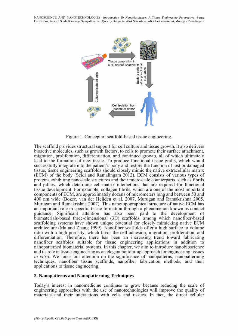
Figure 1. Concept of scaffold-based tissue engineering. The scaffold provides structural support for cell culture and tissue growth. It also delivers bioactive molecules, such as growth factors, to cells to promote their surface attachment, migration, proliferation, differentiation, and continued growth, all of which ultimately lead to the formation of new tissue. To produce functional tissue grafts, which would successfully integrate into the patient’s body and restore the function of lost or damaged tissue, tissue engineering scaffolds should closely mimic the native extracellular matrix (ECM) of the body (Seidi and Ramalingam 2012). ECM consists of various types of proteins exhibiting nanoscale structures and their microscale counterparts, such as fibrils and pillars, which determine cell-matrix interactions that are required for functional tissue development. For example, collagen fibrils, which are one of the most important components of ECM, are approximately dozens of micrometers long and between 50 and 400 nm wide (Bozec, van der Heijden et al. 2007, Murugan and Ramakrishna 2005, Murugan and Ramakrishna 2007). This nanotopographical structure of native ECM has an important role in specific tissue formation through a phenomenon known as contact guidance. Significant attention has also been paid to the development of biomaterials-based three-dimensional (3D) scaffolds, among which nanofiber-based scaffolding systems have shown unique potential for closely mimicking native ECM architecture (Ma and Zhang 1999). Nanofiber scaffolds offer a high surface to volume ratio with a high porosity, which favor the cell adhesion, migration, proliferation, and differentiation. Therefore, there has been an increasing trend toward fabricating nanofiber scaffolds suitable for tissue engineering applications in addition to nanopatterned biomaterial systems. In this chapter, we aim to introduce nanobioscience and its role in tissue engineering as an elegant bottom-up approach for engineering tissues in vitro. We focus our attention on the significance of nanopatterns, nanopatterning techniques, nanofiber tissue scaffolds, nanofiber fabrication methods, and their applications to tissue engineering. 2. Nanopatterns and Nanopatterning Techniques Today’s interest in nanomedicine continues to grow because reducing the scale of engineering approaches with the use of nanotechnologies will improve the quality of materials and their interactions with cells and tissues. In fact, the direct cellular
@Encyclopedia Of Life Support Systems(EOLSS)
NANOSCIENCE AND NANOTECHNOLOGIES- Introduction To Nanobioscience: A Tissue Engineering Perspective -Serge Ostrovidov, Azadeh Seidi, Kaarunya Sampathkumar, Queeny Dasgupta, Alok Srivastava, Ali Khademhosseini, Murugan Ramalingam
environment is in nanometric scale, including the ECM with which cells are familiar and in which they evolve. Mimicking this natural environment by producing materials with nanofeatures and nanosignals to guide and direct cellular behavior is therefore of the highest importance for tissue engineering applications. Numerous techniques are now available for the production of nanostructures and nanotextured or nanopatterned surfaces, including polymer demixing, nanografting, dip-pen nanolithography, conductive atomic force microscopy, nanocontact printing, nanoimprint lithography, photolithography, laser holography, electron beam lithography, colloidal lithography, UV-assisted capillary force lithography, soft lithography, polymer templating, DNA templating, metal anodization, molecular beam epitaxy, self-assembly, electrospinning, nanophase coating, and glancing angle deposition. Rather than discussing these techniques in detail, we refer those readers who are interested in learning further details to several comprehensive articles (Engel, Michiardi et al. 2008; Dolatshahi-Pirouz, Nikkhah et al. 2011, Seidi and Ramalingam 2012). In the following section, we will focus on the different types of nanopatterns used in basic cell/tissue studies and their effects on cell behavior in terms of cell adhesion, migration, proliferation, differentiation, and continued function, which are all important parameters for the success of functional tissue engineering. 2.1. Physical Nanopatterns Physical patterning refers to the modification of biomaterial substrates with a pre-defined texture by modulating their size and shape. Physical substrate characteristics, such as stiffness, roughness, and topography, have a significant influence on the regulation of cell behavior, particularly cell adhesion, migration, proliferation, and differentiation. With the introduction of micro/nanofabrication techniques into the life sciences, significant evidence has been gathered that cells sense and respond to microscale and nanoscale features (Evans, Britland et al. 1999). Thus, a recent study showed that myoblasts aligned to 10 – 15 nm diameter cellulose nanowhiskers ( )Dugan, Gough et al. 2010 . The mechanism by which cells sense these physical cues is not clearly known. However, there is some evidence that filopodia are involved in sensing such cues because they extend in front of cells and probe nearby nanotopographic features (Lim, Hansen et al. 2005). These nanopatterns have major effects on cell adhesion, orientation, shape, proliferation, migration, differentiation, signaling cascades, and gene activation (Lim and Donahue 2007). They may have different forms, such as columns, islands, pits, protrusions, or nodes, and can also be anisotropic structures, such as patterns of ridges/grooves or isotropic-like nanoislands, homogeneously covering the substrate surface. Many studies using patterns of ridges/grooves have shown that cell alignment with the pattern increases with increasing groove depth while decreasing with increasing groove width or pitch, which is the sum of the groove and ridge width ( Zhu, Lu et al. 2005). Thus, Yim et al. observed cell and nuclei alignment, elongated cell shape, and reduced cell proliferation when smooth muscle cells (SMCs) were cultured on nanograting with 350 nm line width and depth (Yim, Reano et al. 2005). In addition, a study by Zhu et al. using nanogrooves with a depth of 60 nm reported that mesenchymal stem cell (MSC)-derived osteoblasts cultured on this substrate showed anisotropic alignment and mineralized matrix (Zhu, Lu et al. 2005). If the groove width has a nanometric size, cells tend to bridge over the top of the ridges rather than reside inside the grooves (Teixeira, Abrams et al. 2003). For example, epithelial cells covered the floor of 2 µm wide grooves, whereas they bridged over 950 nm wide grooves. Studies using other nanostructures have shown that fibroblast adhesion was improved on 13 nm-high island nanotextured surfaces but were impaired on 95 nm high islands (Dalby, Giannaras et al. 2004). Lim et al. also observed better adhesion and differentiation of osteoblasts cultured on 11 nm high island nanotextured surfaces than on 85 nm high islands (Lim, Hansen et al. 2005). Although cell adhesion cannot occur across pits, and cells must settle in the inter-pit area, by using
@Encyclopedia Of Life Support Systems(EOLSS)
NANOSCIENCE AND NANOTECHNOLOGIES- Introduction To Nanobioscience: A Tissue Engineering Perspective -Serge Ostrovidov, Azadeh Seidi, Kaarunya Sampathkumar, Queeny Dasgupta, Alok Srivastava, Ali Khademhosseini, Murugan Ramalingam
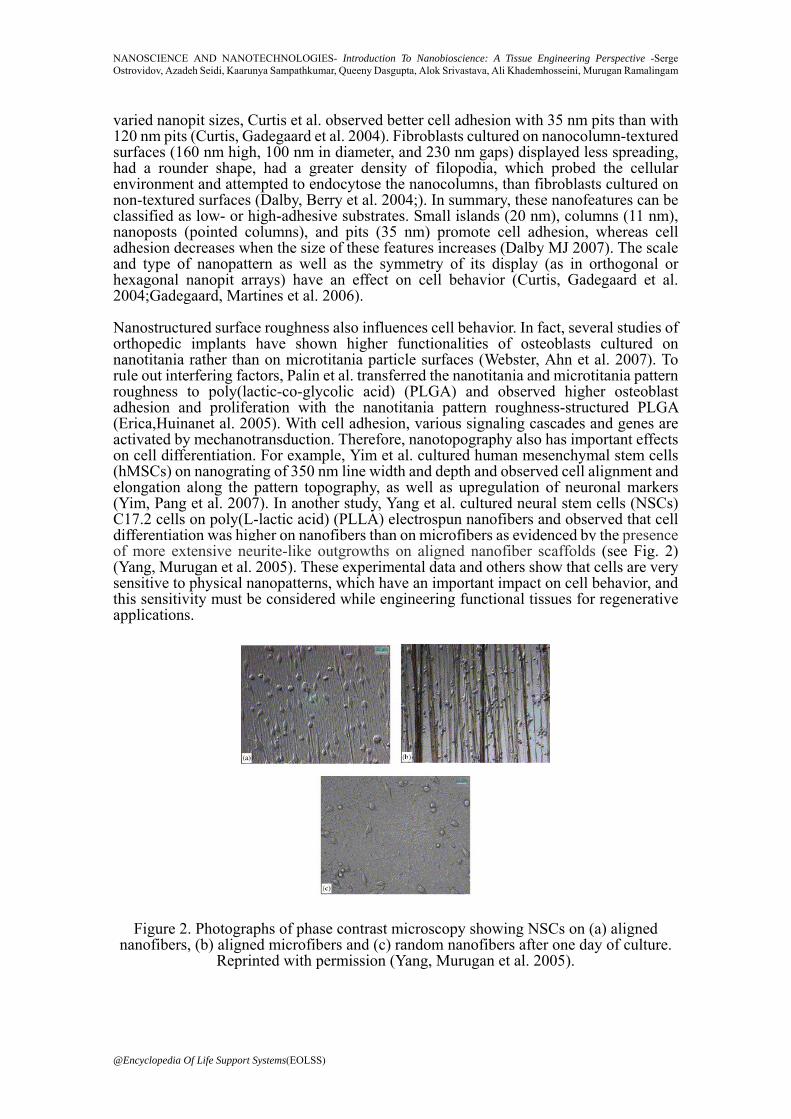
varied nanopit sizes, Curtis et al. observed better cell adhesion with 35 nm pits than with 120 nm pits (Curtis, Gadegaard et al. 2004). Fibroblasts cultured on nanocolumn-textured surfaces (160 nm high, 100 nm in diameter, and 230 nm gaps) displayed less spreading, had a rounder shape, had a greater density of filopodia, which probed the cellular environment and attempted to endocytose the nanocolumns, than fibroblasts cultured on non-textured surfaces (Dalby, Berry et al. 2004;). In summary, these nanofeatures can be classified as low- or high-adhesive substrates. Small islands (20 nm), columns (11 nm), nanoposts (pointed columns), and pits (35 nm) promote cell adhesion, whereas cell adhesion decreases when the size of these features increases (Dalby MJ 2007). The scale and type of nanopattern as well as the symmetry of its display (as in orthogonal or hexagonal nanopit arrays) have an effect on cell behavior (Curtis, Gadegaard et al. 2004;Gadegaard, Martines et al. 2006). Nanostructured surface roughness also influences cell behavior. In fact, several studies of orthopedic implants have shown higher functionalities of osteoblasts cultured on nanotitania rather than on microtitania particle surfaces (Webster, Ahn et al. 2007). To rule out interfering factors, Palin et al. transferred the nanotitania and microtitania pattern roughness to poly(lactic-co-glycolic acid) (PLGA) and observed higher osteoblast adhesion and proliferation with the nanotitania pattern roughness-structured PLGA (Erica,Huinanet al. 2005). With cell adhesion, various signaling cascades and genes are activated by mechanotransduction. Therefore, nanotopography also has important effects on cell differentiation. For example, Yim et al. cultured human mesenchymal stem cells (hMSCs) on nanograting of 350 nm line width and depth and observed cell alignment and elongation along the pattern topography, as well as upregulation of neuronal markers (Yim, Pang et al. 2007). In another study, Yang et al. cultured neural stem cells (NSCs) C17.2 cells on poly(L-lactic acid) (PLLA) electrospun nanofibers and observed that cell differentiation was higher on nanofibers than on microfibers as evidenced by the presence of more extensive neurite-like outgrowths on aligned nanofiber scaffolds (see Fig. 2) (Yang, Murugan et al. 2005). These experimental data and others show that cells are very sensitive to physical nanopatterns, which have an important impact on cell behavior, and this sensitivity must be considered while engineering functional tissues for regenerative applications.
Figure 2. Photographs of phase contrast microscopy showing NSCs on (a) aligned nanofibers, (b) aligned microfibers and (c) random nanofibers after one day of culture.
Reprinted with permission (Yang, Murugan et al. 2005).
@Encyclopedia Of Life Support Systems(EOLSS)
NANOSCIENCE AND NANOTECHNOLOGIES- Introduction To Nanobioscience: A Tissue Engineering Perspective -Serge Ostrovidov, Azadeh Seidi, Kaarunya Sampathkumar, Queeny Dasgupta, Alok Srivastava, Ali Khademhosseini, Murugan Ramalingam
2.2. Chemical Nanopatterns Chemical patterns refer to the modification of biomaterial substrates with patterns of different chemicals or biochemical reagents. Chemical nanopatterning can be realized by nanopatterns of chemical moieties (e.g., self-assembled monolayers (SAMs)) or biological moieties (e.g., RGD peptides). Patterning of ECM proteins, such as fibronectin, laminin, vitronectin, or collagen, to promote cell adhesion is of great interest for tissue engineering. Each ECM protein induces a specific binding with integrins (e.g., fibronectin preferentially binds α5β1, vitronectin binds αvβ3, and collagen binds α2β1). It has been proposed that protein deposition plays a role in contact guidance. Following this hypothesis, local micro-nanotopography may interfere with protein adsorption or change of protein’s adhesive functionality, leading to discontinuous protein deposition and local protein concentration (van Kooten and von Recum 1999). However, this hypothesis has not been confirmed, and other studies have shown that protein deposition does not localize preferentially with topographic discontinuities, while cells sense topography irrespective of protein deposition (Wilson, Clegg et al. 2005).
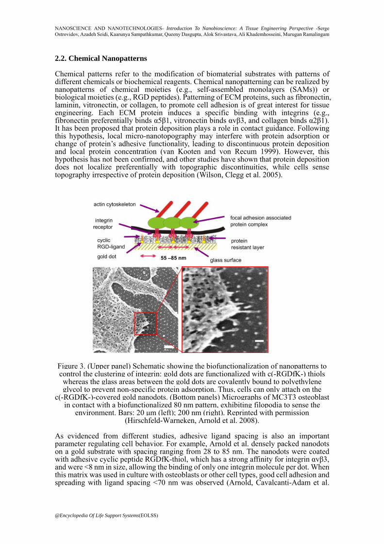
Figure 3. (Upper panel) Schematic showing the biofunctionalization of nanopatterns to control the clustering of integrin: gold dots are functionalized with c(-RGDfK-) thiols whereas the glass areas between the gold dots are covalently bound to polyethylene glycol to prevent non-specific protein adsorption. Thus, cells can only attach on the
c(-RGDfK-)-covered gold nanodots. (Bottom panels) Micrographs of MC3T3 osteoblast in contact with a biofunctionalized 80 nm pattern, exhibiting filopodia to sense the
environment. Bars: 20 μm (left); 200 nm (right). Reprinted with permission (Hirschfeld-Warneken, Arnold et al. 2008).
As evidenced from different studies, adhesive ligand spacing is also an important parameter regulating cell behavior. For example, Arnold et al. densely packed nanodots on a gold substrate with spacing ranging from 28 to 85 nm. The nanodots were coated with adhesive cyclic peptide RGDfK-thiol, which has a strong affinity for integrin αvβ3, and were <8 nm in size, allowing the binding of only one integrin molecule per dot. When this matrix was used in culture with osteoblasts or other cell types, good cell adhesion and spreading with ligand spacing <70 nm was observed (Arnold, Cavalcanti-Adam et al.
@Encyclopedia Of Life Support Systems(EOLSS)
NANOSCIENCE AND NANOTECHNOLOGIES- Introduction To Nanobioscience: A Tissue Engineering Perspective -Serge Ostrovidov, Azadeh Seidi, Kaarunya Sampathkumar, Queeny Dasgupta, Alok Srivastava, Ali Khademhosseini, Murugan Ramalingam
2004). Other studies have also shown that cell activation is increased when ligand spacing decreases over the range of 70 to 10 nm (Cavalcanti-Adam, Volberg et al. 2007). Gradients of ligand density have also been used to investigate cellular adhesion, spreading, and migration. For example, Hirschfeld-Warneken et al. cultured osteoblasts on a gradient of spaced nanodots (see Fig. 3). These nanodots had a diameter of 6 nm and were coated with cyclic RGDfK. The researchers observed an increase in cell adhesion and spreading with decreased nanodot spacing, while nanodot spacing >73 nm reduced cell adhesion and spreading (Hirschfeld-Warneken, Arnold et al. 2008). As the gradient of binding site spacing had a strength of Δ15 nm/mm and induced cell polarization, by analyzing cell morphology, they were able to determine that an osteoblast cell can sense a 1 nm spacing difference between its front and back (Hirschfeld-Warneken, Arnold et al. 2008). During displacement, a cell binds to a new position at its front while unbinding from positions at its back; thus, adhesive ligand spacing is also important for cell motility. Maheshwari et al. showed that cell migration depends not only on the spacing between binding sites but also on the number of adhesive ligands on the site (Maheshwari, Brown et al. 2000). Experimental studies with gradients of adhesive ligand density have shown that cell migration increases toward higher ligand density and adhesiveness up to a threshold point, above which it reaches a plateau or decreases (Smith, Elkin et al. 2006). Hydrogels, a type of biomaterial that has a network of hydrophilic polymer chains, have also been used to study cell behavior, particularly 2D and 3D cell adhesion, largely because the number and repartition of adhesive ligands presented to cells can be tuned. Huebsch et al. showed that hMSCs encapsulated in an alginate hydrogel containing RGD peptides mechanically reorganized ligand presentation near the integrins at a nanoscale level when the flexibility of the substrate allowed such behavior (Huebsch, Arany et al. 2010). Nanopatterns can also be realized by self-assembly, and networks of nanofibers made of self-assembled amphiphilic peptides can be functionalized to direct cellular behavior (Khademhosseini, Rajalingam et al. 2010). For example, Silva et al. synthesized a hydrogel with incorporated peptide isoleucine-lysine-valine-alanine-valine (IKVAV), which promotes neurite outgrowth. When murine neural progenitor cells (NPCs) were encapsulated, the researchers observed rapid stem cell differentiation into neurons and repression of astrocyte development (Silva, Czeisler et al. 2004). Wang et al. functionalized RADA16-I peptide by coupling it with RGD peptide to increase endothelial cell adhesion and with vascular endothelial growth factor (VEGF) mimicking motifs to induce angiogenesis. Cultured with HUVECs, this self-assembled nanofiber scaffold enhanced endothelial cell adhesion, migration, proliferation, and induced capillary formation (Wang, Horii et al. 2008). Self-assembly technology has also been applied in vivo to the release of basic fibroblast growth factor (bFGF). When amphiphilic peptide and bFGF solution were injected subcutaneously into a mouse, a clear hydrogel formed that induced prolonged bFGF release with significant angiogenesis (Hosseinkhani, Hosseinkhani et al. 2006). Other studies have used self-assembly to culture synthetic dermis and synthetic skin (Kao, Kadomatsu et al. 2009), for bone regeneration (Yoshimi, Yamada et al. 2009), neural regeneration (Ellis-Behnke, Liang et al. 2006; Gelain, Bottai et al. 2006), hemostasis (Ellis-Behnke, Liang et al. 2006) and other biomedical applications (Ellis-Behnke and Jonas 2011). Another nanopattern type currently used in orthopedic applications is made using nanophase deposition. Nanophase coating by sol-gel reaction or pulse electrodeposition is currently used to enhance the osteointegration of a material by coating it with hydroxyapatite (Caruso and Antonietti 2001;Saremi and Golshan 2007). Delivering chemical and topographical signals together to the targeted site is desirable for tissue engineering applications. Some studies have showed that chemical and topographical cues can compete or have synergetic effects on cells. For example, Britland et al. showed that BHK cells preferred to align on chemical rather than topographical cues when cultured on groove/ridge patterns overlaid orthogonally with continuous adhesive protein strips (Britland, Morgan
@Encyclopedia Of Life Support Systems(EOLSS)

NANOSCIENCE AND NANOTECHNOLOGIES- Introduction To Nanobioscience: A Tissue Engineering Perspective -Serge Ostrovidov, Azadeh Seidi, Kaarunya Sampathkumar, Queeny Dasgupta, Alok Srivastava, Ali Khademhosseini, Murugan Ramalingam
et al. 1996). However, when both cues were presented to cells in parallel, cell alignment was synergistically enhanced. In contrast, when Charest et al. cultured MC3T3-E1 osteoblasts on embossed groove/ridge patterns overlaid orthogonally with discontinuous printed fibronectin lanes, they observed that cells aligned on the topographical cues (Charest, Eliason et al. 2006). In this section, we have highlighted different aspects of nanopatterns and nanopatterning techniques. Physical features, such as topography, size, symmetrical order or disorder, roughness, and 2D or 3D scaffolding environments, and chemical features, such as adhesive ligand number and spacing, SAMs, protein or nanophase coating, and functionalized peptides, all have a strong impact on cell behavior and must be considered for tissue engineering applications. In vivo, native ECM is a 3D map full of biophysical and biochemical cues that guide and regulate such cell behaviors as adhesion, proliferation, differentiation, and gene expression. Mimicking ECM with respect to the way in which information is delivered to cells is still far from being accomplished. Therefore, mimicking the environmental complexity familiar to cells by integrating ECM nanofeatures into scaffolding systems is necessary and will enhance cell functionality. For example, increasing cell adhesion or favoring stem cell differentiation by the use of nanopatterned surfaces is already advantageous from a tissue engineering perspective. Nanofeature-patterned surfaces may also be more easily accepted by host tissues, reducing fibrous encapsulation. Combining physical and chemical cues on nanopatterned scaffolds offers a way to induce multiple synergetic cellular signaling responses and heterotypic cell stimulation. Mixing micro- and nanofeatures will increase the scaffold’s potential. To mimic 3D natural matrix more thoroughly, the adjunction of temporal control on signal delivery will be required. With advances in nanoscience and nanotechnology, patterning techniques are currently evolving toward biomedical applications that could help to build biomimetically functional matrix suitable for tissue engineering. In the following sections, current methods of fabricating nanofiber scaffolds as promising nanobiomaterials for tissue engineering applications will be discussed. 3. Methods of Fabricating Nanofiber Scaffolds There are three major techniques currently utilized for the fabrication of nanofiber scaffolds: electrospinning, self-assembly, and phase separation. Among them, electrospinning is the most widely used technique for fabricating tissue engineering scaffolds. 3.1. Electrospinning Electrospinning is a versatile technique for producing nanofibers from polymers and their composites. As explained in Murugan and Ramakrishna 2006, this technique was introduced almost a century ago. At that time, it was named “electrostatic spray” or “electrostatic spinning” and was later renamed as “electrospinning” in the 90’s. However, only a decade ago, this method attracted a great deal of attention from researchers fabricating scaffolds for tissue engineering applications. The particular merit of this technique is that it allows building scaffolds with structural features quite similar to those of native ECM. It is also a more versatile technique than other conventional scaffold methodologies. For example, nanofibers with spatial orientation, high aspect ratios, and large surface areas can be produced, while allowing control over pore geometry. These favorable characteristics directly influence the cell adhesion, migration, contact guidance, and transportation of oxygen and nutrients to the cells. On this base, electrospun nanofibers could serve as an optimal tissue scaffold providing spatial environments for the growth of new functional tissue with appropriate physiological metabolic functions.
@Encyclopedia Of Life Support Systems(EOLSS)
NANOSCIENCE AND NANOTECHNOLOGIES- Introduction To Nanobioscience: A Tissue Engineering Perspective -Serge Ostrovidov, Azadeh Seidi, Kaarunya Sampathkumar, Queeny Dasgupta, Alok Srivastava, Ali Khademhosseini, Murugan Ramalingam
sialoprotein, and osteocalcin expression Peptide amphiphile : Peptide-based molecules consisting of a hydrophobic tail and
Photolithography : A process used in microfabrication that utilizes a light source to transfer a geometric pattern from a photomask to a light-sensitive chemical photoresist on substrate Porogen : Particles, such as salt crystals, used to create pores in tissue engineering scaffolds
corresponding tissue Self-assembled monolayer (SAM) : Molecular assemblies that form spontaneously into large ordered domains Self-assembly : The process by which molecules adopt a defined arrangement without guidance or management from an outside source
Soft tissue : Tissues, such as tendons, ligaments, and fascia, that connect, support,
Bibliography Altman, G. H., F. Diaz, C. Jakuba, T. Calabro, R. L. Horan, J. Chen, H. Lu, J. Richmond, D. L. Kaplan (2003). Silk-based biomaterials. Biomaterials 24, 401-416. [This paper reviews importance of silk-based biomaterials for a variety of biomedical applications.]
Arnold, M., E. A. Cavalcanti-Adam, R. Glass, J. Blummel, W. Eck, M. Kantlehner, H. Kessler, J.P. Spatz (2004). "Activation of Integrin Function by Nanopatterned Adhesive Interfaces." ChemPhysChem 5(3): 383-388. [This paper determined that [58-73 nm] is the maximal length range between integrins for effective cell adhesion.]
Bozec, L., G. van der Heijden, M. Horton (2007). “Collagen Fibrils: Nanoscale Ropes.” Biophys J 92(1): 70–75. [This paper provides a new insight on the structure of the collagen fibril which will have diverse applications in the design and fabrication of scaffolds for tissue engineering.]
Britland, S., H. Morgan, B. Wojiak-Stodart, M. Riehle, A. Curtis, C. Wilkinson (1996). "Synergistic and Hierarchical Adhesive and Topographic Guidance of BHK Cells." Experimental Cell Research 228(2): 313-325. [This paper explored the effects of groove depth [0.1-6mm] with/without adhesive cue on BHK cell alignment.]
Cavalcanti-Adam, E. A., T. Volberg, A. Micoulet, H. Kessler, B. Geiger, J. P. Spatz (2007). "Cell Spreading
@Encyclopedia Of Life Support Systems(EOLSS)
NANOSCIENCE AND NANOTECHNOLOGIES- Introduction To Nanobioscience: A Tissue Engineering Perspective -Serge Ostrovidov, Azadeh Seidi, Kaarunya Sampathkumar, Queeny Dasgupta, Alok Srivastava, Ali Khademhosseini, Murugan Ramalingam
and Focal Adhesion Dynamics Are Regulated by Spacing of Integrin Ligands." Biophysical Journal 92(8): 2964-2974. [This paper showed that 58 nm or less spacing between RGD adhesive peptide molecules is necessary for stable focal adhesion formation and fibroblast cell spreading.]
Charest, J. L., M. T. Eliason, A. J. Garcia, W. P. King (2006). "Combined microscale mechanical topography and chemical patterns on polymer cell culture substrates." Biomaterials 27(11): 2487-2494. [This paper presented an easy way to fabricate grooves/ridges topographical cue by hot embossing, with/without chemical cue and studied the combination of both cues on osteoblasts.]
Curtis, A. S. G., N. Gadegaard, M. J. Dalby, M. O. Riehle, C. D. W. Wilkinson, G. Aitchison (2004). "Cells react to nanoscale order and symmetry in their surroundings." IEEE Transactions on NanoBioscience 3(1): 61-65. [This paper studied the fibroblast cell adhesion on symetrical arrays of nanopits with diameter and gap ranging, respectively, from [35-120nm] and [100-300 nm.]
Dalby MJ (2007). "Cellular response to low adhesion nanotopographies." Int J Nanomedicine 2(3): 373–381. [This review discussed on how cell sense their topographical environment and how cells respond to low-adhesion nanotopographies.]
Dalby, M. J., C. C. Berry, M. O. Riehle, D. S. Sutherland, H. Agheli, A. S. G. Curtis (2004). "Attempted endocytosis of nano-environment produced by colloidal lithography by human fibroblasts." Experimental Cell Research 295(2): 387-394. [This report studied the fibroblast cell interactions to environment and showed that nanocolumns (heigh 160 nm, Ø 100nm, gap 230) induced endocytosis.]
Dalby, M. J., D. Giannaras, M. O. Riehle, N. Gadegaard, S. Affrossman, A. S. G. Curtis (2004). "Rapid fibroblast adhesion to 27nm high polymer demixed nano-topography." Biomaterials 25(1): 77-83. [This paper studied the fibroblast cell responses on 27 nm high nanoislands patterned surface.]
Dolatshahi-Pirouz, A., M. Nikkhah, K. Kolind, M. R. Dokmeci, A. Khademhosseini (2011). "Micro- and Nanoengineering Approaches to Control Stem Cell-Biomaterial Interactions." Journal of Functional Biomaterials 2(3): 88-106. [This review presented micro and nanoengineering techniques used to fabricate biomaterials allowing the control of stem cell differentiation.]
Dugan, J. M., J. E. Gough, S. J. Eichhorn (2010). "Directing the Morphology and Differentiation of Skeletal Muscle Cells Using Oriented Cellulose Nanowhiskers." Biomacromolecules11 (9): 2498-2504. [This paper showed that myoblast aligned and differentiated on 15 nm diameter nanowhiskers.]
Ellis-Behnke, R. and J. B. Jonas (2011). "Redefining tissue engineering for nanomedicine in ophthalmology." Acta Ophthalmologica 89(2): e108-e114. [This review discussed about the control of tissue genesis /anti-genesis in opthalmology via the use of nano-features].
Ellis-Behnke, R. G., Y.-X. Liang, D. K. C. Tay, P. W. F. Kau, G. E. Schneider, S. Zhang, W. Wu, K-F. So (2006). "Nano hemostat solution: immediate hemostasis at the nanoscale." Nanomedicine: Nanotechnology, Biology and Medicine 2(4): 207-215. [This paper reported on the use of a self-assembling solution, which induced immediate hemostasis when applied on a wound].
Engel, E., A. Michiardi, M. Navarro, D. Lacroix, J. A. Planell (2008). "Nanotechnology in regenerative medicine: the materials side." Trends in Biotechnology 26(1): 39-47. [This review presented different techniques to fabricate biomaterials with nano-features for regenerative medicine applications.]
Palin E., H. Liu, T. J. Webster (2005). "Mimicking the nanofeatures of bone increases bone-forming cell adhesion and proliferation." Nanotechnology16(9): 1828-1835. [This paper showed that the surface roughness of nanophase titania compared to conventional titania induced higher osteoblast functions.]
Evans, D. J. R., S. Britland, P. M. Wigmore (1999). "Differential response of fetal and neonatal myoblasts to topographical guidance cues in vitro." Development Genes and Evolution 209(7): 438-442. [This paper studied the cellular response of fetal and neonatal myoblasts cultured on different depth (0.04-0.14 and 2.3-6 mm)of grooves micropatterns.]
Gadegaard, N., E. Martines, M. O. Riehle, K. Seunarine, C. D. W. Wilkinson (2006). "Applications of nano-patterning to tissue engineering." Microelectronic Engineering 83(4-9): 1577-1581. [This paper gave some examples of techniques to prepare polymeric biomaterials having nano-cues.]
@Encyclopedia Of Life Support Systems(EOLSS)
NANOSCIENCE AND NANOTECHNOLOGIES- Introduction To Nanobioscience: A Tissue Engineering Perspective -Serge Ostrovidov, Azadeh Seidi, Kaarunya Sampathkumar, Queeny Dasgupta, Alok Srivastava, Ali Khademhosseini, Murugan Ramalingam
Gelain, F., D. Bottai, A. Vescosi, S. Zhang (2006). "Designer Self-Assembling Peptide Nanofiber Scaffolds for Adult Mouse Neural Stem Cell 3-Dimensional Cultures." PLoS ONE1(1): e119. [This paper reported on the use as scaffold of functionalized peptides, which undergo by sef-assembly into nanofiber structure, allowing cell maintenance and inducing specific cell differentiation.]
Hartgerink, J. D., E. Beniash, S. I. Stupp (2001). "Self-assembly and mineralization of peptide-amphiphile nanofibers." Science 294(5547): 1684-1688. [In this paper the structural orientation between collagen and hydroxyapatite observed in bone is mimicked by using the self-assembly of a pH sensitive peptide.]
Hartgerink, J. D., E. Beniash, S. I. Stupp (2002). "Peptide-amphiphile nanofibers: a versatile scaffold for the preparation of self-assembling materials." Proc Natl Acad Sci U S A 99(8): 5133-5138. [This paper described twelves derivative-peptides and three different ways to fabricate nanofibers by self-assembly.]
Hirschfeld-Warneken, V. C., M. Arnold, A. Cavalcanti-Adam, M. Lopez-Garcia, H. Kessler, J. P. Spatz (2008). "Cell adhesion and polarisation on molecularly defined spacing gradient surfaces of cyclic RGDfK peptide patches." European Journal of Cell Biology 87(8-9): 743-750. [This paper studied the polarization of osteoblast cells cultured on a density gradient of gold nanodots functionalized by cyclic RGD ligands.]
Hosseinkhani, H., M. Hosseinkhani, A. Khademhosseini, H. Kobayashi, Y. Tabata (2006). "Enhanced angiogenesis through controlled release of basic fibroblast growth factor from peptide amphiphile for tissue regeneration." Biomaterials 27(34): 5836-5844. [This paper reported on the promotion of angiogenesis by the use in vivo in mice of injectable sef-assembly peptide and growth factor solutions.]
Huebsch, N., P. R. Arany, A. S. Mao, D. Shvartsman, O. A. Ali, S. A. Bencherif, J. Rivera-Feliciano, D. J. Mooney (2010). "Harnessing traction-mediated manipulation of the cell/matrix interface to control stem-cell fate." Nat Mater 9(6): 518-526. [This paper showed that cells probed and changed their ECM matrix environment at the level of adhesion ligands, which in turn had effects on the stem cell fate.]
Kao, B., K. Kadomatsu, Y. Hosaka (2009). "Construction of Synthetic Dermis and Skin Based on a Self-Assembled Peptide Hydrogel Scaffold." Tissue Engineering Part A15(9): 2385-2396. [This paper reported on the whole formation of a synthetic skin with 3D hierarchical cell layers formation and fibroblasts-keratinocytes interactions, on a self-assembled peptide hydrogel scaffold.]
Khademhosseini, A., B. Rajalingam, S. Jinno, R. Langer (2010). Nanoengineered Systems for Tissue Engineering and Regeneration. Nanotechnology, Wiley-VCH Verlag GmbH & Co. KGaA: 361-384. [This book chapter discussed about the use of nanomaterials to control stem cell differentiation for generating 3-D tissues. Other applications of nanotechnology are also discussed.]
Langer, R. and J. P. Vacanti (1993). "Tissue engineering." Science 260(5110): 920-926. [This paper discussed about the foundations and challenges of tissue engineering.]
Lee, C. H., H. J. Shin, I. H. Cho, Y.-M. Kang, I. A. Kim, K.-D. Park, J.-W. Shin (2005). "Nanofiber alignment and direction of mechanical strain affect the ECM production of human ACL fibroblast." Biomaterials 26(11): 1261-1270. [This paper showed that aligned nanofibers produced by electrospinning provided preferable biomimetic sustrate for human ACL fibroblasts culture.]
Li, W. J., R. Tuli, C. Okafor, A. Derfoul, K. G. Danielson, D. J. Hall, R. S. Tuan (2005). "A three-dimensional nanofibrous scaffold for cartilage tissue engineering using human mesenchymal stem cells." Biomaterials 26(6): 599-609. [This paper showed that a substrate of aligned PCL nanofibers supported the formation of cartilage after mesenchymal stem cells differentiation to chondrocytes.]
Li, X., J. Xie, J. Lipner, X. Yuan, S. Thomopoulos, Y. Xia (2009). "Nanofiber scaffolds with gradations in mineral content for mimicking the tendon-to-bone insertion site." Nano Lett 9(7): 2763-2768. [This paper proposed a method to fabricate electrospun nanofiber scaffold with calcium phosphate gradient.]
Liao, S., R. Murugan, C. K. Chan, S. Ramakrishna (2008). "Processing nanoengineered scaffolds through electrospinning and mineralization suitable for biomimetic bone tissue engineering." J Mech Behav Biomed Mat 1, 252-260. [This paper describes electrospinning apparatus for generating nanofibrous scaffolds.]
Lim, J. Y. and H. J. Donahue (2007). "Cell Sensing and Response to Micro- and Nanostructured Surfaces Produced by Chemical and Topographic Patterning." Tissue Engineering13(8): 1879-1891. [This review discussed about the effects on cells of chemical and topographical micro- and nanopatterning.]
@Encyclopedia Of Life Support Systems(EOLSS)

NANOSCIENCE AND NANOTECHNOLOGIES- Introduction To Nanobioscience: A Tissue Engineering Perspective -Serge Ostrovidov, Azadeh Seidi, Kaarunya Sampathkumar, Queeny Dasgupta, Alok Srivastava, Ali Khademhosseini, Murugan Ramalingam
Lim, J. Y., J. C. Hansen, C. A. Siedlecki, J. Runt, H. J. Donahue (2005). "Human foetal osteoblastic cell response to polymer-demixed nanotopographic interfaces." Journal of The Royal Society Interface 2(2): 97-108. [This paper studied the cellular behavior and response of human foetal osteoblasts when they were cultured on surface patterned by nanoislands of different heights (11, 38, 85 nm).]
Loscertales, I. G., A. Barrero, I. Guerrero, R. Cortijo, M. Marquez, A. M. Ganan-Calvo (2002). "Micro/nano encapsulation via electrified coaxial liquid jets". Science 295, 1695-1698. [ This paper describes the fabrication of core/shell nanofibers by using coaxial capillaries.]
Ma, P. X. and R. Zhang (1999). "Synthetic nano-scale fibrous extracellular matrix." J Biomed Mater Res 46(1): 60-72. [This paper studied the physical characteristic of a nano-fibers scaffold obtained by thermal induced gelation, solvent exchange and freeze drying.]
Maheshwari, G., G. Brown, D. A. Lauffenburger, A. Wells, L. G. Griffith (2000). "Cell adhesion and motility depend on nanoscale RGD clustering." Journal of Cell Science 113(10): 1677-1686. [This paper reported that fibroblasts migration on a surface patterned by spots of adhesive ligand depended on the ligand density in each spot.]
Malkar, N. B., J. L. Lauer-Fields, D. Juska, G. B. Fields (2003). "Characterization of peptide-amphiphiles possessing cellular activation sequences." Biomacromolecules 4(3): 518-528. [This paper studied 21 derivatives of a peptide-amphiphile for their angiogenesis induction potential.]
Murugan, R. and S. Ramakrishna (2006). "Nano-featured scaffolds for tissue engineering: a review of spinning methodologies." Tissue Eng 12(3): 435-447. [This didactic review discussed about the electrospinning methodology and gave examples of different polymers synthetic or natural, which have been electrospun for tissue engineering applications.]
Murugan, R. and S. Ramakrishna (2005). "Development of nanocomposites for bone grafting" Comp Sci Tech 65: 2385-2406. [This article discussed the development of nanocomposites focusing on the impact and recent trends in the field of bone grafting.]
Murugan, R. and S. Ramakrishna (2007). "Design strategies of tissue engineering scaffolds with controlled fiber orientation" Tissue Eng13(8): 1845-1866. [This review discussed about the design strategies of tissue engineering scaffolds with control over the orientation of the nanofibers and their effect on controlling cell behavior.]
Prabhakaran, M. P., D. Kai, L. Ghasemi-Mobarakeh, S. Ramakrishna (2011). "Electrospun biocomposite nanofibrous patch for cardiac tissue engineering." Biomed Mater6, doi:10.1088/1748-6041/1086/1085/055001. [This paper explores the potential of nano fibrous scaffolds as cardiac patches for injured myocardium, with the potential of promoting cardiac remodeling.]
Ramalingam, M., M. F. Young, V. Thomas, L. Sun, L. C. Chow, C. K. Tison, K. Chatterjee, W. C. Miles, C. G. Simon (2013). "Nanofiber scaffold gradients for interfacial tissue engineering." J Biomater Appl. [This paper proposed a new method to fabricate gradient electrospun by using a two spinnerets device.]
Ramay, H. R. and M. Zhang (2004). "Biphasic calcium phosphate nanocomposite porous scaffolds for load-bearing bone tissue engineering." Biomaterials 25(21): 5171-5180. [This paper reported on the fabrication of a β-tricalcium phosphate matrix/hydroxyapatite nanofiber composite scaffold and its application for bone tissue engineering.]
Saremi, M. and B. M. Golshan (2007). "Microstructural study of nano hydroxyapatite coating obtained by pulse electrodeposition process on Ti-6Al-4V." Transactions of the Institute of Metal Finishing85(2): 99-102. [This paper studied the electrodeposition of nano-sized hydroxyapatite and showed that the obtained coating contained three phases of calcium phosphate.]
Seidi, A., M. Ramalingam, I. Elloumi-Hannachi, S. Ostrovidov, A. Khademhosseini (2011). "Gradient biomaterials for soft-to-hard interface tissue engineering." Acta Biomater 7(4): 1441-1451. [This review discussed recent developments in the fabrication of gradient biomaterials and their application in interface tissue engineering.]
Seidi, A. and M. Ramalingam (2012). "Cell patterning technologies for tissue engineering." Biomedical materials and diagnostic devices. A. Tiwari, M. Ramalingam, H. Kobayashi, A. P. F. Turner, U.S.A, Wiley.
@Encyclopedia Of Life Support Systems(EOLSS)
NANOSCIENCE AND NANOTECHNOLOGIES- Introduction To Nanobioscience: A Tissue Engineering Perspective -Serge Ostrovidov, Azadeh Seidi, Kaarunya Sampathkumar, Queeny Dasgupta, Alok Srivastava, Ali Khademhosseini, Murugan Ramalingam
[This chapter focuses on recent advances in the engineering of patterned co-cultures of various cell types, and their applications in restoring biological function of corresponding tissues]
Seidi, A. and M. Ramalingam (2012). "Protocols for biomaterial scaffold fabrication." Integrated biomaterials for biomedical technology. M. Ramalingam, Z. Haydar, S. Ramakrishna, H. Kobayashi, Y. Haikei, U.S.A., Wiley. [This chapter describes the characteristics and major fabrication protocols of biocompatible tissue engineering scaffolds]
Sharma, C., S. Gautam, A. K. Dinda, N. C. Mishra (2011). "Cartilage tissue engineering: current scenario and challenges." Advanced Materials Letters 2, 90-99 [This paper highlightens the advances in cartilage tissue engineering by throwing light on cell sources, scaffold materials as well as on growth factors used so far in cartilage tissue engineering]
Sharma, Y., A. Tiwari, S. Hattori, D. Terada, A. K. Sharma, M. Ramalingam, H. Kobayashi (2012). "Fabrication of conducting electrospun nanofibers scaffold for three-dimensional cells culture." International journal of biological macromolecules 51, 627-631[This study demonstrates the fabrication and characterization of polyaniline–carbon nanotube/poly(N-isopropyl acrylamide-co-methacrylic acid) (PANI–CNT/PNIPAm-co-MAA) composite nanofibers and PNIPAm-co-MAA nanofibers suitable as a three-dimensional (3D) conducting smart tissue scaffold using electrospinning]
Shin, M., H. Yoshimoto, J. P. Vacanti (2004). "In vivo bone tissue engineering using mesenchymal stem cells on a novel electrospun nanofibrous scaffold." Tissue Eng 10(1-2): 33-41. [This paper showed in a rat model the development in vivo of bone graft on electrospun nanofibrous scaffold.]
Silva, G. A., C. Czeisler, K. L. Niece, E. Beniash, D. A. Harrington, J. A. Kessler, S. I. Stupp (2004). "Selective Differentiation of Neural Progenitor Cells by High-Epitope Density Nanofibers." Science 303(5662): 1352-1355. [This paper showed the selective differentiation of neural progenitor cells (NPCs) into neurons, when NPCs were cultured on a nanofifer scaffold fabricated by self-assembly, which presented the neurite promoting peptide IKVAV.]
Smith, J. T., J. T. Elkin, W. M. Reichert (2006). "Directed cell migration on fibronectin gradients: Effect of gradient slope." Experimental Cell Research 312(13): 2424-2432. [This paper studied the migration of endothelial cells when cells were cultured on fibronecting gradients of different slopes.]
Teixeira, A. I., G. A. Abrams, P. J. Bertics, C. J. Murphy, P. F. Nealey (2003). "Epithelial contact guidance on well-defined micro- and nanostructured substrates." Journal of Cell Science 116(10): 1881-1892. [This paper showed that human corneal epithelial cells aligned when they were cultured on groove/ridge nanopatterns with 70nm features.]
van Kooten, T. G. and A. F. von Recum (1999). "Cell adhesion to textured silicone surfaces: the influence of time of adhesion and texture on focal contact and fibronectin fibril formation." Tissue Engineering 5(3): 223-240. [This paper studied the cellular behavior of fibroblasts or endothelial cells cultured on groove (0.5μm)/ridge (2,5 or 10 μm) patterns.]
Wang, X., A. Horii, S. Zhang (2008). "Designer functionalized self-assembling peptide nanofiber scaffolds for growth, migration, and tubulogenesis of human umbilical vein endothelial cells." Soft Matter 4(12): 2388-2395. [This paper reported on the development of two new functionalized self-assembling peptide nanofiber scaffold by coupling RADA 16-I with angiogenic motif.]
Webster, T., E. Ahn, K. (2007). Nanostructured Biomaterials for Tissue Engineering Bone Tissue Engineering II, Springer Berlin / Heidelberg. Lee, D. Kaplan 103: 275-308. [This book chapter discussed about nano-phase materials applied to bone tissue engineering.]
Wilson, C. J., R. E. Clegg, D. I. Leavesley, M. J. Pearcy (2005). "Mediation of Biomaterial-Cell Interactions by Adsorbed Proteins: A Review." Tissue Engineering 11(1-2): 1-18. [This review discussed about the mediation of the cell responses to biomaterials by adsorbed proteins in the context of bone tissue engineering.]
Xu, C. Y., R. Inai, M. Kotaki, S. Ramakrishna (2004). "Aligned biodegradable nanofibrous structure: a potential scaffold for blood vessel engineering." Biomaterials 25(5): 877-886. [This paper reported the fabrication by electrospinning of a copolymer nanofibrous scaffold which was a good substrate for the
@Encyclopedia Of Life Support Systems(EOLSS)
NANOSCIENCE AND NANOTECHNOLOGIES- Introduction To Nanobioscience: A Tissue Engineering Perspective -Serge Ostrovidov, Azadeh Seidi, Kaarunya Sampathkumar, Queeny Dasgupta, Alok Srivastava, Ali Khademhosseini, Murugan Ramalingam
culture of human coronary artery smooth muscle cells.]
Yang, F., R. Murugan, S. Wang, S. Ramakrishna (2005). "Electrospinning of nano/micro scale poly(L-lactic acid) aligned fibers and their potential in neural tissue engineering." Biomaterials 26(15): 2603-2610. [This paper showed that neural stem cells differentiated at higher rate when cultured on nanofiber compared to microfiber scaffolds.]
Yang, F., C. Y. Xu, M. Kotaki, S. Wang, S. Ramakrishna (2004). "Characterization of neural stem cells on electrospun poly(L-lactic acid) nanofibrous scaffold." J Biomater Sci Polym Ed 15(12): 1483-1497. [This paper reported on the fabrication of a poly(L-lactic acid) scaffold by electrospinning, which showed interesting properties for culturing neural stem cells.]
Yim, E. K. F., S. W. Pang, K. W. Leong (2007). "Synthetic nanostructures inducing differentiation of human mesenchymal stem cells into neuronal lineage." Experimental Cell Research 313(9): 1820-1829. [This paper showed that nanotopography (nanogratings of 350 nm width) can direct the differentiation of mesenchymal stem cells.]
Yim, E. K. F., R. M. Reano, S. W. Pang, A. F. Yee, C. S. Chen, K. W. Leong (2005). "Nanopattern-induced changes in morphology and motility of smooth muscle cells." Biomaterials 26(26): 5405-5413. [This paper showed that bovine pulmonary artery smooth muscle cells aligned when cultured on nanograting with 350 nm linewidth.]
Yoshimi, R., Y. Yamada, K. Ito, S. Nakamura, A. Abe, T. Nagasaka, K. Okabe, T. Kohgo, S. Baba, M. Ueda (2009). "Self-Assembling Peptide Nanofiber Scaffolds, Platelet-Rich Plasma, and Mesenchymal Stem Cells for Injectable Bone Regeneration With Tissue Engineering." Journal of Craniofacial Surgery 20(5): 1523-1530, 1510.1097/SCS.1520b1013e3181b1509b1527e. [This paper reported on the use of a self-assembling peptide (PuraMatrix) scaffold for the culture of dog mesenchymal stem cells.]
Yu, Y. C., V. Roontga, V. A. Daragan, K. H. Mayo, M. Tirrell, G. B. fields (1999). "Structure and dynamics of peptide-amphiphiles incorporating triple-helical proteinlike molecular architecture." Biochemistry 38(5): 1659-1668. [This paper studied the precise environment of the biologically active sequences in a peptide-amphiphile system.]
Yu, Y. C., M. Tirrell, G. B. Fields (1998). "Minimal lipidation stabilizes protein-like molecular architecture." J Am Chem Soc 120(39): 9979-9987. [This paper studied the stability and peptide self-assembly driven by their hydrophobic tails.]
Zhu, B., Q. Lu, J. Yin, J. Hu, Z. Wang (2005). "Alignment of Osteoblast-Like Cells and Cell-Produced Collagen Matrix Induced by Nanogrooves." Tissue Engineering 11(5-6): 825-834. [This paper showed that 60 nm depth grooves induced mesenchymal stem cells to align and favored their differentiation.]
Zong, X., K. Kim, D. Fang, S. Ran, B. S. Hsiao, B. Chu (2002). "Structure and process relationships of electrospun biodegradable nanofiber membrane." Polymer 43(9): 4403-4412. [This paper studied the effects of different processing parameters during electrospinning on the fiber structure formation.]
Biographical Sketches Dr. Serge Ostrovidov received his Ph.D. degree in biology and health from Nancy University, France, for which he worked on the antioxidative properties of new selenated molecules. From 1998 to 2001, he worked in immunology at Northwestern University, Chicago, USA, on the role of oxidative metabolites in altering the immune response. He moved then to Japan where he worked on BioMEMS during several years at the University of Tokyo. During 5 years he was a scientist at Pentax Corporation working mainly on biomaterials as hydroxyapatite and polyethylene terephthalate. He is currently working on biomaterials and tissues engineering in the Khademhosseini laboratory at WPI-Advanced Institute for Materials Research in Tohoku University, Sendai, Japan. Dr. Azadeh Seidi is a Biochemist at Okinawa Institute of Science and Technology, Japan. Since earning her PhD from Tokyo Institute of Technology, she has focused her activities on biomedical researches, both on biochemical and engineering levels.
@Encyclopedia Of Life Support Systems(EOLSS)
NANOSCIENCE AND NANOTECHNOLOGIES- Introduction To Nanobioscience: A Tissue Engineering Perspective -Serge Ostrovidov, Azadeh Seidi, Kaarunya Sampathkumar, Queeny Dasgupta, Alok Srivastava, Ali Khademhosseini, Murugan Ramalingam
Ms. Deepti Rana is a M.Tech (Nanotechnology) student from Amity University, India. She is currently doing her project work at the Centre for Stem Cell Research, Christian Medical College Campus, India. Her research interests include the development of biomaterials for translational stem cell research. Ms. Kaarunya Sampathkumar is a Junior Research Fellow at the Centre for Stem Cell Research, Christian Medical College Campus, India. She received her M.Tech degree in Medical Nanotechnology from SASTRA University. Her research interests include the development and characterization of nanobiomaterials for stem cell engineering, tissue regeneration, and smart drug delivery. Ms. Queeny Dasgupta is a Junior Research Fellow at the Centre for Stem Cell Research, Christian Medical College Campus, India. She received her M.Tech degree in Nanotechnology from Indian Institute of Technology Roorkee. Her research interests include the development and characterization of nanobiomaterials for stem cell research, tissue engineering, and drug delivery. Dr. Alok Srivastava, MD, FRACP, FRCPA, FRCP is Professor of Medicine at the Christian Medical College, Vellore, India. He is the head of the department of Haematology and the Center for Stem Cell Research which is unit of inStem, Bengaluru. He is among the pioneers who have developed stem cell transplantation in India and is responsible for initiating the Indian Stem Cell Transplant registry. He has been the co-chair of the Task Force for Stem Cell Research and Regenerative Medicine of the Department of Biotechnology, Ministry of Science and Technology, Government of India and is also the chair of the National Apex Committee for Stem Cell Research and Therapy of the Department of Health Research of the Ministry of Health, Government of India. He also serves as the Vice-chair for Asia, Africa and Australia on the Center for International Blood and Marrow Transplantation Research advisory committee. His research involves both basic and clinical aspects of hematopietic stem cells. He has also been involved with research in hemostasis, both clinical and genetic aspects and is currently coordinating the development of gene therapy for hemophilia in India. Dr. Ali Khademhosseini is an internationally recognized bioengineer regarded for his contributions and research in the area of biomaterials and tissue engineering. Currently he is an associate professor at Harvard University and holds appointments at the Harvard-MIT Division of Health Sciences Technology, Brigham & Women's Hospital and Tohoku University. Also he is an associate faculty of the Wyss Institute and Harvard Stem Cell Institute. His research is based on developing micro and nanoscale technologies to control cellular behavior, developing microscale biomaterials and engineering systems for tissue engineering, drug discovery and cell-based biosensing. Dr. Murugan Ramalingam is an Associate Professor at the Centre for Stem Cell Research, A unit of Institute for Stem Cell Biology and Regenerative Medicine-Bengaluru, Christian Medical College Campus, India. Concurrently he is an Adjunct Associate Professor at the Tohoku University, Japan. Prior to joining the CSCR, he was an Associate Professor of Biomaterials and Tissue Engineering at the Institut National de la Santé et de la Recherche Médicale, Faculté de Chirurgie Dentaire, Université de Strasbourg, France. He has worked at the WPI Advanced Institute for Materials Research, Japan, as an Assistant Professor. He has also worked at the National Institute of Standards and Technology, USA and the National Institutes of Health, USA, under the U.S. National Academies Associateship program. He received his Ph.D. in Biomaterials from the University of Madras. He has also undergone training in Ethical and Policy issues on Stem Cells from Harvard University, USA, and in Operations Management from the University of Illinois-Chicago. His current research interests are focused on the development of multiphase biomaterials, through conventional to nanotechnology to biomimetic approaches, cell patterning, stem cell differentiation, and tissue engineering. He is the author of over 175 publications, including peer-reviewed journal papers, conference proceedings, book chapters, authored books, edited books, and patents relevant to biomaterials, stem cells, and tissue engineering. He has organized several international conferences and chaired Biomaterials, Nanotechnology and Tissue Engineering sessions. He also serves as a board member of several international scientific and research committees in various public and private bodies and grant reviewer of international funding agencies. He serves on the editorial boards of multiple biomaterials and tissue engineering-related journals, including as the Editor-in-Chief of the Journal of Biomaterials and
@Encyclopedia Of Life Support Systems(EOLSS)

NANOSCIENCE AND NANOTECHNOLOGIES- Introduction To Nanobioscience: A Tissue Engineering Perspective -Serge Ostrovidov, Azadeh Seidi, Kaarunya Sampathkumar, Queeny Dasgupta, Alok Srivastava, Ali Khademhosseini, Murugan Ramalingam
Tissue Engineering, and the Journal of Bionanoscience. He is a recipient of several prestigious fellowships and awards, including CSIR Fellowship (India), SMF Fellowship (Singapore), NRC National Academies Fellowship (USA), Nationale Professeur des Universités (France), Fellow of Institute of Nanotechnology (UK) and Fellow of Royal Society of Chemistry (UK).
@Encyclopedia Of Life Support Systems(EOLSS)