involvement of peroxisome proliferator-activated receptor
TRANSCRIPT
Abstract
Background: Apelin is an adipokine that revealed numer-ous renoprotective actions; however, its effect in diabeticnephropathy is controversial.
Aim of Study: The objectives of the current study wereto determine the action of apelin administration on nephropathyin type 2 diabetic rat model and to clarify its possible mech-anisms and if PPARs activation is involved in these mecha-nisms.
Material and Methods: Thirty adult male wistar rats weresubdivided into control, diabetic and diabetic-apelin treatedrats. Type 2 diabetes was induced via a high-fat diet togetherwith a single low-dose of streptozotocin. Apelin was injectedintraperitoneally for 10 weeks. Serum glucose, insulin andlipid profile were estimated. Homeostatic Model Assessmentfor Insulin Resistance (HOMA-IR) was calculated. Renalfunctions were evaluated by serum urea and creatinine, urinaryalbumin excretion, urinary N-acetyl-beta-D-glucosaminidase(NAG) activity and histopathological inspection. Renal tissuehomogenate was assessed for Superoxide Dismutase (SOD),Malondialdehyde (MDA), Nitric Oxide (NO) content andPPAR-α and PPAR-γ gene expression.
Results: Apelin administration to diabetic rats improvedhyperglycemia, insulin resistance, dyslipidemia, renal functionparameters and pathological lesions in the kidney that resultedfrom induction of diabetes. It elevates renal SOD and NO,decreased MDA and increased PPAR-α and γ gene expressionin comparison to diabetic rats.
Conclusions: Apelin administration to diabetic rats im-proved insulin resistance, hyperglycemia, dyslipidemia andrenal functions which may be partially via its antioxidantproperties and NO dependent mechanism that struggled theharmful properties of diabetes on the kidney. Besides, apelininduced upregulation of both PPARα/γ genes expression thatcould be involved in the renoprotective effect of apelin. Moreinvestigations for the mechanism by which apelin acts onPPARα/γ are recommended.
Key Words: Apelin – PPARs – Diabetic nephropathy – Oxida-tive stress.
Med. J. Cairo Univ., Vol. 88, No. 3, June: 1165-1174, 2020www.medicaljournalofcairouniversity.net
Involvement of Peroxisome Proliferator-Activated Receptor-α and γin Apelin Action on the Kidney in Type 2 Diabetic Rat Model
RANDA S. GOMAA, M.D.*; NEVERTYTY M. MAHMOUD, M.D.** and JEHAN SAEED, M.D.***
The Departments of Medical Physiology*, Pharmacology** and Internal Medicine***, Faculty of Medicine,Zagazig University
1165
Introduction
DIABETIC Kidney Disease (DKD) is a commonconsequence of Diabetes Mellitus (DM), in whichchronic hyperglycemia causes changes of hemody-namic and metabolic pathways that leads to dys-function of renal and vascular tissues [1]. It consti-tutes a common public health affair, with aworldwide prevalence of 8-16% [2].
Apelin; a newly discovered adipocytokine, isan endogenous ligand of seven-transmembrane Gprotein-coupled receptor APJ [3] that is widelyexpressed in several tissues, such as heart, lung,kidney, and even tumor tissues [4]. Apelin/APJsystem have pleiotropic effects on physiologicaland pathological processes. It has a protectiveaction in cardiovascular diseases [5] and couldcontrol gastrointestinal function, insulin sensitivityand immune function [6]. Recently, it has beenpostulated that apelin/APJ system plays an essentialrole in kidney diseases, however, its role in DKDis controversial. It was reported that apelin-13 hasa protecting action on the kidney of diabetic micevia antioxidant mechanism [7]. Moreover, directcorrelation between apelin levels and the renalfunction parameter was observed in type 2 diabeticpatients, so renal function improves as apelinincreases. In addition, apelin is associated with theprogress of renal disease as patients with lowerapelin levels being more likely to start a depurativetechnique. This might be related to possible reno-protective role of apelin [8]. On the other hand,apelin administration to kk-Ay mice that is ananimal model for Diabetic Nephropathy (DN)induces podocyte dysfunction and promotes DN[9] and increased apelin level in diabetic patientsencouraged the progression of DN by inhibitingautophagy in podocytes [10].
Correspondence to: Dr. Randa S. Gomaa,E-Mail: [email protected]

(n=10) and experimental (n=20) groups. Rats incontrol group were fed normal diet (25% cornstarch, 24.5% whole wheat flour, 23.3% refinedwheat flour, 20% soybean meal, 2% vegetable oil,2% fish powder, 1% calcium bicarbonate, 2%vitamin and mineral mix, 0.2% sodium chloride).Experimental rats were fed a high-fat diet contain-ing 66.5% normal diet, 10% cow fat, 20% sucrose,2.5% cholesterol, and 1.0% cholate. Both foodformulae were obtained from Faculty of Agricul-ture, Zagazig University. Both groups had freeaccess to water. After 8 weeks, the experimentalrats were given a single intraperitoneal injectionof streptozotocin (STZ) (30mg/kg, in citrate bufferpH 4.4) (Sigma-Aldrich, St. Louis, MO, USA) [19].Control animals were injected with equivalentvolume of vehicle citrate buffer (pH 4.4; 1ml/kg).Seven days following STZ injection, Fasting BloodGlucose (FBG) levels were tested and the rats withhigh FBG (≥200mg/dl) were considered to bediabetic rats. The well-established diabetic ratswere then randomly divided into two equal sub-groups (n=10): Untreated Diabetes Mellitus (DM)group and diabetic-apelin treated (AP) group. Therats in AP group were received once daily intra-peritoneal injection of apelin-13 (0.1µmol/kg, insaline) (Sigma-Aldrich, St. Louis, MO, USA) for10 weeks [20]. Both control and diabetic rats weretreated with normal saline intraperitoneally. Therats in AP group and DM group were given HFdiet sequentially.
Sample collection and storage:
At the end of experiment, animals were accom-modated in metabolic cage to collect 24h urinesamples. The urine samples were then centrifugedfor 10min to remove any debris and stored at –80ºC.After urine collection all rats were sacrificed after12 hours of fasting under anesthesia (chloral hy-drate) inhalation. Blood samples were obtained byexsanguination at the time of scarification, collectedand allowed to clot for 2 hours at room temperaturebefore centrifugation. Sera were stored at –20ºCuntil analysis. Repeated freezing and thawing wasavoided. The kidneys were excised from each ratthen washed with cold saline. Right kidneys werefixed in 10% buffered formalin solution at roomtemperature for histopathological studies. Leftkidneys were divided and stored at –80ºC underliquid nitrogen for biochemical and gene expressionanalysis.
Determination of metabolic parameters:
The sera were examined for level of insulin byELISA kits (Bio Basic INC, USA) and glucose byenzymatic colorimetric assay using commercial
Peroxisome Proliferator-Activated Receptors(PPARs) are nuclear hormone-activated receptorswith three different subtypes that are PPAR-α,PPAR-β/δ, and PPAR-γ [11]. PPARs are consideredto be critical regulating factor for many biologicprocesses, as insulin sensitivity, lipid metabolism,immune reactions, and cell growth and differenti-ation [12]. They have role in the pathogenesis ofmetabolic syndrome associated diseases includingdyslipidemia, atherosclerosis, insulin resistance,hypertension, obesity, and microalbuminuria [13].The PPARγ agonist thiazolidinediones that usedas insulin-sensitizers in the treatment of DM havebeen confirmed to significantly reduce albuminuriain type 2 diabetic patients [14]. Besides, the fibratethat is anti-hypercholesterolemia therapy has beenshown to stimulate PPAR-α and decrease urinaryalbumin excretion in type 2 diabetic patients [15].However, the influence of PPARs agonists on renalfunctions was reported to be a controversial [16,17].
On the other hand, adipokines including apelinmight activate different signaling pathways, likeAMP-activated protein kinase (AMPK), phosphati-dyl inositol 3-kinase/protein kinase B (PI3K/Akt),and PPARs [18].
The objectives of the current study were todetermine the action of apelin administration onnephropathy in type 2 diabetic rat model and toclarify its possible mechanisms and if PPARsactivation is involved in these mechanisms.
Material and Methods
Experimental animals:A total number of 30 adult male wistar rats
aged between 10 and 12 weeks (200±20g) wereused as experimental animals in the present inves-tigation and were purchased from Animal Houseof Faculty of Veterinary Medicine, Zagazig Uni-versity, Egypt. The animals were housed in standardcages (five rats/cage). They were maintained undercontrolled room temperature (24±2ºC) and humidity(55%±5%) with 12h light and 12h dark cycle andwere fed on a standard diet with free access towater. All experimental procedures and protocolswere following the guide for the care and use oflaboratory animals (8th edition, National AcademiesPress) and have been reviewed and approved byZagazig University Institutional Animal Care UnitCommittee (ZU-IACUC; Sharkia; Egypt) withapproval number: ZU-IACUC/3/F/173/2019.
Experimental protocol:Following acclimatization for one week, rats
were randomly divided into two groups: Control
1166 Effect of Apelin on Kidney of Diabetic Rats
kits (Cayman chemicals, USA). The homeostaticmodel assessment for insulin resistance (HOMA-IR) was calculated from the formula: [HOMA-IR= Insulin (mIU/L) X glucose (mg/dL)/405] [21].For lipid profile assessment, Total Cholesterol(TC), HDL-cholesterol, LDL-cholesterol and Trig-lycerides (TG) levels using enzymatic colorimetricassays by commercial kits (Spectrum DiagnosticsCairo, Egypt) were examined.
Evaluation of kidney functions:
Serum urea levels were determined colorimet-rically by available commercial kits (SpectrumDiagnostics Cairo, Egypt). Serum and urinarycreatinine were determined enzymatically by color-imetric detection using commercial kits (SpectrumDiagnostics Cairo, Egypt). Urinary albumin wasmeasured colorimetrically by rat albumin ELISAkit (Abcam Co. Shanghai, China). Urinary N-acetyl-β-D-glucosaminidase (NAG) activity wasassessed by spectrophotometric determination ofthe enzymatically released p-nitrophenol from 4-nitrophenyl N-acetyl-β-D-glucosaminide substrate(Cusabio, TX, USA) and the enzyme activity wasrelated to urinary creatinine concentration andexpressed as µmol/h/mg creatinine.
Determination of renal oxidative stress parameters:
Kidney tissue was homogenized in 5mL coldbuffer (50mM potassium phosphate pH 7.4, 1mMEDTA and 1mL/L Triton X-100). Renal tissuehomogenate was assessed for Superoxide Dis-mutase (SOD) activity and Malondialdehyde(MDA) content using commercially available kits(Bio diagnostic, Cairo, Egypt). SOD activity forassessment of antioxidant activity was measuredby modified spectrophotometric assay based onthe inhibition of NADH-dependent Nitro-Blue-Tetrazolium reduction (NBT) by SOD. SOD activ-ity was expressed as U/mg tissue, whereas oneunit of enzyme activity is defined as the amountof enzyme that gave 50% inhibition of NBT reduc-tion in one minute [22]. MDA, for monitoring lipidperoxidation, was evaluated by a method thatdepends on the reaction between MDA with thio-barbituric acid and the color developed was meas-ured spectrophotometrically. The MDA concentra-tion was expressed as nmol/mg tissue [23].
Determination of renal endothelial function:
Renal tissue homogenate was assessed for nitricoxide content by colorimetric detection kits (EnzoLife Sciences, Inc. USA), the stable oxidation endproducts of Nitric Oxide (NO), nitrite and nitratewere measured after the reduction of nitrate tonitrite by copperized cadmium granules. Quantita-
tion of nitrite was based on the Griess reaction andthe absorbance of developed color was measuredspectrophotometrically [24]. The NO concentrationwas expressed as nmol/mg tissue.
Real-time reverse transcription polymerasechain reaction (RT-PCR) for the relative quantifi-cation of peroxisome proliferator activated recep-tors (PPAR-α) and (PPAR-γ):
Total RNA was extracted from kidney homoge-nate by the ribozol RNA extraction reagent (Am-resco, Solon, Cleveland, Ohio, USA) as the man-ufacturer's instructions. cDNAs were synthesizedby the SensiFAST TM cDNA synthesis kit (Bioline,London, UK) at 42ºC for 15min then at 85ºC for5min followed by rapid cooling by ice. Real-timePCR was performed using 10µl of SYBER GreenQPCR Mix (SensiFAST TM SYBER Lo-ROX Kit,Bioline London, UK). The SYBER green data wereanalyzed with a relative quantification to Glycer-aldehyde-3-Phosphate Dehydrogenase (GAPDH)as the reference gene. The sets of primers usedwere as follows:- PPAR-α- Forward primer: 5'-ACGATGCTGTCCTCCTTGATG-3',
- Reverse primer: 5'-GCGTCTGACTCGGTCTTCTTG-3'
- PPAR-γ- Forward primer: 5'-ATTCTGGCCCACCAACTTCGG-3'
- Reverse primer: 5'-TGGAAGCCTGATGCTTTATCCCCA-3'
- GAPDH
- Forward primer: 5'-GTCGGTGTGAACGGATTTG-3'
- Reverse primer: 5'-CTTGCCGTGGGTAGAGTCAT-3'
The relative gene expression ratio is calculatedfrom the real-time PCR by the 2–∆∆Ct method[25].
Histopathological examination of kidney tissue:Kidney samples were fixed in 10% formalin,
paraffin-embedded, cut into 5-micron sectionsdeparaffinized and hydrated in descending seriesof ethyl alcohol. Sections were stained with hema-toxylin (H) and eosin (E) stains, dehydrated andcleared in xylene. The slides were examined blindlyby expert pathologist who was blind to the exper-imental profiles using light microscope (OlympusElectron Microscope, Olympus, Japan) and photo-graphed.
Statistical analysis:The results are presented as descriptive statistics
(mean ± standard deviation). Statistical analysiswas performed using the Statistical Package forSocial Science (SPSS) version 25 (SPSS, Inc.,IBM Company, Chicago, IL, USA). The normal
Randa S. Gomaa, et al. 1167

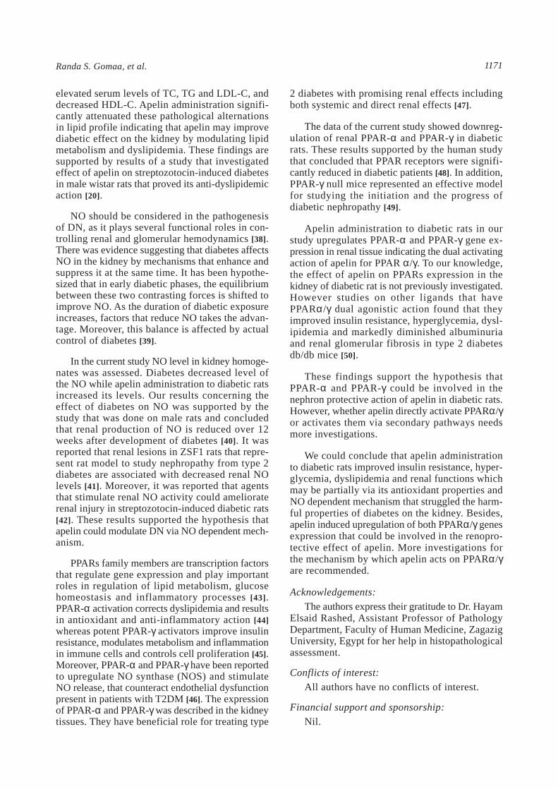
content was (15.3±4), (26.3±6.2) & (19.4±3.4)nmol/mg tissue in control, DM and AP groupsrespectively.
Induction of diabetes induced significant de-crease in renal tissue SOD activity in DM group(p<0.001); with significant increase in renal tissueMDA content in DM group (p<0.001) in compar-ison to control one. Apelin administration to dia-betic rats induced significant increase in renaltissue SOD activity (p<0.001) and significantdecrease in MDA content (p=0.003) when com-pared with DM group Fig. (1).
distribution of data from each group was confirmedusing the Kolmogorov-Smirnov normality test.Since the test indicated that variables followednormal distribution, comparisons among the exper-imental groups were analyzed by one-way analysisof variance (ANOVA) followed by Least Signifi-cance Differences (LSD) test to evaluate statisticaldifference between two groups. p-value <0.05 wasconsidered to be statistically significant.
Results
Effect of apelin on glucose metabolism, lipidprofile & renal function:
Induction of diabetes induced significant in-crease in glucose, HOMA-IR, TG, TC, LDL-C,serum urea and creatinine, urine albumin excretion,and urinary NAG activity with decrease in insulinand HDL-C in DM in comparison to control one(p<0.001). Apelin administration to diabetic ratsinduced significant decrease in glucose, TG, LDL-C (p<0.001), HOMA-IR (p=0.02), TC (p=0.004),serum urea and creatinine (p<0.001) and urinaryalbumin secretion and NAG activity (p<0.001) withincrease in insulin (p=0.007) and HDL-C (p<0.001)when compared with DM group (Table 1).
1168
Table (1): Effect of apelin administration to diabetic rats onglucose metabolism, lipid profile and renal functionparameters.
n=10 in each group.Data are represented as mean ± SD.HDL-CHOMA-IRLDL-CNAGTCTG
Significance (p<0.05):(a) Significant when compared with control group,(b) significant when compared with DM group.
211.1±11.4a,b18.8±3.5a,b8.9±0.64a,b87±9.7a,b182±10.8a,b40±6.7a,b125.1±5.2a,b33.6±4.3a,b1.45±0.39a,b
31.8±6.3a,b
0.057±0.008a,b
AP
251.9±15.3a15.1±2.3a9.4±0.8a128.5±11.6a205.8±24.5a26.3±4.5a150.6±6.9a52.8±4.9a3.42±0.5a
52.4±5.7a
0.16±0.03a
DM
• Glucose (mg/dL)• Insulin (mIU/L)• HOMA-IR• TG (mg/dL)• TC (mg/dL)• HDL-C (mg/dL)• LDL-C (mg/dL)• Serum urea (mg/dl)• Serum creatinine
(mg/dl)• Urinary albumin
excretion (mg/24h)• Urinary (NAG)
activity (µmol/h/mgcreatinine)
96.8±6.425.4±2.76.2±0.774.7±5.6133.4±9.258±6.762.8±724±4.40.87±0.07
25.4±4.4
0.027±0.006
Control
: High-Density Lipoprotein-Cholesterol.: Homeostasis Model Assessment-Insulin Resistance.: Low-Density Lipoprotein-Cholesterol.: N-acetyl-β-D-glucosaminidase.: Total Cholesterol.: Triglyceride.
Effect of apelin on renal oxidative stress pa-rameters:
Renal tissue SOD activity was (99.3±9.2),(71.6±7.2) & (87.9±6.3) U/mg tissue and MDA
120110100
908070605040302010
0
SO
D (
U/m
g ti
ssue
)
Control DM AP
*
*#(A)
40
30
20
10
0
MD
A (
nmol
/mg
tiss
ue)
Control DM AP
*
#
(B)
Fig. (1): Effects of apelin on (A) superoxide dismutase (SOD),(B) malondialdehyde (MDA) oxidative parametersin the kidney tissues of diabetic rats.
n=(10) in each group.Data are represented as mean ± standard deviation.Significance (p<0.05):(*) Significant when compared with control group.(#) Significant when compared with DM group.
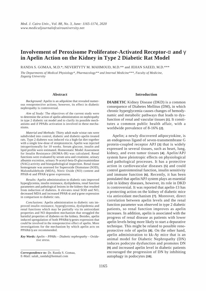
Effect of apelin on renal nitric oxide content:Renal tissue NO content was (18.2±3.6),
(9.1±1.85) & (12.2±2.3) nmol/mg tissue in control,DM and AP groups respectively.
Induction of diabetes induced significant de-crease in renal tissue NO in DM group (p<0.001)in comparison to control one while apelin admin-istration to diabetic rats induced significant increasein its level (p=0.023) when compared with DMgroup Fig. (2).
Effect of Apelin on Kidney of Diabetic Rats
Randa S. Gomaa, et al. 1169
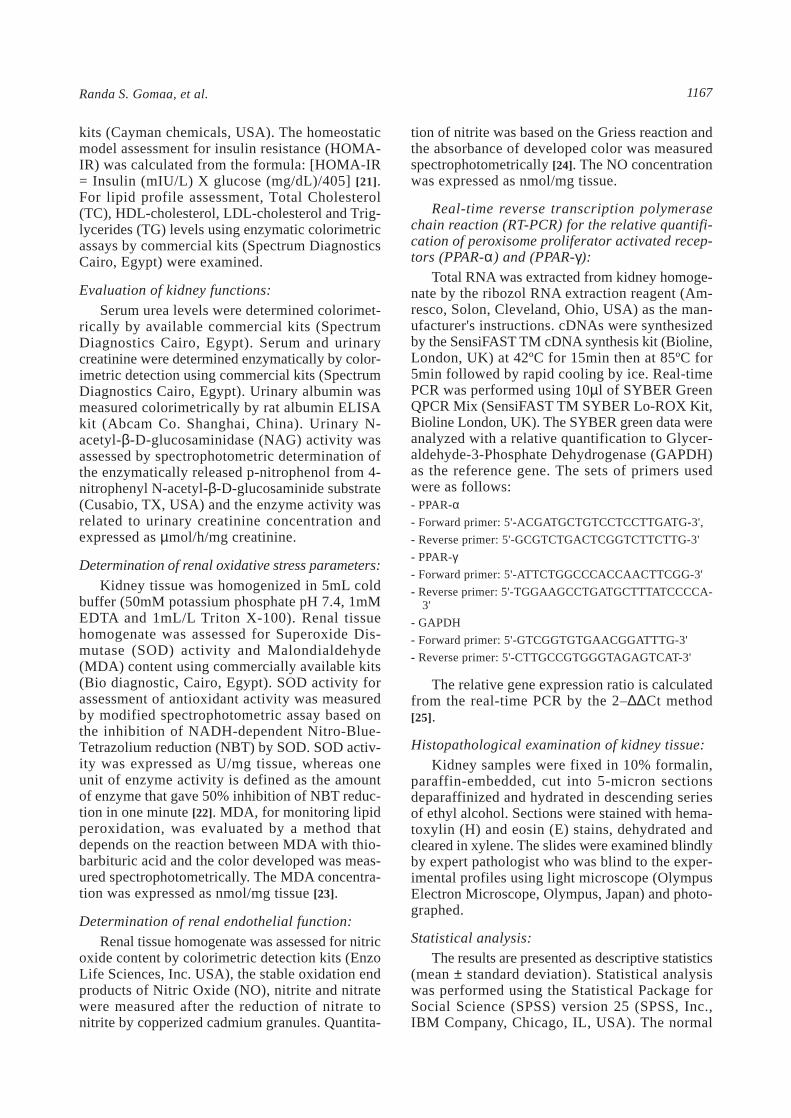
Effect of apelin on renal PPAR-α and PPAR-γexpression:
Relative gene expression of PPAR-α was (3.37±0.45), (2.23±0.43) & (2.65±0.26) and PPAR-γ was(2.43±0.25), (1.66±0.35) & (1.95±0.26) in control,DM and AP groups respectively.
Induction of diabetes induced significant de-crease in renal tissue PPAR-α and PPAR-γ expres-sion in DM group (p<0.001) in comparison tocontrol one. Apelin administration to diabetic ratsinduced significant increase in renal tissue PPAR-α and PPAR-γ expression (p=0.024, p=0.035) whencompared with DM group Fig. (3).
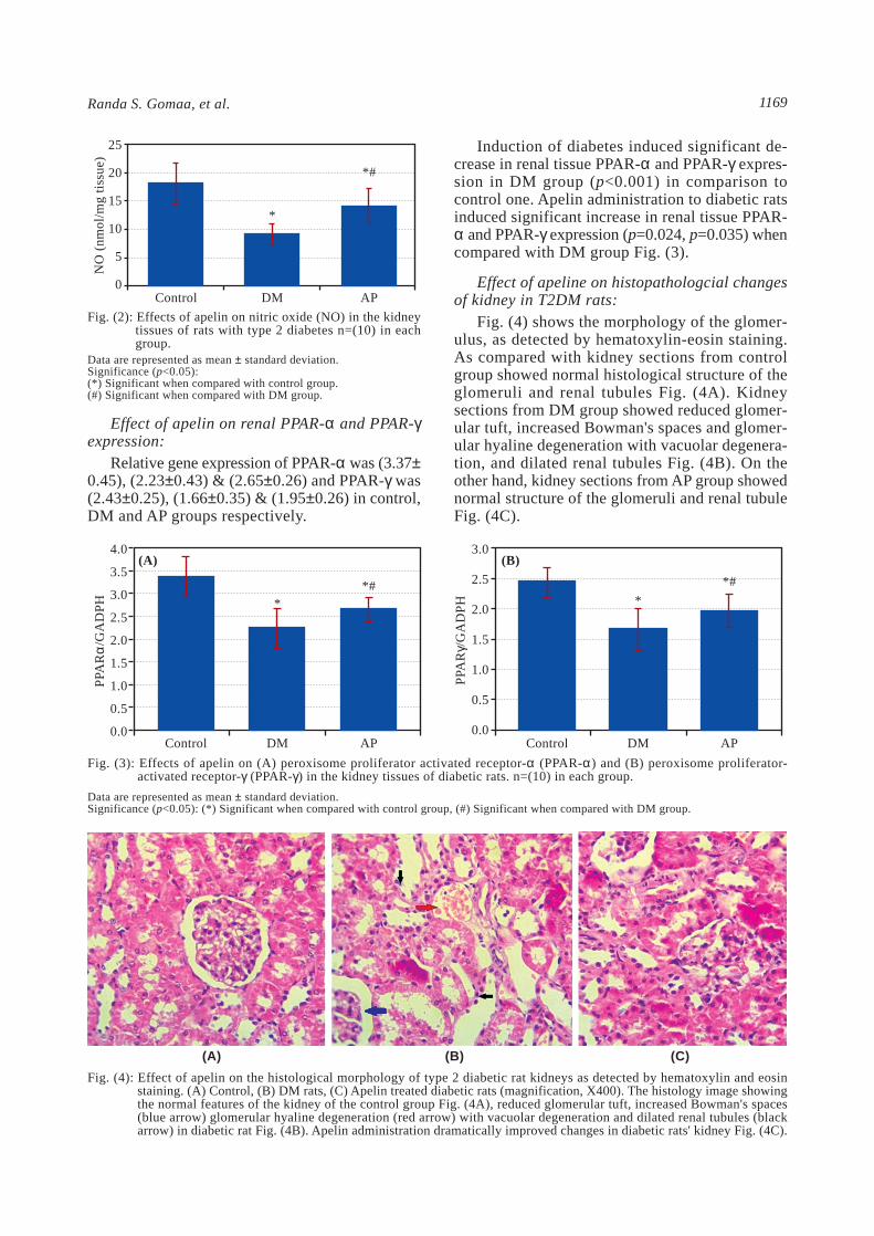
Effect of apeline on histopathologcial changesof kidney in T2DM rats:
Fig. (4) shows the morphology of the glomer-ulus, as detected by hematoxylin-eosin staining.As compared with kidney sections from controlgroup showed normal histological structure of theglomeruli and renal tubules Fig. (4A). Kidneysections from DM group showed reduced glomer-ular tuft, increased Bowman's spaces and glomer-ular hyaline degeneration with vacuolar degenera-tion, and dilated renal tubules Fig. (4B). On theother hand, kidney sections from AP group showednormal structure of the glomeruli and renal tubuleFig. (4C).
25
20
15
10
5
0
NO
(nm
ol/m
g ti
ssue
)
Control DM AP
*
*#
Fig. (2): Effects of apelin on nitric oxide (NO) in the kidneytissues of rats with type 2 diabetes n=(10) in eachgroup.
Data are represented as mean ± standard deviation.Significance (p<0.05):(*) Significant when compared with control group.(#) Significant when compared with DM group.
Fig. (4): Effect of apelin on the histological morphology of type 2 diabetic rat kidneys as detected by hematoxylin and eosinstaining. (A) Control, (B) DM rats, (C) Apelin treated diabetic rats (magnification, X400). The histology image showingthe normal features of the kidney of the control group Fig. (4A), reduced glomerular tuft, increased Bowman's spaces(blue arrow) glomerular hyaline degeneration (red arrow) with vacuolar degeneration and dilated renal tubules (blackarrow) in diabetic rat Fig. (4B). Apelin administration dramatically improved changes in diabetic rats' kidney Fig. (4C).
4.0
3.5
3.0
2.5
2.0
1.5
1.0
0.5
0.0
PPA
Rα/
GA
DP
H
Control DM AP
*
(A)
*#
3.0
2.5
2.0
1.5
1.0
0.5
0.0Control DM AP
*
(B)
PPA
Rγ/
GA
DP
H
*#
Fig. (3): Effects of apelin on (A) peroxisome proliferator activated receptor-α (PPAR-α) and (B) peroxisome proliferator-activated receptor-γ (PPAR-γ) in the kidney tissues of diabetic rats. n=(10) in each group.
Data are represented as mean ± standard deviation.Significance (p<0.05): (*) Significant when compared with control group, (#) Significant when compared with DM group.
(A) (B) (C)

level in diabetic kk-Ay mouse that used as a poly-genic model for human type 2 diabetes mellituspromoted the progression of renal dysfunction byinducing podocyte dysfunction [10]. Moreover,Guo et al., found that apelin stimulates the progressof DN by inducing podocyte dysfunction and ab-normal glomeruli angiogenesis and aggravatesalbuminuria and glomerular basement membranethickening in kk-Ay mice [9]. Whether apelin pre-vents or aggravates the progress of DN is contro-versial.
Hyperglycemia is considered to be the mainpathological finding of DM and it causes most ofdiabetic-related complications. Chronic hypergly-cemia results in overproduction of Reactive OxygenSpecies (ROS) [30] that resulted in damaging cellmembranes and inactivating endogenous antioxi-dants [31]. SOD is one of endogenous antioxidantmolecules that counteract ROS-mediated renalinjury and it is severely decreased in patients withT2DN [32]. Controlling hyperglycemia remains themajor therapeutic strategy as it has a central rolein starting the ROS-mediated pathway so this helpsthe amelioration of oxidative stress [33].
In the current study, hyperglycemia was signif-icantly reduced by apelin demonstrating that con-trolling hyperglycemia is important for apelin-mediated renoprotective effect. Furthermore, SODand MDA levels in kidney homogenates weredetermined. In the diabetic group, decreased levelsof the SOD was accompanied by an increase inMDA levels. Apelin administration increased renalSOD activity and reduced MDA levels. Theseresults suggested that apelin may exert a renopro-tective effect via antioxidant activity.
These data are supported by other studies thatproved anti-hyperglycemic action of apelin indiabetic rats [34]. Moreover, Day et al., suggestedthat renoprotective effects of apelin-13 in diabeticmice could be due to motivation of antioxidantmechanisms [7]. The antioxidant action of apelinis considered to be independent on its anti-hyperglycemic action as it was reported that apelin-13 applied after renal ischemia reperfusion aug-mented the antioxidant enzyme activity, inhibitedthe lipid oxidation and enhanced the renal functionsin non-diabetic rats [35].
Hyperlipidemia is a common finding with type2 DM and is known as a risk factor for DN as itmay aggravate glomerular injury [36]. It was dem-onstrated that diabetic dyslipidemia is associatedwith deterioration in kidney function and albuminu-ria [37]. In the present study, diabetic rats exhibited
Discussion
In the present study, the action of apelin onkidney was investigated in a high-fat diet, low-dose STZ-induced rat model of T2DM. The resultsof our study revealed that apelin could lower serumurea and creatinine, decrease urinary NAG activityand improve albuminuria and the renal histopatho-logical lesions in diabetic rats. At the same time,apelin administration to diabetic rats can decrease,glucose levels and insulin resistance, improveinsulin level and dyslipidemia, decrease renal tissueMDA and increase SOD activity, NO concentrationand PPAR-α and PPAR-γ gene expression in renaltissue of diabetic rats.
STZ is widely used to induce diabetes in theexperimental animals as it has toxic effects onpancreatic β-cells as it is employed as glucoseanalogues that tend to accumulate in pancreaticbeta cells through glucose transporter 2 resultingin reduction in blood insulin levels, and hypergly-cemia that imitating DM pathology [26]. STZ dosesrequired in DM models remain variable. It wasconfirmed that high-dose STZ induced direct ne-phrotoxicity, making it difficult to distinguishbetween the direct toxic effect of STZ and thelesions resulted from STZ-induced DM [27].
In the current study, insulin resistance wasmade via a high-fat diet for 8 weeks, followed bya single low dose of STZ (30mg/kg) to inducehyperglycemia for one week then the same feedingregimen was continued for 10 weeks to developdiabetes complications in rat model. It has beensuggested that diabetic lesion in the kidney isstarted at least 3 weeks after STZ injection as bythis time the kidneys recovered from the acutemild STZ nephrotoxic effects [28].
Kidney damage was assessed in the presentstudy via elevated serum urea and creatinine, albu-minuria, increased urinary NAG activity and his-tological examination of the diabetic kidney. Apelinadministration improved the majority of these renaldysfunctions, indicating the nephroprotective prop-erties of apelin in rats with T2DM.
These findings are supported by results of otherresearches that postulated that apelin treatmentdefeats diabetes-induced renal histological changesand controls the diabetes-induced renal inflamma-tion and albuminuria [7]. Moreover, apelin couldefficiently suppress the progress of nephropathyin Akita mouse, a spontaneous type 1 diabeticmodel [29]. However, Lui and his colleagues pro-posed an oppositional opinion that higher apelin
1170 Effect of Apelin on Kidney of Diabetic Rats
elevated serum levels of TC, TG and LDL-C, anddecreased HDL-C. Apelin administration signifi-cantly attenuated these pathological alternationsin lipid profile indicating that apelin may improvediabetic effect on the kidney by modulating lipidmetabolism and dyslipidemia. These findings aresupported by results of a study that investigatedeffect of apelin on streptozotocin-induced diabetesin male wistar rats that proved its anti-dyslipidemicaction [20].
NO should be considered in the pathogenesisof DN, as it plays several functional roles in con-trolling renal and glomerular hemodynamics [38].There was evidence suggesting that diabetes affectsNO in the kidney by mechanisms that enhance andsuppress it at the same time. It has been hypothe-sized that in early diabetic phases, the equilibriumbetween these two contrasting forces is shifted toimprove NO. As the duration of diabetic exposureincreases, factors that reduce NO takes the advan-tage. Moreover, this balance is affected by actualcontrol of diabetes [39].
In the current study NO level in kidney homoge-nates was assessed. Diabetes decreased level ofthe NO while apelin administration to diabetic ratsincreased its levels. Our results concerning theeffect of diabetes on NO was supported by thestudy that was done on male rats and concludedthat renal production of NO is reduced over 12weeks after development of diabetes [40]. It wasreported that renal lesions in ZSF1 rats that repre-sent rat model to study nephropathy from type 2diabetes are associated with decreased renal NOlevels [41]. Moreover, it was reported that agentsthat stimulate renal NO activity could amelioraterenal injury in streptozotocin-induced diabetic rats[42]. These results supported the hypothesis thatapelin could modulate DN via NO dependent mech-anism.
PPARs family members are transcription factorsthat regulate gene expression and play importantroles in regulation of lipid metabolism, glucosehomeostasis and inflammatory processes [43].PPAR-α activation corrects dyslipidemia and resultsin antioxidant and anti-inflammatory action [44]whereas potent PPAR-γ activators improve insulinresistance, modulates metabolism and inflammationin immune cells and controls cell proliferation [45].Moreover, PPAR-α and PPAR-γ have been reportedto upregulate NO synthase (NOS) and stimulateNO release, that counteract endothelial dysfunctionpresent in patients with T2DM [46]. The expressionof PPAR-α and PPAR-γ was described in the kidneytissues. They have beneficial role for treating type
2 diabetes with promising renal effects includingboth systemic and direct renal effects [47].
The data of the current study showed downreg-ulation of renal PPAR-α and PPAR-γ in diabeticrats. These results supported by the human studythat concluded that PPAR receptors were signifi-cantly reduced in diabetic patients [48]. In addition,PPAR-γ null mice represented an effective modelfor studying the initiation and the progress ofdiabetic nephropathy [49].
Apelin administration to diabetic rats in ourstudy upregulates PPAR-α and PPAR-γ gene ex-pression in renal tissue indicating the dual activatingaction of apelin for PPAR α/γ. To our knowledge,the effect of apelin on PPARs expression in thekidney of diabetic rat is not previously investigated.However studies on other ligands that havePPARα/γ dual agonistic action found that theyimproved insulin resistance, hyperglycemia, dysl-ipidemia and markedly diminished albuminuriaand renal glomerular fibrosis in type 2 diabetesdb/db mice [50].
These findings support the hypothesis thatPPAR-α and PPAR-γ could be involved in thenephron protective action of apelin in diabetic rats.However, whether apelin directly activate PPARα/γor activates them via secondary pathways needsmore investigations.
We could conclude that apelin administrationto diabetic rats improved insulin resistance, hyper-glycemia, dyslipidemia and renal functions whichmay be partially via its antioxidant properties andNO dependent mechanism that struggled the harm-ful properties of diabetes on the kidney. Besides,apelin induced upregulation of both PPARα/γ genesexpression that could be involved in the renopro-tective effect of apelin. More investigations forthe mechanism by which apelin acts on PPARα/γare recommended.
Acknowledgements:
The authors express their gratitude to Dr. HayamElsaid Rashed, Assistant Professor of PathologyDepartment, Faculty of Human Medicine, ZagazigUniversity, Egypt for her help in histopathologicalassessment.
Conflicts of interest:
All authors have no conflicts of interest.
Financial support and sponsorship:
Nil.
Randa S. Gomaa, et al. 1171

J.: Can reduction in hypertriglyceridaemia slow progres-sion of microalbuminuria in patients with non-insulin-dependent diabetes mellitus? Eur. J. Clin. Invest., 27:997-1002, 1997.
16- SALGUEIRO G., BELTRÁN L.M., TORRES R.J. andPUIG J.G.: Fenofibrate increases serum creatinine in apatient with familial nephropathy associated to hyperuri-cemia. Nucleosides Nucleotides Nucleic Acids., 33: 181-4, 2014.
17- BALAKUMAR P., VARATHARAJAN R., NYO Y.H.,RENUSHIA R., RAAGINEY D., OH A.N., AKHTARS.S., RUPESHKUMAR M., SUNDRAM K. andDHANARAJ S.A.: Fenofibrate and dipyridamole treat-ments in low-doses either alone or in combination bluntedthe development of nephropathy in diabetic rats. PharmacolRes., 90: 36-47, 2014.
18- DUPONT J., REVERCHON M., CLOIX L., FROMENTP. and RAMÉ C.: Involvement of adipokines, AMPK,PI3K and the PPAR signaling pathways in ovarian follicledevelopment and cancer. Int. J. Dev. Biol., 56: 959-67,2012.
19- ZHANG S., XU H., YU X., WU Y. and SUI D.: Metforminameliorates diabetic nephropathy in a rat model of low-dose streptozotocin-induced diabetes. Exp. Ther. Med.,14: 383-90, 2017.
20- HEGAB I.I.: Ameliorative effect of apelin on streptozo-tocin-induced diabetes and its associated cardiac hyper-trophy. Alexandria J. Med., 54: 119-27, 2017.
21- SINGH B. and SAXENA A.: Surrogate markers of insulinresistance: A review. World J. Diabetes, 1: 36-47, 2010.
22- KAKKAR P., DAS B. and VISWANATHAN P.N.: Amodified spectrophotometric assay of superoxide dis-mutase. Indian J. Biochem. Biophys., 21: 130-2, 1984.
23- OHKAWA H., OHISHI N. and YAGI K.: Assay for lipidperoxides in animal tissues by thiobarbituric acid reaction.Anal. Biochem., 95: 351-8, 1979.
24- SASTRY K., MOUDGAL R., MOHAN J., TYAGI J. andRAO G.: Spectrophotometric determination of serumnitrite and nitrate by copper-cadmium alloy. Anal. Bio-chem., 306: 79-82, 2002.
25- VANGUILDER H.D., VRANA K.E. and FREEMANW.M.: Twenty-five years of quantitative PCR for geneexpression analysis. BioTechniques, 44: 619-26, 2008.
26- LENZEN S.: The mechanisms of alloxan-and streptozo-tocin-induced diabetes. Diabetologia, 51: 216-26, 2008.
27- TAY Y.C., WANG Y., KAIRAITIS L., RANGAN G.K.,ZHANG C., and HARRIS D.C.: Can murine diabeticnephropathy be separated from superimposed acute renalfailure? Kidney Int., 68: 391-8, 2005.
28- KRAYNAK A.R., STORER R.D., JENSEN R.D., KLOSSM.W., SOPER K.A., CLAIR J.H., DeLUCA J.G., NI-CHOLS W.W. and EYDELLOTH R.S.: Extent and per-sistence of streptozotocininduced DNA damage and cellproliferation in rat kidney as determined by in vivo alkalineelution and BrdUrd labeling assays. Toxicol. Appl. Phar-macol., 135: 279-86, 1995.
29- CHEN H., LI J., JIAO L., PETERSEN R.B., LI J., PENGA., ZHENG L. and HUANG K.: Apelin inhibits thedevelopment of diabetic nephropathy by regulating histoneacetylation in Akita mouse. J. Physiol., 592: 505-21, 2014.
References
1- JHA J.C., BANAL C., CHOW B.S.M., COOPER M.E.and JANDELEIT-DAHM K.: Diabetes and kidney disease:Role of oxidative stress. Antioxid Redox Signal., 25: 657-84, 2016.
2- JHA V., GARCIA-GARCIA G., ISEKI K., LI Z., NAI-CKER S., PLATTNER B., SARAN R., WANG A.Y. andYANG C.W.: Chronic kidney disease: Global dimensionand perspectives. Lancet., 382: 260-72, 2013.
3- LIU C., SU T., LI F., LI L., QIN X., PAN W., FENG F.,CHEN F., LIAO D. and CHEN L.: PI3K/Akt signalingtransduction pathway is involved in rat vascular smoothmuscle cell proliferation induced by apelin-13. ActaBiochim Biophys Sin (Shanghai), 42: 396-402, 2010.
4- HUANG Z., WU L. and CHEN L.: Apelin/APJ system:A novel potential therapy target for kidney disease. J.Cell Physiol., 233: 3892-900, 2018.
5- YU X.H., TANG Z.B., LIU L.J., QIAN H., TANG S.L.,ZHANG D.W., TIAN G.P. and TANG C.K.: Apelin andits receptor APJ in cardiovascular diseases. Clin. Chim.Acta., 428: 1-8, 2014.
6- CASTAN-LAURELL I1., DRAY C., KNAUF C., KUN-DUZOVA O. and VALET P.: Apelin, a promising targetfor type 2 diabetes treatment. Trends. Endocrinol. Metab.,23: 234-41, 2012.
7- DAY R.T., CAVAGLIERI R.C. and FELIERS D.: Apelinretards the progression of diabetic nephropathy. Am. J.Physiol. Renal. Physiol., 304: F788-F800, 2013.
8- SILVA A.P., FRAGOSO A., SILVA C., VIEGAS C.,TAVARES N., GUILHERME P., SANTOS N., RATOF., CAMACHO A., CAVACO C., PEREIRA V., FAÍSCAM., ATAÍDE J., JESUS I. and NEVES P.: What is therole of apelin regarding cardiovascular risk and progressionof renal disease in type 2 diabetic patients with diabeticnephropathy? Biomed. Res. Int., 247649, 2013.
9- GUO C., LIU Y., ZHAO W., WEI S., ZHANG X., WANGW. and ZENG X.: Apelin promotes diabetic nephropathyby inducing podocyte dysfunction via inhibiting proteas-ome activities. J. Cell Mol. Med., 19: 2273-85, 2015.
10- LIU Y., ZHANG J., WANG Y. and ZENG X.: Apelininvolved in progression of diabetic nephropathy by inhib-iting autophagy in podocytes. Cell Death Dis., 8: e3006,2017.
11- BROEDERS N. and ABRAMOWICZ D.: Peroxisomeproliferator-activated receptors (PPARs): Novel therapeutictargets in renal disease. Kidney Int., 60: 14-30, 2002.
12- DESVERGENE B. and WAHLI W.: Peroxisome prolifer-ator-activated receptor: Nuclear control of metabolism.Endocr. Rev., 20: 649-88, 1999.
13- WILLSON T.M., LAMBERT M.H. and KLIEWER S.A.:Peroxisome proliferator-activated receptor gamma andmetabolic disease. Annu. Rev. Biochem., 70: 341-67,2001.
14- SARAFIDIS P.A., STAFYLAS P.C., GEORGIANOS P.I.,SARATZIS A.N. and LASARIDIS A.N.: Effect of thia-zolidinediones on albuminuria and proteinuria in diabetes:A meta-analysis. Am. J. Kidney Dis., 55: 835-47, 2010.
15- SMULDERS Y.M., VAN EEDEN A.E., STEHOUWERC.D., WEIJERS R.N., SLAATS E.H. and SILBERBUSCH
1172 Effect of Apelin on Kidney of Diabetic Rats
30- MATOUGH F.A., BUDIN S.B., HAMID Z.A., ALWA-HAIBI N. and MOHAMED J.: The role of oxidative stressand antioxidants in diabetic complications. Sultan QaboosUniv. Med. J., 12: 5-18, 2012.
31- YAN L.J.: Analysis of oxidative modification of proteins.Curr. Protoc. Protein Sci., Chapter, 14: Unit. 14.4, 2009.
32- VOLPE C.M.O., VILLAR-DELFINO P.H., DOS ANJOSP.M.F. and NOGUEIRA-MACHADO J.A.: Cellular death,reactive oxygen species (ROS) and diabetic complications.Cell Death Dis., 9: 119-27, 2018.
33- SHAO N., KUANG H.Y., WANG N., GAO X.Y., HAOM., ZOU W. and YIN H.Q.: Relationship between Oxi-dant/Antioxidant Markers and Severity of Microalbuminu-ria in the Early Stage of Nephropathy in Type 2 DiabeticPatients. J. Diabetes Res., 232404, 2013.
34- AKCıLAR R., TURGUT S., CANER V., AKCıLAR A.,AYADA C., ELMAS L. and ÖZCAN TO.: The effects ofapelin treatment on a rat model of type 2 diabetes. Adv.Med. Sci., 60: 94-100, 2015.
35- BIRCAN B., ÇAKıR M., KıRBA_ S. and GÜL H.F.:Effect of apelin hormone on renal ischemia/reperfusioninduced oxidative damage in rats. Ren. Fail., 38: 1122-8, 2016.
36- GAI Z., WANG T., VISENTIN M., KULLAK-UBLICKG.A., FU X. and WANG Z.: Lipid Accumulation andChronic Kidney Disease. Nutrients, 11: 722-42, 2019.
37- BALAKUMAR P., KADIAN S. and MAHADEVAN N.:Are PPAR alpha agonists a rational therapeutic strategyfor preventing abnormalities of the diabetic kidney?Pharmacol. Res., 65: 430-6, 2012.
38- LEE J.: Nitric oxide in the kidney: Its physiological roleand pathophysiological implications. Electrolyte BloodPress., 6: 27-34, 2008.
39- KOMERS R. and ANDERSON S.: Paradoxes of nitricoxide in the diabetic kidney. Am. J. Physiol. Renal.Physiol., 284: F1121-F1137, 2003.
40- MAHMOUD A.M., ASHOUR M.B., ABDEL-MONEIMA. and AHMED O.M.: Hesperidin and naringin attenuatehyperglycemia-mediated oxidative stress and proinflam-matory cytokine production in high fat fed/streptozotocin-induced type 2 diabetic rats. J. Diabetes Complications,26: 483-90, 2012.
41- PRABHAKAR S., STARNES J., SHI S., LONIS B. andTRAN R.: Diabetic nephropathy is associated with oxi-dative stress and decreased renal nitric oxide production.J. Am. Soc. Nephrol., 18: 2945-52, 2007.
42- ZHOU S.J., BAI L., LV L., CHEN R., LI C.J., LIU X.Y.,YU D.M. and YU P.: Liraglutide ameliorates renal injuryin streptozotocin-induced diabetic rats by activatingendothelial nitric oxide synthase activity via the down-regulation of the nuclear factor-κB pathway. Mol. Med.Rep., 10: 2587-94, 2014.
43- JAY M.A. and REN J.: Peroxisome proliferator-activatedreceptor (PPAR) in metabolic syndrome and type 2 dia-betes mellitus. Curr. Diabetes. Rev., 3: 33-9, 2007.
44- PLUTZKY J.: Preventing type 2 diabetes and cardiovas-cular disease in metabolic syndrome: The role of PPARalpha. Diab. Vasc. Dis. Res., 4: S12-4, 2007.
45- WANG L., WALTENBERGER B., PFERSCHY-WENZIGE.M., BLUNDER M., LIU X., MALAINER C.,BLAZEVIC T., SCHWAIGER S., ROLLINGER J.M.,HEISS E.H., SCHUSTER D., KOPP B., BAUER R.,STUPPNER H., DIRSCH V.M. and ATANASOV A.G.:Natural product agonists of peroxisome proliferator-activated receptor gamma (PPARγ): A review. Biochem.Pharmacol., 1; 92: 73-89, 2014.
46- GOYA K., SUMITANI S., XU X., KITAMURA T.,YAMAMOTO H., KUREBAYASHI S., SAITO H., KOU-HARA H., KASAYAMA S. and KAWASE I.: Peroxisomeproliferator-activated receptor alpha agonists increasenitric oxide synthase expression in vascular endothelialcells. Arterioscler. Thromb. Vasc. Biol., 24: 658-63, 2004.
47- GUAN Y.: Peroxisome proliferator-activated receptorfamily and its relationship to renal complications of themetabolic syndrome. J. Am. Soc. Nephrol., 15: 2801-15,2004.
48- AFZAL N., HASSAN M., FATIMA S., TARIQ S. andQAYUM I.: Expression Of Peroxisome-Proliferator Ac-tivated Receptors-γ In Diabetics, Obese And NormalSubjects. J. Ayub. Med. Coll Abbottabad., 28: 130-4,2016.
49- TOFFOLI B., GILARDI F., WINKLER C., SODERBERGM., KOWALCZUK L., ARSENIJEVIC Y., BAMBERGK., BONNY O. and DESVERGNE B.: Nephropathy inPparg-null mice highlights PPARγ systemic activities inmetabolism and in the immune system. PLoS One, 12(2): e0171474, 2017.
50- CHA D.R., ZHANG X., ZHANG Y., WU J., SU D., HANJ.Y., FANG X., YU B., BREYER M.D. and GUAN Y.:Peroxisome proliferator activated receptor alpha/gammadual agonist tesaglitazar attenuates diabetic nephropathyin db/db mice. Diabetes, 56 (8): 2036-45, 2007.
Randa S. Gomaa, et al. 1173
1174 Effect of Apelin on Kidney of Diabetic Rats
αγ