issn: 2233-601x (print) issn: 2093-6516 (online) early ... · the mean quantity of drainage from...
TRANSCRIPT

ISSN: 2233-601X (Print) ISSN: 2093-6516 (Online)
− 165 −
Department of Cardiovascular Surgery, Ahi Evren Thorax Cardiovascular Surgery Education and Research HospitalReceived: November 9, 2015, Revised: February 6, 2016, Accepted: February 12, 2016, Published online: June 5, 2016
Corresponding author: Muhammet Onur Hanedan, Department of Cardiovascular Surgery, Ahi Evren Thorax Cardiovascular Surgery Education
and Research Hospital, So uksu Mah. Vatan Cad. No. 9 Ahi Evren Hastanesi 61040 Ortahisar, Trabzon, Türkiye(Tel) 90-505-799-5155 (Fax) 90-462-231-0483 (E-mail) [email protected]
C The Korean Society for Thoracic and Cardiovascular Surgery. 2016. All right reserved.CC This is an open access article distributed under the terms of the Creative Commons Attribution Non-Commercial License (http://creative-
commons.org/licenses/by-nc/4.0) which permits unrestricted non-commercial use, distribution, and reproduction in any medium, provided the original work is properly cited.
Early Outcomes of Sutureless Aortic Valves
Muhammet Onur Hanedan, M.D., lker Matarac , M.D., Mehmet Ali Yürük, M.D., Tan l Özer, M.D., Ufuk Sayar, M.D., Ali Kemal Arslan, M.D., U ur Ziyrek, M.D., Murat Yücel, M.D.
Background: In elderly high-risk surgical patients, sutureless aortic valve replacement (AVR) should be an alter-
native to standard AVR. The potential advantages of sutureless aortic prostheses include reducing cross-clamping and
cardiopulmonary bypass (CPB) time and facilitating minimally invasive surgery and complex cardiac interventions,
while maintaining satisfactory hemodynamic outcomes and low rates of paravalvular leakage. The current study re-
ports our single-center experience regarding the early outcomes of sutureless aortic valve implantation. Methods:
Between October 2012 and June 2015, 65 patients scheduled for surgical valve replacement with symptomatic aortic
valve disease and New York Heart Association function of class II or higher were included to this study. Perceval
S (Sorin Biomedica Cardio Srl, Sallugia, Italy) and Edwards Intuity (Edwards Lifesciences, Irvine, CA, USA) valves
were used. Results: The mean age of the patients was 71.15±8.60 years. Forty-four patients (67.7%) were female.
The average preoperative left ventricular ejection fraction was 56.9±9.93. The CPB time was 96.51±41.27 minutes and
the cross-clamping time was 60.85±27.08 minutes. The intubation time was 8.95±4.19 hours, and the intensive
care unit and hospital stays were 2.89±1.42 days and 7.86±1.42 days, respectively. The mean quantity of drainage
from chest tubes was 407.69±149.28 mL. The hospital mortality rate was 3.1%. A total of five patients (7.69%)
died during follow-up. The mean follow-up time was 687.24±24.76 days. The one-year survival rate was over 90%.
Conclusion: In the last few years, several models of valvular sutureless bioprostheses have been developed. The
present study evaluating the single-center early outcomes of sutureless aortic valve implantation presents the results
of an innovative surgical technique, finding that it resulted in appropriate hemodynamic conditions with acceptable
ischemic time.
Key words: 1. Prosthesis design
2. Heart valve prosthesis implantation
3. Bioprosthesis
INTRODUCTION
The increase in life expectancy among the general pop-
ulation has resulted in an increase in the prevalence of pa-
tients with valvular heart disease eligible for aortic valve re-
placement (AVR) [1]. The most effective treatment for pa-
tients with severe symptomatic aortic stenosis is surgical re-
placement of the valve. Valve replacement improves left ven-
tricular (LV) systolic and diastolic function by reducing LV
hypertrophy, and thereby results in better clinical outcomes
[2]. Given the increasing number of comorbidities and the in-
creasing age of patients, a tendency has emerged to use bio-
logical valve implants, avoiding the need for long-term anti-
coagulation therapy [3]. In comparison with stented biopro-
Korean J Thorac Cardiovasc Surg 2016;49:165-170 □ Clinical Research □
http://dx.doi.org/10.5090/kjtcs.2016.49.3.165
Muhammet Onur Hanedan, et al
− 166 −
stheses and mechanical valves, stentless bioprostheses provide
a significant reduction in transvalvular pressure gradients.
However, they are more difficult to insert, with increased
cross-clamping time [4].
AVR with any kind of bioprosthesis is the preferred method,
especially in older patients, due to satisfactory hemodynamic
performance and postoperative durability without warfarin-re-
lated complications [2]. Transcatheter aortic valve implantation
(TAVI) procedures have been developed and extensively used
in high-risk patients considered to be ineligible for standard
surgery using cardiopulmonary bypass (CPB). However, it re-
mains necessary to improve the quality and safety of this
procedure due to the potential for serious complications, such
as pacemaker implantation, paravalvular leakage (PVL), and a
high incidence of neurologic events [5].
As a cardiac valve substitute, sutureless prostheses reduce
the need for sutures after annular decalcification, thereby re-
ducing aortic cross-clamping and CPB duration and facilitat-
ing a minimally invasive approach [6]. Sutureless aortic bio-
prosthesis implantation is a feasible alternative for high-risk
patients with aortic valve disease [7]. The current study re-
ports our single-center experiences regarding the early out-
comes of sutureless aortic valve implantation.
METHODS
1) Patients
Between October 2012 and June 2015, 65 patients were in-
cluded in this study. The inclusion criteria were severe symp-
tomatic aortic valve disease, New York Heart Association
function of class II or higher, and being scheduled for surgi-
cal valve replacement. All patients gave written informed
consent except in cases of emergency treatment. This study
was approved by our local ethics committee.
Three different rapid deployment valves are currently ap-
proved for clinical use in Europe: the Enable (Medtronic,
Minneapolis, MN, USA), the Perceval S (SorinBiomedica
Cardio Srl, Sallugia, Italy), and the Edwards Intuity (Edwards
Lifesciences, Irvine, CA, USA) valves. Several thousand pa-
tients have been treated with these devices to date. In our
study, we used the Perceval S in 28 patients (43.08%) and
the Edwards Intuity in 37 patients (56.92%).
In January 2012, sutureless valves began to be marketed in
Turkey. At this time, we started to use sutureless valves in
high-risk patients and patients who need concomitant proce-
dures. In high-risk groups like our population, the choice of
valve must be optimal.
2) Operative technique
All procedures were performed by one expert surgeon.
Under general anesthesia and orotracheal intubation, all pa-
tients undergoing AVR were placed on CPB after a mini-
mally invasive approach or full sternotomy. Myocardial pro-
tection was achieved by the antegrade and retrograde admin-
istration of blood cardioplegic solution on induction and con-
tinued with the retrograde administration of cold-blood car-
dioplegic doses every 20 minutes in accordance with our in-
stitution’s routine protocol, and a final warm-blood dose was
administered before releasing the cross-clamp.
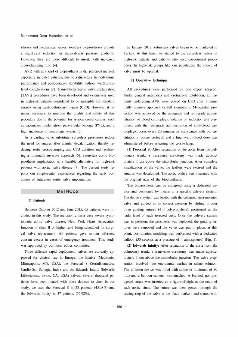
(1) Perceval S: After separation of the aorta from the pul-
monary trunk, a transverse aortotomy was made approx-
imately 1 cm above the sinotubular junction. After complete
visualization of the valve, the leaflets were excised and the
annulus was decalcified. The aortic orifice was measured with
the original sizer of the bioprosthesis.
The bioprosthesis can be collapsed using a dedicated de-
vice and positioned by means of a specific delivery system.
The delivery system was loaded with the collapsed stent-mounted
valve and guided to its correct position by sliding it over
three guiding sutures (4–0 polypropylene), positioned at the
nadir level of each resected cusp. Once the delivery system
was in position, the prosthesis was deployed, the guiding su-
tures were removed and the valve was put in place; at this
point, post-dilation modeling was performed with a dedicated
balloon (30 seconds at a pressure of 4 atmospheres) (Fig. 1).
(2) Edwards intuity: After separation of the aorta from the
pulmonary trunk, a transverse aortotomy was made approx-
imately 1 cm above the sinotubular junction. The valve prep-
aration involved two one-minute washes in saline solution.
The inflation device was filled with saline (a minimum of 30
mL) and a balloon catheter was attached. A braided, non-ple-
dgeted suture was inserted as a figure-of-eight at the nadir of
each aortic sinus. The suture was then passed through the
sewing ring of the valve at the black markers and snared with

Sutureless Aortic Valves
− 167 −
Fig. 1. Intraoperative view of the Perceval S (Sorin Biomedica
Cardio Srl, Sallugia, Italy).
Fig. 2. Intraoperative view of the Edwards Intuity (Edwards Lifes-
ciences, Irvine, CA, USA).
a tourniquet. The valve was then lowered into place in the
annulus, using a gentle back-and-forth rocking motion, while
pulling up on the guiding sutures. Once the valve was prop-
erly positioned, its position was secured with the suture tour-
niquets. The balloon catheter was inserted through the hold-
ing device and locked into place. Saline was injected until
the appropriate pressure was achieved (between 3 and 5 at-
mospheres, depending on the valve size). The target inflation
pressure was maintained for 10 seconds and then the balloon
was deflated. The three prolene sutures on the valve holder
were cut and the entire holding device and balloon were care-
fully removed. The three guiding sutures were tied and cut.
The patency of the coronary ostia was confirmed (Fig. 2).
The surgical procedure was completed with the closure of
the transverse aortotomy for the Perceval S and Edwards
Intuity devices or with the other possible associated procedures.
In cases of associated coronary artery bypass grafting (CABG),
the distal anastomosis preceded the implantation of the pros-
thesis but followed the aortotomy and annulus sizing. Transe-
sophageal echocardiography was performed during the proce-
dure to evaluate the preimplantation measurements and pros-
thetic function. All patients underwent transthoracic echo-
cardiography at discharge.
3) Statistical analysis
Statistical evaluation was performed using SPSS ver. 16.0
(SPSS Inc., Chicago, IL, USA). The results were reported as
mean±standard deviation for quantitative variables and as per-
centages for categorical variables, and the groups were com-
pared using the paired-samples t-test for continuous variables.
Kaplan-Meier curves were used for survival analysis. All p-val-
ues <0.05 were considered to indicate statistical significance.
RESULTS
From October 2012 and June 2015, we inserted sutureless
aortic valves in 65 patients. Their mean age was 71.15±8.60
years. Forty-four (67.7%) of them was female. The average
preoperative LV ejection fraction was 56.9±9.93. The demo-
graphic details of the patients are summarized in Table 1.
The operative and postoperative data are summarized in
Table 2. The Edwards Intuity was used in 37 patients (56.92%)
and the Perceval S was used in 28 patients (43.08%). The
CPB time was 96.51±41.27 minutes and the cross-clamping
time was 60.85±27.08 minutes. The intubation time was
8.95±4.19 hours, and the intensive care unit (ICU) and hospi-
tal stays were 2.89±1.42 days and 7.86±1.42 days, respec-
tively. The mean quantity of drainage from the chest tubes
was 407.69±149.28 mL. The operative and postoperative vari-
ables of the AVR patients are also given in Table 2. No pa-
tients required early postoperative re-exploration due to car-

Muhammet Onur Hanedan, et al
− 168 −
Table 1. Preoperative and perioperative characteristics of the patients
(sutureless valves, n=65)
Characteristic Value
Gender
Female 44 (67.7)
Male 21 (32.3)
Age (yr) 71.15±8.60
Body surface area (m2) 1.70±0.18
Ejection fraction (%) 56.9±9.93
New York Heart Association class 2.83±0.6
Canada classification score 2.77±0.63
EuroScore II 2.79±1.52
Hypertension 44 (67.7)
Diabetes mellitus 16 (24.6)
Smoke 15 (23.1)
Cerebrovascular disease 6 (9.2)
Peripheral vascular disease 0
Carotid artery disease 3 (4.6)
Chronic obstructive pulmonary disease 7 (10.8)
Renal failure 1 (1.5)
Values are presented as number (%) or mean±standard deviation.
Table 2. Operative and postoperative variables
Variable Sutureless valve (n=65) Isolated sutureless valve (n=37)Conventional aortic valve
replacement (n=69)
Operation time (min) 212.48±65.19 190.03±55.95 345±101.85
Cardiopulmonary bypass time (min) 96.51±41.27 67.22±20.57 173.04±63.03
Cross-clamping time (min) 60.85±27.08 48.86±19.43 105.50±41.40
Intubation time (hr) 8.95±4.19 8.47±4.34 9.80±4.15
Inotropic agent 35 (53.8) 13 (35.1) 30 (43.0)
Drainage (mL) 407.69±149.28 412.16±149.26 650.38±34
Erythrocyte suspension (units) 2.32±3.21 1.76±2.13 3.9±2.99
Intensive care unit stay (day) 2.89±1.42 2.23±1.12 3.25±1.25
Hospital stay (day) 7.86±1.42 6.47±1.15 12.20±3.25
Minimally invasive approach 0 5 (13.51) 0
Values are presented as mean±standard deviation or number (%).
Table 3. Additional procedures (sutureless valves, n=65)
Variable Value
CABG 20 (30.77)
Mitral ring annuloplasty+CABG 1 (1.54)
Ascending aortic surgery 3 (4.62)
Ascending aortic surgery+CABG 3 (4.62)
Mitral valve replacement 1 (1.54)
Values are presented as number (%).
CABG, coronary artery bypass grafting.
diac tamponade, bleeding, or any other reason. No serious
bleeding, need for excessive blood transfusion, or thromboem-
bolic complications occurred.
The additional procedures are summarized in Table 3. The
preoperative and postoperative echocardiographic variables are
given in Table 4. The postoperative hemodynamic measure-
ments were favorable, with low peak and mean gradients.
The preoperative maximum aortic gradient was 70.90±24.91
mmHg and the postoperative maximum aortic gradient was
19.92±3.93 mmHg (p=0.000). The preoperative mean gradient
was 44.46±15.83 mmHg and the postoperative mean gradient
was 8.43±2.76 mmHg (p=0.000). No statistically significant
differences were found between the preoperative and post-
operative values of the ejection fraction and the LV end dia-
stolic and systolic diameters.
Two patients (3.1%) died during their hospital stay. One of
them was an 80-year-old man who died on the eighth day
due to acute renal failure and the other was an 85-year-old
woman who died on the third day. She underwent an urgent
repeat AVR after a TAVI procedure due to acute aortic
insufficiency. A total of five patients (7.69%) died during fol-
low-up. The mean follow-up time was 687.24±24.76 days.
The one-year survival rate was over 90%. During follow-up,
no patients had any kind of atrioventricular conduction block
in need of transient or permanent pacemaker placement, and
none underwent reoperation due to bioprosthesis dysfunction.

Sutureless Aortic Valves
− 169 −
Table 4. Echocardiographic data of patients
Variable Preoperative Postoperative p-value
Ejection fraction (%) 57.08±9.92 56.92±9.55 0.788
Left ventricular end diastolic diameter (mm) 49.72±5.60 49.31±5.25 0.159
Left ventricular end systolic diameter (mm) 32.52±6.68 32.06±6.43 0.087
Interventricular septum (mm) 12.98±2.08 12.83±2.00 0.267
Posterior wall (mm) 12.66±2.01 12.57±2.02 0.458
Maximum aortic gradient (mmHg) 70.90±24.91 19.92±3.93 0.000
Mean aortic gradient (mmHg) 44.46±15.83 8.43±2.76 0.000
Values are presented as mean±standard deviation. All p-values <0.05 were considered to indicate statistical significance.
DISCUSSION
This study included 65 patients who received a sutureless
aortic bioprosthesis at our institution. In the last few years,
several models of valvular sutureless bioprostheses have been
developed [8]. In this study, we described our experience
with sutureless aortic valve prostheses, and our preliminary
results demonstrated good clinical and hemodynamic outcomes.
It is well established in the cardiothoracic surgical literature
that extended CPB and aortic cross-clamping durations are
significant independent risk factors for mortality and morbid-
ity in cardiac surgery [9]. A recent retrospective analysis of
979 patients with aortic valve stenosis demonstrated that aort-
ic cross-clamping time was a significant independent predictor
of cardiovascular morbidity [10]. Therefore, any technique
that shortens cross-clamping or CPB time potentially de-
creases the risk of complications, thereby reducing long-term
mortality. Sutureless AVR offered a reduction in the CPB
and cross-clamping time. This is also an advantage in patients
requiring an additional procedure, such as concomitant CABG
in older patients. In our study, 28 patients (43.08%) under-
went an additional procedure. In high-risk patients undergoing
combined surgery with a prolonged surgical time, as well as
in patients undergoing reintervention, the use of sutureless bi-
oprostheses is particularly valuable due to the considerable re-
duction in the implantation time [11]. Choi et al. [12] studied
conventional prostheses performed with a continuous suture
technique to reduce CPB and cross-clamping times. They de-
creased the CPB and cross-clamping times to 150.6±34.6 mi-
nutes and 120.1±29.1 minutes, respectively. When compared
to our study, they reported a longer time for both variables.
PVL can be a result of inadequate sizing and positioning
or due to inappropriate decalcification of the annulus [4].
Correct positioning of the prosthesis can be time-consuming
and must be carried out accurately. PVL must be checked by
intraoperative transesophageal echocardiography. Recent evi-
dence from TAVI trials has demonstrated a significant corre-
lation between PVL and poorer outcomes. PVL was demon-
strated to be a significant predictor of one-year mortality,
even after multivariable adjustment. Unlike TAVI and sim-
ilarly to conventional AVR, the sutureless AVR approach in-
volves excision of the calcified valve and prosthesis place-
ment under direct visualization on a still heart, which may re-
duce the risk of misplacement and PVL [13].
We observed significant reductions in the maximum and
mean gradients postoperatively. This may reduce the risk of
patient-prosthesis mismatch (PPM). In the study by Minh et
al. [14] regarding sutureless valves, the mean transaortic gra-
dient was 11.1±4.6 mmHg, similar to our findings. In a re-
cent randomized trial comparing the Edwards Intuity suture-
less valve with a conventional stented bioprosthesis, a sig-
nificantly lower mean transvalvular gradient (8.5 mmHg vs.
10.3 mmHg) and lower PPM (0% vs. 15%) was found for
the sutureless cohort [15].
Pollari et al. [3] found shorter ICU stays, hospital stays,
and intubation times in the sutureless group then in the stent-
ed group, corresponding to our observations. Better operative
variables associated with sutureless AVR resulted in shorter
ICU and hospital stays and intubation times. Similarly to
their study, our results also suggest a less frequent need for
blood transfusions [3].
AVR with a bioprosthesis is preferred, especially in older

Muhammet Onur Hanedan, et al
− 170 −
populations, due to its satisfactory hemodynamic performance
without warfarin-related complications [2]. Recent published
series of conventional AVR procedures performed in elderly
patients have shown operative mortality rates ranging from
4% to 10% [16]. In our study, the hospital mortality rate was
3.1% and the one-year survival rate was approximately 90%
in this high-risk population.
In our study, five patients underwent a minimally invasive
approach. Sutureless AVR can be performed through a mini-
mal incision, as demonstrated by several recently published
case series that have shown excellent clinical and hemody-
namic results [17].
The major limitation of this study is that it was based on
data from a single institution and a very limited number of
cases. There was no control group, and this was a descriptive
study. This study showed only early outcomes, and it remains
necessary to obtain data documenting long-term performance.
In conclusion, current evidence suggests that sutureless
AVR will become the first choice of procedure in the elderly
high-risk population, with its major advantages being a reduc-
tion in cross-clamping and CPB duration. This single-center
experience in sutureless aortic valve implantation reflects the
implementation of an innovative surgical treatment, resulting
in appropriate hemodynamic conditions with acceptable ische-
mic time.
CONFLICT OF INTEREST
No potential conflict of interest relevant to this article was
reported.
REFERENCES
1. Santarpino G, Pfeiffer S, Schmidt J, Concistre G, Fischlein
T. Sutureless aortic valve replacement: first-year single-cen-
ter experience. Ann Thorac Surg 2012;94:504-8.
2. Altintas G, Diken AI, Hanedan O, et al. The Sorin Freedom
SOLO stentless tissue valve: early outcomes after aortic
valve replacement. Tex Heart Inst J 2013;40:50-5.
3. Pollari F, Santarpino G, Dell’Aquila AM, et al. Better
short-term outcome by using sutureless valves: a propen-
sity-matched score analysis. Ann Thorac Surg 2014;98:611-6.
4. Folliguet TA, Laborde F, Zannis K, Ghorayeb G, Haverich
A, Shrestha M. Sutureless perceval aortic valve replacement:
results of two European centers. Ann Thorac Surg 2012;93:
1483-8.
5. Zahn R, Gerckens U, Grube E, et al. Transcatheter aortic
valve implantation: first results from a multi-centre real-wo-
rld registry. Eur Heart J 2011;32:198-204.
6. Phan K, Tsai YC, Niranjan N, et al. Sutureless aortic valve
replacement: a systematic review and meta-analysis. Ann
Cardiothorac Surg 2015;4:100-11.
7. Eichstaedt HC, Easo J, Harle T, Dapunt OE. Early sin-
gle-center experience in sutureless aortic valve implantation
in 120 patients. J Thorac Cardiovasc Surg 2014;147:370-5.
8. Breitenbach I, Wimmer-Greinecker G, Bockeria LA, et al.
Sutureless aortic valve replacement with the Trilogy Aortic
Valve System: multicenter experience. J Thorac Cardiovasc
Surg 2010;140:878-84, 884.e1.
9. Chalmers J, Pullan M, Mediratta N, Poullis M. A need for
speed?: bypass time and outcomes after isolated aortic valve
replacement surgery. Interact Cardiovasc Thorac Surg
2014;19:21-6.
10. Ranucci M, Frigiola A, Menicanti L, Castelvecchio S, de
Vincentiis C, Pistuddi V. Aortic cross-clamp time, new pros-
theses, and outcome in aortic valve replacement. J Heart
Valve Dis 2012;21:732-9.
11. Task Force on Myocardial Revascularization of the European
Society of Cardiology (ESC) and the European Association
for Cardio-Thoracic Surgery (EACTS); European Association
for Percutaneous Cardiovascular Interventions (EAPCI), Kolh
P, et al. Guidelines on myocardial revascularization. Eur J
Cardiothorac Surg 2010;38 Suppl:S1-S52.
12. Choi JB, Kim JH, Park HK, et al. Aortic valve replacement
using continuous suture technique in patients with aortic valve
disease. Korean J Thorac Cardiovasc Surg 2013;46:249-55.
13. Di Eusanio M, Phan K. Sutureless aortic valve replacement.
Ann Cardiothorac Surg 2015;4:123-30.
14. Minh TH, Mazine A, Bouhout I, et al. Expanding the in-
dication for sutureless aortic valve replacement to patients
with mitral disease. J Thorac Cardiovasc Surg 2014;148:1354-9.
15. Borger MA, Moustafine V, Conradi L, et al. A randomized
multicenter trial of minimally invasive rapid deployment ver-
sus conventional full sternotomy aortic valve replacement.
Ann Thorac Surg 2015;99:17-25.
16. Di Eusanio M, Fortuna D, De Palma R, et al. Aortic valve
replacement: results and predictors of mortality from a con-
temporary series of 2256 patients. J Thorac Cardiovasc Surg
2011;141:940-7.
17. Mazine A, Teoh K, Bouhout I, et al. Sutureless aortic valve
replacement: a Canadian multicentre study. Can J Cardiol
2015;31:63-8.