ivus principle and image interpretation liu jian, m.d. peking university people’s hospital
TRANSCRIPT

IVUS Principle and Image interpretationIVUS Principle and Image interpretation
Liu Jian, M.D.
Peking University People’s Hospital

Ultrasound PrinciplesUltrasound Principles
Rotating Element
Drive Shaft
Multi-Element Array
• Two types of imaging systemsTwo types of imaging systems Mechanical (rotating transducer)Mechanical (rotating transducer) Electronic arrayElectronic array
Basic principles (I)Basic principles (I)
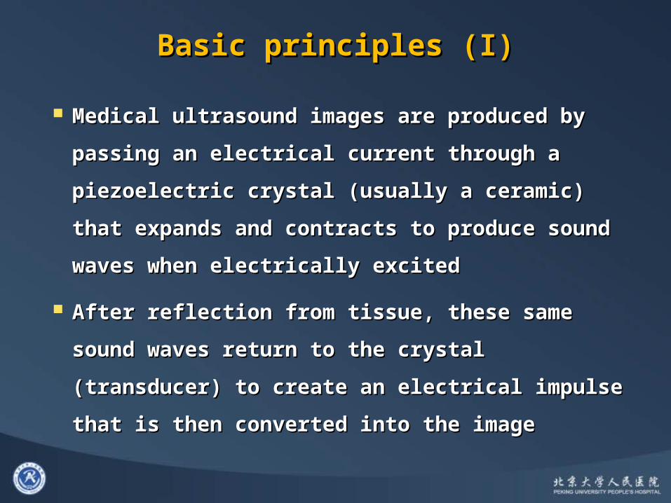
Medical ultrasound images are produced by passing Medical ultrasound images are produced by passing
an electrical current through a piezoelectric crystal an electrical current through a piezoelectric crystal
(usually a ceramic) that expands and contracts to (usually a ceramic) that expands and contracts to
produce sound waves when electrically excitedproduce sound waves when electrically excited
After reflection from tissue, these same sound waves After reflection from tissue, these same sound waves
return to the crystal (transducer) to create an return to the crystal (transducer) to create an
electrical impulse that is then converted into the electrical impulse that is then converted into the
imageimage

The beam remains fairly parallel for a distance The beam remains fairly parallel for a distance (near field) and then begins to diverge (far (near field) and then begins to diverge (far field)field)• The quality of ultrasound images is better in the The quality of ultrasound images is better in the
near field because the beam is more parallel and near field because the beam is more parallel and the resolution greaterthe resolution greater
• The length of the near field is expressed by the The length of the near field is expressed by the equation L = r2 / , where L is the length of the near equation L = r2 / , where L is the length of the near field, r is the radius of the transducer, and is the field, r is the radius of the transducer, and is the wavelengthwavelength
• Therefore, larger transducers with lower Therefore, larger transducers with lower frequencies are used for examination of large frequencies are used for examination of large vessels because they create a deeper near field vessels because they create a deeper near field
Basic principles (II)Basic principles (II)
Ultrasound PrinciplesUltrasound Principles
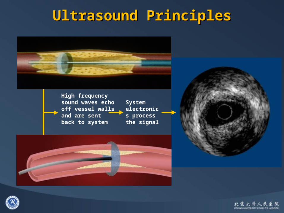
High frequency sound waves echo off vessel walls and are sent back to system
System electronics process the signal
Image quality can be described by two Image quality can be described by two important factorsimportant factors• Spatial resolution (Spatial resolution ( 空间分辨率空间分辨率 ))
– Ability to discriminate small adjacent objects within the Ability to discriminate small adjacent objects within the imageimage
– For a 30-40MHz IVUS transducer the typical resolution is For a 30-40MHz IVUS transducer the typical resolution is 80-100 microns axially and 200-250 microns laterally 80-100 microns axially and 200-250 microns laterally
• Contrast resolutionContrast resolution (对比度分辨率)(对比度分辨率)
Basic principles (III)Basic principles (III)
Ultrasound PrinciplesUltrasound Principles
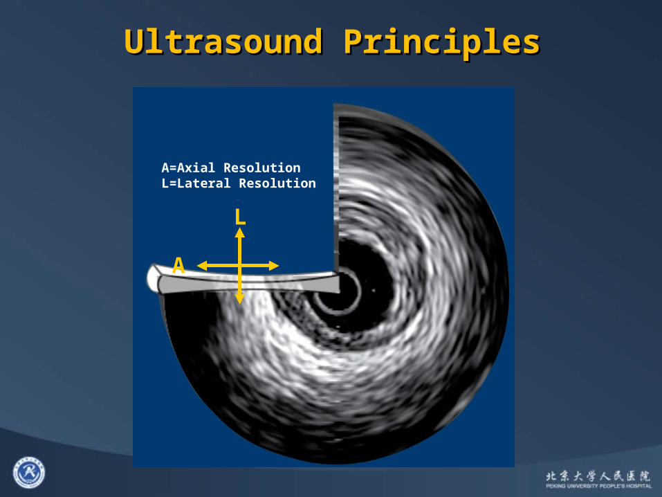
A
L
A=Axial ResolutionL=Lateral Resolution
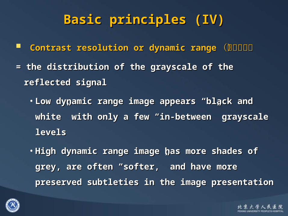
Contrast resolution or dynamic rangeContrast resolution or dynamic range (动态范围)(动态范围)
= the distribution of the grayscale of the reflected signal = the distribution of the grayscale of the reflected signal
• Low dynamic range image appears “black and Low dynamic range image appears “black and
white” with only a few “in-between” grayscale levelswhite” with only a few “in-between” grayscale levels
• High dynamic range image has more shades of grey, High dynamic range image has more shades of grey,
are often “softer,” and have more preserved are often “softer,” and have more preserved
subtleties in the image presentationsubtleties in the image presentation
Basic principles (IV)Basic principles (IV)
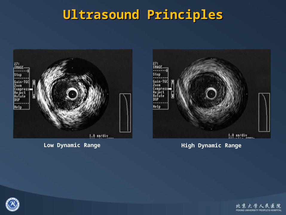
Low Dynamic Range High Dynamic Range
Ultrasound PrinciplesUltrasound Principles
Basic principles (V)Basic principles (V)
Ultrasound will bounce off of some vascular Ultrasound will bounce off of some vascular structures and pass through othersstructures and pass through others• A structure’s acoustic impedanceA structure’s acoustic impedance (声阻抗)(声阻抗)
(density) determines if ultrasound will bounce off (density) determines if ultrasound will bounce off or travel through the structureor travel through the structure
• If sound bounces off the structure, and returns to If sound bounces off the structure, and returns to the transducer, it will appear ‘white’ on the screenthe transducer, it will appear ‘white’ on the screen
• Very dense material, like calcium, will reflect all the Very dense material, like calcium, will reflect all the ultrasound (appear very white) and not allow any ultrasound (appear very white) and not allow any ultrasound to pass through (producing a black ultrasound to pass through (producing a black acoustic shadow beyond the calcium)acoustic shadow beyond the calcium)

ArtifactsArtifacts
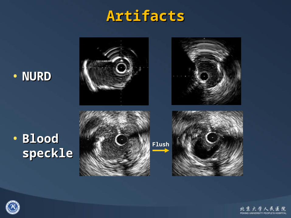
• Nearfield artifactsNearfield artifacts RingdownRingdown(环晕)(环晕) Blood speckle is more intense with higher frequency Blood speckle is more intense with higher frequency
transducers and slows blood flow limiting the ability to transducers and slows blood flow limiting the ability to differentiate lumen from tissue (especially “soft” plaque, differentiate lumen from tissue (especially “soft” plaque, neointima, and thrombus) neointima, and thrombus)
• Flushing contrast or saline through the guiding Flushing contrast or saline through the guiding catheter may clear the lumen and help to identify catheter may clear the lumen and help to identify tissue borderstissue borders
• Motion artifactsMotion artifacts NURD (Non-Uniform Rotation Distortion )NURD (Non-Uniform Rotation Distortion ) 不均匀旋转伪像不均匀旋转伪像 Axial catheter motion with cardiac cycleAxial catheter motion with cardiac cycle
ArtifactsArtifacts
Flush
• NURDNURD
• Blood Blood specklespeckle
Artifacts (II)Artifacts (II)
Mistaking guiding catheter for aorto-ostial Mistaking guiding catheter for aorto-ostial
stenosisstenosis
Position artifactsPosition artifacts
Catheter obliquityCatheter obliquity
Catheter eccentricityCatheter eccentricity
Vessel curvatureVessel curvature

Mistaking guiding catheter for aorto-ostial stenosisMistaking guiding catheter for aorto-ostial stenosis
Side Lobes
Guiding catheterGuiding Catheter
IVUS images interpretationIVUS images interpretation

Plaque ID and CharacteristicsPlaque ID and Characteristics
• AppearanceAppearance Intimal diseaseIntimal disease
• Plaque is dense and will appear Plaque is dense and will appear ‘white’‘white’
MediaMedia
• Made of homogeneous smooth Made of homogeneous smooth muscle cells and does not reflect muscle cells and does not reflect ultrasound (appears dark)ultrasound (appears dark)
Adventitia Adventitia
• Has ‘sheets’ of collagen that Has ‘sheets’ of collagen that reflect a lot of ultrasound reflect a lot of ultrasound (appears white)(appears white)

Normal Diseased
Plaque ID and CharacteristicsPlaque ID and Characteristics

CalciumCalcium
Bright echoes (brighter than the adventitia)Bright echoes (brighter than the adventitia) Obstructs the penetration of ultrasound Obstructs the penetration of ultrasound
(acoustic shadowing)(acoustic shadowing)
Only the leading edge is detected and thickness cannot be determined
Results in reverberations—the oscillation of ultrasound between transducer and calcium causing repeating ‘arcs’

CalciumCalcium
Calcium is Calcium is quantifiedquantified by measuring the by measuring the “arc” it encompasses“arc” it encompasses
Superficial
DeepDeep80°80°
Calcium is classified by its location within the plaque
• Superficial calcium is closer to the lumen than to the adventitia
• Deep calcium is closer to the adventitia than to the lumen
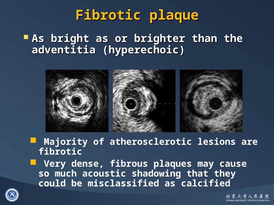
Fibrotic plaqueFibrotic plaque
As bright as or brighter than the adventitia As bright as or brighter than the adventitia (hyperechoic)(hyperechoic)
Majority of atherosclerotic lesions are fibrotic Very dense, fibrous plaques may cause so much
acoustic shadowing that they could be misclassified as calcified
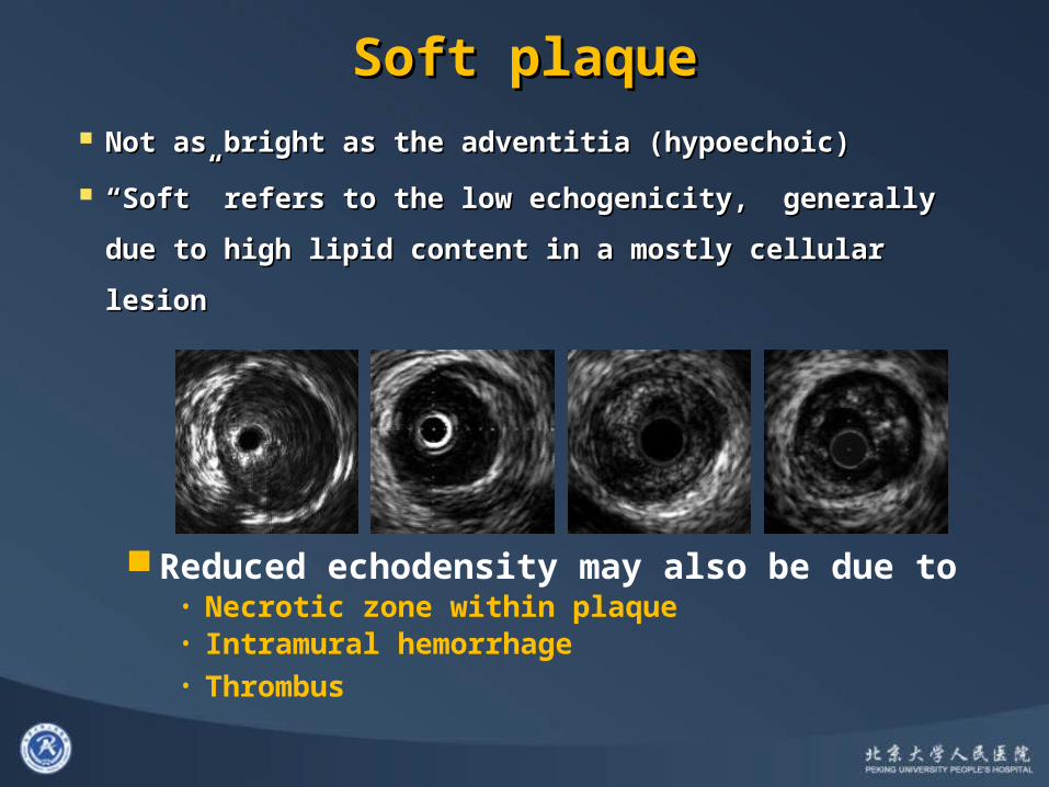
Soft plaqueSoft plaque
Not as bright as the adventitia (hypoechoic)Not as bright as the adventitia (hypoechoic)
““Soft” refers to the low echogenicity, generally due Soft” refers to the low echogenicity, generally due
to high lipid content in a mostly cellular lesionto high lipid content in a mostly cellular lesion
Reduced echodensity may also be due to• Necrotic zone within plaque• Intramural hemorrhage• Thrombus

Vulnerable plaqueVulnerable plaque
Fibrous Cap Lipid Core

ThrombusThrombus
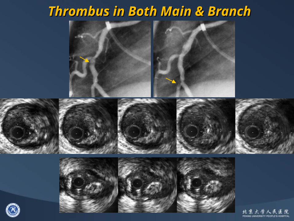
Thrombus in Both Main & BranchThrombus in Both Main & Branch

MLA
End of rupture
Fibrous cap
Plaque RupturePlaque Rupture
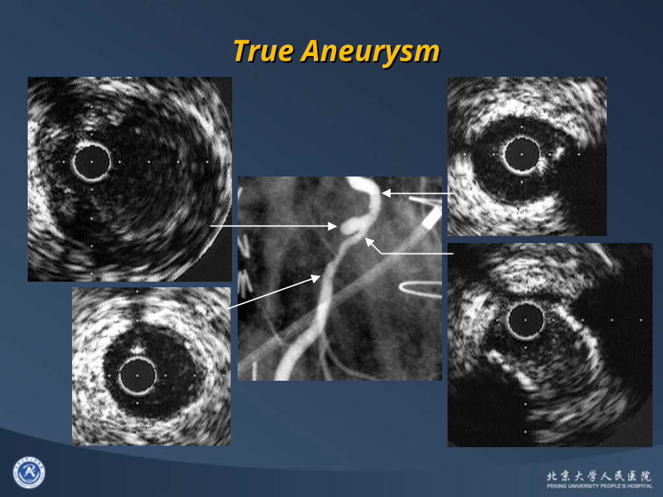
True Aneurysm True Aneurysm

Pseudo Aneurysm Pseudo Aneurysm
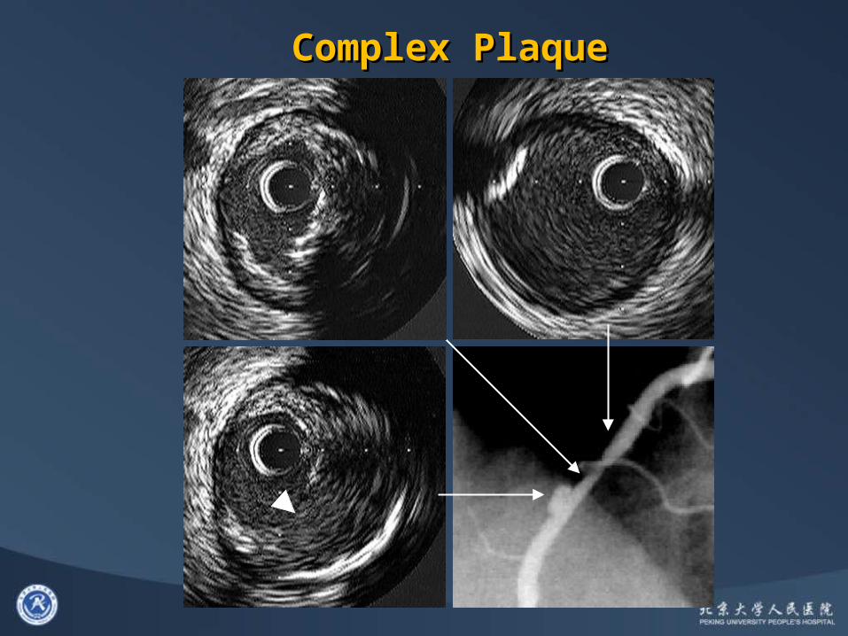
Complex Plaque Complex Plaque

Proximal
Rupture with ThrombusRupture with Thrombus
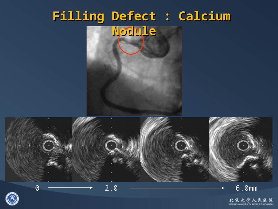
0 2.0 6.0mm
Filling Defect : Calcium Nodule Filling Defect : Calcium Nodule

““Muscle Bridge”Muscle Bridge”
DiastolicDiastolic SystolicSystolic
Inside the muscle bridge, Inside the muscle bridge, no plaque at all!no plaque at all!

Proximal
5555y.o. Female, AMIy.o. Female, AMI
“Spontaneous Dissection”

A B CD
BA C D
42y.o. Male, Unstable AP42y.o. Male, Unstable AP
Normal Artery, HematomaMaehara et al. AJC 2002

Thank you for your attention!