j. thomas lambrecht (editor) oral and implant · pdf [email protected] prof. dr....
TRANSCRIPT

J. Thomas Lambrecht (Editor)
Oral andImplant Surgery:Principles and Procedures
In collaboration with:A. Dunsche, R. Ewers, A. Filippi,
B. Hoffmeister, Th. Kreusch, K. Wangerin
Berlin, Chicago, Tokyo, Barcelona, Istanbul, London, Milan,Moscow, New Delhi, Paris, Peking, Prague, São Paulo and Warsaw
v
Dedication
The Kiel Connection dedicate this book
to their teacher
Prof. Dr. med. Dr. med. dent. Franz HärleDirector of Department
of Oral and Maxillofacial Surgery, Kiel University 1980–2004
and Dean of the Medical Faculty of Kiel University 1991–1992

Outpatient surgery performed by oral surgeons and
maxillofacial surgeons has been subject to various
influences and exposed to new trends in recent years:
• patients are getting older and older
• owing to the associated increase in systemic dis-
eases, the operations are becoming more complex
and overall treatment involves more risk
• the forensic implications are more signigficant
• innovations in operating technique and materials
have made surgical procedures possible that place
high demands on the infrastructure, logistics and
expertise of the treatment team
• the forms of doctor–patient communication have
changed as a result of the Internet; patients no
longer look for their doctor or dentist in the phone
book but on their website and they no longer
attend a consultation uninformed, but are focused
and fully informed.
It is not easy to address all these points in a single text-
book or reference book.
The book presents examples of intraoral opera-
tions, in many cases showing the step-by-step operat-
ing procedure, which can obviously vary from patient
to patient and from clinic to clinic. A whole chapter is
dedicated to endosseous implantology, contrasting it
with tooth-preserving surgery as an alternative
approach.
A selection of cases is also presented, which must
be aligned with the self-assessment and proficiency of
each particular surgeon. A central theme of ‘safety’
runs throughout the entire book, from the principles of
preoperative information to patients, to intraoperative
complications and their possible consequences
through to dealing with at-risk patients. This should,
therefore, interest anyone already working in or cur-
rently studying this specialist field. Unfortunately,
ever-expanding knowledge means some omissions are
inevitable in order to keep within the scope of this
book. Readers may wish to refer to the extensive spe-
cialist literature published by Quintessence.
The bow ties on the cover were hand-made in
Vienna. The bow tie in the center is printed with the
logo of the Congress of the German Society of Oral and
Maxillofacial Surgery, which was held in Kiel in 1994
and chaired by Professor Härle.
The Kiel Connection – former senior residents and
lecturers of Kiel University Department of Oral and
Maxillofacial Surgery – were generously supported in
producing this book by Prof. Dr. A. Filippi, Basel,
Switzerland, and colleagues from Vienna University
Hospital, Dr. H. Fahrenholz, Dr. Dr. A. Reichwein, Dr. K.
Schicho, Dr. K. Sinko, Mag. E. Stein and Prof. Dr. D.
Turhani.
We are grateful to all our colleagues who con-
tributed their illustrations to the book. As well as our
dental assistants and medical consultants, the follow-
ing people have helped to make this book a reality: Dr.
C. Bernsmeier, Prof. Dr. W. Ummenhofer, Prof. Dr. Dr.
M. Kunkel and Prof. Dr. N. Zitzmann. Last, but not
least, we would like to thank all our secretaries, espe-
cially Ms. G. Oertlin and Ms. B. Olufsen, and give spe-
cial thanks to Ms. S. Holmes for her superb translation.
We would also like to thank Quintessence and its
staff for the flexible, swift and ever-friendly support
they provide.
J. Thomas Lambrecht
vii
Preface
Preface

ix
Authors
Prof. Dr. Dr. H. Anton Dunsche
Director of Clinic for Oral and Maxillofacial Surgery
Städtisches Klinikum Karlsruhe
Moltkestrasse 120, D-76133 Karlsruhe, Germany
Prof. Dr. Dr. Rolf Ewers
Chairman of the University Hospital of Cranio-,
Maxillofacial and Oral Surgery
Medical University of Vienna
Währingerstrasse 18-20, A-1090 Vienna, Austria
Prof. Dr. Andreas Filippi
Clinic for Oral Surgery, Oral Radiology and Oral
Medicine
University of Basel
Hebelstrasse 3, CH-4056 Basel, Switzerland
Prof. Dr. Dr. Bodo Hoffmeister
Director of Clinic for Maxillary Surgery and Plastic
Facial Surgery
Charité – Universitätsmedizin Berlin
Campus Benjamin Franklin
Hindenburgdamm 30, D-1220 Berlin, Germany
Prof. Dr. Dr. Thomas Kreusch
Professor of Oral and Maxillofacial Surgery
Department of Oral and Maxillofacial Plastic Surgery
Head and Neck Centre
Asklepios Klinik Nord, Campus Heidberg
Tangstedter Landstrasse 400, D-22417 Hamburg,
Germany
Prof. Dr. Dr. J. Thomas Lambrecht
Clinic Director
Clinic for Oral Surgery, Oral Radiology and Oral
Medicine
University of Basel
Hebelstrasse 3, CH-4056 Basel, Switzerland
Prof. Dr. Dr. Dr. h. c. Konrad Wangerin
Department of Oral and Maxillofacial Surgery
Marienhospital Stuttgart
Böheimstr. 37, D-70199 Stuttgart, Germany
Authors

Anton Dunsche
1 Patient information, preoperative preparationand medication ........................................................1
1.1 Patient information ..........................................................................................................3
1.1.1 Patient's right to choose and informed consent ..................................................................................................3
1.1.2 The purpose of patient information ..........................................................................................................................3
1.1.3 Timing of information........................................................................................................................................................4
1.1.4 Information for non-competent patients and minors ......................................................................................4
1.2 Preoperative preparation ..................................................................................................5
1.2.1 Instruments ............................................................................................................................................................................5
1.2.2 Hygiene: accident prevention ........................................................................................................................................5
1.2.3 Disinfection ..........................................................................................................................................................................10
1.2.4 Sterilization ..........................................................................................................................................................................11
1.2.5 Equipment infrastructure..............................................................................................................................................12
1.3 Medication in the perioperative period........................................................................16
1.3.1 Sedatives ................................................................................................................................................................................16
1.3.2 Local anesthetics ..............................................................................................................................................................16
1.3.3 Analgesics..............................................................................................................................................................................17
1.3.4 Antibiotics ............................................................................................................................................................................18
References ......................................................................................................................20
Thomas Kreusch and J. Thomas Lambrecht
2 Simple and complicated tooth extraction ........212.1 General principles ..........................................................................................................23
2.1.1 Indications for tooth removal......................................................................................................................................23
2.1.2 Contraindications to tooth removal ........................................................................................................................23
2.1.3 Anesthesia ............................................................................................................................................................................23
xi
Contents
Contents
2.1.4 Preparation of the patient ............................................................................................................................................23
2.1.5 Instruments ..........................................................................................................................................................................24
2.1.6 Inspecting the extracted tooth ..................................................................................................................................26
2.1.7 Treating the tooth socket ..............................................................................................................................................26
2.1.8 Aftercare ................................................................................................................................................................................27
2.2 Special procedure ..........................................................................................................28
2.2.1 Position of practitioner and patient ........................................................................................................................28
2.2.2 Removal of primary teeth..............................................................................................................................................28
2.2.3 Removal of anterior teeth..............................................................................................................................................29
2.2.4 Removal of premolars......................................................................................................................................................29
2.2.5 Removal of molars ............................................................................................................................................................30
2.2.6 Serial extractions ..............................................................................................................................................................32
2.3 Complications..................................................................................................................34
2.3.1 Slipped instruments ........................................................................................................................................................34
2.3.2 Lesions to adjacent teeth ..............................................................................................................................................34
2.3.3 Crown fracture ....................................................................................................................................................................34
2.3.4 Root fracture ........................................................................................................................................................................34
2.3.5 Thermal damage caused by rotary instruments................................................................................................35
2.3.6 Nerve lesions........................................................................................................................................................................35
2.3.7 Oro-antral perforation ....................................................................................................................................................35
2.3.8 Maxillary antral cysts ......................................................................................................................................................41
2.3.9 Displacement of teeth/parts of teeth into soft tissue ....................................................................................43
2.3.10 Dry socket ............................................................................................................................................................................43
2.4 Pre-implantology tooth removal ..................................................................................44
2.4.1 Gentle tooth extraction ..................................................................................................................................................44
2.4.2 Stabilization of the alveolar process ........................................................................................................................44
References ......................................................................................................................46
J. Thomas Lambrecht and Bodo Hoffmeister
3 Surgical removal of unerupted and displaced teeth ..............................................49
3.1 Mandibular third molars ................................................................................................51
3.1.1 Indications for surgical removal ................................................................................................................................51
3.1.2 Contraindications..............................................................................................................................................................54
3.1.3 Preoperative diagnosis ....................................................................................................................................................54
xii
Contents

3.1.4 Preoperative medication................................................................................................................................................56
3.1.5 Risks ........................................................................................................................................................................................56
3.1.6 Surgical technique ............................................................................................................................................................57
3.1.7 Aftercare ................................................................................................................................................................................60
3.1.8 Complications caused by third molar removal ..................................................................................................60
3.1.9 Complications caused by leaving third molars in place ................................................................................65
3.2 Maxillary third molars....................................................................................................67
3.3 Maxillary canines ............................................................................................................70
3.4 Other retained teeth ......................................................................................................72
References ......................................................................................................................76
J. Thomas Lambrecht and Andreas Filippi
4 Tooth-preserving surgical procedures ................814.1 Periradicular surgery ......................................................................................................83
4.1.1 Aims ........................................................................................................................................................................................83
4.1.2 Indications and contraindications............................................................................................................................83
4.1.3 Operating technique ........................................................................................................................................................86
4.1.4 Hemostasis............................................................................................................................................................................87
4.1.5 Retrograde filling................................................................................................................................................................89
4.1.6 Wound closure ....................................................................................................................................................................91
4.1.7 Aftercare and prognosis ................................................................................................................................................91
4.2 Intentional replantation ................................................................................................95
4.2.1 Indications and contraindications............................................................................................................................95
4.2.2 Operating technique ........................................................................................................................................................95
4.3.3 Aftercare and prognosis ..............................................................................................................................................100
4.3 Transplantation ............................................................................................................102
4.3.1 Indications, contraindications and planning ....................................................................................................102
4.3.2 Tooth transplantation technique ............................................................................................................................103
4.3.3 Special features of third molar transplantation ..............................................................................................104
4.3.4 Splinting ..............................................................................................................................................................................105
4.3.5 Aftercare and prognosis ..............................................................................................................................................105
4.4 Corrective surgery ........................................................................................................107
4.4.1 Hemisection ......................................................................................................................................................................107
4.4.2 Premolarization ..............................................................................................................................................................109
xiii
Contents

4.4.3 Root amputation ............................................................................................................................................................109
4.4.4 Crown lengthening ........................................................................................................................................................112
4.4.5 Exposure ..............................................................................................................................................................................112
References......................................................................................................................117
Konrad Wangerin
5 Cysts, bone lesions,odontogenic tumors ............................................121
5.1 Odontogenic cysts ........................................................................................................125
5.1.1 Infant gingival cysts ......................................................................................................................................................125
5.1.2 Follicular cyst ....................................................................................................................................................................125
5.1.3 Eruption cyst ....................................................................................................................................................................129
5.1.4 Lateral periodontal cyst ..............................................................................................................................................129
5.1.5 Gingival cyst of adults ..................................................................................................................................................130
5.1.6 Glandular odontogenic cyst ......................................................................................................................................130
5.2 Non-odontogenic cysts................................................................................................132
5.2.1 Nasopalatine duct cyst ................................................................................................................................................132
5.2.2 Nasolabial cyst..................................................................................................................................................................135
5.2.3 Fissural cyst........................................................................................................................................................................135
5.3 Cysts of inflammatory origin ......................................................................................136
5.3.1 Radicular cyst....................................................................................................................................................................136
5.3.2 Residual cyst ......................................................................................................................................................................139
5.3.3 Periodontal cyst ..............................................................................................................................................................139
5.3.4 Occlusion cyst ..................................................................................................................................................................139
5.4 Operating techniques ..................................................................................................141
5.4.1 Cystostomy (Partsch I) ................................................................................................................................................141
5.4.2 Cystectomy (Partsch II) ..............................................................................................................................................145
5.4.3 Biological filling materials ..........................................................................................................................................146
5.4.4 Alloplastic and synthetic filling materials ..........................................................................................................151
5.5 Bone lesions ..................................................................................................................153
5.5.1 Osteolytic lesions ............................................................................................................................................................153
5.5.2 Osteoplastic lesions ......................................................................................................................................................157
xiv
Contents

5.6 Odontogenic tumors ....................................................................................................162
5.6.1 Odontoma ..........................................................................................................................................................................162
5.6.2 Keratocystic odontogenic tumor ............................................................................................................................163
References ....................................................................................................................165
Andreas Filippi
6 Traumatology of permanent teeth ..................1696.1 Classification and nomenclature of dental injuries ..................................................171
6.2 Epidemiology ................................................................................................................173
6.3 Action at site of accident............................................................................................174
6.4 Prevention of dental injuries ......................................................................................176
6.4.1 Fabricating a multilayer mouthguard ..................................................................................................................176
6.5 Post-traumatic diagnostic procedure ........................................................................179
6.6 Prognosis of dental injuries ........................................................................................181
6.7 Post-traumatic treatment............................................................................................182
6.7.1 General treatment ..........................................................................................................................................................182
6.7.2 Intra/extra-alveolar fractures of the teeth ..........................................................................................................184
6.7.3 Concussion and loosening of the teeth................................................................................................................186
6.7.4 Dislocation ........................................................................................................................................................................187
6.7.5 Intrusion ..............................................................................................................................................................................188
6.7.6 Avulsion................................................................................................................................................................................192
6.7.7 Post-traumatic splinting..............................................................................................................................................195
6.7.8 General medicinal treatment after dental trauma ........................................................................................200
6.7.9 Anti-resorptive and regenerative medicinal treatments..............................................................................200
6.8 Late sequelae after dental trauma..............................................................................203
6.8.1 Late sequelae in the pulp ............................................................................................................................................203
6.8.2 Late sequelae in the periodontium ........................................................................................................................206
References ....................................................................................................................220
xv
Contents
Anton Dunsche and J. Thomas Lambrecht
7 Intraoral soft tissue surgery ..............................2257.1 Biopsy ............................................................................................................................227
7.1.1 Excisional biopsy ............................................................................................................................................................227
7.1.2 Incisional biopsy ..............................................................................................................................................................229
7.1.3 Punch biopsy ....................................................................................................................................................................230
7.1.4 Brush biopsy ......................................................................................................................................................................232
7.2 Periodontal surgery ......................................................................................................233
7.2.1 Gingivectomy ....................................................................................................................................................................233
7.2.2 Mucogingival surgery ....................................................................................................................................................234
7.2.3 Flap surgery........................................................................................................................................................................235
7.3 Pre-prosthetic surgery ................................................................................................243
7.4 Surgery of minor salivary glands ................................................................................249
7.5 Abscess incision ............................................................................................................258
7.6 Benign tumors ..............................................................................................................262
7.7 Epithelial precursor lesions..........................................................................................266
References ....................................................................................................................270
Rolf Ewers
8 Implant surgery ....................................................2718.1 Basic principles ............................................................................................................273
8.1.1 Indications and contraindications of endosseous implants......................................................................273
8.1.2 Principles of planning by Harald Fahrenholz....................................................................................................273
8.1.3 Imaging techniques........................................................................................................................................................275
8.2 Clinical requirements ..................................................................................................280
8.2.1 Bone quality ......................................................................................................................................................................280
8.2.2 Bone supply........................................................................................................................................................................281
8.2.3 Osseointegration and functional ankylosis ........................................................................................................283
8.2.4 Loading ................................................................................................................................................................................283
8.2.5 Soft tissue............................................................................................................................................................................285
8.2.6 Esthetics by Harald Fahrenholz ..............................................................................................................................286
8.2.7 Incisions ..............................................................................................................................................................................289
xvi
Contents
8.3 Standard clinical situations ........................................................................................292
8.3.1 Single-tooth gap in anterior region ........................................................................................................................292
8.3.2 Lateral edentulous gap ................................................................................................................................................295
8.3.3 Free-end situation in the maxilla ............................................................................................................................298
8.3.4 Free-end situation in the mandible........................................................................................................................300
8.3.5 Partially edentulous mandible..................................................................................................................................301
8.3.6 Edentulous maxilla ........................................................................................................................................................303
8.3.7 Edentulous mandible ....................................................................................................................................................304
8.4 Augmentation ..............................................................................................................306
8.4.1 Principles of augmentation........................................................................................................................................306
8.4.2 Guided bone regeneration ..........................................................................................................................................312
8.4.3 Ridge preservation..........................................................................................................................................................313
8.4.4 Defect filling ......................................................................................................................................................................325
8.4.5 Condensing ........................................................................................................................................................................327
8.4.6 Bone splitting ....................................................................................................................................................................328
8.4.7 Inlay graft ............................................................................................................................................................................337
8.4.8 Sinus lift (sinus graft) ....................................................................................................................................................350
8.4.9 Onlay graft ..........................................................................................................................................................................360
8.4.10 Distraction..........................................................................................................................................................................374
8.5 Implant navigation ......................................................................................................378
8.5.1 Basic principles by Astrid Reichwein and Kurt Schicho..............................................................................378
8.5.2 Achievable accuracy levels by Astrid Reichwein and Kurt Schicho ......................................................380
8.5.3 Computer-aided planning and template fabrication by Rolf Ewers......................................................380
8.6 Implants for orthodontics by Klaus Sinko and J. Thomas Lambrecht ....................391
8.6.1 Development ....................................................................................................................................................................391
8.6.2 Micro-plates with micro-screws ..............................................................................................................................392
8.6.3 Palatal implant ................................................................................................................................................................393
8.7 Tissue engineering by Dritan Turhani and Elisabeth Stein ......................................396
8.7.1 Basic principles ................................................................................................................................................................396
8.7.2 Tissue engineering of bone ........................................................................................................................................396
8.7.3 Tissue engineering with hydroxyapatite..............................................................................................................397
8.8 Complications ..............................................................................................................400
8.8.1 Suture dehiscence ..........................................................................................................................................................400
8.8.2 Nerve injuries ....................................................................................................................................................................401
8.8.3 Mucosal complications ................................................................................................................................................403
8.8.4 Peri-implantitis ................................................................................................................................................................407
References ....................................................................................................................414
xvii
Contents
J. Thomas Lambrecht
9 At-risk patients and emergencies ....................4219.1 Cardiovascular diseases ................................................................................................424
9.1.1 Endocarditis ......................................................................................................................................................................424
9.1.2 Coronary heart diseases ..............................................................................................................................................426
9.1.3 Arterial hypertension ....................................................................................................................................................428
9.2 Lung diseases ................................................................................................................430
9.2.1 Bronchial asthma ............................................................................................................................................................430
9.2.2 Chronic obstructive pulmonary disease..............................................................................................................430
9.2.3 Tuberculosis ......................................................................................................................................................................430
9.3 Liver, kidneys and gastrointestinal tract ....................................................................431
9.3.1 Liver diseases ....................................................................................................................................................................431
9.3.2 Kidney diseases ................................................................................................................................................................431
9.3.3 Gastrointestinal diseases ............................................................................................................................................431
9.4 Diabetes mellitus ..........................................................................................................432
9.5 Blood diseases ..............................................................................................................433
9.5.1 Hemorrhagic diathesis ................................................................................................................................................433
9.5.2 Patients taking anticoagulants ................................................................................................................................434
9.6 Viral diseases ................................................................................................................437
9.6.1 HIV infection ....................................................................................................................................................................437
9.6.2 Hepatitis B infection......................................................................................................................................................437
9.6.3 Hepatitis C infection......................................................................................................................................................439
9.6.4 Herpes simplex virus ....................................................................................................................................................439
9.7 Allergies ........................................................................................................................440
9.8 Joint diseases ................................................................................................................441
9.8.1 Rheumatoid arthritis ....................................................................................................................................................441
9.8.2 Joint prostheses in osteoarthritis ............................................................................................................................442
9.9 Neurological and mental illnesses ..............................................................................443
9.9.1 Neurological disorders..................................................................................................................................................443
9.9.2 Mental illnesses................................................................................................................................................................445
xviii
Contents

9.10 Pregnancy......................................................................................................................447
9.10.1 Physiological changes ..................................................................................................................................................447
9.10.2 Changes in the orofacial area....................................................................................................................................447
9.10.3 Guidelines on dental treatment ..............................................................................................................................447
9.10.4 Infections during pregnancy......................................................................................................................................448
9.11 Emergencies ..................................................................................................................449
9.11.1 Monitoring..........................................................................................................................................................................449
9.11.2 Emergency equipment..................................................................................................................................................450
9.11.3 Emergency measures ....................................................................................................................................................451
References ....................................................................................................................459
Appendix ..............................................................465Abbreviations ................................................................................................................467
Acknowledgments and source of illustrations ..........................................................468
Internet links ................................................................................................................469
Index ..............................................................................................................................472
xix
Contents
388
Implant surgery8
of error because inaccuracies at least remain ‘consis-
tent’; there is no accumulation of errors in the course
of the individual working steps.
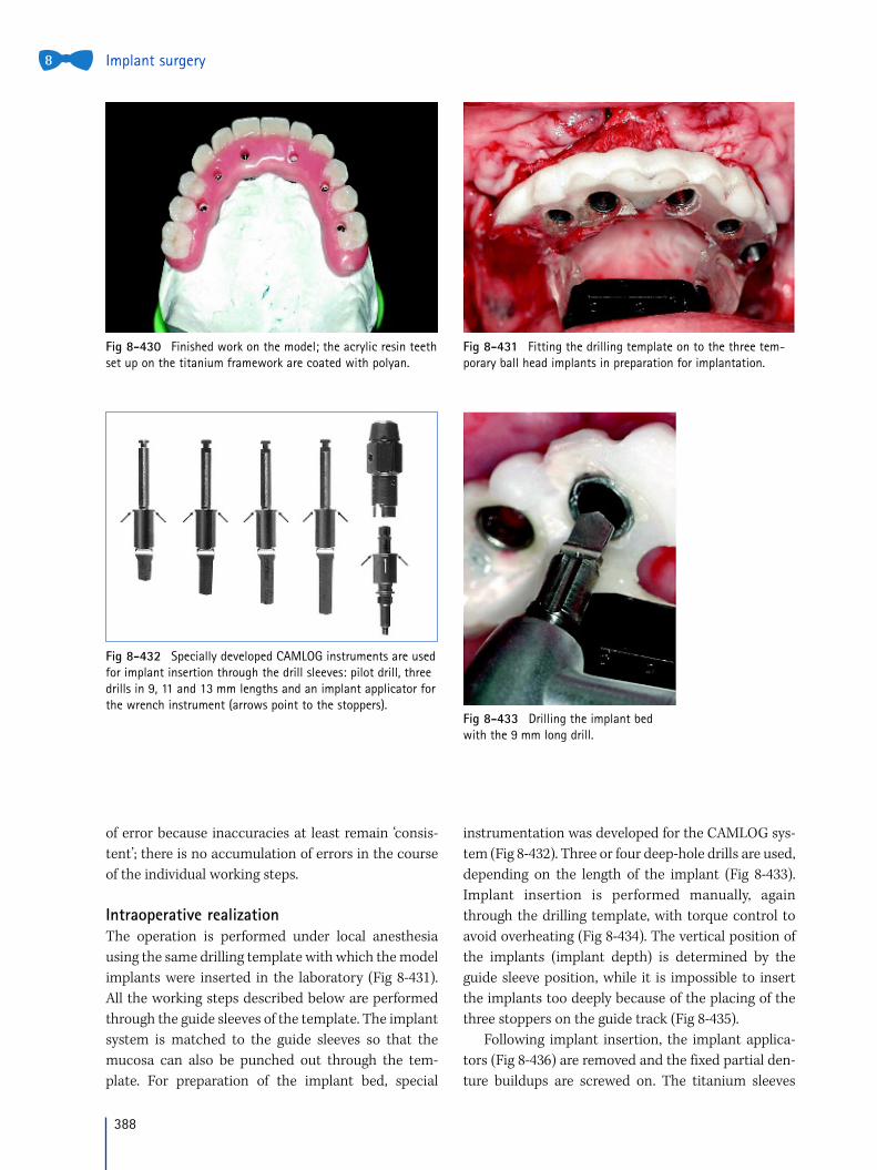
Intraoperative realizationThe operation is performed under local anesthesia
using the same drilling template with which the model
implants were inserted in the laboratory (Fig 8-431).
All the working steps described below are performed
through the guide sleeves of the template. The implant
system is matched to the guide sleeves so that the
mucosa can also be punched out through the tem-
plate. For preparation of the implant bed, special
instrumentation was developed for the CAMLOG sys-
tem (Fig 8-432). Three or four deep-hole drills are used,
depending on the length of the implant (Fig 8-433).
Implant insertion is performed manually, again
through the drilling template, with torque control to
avoid overheating (Fig 8-434). The vertical position of
the implants (implant depth) is determined by the
guide sleeve position, while it is impossible to insert
the implants too deeply because of the placing of the
three stoppers on the guide track (Fig 8-435).
Following implant insertion, the implant applica-
tors (Fig 8-436) are removed and the fixed partial den-
ture buildups are screwed on. The titanium sleeves
Fig 8-430 Finished work on the model; the acrylic resin teethset up on the titanium framework are coated with polyan.
Fig 8-431 Fitting the drilling template on to the three tem-porary ball head implants in preparation for implantation.
Fig 8-433 Drilling the implant bedwith the 9 mm long drill.
Fig 8-432 Specially developed CAMLOG instruments are usedfor implant insertion through the drill sleeves: pilot drill, threedrills in 9, 11 and 13 mm lengths and an implant applicator forthe wrench instrument (arrows point to the stoppers).

389
8
can be adapted to the implants with another screw
connection. In this way, it is possible to ensure ten-
sion-free insertion of the prosthetic work already com-
pleted preoperatively (Figs 8-437 and 8-438).
Before final insertion of the prosthetic work, a try-
in and verification of the exact accuracy of fit (over the
ball-head implants) takes place. Then the prosthetic
work with titanium sleeves is finally bonded in the
patient’s mouth. After the bonding agent has set, the
prosthetic work (e.g. a long-term provisional) can be
unscrewed. The ball-head implants in the alveolar
process and the matrices in the prosthetic work are
then removed.
Implant navigation 5
Fig 8-434 Inserting the implants through the sleeves in thedrilling template with the torque wrench.
Fig 8-435 The depth of implant insertion is mechanicallydetermined by three stoppers (arrows).
Fig 8-436 The six implants are placed; the drilling templatewith the black base stone is still in situ.
Fig 8-437 Fitting the implants with titanium sleeves, whichare intended to receive the long-term provisional appliance. Inthis case, a titanium bar is inserted. The temporary ball-headimplants are still required for positioning the long-term provi-sional appliance.
Fig 8-438 The framework is fitted, which can be bonded inplace. Performing this work intraorally is a prerequisite fortension-free seating of the fixed partial denture.
408
Implant surgery8
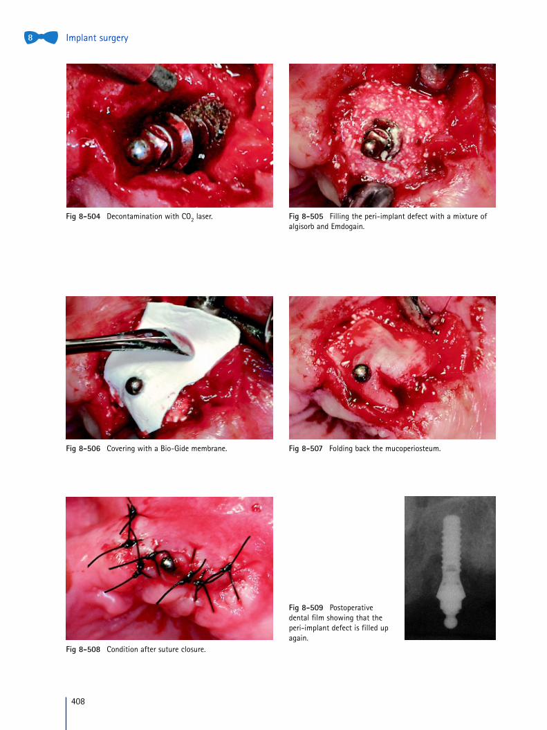
Fig 8-504 Decontamination with CO2 laser. Fig 8-505 Filling the peri-implant defect with a mixture ofalgisorb and Emdogain.
Fig 8-506 Covering with a Bio-Gide membrane. Fig 8-507 Folding back the mucoperiosteum.
Fig 8-508 Condition after suture closure.
Fig 8-509 Postoperativedental film showing that theperi-implant defect is filled upagain.
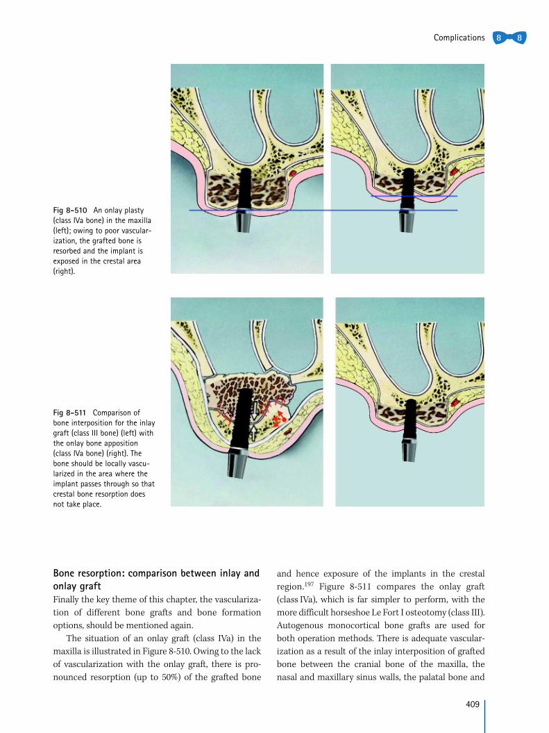
Bone resorption: comparison between inlay andonlay graftFinally the key theme of this chapter, the vasculariza-
tion of different bone grafts and bone formation
options, should be mentioned again.
The situation of an onlay graft (class IVa) in the
maxilla is illustrated in Figure 8-510. Owing to the lack
of vascularization with the onlay graft, there is pro-
nounced resorption (up to 50%) of the grafted bone
and hence exposure of the implants in the crestal
region.197 Figure 8-511 compares the onlay graft
(class IVa), which is far simpler to perform, with the
more difficult horseshoe Le Fort I osteotomy (class III).
Autogenous monocortical bone grafts are used for
both operation methods. There is adequate vascular-
ization as a result of the inlay interposition of grafted
bone between the cranial bone of the maxilla, the
nasal and maxillary sinus walls, the palatal bone and
Complications
Fig 8-510 An onlay plasty(class IVa bone) in the maxilla(left); owing to poor vascular-ization, the grafted bone isresorbed and the implant isexposed in the crestal area(right).
Fig 8-511 Comparison ofbone interposition for the inlaygraft (class III bone) (left) withthe onlay bone apposition(class IVa bone) (right). Thebone should be locally vascu-larized in the area where theimplant passes through so thatcrestal bone resorption doesnot take place.
409
8 8