janette ahola evidence based postoperative physiotherapy …
TRANSCRIPT

Janette Ahola
EVIDENCE BASED POSTOPERATIVE PHYSIOTHERAPY
MANAGEMENT OF ADOLESCENT IDIOPATHIC SCOLIOSIS
- A LITERATURE REVIEW
Degree Programme in Physiotherapy 2015

EVIDENCE BASED POSTOPERATIVE PHYSIOTHERAPY MANAGEMENT
OF ADOLESCENT IDIOPATHIC SCOLIOSIS - A LITERATURE REVIEW
Ahola, Janette
Satakunnan ammattikorkeakoulu, Satakunta University of Applied Sciences
Degree Programme in Physiotherapy
November 2015
Supervisor: Kangasperko, Maija
Number of pages: 55
Appendices: 1
Keywords: scoliosis, adolescent idiopathic scoliosis, scoliosis management, post-
operative physiotherapy, orthopaedic bracing
____________________________________________________________________
The purpose of this thesis was to gather evidence-based and up-to-date studies about
Adolescent Idiopathic Scoliosis (AIS) and its’ post-operative physiotherapy man-
agement. The thesis was made for Satakunta Central Hospital (Satakunnan
keskussairaala). The need for such topic came from the physiotherapists working at
the paediatric outpatient clinic. They wanted to have concrete updated information of
post-operative physiotherapy management for scoliosis surgery patients.
The theoretical part of the thesis consists of basic anatomy of the spine, different
forms of scoliosis and the deficits effects on the human and both the conservative
and surgical management of scoliosis.
The data collection was done in the form of a literature review. The search process
was performed using Pubmed, EBSCO and ScienceDirect as databases. The litera-
ture review was conducted in autumn 2015. Eight studies remained after applying in-
and exclusion criteria.
The results varied greatly and showed that there are several areas in which physio-
therapists could offer their professional skills: restoring patients’ dynamic breathing
and sensorimotor postural control, instructing a preoperative weight management
program, inquiring what could be the reasons behind the reduced time spent in sports
and the late full-time return to school.
CONTENTS
1 INTRODUCTION ........................................................................................................ 4
2 ANATOMY OF THE SPINE....................................................................................... 6
2.1 Anatomy and Physiology of the Spine ................................................................. 6
2.2 A Vertebra and an Intervertebral Disc .................................................................. 7
3 SCOLIOSIS .................................................................................................................. 9
3.1 Biomechanical Changes of the Spine In Scoliosis .............................................. 12
3.2 Structural Scoliosis ............................................................................................. 13
3.2.1 Idiopathic Scoliosis ...................................................................................... 13
3.2.2 Congenital Scoliosis ..................................................................................... 14
3.2.3 Neuromuscular Scoliosis .............................................................................. 15
3.3 Non-structural Scoliosis ...................................................................................... 16
4 TREATMENT OF ADOLESCENT IDIOPATHIC SCOLIOSIS ............................. 17
4.1 Conservative management .................................................................................. 17
4.1.1 Screening, Follow-up and Prognosis of AIS ................................................ 17
4.1.2 Orthopedic Bracing ...................................................................................... 21
4.1.3 Therapeutic Exercising ................................................................................. 25
4.2 Scoliosis Operations ........................................................................................... 27
5 POST-OPERATIVE MANAGEMENT ..................................................................... 30
6 A LITERATURE REVIEW ....................................................................................... 31
6.1 Literature Review Process .................................................................................. 32
6.2 Evidence Based Practice and Studies ................................................................. 33
6.3 Purpose and Aim of Thesis ................................................................................. 34
7 RESULTS ................................................................................................................... 35
7.1 The Search Strategy ............................................................................................ 35
7.2 Study Selection ................................................................................................... 36
7.3 Current Evidence Based Physiotherapy Management Options .......................... 44
8 CONCLUSION .......................................................................................................... 47
9 DISCUSSION ............................................................................................................ 48
REFERENCES ................................................................................................................ 51
APPENDICES

4
1 INTRODUCTION
Adolescent Idiopathic scoliosis is a three-dimensional deformity of the spine. In Fin-
land 0,2% of the population have scoliosis, which requires medical attention. (Nis-
sinen, et al. … 1993, 1.) There are approximately 250 new scoliosis cases discovered
every year in Finland from which 80% are idiopathic scoliosis, one of the subgroups
of structural scoliosis (Pihlajamäki & Ruuska 2010, 1). Adolescent Idiopathic scolio-
sis, which is the largest type of idiopathic scoliosis and which requires medical
treatment is ten times more common in girls than in boys. (Helenius 2015, 1.)
The treatment of scoliosis is either conservative or operative depending on the mag-
nitude of the curve. Both conservative and surgical treatment methods of scoliosis
have been well studied. Conservative management consists of screening of scoliosis,
follow-up and orthopaedic bracing. (Helenius 2013, 1.) When conservative treatment
measures fail, non-conservative management, a spinal fusion operation is performed.
(Helenius 2009, 1169.) Scoliosis surgery is the only treatment method for scoliosis,
which can correct the already curved spine (Helenius 2009, 1169; Ryöppy 1997,
120). Great results have been accomplished with scoliosis surgeries (Pihlajamäki &
Ruuska 2010, 16).
What has not yet been fully studied is the postoperative management of Adolescent
Idiopathic Scoliosis. This thesis aims to find out what could be the areas in which
physiotherapy management would be needed. Currently, the only physiotherapy
management that is offered to the patients is during the first week after the surgery in
a hospital ward. After the patients return from hospital to home, the only manage-
ment they get is the orthopaedist’s controls. (Helenius & Pajulo 2015, 1785.)
The purpose of this thesis is to gather evidence-based and up-to-date researches
about AIS and its’ post-operative physiotherapy management. The client of the thesis
is Satakunta Central Hospital (Satakunnan Keskussairaala). The need for such topic
came from the physiotherapists working at the paediatric outpatient clinic. They
wanted to have concrete updated information of post-operative physiotherapy man-
agement for scoliosis surgery patients. This literature review aims to give helpful in-

5
formation, which would benefit the physiotherapists in their clinical work. (Lemp-
inen 2015.)

6
2 ANATOMY OF THE SPINE
Tortora and Derrickson define anatomy as “the science of body structures and the
relationships among them” (Tortora & Derrickson 2011, 2). This chapter will focus
on regional anatomy, referring to a certain body part, which in this thesis indicates to
the spine of a human. Knowledge of a healthy spine and its’ structure is needed to
later define a spine with a defect, such as scoliosis. (Tortora & Derrickson 2011, 2.)
2.1 Anatomy and Physiology of the Spine
The spine or vertebral column is shaped of twenty-four individual bones called ver-
tebrae (singular: vertebra). It is divided into three regions, which are the following:
cervical (neck), thoracic (chest) and lumbar (low back). Cervical spine is shaped of 7
individual vertebras while thoracic spine of 12 and lumbar spine of 5 vertebras.
There are typically four curves in the spine: cervical lordosis, thoracic kyphosis,
lumbar lordosis and sacral kyphosis (picture 1). While cervical and lumbar curves are
convex, oppositely thoracic and sacral curves are concave relative to the front of the
body. This specific curving of the spine enables the spine to be flexible, mobile and a
help in maintaining the balance of the body. Yet it functions as a supporting and
strong structure of the body. These curves are most visible observed from the lateral
aspect. (Tortora & Derrickson 2011, 233.)
Vertebral columns lowest regions are sacrum, which is formed of 5 fused vertebras
and coccyx, the tailbone fused of four vertebras. Neither of them are mobile parts of
the spine and thus will not be discussed thoroughly in this chapter. Other bones that
complete the skeleton of the trunk of the body are the ribs and the sternum. A human
spine is approximately 71 centimetres long in male adults and 61 centimetres long in
female adults, covering two-fifths of the length of the body. In addition to being
composed of bony structures, the vertebral column is formed of connective tissue.
(Tortora & Derrickson 2011, 233.)
The purpose of the spine is to protect the spinal cord, function as a flexible rod and
shock absorber, support the head and work as an attachment site for the ribs, pelvic

7
girdle and the many back and core muscles. Its’ movements are flexion (forward
bend), later flexion (side bend), extension (backward bend) and rotations. (Tortora &
Derrickson 2011, 233.)
Different conditions and general progression of old age might disturb the spine’s
normal curves. The most common abnormalities of the spine are excessive kyphosis,
lordosis and scoliosis, the latter will be discussed throughout the thesis. (Tortora &
Derrickson 2011, 234.)
Picture 1. The anatomy of the spine, showing four natural curves and five regions of
the spine. (Website of the Spine Universe 2015)
2.2 A Vertebra and an Intervertebral Disc
A typical vertebra consists of a vertebral body, vertebral arch and many processes
(picture 2). The vertebrae differ in shape and size depending on which region they
are located in (Agur & Dalley 2012, 292). The vertebral body is the thickest and

8
strongest part of a vertebra and is shaped as a disc. On the top and bottom of every
vertebral body there are intervertebral discs attached to them, which is made possible
by the rough surface of the vertebral body. The vertebral body is the weight-bearing
part of the vertebra and is well suited for this purpose by being thick in shape. It is
the most anterior part of the vertebra. (Tortora & Derrickson 2011, 235.)
The vertebral arch includes both pedicles and laminas. Pedicles are two processes,
which protrude posteriorly from the vertebral body and are short and thick in shape.
They unite to the flat layers, laminas and together form the vertebral arch. The verte-
bral body and the vertebral arch together shape a vertebral foramen, an empty space
between them. When there are many vertebras stacked on top of each other, together
they formulate vertebral canal or spinal canal, which enables the spinal cord to go
through the vertebra and surround important structures. In addition to the spinal cord,
blood vessels, spinal nerves, areolar connective tissue and adipose tissue go through
the canal. (Tortora & Derrickson 2011, 235.)
There are altogether seven processes that protrude from the vertebral arch. Where a
lamina and a pedicle are united a transverse process extends on both lateral sides.
These are the most lateral parts of the vertebra. A spinous process, a single and the
most prominent process, continues from where the laminae end. It is the most poste-
rior part of the vertebra and often can be seen posteriorly in a human. Many muscles
are attached to the transverse processes and the spinous processes. There are four
more processes, which attach to the other vertebral processes superiorly and inferior-
ly: two superior articular processes and two inferior articular processes both known
as facets. (Tortora & Derrickson 2011, 234.)
Intervertebral discs are soft, pulpy and elastic structures, which work as pads be-
tween the vertebrae. Its’ main purpose is to absorb vertical shock and to enable
movement by being flattened and broadened depending on the pressure it receives
from other surrounding structures. The disc consists of fibrocartilage layer, the annu-
lus fibrosus that covers a softer inner substance called nucleus pulposus. Because the
discs contain a lot of fluid, typically, during the day the discs lose water from their
cartilage affecting the height of the intervertebral disc. This is the reason why we are
shorter after the strenuous day. During the night the discs get rehydrated affecting in

9
us so that we attain our full height again in the mornings. The discs are avascular
meaning that they get their blood supply from surrounding structures. Exercising en-
ables better blood flow to these structures, which increase both the nutrient and the
blood flow also to the intervertebral discs. (Tortora & Derrickson 2011, 234.) Scolio-
sis causes pathological effects on these structures (Website of UW medicine, De-
partment of Scoliosis).
Picture 2. A typical thoracic vertebra. (Principles of Anatomy and Physiology, 2011)
3 SCOLIOSIS
The natural curves of the spine are cervical lordosis, thoracic kyphosis and lumbar
lordosis, which can be seen when observed from the side. The curves enhance the
spine to be flexible and mobile. When there is scoliosis involved, the natural curves
of the spine are imbalanced and the optimal flexibility is lost. (Ryöppy 1997, 119.)
Scoliosis is a condition in which the spine curves laterally and vertebral bodies rotate
in addition causing a three-dimensional deformity in the thoracic or lumbar spine. A
healthy spine looks straight when observing a human being from behind, but with
scoliosis patient, the spine forms a ”C” or ”S” -shape depending on the amount of the

10
curves. In Cobb angle measurement the curve has to be over 10° to be defined as sco-
liosis. (Helenius 2009, 1168.)
Scoliosis can be also noticed with a bare eye as trunk asymmetry. Uneven horizontal
level between the tips of the ears, shoulders, posterior angles of the scapulas, waist
curves, posterior-anterior iliac crests and knees can suggest that a person has a scoli-
osis. In these cases further assessment needs to be done to determine the severity and
type of the scoliosis. (Kerttula, Schlenzka & Tallroth 2004, 2298.)
In scoliosis, there can be one or more curves: only one primary curve or a double
curve, which consists of a primary curve and one or more secondary curves. The
secondary curves try to compensate the more severe curve by turning to the opposite
direction evening out the primary curve and thus balancing the spine (picture 3). Re-
gardless of the scoliosis, the spine tries to find a balanced posture, aiming to keep the
head in a fairly good position. (Pihlajamäki & Ruuska 2010, 4.)
Picture 3. Different types of scoliosis curves, demonstrating spines with both single
curve and double curves. (Website of Broadway Chiropratctic and Wellness Center
New York 2015)
Scoliosis is often classified based on the location of the primary curve where the
apex is the most curved part of the spine. It is also the part, which is the furthest
away from the midline of the spine (Pihlajamäki & Ruuska 2010, 2). In thoracic sco-

11
liosis the apex is between T2-T11, in thoraco-lumbar scoliosis between T12-L1, in
lumbar scoliosis the apex is between L2-L4 and in lumbo-sacral scoliosis the apex is
between L5-S1 (picture 4). The curvature in cervical spine is called torticollis rather
than scoliosis. In torticollis, the apex of the curve is between the vertebras C1-C6.
(Magee 2008, 478.)
Picture 4. A demonstration of the classification of scoliosis based on the location of
the apex of the curve. (Own files)
Scoliosis is most often symptomless. Only every fourth person suffering from scolio-
sis has back pain typically in the thoracic spine, between the scapulas or in the lum-
bar spine (Helenius 2015, 1). The occurrence of back pain in adult scoliosis patients
is the same as with those who do not have scoliosis. Still, if a scoliosis patient has
back pain, it is more difficult to treat it. (Hakkarainen 2007, 1.) One more rare symp-
tom can be spondylolisthesis, a forward displacement of a vertebra. Scoliosis is di-
vided into two main groups: structural scoliosis and non-structural or functional sco-
liosis. (Kerttula, Schlenzka & Tallroth 2004, 2298.)

12
3.1 Biomechanical Changes of the Spine In Scoliosis
The curve of the spine can be either convex (referring to the hump side) or concave
(referring to the hollow side). In thoracic structural scoliosis the vertebral bodies ro-
tate towards the side of the convexity (picture 5). From this effect the spinous pro-
cesses of the vertebraes oppositely rotate to the concave side. This rotation of the
spine causes the vertebral bodies to push the ribs posteriorly narrowing the ribcage
on the convex side and causing a rib hump on the posterior side (picture 6). This
cosmetically poor appearance and deformity is called razorback spine. This is char-
acteristic for scoliosis patients and becomes more visible in a flexed position. On
contrary the ribs on the concave side move anteriorly causing a hollow cave on the
back and a widening on the ribcage. (Magee 2008, 478.)
Picture 5. Demonstration of the effects of concavity-convexity of the spine and the
effect of it to the muscles. (Website of Chiropractic Specialty Centre 2015.)

13
Picture 6. Demonstration of the effects of scoliosis in thoracic spine and ribcage.
(Website of Chiropractic Specialty Center 2015)
3.2 Structural Scoliosis
As the name structural scoliosis suggests, there is a permanent change in the struc-
ture of the bones, in this case vertebras, which formulate the spine. This means that
the process can only be slowed done, not fully corrected even with a spinal correc-
tion surgery. Structural scoliosis is divided into subgroups according to how it has
been formed: congenital, idiopathic and neuromuscular scoliosis. Approximately 70-
90% of structural scoliosis are categorized as idiopathic. (Kerttula, Schlenzka &
Tallroth 2004, 2298.)
3.2.1 Idiopathic Scoliosis
Idiopathic scoliosis is divided into Infantile, Juvenile and Adolescent idiopathic sco-
liosis (AIS) depending on the age of the onset of scoliosis, the latter being the most
common scoliosis of all of the scoliosis subgroups. Idiopathic scoliosis is often first
diagnosed in adolescents. (Crowther 1999, 139.) It is also found in infants and juve-
niles, but is far less common. The main focus of the thesis will be on AIS because it
is most common of all the scoliosis subgroups. There are approximately 200 new
AIS cases appearing every year in Finland. (Hakkarainen 2007,1.) There is no known

14
cause for Idiopathic scoliosis, thus the word ”idiopathic” translating to ”of unknown
cause”. In idiopathic scoliosis the curve is normally a double curve shaped as an S-
letter. (Kerttula, Schlenzka & Tallroth 2004, 2298.)
Infantile Idiopathic scoliosis (IIS) appears and is diagnosed before the third year of
life (Kerttula, Schlenzka & Tallroth 2004, 2298). Typically there is only one primary
curve, which is convex to the left (Adams & Hamblen 2001, 179). Infantile Idio-
pathic scoliosis is more common in the male population than in the female with the
ratio of 3:2. Infantile Idiopathic scoliosis is the least common type of the idiopathic
scoliosis. (Helenius & Parkkila 2008, 2541.) Infantile Idiopathic scoliosis can worsen
rapidly in a few months. This is the reason why it is extremely essential for IIS pa-
tients to get the needed medical care, which most often is more than one scoliosis
surgery or continuous orthopaedic plastering. The treatment has to slower down the
scoliotic curving yet still secure the continuing growth of the spine, which makes it
challenging (Helenius & Pajulo 2015, 1785.) Juvenile idiopathic scoliosis is diag-
nosed between the ages of four to nine years. (Kerttula, Schlenzka & Tallroth 2004,
2298).
Adolescent idiopathic scoliosis is a three-dimensional deformity, which appears in
the late childhood or the early adulthood, from the age of ten to the end of growth
spurt. It is only known that AIS is connected to a hormonal development since it
most commonly occurs during the age period when the child is having a growth
spurt, commonly between the ages of 10 and 12 years. (Adams & Hamblen 2001,
180.) There is a progressive increase in the curvature until the cessation of the skele-
tal growth. As most of the other scoliosis subgroups, also AIS is not symptomatic.
Adolescent Idiopathic scoliosis that requires medical attention and treatment is ten
times more common in girls than in boys. (Helenius 2015, 1.) AIS in progresses
more likely in girls than it does in boys (Pihlajamäki & Ruuska 2010, 5).
3.2.2 Congenital Scoliosis
Congenital scoliosis is caused by vertebral anomalies, which can be radiographically
seen in a newborn baby. Although the anomalies are present already at birth, there
15
might not be a clear curvature in the spine until the adolescent years. The spine is
typically sharply curved in congenital scoliosis. It is unusual that congenital scoliosis
runs in families. (Weinstein 2001, 161.) Ten percent of the scoliosis cases are con-
genital (Kerttula, Schlenzka & Tallroth 2004, 2298).
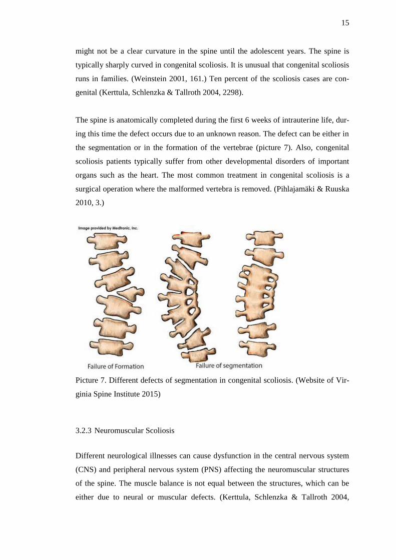
The spine is anatomically completed during the first 6 weeks of intrauterine life, dur-
ing this time the defect occurs due to an unknown reason. The defect can be either in
the segmentation or in the formation of the vertebrae (picture 7). Also, congenital
scoliosis patients typically suffer from other developmental disorders of important
organs such as the heart. The most common treatment in congenital scoliosis is a
surgical operation where the malformed vertebra is removed. (Pihlajamäki & Ruuska
2010, 3.)
Picture 7. Different defects of segmentation in congenital scoliosis. (Website of Vir-
ginia Spine Institute 2015)
3.2.3 Neuromuscular Scoliosis
Different neurological illnesses can cause dysfunction in the central nervous system
(CNS) and peripheral nervous system (PNS) affecting the neuromuscular structures
of the spine. The muscle balance is not equal between the structures, which can be
either due to neural or muscular defects. (Kerttula, Schlenzka & Tallroth 2004,

16
2298.) This type of scoliosis causes clear malpositions and asymmetry in the trunk.
Neuromuscular scoliosis is a condition, which worsens by time and in the worst cas-
es causes an early death. (Ryöppy 1997, 121.)
What makes treating neuromuscular scoliosis especially difficult is the fact that the
spine might continue curving even after the growth period has ended (Pihlajamäki &
Ruuska 2010, 3). Not to mention the fact, that the decision to operate a neurological-
ly disabled child takes more time than in other scoliosis cases. Surgery is the most
commonly used treatment method for neuromuscular scoliosis but also using casts
and orthotics can be a treatment option as well though neuromuscular scoliosis pa-
tients cannot use hard plastic orthotics such as the Boston brace. (Helenius & Pajulo
2015, 1785.) There are multiple conditions that can be behind neuromuscular scolio-
sis, those include Cerebral Palsy (CP), Polio, Myelomeningocele (Spina Bifida),
Muscular Dystrophies, spinal muscular atrophies, spinal injury and stroke (Ryöppy
1997, 141; Pihlajamäki & Ruuska 2010, 3). Neuromuscular scoliosis is most typical-
ly seen as a curve shaped as a C-letter (Kerttula, Schlenzka & Tallroth 2004, 2298).
3.3 Non-structural Scoliosis
Non-structural scoliosis, also known as secondary or functional scoliosis, is caused
by a functional disturbance where the body compensates the bodily movements from
pain or some other irritation thus causing an unwanted curve to the spine (Crowther
1999, 139). In forward flexion the deformity disappears if the scoliosis is non-
structural, while in structural scoliosis the posterior rib hump becomes more promi-
nent. There is often no vertebral rotation accompanied with the lateral curve as it is
in structural scoliosis. Functional scoliosis is a secondary symptom to e.g. poor pos-
ture, nerve root irritation, and inflammatory condition of the spine, leg length dis-
crepancy, hip contracture, hysteria or acute lumbago. When the primary cause of the
scoliosis is found and treated, the spine straightens automatically finding its’ natural
kyphotic and lordotic curves. (Magee 2008, 478.) Pain is more prominent in non-
structural scoliosis than in structural scoliosis (Helenius 2013, 1).
17
4 TREATMENT OF ADOLESCENT IDIOPATHIC SCOLIOSIS
The treatment of scoliosis can be either conservative management including the
screening of patients, brace treatment and scoliosis specific therapeutic exercising or
operative management. The treatment, which will be applied, depends on the magni-
tude of the scoliosis curve. The professionals assess the patients and offer the best-
suited options for them. (Helenius 2013, 1.)
4.1 Conservative management
Conservative management is defined on the Website of the Free Medical Dictionary
as “treatment designed to avoid radical medical therapeutic measures or operative
procedures” (The Website of Medical Dictionary 2015). In scoliosis this refers to
treatment that does not involve an orthopaedic surgery. Conservative management of
scoliosis consists of screening and follow-up of new cases, orthopaedic bracing and
sometimes therapeutic exercising. (Helenius 2009, 1169.)
4.1.1 Screening, Follow-up and Prognosis of AIS
Even 4% of the adolescents have a curved spine, which meet the requirements of
scoliosis (Kerttula, Schlenzka & Tallroth 2004, 2298). This is the reason why in ele-
mentary and junior high schools scoliosis screenings are done to find those with sco-
liosis. This is essential so that the children who have scoliosis would be referred to
receive treatment as soon as possible. In puberty, the growth periods can be rapid and
the worsening can happen in a short time. (Kerttula, Schlenzka & Tallroth 2004,
2298.) A postponed diagnose worsens the effect of conservative treatment and a sur-
gery is most likely needed. The authors Kerttula, Schlenzka and Tallroth speculate
that the screening of scoliosis is not functioning properly in Finland and is yet to be
improved, the reason being the decreased number of cases in the need of orthotic
bracing and the stabile prevalence of the condition. (Kerttula, Schlenzka & Tallroth
2004, 2298.)

18
The Adam’s forward bending test is a common screening method (picture 8). It is the
most powerful determinant of the occurrence of scoliosis (Nissinen 1996, Abstract;
Kerttula, Schlenzka & Tallroth 2004, 2298.) Some curves are yet so small and unno-
ticeable in standing positions that only forward bending reveals the scoliosis (Kerttu-
la, Schlenzka & Tallroth 2004, 2298). In the test the patient is asked to bend down
slowly while keeping the palms together. The therapist observes for trunk asymmetry
or a rib hump, which becomes more dominant in a flexed position. If there is some
seen asymmetry, the therapist may proceed to measure the inclination angle with a
scoliometer (picture 9), which will help to determine which patients need to have ra-
diographs taken of. (Horne, Flannery & Usman 2014, 193.) If the result from a scoli-
ometer is less than six degrees, a check-up every four or six months is enough a
treatment. If the degree is 6° or more, x-rays are taken. (Kerttula, Schlenzka &
Tallroth 2004, 2298.)
Picture 8. Adam’s forward bending test is often used for screening of scoliosis. The
arrows point on the clearly visible rib hump, which becomes more dominant in a
flexed position. (From the website of American Family Physician 2015)

19
Picture 9. Measuring the inclination angle of the spine with a scoliometer (From the
website of American Family Physician 2015).
There are many methods to measure the angle of scoliosis. Measuring the magnitude
is a concrete method for following the development of the curving. When the curve
goes over a certain angle the needed treatment is started depending on the severity of
the curvature. The most common method for assessing the curve is the Cobb method,
which has been in common use since 1948 after Doctor John R Cobb developed it. It
is also most common scoliosis measurement method in Finland. (Helenius 2009,
1168; Kerttula, Schlenzka & Tallroth 2004, 2298.)
The Cobb angle (picture 10) is measured from an x-ray picture, which is to be taken
while the patient is standing. Two extended lines are drawn linearly with the upper
border of the top vertebra and bottom border of the end vertebra, which are the first
and last vertebras that are tilted and involved in the curve. Then two lines are drawn,
which are perpendicular to the first two lines and the crossing of these lines defines
the Cobb angle. (Helenius 2009, 1168.)
The spine is defined with scoliosis if the Cobb angle is 10° or more from a posterior-
anterior (PA) x-ray, which is a key element in diagnostics (Helenius 2009, 1168;
Kerttula, Schlenzka & Tallroth 2004, 2298). The angle of the spine calculated with
the Cobb method defines what kind of treatment the professionals need to prescribe
to the patient based on the guidelines (table 1). Angles between 15-20° are consid-
ered to be that of a mild scoliosis, needing regular check-ups every 3-4 months for
following the development and possible progression of the curve until the cessation

20
of the bone after which the progression normally stops. If the angle is 20-40° more
medical attention by the specialist is needed and commonly first conservative man-
agement, bracing, is recommended to slow down the progression of the curve.
(Crowther 1999, 139.)
Picture 10. Cobb method, a tool with which on can define the angular measurement
of the curve of scoliosis (From the website of American Family Physician 2015 and
from the website of Posturetek 2015).
The prognosis of scoliosis depends on the onset and the location of the scoliosis.
Lumbar scoliosis typically has a better prognosis when compared to thoracic scolio-
sis, where the deformity is also more visible due to the rib hump. The later the onset,
the more likely it is for the medical treatment to help and slow down the curving. Al-
so, in this situation there is less time for the spine to curve before the curving stops
after the end of the period of spinal growth. (Adams & Hamblen 2001, 181.)
AIS patients whose curves are 50° or more require a spinal operation (Helenius 2009,
1168). Curves that are over 50° tend to continue curving even after bone has ossified.
A scoliosis is defined as severe scoliosis if the Cobb angle is over 70°. (Helenius &
Pajulo 2015, 1785.) If the curve is greater than 70° there is an 80% chance that the
person has a restrictive lung disease and the lung capacity is limited as a result of the

21
severe deformity. Patients with curves over 70°, which are left without medical atten-
tion, have a greater risk of early death. (Helenius 2013, 63; Helenius & Pajulo 2015,
1785.) Most commonly these patients have neuromuscular scoliosis instead of idio-
pathic scoliosis, which rarely reaches the angle of a severe level. Also, Finnish
screening system is so efficient that most of the AIS cases are identified before they
reach the operative level (>40°). Only in rare cases, such as teenage pregnancy or
eating disorder where the young avoids public health care check-ups, AIS develops
into a severe one reaching 70° angle. (Helenius & Pajulo 2015, 1785.)
Table 1. Cobb’s angle in relation to the recommended treatment method. The rec-
ommendations vary depending on the references (Helenius & Pajulo 2015, 1785).
4.1.2 Orthopedic Bracing
Orthotic bracing is the most common conservative treatment method used worldwide
for managing and slowing down the further curving of scoliosis in adolescent Idio-
pathic patients (Weinstein 2001, 371). There is no evidence that using a brace would
straighten the curved spine but rather slow down the condition and maintain the
curve at the starting level. The total effect of maximal correction using bracing has
been studied to be 10°. (Helenius 2009, 1168.)
There are multiple braces developed for different scoliosis types, each brace is cus-
tom-made and individually designed to meet the clients’ needs. Ideally, a brace is
prescribed to prevent the need for surgical operations, if the scoliosis is still in main-
tainable angle. A successful bracing is achieved with a good co-operation between
the patient, the parents and the professional team. It has been estimated that the gen-
eral effect of bracing treatment for the back is approximately 10° of the natural de-
Cobb’s angle Treatment method
10-20° Follow-up
25-40°(45°) Bracing
>40°(-50°) Scoliosis surgery

22
velopment. Orthopaedic bracing is started when the curvature exceeds 20-25° in
Cobb angle and is normally continued until the end of the bone growth if there is no
sudden worsening of the condition. (Helenius 2009, 1169.)
Adolescents and children are most often the ones required to wear the orthotics. Idi-
opathic scoliosis affects patient’s self image and confidence. Braces are often un-
comfortable and too noticeable, which makes accepting this management method
difficult for the young clients, who are already sensitive of their appearance. Also, in
that age the clients are continuously growing in height and especially girls are devel-
oping more feminine features with hips growing wider and breast growth making the
fitting more challenging. (Chan et al. … 2008, 137-139; Carvalho de Abreu et al. …
2012, 1.)
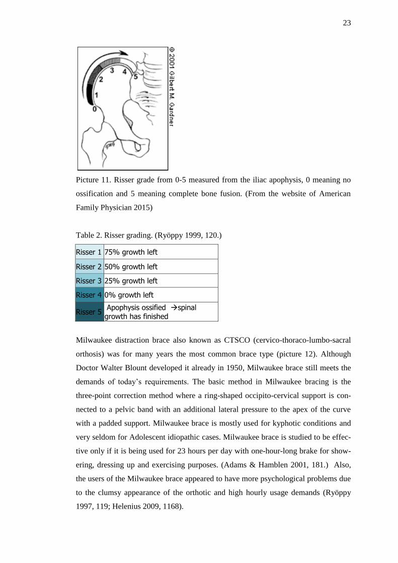
Bone maturity can be diagnosed from an x-ray picture with two different methods.
One is a Risser grading method (picture 11) where an x-ray is taken of the iliac crest.
The bone growth has stopped, if the grade is 4 and fully ossified if the grade is 5.
Grades less than four mean that there is still some growth left (table 2). (Ryöppy
1999, 120.) Another method for assessing the grade of bone maturity with which one
can determine how much growth there will be expected is Greulich-Pylen (G-P)
method. In this method an x-ray is taken of the left hand and similar grading is appli-
cable. These are essential tools for giving a prognosis to the whole curving process.
(Kerttula, Schlenzka & Tallroth 2004, 2298.)
23
Picture 11. Risser grade from 0-5 measured from the iliac apophysis, 0 meaning no
ossification and 5 meaning complete bone fusion. (From the website of American
Family Physician 2015)
Table 2. Risser grading. (Ryöppy 1999, 120.)
Risser 1 75% growth left
Risser 2 50% growth left
Risser 3 25% growth left
Risser 4 0% growth left
Risser 5 Apophysis ossified spinal growth has finished
Milwaukee distraction brace also known as CTSCO (cervico-thoraco-lumbo-sacral
orthosis) was for many years the most common brace type (picture 12). Although
Doctor Walter Blount developed it already in 1950, Milwaukee brace still meets the
demands of today’s requirements. The basic method in Milwaukee bracing is the
three-point correction method where a ring-shaped occipito-cervical support is con-
nected to a pelvic band with an additional lateral pressure to the apex of the curve
with a padded support. Milwaukee brace is mostly used for kyphotic conditions and
very seldom for Adolescent idiopathic cases. Milwaukee brace is studied to be effec-
tive only if it is being used for 23 hours per day with one-hour-long brake for show-
ering, dressing up and exercising purposes. (Adams & Hamblen 2001, 181.) Also,
the users of the Milwaukee brace appeared to have more psychological problems due
to the clumsy appearance of the orthotic and high hourly usage demands (Ryöppy
1997, 119; Helenius 2009, 1168).

24
Picture 12. Milwaukee Brace demonstrating the three-point correction method.
(From the website of The Free Dictionary By Farlex 2015)
The Boston brace (picture 13) was developed soon after the Milwaukee brace in
1960 when the need arose for creating a brace that would be accepted better by its’
users. Boston brace is most commonly used for lumbar or thoraco-lumbar curves
where the apex is located below Th-9 level, thus the name thoraco-lumbo-sacral or-
thosis (TLSO). Boston braces mechanism is to flatten the excessive lumbar lordosis
with only a two-point correction. This controls the lateral curving of the spine. What
makes Boston brace more accepted compared to the Milwaukee brace is that it is an
underarm brace, which is more unnoticeable compared to Milwaukee brace and is
much easier to hide under clothing. Another underarm brace, Lyon brace, is instead
used for curves with apex located in thoracic or thoraco-lumbar area. (Crowther
1999, 139-140.)

25
Picture 13. Boston brace, front and back view. (From the website of Orthotics Pros-
thesis New England 2015)
It is possible that in some cases, there are more than one braces prescribed for one
patient. Typically the Providence or Charleston bending brace are offered to give
some extra support during the night-time additionally. There are also many newly
developed optional braces on the market, which offer more elasticity and are more
comfortable for its’ user. The evidence of their efficiency is yet to be proved.
(Crowther 1999, 139-140.)
4.1.3 Therapeutic Exercising
There is no concrete evidence that exercising alone is helpful for slowing down the
curving of the spine (Adams & Hamblen 2001, 183; Weinstein 2001, 371). Structural
scoliosis cannot be managed with only physiotherapy alone. Oppositely, the symp-
toms in non-structural scoliosis can be relieved by physiotherapy, such as nerve root
irritation. (Hakkarainen 2007,1.) More studies are needed to find out what are the
effects of exercising for scoliosis patients (Helenius 2013, 1).
Still, individually designed exercising programs are found to be efficient for scoliosis
patients in all the phases of scoliosis treatment. The scoliosis patients should not be
restricted from physical activities and exercising. It has also been found, that people
26
with scoliosis have more problems in their balance than those without scoliosis.
(Hakkarainen 2007,1.)
The client who wears a brace is encouraged to exercise daily with and without the
brace. Exercising without a brace is recommended to prevent the total dependency on
an external support and rather activate the core muscles to work as an internal corset.
It is also recommended to exercise with the brace on to activate the core muscles in-
side the brace and to keep the correct posture throughout the day, not only during the
short period during the off-time. The muscle force takes away the extra load from the
bony structures, which already suffer from asymmetric loading. (Romano, et al. …
2015, 3.)
The main idea in exercising for scoliosis patients is to correct the motor control strat-
egies of movement and posture and to turn them into automatic patterns. The aim is
that through patient education and active muscle training, the patient is able to cor-
rect his posture automatically and achieve the needed stability and improve symmet-
rical spinal alignment. One recommended and concrete exercising method is an ac-
tive self-correction, where the patient tries to correct the posture as well as he or she
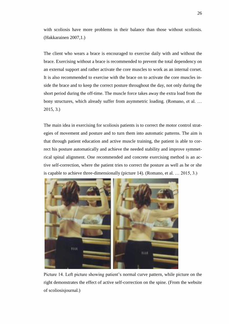
is capable to achieve three-dimensionally (picture 14). (Romano, et al. … 2015, 3.)
Picture 14. Left picture showing patient’s normal curve pattern, while picture on the
right demonstrates the effect of active self-correction on the spine. (From the website
of scoliosisjournal.)

27
4.2 Scoliosis Operations
When the Cobb angle is 40-50° or greater and there is still growth of the spine ex-
pected, spinal fusion operation is performed (Helenius 2009, 1169.) In addition,
those who are diagnosed with scoliosis who are almost at the end of their bone
growth period with curves, which are great in magnitude, are also directed to surgical
treatment (Pihlajamäki & Ruuska 2010, 15).
The purpose of the scoliosis surgery is to fuse all the needed joints of the vertebras
involved in the primary curve, thus the name spinal fusion. After the instrumentation
of the apex of the spine, the instrumented area is also ossified to secure the long last-
ing end result. (Ryöppy 1997, 120.) Normally, the rods or other metal instrumenta-
tions used in the operation are permanently left on the operated area (Pihlajamäki &
Ruuska 2010, 15; Ryöppy 1997, 121). Scoliosis surgery is the only treatment method
for scoliosis, which can correct the already curved spine (Helenius 2009, 1169;
Ryöppy 1997, 120).
The aim of the surgery is to slow down or to stop further development of curving.
Depending on the operation method and the individual, the operation can straighten
the spine up to 50°-60° from the original curve. The orthopaedist always assesses
individually, how much correction can be done in safe measures. (Pihlajamäki &
Ruuska 2010, 15.) Other goals of the surgery are: to achieve a good balance in the
spine between the saggital and coronal planes, to achieve an ideal yet safe amount of
correction in the spine, to stop the further curving of the spine and to get the patient
to return to his normal function as soon as possible after surgery. Importantly, the
spine will not be corrected to achieve a good-looking and cosmetic result, thus the
spine will not necessarily be vertically straight. (Weinstein 2001, 408.)
Before the operation, a standing posterior–anterior (PA) x-rays are taken of the spine.
Additionally, x-rays with patient bending laterally are also taken. These x-rays help
in defining the primary curve and how large an area will be operated and ossified.
The surgical treatment method will be also decided based on the x-rays. (Pihlajamäki
& Ruuska 2010, 15.)

28
There are multiple surgery techniques for scoliosis operations. The first surgical
method in treating scoliosis was the Harrington rod method, which was developed in
1962. It is a posteriorly performed technique, in which two metal rods are inserted in
the concavity of the curve between two hooks. The field of scoliosis treatment has
advanced since, thus new and much better techniques have been developed. (Heleni-
us & Pajulo 2015, 1785.)
How the selection of a surgery method is made, depends on the area of the apex and
the preferences of the operating surgeon. The operation may be done posteriorly, an-
teriorly or with these two methods combined by placing an internal corrective im-
plant to the spine. Lumbar scoliosis is typically operated anteriorly while both tho-
racic and double curve scoliosis are operated posteriorly. (Pihlajamäki & Ruuska
2010, 15.)
The advantage in posterior techniques, such as the Luque technique or Cotrel-
Dubousset (C-D) instrumentation, is the higher possibility of correcting the rib hump
in thoracic spine (picture 15) and securing the function of the spine. Posterior surgery
methods are the most beneficial for greater curves while anterior methods are better
for shorter curves. (Crowther 1999, 141.)
In anterior methods, such as Zielke instrumentation method, the curve can be
straightened more successfully on a smaller area in the lumbar spine. This means that
there are more segments left without fixation leaving the spine more mobile. The
downside of an anterior surgery is the need to cut through the ribs. A pedicle instru-
mentation method provides an option where the ribs are not cut. (Crowther 1999,
141.)
The combination of posterior and anterior operation is used more rarely and only in
special occasions e.g. in adult patients or in severe scoliosis cases (Crowther 1999,
141; Helenius & Pajulo 2015, 1785). Nowadays, this type of operation, which is per-
formed from both sides have become more rare after the studies have shown several
contraindications of this surgery method. Restricted lung capacity, extremely long
operation duration, and vascular paraplegia are some of the contraindications. Some-
times in severe scoliosis cases where the apex is sharply curved, removing a part or a

29
whole vertebra is a better option, when other techniques are not sufficient enough.
This provides up to 65% correction. (Helenius & Pajulo 2015, 1785.)
Scoliosis operations are challenging and time consuming operations that require
proper preparation and special equipment. This is the reason why the operations are
centralized to certain hospitals with operation theatres designed and specialized for
scoliosis surgeries with hygienic facilities and experienced orthopaedists. In Finland,
the hospitals that offer scoliosis surgery are centralised to Tampere, Oulu, Helsinki
and Turku University Hospitals. (Pihlajamäki & Ruuska 2010, 16.)
Picture 15. Pre- and postoperative radiographs showing the obtained correction in the
thoracic scoliosis. (Blondel et al. … 2012, 1969)

30
5 POST-OPERATIVE MANAGEMENT
Scoliosis patients need extra monitoring after their surgery in paediatric intensive
care unit, (ICU) (Hartikainen 2004, 293). The reason for ICU care is the need for
proper respiratory management. After the one-day-long surgery the patient spends
approximately one to two weeks the ward. They are given pain medication and the
operation wound is taken care of to rule out any possible complications. Proper pain
management is the key element for fast recovery. Breathing exercises can be started
as soon as possible after the pain medication has started affecting. (Helenius & Pa-
julo 2015, 1785.)
Rotational movements are restricted for some time to secure the proper healing of the
surgery area. A physiotherapist, who is part of the multiprofessional team, gives
some specific rehabilitative exercises for the patient. It is important to be able to
walk and stand as soon as possible after the operation to restore the normal function
of the body such as the activation of the lungs. The aim of rehabilitation is that sitting
would be possible during the first day and standing after second day post-operation.
The physiotherapist in charge gives breathing exercises for the patient, which helps
in removing unwanted fluids from the lungs. (Vartiainen & Ylipukki 2010, 19-20.)
Thoraco-lumbo-sacral orthosis (TLSO) may be prescribed post-operatively to immo-
bilize and secure the healing process of the surgery wound. This has been found to
decrease some post-operative discomfort. The length of the period of using the TLSO
may vary from 8 weeks to even 6 months. (Crowther 1999, 141.) After scoliosis op-
eration, the patient will have follow-ups until 1 year to 4 years post-operation or until
the growth period has ended. Great results have been accomplished with scoliosis
surgeries. (Pihlajamäki & Ruuska 2010, 16.)
Physical activities are restricted for the first six months after surgery. Some ortho-
paedist recommend light activities such as jogging already 3 months post-
operatively. The spine ossifies fully around one year after surgery, this is why high
velocity contact sports are restricted up to one year post-operatively to prevent any
accidents on the operated area. Because the spine is fused, it loses its’ ability to bend

31
and function normally. It might take long time for the patient to get used to the new
spine and how it functions. (Tarrant et al. … 2014, 1471.)
Complications are uncommon in scoliosis surgeries (Pihlajamäki & Ruuska 2010,
15). Sometimes re-operations are performed, when the first operation has failed or
some other complications appear. Still, all operations carry a risk of neurological
complication. (Adams & Hamblen 2001, 183.) Surgeries for severe scoliosis carry a
greater risk of complications. These complications include haemorrhage (excessive
bleeding), hyperthermia and infections due to long operation times and complications
of the respiration and lungs. (Helenius & Pajulo 2015, 1785.) Although there has
been some great surgical improvement in scoliosis correction operations, the focus
should not be only on the angular amount of spinal correction but instead on the
quality of life of the patients suffering from AIS and their perception of the deformi-
ty (Han, Xu, Yang, Yao & Zhang 2015, 12-16).
6 A LITERATURE REVIEW
According to Johansson et al (2007, 3) a literature review provides an entity of a cer-
tain subject for the reader. It gives an idea of how much the subject has been already
studied before and what kind of evidence there is existing of the selected subject. It is
important to remember that literature review can be a large review including several
studies or a small review with only two studies. Also, a literature review is just a
study based on either one professional’s or professional group’s point of view. A
common feature in all literature reviews is that there has to be at least some studies
made of the chosen subject area. Literature review most often concentrates on studies
that have been made during a certain time period and it is recommended to update
the review when new studies are made of that same subject. (Johansson et al. …
2007, 2.)

32
6.1 Literature Review Process
According to Johansson et al (2007,5) the literature review procedure can be divided
into three different phases: planning, execution and reporting phase (table 3). First, in
the planning phase, before the actual research is started the researcher analyzes if
there is a need for a literature review of that specific subject. If there is such a need,
the research procedure starts with defining one to three research questions to which
the whole research in the end gives an answer for. The researcher needs a problem
that can be solved. Also, in this phase the in- and exclusion criteria is defined. (Jo-
hansson et al. … 2007, 2.)
In the second phase, the execution phase, the researcher performs the research using
the defined search terms in the wanted databases. After this, the researcher reviews
all the articles and applies the in- and exclusion criteria on the studies, and this way
determines, which studies will be kept. The criteria can help in defining the partici-
pants, intervention, outcomes or the design of the studies. Also, writing down the
search history in the form of a figure or a table is a good way to inform a systematic
research procedure. The more detailed the study process has been described in the
process, the more reliable and traceable it is. (Johansson et al. … 2007, 6-7.)
In the last phase, the results will be presented and a conclusion drawn of the studies
involved in the review. Literature reviews differ widely from their quality as any
other studies. They are evidence-based studies, which critically analyze the available
studies. In the end it tries to define why the review was important and how it adds up
with the previously made studies. Typically a literature review also provides a good
theoretical background for the main subject of the review. (Johansson et al. … 2007,
7 & 58.)

33
Table 3. The three different phases of writing a literature review according to Jo-
hansson et al (2007, 5).
Phases Description
1. Planning Reviewing previous articles
Analyzing the need for a review
Defining 1-3 study questions
Choosing databases and search
terms
Determining the inclusion and
exclusion criterion
2. Execution Performing the search
Choosing the studies
Writing down the research history
3. Reporting Reporting the outcomes
Writing a conclusion
6.2 Evidence Based Practice and Studies
Evidence-based practice (EBP) is a combination of clinical expertise opinion, scien-
tific evidence and client or patient perspective on a certain subject that provides a
high-quality service (picture 16). The aim of EBP is to provide a client-centred prac-
tice and to offer the best up-to-date service based on the recent evidence, which is
optimal to the situation of the client including his or her individual preferences. EBP
is a process that needs continually updating and evolving to meet the continuously
changing requirements. (Website of American Speech-Language-Hearing Associa-
tion 2015.)
A literature review is an evidence-based study. The key steps in a study, which is ev-
idence based, are the following four: (1) framing the clinical question, (2) finding the
evidence, (3) assessing the evidence and (4) making a clinical conclusion based on
the evidence. These requirements are met also in a literature review. (Website of
American Speech-Language-Hearing Association 2015.)

34
Picture 16. Evidence based practice and it’s integration. (From the website of Evi-
dence Based Practice 2015)
6.3 Purpose and Aim of Thesis
The purpose of this thesis is to gather evidence-based and up-to-date researches
about AIS and its’ post-operative physiotherapy management. The client of the thesis
is Satakunta Central Hospital (Satakunnan Keskussairaala), which is located in the
west side of Finland, a city called Pori and is part of Satakunta Hospital District (Sa-
takunnan sairaanhoitopiiri). The need for such topic came from the physiotherapists
working at the paediatric outpatient clinic. They wanted to have concrete updated
information of post-operative physiotherapy for scoliosis surgery patients. The aim is
that the literature review could be a tool of which the physiotherapists would benefit
from and would help in their clinical work. The study question for the thesis is the
following question:
1. What are the current evidence-based physiotherapy recommendations for
post-operative management after AIS surgery?

35
7 RESULTS
Before the research strategy was carried out, the author read a few other literature
reviews to get an idea of their structure and the needed procedures. A staff member
of the University library gave a private lesson of research databases and their search
methods. The first step in writing a literature review is a thorough planning before
the actual database research, which was accordingly carried out. (Johansson et al. …
2007, 5.)
7.1 The Search Strategy
The database search was made on the 23.10.2015 and 24.10.2015. The search terms,
which were used in the search were “adolescent idiopathic scoliosis” and “postop-
erat*” combined with “rehabilitation”, “physiotherapy” or “physical therapy”, “man-
agement”, “scoliosis operation” or “scoliosis surgery”. The databases, which were
chosen, were Pubmed, Ebsco and ScienceDirect. Table 4 demonstrates the search
results graphically.
Table 4. The results with different key word combinations from the used databases.
Entry terms Pubmed ScienceDirect Ebsco
"Adolescent Idiopathic
scoliosis" AND postopera-
ti*
AND rehabilitation 34 118 112
AND physiotherapy OR
"physical therapy" 6 130 168
AND management 121 312 3
AND "scoliosis surgery"
OR "scoliosis operation" 87 184 5
TOTAL 248 744 288

36
7.2 Study Selection
Figure 1, the flowchart, demonstrates how the study selection was made after the
search procedure was done with the selected search terms. At first, there were 1280
hits from the three selected databases. From that amount, 1024 studies were removed
after applying exclusion criteria on the studies leaving 256 studies to be remained.
First off, the studies had to be applied for human beings. Secondly, the studies had to
be recent, between the years 2010-2015. Other inclusion criteria consisted of the fol-
lowing requirements: the studies had to be made of young AIS patients, they had to
concentrate on post-operative management and the study language had to be English.
Also, the studies were excluded if they focused on pain, surgical complications, med-
ical procedures and medication (such as anaesthesias), surgery techniques and were
review articles or chapters of books instead of studies. After this, all the duplicates
were removed leaving only eight studies in the review, which were involved in the
final data analysis.

37
Figure 1: Flow chart of the study selection.

38
Table 5: Summary of the included studies.
Title, Author &
Publication year
Purpose/Objective Design Subjects Outcomes/Results Relevance to
study question
“Timing and Pre-
dictors of Return to
Short-term Func-
tional Activity in
Adolescent Idio-
pathic Scoliosis Af-
ter Posterior Spinal
Fusion”
Tarrant et al 2014
To assess the timing and predic-
tors of return to short-term func-
tional activity, school or college,
in patients with AIS after poste-
rior spinal fusion (PSF). Also to
demonstrate actual versus antici-
pated timing of return to school
and physical activities post-
operatively.
A Prospective
Study
Subjects with AIS
who underwent
PSF, total of 77
subjects
The majority of pa-
tients with AIS can
expect to return to
school/college full-
time by 16 weeks
(77.3%), later than it
has been recom-
mended. The median
time was 10 weeks.
If the curve was
greater than 70° in
addition with post-
operative weight loss
more than 5 kg and
minor respiratory
complications the
incidence predicted a
delay in return to
school. The return to
unrestricted physical
activities (PAs) was
studied to be at 52
weeks. Only 3 % did
not return to physical
activities due to
Return to func-
tional activities
happened after 52
weeks. By 24
weeks 51,4% and
by 52 weeks
88,5% of subjects
had returned to
unrestricted PAs
aka contact and
competitive sports.
The subjects can
return to part-time
PAs earlier, which
included gentle
swimming, jog-
ging, cycling, pila-
tes and other non-
contact sports. Re-
turn to school and
college happened
later in most of the
cases than is rec-
ommended in
guidelines and

39
chronic back pain. post-operative
leaflets (4-6
weeks).
“Patient Factors
are Associated With
Poor Short-term
Outcomes After
Posterior Fusion for
AIS”
Basques et al 2014
To find out what is the frequency
of and what factors are associat-
ed with postoperative (1) adverse
events (AEs), (2) extended length
of stay (LOS) and (3) readmis-
sion in patients and total hospital
cost.
A Prognostic
Study
Patients aged 11
to 18 years old
who underwent
PSF, total of 733
subjects from
which 30,6%
were overweight
or obese.
(1) Out of the pa-
tients 3,7% had AEs
including nervous
injury or an infection
and severe AEs were
associated with
overweight or obesi-
ty. (2) Extended LOS
occurred for 8,2% of
the patients and was
associated with
greater than 13 levels
instrumented and if
the operative time
was extended over
365 minutes. (3) Re-
admission occurred
for 1,5% of the pa-
tients.
Obesity stands as a
risk for post-
operative AEs.
“Reciprocal Sagit-
tal Alignment
Changes After Pos-
terior Fusion in the
Setting of AIS”
Blondel et al 2012
To evaluate sagittal plane recip-
rocal changes leading to flatback
after posterior spinal fusion in
the setting of AIS.
A Retrospective
Study
Thirty consecu-
tive adolescents
(mean age
14.6 years) with
AIS Lenke curves
1, 2 or 3
Between preopera-
tive and post-
operative (24 month)
follow-up a signifi-
cant reduction of
Cobb angle was ob-
served (53.6° vs.
These results un-
derline the neces-
sity to restore op-
timal thoracic ky-
phosis, whatever
the surgical strate-
gy used, in order

40
17.2°). A significant
improvement of the
instrumented thorac-
ic kyphosis (TK) was
noted as well as in
lumbar lordosis (LL),
which occurred at 3
months post-
operatively. TK cor-
rection was correlat-
ed with the im-
provement of LL.
to offer adoles-
cents suffering
from AIS optimal
sagittal alignment
as they mature into
adulthood.
“What is the Influ-
ence of Surgical
Treatment of AIS
on Postural Con-
trol?”
Carvalho de Abreu et
al 2012
To evaluate the effect of surgical
treatment on the control of up-
right balance in AIS. The mean
amplitude and velocity of the
centre of pressure (COP) evalua-
tions in the anterior-posterior
(AP) and medial-lateral (ML)
directions were obtained before
surgery and at 7, 30, 60, 90-days
in an upright position.
A Comparative
Study
Thirty adolescents
divided into two
groups: Group C
(n=15) without
scoliosis and
Group S (n=15)
with scoliosis.
Group S had larger
AP and ML mean
amplitude and mean
velocity before and
after surgery com-
pared with group C.
This suggests that a
sensory motor im-
pairment or sensory
integration problem
could explain the
balance control alter-
ations more than
biomechanical fac-
tors in the AIS.
Physiotherapists
could work post-
operatively to re-
duce sensory mo-
tor impairment.

41
“Scoliosis Surgery
in Patients With
AIS Does Not Alter
Lung Volume”
Sarwahi et al 2014
To determine the change in lung
volume after the surgical correc-
tion of scoliosis using a volumet-
ric reconstruction of lung volume
from computed tomography (CT)
scans. Previously published CT-
based volumetric studies in pa-
tients with scoliosis have previ-
ously shown differences in lung
volume and lung volume ratio,
when compared with a normal
population. To date, no study
proves this.
A Retrospective
Study
A total of 29 pa-
tients (23 females,
6 males) with AIS
who had pre- and
postoperative CT-
scans on file. Me-
dian preoperative
major Cobb angle
was 53.2°.
Neither total lung
volume nor left/right
lung volume ratio
changed significantly
postoperatively. Sur-
gery did not signifi-
cantly change total
lung volume or the
ratio between R and
L lung volumes.
Postoperatively the
mean Cobb angle
value was 15° result-
ing in 70% Cobb
correction. In short,
this study concludes
that surgical correc-
tion of AIS improves
the thoracic sym-
metry caused by sco-
liosis but does not
change lung volume.
Physiotherapists
could work on
post-operative dy-
namic breathing
exercises such as
deep expiration
and inspiration
involving move-
ment from the
chest and rib cage.
“Level of Play: Re-
turn to Sports Fol-
lowing Surgery for
AIS”
Lonner et al 2014
To assess the return to sports par-
ticipation in operatively treated
AIS patients via post-operative
questionnaires.
A Retrospective
Study
Operative AIS
patients present-
ing for their 1- or
2-year postopera-
tive visits, 38 sub-
jects in total, me-
dian age 14.2 yrs.
36/38 returned to
their sports after sur-
gery. Median times
to begin training af-
ter surgery was 3-6
months and to fully
return to sports was
Physiotherapists
who work postop-
eratively with the
patients could via
interview find out
what could be the
reasons why the

42
6-12 months. Among
36 patients 29 were
able to return to their
previous or higher
level competitive
sports. Time at play
was significantly de-
creased after surgery.
There were no signif-
icant changes in pa-
tient-perceived phys-
ical potential in
sports.
decrease has hap-
pened.
“Increased Body
Mass Index Nega-
tively Affects Pa-
tient Satisfaction
After a Posterior
Spinal Fusion and
Instrumentation for
AIS”
De la Rocha,
McClung & Sucato
2014
To review the influence of BMI
on the follow-up clinical and
functional outcomes after poste-
rior-only fusion (PSF) and in-
strumentation for adolescent idi-
opathic scoliosis in a larger pa-
tient cohort.
A Retrospective
Study
Patients treated
with PSF for ado-
lescent idiopathic
scoliosis from
2002 to 2009 at a
single institution.
There were 3 cat-
egories: under-
weight (UW),
normal weight
(NML), and
overweight (OW).
A total of 459 pa-
tients at an aver-
age of 15.0 years.
At follow-up, pre-
operative overweight
adolescents reported
more pain and lower
mental, activity, and
appearance domain
scores after surgery
than UW and NML
patients despite equal
percent curve correc-
tion and less post-
operative physical
activity.
This information
may help the pro-
fessional with pre-
operative counsel-
ing of OW patients
by stressing that
their own assess-
ment of outcome
is influenced by
BMI, which may
help promote a
healthy weight
management pro-
gram in this pa-
tient group.

43
“The Biomechani-
cal Effects of Spinal
Fusion on the Sa-
cral Loading in
AIS”
Pasha et al 2015
To evaluate the impact of the
scoliosis spinal fusion on the
transferred load between the
spine and pelvis.
A Comparative
Study
Total of 9 AIS
female subjects
who have under-
gone PSIF, on
average 15 years
old.
The impact of the
surgical spine correc-
tion on the biome-
chanical loading on
the sacrum was high-
lighted in AIS sub-
groups. The position
of the stress distribu-
tion centroid on the
sacrum superior end-
plate to the central
hip vertical axis was
significantly differ-
ent between pre- and
post-operative pa-
tients.
The impact of the
scoliosis spinal
fusion on the
transferred load
between the spine
and pelvis was
evaluated. The
biomechanical
loading of the sa-
crum endplate was
related to the post-
operative postural
balance and com-
pensatory changes
in the spino-pelvic
alignment after
scoliosis surgery.

44
7.3 Current Evidence Based Physiotherapy Management Options
The results presented in the studies varied quite largely between each other and not
just one common result topic could be found. The research question appeared to be
variable and rather exact compared to the results given by the studies, which were
not concrete or practical, as the author had assumed. At this point it was too late to
change the research question. Regardless, interesting results were found. The results
presented here are only suggestions what kind of physiotherapy could be applied for
the AIS patients based on the problems found from the studies. Further studies are
needed to discover what could be the more specific and practical methods of physio-
therapy. (Tarrant et al. … 2014, 1471-1477; Lonner et al. … 2014, 48S; Sarwahi et
al. … 2014, E399-E405; De la Rocha, McClung & Sucato 2014, 208-213; Basques et
al. … 2015, 286-294; Pasha et al. … 2015, 981-987; Carvalho de Abreu et al. …
2012, 886-590; Blondel et al. … 2012, 1964-1971.)
There were nevertheless some similarities between the eight studies. Two of the stud-
ies focused on the return to school and sports of AIS patients postoperatively. Two
studies focused on the effects of obesity or increased BMI on postoperative man-
agement. One study studied about the changes in lung volume while the last three
studies focused on postural control changes and loading in AIS patients. (Tarrant et
al. … 2014, 1471-1477; Lonner et al. … 2014, 48S; Sarwahi et al. … 2014, E399-
E405; De la Rocha, McClung & Sucato 2014, 208-213; Basques et al. … 2015, 286-
294; Pasha et al. … 2015, 981-987; Carvalho de Abreu et al. … 2012, 886-590;
Blondel et al. … 2012, 1964-1971.)
Tarrant et al and Lonner et al both studied the prevalence and timing at which AIS
patients return to school or sports after their surgeries. They both found out that the
overall prevalence is good. In the study group of Tarrant et al, 88,5% of the young
patients had returned to contact or competitive sports at one-year time post-
operation. Only 4,3% of the group did not return at all to sports due to chronic back
pain. The equivalent number in the study group of Lonner et al was 95% of the pa-
tients returning to sports between six months to one year after the surgery. Only 5%

45
similarly did not return to sports. In Tarrant et al study over half (51,4%) of the pa-
tients could return to higher intensity sports, meaning competitive and contact sports,
already at 6 months. In Lonner et al study, moderate intensity sports, including light
swimming, jogging, pilates, were started after 3 months. This proves that physiother-
apists should promote that it is common, safe and highly recommended to return
back to physical activities. (Tarrant et al. … 2014, 1471-1477; Lonner et al. … 2014,
48S.)
In the study of Lonner et al, the interesting finding was the fact that although the re-
turn to old sports hobbies was common, the time spent and the intensity with which
the young performed the sports had decreased compared to their pre-operative per-
formance. The young themselves did not feel that their sports abilities had decreased
due to the surgery. The physiotherapist could motivate the young to continue their
sports hobbies as they used to and to find out what factors could be behind in the di-
minished amount spend at the sports hobbies. (Tarrant et al. … 2014, 1471-1477;
Lonner et al. … 2014, 48S.)
Although the return to sports was relatively good in these two studies, there were
some other problems concerning the patients’ short-term functional ability. Tarrant et
al studied that the majority of the patients returned to school full-time at 16 weeks
postoperatively. This is rather late when it is compared with the guidelines and leaf-
lets given to patients at the clinics, where the recommendation is only 4-6 weeks.
Thus, the actual versus anticipated return differ widely. Also, a curve bigger than 70°
combined with other surgical complications appeared to be a risk factor. Physiother-
apists could via interview find out what are the subjective challenges of the patients
in short term functional ability that could hinder them from returning back to school.
(Tarrant et al. … 2014, 1471-1477; Lonner et al. … 2014, 48S.)
Sarwahi et al studied the total lung volume of postoperative AIS patients. They found
out that the deformity correction leads only to the symmetry improvement of the tho-
racic architecture and joint mechanisms. It does not alter the total lung volume. In
many studies it has been assumed, not proven, that AIS surgery increases total lung
volume, which this study demonstrates to be incorrect. The problem appears to be a
dynamic rather than a static problem. Physiotherapists could work on post-operative

46
dynamic breathing exercises such as deep expiration and inspiration involving
movement from the chest and rib cage. (Sarwahi et al. … 2014, E399-E405.)
Two study groups, De la Rocha, McClung & Sucato and Basques et al studied the
effect of high BMI scores and adverse events connected to obesity on patients after
AIS operation. In De la Rocha et al study, the adolescents who were overweight re-
ported to have worse scores in mental, activity level and appearance domain scores
post-operatively compared with the normal weight or under weight adolescents. In-
terestingly the amount of correction was the same between the study groups and the
obese patients exercised less than the other two groups. Basques et al found out that
obesity stands as a risk for post-operative adverse events. This information may help
the professional with preoperative counseling of overweight patients by stressing that
their own assessment of outcome is influenced by BMI, which may help promote a
healthy weight management program in this patient group. (De la Rocha, McClung
& Sucato 2014, 208-213; Basques et al. … 2015, 286-294.)
The last three studies of Pasha et al, Carvalho de Abreu et al and Blondel et al, con-
centrated on the effects of spinal fusion on the postural changes and biomechanics on
the spinal alignment. Pasha et al found out that the transfer load between spine and
pelvis go through a lot of changes. There appears to be an increased amount of load-
ing on sacrum after the fusion of the spine. Carvalho de Abreu et al studied that pos-
tural control changes are due to a sensorimotor impairment instead of biomechanical
changes. Blondel et al proved that thoracic kyphosis correction improves lumbar lor-
dosis and thus prevents the patient to have a flat lower back. This underlines the ne-
cessity to restore a natural thoracic kyphosis so that it is possible to also obtain a
lumbar lordosis. Physiotherapists can work in these areas to help the AIS patients to
restore a spine, which follows good biomechanical function and better postural con-
trol. (Pasha et al. … 2015, 981-987; Carvalho de Abreu et al. … 2012, 886-590;
Blondel et al. … 2012, 1964-1971.)

47
8 CONCLUSION
The purpose of the review was to find out the post-operative challenges of AIS pa-
tients to which physiotherapy management would be necessary to after scoliosis sur-
gery. The search process was successful: eight studies remained until the end. The
studies focused on different aspects of postoperative management of scoliosis: the
return to short-term function, the effects of obesity or increased BMI, the alteration
in lung volume as well as postural changes and loading in AIS patients. The results
showed multiple areas in which physiotherapy could be needed at.
The results proved that the patients’ return to sports is likely but the intensity and the
amount spent performing the sports had decreased. Also the return to school full-time
happened later than expected. The professionals could find out what factors could be
behind these findings and apply the needed therapy methods accordingly. It was also
found out that spinal fusion offers only symmetrical improvement to the thoracic
structures but does not increase lung volume itself. Dynamic breathing exercises
could be applied to improve the lung volume and mobility of the chest area. One re-
sult showed that obesity stands as a risk for postoperative adverse events and de-
creased postoperative patient satisfaction. This could be prevented with preoperative
physiotherapeutic counselling and a healthy weight management program. The pos-
tural changes are great in scoliosis surgery: the loading line of the body changes thus
overloading sacrum and lower back. In addition there might be a sensorimotor prob-
lem leading to postural control problems. Restoring a natural thoracic kyphosis helps
in obtaining a natural lumbar lordosis. Physiotherapists could offer their knowledge
in restoring a functioning spine and help in improving postural control and correct
biomechanical alignment of the spine. In short, there are a lot of areas in which phys-
iotherapy management would be essential. (Tarrant et al. … 2014, 1471-1477; Lon-
ner et al. … 2014, 48S; Sarwahi et al. … 2014, E399-E405; De la Rocha, McClung
& Sucato 2014, 208-213; Basques et al. … 2015, 286-294; Pasha et al. … 2015, 981-
987; Carvalho de Abreu et al. … 2012, 886-590; Blondel et al. … 2012, 1964-1971.)

48
9 DISCUSSION
The thesis process started in November 2014. Together with the thesis instructor, a
possible client, Satakunta Central Hospital was contacted and asked for a thesis top-
ic. A reply came soon after. Two topics were offered from which one of the topics
was picked out based on my own interests. This lead to a meeting with a physiother-
apist, Riikka Lempinen who works at the pediatric outpatient clinic. After discussing
with her, slowly the thesis process began with brainstorming and planning.
In the beginning, the main idea of the thesis was to conduct a survey for young AIS
patients who had gone through a scoliosis surgery within a year. The survey would
have been a modified version of scoliosis research society’s SRS-30 questionnaire.
The focus would have been on post-operative satisfaction, self-image and the possi-
ble demand for an increased amount of physiotherapy consultation. The aim was to
find out if the patients themselves felt a greater need for post-operative physiothera-
py than they had been offered from Satakunta Central Hospital’s behalf at that time.
Due to permit allowances, this plan could not be carried out without making some
bigger alterations suggested by the hospital staff. The deadline was getting nearer
which was one of the main reasons why these alterations could not be carried out.
Together with the tutoring teacher the future possibilities of the thesis were dis-
cussed. This resulted in the fact that the thesis study design was switched then into a
literature review. This sudden and unexpected change was probably one of the big-
gest challenges in the whole thesis process.
The change in the study design meant that some work, which had been already done
had to be left behind. Still, the switch happened surprisingly smoothly. The most im-
portant issue was the fact that the same thesis theory could be used regardless of the
sudden change. Hardly any modifications had to be made on the theory itself. Writ-
ing the theory of this thesis went rather well. In June 2015, some literature and Inter-
net references were collected from local libraries and Internet databases. The theory
was written during the late summer and autumn of 2015.

49
The switch in the study design happened quite late in the thesis process, only in Oc-
tober, one month before the deadline. This meant that the pace at which the thesis
had been written so far was not enough for reaching the goal, the deadline, on time.
The pace had to be sped up. The literature review process was conducted in mid-
October. Regardless of having a tighter schedule due to an overlap of the final clini-
cal practice and writing the thesis, in the end I reached my goal and could finish the
thesis exactly on time.
Conducting a literature review was a time-consuming yet an interesting process. It
was a rich learning experience especially because it was the first literature review
that I had to write. I had to familiarize myself with the whole concept of writing a
literature review. The process was made easier with the consultation of the tutoring
teacher, Mrs Maija Kangasperko and the University library staff. After this, the data-
base search and the study selection were performed without any major difficulties.
Gladly, there were surprisingly many articles, which passed the inclusion criteria.
The most challenging phase in writing the review was to write down the results of
the eight studies that were included. The challenge laid partly in the fact that the
studies focused on rather different areas. There was no common feature in all the re-
maining studies. It was surprisingly challenging to write down the results clearly
when even the focus area of the studies varied so widely. As an author, I find that the
conclusion of the thesis is not as impact as I would have wished for. I had a different
vision of the results when deciding the study question. Perhaps the study question
was not ideally formed or the exclusion criteria could have been even stricter leaving
fewer studies to the final stage, which might have made the result of the literature
review clearer and more reader-friendly.
Also, one of the biggest downsides of this thesis was the fact that no assessment of
the studies was made. The studies were not randomized control trials (RCTs) or other
studies with high level of evidence scores. PEDro-scaling (Physiotherapy Evidence
Based Database scaling system) could not be used for other than RCT studies. This
means that this thesis does not present as high evidence in its results as it could have,
if only RCTs or other high-evidence studies had been used.
50
For further research, the thesis could be continued as it had been originally planned.
A questionnaire could be conducted for the young AIS patients to find out their satis-
faction after surgery, return to sports, their need for physiotherapy or some other key
element of post-operative physiotherapy. One possible study topic, which stood up
from the results, could be based on the fact that the young return to school much later
than is proposed in the guidelines. The study could find out what are the reasons that
hinder the postoperative patients from returning to school.
The results of this study work as a wide base for a further research with multiple in-
teresting options to focus on. Because the AIS patients are under aged, very detailed
and strict ethical aspects have to be taken into consideration in conducting the study.
A permit from an ethical committee is needed for implementing such a study.

51
REFERENCES
Adams, J. C. & Hamblen, D. L. 2001. Outline of Orthopaedics, 13th edition. London:
Churhill Livingstone.
Agur, A. & Dalley, A. 2012. Grant’s Atlas of Anatomy, 13th edition. Lippincott Wil-
liams & Wilkins.
Basques B. A., Bohl, D. D., Golinvaux, N. S., Smith, B. G. & Grauer, J. N. 2014.
Patient Factors are Associated With Poor Short-term Outcomes After Posterior Fu-
sion for Adolescent Idiopathic Scoliosis. Clinical Orthopaedics and Related Re-
searches(2015) 473;286-294.
Blondel, B., Lafage, V., Schwab, F., Farcy, J. P., Bollini, G. & Jouve, J. L. Sagittal
Alignment Changes After Posterior Fusion in the Setting of Adolescent Idiopathic
Scoliosis. European Spine Journal 2012 Oct; 21(10): 1964–1971.
Buttermann, G. L. & Mueller, S. A. 2015. Surgery Outcomes of Adolescent Idio-
pathic Scoliosis Surgery Related to Age and Length of Follow-up. The Spine Journal
2015, 7, 162.
Carvalho de Abreu, D. C,. Gomes, M. M., Rocha de Santiago, H. A., Pereira da Silva
Herrero, C. F., Porto, M. A & Defino, H. L. A. 2012. What is the Influence of Surgi-
cal Treatment Of Adolescent Idiopathic Scoliosis on Postural Control? Gait & Pos-
ture 36 (2012) 586-590.
Chan, C. Y. W., Kwan, M. K., Saw, L. B., Deepak, A. S., Chong, C. S., Liew, T. M.
& Lee, C. S. 2008. Post-operative Health Related Quality of Life Assessment in Sco-
liosis Patients. Med J Malaysia No2, 137-139.
Crowther, C. L. 1999. Primary Orthopedic Care. USA: Mosby.
Haher, T., Gorup, M., Shin, T., Homel, P., Merola, A., Grogan, D., Pugh, L., Lowe,
T. & Murray, M. 1999. Results of the Scoliosis Research Society Instrument for
Evaluation of Surgical Outcome in Adolescent Idiopathic Scoliosis: A Multicenter
Study of 244 Patients. Spinejournal vol 24, issue 14, 1435.
Hakkarainen, H. 2007. Nuoruusiän idiopaattinen skolioosi: itseopiskelumateriaali
fysioterapeuttiopiskelijoille. Thesis. Helsinki: Stadia. Helsingin ammattikorkeakoulu.
Han, J., Xu, Q., Yang, Y., Yao, Z. & Zhang, C. 2015. Evaluation of quality of life
and risk factors affecting quality of life and risk factors affecting quality of life in
adolescent idiopathic scoliosis. Intractable Rare Diseases Research 4, 12-16.
Hartikainen, S. 2004. Ortopedia. Lasten ja nuorten hoitotyon kasikirja. Tammi,
Jyväskyla.
52
Helenius, I. & Pajulo, O. 2015. Vaikean skolioosin hoito. Lääketieteellinen
Aikakauslehti Duodecim 131(19):1785-91. Referred 13.10.2015
http://www.terveysportti.fi.lillukka.samk.fi/dtk/ltk/koti?p_artikkeli=duo12461&p_ha
ku=skolioosi
Helenius, I. 2013. Anterior surgery for adolescent idiopathic scoliosis. Journal of
Children’s Orthopaedics 7, 63-68.
Helenius I. 2009. Kasvuikäisen selkäongelmien kirurginen hoito. Journal of Duodec-
im 11, 1168-1175. Referred 30.6.2015.
http://www.terveysportti.fi/xmedia/duo/duo98098.pdf
Helenius, I. & Parkkila, T. 2008. Varhaislapsuuden skolioosin vanhat ja uudet hoi-
tomenetelmat. Lääketieteellinen aikakausikirja Duodecim 124 (22). 2541– 46.
Helenius, I. 2015. Skolioosi. Lääkärikirja Duodecim. Referred 18.9.2015.
http://www.terveyskirjasto.fi/terveyskirjasto/tk.koti?p_artikkeli=dlk00836&p_haku=
selän%20virheasen%2a
Horne, J. P., Flannery, R. & Usman, S. 2014. Adolscent Idiopathic Scoliosis: Diag-
nosis and Management. American Family Physician 1, 193-198. Referred 15.9.2015.
http://www.aafp.org/afp/2014/0201/p193.html
Johansson, K., Axelin, A., Stolt, M. & Ääri, R-L. 2007. Systemaattinen
kirjallisuuskatsaus ja sen tekeminen. Turku: Turun Yliopisto.
Kerttula, L., Schlenzka, D. & Tallroth, K. 2004. Skolioosin kuvantaminen.
Lääketieteellinen aikakauskirja Duodecim 120(19):2298-305. Referred 13.10.2015.
http://www.terveysportti.fi.lillukka.samk.fi/dtk/ltk/koti?p_artikkeli=duo94541&p_ha
ku=skolioosi
Lempinen, R. Fysioterapeutti. 2014 & 2015. Interview. Interviewer Ahola, J. Pori.
Lonner , B. S., Ren, Y., Rieger, M., Petrizzo, A., Rogers, P. & Toombs, C. 2014.
Level of Play: Return to Sports Following Surgery for Adolescent Idiopathic Scolio-
sis. The Spine Journal. 8, 126.
Magee, D. J. 2008. Orthopedic Physical Assessment, 5th Edition. Canada: Saunders.
Nissinen, M. 1996. The Growth of the Adolescent Back. A three year cohort study.
Helsinki: University of Helsinki.
Nissinen, M., Heliovaara, M., Ylikoski, M. & Poussa, M. Trunk asymmetry and
screening for scoliosis: a longitudinal cohort study of pubertal schoolchildren. Acta
Pediatr 1993;82:77–82.
Pasha, S., Aubin, C-E., Labelle, H., Parent, S. & Mac-Thiong, J-M. 2015. The Bio-
mechanical Effects of Spinal Fusion on the Sacral Loading in Adolescent Idiopathic
Scoliosis. Journal of Clinical Biomechanics 30, 981-987.

53
Pihlajamäki, S. & Ruuska, A. 2010. Mutkat suoriksi: Selvitystyö lapsuus- ja
nuoruusiän idiopaattisen skolioosin hoitokäytänteistä Suomessa. Thesis. Helsinki:
Metropolia Ammattikorkeakoulu. Referred 30.6.2015.
Romano, M., Negrini, A., Parzini, S., Tavernaro, M., Zaina, F., Donzelli, S. &
Negrini. S. 2015. SEAS (Scientific Exercises Approach to Scoliosis): a modern and
effective evidence based approach to physiotherapy specific scoliosis exercises.
Journal of Scoliosis 10, 3. Referred 15.9.2015. http://www.ncbi.nlm.nih.gov
Ryöppy, S. 1997. Lasten Ortopedia. Jyväskylä: Gummerus Kirjapaino.
Sarwahi, V., Sugarman, E. P., Wollowick, A. L., Amaral, T. D., Harmon, E. D. &
Thornhill, B. 2014. Scoliosis Surgery in Patients With Adolescent Idiopathic Scolio-
sis Does Not Alter Lung Volume. The Journal of Spine Vol. 39, 6, pp. E399-E405.
Tarrant, R. C., O’Loughlin P. F., Lynch, S. Queally, J. M., Sheeran, P., Moore, D. P.
& Kiely, P. J. 2012. Timing and Predictors of Return to Short-term Functional Activ-
ity in Adolescent Idiopathic Scoliosis After Posterior Spinal Fusion. Journal of
Spine. Vol 39, 18, pp.1471-1478.
Tortora, G. & Derrickson, B. 2011. Principles of Anatomy and Physiology, 13th edi-
tion. John Wiley & Sons, Inc.
Vartiainen, A. & Ylipukki, J. 2010. Skolioosileikkauksesta toipuva nuori
sairaalahoidossa. Thesis. Oulu: Oulun seudun ammattikorkeakoulu.
Website of the Scoliosis Research Society. Referred 19.8.2015. http://www.srs.org
Website of American Family Physician. Referred 15.9.2015. http://www.aafp.org
Website of Chiropractic Specialty Center. Referred 15.9.2015.
http://www.mychiro.com.my
Website of Bethesda Clinic. Referred 15.9.2015. http://www.bethesda-clinic.com
Website of the Free Dictionary. Referred 15.9.2015. http://medical-
dictionary.thefreedictionary.com
Website of Broadway Chiropratctic and Wellness Center New York. Referred
17.7.2015. http://www.bcwnyc.com
Website of Orthotics Prosthesis New England. Referred 16.9.2015.
http://www.orthoticsprostheticsne.com/home/pediatric-orthotics/scoliosis
Website of Posturetek. Referred 16.9.2015.
http://www.posturetek.com/en/scoliosis.html
Website of Virginia Spine Institute. 2015. Referred 15.10.2015.
http://www.spinemd.com/symptoms-conditions/congenital-scoliosis
Website of Spinal Universe 2015. Referred 15.10.2015.
http://www.spineuniverse.com/anatomy

54
Website of Scoliosisjournal 2015. Referred 1.11.2015.
http://www.scoliosisjournal.com/content/supplementary/1748-7161-7-1-s3.pdf
Website of Evidence based practice wikispaces 2015. Referred 1.11.2015
https://evidence-based-practice.wikispaces.com
Website of American Speech-Language-Hearing Association 2015. Referred
1.11.2015. http://www.asha.org/members/ebp/
Website of UW medicine, Department of Scoliosis 2015. Referred 6.11.2015.
http://www.rad.washington.edu
Weinstein, S. L. 2001. The Pediatric Spine: Principles and Practice, 2nd Edition.
USA: Lippincott Williams & Wilkins

APPEND
IX 1
APPENDIX 1. Thesis process.
November 2014 Finding a thesis topic
December 2014 Meeting the representative of Satakunta
central hospital
February 2015 – May 2015 Clinical practice abroad
June 2015 A new meeting at the hospital, gather-
ing references
August 2015 Beginning to write theory
September 2015 Writing theory and applying for permis-
sion from ethical committee
October 2015 Sudden change in the research method:
from a questionnaire into a systematic
review, writing theory.
November 2015 Finalizing thesis and the presentation
seminar