jason marc goldstein - university of georgia marc goldstein the isolation, characterization and...
TRANSCRIPT

JASON MARC GOLDSTEINThe Isolation, Characterization and Cloning of Three Novel Peptidases From
Streptoccocus gordonii: Their Potential Roles in Subacute Bacterial Endocarditis(Under the Direction of JAMES TRAVIS)
Streptococcus gordonii is generally considered a benign inhabitant of the oral
microflora yet is a primary etiological agent in the development of subacute bacterial
endocarditis (SBE), an inflammatory state that propagates thrombus formation and tissue
damage on the surface of heart valves. Colonization and adherence mechanisms have
been identified, yet factors necessary to sustain growth remain unidentified. Strain FSS2
produced three extracellular aminopeptidase activities during growth in neutral pH-
controlled batch cultures. The first included a serine-class dipeptidyl-aminopeptidase, an
x-Pro DPP (Sg-xPDPP) found as an 85 kDa monomer by SDS-PAGE while appearing as
a homodimer under native conditions. Kinetic studies indicated a unique and stringent x-
Pro specificity comparable to the DPPIV/CD26 and lactococcal x-Pro DPP families.
Isolation of the full-length gene uncovered a 759-amino acid polypeptide with a mass of
87,115 Da and theoretical pI of 5.6. Significant homology was found with PepX gene
family members from Lactobacillus ssp. and Lactococcus ssp., and putative streptococcal
x-Pro DPPs. The second activity was a putative serine-class arginine aminopeptidase (Sg-
RAP) with some cysteine-class characteristics. It was found as a protein monomer of 70
kDa under denaturing conditions. Nested PCR cloning enabled the isolation of a 324 bp-
long DNA fragment encoding the protein’s 108 amino acid N-terminus. Culture activity
profiles and N-terminal sequence analysis indicated the release of this protein from the
cell surface. Homology was found with a putative dipeptidase from Streptococcus
pyogenes and non-specific dipeptidases from Lactobacillus heleveticus and Lactococcus

lactis. The third peptidase belonged to an extracellular, metallo-class dipeptidase found
as a 55 kDa monomer as determined by SDS-PAGE and gel filtration analysis. Kinetic
studies indicated degradation of various hydrophobic dipeptides except for an x-Pro
sequence. Lesser activity was detected against the N-terminus of hydrophobic tripeptides.
Isolation and sequence analysis of the full-length gene indicated a 467-amino acid
polypeptide with a mass of 51,114 Da and theoretical pI of 4.8. Homology was found
with the PepV gene family coding for cytoplasmic, non-specific dipeptidases from
Lactobacillus and Lactococcus ssp. Collectively, these aminopeptidases may serve as
critical factors during arginine acquisition, degradation of proline peptides, hydrolysis of
di-, tri- and oligopeptides and maintenance of amino acid pools. During SBE pathology
this peptidase system would serve to sustain growth in vivo, functioning in proteolysis of
host protein and potentially modulating biologically active peptides.
INDEX WORDS: Streptococcus gordonii, Streptococcus sanguinis, Streptococcus
sanguis, Streptococcus viridans, infective endocarditis, subacute bacterial endocarditis,
native valve endocarditis, bacterial inflammation, viridans streptococci, protease,
proteinase, aminopeptidase, dipeptidase, dipeptidyl-peptidase, PepX, PepV, ADI, RAP

THE ISOLATION, CHARACTERIZATION AND CLONING OF THREE
NOVEL PEPTIDASES FROM STREPTOCOCCUS GORDONII: THEIR
POTENTIAL ROLES IN SUBACUTE BACTERIAL ENDOCARDITIS
by
JASON MARC GOLDSTEIN
B.S., The University of Georgia, 1996
A Dissertation Submitted to the Graduate Faculty of The University of Georgia in
Partial Fulfillment of the Requirements for the Degree
DOCTOR OF PHILOSOPHY
ATHENS, GEORGIA
2001

©2001
Jason Marc Goldstein
All Rights Reserved

THE ISOLATION, CHARACTERIZATION AND CLONING OF THREE
NOVEL PEPTIDASES FROM STREPTOCOCCUS GORDONII: THEIR
POTENTIAL ROLES IN SUBACUTE BACTERIAL ENDOCARDITIS
by
JASON MARC GOLDSTEIN
Approved:
Major Professor: James Travis
Committee: Alan Przybyla Robert Woods David Puett Harry Dailey
Electronic Version Approval:
Gordhan L. PatelDean of Graduate SchoolThe University of GeorgiaDecember 2001

DEDICATION
This Dissertation is dedicated to my family:
My parents Nathan Goldstein and Sybil Goldstein. For their love and compassion all my
life. Their unconditional emotional, intellectual and financial support forms the basis for
all my accomplishments.
Karen. For her love, encouragement, honesty and promise of a full life.
To the memory of my grandparents Herman and Hilda Goldstein, and Jack and Frieda
Solin.

v
ACKNOWLEDGEMENTS
I would like to express my sincere appreciation and gratitude to my major professor and
mentor, Professor James Travis. As a teacher of science and of life, he greatly changed
the course of my career and outlook on the world. The experiences I have gained from
him and his laboratory have been valuable and joyous.
I also thank the following people for their contributions:
The members of my advisory committee: Professor J. David Puett, Professor Harry
Dailey, Professor Alan E. Przybyla, and Professor Robert Woods, for their support and
encouragement during my graduate education.
Dr. John Mayo for his wisdom, guidance and friendship during my graduate experience,
as well as the origins of my dissertation.
Dr. Tomek Kordula for his immense support in my scientific endeavors, shared
philosophies and tremendous comradery during our times together.
Jonathan Moon for his sincere friendship, support and advice in all things and shared
experiences during our tenure in the Travis Laboratory.
The co-authors of my manuscripts: Dr. Daniel Nelson and Dr. Agnieska Banbula for their
support, generosity, friendships and expertise.
Dr. Jan Potempa, Dr. Nancy Matheson, Maryta Stuzyeska, Dr. Marcin Bugno and other
colleagues in the lab.

vi
Also, I would like to thank Dr. Jan Pohl, Emory University Microchemical Facilty and
Dr. Pawel Mac, Jagiellonian University, for sequencing; Todd Hoopes and Alex Tracy,
Department of Biochemistry and Molecular Biology, UGA, for protein chromatography
and analysis, and Dr. Ron Orlando and Dan King, CCRC, UGA, for mass spectrometry.
vii
PREFACE
The experimental work described in this dissertation was performed in the laboratory of
James Travis at the Department of Biochemistry and Molecular Biology, The University
of Georgia, Athens, Georgia, USA from January 1997 to June 2001.
These studies represent original work by the author and have not been submitted in any
other form to another university. This dissertation discusses the data of three manuscripts,
Chapter 2 published by Infection and Immunity in September 2001, Chapter 3 was
submitted to Infection and Immunity during September 2001 and was under review, and
Chapter 4 was in manuscript form at the time this dissertation was submitted to Graduate
Faculty of the University of Georgia. Where use was made of the work of others it has
been duly noted in the text.

viii
TABLE OF CONTENTS
Page
ACKNOWLEDGMENTS ...................................................................................................v
PREFACE.......................................................................................................................... vii
LIST OF TABLES .............................................................................................................. xi
LIST OF FIGURES ........................................................................................................... xii
ABBREVIATIONS ........................................................................................................... xv
CHAPTER
1 INTRODUCTION ..............................................................................................1
Infective Endocarditis ...................................................................................1
Subacute Bacterial Endocarditis ...................................................................4
The Vegetation and Biofilm Growth ............................................................9
The Mitis Group, Streptococcus gordonii and Isolate FSS2 ......................18
Metabolism and the Arginine Deiminase Pathway.....................................22
Amino Acid and Peptide Transport ...........................................................26
Hypothesis for Proteolytic Activity in the Vegetation................................29
2 NOVEL EXTRACELLULAR X-PROLYL DIPEPTIDYL-PEPTIDASE
(xPDPP) FROM STREPTOCOCCUS GORDONII FSS2: AN EMERGING
SUBFAMILY OF VIRIDANS STREPTOCOCCAL X-PROLYL DPPs ........35
Abstract .......................................................................................................36
Introduction................................................................................................37

ix
Experimental Procedures: Materials ..........................................................39
Experimental Procedures: Methods ...........................................................39
Results ........................................................................................................46
Discussion..................................................................................................69
References ..................................................................................................74
3 AN EXTRACELLULAR ARGININE AMINOPEPTIDASE (RAP) FROM
STREPTOCOCCUS GORDONII FSS2 .............................................................81
Abstract .......................................................................................................82
Introduction................................................................................................83
Experimental Procedures: Materials ..........................................................85
Experimental Procedures: Methods ...........................................................85
Results ........................................................................................................90
Discussion................................................................................................107
References ................................................................................................113
4 AN EXTRACELLULAR DI/TRI-PEPTIDASE (PEPV) FROM
STREPTOCOCCUS GORDONII FSS2 ..........................................................121
Abstract .....................................................................................................122
Introduction..............................................................................................123
Experimental Procedures: Materials ........................................................123
Experimental Procedures: Methods .........................................................124
Results ......................................................................................................129
Discussion................................................................................................145

x
References .................................................................................................149
5 DISCUSSION................................................................................................152
Characterization of xPDPP, RAP and PepV............................................154
Role of S. gordonii Peptidases in Vegetation Environment ....................157
Environmental Effects on S. gordonii Peptidases ....................................159
Synergy Between S. gordonii Peptidases and Their Potential
Physiological Effects ...............................................................................163
Summary and Conclusions ......................................................................165
Future Research........................................................................................167
BIBLIOGRAPHY............................................................................................................168

xi
LIST OF TABLES
Table 1.1 Frequency of various organisms causing native valve endocarditis ..............8
Table 1.2 Potential resources and growth factors pertinent to S. gordonii FSS2
growth in physiological environments ...................................................31
Table 2.1 Purification of S. gordonii x-Pro DPP .......................................................50
Table 2.2 Relative amidolytic activity of S. gordonii x-Pro DPP against various
substrates ....................................................................................................56
Table 2.3 Cleavage specificity of S. gordonii x-Pro DPP on peptide substrates ........57
Table 2.4 Inhibition profile of S. gordonii x-Pro DPP ...............................................58
Table 3.1 Purification of S.gordonii RAP...................................................................96
Table 3.2 Relative amidolytic activity of S. gordonii RAP against various
substrates ...................................................................................................100
Table 3.3 Cleavage specificity of S. gordonii arginine aminopeptidase on
peptide substrates ......................................................................................101
Table 3.4 Inhibition profile of S. gordonii RAP ........................................................103
Table 4.1 Cleavage specificity of S. gordonii PepV on peptide substrates .............136
Table 4.2 Inhibition profile of S. gordonii PepV ......................................................138
Table 5.1 Comparison of Sg-peptidases: xPDPP, RAP and PepV ...........................155

xii
LIST OF FIGURES
Figure 1.1 Categorization of selected microbial etiologies of native and
prosthetic valve endocarditis...................................................................3
Figure 1.2 The anatomic location of vegetations in SBE .........................................6
Figure 1.3 Pathogenesis of an infected vegetation.................................................14
Figure 1.4 Platelet activation by viridans streptococci ..........................................15
Figure 1.5 Colonies emeshed in fibrin-platelet vegetation.....................................17
Figure 1.6 Phylogenetic relationships among 34 Streptococcus species by
16S rRNA gene sequence analysis ......................................................20
Figure 1.7 The arginine deiminase (ADI) pathway ................................................24
Figure 1.8 Cell- free endopeptidase activity from S. gordonii FSS2 culture........... 34
Figure 2.1 S. gordonii FSS2 growth and activity curve from a pH 7.5-
controlled culture supplemented with 50 mM glucose and
3.5 mM arginine ......................................................................................48
Figure 2.2 Extracellular and cell-surface Sg-xPDPP activities under variable
growth conditions .....................................................................................49
Figure 2.3 SDS-PAGE of fractions from the purification of S. gordonii x-Pro
DPP and the autoradiography of the purified peptidase ...........................53
Figure 2.4 Multiple sequence alignment of S. gordonii x-Pro DPP (Sg xPDPP),
putative streptococcal Pep X genes and other bacterial homologues .....62
Figure 2.5 Copy number of S. gordonii x-Pro DPP (Sg- xPDPP) gene in strain
xiii
FSS2 as revealed by restriction endonuclease analysis ............................67
Figure 2.6 Conservation of Sg xPDPP in viridans streptococcus (mitis group)........68
Figure 3.1 S. gordonii FSS2 growth and activity curve from a pH 7.0-controlled
culture supplemented with 50 mM glucose and 3.5 mM arginine............93
Figure 3.2 Extracellular and cell-surface RAP activities under variable growth
conditions ..................................................................................................94
Figure 3.3 SDS-PAGE of fractions from the purification of S. gordonii arginine
aminopeptidase .........................................................................................98
Figure 3.4 N-terminal sequence of S. gordonii arginine aminopeptidase (RAP)
deduced from FSS2 genome ...................................................................105
Figure 3.5 Copy number of S. gordonii RAP (RAP) gene in strain FSS2 as
Revealed by restriction endonuclease analysis ......................................106
Figure 4.1 S. gordonii FSS2 growth and Sg-PepV activity curve from a pH
7.5- controlled culture supplemented with 50 mM glucose and
3.5 mM arginine ......................................................................................131
Figure 4.2 SDS-PAGE (silver stained) of purified Sg-x-Pro DPP and Sg-PepV....133
Figure 4.3 Activity of S. gordonii PepV against dipeptide substrates .....................135
Figure 4.4 Multiple sequence alignment of S. gordonii PepV (Sg PepV) and
bacterial homologues ..............................................................................141
Figure 4.5 Conservation of Sg PepV in viridans streptococcus strains....................144
Figure 5.1 Model of streptococcal proteases in SBE vegetation ..............................158

xiv
ABBREVIATIONS
α1-PI -human α1-proteinase inhibitor (also known as α1-antitrypsin)
ABE –acute bacterial endocarditis
ADI - arginine deiminase
AT III - human antithrombin III
ATCC – American Tissue Culture Collection
ATR –acid tolerance response
CK -carbamate kinase
CP -carbamoyl phosphate
DFP -di-isopropyl fluorophosphate
DPP –dipeptidyl peptidase
DTT -dithiothreitol
E-64 -L-trans-epoxysuccinyl-leucylamide-(4-guanidino)-butane
EDTA –ethylenediaminetetraacetic acid
FPLC - fast protein liquid chromatography
GAPDH –glyceraldehyde-3- phosphate dehydrogenase
GSR- growth stress response
HNE -human neutrophil elastase
HPLC - high pressure liquid chromatography
IE –infective endocarditis
xv
IL -interleukin
IVET - in vivo expression technology
LTA -lipoteichoic acid
MALDI-TOF – matrix assisted laser desorption ionization-time of flight
MW -molecular weight
NTBE - nonbacterial thrombotic endocarditis
NVE –native valve endocarditis
ORF -open reading frame
OCT - ornithine carbamoyltransferase
PAAP - reticuloendothelial system
PAGE - polyacrylamide gel electrophoresis
PCR -polymerase chain reaction
PEPV –di/tri-peptidase
pI –isoelectric point
PMSF -phenylmethylsulfonyl fluoride
pNA -para-nitroanilide
PVDF -polyvinylidene difluoride
PVE –prosthetic valve endocarditis
RAP –arginine aminopeptidase
RES - reticuloendothelial system
Sar –N-methyl glycine
SBE –subacute bacterial endocarditis
SDS -sodium dodecyl sulfate

xviSuc -succinyl
TFA - trifluoroacetic acid
TLCK -N-α-p-tosyl-L-lysine-chloromethyl ketone
TPCK -N-α-p-tosyl-L-phenylalanine-chloromethyl ketone
TPP –tripeptidyl-peptidase
XP-DPP – x-prolyl dipeptidyl peptidase
Z -benzyloxycarbonyl

1
CHAPTER 1
INTRODUCTION
Infective Endocarditis
The disease state referred to as infective endocarditis (IE) has many faces and
encompasses a plethora of symptoms and manifestations in humans. The resulting
infection can originate from a variety of microbial species and fluctuate between several
degrees of intensity. Jean-Baptiste Bouillaud introduced the term endocarditis between
1824 and 1835 (Major, 1945). Yet it was William Osler who studied the disease
extensively, and in his hallmark Goulstonian lectures of 1885 provided major
contributions to the knowledge of the natural history, pathogenesis and pathology of the
disease (Osler, 1909). Additionally, the efforts of other contemporary scientists: Lehnarz
(Major, 1945), Blumer (Blumer, 1923) and Beeson et.al. (Blumer, 1923) shaped the
medical perception of IE into the 20th Century. The post-antibiotic knowledge of the
disease was first summarized in 1955, upon Kerr’s comprehensive publication on
subacute bacterial endocarditis (SBE) (Kerr, 1955).
Before the advent of antibiotics, IE was invariably fatal. With the discovery of
anti-microbial agents, patients could make a full recovery before extensive tissue
destruction and systemic infection occurred. Additionally, the improvements in surgical
techniques and valve replacement have warded off the disease’s death sentence. Yet the
emergence of antibiotic-resistant microorganisms and shifting epidemiological groups
(increased median age, increased proportion of acute cases resulting from nosocomial

2
infections, lowered mortality of children with congenital heart disease, etc.) make this
life-threatening entity a continuing reality (Mounzer et al., 2000). Currently the overall
mortality rate remains just over 10% (Watanakunakorn and Burkert, 1993). Various
studies in developed countries have estimated the incidence of IE to be 0.7 to 6.8 cases
per 100,000 person years. This would result in the range of 4000 to 15,000 new cases per
year in the United States (Hogevik et al., 1995) (Drangsholt, 1998).
IE is caused by microbial infection of the endothelial lining of the heart and is
characterized by a lesion, known as the vegetation, which usually develops on the heart
valve but can appear elsewhere on the endocardium. The disease is classified upon two
sets of criteria that are defined by: 1) the condition and type of infected valve and 2) the
relative virulence of the infecting organism. Infection of a heart valve that was either
previously normal or damaged by congenital or acquired disease is termed native valve
endocarditis (NVE). Infection of an artificial heart valve is termed prosthetic valve
endocarditis (PVE) (Sande et al., 2001). Figure 1.1 diagrams the microbial etiologies
upon both types of valves. The second classification encompasses subacute bacterial
endocarditis (SBE) and acute bacterial endocarditis (ABE). SBE progresses over a
duration of more than six weeks and is usually caused by organisms of low virulence
possessing limited ability to infect other types of tissue. The disease begins insidiously
with the presentation of fever, malaise and spleen enlargement, followed by more
complicated symptoms (Hermans, 1982). On the other hand, ABE is most often caused
by more pathogenic organisms (i.e. Staphylococcus aureus) capable of producing
infection at several other body sites. This form develops in less than six weeks and
follows a rapidly changing clinical course that presents an uncomplicated diagnosis

3
(Korzenioswki and Kaye, 1992). NVE can be characterized as acute or subacute
infections while PVE appears to result from organisms associated with ABE in the early
stage of infection followed by those from SBE in latter stages (Sande et al., 2001). Those
organisms that are associated with ABE and early PVE derive mostly from the skin flora
(coagulase-negative and coagulase-positive Staphylococci, Pseudomonas ssp. and fungi).
Intravenous drug users pose a high risk for ABE and those organisms that cause drug-
related endocarditis most frequently originate from the addict’s skin (Tuazon and
Sheagren, 1974).
Figure 1.1 Categorization of Selected Microbial Etiologies of Native and Prosthetic
Valve Endocarditis (Mounzer et al., 2000).
NVE, native valve endocarditis; PVE, prosthetic valve endocarditis
IDU, intravenous drug use; GNR, gram-negative rods

4
Subacute Bacterial Endocarditis
Unlike ABE, which can occur spontaneously on normal endothelium, those
organisms that promote SBE are dependent upon native valves with previous damage.
Therefore most patients who develop IE have some type of pre-existing heart condition.
Those cardiac abnormalities that compromise valve integrity and that pose a relatively
high risk for SBE include: cyanotic congenital heart disease, aortic valve disease, mitral
regurgitation and stenosis, patent ductus arteriosus, ventricular septal defect, coarctation
of the aorta, prosthetic heart valves and previous damage from IE (Dajani et al., 1991).
An increase in the elderly population has put this group at higher risk for infection and
increased mortality based upon underlying degenerative defects and calcified heart tissue
(Werner et al., 1996). These phenomena ultimately result in the factors that define the
epidemiology and pathology of this disease. This can be defined by 1) sites and nature of
endocardium that promotes pathogenesis, 2) type and source of infecting microorganism
found during bacteremia, 3) insults or procedures which initiate the infection and 4)
extra-cardiac complications associated with SBE.
The development of SBE requires a complex interaction between the vascular
endothelium of the host, its hemodynamic response and the presence of circulating
bacteria. Normal endothelium is non-thrombogenic and poorly receptive to attachment by
most bacterial species (Rodgers et al., 1983). The sterile vegetation termed nonbacterial
thrombotic endocarditis (NTBE) must develop prior to infection. The force of a
circulatory jet stream presumably denudes the endothelial surface exposing the basement
membrane, namely a collagen matrix. This is an environment that stimulates hemostasis,
leading to the deposition of a fibrin/ platelet clot (Lopez et al., 1987). These microscopic
5
thrombi may embolize away harmlessly or may be stabilized and grow by deposition of
fibrin and more platelets to form macroscopic vegetations (Sande et al., 2001). The
genesis of NTBE and the potential of bacteria to circulate upon those sites are dependent
on physical properties of the heart. The frequency of involvement of each valve is
directly proportional to the mean blood pressure upon it, thus the left side of the heart is
more often involved than the right (Lepeschkin, 1952). Vegetations are usually located
on the downstream side of anatomic abnormalities in the heart or great vessels.
Vegetations usually arise at a site where blood flows from a high-pressure source (e.g. the
left ventricle) through a narrow orifice (e.g. stenotic aortic valve) into a low-pressure sink
(e.g. aorta) (Sande et al., 2001). Experiments revealed that bacteria carried in an aerosol
flowing through a constricted tube into an area of low pressure were deposited into the
walls of a tube immediately beyond the constriction due to Venturi pressure effects and
turbulence (Rodbard, 1963). Figure 1.2 presents the most common locations and
incidences of vegetations that predispose SBE. A high velocity regurgitation jet stream
(depicted by the arrow) is shown to pass through an incompetent aortic valve into a low-
pressure sink (A). Similar situation occurs at the mitral valve (B), while jet flow lesions
occur at the chordae tendineae (C) and left atrium (D). 85% of SBE cases involve the
left-sided valves, with the aortic (15-26% frequency), mitral (38-45%) and aortic/ mitral
(23-30%) most often affected (Buchbinder and Roberts, 1972).
The bacterial species that have best evolved to colonize and infect sterile
vegetations are inextricably linked to areas in the body that appear healthy and have no
apparent pathologies. The mandatory presence of NTBE and benign nature of etiological
bacteria categorize SBE as an opportunistic disease. The disease process requires

6
microbial colonization to the endocardial surface followed by multiplication and
persistence at that site. Those species can efficiently adhere to the sterile vegetation,
resist host defense mechanisms and retrieve materials for growth, can potentially
Figure 1.2 The Anatomic Location of Vegetations in SBE (Rodbard, 1963)
A, aortic valve B, mitral valve C, chordae tendineae
D, atrial endocardium , high-velocity regurgitant stream

7
establish a niche on the traumatized endocardium. The range of microbial species that can
cause IE is quite large, yet only a few microorganisms account for infections of native
valves and even fewer are responsible for SBE. On native valves Streptococci ssp. and
coagulase-positive Staphylococci cause more than 80% of cases. Table 1.1 displays the
frequency of various organisms to cause NVE. Streptococci produce far more cases than
other organisms, with the α hemolytic, viridans streptococci group playing the biggest
role. These bacteria are ubiquitous, normal inhabitants of the oropharynx and considered
low-grade pathogens, rarely causing disease outside of the bloodstream.
Phylogenetically-related members within the mitis group of the viridans (S. mitis, S.
sanguinis, S. gordonii and S. oralis) represent 45% of all NVE cases and 70% of SBE
(Roberts et al., 1979). Their ability to cause disease rests in a tendency to enter the
bloodstream and adhere to the endocardium rather than a reliance on virulence factors.
Bacteremia from the oral cavity often follows dental procedures associated with bleeding
that includes: tooth extraction (60% of procedures resulting in bacteremia), periodontal
surgery (88%), brushing teeth or irrigation (40%) and tonsillectomy (35%) (Durack,
1995). Bacteria that inhabit the gastrointestinal and genitourinary tracts, S. bovis (Group
D streptococci) and Enterococcus faecalis, account for 20% of NVE cases. Their
isolation in culture is often associated with diagnostic and surgical procedures of those
systems (Durack, 1995). The bulk of remaining NVE cases result from the invasive
Staphylococcus aureus that originate from the skin and enter the bloodstream via
intravenous drug usage or IV catheters in a nosocomial environment. As a professional
pathogen expressing numerous virulence factors, S. aureus infects undamaged tissue and
generally leads to ABE on the right side of the heart (Watanakunakorn et al., 1973).

8
Table 1.1 Frequency of Various Organisms Causing Native Valve Endocarditis(Sande et al., 2001)
Organism NVE%
Streptococci 60
Viridans, α-hemolytic 35
Streptococcus bovis 10
Enterococcus faecalis 10
Other streptococci <5
Staphylococci 25
Coagulase-positive 23
Coagulase-negative <5
Gram-negative aerobicbacilli
<5
Fungi <5
Miscellaneous bacteria <5
Diphtheroids >1
Other anaerobes <1
Rickettsiae <1
Chlamydiae <1
Polymicrobial infection <1
Culture-negativeendocarditis
5-10

9
A feature of viridans streptococci is their ability to play a significant role on the
pathogenesis of SBE, yet display a low invasive capacity. Minimal pathogenicity,
coupled with a small inoculum, is probably responsible for the long delay between the
initiation of the infectious process and the clinical manifestations of the disease.
Agglutinating, complement-fixing, bactericidal antibodies and cryoglobulins have been
identified in patients with SBE (Cordeiro et al., 1965). Their presence suggests that the
immune system plays an important role in the pathogenesis and clinical course of
endocarditis. The immune system in these patients is altered in three major areas: (1)
hypergammaglobinemia caused by specific polyclonal B-cell hyperactivity, (2) the
presence of circulating antigen-antibody complexes and (3) the immune stimulation of an
inflammatory response directed against the patient’s tissues (Phair and Clarke, 1979).
Injury of tissue caused by circulating immune complexes has been responsible for many
of the clinical symptoms. The presence of aggregates and end organ damage lead to the
development of glomerulonephritis in infected patients (Keslin et al., 1973). Peripheral
manifestations of SBE include: purpuric lesions, Osler nodes, Roth spots and subungal
hemorrhages. These may be due, in part, to a vasculitis caused by circulating immune
complexes (Alpert et al., 1976). Cerebral emboli, kidney infarction and mycotic
aneurysms develop in more than a third of patients with SBE (Morgan and Bland, 1959).
The Vegetation and Biofilm Growth
The threat posed by viridans streptococci is a consequence of their physical
exodus from the oropharynx and skill in adapting to a new microenvironment. This
transition is contingent upon the presence of a biofilm and the ability to maintain its
integrity during infection. Within dental plaque, bacterial species specifically attach to

10
different surfaces and co-aggregate with specific partners (Whittaker et al., 1996).
Regardless of its location, the bacterial biofilm exists as a structured community of cells
enclosed in a self-produced polymeric matrix and adherent to an inert or living surface
(Costerton et al., 1999). In the case of SBE, sessile (community bound) bacteria of the
plaque become planktonic (free living individual) during transit through circulation. After
encountering the vegetation, they colonize the surface and start to re-exist as sessile in the
presence of numerous individuals within protected surroundings. On one hand, the
lifestyle change from plaque resident to heart dweller is a journey of great consequence
and peril. On the other hand and in a more simplistic view, the planktonic phase is merely
a means to an end from one surface to another.
It has become increasingly evident that the oral cavity can act as the origin for the
dissemination of bacteria to the heart, lungs and peripheral blood capillary system, and is
common less than 1 min after an oral procedure (Kilian, 1982). The oral cavity has
several barriers to bacterial penetration from dental plaque into the tissue, including the
surface epithelium, defensins, electrical barrier, antibody-forming cells and the
reticuloendothelial system (RES) (Loesche, 1994). Those organisms involved in transient
bacteremia are usually eliminated by the RES within minutes and are generally
asymptomatic to the host (Kilian, 1982). Nevertheless, multiple animal models (rats,
rabbits, and pigs) subjected to trauma have histological evidence of endocarditis under
experimental conditions (Durack et al., 1973). A contentious debate has surfaced
pertaining to the correlation between dental procedures and susceptibility to SBE. Over
50% of all endocarditis cases are not associated with either a procedural or infectious
event 3 months prior to developing symptoms (Li et al., 2000). A population-base study

11
found no increased prevalence for IE among dental patients with or without valvular
abnormalities, concluding that bacteremia is part of daily low-grade bacteremia,
coincidental with brushing and chewing, and therefore few cases would be preventable
with antibiotic prophylaxis (Strom et al., 1998). A newly proposed causal model predicts
that an early bacteremia may prime the endothelial surface of the heart over many years
and promote valve thickening. This would render the valve susceptible to adherence and
colonization by a later bacteremia that would culminate over a few weeks into fulminant
infection (Drangsholt, 1998).
There are three steps critical for infection of the sterile vegetation leading up to
SBE pathology. Bacterial adherence, platelet activation and fibrin overlaying are
generally described in the schematic of Figure 1.3. The end result is friable white or tan
masses of variable sizes that are situated along the lines of valve closures. Their
composition includes clumps of microorganisms, a meshwork of fibrin-platelet bridges,
occasional erythrocytes and a few leukocytes (Ferguson et al., 1986). Unimpaired
bacterial growth can result in extremely high colony counts of 109 to 1010 bacteria per
gram of tissue (Durack, 1975). Microbial adhesion that can be mediated by a variety of
surface components and receptors, acts as a virulence factor for the colonization of the
endothelium. Adherence rates upon damaged aortic valve leaflets were measured for
several species and after Enterococci ssp., viridans streptococci showed the highest rates
with 400x greater adherence than E. coli (Gould et al., 1975). Dextran (hydrated
extracellular polysaccharide) has been implicated in attachment to dental enamel but
more importantly has been shown to correlate directly with the ability to produce IE in
the rabbit model (Scheld et al., 1978). The ubiquitous presence of fibronectin on damaged

12
tissue enables a surface binding protein, Fim A, to aid in colonization. 80% of IE
streptococci express this binding protein on the cell surface to reduce, ostensibly to coat
the surface with host protein and minimize a host immune response (Kuusela et al.,
1985). Additionally, the streptococcal cell wall component, lipoteichoic acid (LTA), has
been implicated as a fibronectin receptor (Nealon et al., 1986). Endothelial damage
exposes the primary components of valve connective tissue, type I and IV collagens, and
the Yad A adhesin is able to exploit such conditions (Herzberg, 1996). Another
mechanism was elucidated in S. gordonii in which two cell-wall associated proteins were
found to bind immobilized laminin, another target present during cell damage (Sommer
et al., 1992).
Those bacteria that successfully bind the endocardium trigger a localized
thrombosis that is the result of both host response and bacterial clotting factors. Pre-
existing fibrin from the sterile vegetation and an inflammatory cytokine response to
foreign bacteria, initiate thromboplastin formation by leukocytes that further propagates
clotting (van Ginkel et al., 1979). Figure 1.4 shows the active role that viridans play in
the activation of platelets that results in fibrin layering constituting macroscopic
vegetations. Because greater than 60% of S. sanguinis strains activate human platelets in
vitro, the described mechanism was elucidated by Herzberg et.al. for this particular
viridans streptococci member (Herzberg, 1996). The cell is thought to come into close
proximity of the platelet via interaction between a cell surface adhesin (SsaB) and an
unidentified ligand of the platelet. Platelet aggregation is stimulated by the action of a
platelet aggregation-associated protein (PAAP) binding to the platelet α2β1 integrin. An
epitope on PAAP mimics the integrin’s physiological ligands, collagen and von

13
Willenbrand factor, thus activating the platelet. The effect is morphological changes,
surface expression of receptors for stimulators, followed by degranulation whereupon
clotting factors and fibrinogen are released, as well as ATP that is potentially hydrolyzed
by a cell surface ATPase on S. sanguinis. The pro-coagulant effect is potentiated by
platelet display of the fibrinogen receptor, GPIIb-IIIa integrin. The resulting fibrin-
platelet network increases in mass as cells colonize and expand layer upon layer of
vegetation.

14
Figure 1.3 Pathogenesis of Infected Vegetation (Mandell and Korzeniowski, 1998)

15
Figure 1.4 Platelet Activation by Viridans Streptococci (Herzberg, 1996)
The newly colonized vegetation represents a unique biofilm environment that is
surrounded by antimicrobial dangers. Planktonic cells, resulting from transient
bacteremia or released from friable pieces of vegetation, will generally succumb to
immune surveillance. The situation inside the fibrin barrier is quite different. Figure 1.5
illustrates colonies of unspecified viridans streptococci emeshed in the fibrin-platelet
meshwork. Those organisms that can resist antimicrobial defense mechanisms multiply
rapidly in the vegetation, soon reaching high numbers and then entering a stationary
growth phase. As the vegetation enlarges, the colonies are gradually buried below the
accumulating layers, and large numbers of cells (109 to 1015 per gram of tissue) are the
consequence of the unimpeded thrombus growth (Durack and Beeson, 1972). A
phenomenon of sessile biofilm communities is their ability to withstand host immune

16
responses in regard to leukocyte access and impeded diffusion of materials, compounded
with slowed growth of the cells. The vegetation provides the bacteria with a “protected or
privileged site” in which polymorphonuclear leukocytes penetrate poorly and are unable
to check colony growth. Polymeric substances are known to retard the diffusion of
antibiotics (Ishida et al., 1998) and solutes generally diffuse at a slower rate within
biofilms than in water (Stewart, 1998). Antimicrobial oxidants produced from the
oxidative bursts of phagocytes may poorly penetrate inside the vegetation, thwarting
further attempts to destroy biofilm organisms (Costerton et al., 1999). Bacteria are
thought to metabolize slowly in the biofim due to their extremely high densities. It is
probable that certain populations experience varied metabolic states with survival in slow
growing or starved states (Brown et al., 1988; Korzenioswki and Kaye, 1992).
Variable biofilm growth aids in the antibiotic resistance of bacteria, particularly
on the efficacy of cell-wall active drugs, i.e. penicillins, cephalosporins and vancomycin.
The vast majority of viridans streptococci strains are sensitive to penicillin and
derivatives when treated in the planktonic state (Wilson et al., 1995). These phenomena
are the logic behind long courses regiments of antibiotics. Nevertheless, recurrent
infection (defined as the return of the symptoms of the disease and positive cultures of
the blood after 6 months of treatment with an effective agent) occurred in 9.5% of SBE
patients (Pankey, 1959). Most relapses occur within a few weeks of the end of treatment,
but viable bacteria can persist in apparently healed vegetations for several months before
a late relapse. SBE patients remain at permanent risk for relapse after the infection is
cured due to compromised integrity of the endocardium associated with vegetation

17
burden on the valve surface as well as the tissue damage from chronic inflammation at
the endothelium (Sande et al., 2001).
Figure 1.5 Colonies Emeshed in Fibrin-Platelet Vegetation (Durack, 1975)

18
The Mitis Group, Streptococcus gordonii and Isolate FSS2
The classification, serological typing and nomenclature associated with S.
gordonii and related species has been a matter of ambiguity for microbiologists and
epidemiologists alike. The term “viridans” has been synonymous with either oral
streptococci or the ability of bacteria to execute the partial clearing (α-hemolysis) of
erythrocytes on agar plates. The adaptation of the second definition has helped to
distinguish them from pathogenic streptococci that perform complete hemolysis (β-
hemolysis). The definition of oral streptococci has refered to those species comprising the
normal human flora of the oropharynx and occasionally the gastrointestinal and
genitourinary tracts (Whiley and Beighton, 1998). Sherman provided the first accepted
classification of the primary divisions of the genus Streptococcus, namely pyogenic,
viridans, lactic and enterococcus. This was based upon biochemical characteristics,
surface antigens and hemolysis. It was also reported that the Lancefield grouping system,
used for the majority of streptococci, was not useful for classifying the viridans
streptococci (Sherman, 1943). The Lancefield markers A (S. pyogenes), B (S.
agalactiae), C (animal pyogenes), etc., were developed as convenient cell-surface
antigenic markers for serotyping the genus. Despite antigenic variation among the
viridans streptococci, a common group antigen was discovered as group H strains that fell
between pyogenic and viridans physiological criteria (Lancefield, 1933). The identity of
this antigen was later found as glycerol teichoic acid (Hamada et al., 1979). Further
classifications have been numerous and contradictory, mainly due to ambiguity and poor
reproduction of biochemical tests, followed by differences in international nomenclature.
Recently, the techniques of genotypic data, mostly in DNA base pairing and 16S rRNA

19
sequence analyses, have enabled the construction of a definitive classification of the
Streptococcus genus. Figure 1.6 outlines the phylogenetic relationships among 34
streptococcus species by 16S rRNA gene sequence analysis. The four groups that
encompass oral streptococci, “anginosus”, “mitis”, “mutans” and “salivarius” are
differentiated from pyogenic and other pathogenic groups, as well as previously know
streptococci that are currently classified as Enterococcus and Lactococcus (Whiley and
Beighton, 1998). The four oral groups now include 18 species that can be separated
further into strain-specific biovars (Kawamura et al., 1995).
Based on 16S rRNA homology, S. gordonii is a member of the mitis group. These
6 species that comprise this group are found within two phylogenetic branches, 1)
sanguinis and parasanguinis and 2) mitis, pneumoniae, oralis and gordonii. Yet based on
biochemical criteria (cell wall polysaccharide constituents, murein linkage, carbohydrate
fermentation) S. gordonii behaves more like S. sanguinis than more closely related
members. A recent study of 151 viridans strains has identified S. gordonii as a new
species from previously identified subspecies and biotypes. S. sanguinis is currently
divided into S. sanguinis sensu stricto and S. gordonii (Kilian et al., 1989).
Statistical analysis for IE etiologies generally fails to provide a detailed
description of viridans streptococci. This has been the result of taxonomic confusion and
convoluted biochemical differentiation. A recent study has provided insight into the
association between individual species and their relative contributions to disease
(Douglas et al., 1993). The identities of 47 strains of oral streptococci, collected from 42
confirmed cases of IE, were revealed. The most common species identified were S.
sanguinis sensu stricto (32%), S. oralis (30%) and S. gordonii (13%). Other related

20
Figure 1.6 Phylogenetic Relationships among 34 Streptococcus Species by 16S rRNA
Gene Sequence Analysis (Whiley and Beighton, 1998)

21
species, including S. mitis and S. parasanguinis, were less common. The authors
speculate that the frequency of the IE producing species in examined cases is the result of
certain shared pathogenic traits, namely dextran-mediated adhesion, platelet aggregation
and adherence to connective tissue. Extracellular dextran is produced from sucrose by all
three species and evidence suggests it promotes experimental endocarditis in the rabbit
model (Baddour et al., 1989). Aforementioned PAAP of S. sanguinis is another
pathogenic feature although its presence within other species and their potential to
activate platelets has not been discussed.
Two strains from the S. sanguinis group (SSG), FSS2 and L50, were compared
based upon their ability to aggregate both human and rat platelets. S. sanguinis FSS2,
reclassified as S. gordonii FSS2 upon reevaluation by new biochemical testing (Harty et
al., 2000), was isolated from the blood culture of a patient with endocarditis (Manning et
al., 1994). Strain L50, provided by Dr. M. Herzberg was isolated from the dental plaque
of a healthy volunteer. Both strains were shown to bind both insoluble human fibrinogen
and fibronectin. But FSS2 was more active than L50 in all assays, especially a 200-fold
greater adherence rate to human platelets, and a threefold difference with fibrinogen and
platelet-fibrin clots. FSS2 aggregated both human and rat platelets to a final value of 85-
90%, measured in lag times of 8 min. and 0.5 min, respectively. L50 was unable to
induce any measurable platelet activation. Although L50 was capable of producing
endocarditis in the rat model, infection with strain FSS2 produced significantly larger
vegetations and embolic spread that manifested principally as multifocal renal abscess.
Comparative studies between the strains showed that platelet activation is not a key event

22
for initial colonization but contributes to the pathologies associated with thrombosis
(Manning et al., 1994).
Metabolism and the Arginine Deiminase Pathway
S. gordonii, in addition to other Gram-positive streptococci, produce lactic acid as
the sole end product in the anaerobic fermentation of glucose-6-phosphate.
Homofermentation also occurs on N-acetyl-glucosamine, esculin, amygdalin, arbutin,
fructose, galactose, lactose, maltose, sialicin and trehalose. Like most other oral
streptococci, S. gordonii can convert sucrose into dextran polymers that constitutes a
sticky, glycocalyx coat and structural element within dental plaque (Whiley and
Beighton, 1998). Although thriving amidst the plaque environment characterized by low
oxygen tension, viridans streptococci are facultative (aerotolerant) anaerobes that are
relatively insensitive to the lethal effects of oxygen. Cells absorb the element via the
flavoprotein oxidase system to produce H2O2, and dispose of this toxic metabolite using
peroxidase as opposed to the catalase system. The inability of S. gordonii to utilize
oxygen as a final electron acceptor, the absence of porphyrin and cytochrome-containing
proteins and the failure to express TCA specific enzymes, commit ATP production by
substrate-level phosphorylation. Therefore, 85-90% of glucose is metabolized by
homofermentation as the result of limited biosynthetic capability. Glycolysis of one
glucose results in the net gain of 2 ATPs and 2 pyruvates, followed by homolactic
fermentation that produces 2 lactate molecules and the oxidation of NADH for recycling.
In contrast, heterofermentation yields only 1 ATP per glucose along with ethanol, CO2
and minor acid products. Homofermenters produce twice the cell biomass as
heterofermenters using the same quantity of glucose. The consequence of this more

23
efficient fermentation is the commitment release of protons and a rapid fall in
intracellular and biofilm pH as lactic acid concentration increases (Brock et al., 1994).
The catabolism of arginine provides S. gordonii with an alternative energy source
to carbohydrates. Among the Gram-positives and members of the Bacillus group, the
arginine deiminase (ADI) pathway accounts for the majority of arginine degradation
(Baumberg, 1993). The pathway has been identified in members of the genera:
Streptococcus, Bacillus, Clostridium and Lactobacillus, and among viridans members: S.
sanguinis, S. mitis and S. gordonii (Floderus et al., 1990).
The ADI Pathway (Figure 1.7) consists of three enzymatic steps that produce
intermediates and end products that serve critical aspects of growth physiology,
metabolism and survival in the plaque environment. Analysis of the catabolic pathway
can illustrate four advantages that are conferred to Gram-positive bacteria. Firstly, one
molecule of ATP is generated from a single arginine via substrate-level phosphorylation
in the final reaction involving carbamate kinase. This underscores the fact that lactic acid
bacteria are unable to carry out electron-transport phosphorylation (Fisher, 1993). This is
perhaps the most important and fundamental role of the pathway when accessibility to
carbohydrates is low and energy demands remain high.

24
Figure 1.7 The Arginine Deiminase (ADI) Pathway (Cunin et al., 1986)
Secondly, the production of ornithine from citrulline, via the ornithine
carbamoytransferase (OCT) reaction, is an essential step in the catabolism of arginine,
proline and glutamate biosynthesis, and arginine intracellular transport. OCT can proceed
in both directions with a steady-state equilibrium that lies in conversion towards citrulline
Ornithine is therefore either a short-lived intermediate or exported from the cell. In

25
resting S. sanguinis 903 cells, ornithine was released from cells upon incubation with free
arginine, arg-containing peptides or saliva. When extracellular citrulline is supplied to
cultures, the compound is neither transformed into ornithine nor involved in cellular
uptake (Abdelal, 1979).
The ADI pathway confers a third advantage to cells when pools of carbamoyl
phosphate (CP) appear as a result of citrulline lysis. The acceptance of free phosphate
during OCT catalysis provides the carbamoyl group with a high-energy phosphate bond.
This has the potential to drive several metabolic events for the cell during substrate-level
phosphorylation reactions. Phosphoryl donation enables the production of glucose-1-
phosphate, facilitating intracellular glycogen stores that maintain viability during
carbohydrate starvation and contributing to glycan formation that forges cellular adhesion
and biofilm formation. The enhanced production of glucose-6-P, as a result of ATP and
CP, permits additional substrate to enter the glycolytic pathway. Additionally, CP
contributes to glucose uptake via the cellular phosphotransferase system. Further
substrate-level phosphorylation drives UMP/pyrimidine synthesis as well as other
phoshorylation events for cell growth. Perhaps the most notable event occurs during the
recycling of ADP to ATP. The action of carbamate kinase (CK) on CP produces ATP,
ammonia and carbon dioxide upon hydrolysis. Studies with S. faecalus reveal that for
accelerated exponential growth, the simultaneous presence of glucose and arginine is
necessary (Simon et al., 1982). Under glucose-limiting conditions in continuous culture,
the fermentation of glucose in the presence of arginine enables S. sanguinis to maintain
efficient growth. The ability of cells to convert arginine into biomass and/or intracellular

26
glycogen explains how this organism can survive and dominate mixed cultures grown in
glucose-limited chemostats (Floderus et al., 1990).
Non-mutans streptococci (predominantly viridans streptococci members) are
pioneer bacteria for dental plaque formation and predominate the microbial flora of
plaque. S. gordonii, S. oralis, S. mitis and S. sanguinis have been studied based upon their
adaptation and tolerance in acidic microenvironments established by more acidogenic
bacteria such as lactobacilli and mutans streptococci. Upon acidification at pH 4.0, S.
gordonii increases (H+)-ATPase-driven pumps and ADI activities, as well as
upregulation of stress proteins (Takahashi and Yamada, 1999). Ammonia production at
the termination point of the ADI pathway serves to neutralize protons generated by
lactate, protect acid sensitive cells and contribute to a temporary pH rise in the saliva. It is
believed that enamel demineralization (an etiological factor for dental caries) results
from the inability of bacteria to generate sufficient base from salivary substrates rather
than excessive glycolysis on the tooth surface (Abelson and Mandel, 1981). Arginine, in
comparison to the other amino acids, is the most effective at counteracting periodic acid
shifts. Ammonia production can occur at low pH values and arginine catabolism occurs at
a pH well below the minimum for viridans growth (pH 4.7) and glycolysis (pH 3.7) in
complex media.
Amino Acid and Peptide Transport
The cellular benefits of the ADI Pathway are ultimately limited to the availability
of free arginine to the growing cell. The acquisition of additional amino acids for
biosynthetic and regulatory systems follows a closely related scenario. This is a function
of 1) mechanisms and efficiency of amino acid and peptide transport, 2) the presence of

27
free arginine and peptides/ proteins in the surrounding environment (plaque, saliva,
thrombic vegetation or bloodstream) and 3) the success of converting arginine-based
substrates into a suitable form for transport (i.e. proteolytic activity).
Arginine transport is regulated by low and high affinity transport systems (Cunin
et al., 1986). Viridans streptococci have two known systems for arginine import: 1) a
high affinity uniporter and 2) the lower affinity arginine/ornithine antiporter. When cells
are grown in the presence of high glucose they require small quantities or arginine for
biosynthetic purposes rather than energy production. An Arg uniporter is driven by pmf-
coupled facilitated transport. It represents a constitutively active, scavenger system for
anabolic (peptide or pyrimidine synthesis) rather than catabolic utilization of the limited
arginine (Konings et al., 1989). An Arg/Orn antiporter has been characterized from lactic
acid bacteria supplemented with galactose and arginine (Konings et al., 1989). This low
affinity transporter accounts for the majority of extracellular arginine import at
concentrations >10 –6 M. It is composed of an Arg/Orn binding protein and a putative
membrane-carrier protein. This system requires neither exogenous metabolic energy nor
a readily available proton motive force. The driving force is supplied by a concentration
gradient formed by intracellular ornithine and extracellular arginine (Konings et al.,
1989). The antiporter system has been described in S. sanguinis and has been implicated
in metabolite exchange (Poolman et al., 1987). This antiporter follows a ping-pong
mechanism for residue exchange and is dependent on high intracellular pools of ornithine
(42 ì M) and dissociation values which favor binding of arginine at common binding sites
at the cell surface (Kd Arg= 6.6 ì M vs orn=62 ì M). Although affinity constants for the
antiporter are significantly lower than for the pmf-driven uniporter, the lower affinity

28
system provides the cell with a high turnover of arginine that is critical during
carbohydrate depletion or environments deficient in suitable fermentable substrates.
Utilization of other types of amino acids is more reliant upon pmf-driven transport
and ATP/phosphate-donor linked transport. The transport of branched chain, bulky and
neutral amino acids are tightly coupled to the pmf and therefore highly dependent on the
regulation of intracellular pH (Konings et al., 1989). The driving force behind the import
of charged residues lies in the metabolic energy from glycolysis or the ADI pathway. In
comparison to antiporter exchange, rates of transport are two orders of magnitude greater
in the presence of an energy source (Poolman et al., 1987).
Many distinct systems of peptide transport exist between bacterial species and can
be defined by the kinetics and character of the translocated substrate. The import
mechanisms elucidated in Gram-positive lactate bacteria have been almost exclusively
from lactococci and lactobacilli. Research has therefore been focused on the amino acid/
peptide content of dairy starter cultures and their applications in food processing. Peptide
uptake by lactococci is an energy-requiring process and is abolished by inhibitors of a
membrane-bound ATPase (Boven and Konings, 1987). By 2000, 15% of the S. gordonii
DL1 genome was sequenced by the Australian Institute of Dental Research (AIDR) and
the University of New South Wales. 15 of the 171 ORFs coding putative proteins of
known function represent ATP-binding cassette (ABC)-transporters as well as
oligopeptide-binding lipoproteins (permeases) that are thought to contribute to this type
of active transport. Lactococci have been shown to possess separate transport systems for
di-, tri-, and oligopeptides that are necessary for transport of peptides 4 to 8 residues
(Kunji et al., 1993). But growth studies on peptide-containing media and uptake of 14[C-]

29
labeled peptides have indicated the size exclusion limit for substrates across the
membrane is four to five residues in length (Law, 1978). In S. gordonii, a solute-binding-
protein complex consisting of membrane-bound lipoproteins was shown to be necessary
for the binding and subsequent uptake of hexa- and heptapeptides (Jenkinson et al.,
1996).
Distinct transport systems for di-, oligo- and anionic peptides have been identified
in E. faecalis. The dipeptide system displays greater affinity and specificity than either
the oligopeptide or amino acid transporters (Nisbet and Payne, 1982). S. sanguinis ATCC
10556 maintains a pathway for the degradation and uptake of X-Pro-Y tripeptides which
permits the import of X-Pro upon proteolysis (Cowman and Baron, 1997). Free L-Pro
cannot be translocated into L. lactis cells (Smid et al., 1989). Although this phenomenon
has not been published for viridans streptococci, generation of this dipeptide form could
be a means to circumvent such a constraint. In general, oral streptococci show extreme
selectivity towards those peptides that are degraded and thus imported. Salivary peptides
containing high concentrations of Arg and Pro are externally cleaved by S. mitis and S.
sanguinis, thereby reducing the need for specific transport systems that cater to longer
forms of these peptides (Rogers et al., 1991).
Hypothesis for Proteolytic Activity in the Vegetation
S. gordonii exists in one temporary environment (bloodstream) and two
permanent ones (oral and vegetation environments) as a circumstance of SBE etiology.
The consequence of this phenomenon is both the intermittent and variable access to
resources. Growth in culture is fastidious due to excess supplies of glucose, peptides,
amino acids, cofactors and salts. A standard, pH-controlled culture environment

30
(complex, undefined media with 50 mM glucose and 3.5 mM arginine) provides
accelerated growth, reaching late-logarithmic phase within 7 hours. This is a stark
contrast to the oral flora where doubling times are approximately 3-7 hours (Sissons et
al., 1995). This type of quantification in tissue is technically difficult because of the lack
of reproducable animal models and the presence of varying biofilm microenvironments.
Stationary growth in the vegetation is characterized by varying degrees of metabolic
activity in which younger colonies are oriented toward the surface and continue to
proliferate, while older, embedded colonies are largely quiescent or dead [Durack, 1972
#66]. Nevertheless, any degree of in vivo survival is highly dependent on the success of
the individual and whole colony. This can be measured in the extraction of sufficient
quantities of nutrients from the local environment. In the oropharynx these can derive
from the carbohydrate-rich meals of the host and to a lesser extent saliva, gingival
crevicular fluid and contents of the damaged epithelium. The oral cavity is subject to
periodic high loadings of dietary carbohydrate with a subsequent low pH as the
carbohydrate is metabolized. During periods of host fast the access to dietary
carbohydrates is limited, and a more neutral pH occurs through buffering of saliva. Under
these conditions the bacterium must scavenge nutrients from salivary and plaque
components, and it has been shown that peptidase and glycosidase enzymes become
upregulated and increasingly important (Harty et al., 2000). These starvation conditions
can be parallelled to life inside the vegetation whereupon fibrinogen and penetrable
constituents of plasma become significant energy sources. Table 1.2 compares the
known concentrations of key elements for the growth of S. gordonii FSS2 within such
relevant physiological environments. This table provides only a rough estimation of the
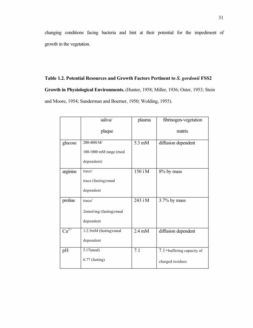
31
changing conditions facing bacteria and hint at their potential for the impediment of
growth in the vegetation.
Table 1.2. Potential Resources and Growth Factors Pertinent to S. gordonii FSS2
Growth in Physiological Environments. (Hunter, 1958; Miller, 1936; Oster, 1953; Stein
and Moore, 1954; Sunderman and Boerner, 1950; Wolding, 1955).
saliva/
plaque
plasma fibrinogen-vegetation
matrix
glucose 200-400ì M/
100-1000 mM range (meal
dependent)
5.3 mM diffusion dependent
arginine trace/
trace (fasting)-meal
dependent
150 ì M 8% by mass
proline trace/
2nmol/mg (fasting)-meal
dependent
243 ì M 3.7% by mass
Ca2+ 1-2.5mM (fasting)-meal
dependent
2.4 mM diffusion dependent
pH 5.17(meal)
6.77 (fasting)
7.1 7.1+buffering capacity of
charged residues

32
Periods of starvation under adverse conditions become a new reality for S.
gordonii in the vegetation. Proteolytic enzymes are postulated to be an intricate part in
the new paradigm for survival. Their indispensable role in other disease states has been
well documented. As the primary agent in development of the chronic inflammatory
disease, periodontitis, P. gingivalis produces a large array of proteolytic enzymes that
have subsequently been the most investigated aspect of the Gram-negative species, both
as a means of nutrient acquisition and as virulence factors in modulating the host immune
response (Travis et al., 2000). The severity of infections by S. pyogenes, an
epidemiological factor in streptococcal toxic shock syndrome (STSS) and necrotizing
fasciitis (NF) cases, has been linked to proteolytic factors. These findings implicate a
cysteine protease (SpeB) in host-pathogen interactions via regulation of the expression of
GAS virulence genes (Kansal et al., 2000).
Studies of proteolytic activities from viridans streptococci (with the exception of
S. pneumoniae) are scarce and their implications in the progression of SBE are non-
existent. Among members of the mitis group, an adhesin-degrading proteinase (Lamont
and Rosan, 1989) and IgA1-specific proteinase (Labib et al., 1978), both from S.
sanguinis, have been identified. Additionally, a trypsin-like endopeptidase from S. oralis
(Lo and Hughes, 1996) and a neutral, metallopeptidase from S. parasanguinis (Froeliger
et al., 1999) were uncovered. S. gordonii has representatives, with a 98 kDa serine
proteinase capable of hydrolyzing collagen and fibrinogen (Juarez and Stinson, 1999)
followed by a putative zinc metalloproteinase (EMBL T11548) and intracellular sortase
involved in protein maturation (EMBL Q9F0P0). S. gordonii FSS2 has exhibited both
cell-surface and secreted activities against chromogenic and fluorogenic substrates for

33
thrombin, collagenase, Xa, chymotrypsin, Hageman, plasmin, kallikrein and Ca2+-
dependent protease (Harty et al., 2000). Cell-free culture supernatant was also tested for
proteolytic activity against whole proteins. Figure 1.8a shows time-based degradation of
Aα and Bβ chains of human fibrinogen while the zymogram in Fig.1.8b reveals several
discrete activities capable of the broad hydrolysis of denatured collagen (gelatin). Both
these and several of the synthetic substrate extracellular activities are significantly
increased upon rise in the terminal pH of batch culture.
The hypothetical model for bacterial growth in the SBE vegetation supports a
state of nutrient stress and a general stress response (GSR) in which environmental cues
have dramatic effects on cellular activity. One such likely response is an exaggerated
drive for the acquisition of peptides and amino acids. Those endoproteinase activities that
have been partially characterized could potentially aid the remodeling and fragmentation
of the surrounding protein matrix. However, enzymes responsible for the turnover of
smaller peptides and amino acids which could benefit cellular transport systems and the
ADI pathway have not yet been identified. This dissertation describes the isolation,
characterization, cloning and sequence analysis of three peptidases secreted by S.
gordonii FSS2 that can potentially meet these challenges.

34
A. B.
Figure 1.8. Cell-free Endopeptidase Activity from S. gordonii FSS2 Culture.
A, Fibronogen degradation assay, 1ì g crude supernatant protein: 20 ì g human
fibrinogen, 37C; markers, crude supernatant, t(hrs)=0, 1, 2, 7 and 24. B, Gelatin
zymograph, 10% substrate in SDS-PAGE, 100 ì g crude supernatant protein, 37C 15hrs.

35
CHAPTER 2
NOVEL EXTRACELLULAR X-PROLYL DIPEPTIDYL-PEPTIDASE
(xPDPP) FROM STREPTOCOCCUS GORDONII FSS2: AN EMERGING
SUBFAMILY OF VIRIDANS STREPTOCOCCAL X-PROLYL DPPs1
1 Goldstein, J.M., A. Banbula, T. Kordula, J.A. Mayo, and J. Travis. 2001. Infection and Immunity. 69(9): 5494-5501Reprinted here with permission of publisher.

36
Abstract
Streptococcus gordonii is generally considered a benign inhabitant of the oral
microflora yet is a primary etiological agent in the development of subacute bacterial
endocarditis (SBE), an inflammatory state that propagates thrombus formation and tissue
damage on the surface of heart valves. Strain FSS2 produced several extracellular
aminopeptidase and fibrinogen-degrading activites during growth in culture. In this report
we describe the purification, characterization and cloning of a serine-class dipeptidyl-
aminopeptidase, an x-Pro DPP (Sg-xPDPP), produced in a pH-controlled batch culture.
Purification of this enzyme by anion-exchange, gel filtration and hydrophobic interaction
chromatography yielded a protein monomer of approximately 85 kDa, as shown by SDS-
PAGE under denaturing conditions. However under native conditions, the protein
appeared to be a homo-dimer based upon gel filtration and PAGE. Kinetic studies
indicated that purified enzyme had a unique and stringent x-Pro specificity that is
comparable to both the DPPIV/CD26 and lactococcal x-Pro DPP families. Nested PCR
cloning from an S. gordonii library enabled the isolation and sequence analysis of the
full-length gene. A 759-amino acid polypeptide with a theoretical molecular mass of
87,115 Da and calculated pI of 5.6 was encoded by this gene. Significant homology was
found with the PepX gene family from Lactobacillus and Lactococcus ssp. and putative
x-Pro DPPs from other streptococcal ssp. Sg-xPDPP may serve as a critical factor for the
sustaining bacterial growth in vivo and furthermore, aid in the proteolysis of host tissue
that is commonly observed during SBE pathology.

37
Introduction
Streptococcus gordonii, classified in the S. mitis group of oral streptococci, is a
well studied member of the viridans family of streptococci (53). These primary
colonizers serve a necessary role in the establishment of microbial communities that are
characteristic of healthy dental plaque. Although considered benign inhabitants of the
oral microflora, viridans members have been implicated in the systemic disease, infective
endocarditis (IE) (9, 26). The progression of this disease state requires: 1) trauma
(congenital or disease-related) to the endothelial valve surface such that it is predisposed
to colonization, 2) adhesion of organisms to the modified valve surface after their entry
into the bloodstream via the oral cavity and 3) the propagation of infected vegetations
consisting of a fibrin-platelet meshwork (50). Despite the uniform susceptibility of these
streptococci to β-lactam antibiotics and their lack of classical streptococcal virulence
factors, they can cause life-threatening disease and/or chronic inflammation with periods
of latency and several defined stages (10, 14)
The ability of these organisms to colonize biofilm surfaces at two distinct
microenvironments has prompted studies of their dynamic metabolism and patterns of
gene expression. Streptococcus sanguinis, studied as model for viridans pathogenesis,
expresses cell surface adhesins and PAAP (platelet aggregation-associated proteins) that
facilitate both colonization and thrombosis at the infected lesion (12, 15). In addition,
metabolic activities have been shown to dictate the availability of cell-surface receptors
manifested during the recruitment of planktonic bacteria to plaque or the attachment to a
new biofilm surface (4, 12). Upon entry into the bloodstream, bacteria undergo a shift in
pH from mildly acidic plaque (6.0-6.5) to the neutral pH (7.3) of the blood (41). In vivo
38
expression technology (IVET) was employed on the S. gordonii rabbit model of IE to
detect genes activated in the new environment with the alkaline shift in pH correlating
with enhanced bacterial growth, upregulation of the msrA oxidative stress gene (52) and
the induction of genes encoding carbohydrate metabolism enzymes, protein transporters
and cell surface proteins (22). The expression and secretion of glycosidase and peptidase
activities, as examined in pH-controlled batch cultures, was found to be down-regulated
by acid growth conditions and up-regulated by growth in a neutral pH environment
supplemented with serum (13) Chemostat growth also uncovered a pH-dependent
thrombin-like activity which was considered more important in the tissue model than on
tooth surfaces (32). This could reflect selective pressure for the organism to adapt novel
enzymatic mechanisms for a changing environment.
It is presumed that S. gordonii obtains necessary protein nutrients from salivary
glycoproteins in the oral cavity, while utilizing plasma proteins when growing on heart
surfaces. This use of plasma proteins by oral streptococci as carbon/nitrogen sources
would ostensibly benefit growth in the vegetation. Certainly, proteolytic and peptide
transport systems for viridans members have been described (1,7,17, 18, 25, 45, 54), and
it has been shown that small peptides can be imported into S. sanguinis while those
exceeding size limitation would require further hydrolysis by endo- and exopeptidases
present on the surface or secreted by these cells (8). One such activity that facilitates
these metabolic requirements, a dipeptidyl peptidase with Gly-Pro-pNa hydrolyzing
activity, has been detected in culture supernatant of S. gordonii (8, 19) yet has presently

39
remained undefined. In this report we describe the purification, characterization and
cloning of a novel extracellular x-Pro dipeptidyl-peptidase (Sg-xPDPP) that derives from
S. gordonii FSS2, a strain previously isolated from the bloodstream of an SBE patient.
Experimental Procedures: Materials
H-Gly-Pro-pNa, H-Arg-Pro-pNa, H-Ala-Ala-pNa, N-Suc-Gly-Pro-pNa, L-Pro-pNa, H-
Gly-Arg-pNa, H-Gly-pNa, Sar-Pro-Arg-pNa, Di-isopropyl fluorophosphate (DFP), Nα-
p-tosyl-L-lysine-chloromethyl ketone (TLCK), Nα-p-tosyl-L-phenylalanine chloromethyl
ketone (TPCK), iodoacetamide, O-phenanthroline, aprotinin, bestatin, apstatin, β-
mercaptoethanol, Gly-Pro-Arg-Pro-amide, sexual agglutination peptide, sleep inducing
peptide, substance P, des-Arg1 bradykinin, bradykinin, kallidin, lymphocyte activating
pentapeptide fragment and fibrin polymerization inhibitor were obtained from Sigma.
Pefabloc SC, phenylmethylsulfonyl fluoride (PMSF), 3,4-dichloroisocoumarin and E-64
from Boehringer Mannheim. Z-Ala-Pro-pNa, H-Ala-Ala-pNa, H-Ala-Phe-pNa, H-Lys-
pNa, H-Arg-pNa, protein kinase C fragment, Arg-Pro and fibrin inhibitory peptide from
Bachem. Protein kinase c peptide from American Peptide Company. Human RANTES,
MIP-1β and GM-CSF from PeproTech. H-Glu-(NHO-Bz) pyrollidide and diprotin A
from Calbiochem. α1 proteinase inhibitor (α1-PI) and α-2 macroglobulin were gifts from
Athens Research and Technology, Athens, Ga. Viridans streptococci strains were a gift
from Dr. Vincent Fischetti at The Rockefeller Institute.
Experimental Procedures: Methods
Bacterial Growth. S. gordonii FSS2 (previously S. sanguinis FSS2 (13,29) was
stored and maintained (at -80°C) as previously described (32). Frozen cells were

40
inoculated into autoclaved medium containing 20 g/L trypticase peptone (BBL), 5 g/L
yeast extract (Difco), 2 g/L NaCl, 0.1 g/L CaCl2, 4 g/L K2HPO4 and 1 g/L KH2PO4. 10
g/L glucose and 0.5 g /L L-arginine were sterile filtered and subsequently added. A static
culture (200 ml) was grown overnight at 37°C and used to inoculate a 4 L starter culture,
in turn used to inoculate a 15 L stirred batch culture which was grown in an atmosphere
of 5% CO2 and 95% N2 at 37°C with the pH held constant at 7.5 by addition of 5 M KOH
using a pH controller (Cole Parmer). Cultures were harvested in early stationary phase at
a point when the bacteria had metabolized all available glucose and the addition of base
had ceased.
Enzymatic Assay. Amidolytic activities of crude samples and purified protease
were measured using the substrate H-Gly-Pro-pNa (1 mM final concentration) in assay
buffer A (50 mM Tris, 1 mM CaCl2 , pH 7.8) at 37°C. Assays were performed in 0.1 ml
on 96 well plates using a thermostated microplate reader, and the release of p-nitroaniline
was measured at 405 nm. (Spectramax, Molecular Devices). Inhibition assays involved
pre-incubation of pure enzyme with inhibitor for 10 min at 37°C followed by
measurement of residual activity. Additional pNa substrates were treated identically.
Enzyme Purification. 14.5 L of cell-free culture filtrate was obtained after
centrifugation (20 min, 4 °C at 6000 x g) of the batch culture. Proteins in the filtrate were
precipitated over several hours at 4 °C with ammonium sulfate to a final concentration of
80% and the precipitate pelleted by centrifugation at 8000 x g for 40 min. Protein pellets
were obtained upon centrifugation as previously described. Pellets were resuspended in
200 ml of buffer A and dialyzed over 2 days (4 °C), with changes, against 40 volumes of
the same buffer. All column chromatography steps were performed at 4 °C except FPLC

41
separations that were done at room temperature. The dialyzed fractions was applied to a
DE52 (Whatman) column (2.5 x 30 cm, 150 ml) equilibrated with buffer A. The column
was washed with 3 column volumes of buffer A at 1ml/min. A gradient from 0 to 1M
NaCl in buffer A was applied over a total volume of 700 ml. Peak activity that eluted was
pooled and concentrated by ultrafiltration to 32ml, using a 10K membrane (Filtron). The
concentrated sample was divided into three equal fractions that were separately loaded
onto a SuperdexTM-75 (Amersham Pharmacia Biotech) HR 10/30 equilibrated with gel
filtration buffer (50 mM Tris, 200 mM NaCl, 1 mM CaCl2, 0.02% sodium azide, pH 7.8)
at 1 ml/min. Peak activities of all runs were combined and concentrated to 22 ml.
Ammonium sulfate was added to the fraction in order to give a final salt concentration of
1 M. This sample was then applied to a phenyl-Sepharose HP column (1.5 x 9 cm, 15 ml)
equilibrated with 200 mM potassium phosphate-1M ammonium sulfate (pH 7.5) at 0.5
ml/min. The column was washed with high salt buffer (120 ml) until reaching an A280
baseline and then subjected to a 250 ml gradient of 1.0 M to 0.0 M ammonium sulfate
utilizing 50 mM potassium phosphate (pH 7.5). Active fractions were pooled and
dialyzed against 20 volumes of buffer A over a 24 hr period. The dialyzed sample was
concentrated to 37 ml and then applied to a Mono Q HR 10/10 FPLC column (Amersham
Pharmacia Biotech) that was equilibrated with buffer A. The column was washed with
equilibration buffer until the base line stabilized. A linear gradient (0-500 mM NaCl in
buffer A) over 100 ml, was applied, and peak enzyme activity eluted between 200-250
mM NaCl. This activity was pooled, dialyzed against 10 volumes of buffer B (25 mM
Tris, 5 mM CaCl2, pH 7.4) and applied to a Cibacron blue Sepharose CL-6B column (1
ml) equilibrated with buffer B. Activity flowed through the matrix and was collected,

42
while bound, non-specific protein was eluted in a high salt buffer B. Final purification to
remove additional contaminants involved the use of a Gly-Pro Sepharose affinity column
(1 ml) Enzyme obtained from the previous step was loaded onto the column, previously
equilibrated with buffer B. It was then washed with 10 column volumes of this buffer and
a linear gradient (0-1 M NaCl in buffer B) over 30 ml was applied. Peak activity eluted
between 150-200 mM NaCl and was concentrated to 2.5 ml using a 10K MicrosepTM.
Protein Determination. Protein concentration was determined using a
bicinchoninic acid BCA reagent kit (Sigma) according to the manufacturer’s protocol.
Electrophoresis and Autoradiography. Enzyme purity was determined by
sodium dodecyl sulfate polyacrylamide gel electrophoresis (SDS-PAGE) using a 10%
acrylamide gel and the Tris-HCl/Tricine buffer system, according to Schägger and von
Jagow (49). Nondenaturing gel electrophoresis (48) in a 4-20% gradient gel and gel
filtration was used to obtain the MW of the native protein. To confirm the identity of Sg-
xPDPP as a serine protease, five micrograms of pure enzyme was incubated with 500nCi
of [1,3-3H] DFP for 30 min at 25 °C in 20 mM HEPES, pH 7.5. Cold DFP (10 mM final
concentration) was used to quench the reaction and labeled protein was run on SDS-
PAGE, followed by fixing in 2,5-diphenyloxazole, drying of the gel and exposure to x-
ray film (XAR, Eastman Kodak) over a 96 hr period. For amino-terminal sequence
analysis, Sg-xPDPP was resolved by SDS-PAGE, followed by electroblotting to a
polyvinylidene difluoride membrane using 10 mM 3-(cyclohexylamino) propanesulfonic
acid, 10% methanol, pH 11(30). The blot was air dried and the band was submitted for
sequencing.

43
Enzyme Kinetics and Specificity. Kinetic values were measured using H-Gly-
Pro-pNa at varying concentrations ranging from 20 µM to 10mM, with a fixed enzyme
concentration at 2 nM, in 100 mM Tris, pH 7.8, at 37 °C. Vmax and Km values were
obtained through Hyperbolic Regression Analysis (shareware from J.S. Easterby,
University of Liverpool, UK). Specificity studies utilized Sg-xPDPP incubated with 5 µg
of peptide in a 1:1000 molar ratio. Reactions occurred in 100 µL volumes with 100 mM
Tris, pH 7.8, at 37 °C for 2 hours. Digestions were terminated by acidification with 10 µL
of 10 M HCl, followed by centrifugation (10,000 x g, 5 min). The entire supernatant was
applied to a reverse phase high pressure liquid chromatography using a LC-18 column
(25 x 4.6 mm, 5µm)(Supelco) equilibrated with 0.1% TFA in HPLC grade water and
developed with an acetonitrile gradient (0-80% in 0.08% TFA over 50 min). Peaks were
manually collected and analyzed by mass spectrometry.
Mass Spectrometry. Peptides and native Sg-xPDPP were analyzed by MALDI-
TOF on a Hewlett-Packard G2030A mass spectrometer. The instrument was operated at
an accelerating voltage of 28 kV, an extractor voltage of 7 kV, and a pressure of 7 x 10⁻⁷
Torr. Samples were dissolved in sinapanic acid and ionized from the probe tip using a
nitrogen laser source. Calibration was performed using mixtures of peptides/proteins of
known molecular masses.
N-terminal and Internal Sequencing. Proteins and peptides were sequenced by
Edman degradation in a model Procise-cLC sequencer (PE Biosytems, Foster City, CA)
operated using the manufacturer’s protocol. For internal sequence, proteins were in-gel
digested with trypsin (Promega, sequence grade) and the peptides extracted (46), with
their masses being determined by reflectron MALDI-TOF mass spectrometry using a

44
Bruker Daltonics ProFlex instrument (42). Selected peptides were sequenced by the
Edman Degradation method.
Cloning of S. gordonii xPDPP Gene . DNA from S. gordonii was purified using
the Purgene DNA Isolation Kit (Gentra, Minneapolis, MN) according to manufacturer’s
instructions. The N-terminal fragment of xPDPP gene was obtained using a PCR
approach and degenerate primers designed to the N-terminal (MRYNQY) and the
internal (NVIDWL) peptides. Two putative N-terminal primers and one internal primer
(all containing BamHI sites) were synthesized (nt-1-dpp;
5’-CGCGGATCCATGCGNTAYAAYCARTA-3’, nt-2-dpp;
5’-CGCGGATCCATGAGRTAYA AYCARTA-3’ and int-1-dpp;
5’-CGCGGATTCARCCARTCDATNACRTT-3’, respectively). PCR was performed
using the Pwo DNA polymerase (Roche Molecular Biochemicals, Indianapolis, IN), 1 µg
of S. gordonii DNA and 500 ng of the primers (94ºC for 1 min, 65ºC for 1 min, 72ºC for
1 min, 32 cycles). A single 965 bp-long PCR product was obtained, gel-purified, digested
with Bam HI, subcloned into the Bam HI site of pUC19 and sequenced. The obtained
sequence was used to search the unfinished S. gordonii database available at The Institute
of Genomic Research (TIGR) (ftp://ftp.tigr.org/pub/data/s_gordonii/). Several
overlapping contigs were found and this approach resulted in the identification of a 2280
bp-long open reading frame encoding S. gordonii xPDPP. Subsequently, two PCR
primers encoding the N-and C-terminus of DPPIV were synthesized
(5’-AGTGGATCCATGCGTTACAACCAGTATAG-3’ and 5’-TTTGGA
TCCTCAAGATTTCTTGTGAGGC-3’) and used in the PCR to obtained full-length
DNA fragment encoding DPPIV. The 2298 bp-long PCR product was gel-purified,

45
digested with Bam HI, subcloned into the Bam HI site of pUC19 and sequenced on both
strands. The obtained sequence was deposited in GeneBank (accession number
AY032733).
Southern Blot Analysis. DNA from S. gordonii PK48, DL1, PK2585, S.
parasanguinis PK2584, S.mitis J27, S. oralis J21, PK34 and S. salivarius ATCC 27945
and was purified using the Purgene DNA Isolation Kit (Gentra, Minneapolis, MN)
according to manufacturer’s instructions. Four µg of bacterial DNA was digested with
each of the restriction enzymes BamHI, EcoRI, HindIII or PstI overnight at 37° C.
Digestion products were separated in 0.7% agarose gels using Standard procedure
(Sambrook). Gels were soaked in denaturation solution (0.5 M NaOH, 1.5 M NaCl) for
30 min followed by neutralization solution (1.5 M Tris pH 7.5, 1.5 M NaCl) for 1 hour.
Separated DNA was transferred onto a nylon membrane (Amersham-Pharmacia Biotech)
by capillary transfer and subsequently fixed by cross-linking (Stratalinker, Stratagene
Inc.). Membranes were prehybridized in 0.5 M phosphate buffer pH 7.2, 7% SDS and 1
mM EDTA at 50C for 1 hour, 32P-labelled DPP cDNA probe was added and incubated
overnight. After hybridization, non-specifically bound radioactivity was removed by a
single wash in 2xSSC and 0.1% SDS at room temperature for 20 min followed by two
subsequent washes in 0.5xSSC and 0.1%SDS at 50C for 20 min. Membranes were
exposed to X-ray films using enhancing screens at -80C for 1-4 hours.

46
Results
Growth and Culture Activity. Preliminary experiments indicated the presence of
an extracellular Gly-Pro-pNa activity in culture media (Fig. 2.1). Detection of this
amidolytic activity in cell-free filtrate was observed during growth in culture
supplemented with 50 mM glucose and 3.5 mM arginine, and was dependent on a pH
maintained in the range of 6.0-8.0. Growth and activity from a 1 L pH-controlled (pH
7.5) batch culture were monitored at hourly intervals. Detection of cell-associated Gly-
Pro-pNa activity occured early in logarithmic phase, as it paralleled the growth curve,
and continued to increase until early stationary phase. Cell-free activity appeared later in
log phase and sharply increased during early stationary phase. The profiles of both
activity forms began to diverge during later stationary phase, between the 8th and 10th
hours of growth. As cell-associated activity decreased by approximately 25%,
extracellular activity steadily increased by roughly 15%. The trend of enzyme shedding
continued beyond the shown timepoints (not shown) at a cost to activity found on the
cell. These observations occurred at a time when bacteria had metabolized all available
glucose and the addition of base had ceased. This phenomenon may be the result of the
permeability in the cell envelope, cell wall turnover and/ or peptidoglycan rearrangement
due to autolysin activities.
Further growth experiments were conducted under similar conditions but varied in
culture environments with respect to pH control, carbohydrates, arginine, Ca2+, strain
type and addition of serum (Fig. 2.2). Measurements were made at the 10th hour of
growth when stationary phase was evident by lack of sugar consumption. The activity
profiles during varied pH-controlled conditions provided evidence for a pH-dependent

47
secretion mechanism. Cell-associated Gly-Pro-pNa activity was detected under all
conditions, with the least activity measured in culture devoid of pH control (final pH of
4.4) and greatest activity found in pH 7.0-controlled culture. Cultures at pH 6.3, 7.5 and
8.0 were similar. Appreciable extracellular activity was evident at pH 7.0 and 7.5, and at
roughly equal values. Although growth at pH 7.0 accounted for the greatest total Gly-
Pro-pNa activity, cells at pH 7.5 contributed their highest proportion of total activity
(51%) to the secreted form. The remaining cultures were controlled at pH 7.0 for
standardization and optimum enzyme expression. Excess supplemented arginine (50 mM)
provided the greatest level of cell-associated activity while it appeared to inhibit the
secretion of protease. Under this condition, less than 2% of the extracellular form was
found versus the 3.5 mM arginine culture. The 25 mM glucose culture was unable to
reach the appropriate stationary phase in the 10 hr. timeframe and thus cannot be
accurately compared. The secretion of enzyme seemed dependent on the addition of Ca2+
(mM) since culture without any exogenously supplied to the media could only produce
cell-surface enzyme. The addition of 25% fetal bovine serum to the starting media
provided a decrease in total activity with no significant difference in the proportion of
secreted activity. Mice-passaged S. gordonii FSS2 assays showed that these cells
produced slightly less total Gly-Pro-pNa activity while a substantial decrease in
extracellular activity (10% of total) occurred. Although, an overall decrease in total
activity was seen in galactose-supplemented cultures, this type of sugar neither repressed
nor activated the release of activity from cells, indicating the absence of a glucose
repressor.

48
0
0.5
1
1.5
2
0 2 4 6 8 10
t(hrs)
OD
560
0
10
20
30
40
50
mO
D/m
in gly-pro-pNa
Fig. 2.1. S. gordonii FSS2 growth and activity curve from a pH 7.5-controlled
culture supplemented with 50 mM glucose and 3.5 mM arginine. Samples (1 ml)
were removed from culture at fixed timepoints and cells were removed from media by
centrifugation (5 min, 4 ºC, 13,000 x g) followed by 2x wash and resuspension into initial
volume with non-supplemented media. Symbols: , whole culture; , cell-free culture
fluid; , whole cells. Assays were performed as previously described using 30ì L of
sample.

49
Fig. 2.2. Extracellular and cell-surface Sg-xPDPP activities under variable growth
conditions. Samples (1 ml) were removed from culture at early stationary phase and
cells were removed from media by centrifugation (5 min, 4 ºC, 13,000 x g) followed by
2x wash and resuspension into initial volume with non-supplemented media. Symbols: ,
cell-free culture fluid; , whole cells. Assays were performed as previously described
using 30ì L of sample. Total cell and cell-free activities for 1L cultures were measured as
OD/min and adjusted for cell density.

50
Enzyme Purification. Earlier growth experiments indicated the presence of an
extracellular Gly-Pro-pNa activity in culture media. Detection of this amidolytic activity
in cell free filtrate was dependent upon growth maintained in a pH range of 6-7.5
(unpublished results). In this report, maximum activity was achieved using a 14.5 L batch
culture held at pH 7.5 by addition of base and harvested during early stationary growth.
An 80% ammonium sulfate precipitation concentrated extracellular proteins to a
workable volume despite the large decrease in enzymatic yield. The following
chromatographic steps (DE52 anion-exchange, S-75 gel filtration and Phenyl Sepahrose
hydrophobic chromatography) successfully removed high and low molecular weight
contaminants, media pigments and peptides/amino acid away from the Gly-Pro-pNa
activity, with a minimal drop in yield. The Mono-Q step increased specific activity by 16
fold, although the yield was reduced fifty percent. CB Sepharose chromatography served
as a negative step, enabling activity to flow through the matrix while higher affinity
proteins and GAPDH(40) remained bound. The final step, utilizing Gly-Pro Sepharose,
resulted in a single, sharp peak of purified protein. Concentrated fractions corresponded
to high specific activity, a greater than 6000-fold purification and 1% yield from the
starting batch culture (Table 2.1).

51
Table 2.1 Purification of S. gordonii x-Pro DPP
aBased on enzymatic activity using H-Gly-Pro-pNa, where 1 unit= 1 µmol of pNa released per sec.bFPLC, fast protein liquid chromatography
Step Volume TotalActivity
a
Total Protein SpecificActivity
Purification
Yield
ml Unitsa mg units/mg fold %
Culture filtrate 14,500 134,117 58,000 2.3 1 100
Ammonium sulfate precip.
320 47,663 9504 5.0 2 36
DE52 anion exchange
32 41,717 3290 12.7 6 31
Superdex 75 22 38,063 2168 17.6 8 28
Phenyl Sepharose 37 35,708 1125 31.7 14 27
Mono-Q, FPLCb 10 17,063 35 487.5 211 13
CB Sepharose, FPLC
15 4180 7 593.0 258 4
Gly-Pro Sepharose, FPLC
3 1370 0.97 14,153 6153 1

52
Physical Properties. SDS-PAGE analysis of the purified enzyme (Figure 2.2)
showed a single protein band, as judged by both Coomassie and silver staining (latter not
shown) with an approximate mass of of 85 kDa. Analysis of the protein by MALDI
revealed a laser intensity peak that corresponded to a mass of 86,977.6 Da. Protein
analyzed on a 4-20% native gradient gel and Superdex 200 gel filtration indicated a
molecular weight between 150-200 kDa, providing evidence for homodimer formation in
the native state. This was ablated by heat treatment, SDS denaturation, and MALDI
analysis. This suggested the dimer was stabilized by weak ionic/ electrostatic conditions.
Isoelectric focusing (data not shown) produced a pI of 4.9 for the native protein. The Gly-
Pro-pNa activity was optimum at pH 8.0 with activity detected over a broad phosphate
buffer range of 5.0 to 10.0. The optimum temperature for Gly-Pro-pNa hydrolysis was
determined to be between 45-50 °C. The enzyme in its pure form was unstable over a 15h
period at 37 °C and 25 °C (complete loss of activity,) and 4 °C (75% loss of activity)
while storage at –20 °C over several months exhibited minimal activity loss.
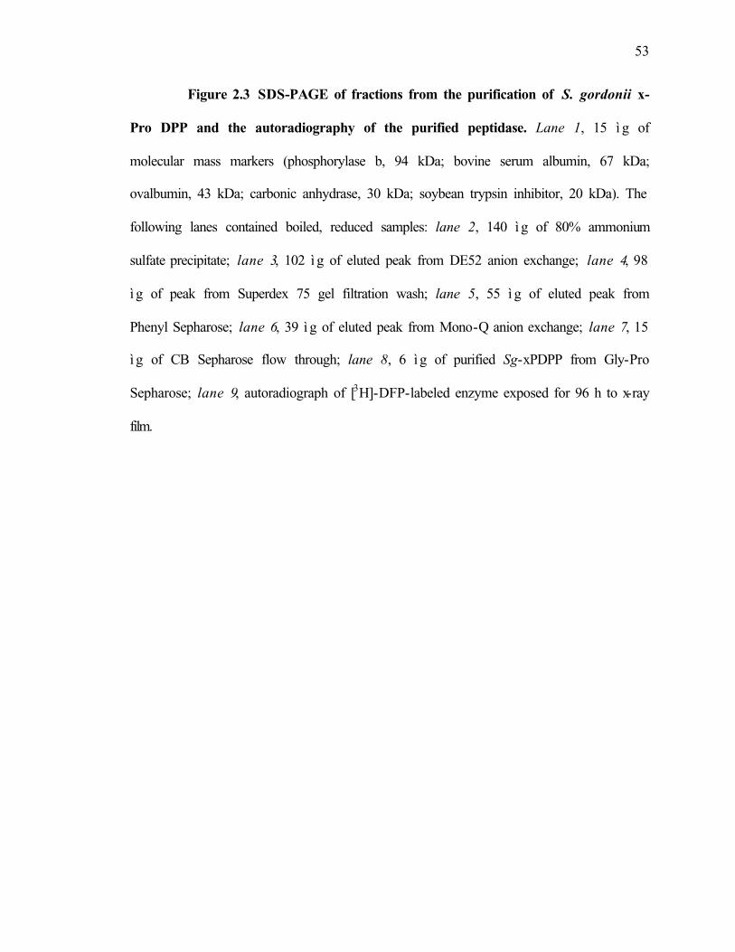
53
Figure 2.3 SDS-PAGE of fractions from the purification of S. gordonii x-
Pro DPP and the autoradiography of the purified peptidase. Lane 1, 15 ì g of
molecular mass markers (phosphorylase b, 94 kDa; bovine serum albumin, 67 kDa;
ovalbumin, 43 kDa; carbonic anhydrase, 30 kDa; soybean trypsin inhibitor, 20 kDa). The
following lanes contained boiled, reduced samples: lane 2, 140 ì g of 80% ammonium
sulfate precipitate; lane 3, 102 ì g of eluted peak from DE52 anion exchange; lane 4, 98
ì g of peak from Superdex 75 gel filtration wash; lane 5, 55 ì g of eluted peak from
Phenyl Sepharose; lane 6, 39 ì g of eluted peak from Mono-Q anion exchange; lane 7, 15
ì g of CB Sepharose flow through; lane 8, 6 ì g of purified Sg-xPDPP from Gly-Pro
Sepharose; lane 9, autoradiograph of [3H]-DFP-labeled enzyme exposed for 96 h to x-ray
film.

54
1 2 3 4 5 6 7 8 9
kDa
94
67
43
30
20

55
Enzyme Specificity. Of the fourteen chromogenic endo- and aminopeptidase
substrates tested on purified enzyme (Table 2.2), only three (H-Gly-Pro-pNa, H-Ala-
Pro-pNa and H-Arg-Pro-pNa) were rapidly hydrolyzed. Weaker activity (< 10%) was
detected against H-Ala-Ala-pNa. A maximum rate of hydrolysis occurred against H-
Gly-Pro-pNa and Michaelis-Menten kinetics were measured using this substrate. A Vm
of 15.2 µmol min-1 and Km of 378 µM was observed with 2 nM of enzyme. To obtain
further information on cleavage specificity, various peptides were tested as substrates
for Sg-xPDPP (Table 2.3). Proteolysis was confirmed on peptides between 4 to 17
residues with a Proline residue in the P1 position. A major impediment for cleavage was
the presence of Pro in the P1’. There were no additional restrictions for amino acids
present in the P2 and P1’sites. The general formula for proteolysis was deduced as NH2-
Xaa-Pro(Ala)-↓-Yaa-(Xaa)n, where Yaa could represent any residue except Pro. The
combined data for synthetic and peptide substrates fit a profile that was consistent with
previous data for Gram-positive bacterial x-Pro-dipeptidyl aminopeptidases (11, 20, 21,
28, 33, 34, 36) and Gram-negative DPPIV families (2, 56). Extended time incubation
with peptides failed to yield additional peptide fragments, indicating the absence of
endopeptidase activity or contaminating aminopeptidases. Proteolysis of CC
chemokines (RANTES and MIP-1β), which presented an unblocked x-Pro N-terminus,
was not observed under identical conditions as peptide substrates. The inability of Sg-
xPDPP to catalyze endo-specific proteolysis was tested on whole protein substrates
(azocasein,

56
Table 2.2 Relative amidolytic activity of S. gordonii x-Pro DPP against
various substrates
1Activity against H-Gly-Pro-pNa hydolysis taken as 100
Substrate (pNa) Relativeactivity1
(%)H-Gly-Pro 100
H-Ala-Pro 81
H-Arg-Pro 63
H-Ala-Ala 8
H-Gly-Arg 0
H-Ala-Phe 0
H-Arg 0
H-Lys 0
H-Ala 0
H-Gly 0
L-Pro 0
Suc-Gly-Pro 0
Z-Ala-Pro 0
Sar-Pro-Arg 0

57
Table 2.3. Cleavage specificity of S. gordonii x-Pro DPP on peptide substrates
↓-cleavage site
Substrate Sequence
Sexual Agglutination Peptide Arg-Gly-Pro-Phe-Pro-Ile
Protein Kinase C Fragment Arg-Phe-Ala-Arg-Lys-Gly-Ala-Leu-Arg-Gln-Lys-Asn-Val
Substance P Arg-Pro-↓-Lys-Pro-↓-Gln-Gly-Leu-Met
Bradykinin Arg-Pro-Pro-Gly-Phe-Ser-Pro-Phe-Arg
des Arg1 Bradykinin Pro-Pro-↓-Gly-Phe-Ser-Pro-Phe-Arg
Dipeptide A Arg-Pro
Sleep Inducing Peptide Trp-Ala-↓-Gly-Gly-Asp-Ala-Ser-Gly-Glu
Fibrin Polymerization Inhibitor Gly-Pro-↓-Arg-Pro
Fibrin Inhibitory Peptide Gly-Pro-↓-Arg-Pro-Pro-Glu-Arg-His-Gln-Ser
Protein Kinase C Peptide Gly-Pro-↓-Lys-Thr-Pro-Gln-Lys-Thr-Ala-Asn-Thr-Ile-Ser-Lys-Phe-Asp-Cys
Kallidin Lys-Arg-Pro-Pro-Gly-Phe-Ser-Pro-Phe-Arg
Lymphocyte Activating Pentapeptide
Leu-Pro-Pro-Ser-Arg

58
gelatin, collagen type IV, fibrinogen). Internal digestion was not observed after standard
incubations and SDS-PAGE analysis (data not shown).
Inhibition and Activation Studies. Autoradiography of the purified Sg-xPDPP
radiolabeled with [1,3-3H] DFP (Figure 1, lane 9) positively identified the protease as a
member of the serine class. Further studies with class-specific inhibitors (Table 2.4)
supported this assignment. Sensitivity was observed to serine protease inhibitors
(Pefabloc, PMSF, and 3,4 Dichloroisocoumarin), while cysteine-class (iodoacetamide
and E64) and metallo-class (EDTA and 1,10-orthophenanthroline) had little or no effect
on activity. Compounds specific for Aminopeptidase B and Leucine Aminopeptidase
(Bestatin) and Aminopeptidase P (Apstatin) were not inhibitory. However, Sg-xPDPP
was highly sensitive to the DPPIV/CD26 specific inhibitors, H-Glu-pyrollidide and
Diprotin A(Ile-Pro-Ile), as evidenced by IC50 values that approximated 100 µM. Human
plasma inhibitors, α-1-proteinase inhibitor and α-2-macroglobulin, had no effect on
enzymatic activity. Sg-xPDPP activity was also inhibited by the heavy metal ions Zn2+
and Co2+, at 0.5 and 5 mM, respectively. Complete inactivation occurred in the presence
of 1% SDS detergent. Activity was unaffected by reducing agents and urea. Gly-Pro-Arg-
Pro-amide served as a competitive inhibitor while the dipeptide, Gly-Pro, provided no
inhibition. In contrast, Gly-Gly moderately stimulated pNa hydrolysis.

59
Table 2.4. Inhibition profile of S. gordonii x-Pro DPP
Inhibitor Concentration % ResidualActivity
DFP 10 mM 0Pefabloc 1 mM 55PMSF 5 mM 513,4-Dichloroisocoumarin
5 mM 0
TLCK 10 mM 96TPCK 10 mM 100Aprotinin 600 µM 97Bestatin 250 µM 99Apstatin 1 mM 100H-Glu-pyrollidide 100µM 56
1 mM 11Diprotin 100 µM 47
1 mM 10E-64 1 mM 85Iodoacetamide 1 mM 100EDTA 1 mM 1061,10-Orthophenanthroline
10 mM 99
L-Cysteine 5 mM 100β-mercaptoethanol 5 mM 87Gly-Gly 5 mM 117Gly-Pro 10 mM 105Gly-Pro-Arg-Pro-amide
4 mM 62
Co2+ 500 µM 57Zn2+ 5 mM 42Ca2+ 5 mM 103SDS 1% 7Urea 2 mM 100

60
Sg-xProDPP Sequence Analysis and Gene Structure . N-terminal and internal
sequences facilitated the construction of degenerate primers. PCR against an FSS2 library
resulted in the isolation of a 960bp fragment that represented the N-terminus of the
protein. This product was used to search the genomic clone of a relatively
uncharacterized S. gordonii strain in the Unfinished Microbial Genomes database, TIGR.
An ORF of 2280bp was identified and was found to correspond to the Sg-xPDPP based
upon the conservation of protein-derived peptides. Primers were designed from the
electronic sequence and a PCR approach was used to obtain a full-length DNA fragment
encoding DPPIV. A 759-amino acid polypeptide with a theoretical molecular mass of
87,115 Da and calculated pI of 5.6 was encoded by this ORF. This is in agreement with
the observed experimental mass while the native state dimer is produced by association
of two identical monomers. There is no evidence for post-translational modifications of
the gene product. The homology search (Figure 2.3) performed using NCBI TBLASTN
tool against EMBL, DDBJ, GenBank and PDB databases indicated that Sg-xProDPP is a
new member of the X-Pro dipeptidyl-peptidase S15 family grouped in the SC clan of
serine peptidases (5). This family has previously consisted of peptidases that derive from
lactic acid bacteria (Lactobacillus ssp. and Lactococcus ssp.), known as PepX (51). Sg-
xProDPP shares the closest homology with putative genes uncovered in Streptococcus
pneumoniae (72% identity) and Streptococcus mutans (57% identity) while maintaining
only 48% and 34% identity with Lactococcus and Lactobacillus, respectively. Sg-
xProDPP displays evolutionary divergence from bacterial members of the DPPIV (S9)
family, Flavobacterium meningosepticum (10% identity) and Porphyromonas gingivalis
(11% identity), although sharing functional similarity. The active site serine has been

61
identified in the PepX gene of L. lactis as Ser 348 inside the motif GKSYLG (6). Thus,
by analogy Ser 347 of Sg-xProDPP is likely to be the active site residue. Streptococcal
xProDPPs and PepX have nearly identical residues flanking the catalytic motif, except
for a hydrophobic amino acid replacing a charged Lys in the second position. Based upon
homology to SC peptidases with Ser-Asp-His active site nomenclature, Asp 574/576 and
His 733 of Sg-xProDPP are likely candidates for the catalytic triad (44). A computer
assisted search for protein localization predicted neither signal sequence sites nor
transmembrane regions in the protein (37). Although lacking an export signal and
predicted to be cytoplasmic, activity detected on the cell surface and within supernatant
revealed over 50% of activity to be extracellular at pH 7.5.
Southern Blot analysis revealed Sg-xPDPP to be a single copy gene (Fig. 2.5). A
2.3 kb ORF was examined in the electronic database for the presence of promoter
elements and additional genes. Three open reading frames were revealed and designated
ORF 1 (760 codons), ORF 2 (277 codons) and ORF 3 (236 codons). Sg-xPDPP is coded
by ORF 1, while ORFs 2 and 3 are oriented upstream on complementary strands in
reverse directions. A ribosome-binding site was identified 5 bp upstream of the start
codon. The putative transcription start site was found 33 bp from the start Met as well as
the TATA (-10 box) and –35 region promoters. Previous growth experiments with
glucose and galactose revealed that the metabolism of glucose did not alter the total
activity profile. The absence of sequence indicating a glucose repression operon
corresponds with this observation. ORF 2 is 118 bp upstream from the start codon of
ORF 1. The protein specified by ORF 2 has close to 70% identity with a hypothetical
30.9 kDa protein found in the PepX 5’ region of L. lactis (31, 38) and shares significant

62
homology to the glycerol uptake facilitator (Glpf) from S. pneumoniae (47). ORF 3 is
1091 bp upstream from ORF 1 and encodes a 236 aa protein with no significant
homology to proteins in the aforementioned databases.
Fig. 2.4. Multiple sequence alignment of S. gordonii x-Pro DPP (Sg xPDPP),
putative streptococcal Pep X genes and other bacterial homologues. Sequences of
Sg-xPDPP (Sgord) deduced from FSS2 genome, putative x-Pro DPPs from gene products
of Streptococcus pneumoniae (Spneu) and Streptococcus mutans (Smut), cloned x-Pro
DPP from Lactococcus lactis lactis 763 (Llac763), Lactococcus lactis cremoris (Lcrem),
Lactobacillus delbrueckii lactis (Ldel17290), and cloned DPPIV from Flavobacterium
meningosepticum (Fmenin) and Porphyromonas gingivalis (Pging) were aligned using
the ClustalW multiple sequence alignment tool according to homology modeling (grey
boxes indicate similarity and black boxes indicate identity). The putative catalytic
residues (serine, aspartic acid and histidine) are denoted (*). The arrow marks a
consensus motif flanking the active site serine in members of the x-Pro DPP family (Pep
X).

63
Sgord 1 MRYNQYSYTK...ASEEVMLDELARLGFTIQ.....TTNSPKENLHHFLQKILFRYQDVNSpneu 1 MRFNQYSYIN...FPKENVLSELKKCGFDLQ.....NTANHKDSLETFLRRFFFTYQDTNSmut 1 MKYNQYSYIG...TSVSQAEKELKELGFQIS.....SQKTNKANLATFVSQVYFHNPDKDLlac763 1 MRFNHFSIVD...KNFDEQLAELDQLGFRWS.....VFWDEKKILKDFLIQSPSD.....Lcrem 1 MRFNHFSIVD...KNFDEQLAELDQLGFRWS.....VFWDEKKILKDFLIQSPSD.....Ldel7290 1 MKYNQYAYVE...TSPEKATEELLAINFLPENYSSLSFSELLAVLTGNVLAEATTRQAKDFmenin 1 MKKKIFSLLS..IAVVAFHGLSAQEITLDKIYSGQYRAKGIS.GIASLNDGENYATIEP.Pging 1 MKRPVIILLLGIVTMCAMAQTGDKPVDLKEITSGMFYARSAGRGIRSMPDGEHYTEMNRE
Sgord 53 .YVLSSWVADQKTDLLTFFQSD..KQLTEEVFYTVALQVLGFAPFVDFDD..VTAFCKEISpneu 53 .YPLSILAADKKTDLLTFFQSE..DELTADIFYTVAFQLLGFSYLVDFED..SDVFRKETSmut 53 .DVFKSIIADSQTDLATFLHSD..RELTEEIFYTIALQLLEFTPYIDFDE..AKTFIKHSLlac763 48 ...MTALQATAELDVIEFLKSS..IELDWEIFWNIALQLLDFVPNFDFEIGKAFEYAKNSLcrem 48 ...MTDLQATAELDVIEFLKSS..IELDWEIFWNIALQLLDFVPNFDFEIGKAFEYAKNSLdel7290 58 .AKLAEFAVDDQTDLAAFLLDTP.TAITASQFANVALQLLGYHPNYDYSLTDPLTCGKKHFmenin 57 .TGIAKYSYKTSQKEKNIVDGS.........FQGYTFSNDESKILLQKSS..Q.SIYRHSPging 61 RTAIVRYNYASGKAVDTLFSVERARECPFKQIQNYEVSSTGHHILLFTDM..E.SIYRHS
Sgord 108 HFPITYG.....NILENLYQLLNTRTKLGNTLIDQLVSEGFIPE...SNDYHFFNGKSLASpneu 108 GFPIIYG.....DLIENLYQLLNTRTKKGNTLIDQLVSDGLIPE...DNDYHYFNGKSLASmut 108 HFPIIFQPK...HFLLNFYQLLGTRTKNGMTLIDKLVSQGFLPA...DNHYRYFNGKSLALlac763 103 NLPQIEAEMTTENIISAFYYLLCTRRKTGMILVEHWVSEGLLPL...DNHYHFFNDKSLALcrem 103 NLPQIEAEMTTENIISAFYYLLCTRRKNGMILVEHWVSEGLLPL...DNHYHFFNDKSLALdel7290 116 ALPAFKDLTSKEELIFTFYRLLNTRSKNGQILLDVMAGKGYFTQFWGEGKFMFFNGKSLPFmenin 104 FLGKFEVK....DLKSRTVVSLNN...ANWIQEPKFSPDGSKVA........FIADNNLFPging 118 YRAAVYDY....DVRRNLVKPLSEH..VGKVMIPTFSPDGRMVA........FVRDNNIF
Sgord 160 TFSSHEAIREVVYVESRVDTDGDGKPDLVKVSIIRPS...YEGQVPAVMTASPYHQGTNDSpneu 160 TFSNQDVIREVVYVESRVDTDQKGLSDLVKVSIIRPR...FDGKIPAIMTASPYHQGTNDSmut 162 TFDTNALIREIVYVEAPLDTDKDGQLDLIKVNIIRPQ...TKAKLPVVMTASPYHQGTNDLlac763 160 TFDSSLLEREVLWVESPVDSEQRGENDLIKIQIIRPK...STEKLPVVMTASPYHLGINDLcrem 160 TFDSSLLEREVLWVESPVDSEQRGENDLIKIQIIRPK...STEKLPVVMTASPYHLGINDLdel7290 176 VFDTSQVIREVVYVQSDLDTDGDGKGDLLPVTVFRPVESQDQLKVPALYTASPYFGGIIDFmenin 149 YQDLNTGKITQITTDGKKNEIINGLGDWVYEEEFGHA......DYYQWNKAGDALVFVRFPging 164 IKKFDFDTEVQVTTDGQINSILNGATDWVYEEEFGVT......NLMSWSADNAFLAFVRS
Sgord 217 KASDKALHNMNVDLSCKNPRTITVQESSIQTIEPQGQASLVEKAEEKLGHIG.SYTLNDYSpneu 217 KASDKALYKMEGELEVKLPHKIELEKPQLNLVQPQGKAELIAEAEEKLTHINSSYTLNDYSmut 219 KGADKKLHQMEGELTVKKAATITVTDSHFQALQVPSSNLPISPAQESFSYID.SYTLNDYLlac763 217 KANDLALHDMNVELEEKTSHEIHVEQKLPQKLSAKAKELPIVDKAPYRFTHGWTYSLNDYLcrem 217 KANDLALHDMNVELEEKTSHEIHVEQKLPQKLSAKAKELPIVDKAPYRFTHGWTYSLNDYLdel7290 236 NVKTNHNVDENLTDATTWTNPKYVAKPLVKSPAPSDQDVPATELATGQSSYG....LNEYFmenin 203 DERKVPEINIPIYYQNLYPKLMTYKYPKAGEENSAVTAYLYQLSSGKSAQLNFG.SSEKYPging 218 DESAVPEYRMPMYEDKIYPEDYTYKYPKAGEKNSTVSLHLYNVADRNTKSVSLP.IDADG
Sgord 276 LLPRGFANLYVSGVGTKDSEGMMTSGDYQQIEAYKNVIDWLNGRCRAFTDHTRQREIKATSpneu 277 FLPRGFANLYVSGVGTKDSTGFMTNGDYQQIEAYKNVIDWLNGRCRAFTDHTRQRQVKADSmut 278 FLARGFANIYVSGVGTKDSDGFMTSGDYAQIESFKAVIDWLNGRTTAYTSHKRDKRVMADLlac763 277 FLTRGFASIYVAGVGTRSSDGFQTSGDYQQIYSMTAVIDWLNGRARAYTSRKKTHEIKASLcremx4 277 FLTRGFASIYVAGVGTRSSDGFQTSGDYQQIYSMTAVIDWLNGRARAYTSRKKTHEIKASLdel7290 292 LLARGFASVFSGAIGNRHGDGIRITGSPEETISQKEVIEWLTGDRVAYTDRTRRFETKASFmenin 262 YIPQLFQTNANDEIVVATANRHQNKVDLLKVNTKTAAVSKLFTETDNAWIETDNLT.MEFPging 277 YIPRIAFTDNADELAVMTLNRLQNDFKMYYVHPKSLVAKLILQDMNKRYVDSDWIQALKF

64
*
Sgord 336 WSNGKVATTGISYLGTMSNGLATTGVDGLEVIIAEAGISSWYNYYRENGLVTSPGGYPGESpneu 337 WSNGKVATTGLSYLGTMSNGLATTGVDGLEVIIAEAGISSWYNYYRENGLVTSPGGYPGESmut 338 WTNGLVATTGKSYLGTMSTGLATTGIKELKVIIAESAISSWYDYYRENGLVCSPGGYPGELlac763 337 WANGKVAMTGKSYLGTMAYGAATTGVEGLELILAEAGISSWYNYYRENGLVRSPGGFPGELcrem 337 WANGKVAMTGKSYLGTMAYGAATTGVEGLEVILAEAGISSWYNYYRENGLVRSPGGFPGELdel7290 352 WCSGNVGMTGRSYLGTLQIAIATTGVKGLKTVVSEAAISSWYDYYREHGLVVAPSECQGEFmenin 321 LDDNSFLWASERDGHRHLYWYDAAGKLKKQVSKGDWEIINYYGYNPKTKEVYIQTTEKGSPging 337 TAGGGFAYVSEKDGFAHIYLYDNKGVMHRRITSGNWDVTKLYGVD.ASGTVFYQSAEESP
Sgord 396 DFESLTELTYSRNLLAGEYLRHNQAYQAYLDQQRKDLERETGDYNQFWHDRNYLIHADKVSpneu 397 DFDSLAELTYSRNLLAGDYIRGNEAHQADLEKVKAQLDRKTGDYNQFWHDRNYLLNAHKVSmut 398 DIDVLTELTYSRNLAAGDYLRNNAQYQKMLAEQVKQIDRTSGDYNQFWQDRNYLPHAHKILlac763 397 DLDVLAALTYSRNLDGADFLKGNAEYEKRLAEMTAALDRKSGDYNQFWHDRNYLINTDKVLcremx4 397 DLDVLAALTYSRNLDGADFLKGNAEYEKRLAEMTAALDRKSGDYNQFWHDRNYLINTDKVLdel7290 412 DMDKLAEVCQS.NLWDGGNFTAKKAYEAEQAELLAAQDRATGQYSDFWESRNYRHHTDGIFmenin 381 INKVVSKLNIN..................TGKTQLLSNAEGNNSAAFSKTFNYFINTSSTPging 396 IRRAVYAIDAK..................GRKTKLSLNV.GTNDALFSGNYAYYINTYSS
Sgord 456 KAEVVFTHGSQDWNVKPLHVYNMFHALPAH.IKKHLFFHNGAHVYINNWQSIDFRESMNASpneu 457 KAEVVFTHGSQDWNVKPLHVYQMFHALPTH.IHKHLFFHNGAHVYMNNWQSIDFRESINASmut 458 KAHVVYTHGLQDWNVKPNQVYYIFNALPEE.IQKHIFLHQGQHVYMHNWQSIDFRESMNALlac763 457 KADVLIVHGLQDWNVTPEQAYNFWKALPEG.HAKHAFLHRGAHIYMNSWQSIDFSETINALcrem 457 KADVLIVHGLQDWNVTPEQAYNFWKALPEG.HAKHAFLHRGAHIYMNSWQSIDFSETINALdel7290 471 KCSWISVHGLNDWNVKPKNVYKIWQKVKQLPVKSHLFLHQGPHYNMNNLVSIDFTDLMNLFmenin 423 AKVPTKY.ILKDANGKDVKELQNNDDLLNK.LKSDNFIAK..EFITIPN..AAG.DQMNAPging 437 AATPTVVSVFRSKDAKELRTLEDNVALRER.LKAYRYNPK..EFTIIKT..QSG.LELNA
Sgord 515 LLSKKLLGHSS..DFDLPPVIWQDNSQAQNWMSLDDFGNQEDYSHFHLGKGSQ...EIRNSpneu 516 LLTKKLLGQET..DFQLPTVIWQDNTAPQTWLSLDNFGGQENCETFSLGQEEQ...AIQNSmut 517 LLSEELLGLQN..HFQLPTIIWQDNSQVQTWKKLNNFG.SHQTRQFSLGQEKK...IIDNLlac763 516 YFVAKLLDRDL..NLNLPPVILQENSKDQVWTMMNDFG.ANTQIKLPLGKTAVSFAQFDNLcrem 516 YFVAKLLDRDL..NLNLPPVILQENSKDQVWTMMNDFG.ANTQIKLPLGKTAVSFAQFDNLdel7290 531 WFVHELLEVENGAYEQWPKVMIQDNLEADEWHAESDWASDLGQASLYLPTADGDLSTVENFmenin 476 WMIKPKNFDPA....KKYPVFMFQYSGPGSQQVANSWDGGNGIWFDMLAQKGYLVVCVDGPging 491 WIVKPIDFDPS....RHYPVLMVQYSGPNSQQVLDRYSFD...WEHYLASKGYVVACVDG
* *Sgord 570 RYSDE........DYNRFAKSYQVFKNELFEGKTQQITLDWTLEQDLFIN.....GPAKLSpneu 571 QYPDK........DFERYGKTYQTFNTELYQGKANQITINLPVTKDLHLN.....GRAQLSmut 571 HYPSS........DFKLYSDDYHAFKHDLFLKKANEIAIDLPIQEDMLVN.....GQIKLLlac763 573 NYDDE........TFKKYSKDFNVFKKDLFENKANEAVIDLELPSMLTIN.....GPVELLcrem 573 HYDDE........TFKKYSKDFNVFKKDLFENKANEAVIDLELPSMLTIN.....GPVELLdel7290 591 GTGQLTFTDLGGTEFKKAGISETDWEYQFISGEEKWAKASLRFESEEFLHPTTLVGRPKVFmenin 532 RGTGF..........RGTKYKKVTYKNLGKYEIEDQITAAKWLGNQSYVD......KSRIPging 544 RGTGA..........RGEEWRKCTYMQLGVFESDDQIAAATAIGQLPYVD......AARI
Sgord 617 KLRLKSSTNKGLISAQLLDYGPAKRLTPIPSLLEPRVMDNGRYYMLDNLMELP.FADTPHSpneu 618 NLRIKSSTNKGLLSAQLLEFGQKKYLQPYPAI............................Smut 618 NLTLKSSSNKGLLSAQVLDYGQKKRFSDLPTILEHNSIDNGQNFSREALRELP.FKKAPYLlac763 620 ELRLKLNDTKGFLSAQILDFGQKKRLEDKVRVKDFKVLDRGRNFMLDDLVELP.LVESPYLcrem 620 ELRLKLNDTKGFLSAQILDFGQKKRLEDKARVKDFKVLDRGRNFMLDDLVELP.LVESPYLdel7290 651 RVRVAANKTVGQLSVALVDLGTRQRLTATPKIFARGNQPFGYRFGADSLQEFVPDKATKAFmenin 576 GIFGWSYG..GYMASLAMTKG.ADVFKMGIAVAPVTNWRFYDSIYTERFLQTPQENKDGYPging 588 GIWGWSYG..GYTTLMSLCRG.NGTFKAGIAVAPVADWRFYDSVYTERFMRTPKENASGY
*Sgord 676 RVITKGFLNLQNRTDLLTVEEVVPNQWM ELSFELQPTIYKLKKGDQLRLVLYTTDFEHTVSpneu ............................................................Smut 677 RIITKGVLNLQNRTDLLTIEDIFPNKWM TITFNLQASLYQLQKGDSLRIVLYTTDFEQTVLlac763 679 QLVTKGFTNLQN.QSLLTVSDLKADEWF TIKFELQPTIYHLEKADKLRVILYSTDFEHTVLcrem 679 QLITKGFTNLQN.QNLLTVSDLKADEWF TIKFELQPTIYHLEKADKLRVILYSTDFEHTVLdel7290 711 KLITKAHMNLQNYQDMKQPSKLEAGQFV DLEFELQPTYYTLPAGAKLGLIIYSTDQGMTKFmenin 633 DLNSPTTYAKLLKGKFLLIHG.TADDNV HFQNSMEFSEALIQNKKQFDFMAYPDKNHSIIPging 645 KMSSALDVASQLQGNLLIVSG.SADDNV HLQNTMLFTEALVQANIPFDMAIYMDKNHSIY

65
Sgord 736 RDKTDYHLSVDMEHSSLSLPHKKS..Spneu ..........................Smut 737 RDNSNYILVVDLAKSTIEIPIA....Llac763 738 RDNRKVTYEIDLSQSKLIIPIESVKNLcrem 738 RDNRKVTYEIDLSQSKLIIPIESVKNLdel7290 771 RPLETEDYTVDLAGTALLLYRK....Fmenin 692 GGNTRPQLYEKMTNYILENL......Pging 704 GGNTRYHLYIRKAKFLFDNL......

66
Genetic Analysis of Sg-xPDPP in S. gordonii FSS2 and Viridans Streptococci.
A Southern Blot of the S. gordonii FSS2 genome (Fig. 2.5) reveals a single hybridizing
band for four of the restriction enzyme digests under high stringency conditions. All
digests show single bands as would be expected for a single copy gene that does not
harbor any digest sites within its sequence. Strains representing the major serovars of S.
gordonii, as well as mitis group members, S. parasanguinis, S. mitis and S. oralis, and
related viridans member, S. salivarius, were screened for xPDPP homologues (Fig 2.6).
The protein was conserved in all S. gordonii strains tested and in S. mitis, but was not
detected in S. parasanguinis, S. oralis and S. salivarius. In the strains in which xPDPP
was detected, the probe hybridized with a 3.0-kb HindIII fragment, except that in S.
gordonii PK48 the band was 7.0-9.0-kb. There is little evidence here for variability in the
xPDPP gene and it appears highly conserved in representative strains of S. gordonii. The
difference with PK48 band size may be due to inaccessability of genomic DNA to the
restriction endonuclease, while the decrease in hybridization intensity is probably the
result of lower DNA sample quantity. We believe the fainter S. mitis intensity is due to
less specific hybridization, hence sequence divergence from S. gordonii. The inability for
other strains to maintain homologues is consistent with phylogenetic relationships
discussed in Figure 1.6. Although, other mitis group members and S. salivarius lack a
close genetic homologue to xPDPP, protein sequence analyses with S. pneumoniae and S.
mutans make it highly probable for protein homologues among these members.

67
Fig. 2.5. Copy Number of S. gordonii x-Pro DPP (Sg- xPDPP) gene in strain
FSS2 as revealed by restriction endonuclease analysis. Lanes: M, markers (kb) 5.0,
3.0, 2.0, 1.5 and 1.0; 1. BamHI, 2. EcoRI, 3. HindIII, 4. Pst 1.
M 1 2 3 4
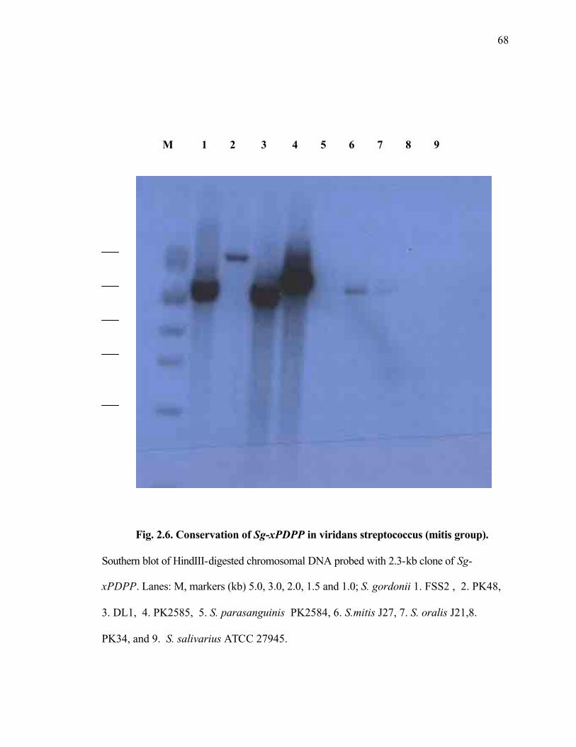
68
Fig. 2.6. Conservation of Sg-xPDPP in viridans streptococcus (mitis group).
Southern blot of HindIII-digested chromosomal DNA probed with 2.3-kb clone of Sg-
xPDPP. Lanes: M, markers (kb) 5.0, 3.0, 2.0, 1.5 and 1.0; S. gordonii 1. FSS2 , 2. PK48,
3. DL1, 4. PK2585, 5. S. parasanguinis PK2584, 6. S.mitis J27, 7. S. oralis J21,8.
PK34, and 9. S. salivarius ATCC 27945.
M 1 2 3 4 5 6 7 8 9

69
Discussion
This report describes the purification, characterization, cloning and sequence
analysis of an extracellular x-Pro dipeptidyl peptidase from S. gordonii FSS2. This
activity has been described in studies concerning tripeptide degradation systems of S.
sanguinis and S. mutans (8) and collagen degradation by S. gordonii Challis in the IE
model (19). Our work presents the first biochemical and genetic studies on purified
enzyme. Filtrate obtained from a pH-controlled culture of S. gordonii FSS2 enabled the
purification of Sg-xPDPP by over 6000 fold. The preparation was determined to be
homogenous and consisted of two identical, non-covalently attached monomers in the
native state. Inactivation studies revealed a serine-class catalytic mechanism in which
DPPIV-family inhibitors appear to be the most effective. Specificity studies were
conducted using para-nitroanalides and peptides smaller than 20 residues. Limited
proteolysis was observed upon incubation with either substrate. The requirement of Pro
or Ala in the penultimate position and prohibition of Pro in the P1’ site define the specific
nature of this peptidase.
Collectively, the biochemical data are consistent with DPPIV-like peptidases
studies from Streptococcus mitis (11), Streptococcus thermophillus (33) and
Streptococcus salivarius (35). Comparison of the Sg-xPDPP protein sequence with
cloned x-Pro dipeptidyl peptidases reveal significant homology (41% average identity)
with PepX gene products of lactic acid bacteria (31, 34). Putative genes for x-Pro
dipeptidyl peptidase were identified in unfinished microbial genomes for S. mutans and S.
pneumoniae and maintained 57% and 72% identity to Sg-xPDPP, respectively. The
consensus sequence, GXSYLG, is present at the active site of PepX and streptococcal

70
members. Phylogenetically-related viridans members, S. pneumoniae and S. gordonii,
maintain hydrophobic residues at the second position which are a contrast to basic
residues conserved in other species. The PepX gene and streptococcal analogue appear to
have diverged from a common ancestor that bears little sequence homology to the
CD26/DPPIV family of eukaryotic and gram negative peptidases. Sg-xPDPP closely
resembles members of the S15 family (EC 3.4.14.11) yet our discovery of streptococcal
members with remarkable evolutionary conservation may warrant a change in
nomenclature.
The localization of peptidases outside of the cell is considered to be a necessary
mechanism for the degradation and intracellular transport of endopeptidase-liberted
peptides. Currently, all reported x-Pro DPP members from Gram-positive bacteria have
been either associated with cytoplasmic, membrane or whole cell extracts (24). Sg-
xPDPP represents the first peptidase from this group isolated from an extracellular
source. Culture conditions revealed the presence of 1 mM Ca2+ rather than trace levels in
complex media to be necessary for export from the cell. The presence of this cation may
alter membrane permeability, non-specifically, or enhance an unknown Ca2+-dependent
mechanism for secretion. Additionally, excess arginine (50 mM) greatly reduced levels
of the secreted protein. The presence of the free amino acid and/or the utilization of the
ADI pathway as a continued source of energy could inactivate an export mechanism
while still promoting the cell-surface localization of the protease. Sequence analysis did
not reveal a N-terminal signal sequence, LPXTG motif (39) or post-translational
modifications that could account for translocation from the cytoplasm. We found the
gene for Sg-xPDPP to be single copy, ruling out the possibility of an altered form of the

71
peptidase. Previous reports have suggested that such enigmatic transport could be the
result of “specific leakage” of cytoplasmic aminopeptidase from cells (51) or the
shedding of protease/peptidoglycan complexes as a consequence of cell wall turnover
(19). In S. gordonii G9B, extracellular protein profiles were altered by changes in pH,
growth medium composition and rate of growth (23). Additionally, the secretion of two
cytoplasmic proteins from S. gordonii, a GAPDH (40) and collagenase (19) were
observed upon growth at a constant, neutral pH. In studies conducted by Harty et. al.,
specific activities of extracellular proteases (thrombin-like, Hageman-factor and
collagenase) from FSS2 increased several fold upon a shift in pH from 6.5 to 7.5
(13).This data is consistent with maximum detection of our Sg-xPDPP activity at a
controlled pH of 7.5. A similar level activity was observed at pH 7.0 but accounted for
lower proportion of the secreted form. Furthermore, the peak of Gly-Pro-pNa activity
occurred during early stationary phase when excess glucose had been exhausted. The
alkaline environment of the buffered vegetation contacting the bloodstream may be more
suitable for enzyme release from the cell. An extracellular peptidase would therefore
better serve the nutritional requirements of the bacteria in a fibrin/ECM surrounding
versus the carbohydrate-rich oral environment.
The biological significance of Sg-xPDPP outside of the cell would serve a
threefold purpose, 1) the production of proline-containing dipeptides that could
subsequently be transported into the cell, 2) a synergy with other secreted proteases that
would permit the degradation of host proteins and 3) the modification of bioactive
peptides at the endothelium. L. lactis and other lactic acid bacteria are unable to import
free Pro which must be transported in a peptide-bound configuration (24). A tripeptide

72
transport system was identified in S. sanguinis that permitted the assimilation of free
amino acids by membrane-bound and intracellular proteolysis. It was further concluded
that x-Pro dipeptides were insufficient for transport (8). This is counterintuitive to the
action of Sg-xPDPP that results in high concentrations of extracellular dipeptides. In S.
gordonii, uptake could be driven by an inwardly directed chemical gradient maintained
by fluctuating levels of extracellular and cell-associated products of peptide hydrolysis.
The limited cleavage specificity of Sg-xPDPP prohibits the independent proteolysis of
constituents within and surrounding the thrombotic vegetation.
Neither type IV collagen nor fibrinogen were suitable substrates for the enzyme.
This contrasts the findings by Juarez and Stinson that reports a Gly-Pro-pNa activity
capable of endopeptidase attack on native collagen (19). Culture supernatant of FSS2
yielded several endopeptidase activities capable of degrading denatured collagen,
fibrinogen and azocasein. Two additional extracellular aminopeptidases, an Arg-specific
aminopeptidase (RAP) and PepV di/tri-peptidase (manuscripts in preparation), would
participate in the acquisition of small peptides from the protein meshwork surrounding
the bacteria.
Although Sg-xPDPP achieved proteolysis of all suitable peptides tested, there is
an apparent restriction of substrate size. Three cytokines tested (RANTES, MIP-1β and
GM-CSF), presenting unblocked x-Pro motifs, failed to undergo cleavage upon extended
incubations and increased E:S molar ratios. This is in contrast to the activities of
DPPIV/CD26 members which facilitate the N-terminal proteolysis of these larger
substrates despite their high catalytic similarity to the Pep X members (16, 43). Sequence
divergence in specificity pockets and the presence of adhesion domains, absent in Gram-

73
positive peptidases, may contribute to this phenomenon. The removal of two dipeptides
from substance P was observed and this heptapeptide product has been found to display
biological activity more potent that the intact peptide (55). Additionally, the concerted
action of an extracellular Arg aminopeptidase and Sg-xPDPP produce a truncated form of
bradykinin that has lost essential residues for receptor activation (3). The combined effect
of these modifications may result in local changes in vascular permeability and smooth
muscle contraction at the infected endothelium. The truncation of fibrin inhibitory
peptides by Sg-xPDPP could have consequences on thrombus formation and the overall
growth of the vegetation. The soluble tetrapeptide (GPRP), representing the N-terminus
of the fibrin α-chain, blocks the polymerization of fibrin monomers leading to blocked
polymerization, decreased clotting and increased clotting times (27). The minimum
structural unit (GPR) is required for inhibitory action. Sg-xPDPP may inactivate
circulating inhibitor in the growing thrombus and alter the balance between
polymerization/fibrinolysis in favor of the growing vegetation. Studies with the mice-
passaged FSS2 strain indicated levels of cell-surface activity close similar to the basic
laboratory strain. Levels of extracellular Sg-xPDPP were roughly 4x lower in the mice-
passaged strain. This may indicate an alteration in secretion mechanism or mutation upon
entry into an animal model. Although this data was counterintuitive to the secretion
hypothesis, regrowth in media could induce new stress responses or alter gene expression
in a manner inconsistent with in vivo human colonization. Future studies will focus on the
expression and knockout of Sg-xPDPP in order to evaluate its relative contributions to
streptococcal virulence and survival at the site of infection.

74
References
1. Andersson, C., M. L. Sund, and L. Linder. 1984. Peptide utilization by oral
streptococci. Infect Immun. 43:555-60.
2. Banbula, A., M. Bugno, J. Goldstein, J. Yen, D. Nelson, J. Travis, and J.
Potempa. 2000. Emerging family of proline-specific peptidases of
Porphyromonas gingivalis: purification and characterization of serine dipeptidyl
peptidase, a structural and functional homologue of mammalian prolyl dipeptidyl
peptidase IV. Infect Immun. 68:1176-82.
3. Bhoola, K. D., C. D. Figueroa, and K. Worthy. 1992. Bioregulation of kinins:
kallikreins, kininogens, and kininases. Pharmacol Rev. 44:1-80.
4. Burne, R. A. 1998. Oral streptococci... products of their environment. J Dent Res.
77:445-52.
5. Chich, J. F. 1998. X-Pro-dipeptidyl peptidase, p. 403-405. In A. J. Barrett, N. D.
Rawlings, and J. F. Woessner (ed.), Handbook of Proteolytic Enzymes. Academic
Press, San Diego, CA.
6. Chich, J. F., M. P. Chapot-Chartier, B. Ribadeau-Dumas, and J. C. Gripon. 1992.
Identification of the active site serine of the X-prolyl dipeptidyl aminopeptidase
from Lactococcus lactis. FEBS Lett. 314:139-42.
7. Cowman, R. A., and S. S. Baron. 1990. Influence of hydrophobicity on
oligopeptide utilization by oral streptococci. J Dent Res. 69:1847-51.
8. Cowman, R. A., and S. S. Baron. 1997. Pathway for uptake and degradation of X-
prolyl tripeptides in Streptococcus mutans VA-29R and Streptococcus sanguinis
ATCC 10556. J Dent Res. 76:1477-84.

75
9. Douglas, C. W., J. Heath, K. K. Hampton, and F. E. Preston. 1993. Identity of
viridans streptococci isolated from cases of infective endocarditis. J Med
Microbiol. 39:179-82.
10. Drangsholt, M. T. 1998. A new causal model of dental diseases associated with
endocarditis. Ann Periodontol. 3:184-96.
11. Fukasawa, K. M., and M. Harada. 1981. Purification and properties of dipeptidyl
peptidase IV from Streptococcus mitis ATCC 9811. Arch Biochem Biophys.
210:230-7.
12. Gong, K., T. Ouyang, and M. C. Herzberg. 1998. A streptococcal adhesion
system for salivary pellicle and platelets. Infect Immun. 66:5388-92.
13. Harty, D. W., J. A. Mayo, S. L. Cook, and N. A. Jacques. 2000. Environmental
regulation of glycosidase and peptidase production by Streptococcus gordonii
FSS2. Microbiology. 146:1923-31.
14. Herzberg, M. C. 1996. Platelet-streptococcal interactions in endocarditis. Critical
Reviews In Oral Biology And Medicine. 7:222-36.
15. Herzberg, M. C., and M. W. Meyer. 1996. Effects of oral flora on platelets:
possible consequences in cardiovascular disease. J Periodontol. 67:1138-42.
16. Hoffmann, T., J. Faust, K. Neubert, and S. Ansorge. 1993. Dipeptidyl peptidase
IV (CD 26) and aminopeptidase N (CD 13) catalyzed hydrolysis of cytokines and
peptides with N-terminal cytokine sequences. FEBS Lett. 336:61-4.
17. Homer, K. A., R. A. Whiley, and D. Beighton. 1990. Proteolytic activity of oral
streptococci. FEMS Microbiol Lett. 55:257-60.

76
18. Jenkinson, H. F., R. A. Baker, and G. W. Tannock. 1996. A binding-lipoprotein-
dependent oligopeptide transport system in Streptococcus gordonii essential for
uptake of hexa- and heptapeptides. J Bacteriol. 178:68-77.
19. Juarez, Z. E., and M. W. Stinson. 1999. An extracellular protease of
Streptococcus gordonii hydrolyzes type IV collagen and collagen analogues.
Infect Immun. 67:271-8.
20. Khalid, N. M., and E. H. Marth. 1990. Purification and partial characterization of
a prolyl-dipeptidyl aminopeptidase from Lactococcus helveticus CNRZ 32. Appl.
Environ. Microbiol. 56:381-388.
21. Kiefer-Partsch, B., W. Bockelmann, A. Geis, and M. Teuber. 1989. Purification
and characterization of an X-prolyl-dipeptidyl aminopeptidase from the cell wall
proteolytic system of Lactococcus lactis subsp. cremoris. Appl Microbiol
Biotechnol. 31:75-78.
22. Kili, A. O., M. C. Herzberg, M. W. Meyer, X. Zhao, and L. Tao. 1999.
Streptococcal reporter gene-fusion vector for identification of in vivo expressed
genes. Plasmid. 42:67-72.
23. Knox, K. W., L. N. Hardy, L. J. Markevics, J. D. Evans, and A. J. Wicken. 1985.
Comparative studies on the effect of growth conditions on adhesion,
hydrophobicity, and extracellular protein profile of Streptococcus sanguinis G9B.
Infect Immun. 50:545-54.
24. Kok, J. 1990. Genetics of the proteolytic system of lactic acid bacteria. FEMS
Microbiol Rev. 7:15-42.
77
25. Konings, W. N., B. Poolman, and A. J. Driessen. 1989. Bioenergetics and solute
transport in lactococci. Crit Rev Microbiol. 16:419-76.
26. Korzenioswki, O. M., and D. Kaye. 1992. Infective Endocarditis, p. 1078-1105.
In E. Braunwald (ed.), Heart Disease, 4th ed.
27. Laudano, A. P., and R. F. Doolittle. 1980. Studies on synthetic peptides that bind
to fibrinogen and prevent fibrin polymerization. Structural requirements, number
of binding sites, and species differences. Biochemistry. 19:1013-9.
28. Lloyd, R. J., and G. G. Pritchard. 1991. Characterization of X-prolyl dipeptidyl
aminopeptidase from Lactococcus lactis subsp. lactis. Journal of Gen. Microbiol.
137:49-55.
29. Manning, J. E., E. B. Hume, N. Hunter, and K. W. Knox. 1994. An appraisal of
the virulence factors associated with streptococcal endocarditis. J Med Microbiol.
40:110-4.
30. Matsudaira, P. 1987. Sequence from picomole quantities of proteins electroblotted
onto polyvinylidene difluoride membranes. J Biol Chem. 262:10035-8.
31. Mayo, B., J. Kok, K. Venema, W. Bockelmann, M. Teuber, H. Reinke, and G.
Venema. 1991. Molecular cloning and sequence analysis of the X-prolyl
dipeptidyl aminopeptidase gene from Lactococcus lactis subsp. cremoris. Appl
Environ Microbiol. 57:38-44.
32. Mayo, J. A., H. Zhu, D. W. Harty, and K. W. Knox. 1995. Modulation of
glycosidase and protease activities by chemostat growth conditions in an
endocarditis strain of Streptococcus sanguinis. Oral Microbiol Immunol. 10:342-
8.
78
33. Meyer, J., and R. Jordi. 1987. Purification and characterization of X-prolyl-
dipeptidyl-aminopeptidase from Lactobacillus lactis and from Streptococcus
thermophilus. J Dairy Sci. 70:738-45.
34. Meyer-Barton, E. C., J. R. Klein, M. Imam, and R. Plapp. 1993. Cloning and
sequence analysis of the X-prolyl-dipeptidyl-aminopeptidase gene (pepX) from
Lactobacillus delbruckii ssp. lactis DSM7290. Appl Microbiol Biotechnol. 40:82-
9.
35. Mineyama, R., and K. Saito. 1991. Purification and characterization of dipeptidyl
peptidase IV from Streptococcus salivarius HHT. Microbios. 67:37-52.
36. Miyakawa, H., S. Kobayashi, S. Shimamura, and T. M. 1991. Purification and
characterization of an X-prolyl dipeptidyl aminopeptidase from Lactobacillus
delbrueckii ssp. bulgaricus LBU-147. J Dairy Sci. 74:2375-2381.
37. Nakai, K., and M. Kanehisa. 1991. Expert system for predicting protein
localization sites in gram-negative bacteria. Proteins. 11:95-110.
38. Nardi, M., M. C. Chopin, A. Chopin, M. M. Cals, and J. C. Gripon. 1991. Cloning
and DNA sequence analysis of an X-prolyl dipeptidyl aminopeptidase gene from
Lactococcus lactis subsp. lactis NCDO 763. Appl Environ Microbiol. 57:45-50.
39. Navarre, W. W., and O. Schneewind. 1999. Surface proteins of gram-positive
bacteria and mechanisms of their targeting to the cell wall envelope. Microbiol
Mol Biol Rev. 63:174-229.
40. Nelson, D., J. M. Goldstein, K. Boatright, D. W. Harty, S. L. Cook, P. J.
Hickman, J. Potempa, J. Travis, and J. A. Mayo. 2001. pH-regulated secretion of
a glyceraldehyde-3-phosphate dehydrogenase from Streptococcus gordonii FSS2:

79
purification, characterization, and cloning of the gene encoding this enzyme. J
Dent Res. 80:371-7.
41. Nolte, W. A. 1982. Defense Mechanisms of the Mouth, p. 245-260. In W. A.
Nolte (ed.), Oral MIcrobiology. C.V. Mosby Company, St. Louis, MO.
42. Pohl, J., F. Hubalek, M. E. Byrnes, K. R. Nielsen, A. Woods, and M. W.
Pennington. 1995. Assignment of three disulfide bonds in ShK toxin: A potent
potassium channel inhibitor from sea anenome Stichodactyla heliatus. Peptide
Science. 1:291-297.
43. Proost, P., I. De Meester, D. Schols, S. Struyf, A. M. Lambeir, A. Wuyts, G.
Opdenakker, E. De Clercq, S. Scharpe, and J. Van Damme. 1998. Amino-terminal
truncation of chemokines by CD26/dipeptidyl-peptidase IV. Conversion of
RANTES into a potent inhibitor of monocyte chemotaxis and HIV-1-infection. J
Biol Chem. 273:7222-7.
44. Rawlings, N. D., L. Polgar, and A. J. Barrett. 1991. A new family of serine-type
peptidases related to prolyl oligopeptidase [letter]. Biochem J. 279:907-8.
45. Rogers, A. H., A. L. Pfennig, N. J. Gully, and P. S. Zilm. 1991. Factors affecting
peptide catabolism by oral streptococci. Oral Microbiol Immunol. 6:72-5.
46. Rosenfeld, J., J. Capdevielle, J. C. Guillemot, and P. Ferrara. 1992. In-gel
digestion of proteins for internal sequence analysis after one- or two-dimensional
gel electrophoresis. Anal Biochem. 203:173-9.
47. Saluja, S. K., and J. N. Weiser. 1995. The genetic basis of colony opacity in
Streptococcus pneumoniae: evidence for the effect of box elements on the
frequency of phenotypic variation. Mol Microbiol. 16:215-27.

80
48. Schägger, H. 1995. Native electrophoresis for isolation of mitochondrial oxidative
phosphorylation protein complexes. Methods Enzymol. 260:190-202.
49. Schägger, H., and G. von Jagow. 1987. Tricine-sodium dodecyl sulfate-
polyacrylamide gel electrophoresis for the separation of proteins in the range from
1 to 100 kDa. Anal Biochem. 166:368-79.
50. Sullam, P. M. 1994. Host-pathogen interactions in the development of bacterial
endocarditis. Current Opinion in Infectious Diseases. 7:304-309.
51. Tan, P. S., B. Poolman, and W. N. Konings. 1993. Proteolytic enzymes of
Lactococcus lactis. J Dairy Res. 60:269-86.
52. Vriesema, A. J., J. Dankert, and S. A. Zaat. 2000. A shift from oral to blood pH is
a stimulus for adaptive gene expression of Streptococcus gordonii CH1 and
induces protection against oxidative stress and enhanced bacterial growth by
expression of msrA. Infect Immun. 68:1061-8.
53. Whiley, R. A., and D. Beighton. 1998. Current classification of the oral
streptococci. Oral Microbiol Immunol. 13:195-216.
54. Willcox, M. D., M. Patrikakis, and K. W. Knox. 1995. Degradative enzymes of
oral streptococci. Aust Dent J. 40:121-8.
55. Yajima, H., and K. Kitagawa. 1973. Studies on peptides. XXXIV. Conventional
synthesis of the undecapeptide amide corresponding to the entire amino acid
sequence of bovine substance P. Chem Pharm Bull (Tokyo). 21:682-3.
56. Yoshimoto, T., and D. Tsuru. 1982. Proline-Specific Dipeptidyl Aminopeptidase
from Flavobacterium meningosepticum. J. Biochem. 91:1899-1906.

81
CHAPTER 3
AN EXTRACELLULAR ARGININE AMINOPEPTIDASE (RAP) FROM
STREPTOCOCCUS GORDONII FSS21
1 Goldstein, J.M., D. Nelson, T. Kordula, J.A. Mayo, and J. Travis, J. 2001.Submitted to Infection and Immunity, ASM Publications.

82
Abstract
Streptococcus gordonii is generally considered to be a benign inhabitant of the oral
microflora. However, it is also a primary etiological agent in the development of subacute
bacterial endocarditis (SBE) producing thrombus formation and tissue damage on the
surface of heart valves. Strain FSS2 of S. gordonii has been found to produce several
extracellular aminopeptidase and fibrinogen-degrading activites during growth in a pH-
controlled batch culture. In this report we describe the purification, characterization and
partial cloning of a predicted serine-class arginine aminopeptidase (RAP) with some
cysteine-class characteristics. Isolation of this enzyme by anion-exchange, gel filtration
and IEF chromatography yielded a protein monomer of approximately 70 kDa, as shown
by MALDI-TOF mass spectrometry, gel filtration and SDS-PAGE under denaturing
conditions. Nested PCR cloning enabled the isolation of a 324 bp-long DNA fragment
encoding the108 amino acid N-terminus of RAP. Culture activity profiles and N-terminal
sequence analysis indicated the release of this protein from the cell surface. Homology
was found with a putative dipeptidase from Streptococcus pyogenes and non-specific
dipeptidases from Lactobacillus heleveticus and Lactococcus lactis. We believe that RAP
may serve as a critical factor for arginine acquisition during nutrient stress in vivo and
also in the proteolysis of host proteins and peptides during SBE pathology.

83
Introduction
Streptococcus gordonii is a frequently researched member of the viridans family
of oral streptococci (55). Viridans streptococci are primary colonizers of human dental
plaque where they serve a critical role in the establishment of microbial communities
associated with good oral health. Although considered benign inhabitants of the oral
microflora, members have been implicated in the systemic disease, subacute bacterial
endocarditis (SBE) (8, 33). The progression of this disease state requires: 1) trauma
(congenital or inflammatory) to the endothelial valve surface such that it is predisposed to
colonization, 2) adhesion of organisms to the modified valve surface after their entry into
the bloodstream via the oral cavity and 3) the propagation of infected vegetations
consisting of bacteria in a fibrin-platelet meshwork (52). Despite the uniform
susceptibility of these organisms to β-lactam antibiotics and their lack of classical
streptococcal virulence factors, they can cause a chronic inflammation and/or life-
threatening disease with periods of latency and recalcitrant infection (9, 18).
The ability of these organisms to colonize biofilm surfaces within two distinct
microenvironments evoked studies into their patterns of gene expression and dynamic
metabolism. Streptococcus sanguinis, studied as a model for viridans pathogenesis, is
known to express cell surface adhesins and a platelet aggregation-associated protein
(PAAP) that facilitate both colonization and thrombosis (16, 19). Upon their entry into
the bloodstream, bacteria undergo a shift in pH from mildly acidic in dental plaque (6.0-
6.5) to neutral pH (7.3) of the blood (44). This is significant since proteins extracted from
Streptococcus oralis grown in batch culture and analyed by 2-D electrophoresis indicates
39 proteins with altered expression at pH 5.2 vs. 7.0 (56). Indeed, in vivo Expression

84
Technology (IVET) conducted on the S. gordonii rabbit model for IE to detect genes
activated in the new environment indicated an upregulation of the msrA oxidative stress
gene (54) and the induction of genes encoding carbohydrate metabolism enzymes, protein
transporters and cell surface proteins (26). The expression and secretion of glycosidase
and peptidase activities, as examined in pH-controlled batch cultures, was found to be
down-regulated by acid growth conditions and up-regulated by growth in a neutral pH
environment supplemented with serum (17).
Survival in vivo is dependent on the ability of the bacterium to remove sufficient
quantities of nutrients from its environment. It is presumed that S. gordonii meets these
needs by degrading salivary carbohydrates and glycoproteins in the oral cavity, while
utilizing plasma proteins when growing on heart valve surfaces. The vegetation biofilm is
known to retard the diffusion of antibiotics (24) and solutes generally diffuse into the
interior at slower rates than in water (51). Thus, access to carbohydrates and plasma
proteins within the vegetation remains problematic. During such conditions where
carbohydrates are scarce, oral streptococci can catabolize arginine via the arginine
deiminase (ADI) pathway, resulting in the synthesis of both ATP and other essential
metabolic precursors (7, 14). The utilization of this amino acid is then prioritized so that
the transport and metabolism of arginine-containing peptides may become particularly
important. Amino acid and peptide transport systems for viridans streptococci members
have been described (2, 5, 10, 25, 31), and it has been shown that amino acids and small
peptides are readily imported while those exceeding size limitations require hydrolysis by
endo- and exopeptidases either present on the surface or secreted by these cells (6). One
such activity that could meet the transport and metabolic challenges in vivo is an arginine

85
aminopeptidase. Such an enzyme has previously been described from cell wall and
cytoplasmic preparations of S. mitis and S. sanguinis (1, 12, 21) but has eluded detailed
investigations. Here, we describe the purification, characterization and partial cloning of
the first reported extracellular arginine aminopeptidase (RAP) derived from S. gordonii
FSS2, a strain previously isolated from the bloodstream of an SBE patient (38).
Experimental Procedures: Materials
H-Gly-Pro-pNa, H-Gly-Arg-pNa, H-Gly-pNa, Ile-Pro-Arg-pNa, L-Leu-pNa, Lys-Arg-
pNa, L-Pro-pNa, Sar-Pro-Arg-pNa, Di-isopropyl fluorophosphate (DFP), Nα-p-tosyl-L-
lysine-chloromethyl ketone (TLCK), iodoacetamide, O-phenanthroline, amastatin,
apstatin,, bestatin, β-mercaptoethanol, L-Cysteine, L-Arginine, L-Lysine, Gly-Pro, Gly-
Gly, angiotensin III, fibronectin binding inhibitor, platelet aggregation inhibitor, sexual
agglutination peptide, substance P, thymopentin fragment, bradykinin, lymphocyte
activating pentapeptide fragment and fibrin polymerization inhibitor were obtained from
Sigma. Pefabloc SC, 3,4-dichloroisocoumarin and E-64 from Boehringer Mannheim.
Anistatin-related peptide, Bz-Arg-pNa, H-Arg-pNa, H-Ala-pNa, H-Ile-pNa, H-Lys-pNa,
H-Val-pNa and protein kinase C substrate were from Bachem. Protein kinase c peptide
was purchased from American Peptide Company and H-Glu-(NHO-Bz) pyrollidide from
Calbiochem. α1 Proteinase inhibitor (α1PI) and α-2 macroglobulin were purchased from
Athens Research and Technology, Athens, Ga.
Experimental Procedures: Methods
Bacterial Growth. S. gordonii FSS2 (previously S. sanguinis FSS2 (17, 38)) was
stored and maintained (at -80°C) as previously described (40). Frozen cells were
inoculated into autoclaved medium containing 20 g/L trypticase peptone (BBL), 5 g/L

86
yeast extract (Difco), 2 g/L NaCl, 0.1 g/L CaCl2, 4 g/L K2HPO4 and 1 g/L KH2PO4. Ten
g/L glucose and 0.5 g /L L-arginine were filter sterilized and subsequently added. A static
culture (200 ml) was grown overnight at 37°C, and a 15L batch culture further inoculated
and grown in an atmosphere of 5% CO2 and 95% N2 at 37°C with the pH held constant at
7.0 by addition of 5M KOH. Cultures were harvested in early stationary phase at a point
when the bacteria had metabolized all available glucose and the addition of base was no
longer required.
Enzymatic Assay. Amidolytic activities of crude samples and purified protease
were measured using the substrate H-Arg-pNa (1 mM final concentration) in Buffer A
(50 mM Tris, 1 mM CaCl2, pH 7.8) at 37°C. Assays were performed on 96 well plates
(0.1 ml) using a thermostated microplate reader, and the release of p-nitroaniline was
measured at 405 nm. (Spectramax, Molecular Devices). Inhibition assays involved pre-
incubation of pure enzyme with inhibitor for 10 min. at 37°C, followed by measurement
of residual activity.
Enzyme Purification. 4 L of cell-free culture filtrate was obtained after
centrifugation (20 min., 4 °C at 6000 x g) of the batch culture. Proteins in the filtrate
were precipitated with (NH4)2SO4 over several hours at 4 °C to a final concentration of
80% and the precipitate pelleted by centrifugation at 8000 x g for 40 min. Pellets were
resuspended in 30 ml of buffer A and dialyzed over 2 days (4 °C), with changes, against
40 volumes of the same buffer. The dialyzed material was applied to a DE52 (Whatman)
column (2.5 x 30 cm, 150 ml) equilibrated with buffer A, which was then washed with 3
column volumes of buffer A at 1ml/min. All protein that eluted in the flow-through was
pooled and concentrated by ultrafiltration. The concentrated sample (10 ml) was loaded

87
onto and eluted from a SuperdexTM-75 (Amersham Pharmacia Biotech) HR 10/30
equilibrated with gel filtration buffer (50 mM Tris, 200 mM NaCl, 1 mM CaCl2, 0.02%
sodium azide, pH 7.8) and eluted with the same buffer at 1 ml/min. Peak activities were
combined and concentrated. The sample (10 ml) was then applied to a Mono Q HR 10/10
FPLC column (Amersham Pharmacia Biotech) equilibrated with buffer A. The column
was washed with equilibration buffer until the base line stabilized, a linear gradient (0-
500 mM NaCl in buffer A) applied, and peak enzyme activity eluted between 200-250
mM NaCl. After pooling and concentration of active fractions, protein was dialyzed
against 25 mM N-methyl piperazine, pH 5.3 and loaded onto a Mono-P 10/5 FPLC
chromatofocusing column equilibrated the same buffer. The column was then washed
with four column volumes of equilibration buffer and a pH gradient developed with 40
ml of 10 x diluted Polybuffer 74 (Amersham Pharmacia Biotech) adjusted to a pH of 3.8.
Peak activity was pooled and concentrated to a volume of 2.0 ml.
Protein Determination. Protein concentration was determined using a
bicinchoninic acid BCA reagent kit (Sigma) according to the manufacturer’s protocol.
Electrophoresis. Enzyme purity was determined by sodium dodecyl sulfate
polyacrylamide gel electrophoresis (SDS-PAGE) using a 10% acrylamide gel and the
Tris-HCl/Tricine buffer system, according to Shagger and von Jagow (48). For amino-
terminal sequence analysis, RAP was resolved by SDS-PAGE, followed by
electroblotting to a polyvinylidene difluoride membrane using 10 mM 3-
(cyclohexylamino) propanesulfonic acid, 10% methanol, pH 11 (39). The blot was air-
dried and subjected to sequencing.
88
Enzyme Kinetics and Specificity. Kinetic values were measured using H-Arg-
pNa and other substrates at varying concentrations [25 µM to 2 mM], with a fixed
enzyme concentration at 34 nM, in 100 mM Tris, pH 7.8, at 37 °C. Vmax and Km values
were obtained through Hyperbolic Regression Analysis (shareware from J.S. Easterby,
University of Liverpool, UK).
Specificity studies utilized RAP incubated with 5 µg of peptide in a 1:1000 [E:S] molar
ratio. Reactions were done in 100 µl volumes with 100 mM Tris, pH 7.8, at 37 °C for 2
hours. Digestions were terminated by acidification with 10 µl of 10 M HCl, followed by
centrifugation (10,000xg, 5 min.). The entire supernatant was applied to a reverse phase
high pressure liquid chromatography using an LC-18 column (25 x 4.6 mm,
5µm)(Supelco) equilibrated with 0.1% TFA in HPLC grade water and developed with an
acetonitrile gradient (0-80% in 0.08% TFA over 50 min.). Peaks were manually collected
and analyzed by mass spectrometry.
Mass Spectrometry. Peptides and native RAP were analyzed by MALDI-TOF
on a Hewlett-Packard G2030A mass spectrometer. The instrument was operated at an
accelerating voltage of 28 kV, an extractor voltage of 7 kV, and a pressure of 7 x 10⁻⁷
Torr. Samples were dissolved in sinapanic acid and ionized from the probe tip using a
nitrogen laser source. Calibration was performed using mixtures of peptides/proteins of
known molecular masses.
Internal Sequencing. Proteins and peptides were sequenced by Edman
degradation in a model Procise-cLC sequencer (PE Biosytems, Foster City, CA) operated
using the manufacturer’s protocol. To obtain internal sequences, proteins were in-gel
digested with trypsin (Promega, sequence grade) and the peptides extracted (47), with

89
their masses being determined by reflectron MALDI-TOF mass spectrometry using a
Bruker Daltonics ProFlex instrument (45). The selected peptides were sequenced by
Edman Degradation.
Cloning of DNA fragment encoding N-terminal region of RAP. DNA from S.
gordonii was purified using the Purgene DNA Isolation Kit (Gentra, Minneapolis, MN)
according to manufacturer’s instructions. The internal peptide sequence
(DLTADGSTLFGR) was used to search the unfinished S. gordonii database available at
The Institute of Genomic Research (TIGR) (ftp://ftp.tigr.org/pub/data/s_gordonii/).
Briefly, DNA sequences were imported into Microsoft Word and searched for a matching
DNA sequence encoding the peptide using wild cards at degenerate code positions. One
matching contig was found that contained a DNA sequence encoding the N-terminus and
internal fragment of RAP. Subsequently, two PCR primers were synthesized
(5’-AGAGGATCCATGAAGAAAATACTGCTC-3’ and
5’-GAAGGATCCGTTACTTCCG TTGTCAC-3’) and used in the PCR to obtain a DNA
fragment encoding the N-terminal fragment of RAP. PCR was performed using the Pwo
DNA polymerase (Roche Molecular Biochemicals, Indianapolis, IN), 1 µg of S. gordonii
DNA and 500 ng of the primers (94ºC for 1 min, 65ºC for 1 min, 72ºC for 1 min, 32
cycles). A single 340 bp-long PCR product was obtained, gel-purified, digested with Bam
HI, subcloned into the Bam HI site of pUC19 and sequenced. The obtained sequence was
deposited in GeneBank under accession number AY052829.
Southern Blot Analysis. Four µg of bacterial DNA was digested with each of the
restriction enzymes BamHI, BglII, SacI, SpeI, EcoRI, HindIII or PstI overnight at 37° C.
Digestion products were separated in 0.7% agarose gels using Standard procedure
90
(Sambrook). Gels were soaked in denaturation solution (0.5 M NaOH, 1.5 M NaCl) for
30 min followed by neutralization solution (1.5 M Tris pH 7.5, 1.5 M NaCl) for 1 hour.
Separated DNA was transferred onto a nylon membrane (Amersham-Pharmacia Biotech)
by capillary transfer and subsequently fixed by cross-linking (Stratalinker, Stratagene
Inc.). Membranes were prehybridized in 0.5 M phosphate buffer pH 7.2, 7% SDS and 1
mM EDTA at 50C for 1 hour, 32P-labelled DPP cDNA probe was added and incubated
overnight. After hybridization, non-specifically bound radioactivity was removed by a
single wash in 2xSSC and 0.1% SDS at room temperature for 20 min followed by two
subsequent washes in 0.5xSSC and 0.1%SDS at 50C for 20 min. Membranes were
exposed to X-ray films using enhancing screens at -80C for 1-4 hours.
Results
Growth and Culture Activity. Preliminary experiments indicated the presence of
an extracellular H-Arg-pNa activity in culture media. Detection of this amidolytic activity
in cell free filtrate was observed during growth in culture supplemented with 50 mM
glucose and 3.5 mM arginine and was dependent on a pH maintained in the range of 6.0-
7.0. Growth and activity from a 1 L pH-controlled (pH 7.0) batch culture were monitored
at hourly intervals (Figure 3.1). Detection of extracellular and cell-associated H-Arg-pNa
activity did not occur until early stationary phase when bacteria had metabolized all
available glucose and the addition of base had ceased. A substantial increase in activity
occurred between hours 6 and 7 when extracellular activity increased fivefold while cell-
associated activity had a slight increase of 1.5 fold. Examination of cultures in stationary
phase revealed a constant level of H-Arg-pNa activity secreted into the medium, whereas
activity on cell-surfaces peaked at 8 hours (approximately ¹ /3 of the activity found in

91
media) and fell to basal (pre-stationary) levels at the final time point. This latter
phenomenon may be the result of the complete export of H-Arg-pNa activity from the
cell surface.
Further growth experiments were conducted under similar conditions but varied in
culture environments with respect to pH control, carbohydrates, arginine, Ca2+, strain
type and addition of serum (Fig. 3.2). Measurements were made at the 10th hour of
growth when stationary phase was evident by lack of glucose consumption. The activity
profiles during varied pH-controlled conditions provided evidence for a dependence on
pH for the expression of H-Arg-pNa activity. Cell-associated and extracellular activities
were detected under all conditions, except those devoid of pH control, with the least
activity measured in more alkaline cultures (pH 7.5 and 8.0). Appreciable extracellular
activity was evident at pH 6.3 and 7.0. Growth at pH 7.0 accounted for the greatest total
activity, while cells at pH 6.5 contributed 30% less. Growth under both these pH
conditions maintained an equal proportion of total activity (70-75%) to the secreted form.
The remaining culture conditions were controlled at pH 7.0 for standardization and
optimum enzyme expression. Excess supplemented arginine (50 mM) provided the
greatest level of total activity while it has little effect on the level of extracellular H-Arg-
pNa. The 25 mM glucose culture was unable to reach the appropriate stationary phase in
the 10 hr. timeframe and thus cannot be accurately compared. The expression of enzyme
seemed dependent on the addition of 1 mM Ca2+ since culture without exogenously
supplied Ca2+ could only produce cell-surface enzyme. Only a slight increase in the
secreted form was detected under this condition. The addition of 25% fetal bovine serum
to the starting media provided a drastic decrease (2.5x) in total activity with a significant

92
drop (¹ /3) in the proportion of secreted activity. Mouse S. gordonii FSS2 assays showed
no significant difference in activity profile. Although, an overall decrease in total activity
was seen in galactose-supplemented cultures, this sugar neither repressed nor activated
the release of activity from cells. This indicates a potential absence of a glucose
repressor.

93
0
0.5
1
1.5
2
0 2 4 6 8 10
t(hrs)
OD
560
0
20
40
60
80
100
120
mO
D/m
in H
-arg-p
Na
Fig. 3.1. S. gordonii FSS2 growth and activity curve from a pH 7.0-controlled
culture supplemented with 50 mM glucose and 3.5 mM arginine. Samples (1 ml)
were removed from culture at fixed timepoints and cells were removed from media by
centrifugation (5 min, 4 ºC, 13,000 x g) followed by 2x wash and resuspension into initial
volume with non-supplemented media. Symbols: , whole culture OD; , cell-free
culture fluid; , whole cells. Assays were performed as previously described using 30ì L
of sample.

94
Fig. 3.2. Extracellular and cell-surface RAP activities under variable growth
conditions. Samples (1 ml) were removed from culture at early stationary phase and
cells were removed from media by centrifugation (5 min, 4 ºC, 13,000 x g) followed by
2x wash and resuspension into initial volume with non-supplemented media. Symbols:
, cell-free culture fluid; , whole cells. Assays were performed as previously described
using 30ì L of sample.

95
Enzyme Purification. Maximum activity for the purpose of isolation was
achieved using a 4 L batch culture held at pH 7.0 with added base and harvested during
early stationary growth. An 80% ammonium sulfate precipitation concentrated
extracellular proteins to a workable volume despite the large decrease in enzymatic yield.
A negative DE52 anion-exchange step then permitted the recovery of most of the
remaining RAP activity. S-75 gel filtration aided primarily in the removal of high and
low molecular weight contaminants, media pigmentation and peptide/amino acid
components. Mono-Q chromatography provided sufficient anionic affinity for RAP
binding to allow for an increase in specific activity by 6 fold after desorption. The final
step, which utilized a Mono-P chromatofocusing column, resulted in a single, defined
peak of purified protein, with the concentrated enzyme corresponding to a specific
activity of 1835 units/mg of protein, a greater than 3500 fold purification and roughly 3%
yield from the starting batch culture (Table 3.1).

96
Table 3.1. Purification of S.gordonii RAP
aBased on enzymatic activity using H-Arg-pNa, which 1 unit= 1 µmol of pNa released per sec.bFPLC, fast protein liquid chromatography
Step Volume TotalActivity
a
TotalProtein
SpecificActivity
Purification
Yield
ml Unitsa mg units/mg fold %
Culture filtrate 3900 8375 15,990 0.524 1 100
Ammonium sulfate precip.
43 1707 433 3.94 7.52 20
DE52 anion exchange
10 3284 176 18.7 36 40
Superdex 75 10 1941 8 241.3 461 23
Mono-Q, FPLCb 1 449 0.3 1496 2855 5.5
Mono-P, FPLC 2 232 0.12 1835 3506 2.8

97
Physical Properties. SDS-PAGE analysis of the purified enzyme showed a single
protein band, as judged by both Coomassie (Figure 3.4) and silver staining (not shown)
with an approximate mass of 70 kDa. Analysis of the protein by MALDI revealed a laser
intensity peak that corresponded to a mass of 69,695.6 Da, while analytical Superdex 200
gel filtration indicated a protein size of 64 kDa and, therefore, a monomeric structure.
Isoelectric focusing produced a pI of 5.6 for the native protein. The H-Arg-pNa activity
was optimum at pH 6.5 with activity detected over a broad phosphate buffer range of 4.0
to 10.0. The enzyme in its pure form was unstable after a 24h period at either 37 °C or 25
°C while affected minimally at 4 °C. However, storage at –20 °C over several weeks
resulted in a major loss in enzyme with complete inactivation after a 2 months period.

98
Fig. 3.3. SDS-PAGE of fractions from the purification of S. gordonii arginine
aminopeptidase . Lane 1,7 molecular mass markers (phosphorlase b, 94 kDa; bovine
serum albumin, 67 kDa; ovalbumin, 43 kDa; carbonic anhydrase, 30 kDa; soybean
trypsin inhibitor, 20 kDa). The following lanes contained boiled, reduced samples: lane 2,
80% ammonium sulfate precipitation; lane 3, flow through from DE52 anion exchange;
lane 4, peak from Superdex 75 gel filtration wash; lane 5, eluted peak from Mono Q; lane
6, purified RAP (arginine aminopeptidase) from Mono-P.
1 2 3 4 5 6 7
94
67
43
30
20

99
Enzyme Specificity. Of the fourteen chromogenic endo- and aminopeptidase
substrates tested on the purified enzyme (Table 3.2), only H-Arg-pNa was rapidly
hydrolyzed. Weaker activity (< 10%) was detected against H-Lys-pNa and H-Leu-pNa.
When Michaelis-Menten kinetics were measured using H-Arg-pNa as substrate, a Vm of
17.44 µmol min-1 and Km of 51 µM were determined.
To obtain further information on cleavage specificity, various peptides (2-13 aa)
were tested as substrates for RAP (Table 3.3). Significantly, all proteolysis was restricted
to peptides with an arginine residue at the N-terminus, with preferential cleavage
occuring when a hydrophobic or uncharged residue occupied the 2nd site. There were no
apparent additional restrictions for amino acids present downstream of the cleavage site.
Extended time incubation with peptides failed to yield additional peptide fragments,
indicating the absence of endopeptidase activity or contaminating aminopeptidases. The
inability of RAP to catalyze endo-specific proteolysis as tested on whole protein
substrates (azocasein, gelatin, collagen type IV, fibrinogen), further supported the role of
the purified enzyme as an exopeptidases with the potential to aid endopeptidases.

100
Table 3.2. Relative amidolytic activity of S. gordonii RAP against various substrates
1Activity against H-Arg-pNa hydrolysis taken as 100
Substrate (pNa) Relativeactivity(%)
H-Arg 100
H-Lys 5
H-Ala 0
H-Gly 0
H-Ile 0
L-Leu 7
L-Pro 0
H-Val 0
H-Gly-Arg 0
H-Gly-Pro 0
Lys-Arg 0
Bz-Arg 0
Ile-Pro-Arg 0
Sar-Pro-Arg 0

101
Table 3.3. Cleavage Specificity of S. gordonii arginine aminopeptidase on Peptide Substrates
↓, cleavage site
Substrate Sequence
Tyrosine Kinase Substrate Arg-Arg-Leu-Ile-Glu-Asp-Ala-Glu-Tyr-Ala-Ala-Arg-Gly
Anistatin Related Peptide Arg-Cys-Arg-Val-His-Cys-Pro
Fibronectin Binding Inhibitor Arg-↓-Gly-Asp-Ser-Pro-Ala-Ser-Ser-Lys-Pro
Sexual Agglutination Peptide Arg-↓-Gly-Pro-Phe-Pro-Ile
Protein Kinase C Substrate Arg-↓-Phe-Ala-Arg-Lys-Gly-Ala-Leu-Arg-Gln-Lys-Asn-Val
Platelet Aggregation Inhibitor Arg-↓-Gly-Glu-Ser
Peptide 7 Arg-↓-Gly
Thymopentin Fragment Arg-Lys-Asp-Val-Tyr
Peptide 9 Arg-↓-Phe
Substance P Arg-↓-Pro-Lys-Pro-Gln-Gly-Leu-Met
Bradykinin Arg-↓-Pro-Pro-Gly-Phe-Ser-Pro-Phe-Arg
Dipeptide A Arg-↓-Pro
Angiotensin III Arg-↓-Val-Tyr-Ile-His-Pro-Ile
Fibrin Polymerization Inhibitor (tetrapeptide)
Gly-Pro-Arg-Pro
Kallidin Lys-Arg-Pro-Pro-Gly-Phe-Ser-Pro-Phe-Arg
Lymphocyte ActivatingPentapeptide
Leu-Pro-Pro-Ser-Arg
102
Inhibition and Activation Studies. Studies with class-specific inhibitors (Table
3.4) supported the assignment of a serine-class mechanism with some cysteine-class
characteristics. Moderate inhibition was observed using serine protease inhibitors
(Pefabloc and to a lesser extent, DFP and 3,4-Dichloroisocoumarin). However, cysteine-
class inhibitors, iodoacetamide and E64, had little or no effect on activity while pCMB, a
sulfhydryl-sensitive reagent, was a potent inhibitor of RAP, indicating the possibility of a
functional cysteine residue near the vicinity of the active site. Reducing agents (cysteine
and β-mercaptoethanol) stimulated activity by approximately 50% and had a more
pronounced effect at lower enzyme concentrations. With metallo-class inhibitors, 1,10-
orthophenanthroline provided no inhibition and EDTA actually stimulated RAP activity
in a similar manner as the reducing agents. Activity was inhibited by the heavy metal ions
Zn2+ and Co2+ at 1mM but was increased in the presence of lower mass mono- and
divalent cations. L-arginine served as a competitive inhibitor while L-lysine gave
minimal inhibition, providing further evidence for the strict specificity in the P1 position.
Compounds specific for eukaryotic arginine aminopeptidase/ Aminopeptidase B (EC
3.4.11.6) and Leucine Aminopeptidase (Bestatin) as well as glutamyl aminopeptidase/
aminopeptidase A (Amastatin) were not inhibitory even at high concentrations. However,
RAP was sensitive to the DPPIV-specific inhibitors, H-Glu-pyrollidide, and the
Aminopeptidase P-specific inhibitor, Apstatin, as evidenced by 98% and 50% inhibition,
respectively, at 100 µM. Human plasma inhibitors, α-1-proteinase inhibitor and α-2-
macroglobulin, had no effect on enzymatic activity.
These results are consistent with previous studies conducted on cell-associated
arginine aminopeptidases from S. sanguinis (12) and S. mitis (21).

103
Table 3.4. Inhibition profile of S. gordonii RAP
Inhibitor Concentration % of Residual Activity
DFP 5 mM 75 10 mM 56
Pefabloc 5 mM 40 10 mM 31
3,4-Dichloroisocoumarin 2 mM 62Amastatin 1 mM 89Bestatin 1 mM 68Apstatin 100µM 50H-Glu-pyrollidide 100µM 2E-64 500 µM 98Iodoacetamide 5 mM 91EDTA 5 mM 1311,10-Orthophenanthroline 1 mM 99pCMB 2 mM 0L-Cysteine 5 mM 148L-Arginine 5 mM 30L-Lysine 5 mM 72β-mercaptoethanol 1 mM 160Gly-Gly 5 mM 92Gly-Pro 5 mM 104Na+ 200 mM 166K+ 5 mM 131Ca2+ 5 mM 150Mg2+ 1mM 93SDS 5% 4Urea 2 M 82
104
RAP Sequence Analysis. The failure to generate a protein sequence from an
apparently blocked N-terminus required the sequencing of internal peptides after trypsin
digestion. This partial structure resulted in the isolation of a peptide DLTADGSTLFGR
which was used to design a degenerate primer. In turn, this was employed to search the
genomic clone of an S. gordonii strain in the Unfinished Microbial Genomes Database,
TIGR. A matching contig (bvs 3948) was identified which comprised an ORF of 324 bp
and included the internal sequence of RAP. N and C-terminal sequences facilitated the
construction of degenerative primers and PCR against a FSS2 library resulted in the
isolation of the 324 bp product that constituted a partial-length DNA fragment. Southern
Blot analysis was performed with the clone and revealed RAP to be a single copy gene.
A 108-amino acid polypeptide with a theoretical molecular mass of 11,934 Da and
representing approximately 17% of the sequence of the intact protein was encoded by this
ORF (Figure 3.4).
The sequence was analyzed for the presence of Gram-positive post-translational
modifications. Predictions indicated an intracellular, positively charged N-terminus, a
hydrophobic membrane insertion domain and a putative site for signal peptide cleavage
(43). This translocation mechanism is well conserved among Bacillus spp. (50) and has
been identified in B. subtilis alkaline protease (49) and Bacillopeptidase F (57). In RAP,
an export signal sequence of 14 hydrophobic residues is located downstream from the N-
terminus, followed by a polar region (denoted by QACS) that provides a cleavage site
after alanine.
The finding of an export signal supports the data for extracellular localization in

105
culture experiments. A protein homology search was performed with the 108 amino acid
fragment using CMR BLAST against EMBL, DDBJ, GenBank and PDB databases.
Results indicated that RAP shares 65% identity and 82% similarity with an unpublished
report of a gene encoding an amylase binding protein B (abpB) from S. gordonii (36).
RAP and abpB have predicted transmembrane domains, share nearly identical export
signals and have masses of approximately 70 kDa upon secretion from the cell. RAP
maintains lesser homology with a putative dipeptidase (TIGR#NTLO1SPL1617) from
Streptococcus pyogenes (44% identity), dipeptidase DA from Lactococcus lactis (34%
identity) and non-specific dipeptidase A from Lactobacillus helveticus (31% identity).
1 10 20 30 40 50
| | | | | |MKKILLRLMFLAMLVALLPVHVAQACSAFIVGKDLTADGSTLFGRTEDYPYAPD
60 70 80 90 100 | | | | |GGRHNQNYVVVPAKTYKDGDKIEDESNGFTYPHLANEMKYTAVYDSDRDNGS
108 |NG
Fig. 3.4. N-terminal sequence of S. gordonii arginine aminopeptidase (RAP)
deduced from FSS2 genome. Single underlined sequence represents the predicted
transmembrane domain. The bold letters mark a conserved signal peptide cleavage site
for Gram-positive bacteria. Double lines represent the internal fragment used to search
the electronic database and generate a 340 bp PCR product.

106
Genetic Analysis of Sg-RAP in S. gordonii FSS2 and Viridans Streptococci. A
Southern Blot of the S. gordonii FSS2 genome (Fig. 3.6) reveals a single hybridizing
band for seven of the restriction enzyme digests under high stringency conditions. All
digests show single bands as would be expected for a single copy gene. Digests were
subjected to electrophoresis, blotted, and probed for the RAP gene utilizing the 0.34 kb
clone corresponding to the N-terminus of the protein.
Fig. 3.5. Copy Number of S. gordonii RAP (Sg- RAP) gene in strain FSS2 as
Revealed by restriction endonuclease analysis. Lanes: 1. BamHI, 2. BglII, 3. EcoRI, 4.
HindIII, 5. PstI, 6. SacI, 7. SpeI.
1 2 3 4 5 6 7

107
Discussion
This report describes the purification, characterization, partial cloning and
sequence analysis of an arginine aminopeptidase from S. gordonii FSS2. While this
activity has been described in studies concerning arginine proteolysis by S. sanguinis and
S. mitis (1, 12, 13, 20, 21, 37), the results presented here are the first biochemical
evidence of an extracellular arginine aminopeptidase (RAP), as well as the only study of
the molecular structure of a viridans streptococcal arginine aminopeptidase. The enzyme,
which was obtained from a pH-controlled culture, was purified over 3500 fold and
determined to be a homogenous monomeric protein with a molecular size of 70 kDa.
Specificity studies conducted using both p-nitroanilides and peptides smaller than 13
residues, indicated only an exopeptidase activity upon incubation with either type of
substrate. Although minor Lys and Leu cleavage occured, efficient aminopeptidase
activity required an Arg in the first position with a non-polar or uncharged residue
preferred in the second site. Inactivation studies propose a serine-class catalytic
mechanism in which x-Pro specific inhibitors appear to be the most effective.
Collectively, the biochemical studies on RAP are consistent with those on
arginine aminopeptidases purified from S. mitis ATCC 9811 and S. sanguinis ATCC 903.
These enzymes represented the first Aminopeptidase B/ arginyl-exopeptidase activities
isolated from a bacterial source. Previous work has identified two activities, a broad
substrate aminopeptidase and an Arg-specific aminopeptidase, each capable of
hydrolyzing arginine-2-naphthylamide when using cytoplasmic and cell wall/ membrane
fractions in cultures undergoing late exponential-early stationary growth (12, 21). These
molecular forms differ from each other with respect to their physical properties,

108
inhibition studies, immunochemical properties and cellular localization. RAP is most
closely identified with the Arg-specific, lower molecular weight form that is associated
with the cell wall and membrane, found to be sensitive to heavy metals, and classified as
a cysteine protease based upon inhibition studies. The inhibition of the S. mitis arginine
aminopeptidase by pCMB and metals is suggested to be indicative of a sulfhydryl group
located near the active site and essential for enzymatic activity (21). Additionally, it was
reported that the stimulating effect of reducing agents was evidence for cysteine
residue(s) prone to oxidation (12). However, the sensitivity of RAP to serine-class
inhibitors, together with general unresponsiveness to cysteine-class inhibitors, questions
such a characterization. The majority of bacterial aminopeptidases, including the broad
substrate Arg-aminopeptidase, are members of the metallo-class and excluding the PepC
family, bacterial cysteine aminopeptidases are a rarity. The elucidation of crystal
structures and site-directed mutatgenesis experiments on bacterial prolyl-iminopeptidase
(PIP) family members indicated that they are serine peptidases resistant to fluoride
compounds, activated by reducing agents and sensitive to mercurial and heavy metal
salts. These features have provided ambiguity during characterization and have
improperly classified them as members of the cysteine class without conclusive evidence
(27). PIP genes from Bacillus coagulans (28) and Lactobacillus lactis (29) were modeled
upon elucidation of the three dimensional structure of Xanthomonas campestris, a model
for the PIP family. The data obtained indicated that the active site was comprised of the
catalytic triad Asp58, Ser101, and His 267, with Cys62 located at the bottom of the active
site about 5Å from the nucleophilic serine, presumably accounting for its cysteine-class

109
behavior (41). However, in the case of RAP, the absence of both a complete sequence
and analogous reactive cysteine, provide inconclusive evidence for serine classification.
The pattern of substrate cleavage exhibited by RAP indicates an aminopeptidase
with a strict specificity for arginine in the first position and a restriction against positively
charged (Arg or Lys) or polar (Cys) residues in the second. However, proteolysis is
favored when non-polar or hydrophobic amino acids are present in that position. There is
neither constraint for residues downstream of the hydrophobic site nor for peptide length,
although longer peptides and proteins were not tested.
The unique specificity of RAP for Arg and Pro-containing substrates may
represent a new paradigm for aminopeptidase catalysis. The selectivity of RAP for
arginine distinguishes it from eukaryotic Aminopeptidase B, which cleaves, equally well,
arginine or lysine from the N-terminus (23). Proteolysis with proline in the second
position is generally reserved solely for members of the Aminopeptidase P family (4).
Nevertheless, RAP functioned on three peptides containing Pro in the second position.
Furthermore, inhibition was observed with both Apstatin and H-Glu-pyrollidide, two
compounds that target those peptidases that can accept a Pro in their specificity pockets.
This report provides the initial sequence data for a viridans streptococcal arginine
aminopeptidase. A homology search using the partial sequence of RAP revealed
significant homology with a group of dipeptidases from Gram-positive sources. An
unclassified, putative dipeptidase from S. pyogenes shares the most identity (44%
identical; 60% similar) and predicted a protein of about 498 amino acids. It was also
suggested to be a cell surface protein with an export signal (VSYACT) similar to that of
RAP (VAQACS). Both L. lactis and L. helveticus express biochemically identical

110
dipeptidases, pepDA (459 aa) and cytosol non-specific dipeptidase 3.4.13.18 (474aa),
which are approximately 30% identical and 47% similar to RAP (11). These enzymes
are biochemically distinct from RAP, specific for dipeptides of variable composition and
isolated from the cytoplasm of prokaryotic cells (34, 35).
Currently, all reported viridans arginine aminopeptidase members have been
either associated with the cytoplasm, cell wall or cell membrane (13, 20). The apparent
export signal discovered in RAP represents the first extracellular protease from S.
gordonii. Similar to Bacillus ssp. exported proteins, RAP maintains N-, H- and C-regions
in its unprocessed form. The consensus cleavage site (QACS), between residues 24-27,
most closely resembles those sites in Bacillus spp. proteins. Extracellular proteases from
B. subtilis and L. lactis (PrsA and PrtM, respectively) maintain SACS sites that are
processed via signal peptidase II (32). An oligopeptide-binding protein from S. gordonii
also shares an analogous AACS site (25). These findings correspond to results obtained
during varying culture conditions. Although total H-Arg-pNa activity was variable, the
percentage of secreted activity was unaltered. Collectively, this data hypothesizes an
export or expression mechanism that is more influenced by the phase of growth than
culture composition.
Although arginine acquisition has been considered an important event inside the
cell or in the space between the membrane and cell wall junction during peptide import,
studies have not focused on extracellular peptidases capable of generating free arginine
on the cell surface or in the surrounding environment. As opposed to broad-substrate
aminopeptidases, the specific arginine aminopeptidase has been implicated in the
generation of a substrate for the arginine deiminase (ADI) pathway (22). Suitable

111
concentrations of extracellular arginine are required for proper function of an antiporter
that exchanges intracellular ornithine, a metabolite of the ADI pathway, for exogenous
arginine (31, 46). The cell-surface localization of the highly specific RAP is
advantageous for the preservation of an energetically favorable concentration gradient. Its
close proximity to the cellular membrane systems involved in amino acid and peptide
transport could help meet nutritional requirements. The secretion of RAP would then
regulate free arginine concentrations proximal to the cell and serve to scavenge proteins
in the local environment, while retention of the broad substrate aminopeptidase might be
necessary for proteolysis of imported peptides and general protein turnover.
. In S. gordonii G9B, extracellular protein profiles were altered by changes in pH,
media composition and rate of growth (30). The release of two cytoplasmic proteins from
S. gordonii, a GAPDH (42) and an x-Pro DPP (15) was observed upon growth at a
constant, neutral pH. This data is consistent with maximum expression of RAP activity at
a controlled pH of 7.0 compared to growth at more alkaline conditions. Regardless of
changes made to culture, between 70-80% of total secreted RAP activity was constantly
released from the cell. Growth and activity curves revealed the peak of H-Arg-pNa
activity to occur during early stationary phase when excess glucose had been exhausted
and a switch to arginine metabolism was evident by ammonia production in culture.
Furthermore, linkage between carbohydrate metabolism and peptide degradation has been
established. Experiments showed the levels of Arg-specific aminopeptidase and the three
enzymes of the ADI pathway were suppressed by glucose via catabolite repression in a
coordinated manner (22). On the contrary, growth experiments with glucose and
galactose failed to show such a carbohydrate-repression effect. A slightly alkaline

112
environment of the blood may be more conducive for expression and release of RAP
from the cell wall compared to the more acidic surroundings within the plaque. An
extracellular RAP may therefore exploit the potential of arginine as an energy source in
the protein-rich vegetation versus the carbohydrate-rich oral environment.
Culture supernatant of FSS2 yielded several endopeptidase activities capable of
degrading denatured collagen, fibrinogen and azocasein, and at least two additional
extracellular aminopeptidases, an x-Pro DPP (15) and PepV di/tri-peptidase (manuscript
in preparation). The concerted action of RAP with these activities would be beneficial for
the acquisition of small peptides and exposure of new cleavage sites within the protein
meshwork surrounding bacteria in the vegetation. We predict that as a consequence of
intense arginine acquisition during physiological stress, RAP may catalyze proteolytic
events that could exacerbate the pathology of SBE. The removal of an N-terminal
arginine from bradykinin and substance P produces truncated forms that have either lost
essential residues for receptor activation (3) are found to display biological activity more
potent than the intact peptide (58). The combined effect of these modifications could
result in local changes in vascular permeability and smooth muscle contraction at the
infected endothelium. The truncation of platelet aggregation inhibitor by RAP could have
consequences on thrombus formation and the overall growth of the vegetation. The
soluble tetrapeptide (RGES), derived from fibrinolysis of the fibrinogen A-gamma chain,
inhibits both platelet aggregation and fibrinogen binding to activated alpha IIb-beta 3
integrin (53). RAP may inactivate circulating inhibitor in the growing thrombus and alter
the balance between polymerization and fibrinolysis in favor of the growing vegetation.

113
Future studies will focus on the expression and knockout of RAP in order to evaluate its
relative contributions to streptococcal virulence and survival at the site of infection.
References
1. Andersson, C., L. E. Linder, M. L. Sund, and H. Lonnies. 1992. Purification
and characterization of an aminopeptidase from Streptococcus mitis ATCC 903.
Curr Microbiol. 25:261-7.
2. Andersson, C., M. L. Sund, and L. Linder. 1984. Peptide utilization by oral
streptococci. Infect Immun. 43:555-60.
3. Bhoola, K. D., C. D. Figueroa, and K. Worthy. 1992. Bioregulation of kinins:
kallikreins, kininogens, and kininases. Pharmacol Rev. 44:1-80.
4. Chen, K. C., and T. M. Buchanan. 1980. Hydrolases from Neisseria
gonorrhoeae. The study of gonocosin, an aminopeptidase-P, a proline
iminopeptidase, and an asparaginase. J Biol Chem. 255:1704-10.
5. Cowman, R. A., and S. S. Baron. 1990. Influence of hydrophobicity on
oligopeptide utilization by oral streptococci. J Dent Res. 69:1847-51.
6. Cowman, R. A., and S. S. Baron. 1997. Pathway for uptake and degradation of
X-prolyl tripeptides in Streptococcus mutans VA-29R and Streptococcus sanguis
ATCC 10556. J Dent Res. 76:1477-84.
7. Cunin, R., N. Glansdorff, A. Pierard, and V. Stalon. 1986. Biosynthesis and
metabolism of arginine in bacteria [published erratum appears in Microbiol Rev
1987 Mar;51(1):178]. Microbiol Rev. 50:314-52.

114
8. Douglas, C. W., J. Heath, K. K. Hampton, and F. E. Preston. 1993. Identity of
viridans streptococci isolated from cases of infective endocarditis. J Med
Microbiol. 39:179-82.
9. Drangsholt, M. T. 1998. A new causal model of dental diseases associated with
endocarditis. Ann Periodontol. 3:184-96.
10. Driessen, A. J., B. Poolman, R. Kiewiet, and W. Konings. 1987. Arginine
transport in Streptococcus lactis is catalyzed by a cationic exchanger. Proc Natl
Acad Sci U S A. 84:6093-7.
11. Dudley, E. G., A. C. Husgen, W. He, and J. L. Steele. 1996. Sequencing,
distribution, and inactivation of the dipeptidase A gene (pepDA) from
Lactobacillus helveticus CNRZ32. J Bacteriol. 178:701-4.
12. Floderus, E., L. Linder, and M. Sund. 1990. Characterization of Membrane-
Associated Arginine Aminopeptidase in Streptococcus sanguis 903. Current
Microbiology. 21:145-149.
13. Floderus, E., and L. E. Linder. 1990. Localization of Aminopeptidases in
Streptococcus sanguis strain 903. Current Microbiology. 20:255-260.
14. Floderus, E., L. E. Linder, and M. L. Sund. 1990. Arginine catabolism by
strains of oral streptococci. Apmis. 98:1045-52.
15. Goldstein, J. M., A. Banbula, T. Kordula, J. A. Mayo, and J. Travis. 2001.
Novel Extracellular x-Prolyl Dipeptidyl-Peptidase (DPP) from Streptococcus
gordonii FSS2: an Emerging Subfamily of Viridans Streptococcal x-Prolyl DPPs.
Infect Immun. 69:5494-501.

115
16. Gong, K., T. Ouyang, and M. C. Herzberg. 1998. A streptococcal adhesion
system for salivary pellicle and platelets. Infect Immun. 66:5388-92.
17. Harty, D. W., J. A. Mayo, S. L. Cook, and N. A. Jacques. 2000. Environmental
regulation of glycosidase and peptidase production by Streptococcus gordonii
FSS2. Microbiology. 146:1923-31.
18. Herzberg, M. C. 1996. Platelet-streptococcal interactions in endocarditis. Critical
Reviews In Oral Biology And Medicine. 7:222-36.
19. Herzberg, M. C., and M. W. Meyer. 1996. Effects of oral flora on platelets:
possible consequences in cardiovascular disease. J Periodontol. 67:1138-42.
20. Hiraoka, B. Y. 1985. Immunochemical properties and intracellular localization of
two molecular forms of arginine aminopeptidase in Streptococcus mitis ATCC
9811. Biochim Biophys Acta. 841:166-72.
21. Hiraoka, B. Y., K. Fukasawa, and M. Harada. 1983. Purification and
characterization of two novel arginine aminopeptidases from Streptococcus mitis
ATCC 9811. J Biochem (Tokyo). 94:1201-8.
22. Hiraoka, B. Y., M. Mogi, K. Fukasawa, and M. Harada. 1986. Coordinate
repression of arginine aminopeptidase and three enzymes of the arginine
deiminase pathway in Streptococcus mitis. Biochem Int. 12:881-7.
23. Hopsu, V. K., K. K. Makinn, and G. G. Glenner. 1966. Characterization of
aminopeptidase B: substrate specificity and affector studies. Arch Biochem
Biophys. 114:567-75.

116
24. Ishida, H., Y. Ishida, Y. Kurosaka, T. Otani, K. Sato, and H. Kobayashi.
1998. In vitro and in vivo activities of levofloxacin against biofilm-producing
Pseudomonas aeruginosa. Antimicrob Agents Chemother. 42:1641-5.
25. Jenkinson, H. F., R. A. Baker, and G. W. Tannock. 1996. A binding-
lipoprotein-dependent oligopeptide transport system in Streptococcus gordonii
essential for uptake of hexa- and heptapeptides. J Bacteriol. 178:68-77.
26. Kili, A. O., M. C. Herzberg, M. W. Meyer, X. Zhao, and L. Tao. 1999.
Streptococcal reporter gene-fusion vector for identification of in vivo expressed
genes. Plasmid. 42:67-72.
27. Kitazono, A., K. Ito, and T. Yoshimoto. 1994. Prolyl aminopeptidase is not a
sulfhydryl enzyme: identification of the active serine residue by site-directed
mutagenesis. J Biochem (Tokyo). 116:943-5.
28. Kitazono, A., T. Yoshimoto, and D. Tsuru. 1992. Cloning, sequencing, and
high expression of the proline iminopeptidase gene from Bacillus coagulans. J
Bacteriol. 174:7919-25.
29. Klein, J. R., U. Schmidt, and R. Plapp. 1994. Cloning, heterologous expression,
and sequencing of a novel proline iminopeptidase gene, pepI, from Lactobacillus
delbrueckii subsp. lactis DSM 7290. Microbiology. 140:1133-9.
30. Knox, K. W., L. N. Hardy, L. J. Markevics, J. D. Evans, and A. J. Wicken.
1985. Comparative studies on the effect of growth conditions on adhesion,
hydrophobicity, and extracellular protein profile of Streptococcus sanguis G9B.
Infect Immun. 50:545-54.
117
31. Konings, W. N., B. Poolman, and A. J. Driessen. 1989. Bioenergetics and
solute transport in lactococci. Crit Rev Microbiol. 16:419-76.
32. Kontinen, V. P., P. Saris, and M. Sarvas. 1991. A gene (prsA) of Bacillus
subtilis involved in a novel, late stage of protein export. Mol Microbiol. 5:1273-
83.
33. Korzenioswki, O. M., and D. Kaye. 1992. Infective Endocarditis, p. 1078-1105.
In E. Braunwald (ed.), Heart Disease, 4th ed.
34. Kunze, N., H. Kleinkauf, and K. Bauer. 1986. Characterization of two
carnosine-degrading enzymes from rat brain. Partial purification and
characterization of a carnosinase and a beta-alanyl-arginine hydrolase. Eur J
Biochem. 160:605-13.
35. Lenney, J. F., S. C. Peppers, C. M. Kucera-Orallo, and R. P. George. 1985.
Characterization of human tissue carnosinase. Biochem J. 228:653-60.
36. Li, L. N., and F. A. Scannapieco. 2001. Identification and Analysis of the
Amtlase-Binding Protein B and Gene from Streptococcus gordonii EMBL
AF354648. SUNY.
37. Linder, L. E., H. Lonnies, and M. L. Sund. 1996. Analysis of sodium dodecyl
sulfate-stable cell wall aminopeptidases in strains of viridans streptococci. FEMS
Microbiol Lett. 143:19-23.
38. Manning, J. E., E. B. Hume, N. Hunter, and K. W. Knox. 1994. An appraisal
of the virulence factors associated with streptococcal endocarditis. J Med
Microbiol. 40(2):110-4.

118
39. Matsudaira, P. 1987. Sequence from picomole quantities of proteins
electroblotted onto polyvinylidene difluoride membranes. J Biol Chem.
262:10035-8.
40. Mayo, J. A., H. Zhu, D. W. Harty, and K. W. Knox. 1995. Modulation of
glycosidase and protease activities by chemostat growth conditions in an
endocarditis strain of Streptococcus sanguis. Oral Microbiol Immunol. 10:342-8.
41. Medrano, F. J., J. Alonso, J. L. Garcia, A. Romero, W. Bode, and F. X.
Gomis-Ruth. 1998. Structure of proline iminopeptidase from Xanthomonas
campestris pv. citri: a prototype for the prolyl oligopeptidase family. Embo J.
17:1-9.
42. Nelson, D., J. M. Goldstein, K. Boatright, D. W. Harty, S. L. Cook, P. J.
Hickman, J. Potempa, J. Travis, and J. A. Mayo. 2001. pH-regulated secretion
of a glyceraldehyde-3-phosphate dehydrogenase from Streptococcus gordonii
FSS2: purification, characterization, and cloning of the gene encoding this
enzyme. J Dent Res. 80:371-7.
43. Nielsen, H., J. Engelbrecht, S. Brunak, and G. von Heijne. 1997. Identification
of prokaryotic and eukaryotic signal peptides and prediction of their cleavage
sites. Protein Eng. 10:1-6.
44. Nolte, W. A. 1982. Defense Mechanisms of th eMouth, p. 245-260. In W. A.
Nolte (ed.), Oral MIcrobiology. C.V. Mosby Company, St. Louis, MO.
45. Pohl, J., F. Hubalek, M. E. Byrnes, K. R. Nielsen, A. Woods, and M. W.
Pennington. 1995. Assignment of three disulfide bonds in ShK toxin: A potent

119
potassium channel inhibitor from sea anenome Stichodactyla heliatus. Peptide
Science. 1:291-297.
46. Poolman, B., A. J. Driessen, and W. N. Konings. 1987. Regulation of arginine-
ornithine exchange and the arginine deiminase pathway in Streptococcus lactis
[published erratum appears in J Bacteriol 1988 Mar;170(3):1415]. J Bacteriol.
169:5597-604.
47. Rosenfeld, J., J. Capdevielle, J. C. Guillemot, and P. Ferrara. 1992. In-gel
digestion of proteins for internal sequence analysis after one- or two-dimensional
gel electrophoresis. Anal Biochem. 203:173-9.
48. Schagger, H., and G. von Jagow. 1987. Tricine-sodium dodecyl sulfate-
polyacrylamide gel electrophoresis for the separation of proteins in the range from
1 to 100 kDa. Anal Biochem. 166:368-79.
49. Sibakov, M., T. Koivula, A. von Wright, and I. Palva. 1991. Secretion of TEM
beta-lactamase with signal sequences isolated from the chromosome of
Lactococcus lactis subsp. lactis. Appl Environ Microbiol. 57:341-8.
50. Simonen, M., and I. Palva. 1993. Protein secretion in Bacillus species. Microbiol
Rev. 57:109-37.
51. Stewart, P. S. 1998. A review of experimental measurements of effective
diffusive permeabilities and effective diffusion coefficients in biofilms.
Biotechnol Bioeng. 59:261-72.
52. Sullam, P. M. 1994. Host-pathogen interactions in the development of bacterial
endocarditis. Current Opinion in Infectious Diseases. 7:304-309.

120
53. Sultan, C., M. Plantavid, C. Bachelot, P. Grondin, M. Breton, G. Mauco, S.
Levy-Toledano, J. P. Caen, and H. Chap. 1991. Involvement of platelet
glycoprotein IIb-IIIa (alpha IIb-beta 3 integrin) in thrombin-induced synthesis of
phosphatidylinositol 3',4'-bisphosphate. J Biol Chem. 266:23554-7.
54. Vriesema, A. J., J. Dankert, and S. A. Zaat. 2000. A shift from oral to blood pH
is a stimulus for adaptive gene expression of Streptococcus gordonii CH1 and
induces protection against oxidative stress and enhanced bacterial growth by
expression of msrA. Infect Immun. 68:1061-8.
55. Whiley, R. A., and D. Beighton. 1998. Current classification of the oral
streptococci. Oral Microbiol Immunol. 13:195-216.
56. Wilkins, J. C., K. A. Homer, and D. Beighton. 2001. Altered Protein
Expression of Streptococcus oralis Cultured at Low pH Revealed by Two-
Dimensional Gel Electrophoresis. Appl Environ Microbiol. 67:3396-405.
57. Wu, X. C., S. Nathoo, A. S. Pang, T. Carne, and S. L. Wong. 1990. Cloning,
genetic organization, and characterization of a structural gene encoding
bacillopeptidase F from Bacillus subtilis. J Biol Chem. 265:6845-50.
58. Yajima, H., and K. Kitagawa. 1973. Studies on peptides. XXXIV. Conventional
synthesis of the undecapeptide amide corresponding to the entire amino acid
sequence of bovine substance P. Chem Pharm Bull (Tokyo). 21:682-3.

121
CHAPTER 4
A NOVEL EXTRACELLULAR DI/TRI-PEPTIDASE (PEP V) FROM
STREPTOCOCCUS GORDONII FSS21
1 Goldstein, J.M., J. Moon, T. Kordula, J.A. Mayo, and J. Travis, J. 2001.To be submitted.

122
Abstract
Generally considered a benign inhabitant of the oral microflora, Streptococcus
gordonii is a primary etiological agent in the development of subacute bacterial
endocarditis (SBE). Strain FSS2, isolated from an endocarditis patient, produced
extracellular gelatin and fibrinogen-degrading activites, as well as an x-Pro dipeptidyl-
peptidase (xPDPP) and arginine aminopeptidase (RAP) during growth in culture. In this
report we describe the purification, characterization and cloning of an extracellular,
metallo-class dipeptidase produced in a pH 7.5 controlled batch culture. Purification of
this enzyme by anion-exchange, gel filtration, hydrophobic interaction and isoelectric
focusing chromatography yielded a protein monomer of approximately 55 kDa, as shown
by SDS-PAGE under denaturing conditions and gel filtration analysis. Kinetic studies
indicated that the purified enzyme was capable of degrading various hydrophobic
dipeptides with the exception of an x-Pro sequence. Lower activity was detected against
the N-terminus of hydrophobic tripeptides. Nested PCR cloning against an S. gordonii
library enabled the isolation and sequence analysis of the full-length gene. A 467-amino
acid polypeptide with a theoretical molecular mass of 51,114 Da and calculated pI of 4.8
was encoded by this gene. Significant homology was found with the PepV gene family
that codes for cytoplasmic, non-specific dipeptidases from Lactobacillus and Lactococcus
ssp. Sg-PepV may serve as a growth factor during the extracellular, cell surface or
cytoplasmic hydrolysis of di- and tripeptides in vivo, and thus maintain sufficient pools of
free amino acids capable of sustaining bacterial growth.

123
Introduction
It is widely believed that S. gordonii obtains necessary protein nutrients from
salivary glycoproteins in the oral cavity, while utilizing plasma proteins when growing on
heart surfaces. This use of plasma proteins by oral streptococci would presumably benefit
growth in the vegetation. Proteolytic and peptide transport systems for viridans members
have been described and it has been shown that amino acids and small peptides can be
imported into S. sanguinis while those exceeding size limitation would require further
hydrolysis by endo- and exopeptidases present on the surface or secreted by these cells
(2). One such enzyme that could facilitate these metabolic requirements is a peptidase
capable of non-specifically degrading dipeptides and tripeptides into free amino acids.
Such an activity has been characterized from whole cell and cytoplasmic extracts of lactic
acid bacteria (4,7,19,20) This activity, known as PepV, has been studied in terms of
fermenting dairy bacteria concerning casein degradation and cheese ripening processes.
In this report we describe the purification, characterization and cloning of a novel
extracellular di/tri-peptidase (Sg-PepV) that derives from S. gordonii FSS2, a strain
previously isolated from the bloodstream of an SBE patient.
Experimental Procedures: Materials
H-Ala-Phe-Pro-pNa, Ala-Phe, Ala-Pro, Ala-Pro-Gly, Ala-Ala, β-Ala-His, Gly-Gly, Gly-
Pro, Gly-Gly-Ala, Gly-Pro-Ala, Ile-Pro, Leu-Gly, Leu-Pro, Lys-Pro, Met-Tyr, Pro-Phe,
Ser-Pro, di-isopropyl fluorophosphate (DFP), N-α-p-tosyl-L-phenylalanine chloromethyl
ketone (TPCK), EDTA, EGTA, iodoacetamide, 1,10-o-phenanthroline, phosphoramidon,
amastatin, apstatin, DTT, β-mercaptoethanol, Gly-Pro-Arg-Pro-amide, sleep inducing
peptide and Cd-ninhydrin were obtained from Sigma. Phenylmethylsulfonyl fluoride

124
(PMSF), 3,4-di-chloroisocoumarin and E-64 from Boehringer Mannheim. Z-Ala-Pro-
pNa, H-Ala-Ala-pNa, H-Ala-Phe-pNa, H-Lys-pNa, H-Arg-pNa and Arg-Pro from
Bachem. Diprotin A from Calbiochem. Anstatin was a gift from Mirjana Grujic at the
Jozef Stefan Institute, Ljublana, Slovenia. Viridans streptococci strains were a gift from
Dr. Vincent Fischetti at The Rockefeller University.
Experimental Procedures: Methods
Bacterial Growth. S. gordonii FSS2 (previously S. sanguinis FSS2 (6,11) was
stored and maintained (at -80°C) as previously described (13). Frozen cells were
inoculated into autoclaved medium containing 20 g/L trypticase peptone (BBL), 5 g/L
yeast extract (Difco), 2 g/L NaCl, 0.1 g/L CaCl2, 4 g/L K2HPO4 and 1 g/L KH2PO4. 10
g/L glucose and 0.5 g /L L-arginine were sterile filtered and subsequently added. A static
culture (200 ml) was grown overnight at 37°C and used to inoculate a 4 L starter culture,
in turn used to inoculate a 15 L stirred batch culture which was grown in an atmosphere
of 5% CO2 and 95% N2 at 37°C with the pH held constant at 7.5 by addition of 5 M KOH
using a pH controller (Cole Parmer). Cultures were harvested in early stationary phase at
a point when the bacteria had metabolized all available glucose and the addition of base
had ceased.
Enzymatic Assay. Amidolytic activities of crude samples and purified protease
were measured using the tripeptide substrate, H-Ala-Phe-Pro-pNa, at a 1 mM final
concentration in assay buffer A (50 mM Tris, 1 mM CaCl2 , pH 7.8) at 37°C. Samples
were incubated with substrate for 10 min followed by the addition of 50 ng of Sg-xPDPP
to obtain release of the Phe-Pro from the reporter group (5). Assays were performed in
0.1 ml in 96 well plates using a thermostated microplate reader, and the release of p-

125
nitroaniline was measured at 405 nm (Spectramax, Molecular Devices). Dipeptidase
activities were determined by the Cd-ninhydrin method (3). Briefly, sample was diluted
into 100 ì l volume of 100 mM Tris (pH 7.8) containing 20 mM of dipeptide, followed by
incubation at 37 °C for 2 hrs. The reaction was terminated by the addition of 50 ì l of the
Cd-ninydrin reagent (in 50% ethanol). After boiling at 100 °C for 5 min., the solution
was cooled and the A570 was measured. Enzymatic activity was reported as the endpoint
absorbance. Inhibition assays were conducted using Leu-Gly by the ninhydrin method.
Pure enzyme was pre-incubated with inhibitor for 10 min at 37°C followed by
measurement of residual activity.
Enzyme Purification. 14.5 L of cell-free culture filtrate was obtained after
centrifugation (20 min, 4 °C at 6000 x g) of the batch culture. Proteins in the filtrate were
treated over several hours at 4 °C with ammonium sulfate to a final concentration of 80%
and the precipitate collected by centrifugation at 8000 x g for 40 min. Pellets were
resuspended in 200 ml of buffer A and dialyzed over 2 days (4 °C), with changes, against
40 volumes of the same buffer. All column chromatography steps were performed at 4 °C
except FPLC separations that were done at room temperature. The dialyzed fractions
were applied to a DE52 (Whatman) column (2.5 x 30 cm, 150 ml) equilibrated with
buffer A. The column was washed with 3 column volumes of buffer A at 1ml/min. A
gradient from 0 to 1M NaCl in buffer A was applied over a total volume of 700 ml. Peak
activity that eluted was pooled and concentrated by ultrafiltration to 32ml, using a 10K
membrane (Filtron). The concentrated sample was divided into three equal fractions that
were separately loaded onto a SuperdexTM-75 (Amersham Pharmacia Biotech) HR 10/30
equilibrated with gel filtration buffer (50 mM Tris, 200 mM NaCl, 1 mM CaCl2, 0.02%

126
sodium azide, pH 7.8) at 1 ml/min. Peak activities of all runs were combined and
concentrated to 22 ml. Ammonium sulfate was added to the fraction in order to give a
final salt concentration of 1 M. This sample was then applied to a phenyl-Sepharose HP
column (1.5 x 9 cm, 15 ml) equilibrated with 200 mM potassium phosphate-1M
ammonium sulfate (pH 7.5) at 0.5 ml/min. The column was washed with high salt buffer
(120 ml) until reaching an A280 baseline and then subjected to a 250 ml gradient of 1.0 M
to 0.0 M ammonium sulfate utilizing 50 mM potassium phosphate (pH 7.5). Active
fractions were pooled and dialyzed against 20 volumes of buffer A over a 24 hr period.
The dialyzed sample was concentrated to 37 ml and then applied to a Mono Q HR 10/10
FPLC column (Amersham Pharmacia Biotech) that was equilibrated with buffer A. The
column was washed with equilibration buffer until the base line stabilized. A linear
gradient (0-500 mM NaCl in buffer A) over 100 ml, was applied, and peak enzyme
activity eluted between 200-250 mM NaCl. This activity was pooled, dialyzed against 10
volumes of buffer B (25 mM Tris, 5 mM CaCl2, pH 7.4) and applied to a Cibacron blue
Sepharose CL-6B column (1 ml) equilibrated with buffer B. Activity flowed through the
matrix and was collected, while bound, non-specific protein was eluted in a high salt
buffer B. Further contaminants were removed using a Gly-Pro Sepharose affinity column
(1 ml) Enzyme obtained from the previous step was loaded onto the column, previously
equilibrated with buffer B. It was then washed with 10 column volumes of this buffer and
a linear gradient (0-1 M NaCl in buffer B) over 30 ml was applied. Peak activity eluted
between 150-200 mM NaCl and was concentrated to 2.5 ml using a 10K MicrosepTM.
The sample (8 ml) was then applied to a Mono Q HR 10/10 FPLC column (Amersham
Pharmacia Biotech) equilibrated with buffer A. The column was washed with

127
equilibration buffer until the base line stabilized, a linear gradient (0-500 mM NaCl in
buffer A) applied, and peak enzyme activity eluted between 200-250 mM NaCl. After
pooling and concentration of active fractions, protein was dialyzed against 25 mM N-
methyl piperazine, pH 5.3 and loaded onto a Mono-P 10/5 FPLC chromatofocusing
column equilibrated the same buffer. The column was then washed with four column
volumes of equilibration buffer and a pH gradient developed with 40 ml of 10 x diluted
Polybuffer 74 (Amersham Pharmacia Biotech) adjusted to a pH of 3.8. Samples were
returned to a pH of 7.8 using 2M Tris. Peak activity was pooled and concentrated to a
volume of 1.0 ml.
Electrophoresis. Enzyme purity was determined by sodium dodecyl sulfate
polyacrylamide gel electrophoresis (SDS-PAGE) using a 10% acrylamide gel and the
Tris-HCl/Tricine buffer system, according to Schägger and von Jagow (17) followed by
silver-staining analysis (1). Sepharose-200 gel filtration was used to obtain the MW of
the native protein. For sequence analysis, Sg-PepV was resolved by SDS-PAGE,
followed by electroblotting to a polyvinylidene difluoride membrane using 10 mM 3-
(cyclohexylamino) propanesulfonic acid, 10% methanol, pH 11 (12). The blot was air-
dried and the band was submitted for sequencing.
Enzyme Kinetics and Specificity. Specificity studies utilized Sg-PepV incubated
with 5 µg of peptide in a 1:1000 (E:S) molar ratio. Reactions occurred in 100 µL volumes
with 100 mM Tris, pH 7.8, at 37 °C for 2 hours. Digestions were terminated by
acidification with 10 µL of 10 M HCl, followed by centrifugation (10,000 x g, 5 min).
The entire supernatant was applied to a reverse phase high pressure liquid
chromatography using a LC-18 column (25 x 4.6 mm, 5µm)(Supelco) equilibrated with

128
0.1% TFA in HPLC grade water and developed with an acetonitrile gradient (0-80% in
0.08% TFA over 50 min). Experimental and control peaks were integrated and %
cleavage was determined.
N-terminal Sequencing. Protein was sequenced by Edman degradation in a
model Procise-cLC sequencer (PE Biosytems, Foster City, CA) operated using the
manufacturer’s protocol
Cloning of S. gordonii PepV Gene . DNA from S. gordonii was purified using the
Purgene DNA Isolation Kit (Gentra, Minneapolis, MN) according to the manufacturers
instructions. The N-terminal peptide sequence (TIDFKAEVEKRREAL) was used to
search the unfinished S. gordonii database available at The Institute of Genomic Research
(TIGR) (ftp://ftp.tigr.org/pub/data/s_gordonii/). Briefly, DNA sequences were imported
into Microsoft Word and searched for a matching DNA sequence encoding the peptide
using wild cards at degenerate code positions. Several overlapping contigs were found
and this approach resulted in the identification of a 1401 bp-long open reading frame
encoding S. gordonii PepV. Subsequently, two PCR primers encoding the N-and C-
termini were synthesized (5’-AGTGGATCCATGACAATTGATTCTAAAGC-3’ and
5’-TTTGGATCCTTATTTGATTAGTTCGTAG-3’) and used in PCR to obtained full-
length DNA fragment encoding Sg-PepV. PCR was performed using the Pwo DNA
polymerase (Roche Molecular Biochemicals, Indianapolis, IN), 1 µg of S. gordonii DNA
and 500 ng of the primers (94ºC for 1 min, 65ºC for 1 min, 72ºC for 1 min, 32 cycles). A
single 1410 bp-long PCR product was obtained, gel-purified, digested with Bam HI,
subcloned into the Bam HI site of pUC19 and sequenced.

129
Southern Blot Analysis. DNA from S. gordonii PK48, DL1, PK2585, S.
parasanguinis PK2584, S.mitis J27, S. oralis J21, PK34 and S. salivarius ATCC 27945
and was purified using the Purgene DNA Isolation Kit (Gentra, Minneapolis, MN)
according to manufacturer’s instructions. Four µg of bacterial DNA was digested with
each of the restriction enzymes BamHI, EcoRI, HindIII or PstI overnight at 37° C.
Digestion products were separated in 0.7% agarose gels using Standard procedure
(Sambrook). Gels were soaked in denaturation solution (0.5 M NaOH, 1.5 M NaCl) for
30 min followed by neutralization solution (1.5 M Tris pH 7.5, 1.5 M NaCl) for 1 hour.
Separated DNA was transferred onto a nylon membrane (Amersham-Pharmacia Biotech)
by capillary transfer and subsequently fixed by cross-linking (Stratalinker, Stratagene
Inc.). Membranes were prehybridized in 0.5 M phosphate buffer pH 7.2, 7% SDS and 1
mM EDTA at 50C for 1 hour, 32P-labelled DPP cDNA probe was added and incubated
overnight. After hybridization, non-specifically bound radioactivity was removed by a
single wash in 2xSSC and 0.1% SDS at room temperature for 20 min followed by two
subsequent washes in 0.5xSSC and 0.1%SDS at 50C for 20 min. Membranes were
exposed to X-ray films using enhancing screens at -80C for 1-4 hours.
Results
Growth and Culture Activity. Preliminary experiments indicated the presence of
an extracellular peptidase activity in culture media (Fig. 4.1). Detection of this activity in
cell- free filtrate was observed during growth in media supplemented with 50 mM
glucose and 3.5 mM arginine and was dependent on a pH maintained in the range of 6.0-
8.0. Growth and activity from a 1 L pH-controlled (pH 7.5) batch culture were monitored
at hourly intervals. Detection of Sg-PepV occurred using Ala-Phe-Pro-pNa as a tripeptide

130
substrate. Activity was detected through the use of culture sample followed by the
addition of 100 ng of pure Sg-xPDPP to hydrolyze Phe-Pro from the chromogenic group.
Cell-associated activity appeared early in logarithmic phase and increased until early
stationary phase. Cell-free activity appeared early in stationary phase and steadily
increased over the next four hours as bacteria metabolized all available glucose and the
addition of base had ceased. An increase in extracellular Sg-PepV occurred between
hours 6 and 10 when activity increased two-fold. Cell-associated activity sharply
increased by 17-fold between hours 3 to 7, paralleling the period of logarithmic growth.
Final measurements at stationary phase revealed a constant level of cell-associated
activity while the secreted form continued to increase. Extracellular activity accounted
for approximately ¼ of the cell-associated activity. This phenomenon may be the result
of end-stage growth characterized by peptidoglycan turnover and shedding of wall-
associated proteins into the media.

131
0
0.5
1
1.5
2
0 1 2 3 4 5 6 7 8 9 10
t(hrs)
OD
560
0
10
20
30
40
mO
D/m
in H
-ala-ph
e-pro
-pN
a
Fig. 4.1. S. gordonii FSS2 growth and Sg-PepV activity curve from a pH 7.5-
controlled culture supplemented with 50 mM glucose and 3.5 mM arginine. Samples
(1 ml) were removed from culture at fixed timepoints and cells were removed from media
by centrifugation (5 min, 4 ºC, 13,000 x g) followed by 2x wash and resuspension into
initial volume with non-supplemented media. Symbols: , whole culture OD; , cell-free
culture fluid; , whole cells. Assays were performed as previously described using 30ì L
of sample.

132
PepV Purification and Physical Properties. Purification of Sg-PepV was the
result of co-purification with Sg-xPDPP. The utilization of an additional, final step,
isoelectric focusing chromatography (Mono-P), enabled the homogenous preparation of
the enzyme. SDS-PAGE analysis of purified protein (Figure 4.2) shows a single protein
band, as judged by silver staining, with an approximate mass of of 50 kDa. Protein
analyzed on a Superdex 200 gel filtration (not shown) indicated a molecular weight
between 50-60 kDa, providing evidence for a monomer in the native state. IEF-PAGE
and Mono-P standardization indicated an approximate pI of 4.3 for the enzyme.

133
Figure 4.2 SDS-PAGE (silver stained) of purified Sg-x-Pro DPP and Sg-
PepV. Lane 1, 5 ì g of molecular mass markers (phosphorylase b, 94 kDa; bovine serum
albumin, 67 kDa; ovalbumin, 43 kDa; carbonic anhydrase, 30 kDa; soybean trypsin
inhibitor, 20 kDa; cytochrome c, 14 kDa). The following lanes contained boiled, reduced
samples of pure protein from their respective final chromatography steps: lane 2, 500 ng
of purified Sg-xPDPP from Gly-Pro Sepharose; lane 3 100 ng of partially purified Sg-
PepV from Mono-P; lane 4, 200 ng of purified Sg-PepV from Mono-P.
1 2 3 4
94
67
43
30
20
14

134
Enzyme Specificity. Of the thirteen dipeptides tested on purified enzyme, using
Cd-ninhydrin detection (Figure 4.3) and HPLC analysis (Table 4.1), five were completely
hydrolyzed. They included Ala-Ala, Ala-Phe, Leu-Gly, Met-Tyr and Pro-Phe. 100%
hydrolysis occurred against Leu-Gly as observed by endpoint analyses. The inability to
obtain and test charged and/or polar dipeptides prohibited a comprehensive
characterization of enzymatic specificity. To obtain further information on substrate
cleavage, tripeptides were tested as against Sg-PepV (Fig. 4.4). Partial proteolysis was
confirmed on three peptides, Gly-Gly-Ala (75% cleavage), Gly-Pro-Ala (20%) and Ala-
Phe-Pro (20%). The presence of residues in the P2’ enabled cleavage between Gly-Gly
and Gly-Pro, which were otherwise prohibited in their dipeptide forms. A major
impediment for the cleavage of all dipeptides was the presence of Pro in the P1’ position.
Such activity is generally reserved for the Prolidase family (21). The collective data for
the proteolysis of dipeptide substrates fits a profile that is consistent with peptidases from
Lactococcus lactis (7) and Lactobacillus delbrueckii (20) that belong to the PepV family.
These dipeptidases hydrolyze both charged/polar and hydrophobic substrates. PepV
members have been termed carnosinases based on their ability to degrade the
physiological dipeptide, Ala-His. Sg-PepV was unable to utilize carnosine as a substrate.
In addition, the ability of Sg-PepV to hydrolyze the N-terminus of tripeptides is
comparable to a non-specific dipeptidase (pepDA) from Lactobacillus helveticus (4).
Extended time incubations with longer peptides (fibrin inhibitory peptide and sleep
inducing peptide) failed to yield additional fragments, indicating size limitation for
activity and potential absence of contaminating aminopeptidases.

135
0
0.2
0.4
0.6
0.8
1
1.2
1.4
Ala-
Ala
Ala-
His
Ala-
Phe
Gly-
Gly
Leu-
Gly
Ala-
Pro
Gly-
Pro
Ile-
Pro
Leu-
Pro
Lys-
Pro
Ser-
Pro
dipept ide (20mM)
OD
57
0(e
xp
-co
ntr
ol)
Figure 4.3. Activity of S. gordonii PepV against dipeptide substrates

136
Table 4.1. Cleavage specificity of S. gordonii PepV on peptide substrates
↓, cleavage site;% cleavage (area experimental peak/ area control peak)
Peptide Sequence % Cleavage
1 Ala-↓-Ala 100
carnosine Ala-His 0
3 Ala-↓-Phe 100
4 Leu-↓-Gly 100
5 Gly-Pro 0
6 Gly-Gly 0
7 Met-↓-Tyr 100
8 Pro-↓-Phe 100
9 Ala-↓-Phe-Pro 20
10 Ala-↓-Pro-Gly 6
11 Gly-↓-Gly-↓-Ala 75
12 Gly-↓-Pro-Ala 20
Fibrin inhibitory peptide
Gly-Pro-Arg-Pro 0
Sleep inducing peptide
Trp-Ala-Gly-Gly- 0 Asp-Ala-Ser-Gly-Glu

137
Inhibition and Activation Studies. Sensitivity was observed to broad, metallo-
class inhibitors (EDTA, EGTA and 1,10-orthophenanthroline), while serine protease
inhibitors (DFP, PMSF and 3,4 Dichloroisocoumarin) and cysteine-class inhibitors
(iodoacetamide and E64) had little or no effect on activity. Phosphoramidon, an inhibitor
of metallo-class endopeptidases, was not effective against PepV. This is to be expected
for metallo-exopeptidases which are unable to accommadate the inhibitor’s L-rhamnose
and phosphate groups in the active site. Compounds specific for Aminopeptidase N
(Anstatin) and DPPIV/CD26 (Diprotin A) were not inhibitory. However, Sg-PepV was
sensitive to inhibitors of Aminopeptidase P (Apstatin) and glutamyl aminopeptidase/
aminopeptidase A (Amastatin) as evidenced by sensitivity in the µM range. Sg-PepV
activity was significantly inhibited by the heavy metal ion Zn2+ (91%) and to a lesser
extent, Ca2+ (65%), although other cations, Mg2+ and Mn2+, stimulated activity. Reducing
agents at 5 mM (DTT and β-mercaptoethanol) activated the peptidase. Urea destabilized
the enzyme and complete inactivation occurred in the presence of 1% SDS detergent and
10 min. boiling.

138
Table 4.2. Inhibition profile of S. gordonii PepV
Inhibitor Concentration %ResidualActivity
DFP 5 mM 90PMSF 2 mM 1003,4-Dichloroisocoumarin 2 mM 97TLCK 10 mM 100Amastatin 350ì M 3Anstatin 50ì M 100Apstatin 500 µM 1Diprotin A 500 µM 98E-64 500 µM 157Iodoacetamide 5 mM 100EDTA 5 mM 2EGTA 5 mM 61,10-o-phenanthroline 5 mM 0Phosphoramidon 1mM 110DTT 5 mM 215β-mercaptoethanol 5 mM 130K+ 5mM 76Ca2+ 5 mM 35Mg2+ 5 mM 136Mn2+ 5mM 143Zn2+ 5 mM 9Urea 1 mM 16SDS 1% 1100°C 10min. 0

139
Sg-PepV Sequence Analysis. The deduced N-terminal sequence was used to
search the genomic clone of a relatively uncharacterized S. gordonii strain in the
Unfinished Microbial Genomes database, TIGR. An ORF of 1401bp was identified and
was found to correspond to the Sg-PepV based upon the conservation of the N-terminal
sequence of the protein. Primers were designed from the electronic sequence and a PCR
approach was used to obtain a full-length DNA fragment encoding Sg-PepV. A 467-
amino acid polypeptide with a theoretical molecular mass of 51,114 Da and calculated pI
of 4.8 was encoded by this gene. There is no evidence for post-translational modifications
of the gene product. The homology search (Figure 4.4) performed using NCBI
TBLASTN tool against EMBL, DDBJ, GenBank and PDB databases indicated that Sg-
PepV is highly conserved with the non-specific dipeptidase M20 family grouped in the
MH clan of metallo-peptidases. This family has previously consisted of carnosinases/di-
tripeptidases that derive from lactic acid bacteria (Lactobacillus ssp. and Lactococcus
ssp.), known as the PepV family (7,19,20) Sg-PepV shares the closest homology with
genes cloned from Lactococcus lactis (60% identity), Lactobacillus helveticus (49%
identity) and Lactobacillus delbrueckii (45%) as well as a hypothetical dipeptidase from
Bacillus subtilis (49%). A putative active site histidine (His 88) has been identified in the
box 1 catalytic site of the PepV gene of L. helveticus. It is found inside the motif
LGIIGHMDVVP (7). By analogy, His 87 of Sg-PepV is likely to be one of the active site
residues. Sg-PepV and known PepV members have nearly identical residues flanking the
catalytic motif, except for FA in place of the consensus IG that are two residues upstream
of the catalytic histidine. Three additional regions of the Sg-PepV sequence conform to
the M20 family of metallopeptidases. These regions, known as box 2, box 3 and box 4,

140
may represent other catalytically or structurally important domains in the protein based
upon a series of conserved charges (7). Sg-PepV maintains high identity with these
signature sequences, namely 69% (box 2), 57% (box 3) and 93% (box 4), and
conservation of 9/9 invariable, charged residues. A recent crystal structure of PepV from
Lactobacillus delbrueckii was solved to 1.8 Å by MAD (Jozic D., Bode W., Huber R.,
and Maskos K., unpublished). The structure consists of two domains, a large catalytic
domain containing two zinc atoms at the active site and a separate small domain that
forms the lid. The catalytic domain was found to have structural similarity with other zinc
dependent exopeptidases. The mechanism of peptide cleavage is believed to be quite
similar in these enzymes and involve a bridging hydroxyl-ion ligand acting as a
nucleophile. Apart from the metal coordinating His, identified as His 87, five residues
(Asp 120, Glu 151, Asp 177, Arg 351 and His 439) have been implicated in critical metal
coordination and nucleophile stabilization within the catalytic pocket. Sg-PepV maintains
all of these essential residues. A computer-assisted search for protein localization
predicted neither signal sequence sites nor transmembrane regions in the protein (14).
Although lacking an export signal and predicted to be cytoplasmic, detection during
stationary phase revealed approximately 25% of Ala-Phe-Pro-pNa activity to be
extracellular.

141
Fig. 4.4. Multiple sequence alignment of S. gordonii PepV (Sg PepV) and bacterial
homologues. Sequences of Sg-PepV deduced from FSS2 genome and cloned PepV
members from Lactococcus lactis MG1363 (Llactdipep), Lactobacillus helveticus
SBT2171 (Lbhelvcarn), Bacillus subtilus (Bsubputpep), Lactobacillus delbrueckii lactis
DSM7290 (Lbdelcarn) and were aligned using the ClustalW multiple sequence alignment
tool according to homology modeling (grey boxes indicate similarity and black boxes
indicate identity). Conserved box regions [box 1, box 2, box 3 and box 4] are in
sequential order and denoted with single lines. The putative catalytic His residue within
box 1 is denoted ( ). Conserved charged residues are denoted ( ). Residues implicated
in the catalytic pocket from the crystal structure of L. delbrueckii PepV are denoted ( ).

142
SgPepV 1 -MTIDSKAEVEKRREALLADLFSLLEINSERDDSKADKEHPFGPGPVQALHKFLEIAERDLlactdipep 1 MTTIDFKAEVEKRKDALMEDLFSLLRIDSAMDMEHADAENPFGPGPRKALDAFLKIAERDLbhelvcarn 1 -MNLDYKKLAAAKKDDILRDLDELISIDSSEDLDNTSAEYPVGPGPVKAMKKFLSFAKRDBsubputpep 1 ---MNWEVEVIRKKEDLIRDTQEFLRINSVMDETTAGPGKPFGEGVNASLTSLLELGEKELbdelcarn 1 -MDLNFKELAEAKKDAILKDLEELIAIDSSEDLENATEEYPVGKGPVDAMTKFLSFAKRD
SgPepV 60 GYETKNVDNYAGHFTFGEGQ----EELGIFAHMDVVPAGSGWNTDPYKPEIID-GKLYARLlactdipep 61 GYTTKNYDNYVGHFEYENGANADAEVLGIIGHLDVVPAGSGWDSNPFEPEIRN-GNLYARLbhelvcarn 60 GFHIKNVDNYAGRVDYGDGE----KRLGIIGHMDVVPAGDGWNTDPFKMLIKD-GKIIGRBsubputpep 58 GFTTKNLDGFAGHIEWGEGD----DIIGVLCHVDVVPPGDGWTSDPFSAEIRN-GRIYARLbdelcarn 60 GFDTENFANYAGRVNFGAGD----KRLGIIGHMDVVPAGEGWTRDPFKMEIDEEGRIYGR
SgPepV 115 GSSDDKGPTMACYYGLKIIKDLGLPVSKRVRFVVGTDEESGWQDMDYYFKHVGLPEPDFGLlactdipep 120 GASDDKGPTVACYYALKILKELNLPLSKKIRFIVGTNEETGWADMDYYFEHCELPLPDFGLbhelvcarn 115 GSADDKGPALAAYYGMLLLKEAGFTPKKKIDFIVGTNEETNWVGIDYYLKHEPTP--DQVBsubputpep 113 GAIDDKGPTMAAFYALKIVKDMNLPLSKRVRMIIGTDEESDWRCVEHYFKHEEMP--TMGLbdelcarn 116 GSADDKGPSLTAYYGMLLLKEAGFKPKKKIDFVLGTNEETNWVGIDYYLKHEPTP--DIV
SgPepV 175 FSPDAEFPIINGEKGNITEYLHFGNE---NTGNAKLHSFTGGLRENMVPESATAVVSGQLLlactdipep 180 FSPDAEFPIINGEKGNITEYLHFSGK---NAGQVVLHSFKAGLAENMVPESATAVIS-GALbhelvcarn 173 FSPDAEYPIINGEQGIYTLILNFKDDA--QTGSVKLEKFTAGIAENVTPQKAYATIS--GBsubputpep 171 FAPDADFPIINAEKGIIDASLLIPHRPNQAEPKAVLVSFQSGLRLNMVPDAAEAVIE--GLbdelcarn 174 FSPDAEYPIINGEQGIFTLEFSFKNDD--TKGDYVLDKFKAGIATNVTPQVTRATIS--G
SgPepV 232 PDLAGLLDAFAKEHQLQYEISTVDEET--YTITIIGKSAHGSTPEDGINGGTYLALLLNQLlactdipep 236 KDLEAALEKFVAEHASKNLRFDLEEADGKATITLYGKSAHGAMPEKGINGATYLTLFLNQLbhelvcarn 229 ADLANIKEQFTKFLTDNNLEGNFEINGDEAKIELTGQGAHASAPQVGRNAATFLGKFLNQBsubputpep 229 PKNEEILSSFKDMLRTTDQKGEAAIENGQLILRMYGLSCHAMEPNNGINAGILLCEFLQQLbdelcarn 230 PDLEAVKLAYESFLADKELDGSFEINDESADIVLIGQGAHASAPQVGKNSATFLALFLDQ
SgPepV 290 FDFGGDAKAYLQVAARVLHEDFTGEKLGIAYTDAKMGALSMNAGVFHFDSSKADNTIALNLlactdipep 296 FDFADGAAAFIKVGAEKLLEDHEGEKLGTAFVDELMENTSMNAGVWSFDEN-GEGKIALNLbhelvcarn 289 FDFAGRDKNYLNFLANVEHEDFKGEKLGVAHHDDLMGDLSSAPSIFEYQAD-GDAILKDNBsubputpep 289 TELDDAGKRFVQVVTDKFSGDTRGKKLDIDCEDEISGELTLNVGTLRYKEG-QGGELGINLbdelcarn 290 YAFAGRDKNFLHFLAEVEHEDFYGKKLGIFHHDDLMGDLASSPSMFDYEHA-GKASLLNN
SgPepV 350 IRYPQGTDPKTIQAGLEKVTGVVSVSLSEHGHTPHYVPTDDELVATLLSVYEKQTGLKGHLlactdipep 355 FRFPQGNSPERMQEILAKLDGVVEVELSKHLHTPHYVPMSDPLVSTLIDVYEKHTGLKGYLbhelvcarn 348 IRYPQGTDPNKMVKQVEEKFSDILTASFNSFEEPHYVPGDDPLVKTLLKVYEHQTGKPGHBsubputpep 348 IRYPVTAESKVIRDTFESASEFELGEFKD--SKPHHVSADHPLVKTLQKVYEGQLGKKADLbdelcarn 349 VRYPQGTDPDTMIKQVLDKFSGILDVTYNGFEEPHYVPGSDPMVQTLLKVYEKQTGKPGH
SgPepV 410 EQVIGGGTFGRLLKRGVAFGAMFPDYVNTMHQANEFTDVEDLFRAAAIYAEAIYELIK--Llactdipep 415 ETIIGGGTFGRLLERGVAYGAMFEGEPDSMHQANEMKPVENIYKAAVIYAEAIYELAK--Lbhelvcarn 408 EVVIGGGTYGRLFKHGVAFGAQPEDAPMVMHQANEYMKVDDLIDSIAIYAEAIYELTKEABsubputpep 406 LISIGGGTYARSLKAGVAFGPLFPGRPDSAHQKDEYIEIDDLLRSTALYAQAIYELAK--Lbdelcarn 409 EVVIGGGTYGRLFERGVAFGAQPENGPMVMHAANEFMMLDDLILSIAIYAEAIYELTKDE

143
Genetic Analysis of Sg-PepV in S. gordonii FSS2 and Viridans Streptococci.
Strains representing the major serovars of S. gordonii, as well as mitis group members, S.
parasanguinis, S. mitis and S. oralis, and a related viridans streptococcal member, S.
salivarius, were screened for PepV homologues (Fig 4.5). The protein was conserved in
all S. gordonii strains tested as well as mitis group members: S. mitis, S. parasanguinis
and S. oralis. There was no apparent homologous gene in S. salivarius. The probe
hybridized with a 3.0-kb HindIII fragment in S. gordonii FSS2 and DL1, while other
strains exhibited positives in digests of different lengths. There is strong evidence here
for the conservation of the PepV gene among S. gordonii strains and other closely related
viridans streptococcci. The broad substrate specificity of PepV and its role in generating
free amino acids are probably indispensable functions for these bacteria. Therefore, there
is a greater likelihood for the conservation of this activity at the genetic level. We
believe the fainter S. mitis and S. oralis intensity is due to less specific hybridization,
hence minor sequence divergence from S. gordonii. Although, S. salivarius lacks a
genetic homologue to PepV, protein sequence analyses with Lactococcus ssp. and
Lactobacillus ssp. make it highly probable for protein homologues among other
streptococcal members.

144
Fig. 4.5. Conservation of Sg-PepV in viridans streptococcus strains. Southern
blot of HindIII-digested chromosomal DNA probed with 2.3-kb clone of Sg-PepV. Lanes:
M, markers (kb) 10.0, 5.0, 3.0, 2.0, 1.5 and 1.0; S. gordonii 1. FSS2 , 2. PK48, 3. DL1,
4. PK2585, 5. S. parasanguinis PK2584, 6. S.mitis J27, 7. S. oralis J21, 8. PK34, and 9.
S. salivarius ATCC 27945.
M 1 2 3 4 5 6 7 8 9

145
Discussion
This report describes the purification, characterization, cloning and sequence
analysis of an extracellular dipeptidase, with some tripeptidase specificity, from S.
gordonii FSS2. This work presents the first biochemical and genetic studies on a purified
dipeptidase from this species. Filtrate obtained from a pH-controlled culture of S.
gordonii FSS2 enabled the homogenous purification of Sg-PepV. An assay that employed
purified Sg-xPDPP and partially purified Sg-PepV was developed with the chromogenic
substrate, Ala-Phe-Pro-pNa. This unconventional technique required the tripeptidase
activity of crude PepV sample to hydrolyze the alanine group, followed by the dipeptidyl-
peptidase activity of pure xPDPP to release the chromogenic group. The final preparation
was determined to be homogenous and consisted of a single monomer in the native state.
Inactivation studies revealed a metallo-class catalytic mechanism in which metal
chelators and aminopeptidase inhibitors appeared to be the most effective. Specificity
studies were conducted using di- and tripeptides. Non-specific proteolysis of hydrophobic
substrates was observed. The impediment of cleavage with Pro in the P1’ site, certain
dipeptide motifs and extended peptides, define the specific nature of this peptidase.
Collectively, the biochemical data are consistent with dipeptidases studied from
Lactococcus lactis MG1363 (7), Lactobacillus helveticus SBT2171 (19), Lactobacillus
delbrueckii lactis DSM7290 (20)] and Lactobacillus helveticus CNRZ32 (4). Comparison
of the Sg-PepV protein sequence with cloned peptidases reveals a significant homology
(51% average identity) with PepV gene products of lactic acid bacteria. The consensus
sequence, LGIIGHxDVVPAG, is present at the proposed active site of PepV. Within this
motif, S. gordonii maintains a Phe at the fourth position and an Ala at the fifth in contrast

146
to the IG sequence conserved in other species. Despite this minor divergence, Sg-PepV
maintains highly conserved charges within all 4 box motifs and preserves biochemical
similarity to members of the M20 family. The elucidation of the PepV crystal structure
reveals that S. gordonii PepV maintains those catalytic residues thought to stabilize zinc
and the water nucleophile. Our discovery represents the first streptococcal member of the
PepV subfamily with sequence conservation that may provide evidence for evolutionary
divergence from a common Gram-positive ancestor.
Sg-PepV has a few unique characteristics that are separable from PepV members.
The localization of peptidases outside of the cell is considered to be a necessary
mechanism for the degradation and intracellular transport of endopeptidase-liberted
peptides. Currently, all reported PepV members and di/tripeptidases from Gram-positive
bacteria have been either associated with cytoplasmic, membrane or whole cell extracts.
Sg-PepV represents the first peptidase from this group isolated in an extracellular form.
Sequence analysis did not reveal an N-terminal signal sequence, LPXTG motif (15) or
post-translational modifications that could account for translocation from the cytoplasm.
We found the gene for Sg-PepV to be single copy, ruling out the possibility of an altered
form of the peptidase. Previous reports have suggested that such enigmatic transport
could be the result of “specific leakage” of cytoplasmic aminopeptidase from cells (18) or
the shedding of protease/peptidoglycan complexes as a consequence of cell wall turnover
(8). In S. gordonii G9B, extracellular protein profiles were altered by changes in pH,
growth medium composition and rate of growth (9). The secretion of two cytoplasmic
proteins from S. gordonii, a GAPDH (16) and collagenase (8) were observed upon
growth at a constant, neutral pH. Additionally, this type of secretion is comparable to

147
that observed for Sg-xPDPP during stationary growth. Altogether, these data are
consistent with maximum detection of Sg-PepV activity at a controlled pH of 7.5. The
appearance of cell-surface Ala-Phe-Pro-pNa activity occurred throughout logarithmic
growth while the peak occured during early stationary phase when excess glucose had
been exhausted. At this point, extracellular activity steadily increased and was trending
toward greater levels at the final time point. Much like pH-controlled culture conditions,
the alkaline environment of the buffered vegetation contacting the bloodstream may be
more suitable for enzyme release from the cell compared to activities within the acidic
plaque. An extracellular peptidase would, therefore, better serve the nutritional
requirements of the bacteria in a fibrin/ECM surrounding versus the carbohydrate-rich
oral environment.
The biological significance of the PepV subfamily is presently unknown. The
study of this peptidase has been in terms of casein degradation by lactic acid bacteria. A
PepV knockout strain was constructed and growth in milk was significantly slower than
that of the wild type, but the strains ultimately reached the same final cell densities (7).
The ability to cleave strictly dipeptides by most PepV members could make this activity
non-essential for cellular activity. Complex proteolytic systems of lactococci and
lactobacilli are characterized by redundant components with several activities capable of
compensating for a single deficiency (10). The cleavage of β-Ala-His (carnosine) and
other unusual dipeptides is crucial for strains that are auxotrophic for His, and suggests
the participation of PepV in certain metabolic pathways that have nothing in common
with casein degradation. Because S. gordonii FSS2 has no known metabolic auxotrophies
nor grows in a casein environment, new roles for PepV should be explored. Analogous to

148
the casein model, bacteria within the biofilm could utilize PepV in conjunction with
endopeptidases, dipeptidyl-peptidases, tripeptidyl-peptidases and oligopeptidases.
Whether during a period of starvation in dental plaque or within the vegetation, the
actions of the protease on peptide substrates may potentiate its physiological role.
Biochemical and kinetic studies show a more likely role for Sg-PepV in dipeptide
degradation, hence a more direct, synergistic relationship with dipeptidyl-peptidases.
PepV may contribute to overall protein turnover and cellular remodeling in the
cytoplasm, with the potential for processing events during protein maturation. The
generation and elevated concentrations of free amino acids, either cytoplasmically or
extracellularly, would benefit antiporter function for the respective residue to be
translocated. Future studies will focus on the expression and knockout of Sg-PepV in
order to evaluate its relative contributions to streptococcal virulence and survival at the
site of infection.

149
References
1. Blum, H., H. Beier, and H. J. Gross. 1987. Improved silver staining of plant
proteins, RNA and DNA in polyacrylamide gels. Electrophoresis. 8:93-99.
2. Cowman, R. A., and S. S. Baron. 1997. Pathway for uptake and degradation of
X-prolyl tripeptides in Streptococcus mutans VA-29R and Streptococcus sanguis
ATCC 10556. J Dent Res. 76(8):1477-84.
3. Doi, E., D. Shibata, and T. Matoba. 1981. Modified colorimetric ninhydrin
methods for peptidase assay. Anal Biochem. 118(1):173-84.
4. Dudley, E. G., A. C. Husgen, W. He, and J. L. Steele. 1996. Sequencing,
distribution, and inactivation of the dipeptidase A gene (pepDA) from
Lactobacillus helveticus CNRZ32. J Bacteriol. 178(3):701-4.
5. Goldstein, J. M., A. Banbula, T. Kordula, J. A. Mayo, and J. Travis. 2001.
Novel Extracellular x-Prolyl Dipeptidyl-Peptidase (DPP) from Streptococcus
gordonii FSS2: an Emerging Subfamily of Viridans Streptococcal x-Prolyl DPPs.
Infect Immun. 69(9):5494-501.
6. Harty, D. W., J. A. Mayo, S. L. Cook, and N. A. Jacques. 2000. Environmental
regulation of glycosidase and peptidase production by Streptococcus gordonii
FSS2. Microbiology. 146(Pt 8):1923-31.
7. Hellendoorn, M. A., B. M. D. Franke-Fayard, I. Mierau, G. Venema, and J.
Kok. 1997. Cloning and Analysis of the pepV Dipeptidase Gene of Lactococcus
lactis MG1363. Journal of Bacteriology. 179(11):3410-3415.

150
8. Juarez, Z. E., and M. W. Stinson. 1999. An extracellular protease of
Streptococcus gordonii hydrolyzes type IV collagen and collagen analogues.
Infect Immun. 67(1):271-8.
9. Knox, K. W., L. N. Hardy, L. J. Markevics, J. D. Evans, and A. J. Wicken.
1985. Comparative studies on the effect of growth conditions on adhesion,
hydrophobicity, and extracellular protein profile of Streptococcus sanguis G9B.
Infect Immun. 50(2):545-54.
10. Kok, J. 1990. Genetics of the proteolytic system of lactic acid bacteria. FEMS
Microbiol Rev. 7(1-2):15-42.
11. Manning, J. E., E. B. Hume, N. Hunter, and K. W. Knox. 1994. An appraisal
of the virulence factors associated with streptococcal endocarditis. J Med
Microbiol. 40(2):110-4.
12. Matsudaira, P. 1987. Sequence from picomole quantities of proteins
electroblotted onto polyvinylidene difluoride membranes. J Biol Chem.
262(21):10035-8.
13. Mayo, J. A., H. Zhu, D. W. Harty, and K. W. Knox. 1995. Modulation of
glycosidase and protease activities by chemostat growth conditions in an
endocarditis strain of Streptococcus sanguis. Oral Microbiol Immunol. 10(6):342-
8.
14. Nakai, K., and M. Kanehisa. 1991. Expert system for predicting protein
localization sites in gram-negative bacteria. Proteins. 11(2):95-110.

151
15. Navarre, W. W., and O. Schneewind. 1999. Surface proteins of gram-positive
bacteria and mechanisms of their targeting to the cell wall envelope. Microbiol
Mol Biol Rev. 63(1):174-229.
16. Nelson, D., J. M. Goldstein, K. Boatright, D. W. Harty, S. L. Cook, P. J.
Hickman, J. Potempa, J. Travis, and J. A. Mayo. 2001. pH-regulated secretion
of a glyceraldehyde-3-phosphate dehydrogenase from Streptococcus gordonii
FSS2: purification, characterization, and cloning of the gene encoding this
enzyme. J Dent Res. 80(1):371-7.
17. Schagger, H., and G. von Jagow. 1987. Tricine-sodium dodecyl sulfate-
polyacrylamide gel electrophoresis for the separation of proteins in the range from
1 to 100 kDa. Anal Biochem. 166(2):368-79.
18. Tan, P. S., B. Poolman, and W. N. Konings. 1993. Proteolytic enzymes of
Lactococcus lactis. J Dairy Res. 60(2):269-86.
19. Tan, P. S. T., M. Sasaki, B. W. Bosman, and T. Iwasaki. 1995. Purification and
Characterization of a Dipeptidase from Lactobacillus helveticus SBT 2171.
Applied and Environmental Microbiology. 63(9):3430-3435.
20. Vongerichten, K. F., J. R. Klein, H. Matern, and R. Plapp. 1994. Cloning and
nucleotide sequence analysis of pepV, a carnosinase gene from Lactobacillus
delbrueckii subsp. lactis DSM 7290, and partial characterization of the enzyme.
Microbiology. 140(Pt 10):2591-600.
21. Yoshimoto, T., K. Tsukumo, N. Takatsuka, and D. Tsuru. 1982. An inhibitor
for post-proline cleaving enzyme; distribution and partial purification from
porcine pancreas. J Pharmacobiodyn. 5(9):734-40.

152
CHAPTER 5
DISCUSSION
Subacute bacterial endocarditis (SBE) is characterized by the microbial
colonization of pre-existing fibrin-platelet vegetations and the subsequent onset of
chronic inflammation. Bacteria are normally destroyed by phagocytes in the blood yet in
those individuals with heart valve abnormalities, immune responses are generally
ineffective (Korzenioswki and Kaye, 1992). This is a direct effect of a physical barrier
and protective environment for oral Gram-positive anaerobes. Histological examinations
of rabbits inflicted with experimental endocarditis were conducted using pathogenic
strains. Lesions were found as a surface to which circulating bacteria adhere and multiply
unimpeded by the host immune response. They are painted as microenvironments that
ostensibly interfere with diapedesis and phagocytosis by infiltrating leukocytes.
Micrographs of vegetations indicate the absence of leukocytes around those colonies
embedded in a fibrin matrix, often personified as a “privileged sanctuary” [Durack, 1972
#64].
Viridans streptococci have developed defined mechanisms for colonization and
thrombus formation, namely cell surface adhesins and activators of platelets (Herzberg,
1996). As an opportunistic pathogen, classical virulence factors have yet to be identified
in S. gordonii. Determinants of virulence (i.e. pneumolysin and a complement/Ab-
resistant capsule) found in the mitis-group member S. pneumoniae, are not in the
molecular repertoire of viridans streptococci (Salyers and Whitt, 1994). As a

153
consequence of its relatively benign nature and ubiquitous habitation in the oropharynx,
research into S. gordonii has focused on exploitation of its innate pathogenesis through
transgenic expression. Recombinant S. gordonii strains are employed as vaccine and/or
protein expression vectors for insertion of foreign genes. Most notably, portions of the
emm6 gene (M protein) of S. pyogenes, P. gingivalis FimA, and tetanus toxin fragment C
have been expressed in dental strains and introduced as an oral vector to illicit protective
immune responses (Franke et al., 2001; Medaglini et al., 2001; Sharma et al., 2001).
Yet studies into other factors that are innate to the bacterium and confer
advantages during vegetation colonization and growth have been absent in the literature.
Viridans members are nutritionally variant streptococci maintaining complex glycosidase
and metabolic systems with no reported auxotrophies (Beighton et al., 1991). These
features enable growth in complex media and assist the pioneer colonization of the tooth
plaque. Proteolytic capability is a vital aspect of nutrient acquisition and comprises those
mechanisms that support hardy growth (e.g. specific needs for transport and the ADI
pathway). Previous reports have found S. gordonii FSS2 to express activities of greater
significance in the vegetation vs. the plaque (Harty et al., 2000; Juarez and Stinson, 1999;
Mayo et al., 1995). Significantly, none of the previously characterized proteases possess a
broad substrate profile or the ability to meet the aforementioned nutritional demands of
cells.
As shown in this dissertation, three amidolytic assays were employed to
specifically detect proteases as potential growth factors. The in vivo importance of the
elucidated peptidases is currently speculative and will be difficult to assess until the
creation of knockout strains and study within experimental animal growth models.

154
Furthermore, in vitro experiments presented here suggest that these enzymes could play a
role in processing and assimilation of the protein milieu and therefore benefit S. gordonii
during in vivo infection. I believe the properties of these peptidases, working in concert
with several undefined endoproteinase activities, play significant roles during SBE.
Characteristics of xPDPP, RAP and PepV
Initial experiments identified several endoproteinase activities capable of acting
on fibrinogen and gelatin. Attempts to purify a putative “fibrinogenase” using the
chromogenic substrate N-methyl-Gly-Pro-Arg-pNa, previously identified as a thrombin-
like activity, were unsuccessful. Additionally, the tripeptidyl-peptidase (TPP) substrate
Ala-Phe-Pro-pNa failed to uncover its respective activity. Both synthetic substrates
yielded activities in culture filtrate, whole cells and after preliminary chromatographic
steps, albeit at low kinetic velocities. Recovery and pooling assays uncovered three
distinct peptidase activities that acted in concert to hydrolyze the substrates (Table 5.1).
The first enzyme, in chapter 2, was characterized and termed as an xPDPP (x-
prolyl-dipeptidylpeptidase) based on the initial cleavage of N-methyl-Gly-Pro, secondary
cleavage of Phe-Pro-pNa and primary cleavage of H-Gly-Pro-pNa. It was classified as an
87 kDa serine peptidase with a unique proline specificity that generated an x-Pro
dipeptide. The activity was detected on cells in early log phase and was found in culture
in late log and early stationary phases.
The second enzyme, in chapter 3, was studied by similar methodology and termed
RAP (arginine aminopeptidase) due to secondary cleavage of Arg from N-methyl-Gly-
Pro-Arg-pNa and primary hydrolysis of H-Arg-pNa. It was classified as a 70 kDa
peptidase with cysteine-class characteristics but with partial evidence for serine

155
classification. It was shown to have a strict preference for Arg at the N-terminus
regardless of peptide length. Its detection on cells and in an extracellular form coincided
during early stationary growth.
The third and final enzyme, described in chapter 4, was found to be primarily a
dipeptidase with tripeptidase activity, and termed PepV. It was identified through its
latter activity, the N-terminal cleavage of Ala from Ala-Phe-Pro-pNa, followed by the
action of xPDPP to release the reporter group. Classified as a 51 kDa metallo-class
peptidase, this enzyme showed a liberal specificity for substrates. The detection of this
activity on whole cells and in media was nearly identical to xPDPP.
Based on biochemical data, all three peptidases were similar to proteolytic
activities found in Gram-positive lactic acid bacteria, predominately Lactococcus ssp. and
Lactobacillus ssp. Among all three enzymes, homologues were found based on sequence
analysis that included L. lactis, S. pyogenes (GAS) and S. pneumoniae. At the genetic
level, homologues were identified among other S. gordonii strains and evolutionary-
related viridans streptococci.

156
Table 5.1. Comparison of Sg Peptidases: xPDPP, RAP and PepV
xPDPP RAP PepV
Molecular Weight
Mechanistic Class
87 kDa
serine
70 kDa
putative serinecysteine-like
51 kDa
metallo
Specificity
Phase Detected on Cell
x-Pro-↓-y-zn
(x-Ala; minor)(y Pro)
early log
Arg- -y-zn
(x=non-polar/hydrophobic)
early stationary
x- -y-(z)(x,y=broad)
(y Pro)
early log
Secretion Phase late log/ early stationary
early stationary early stationary
Regulation Arg; pH;Ca2
+; carbohy.Arg; pH;
Ca2+; carbohy.
?
Putative Physiological Function Proassimilationpeptide-digestion/modulation
ADI pathwaypeptide-digestion/modulation
Free amino acids
Homologues (sequence)
(Southern)
S. pneumoniae(putativexPDPP)
S. gordoniiS. mitis
S. pyogenes (putativedipep.)
?
L. lactis PepV
all tested mitis group

157
Role of S. gordonii Peptidases in Vegetation Environment
The conditions within plaques provide variable concentrations of resources that
hinge on dietary activities of the host. Despite these fluctuations, ample supplies of
carbohydrates (i.e. sucrose) can be expected at mealtimes while periods of fasting
predicate alternative energy sources (i.e. arginine). Table 1.2 contrasts levels of energy
sources between the oral environment, plasma and vegetation. Although levels of glucose
in plasma can reach 5 mM, concentrations experienced in the oral environment during
meals are always greater. Furthermore, and with reasonable certainty, actual levels
reaching cells in the vegetation matrix are less than 5 mM based on diffusion
phenomenon inside the biofilm. Under states of nutrient stress and a general stress
response (GSR), instead of growing slowly, cells may continue to proliferate, adapt to
resistant forms or enter quiescent states. To grow continuously, bacteria need a constant
supply of all the resources that are convertible into all the constituents necessary for cell
propagation. The dogma associated with this rule provides for extracellular processing
and uptake, which includes: 1) the degradation of a substance in the medium by enzymes
secreted by the cell to convert certain macromolecules to utilizable forms, 2) diffusion of
the resources into the cell from the environment and 3) movement of the cell to the
resources (Koch, 1997). Contrary to the third point, physical confinement of deeply
embedded cells (and to a lesser extent partially mobile cells closer to the surface) forces
the movement of resources to the cell if growth is to continue. This, in turn, prioritizes
the role of extracellular proteases if sufficient resource diffusion is to be maintained. The
model for streptococcal proteases in the SBE vegetation (Figure 5.1) is believed to meet
these demands. The degradation of fibrin(ogen) by secreted endopeptidases would

158
loosen up the matrix and generate peptide fragments. S. gordonii would encounter
substrate at its surface or continue to employ extracellular endopeptidases for continued
fragmentation. Exopeptidases, such as xPDPP, RAP and PepV, are now in a position to
attack smaller peptides creating greater concentrations of substrate. Their combined
effort, with one another and endopeptidases, provides one mechanism in the exaggerated
drive for alternative energy sources (Arg) and biosynthetic precursors (x-Pro dipeptides
and free amino acids) during periods of nutrient stress.
Figure 5.1. Model of streptococcal proteases in SBE vegetation.

159
Environmental Effects on S. gordonii Peptidases
Evidence to support this model has manifested in the forms of in vivo protein
expression and the effects of pH on protein regulation and export. An environmental
change encountered upon entering the bloodstream from the oral cavity is an increase in
pH, 7.3 vs. 6.2, respectively. An S. gordonii CH1 genomic library was used to select for
promoters upregulated by this shift (Vriesema et al., 2000; Vriesema et al., 2000). A
plasmid-based selection system for the isolation of inducible promoters was designed
partly in analogy to the in vivo expression technology (IVET) system. This chromosomal
integration system had previously enabled the selected of 13 inducible promoters from
S. gordonii V288 in a rabbit model of IE. The genes controlled by the isolated promoters
encode proteins involved in different cellular functions, including rapid bacterial growth
and resistance to host defense (Herzberg et al., 1997). Work with the pH-regulated
promoter showed that S. gordonii could sense an increase in environmental pH as a signal
for bacterial gene activation. Five clones with inducible promoters showed homology to
promoters from the hydA gene of C. acetobutylicum, cysteine synthase gene from
B. subtilis and msrA gene (methionine sulfide reductase) found in many prokaryotic and
eukaryotic species (Vriesema et al., 2000; Vriesema et al., 2000).
The phenomenon of pH regulation among oral streptococci has been explored at
the protein level. Initial studies focused on pH variation in the context of acid tolerance
response (ATR) during growth in the oral cavity. This concept allows for the adaptation
of weakly cariogenic strains (viridans streptococci) in acidic environments (< pH 5.0)
facilitated during periods of excess carbohydrate fermentation by acidogenic strains (S.

160
mutans and Lactobacillus spp.) (Burne, 1998). Two-dimensional gel electrophoresis
showed that ATR affected the de novo synthesis or upregulation of 39 proteins from S.
oralis. Soluble, cellular proteins were extracted from growth in batch culture at pH 5.2
and 7.0, and the resulting peptide mass fingerprints from the 2D-gel were compared to a
genomic database from S. pneumoniae. The expression of proteins from several
functional categories, including stress proteins and components of intermediary
metabolism, were modulated as a result of growth at low pH (Wilkins et al., 2001).
The synthesis of cell-associated and secreted proteins from S. gordonii FSS2 is
also influenced by pH and the availability of energy sources. Many of the major
glycosidases synthesized by the bacteria were down-regulated by acid growth conditions.
Studies in chemostatic, glucose limited cultures at a dilution rate of 0.10 h-1 showed a
pattern for specific activities hydrolyzing glycosidase and proteinase substrates.
Activities were generally highest in cells grown at pH 6.5 and equal or lower in cells
grown at pH 5.5 or 7.5. One exception was the Sar-Pro-Arg–pNa activity that was 5-fold
higher in cells grown at pH 7.5 than in cells grown at pH 5.5 (Mayo et al., 1995).
Controlling the pH at 7.5 in stirred batch cultures led to an increase in cell-associated and
secreted protein concentrations, during both late exponential and stationary phases, by
68% and 125%, respectively, compared with similar cultures without pH control.
Extracellular proteolytic activities were significantly increased when the pH was
maintained at 6.0 or 7.5, indicating modulation of enzyme activity by pH (Harty et al.,
2000). Growth at defined pH values showed the secretion of S. gordonii FSS2 GAPDH
was regulated by environmental pH. The protein was primarily surface-associated at pH
6.5 but shifted to a >90% secreted form during growth at pH 7.5 (Nelson et al., 2001).

161
The expression and secretion data for xPDPP was consistent with previous data for an
alkaline pH response. Total pNa activity (cell surface + extracellular) reached a
maximum at pH 7.0 and the greatest amount of secreted activity occurred at pH 7.5.
Shifts in pH to 6.3 or 8.0 permitted the release of less than 10% activity found at 7.5.
Total activity with no pH control (final pH 4.5) was 5x less than control at pH 7.0.
Additionally, both extracellular xPDPP and PepV activities were increased during late
exponential and stationary phase, identical to the properties of Sar-Pro-Arg-pNa observed
in batch culture. The maximum expression of RAP occurred at a lower pH range (pH 6.3
and 7.0) while activity was dwarfed at pH 7.5 and 8.0, and non-existent during the
absence of pH control. The secretion mechanism for RAP was independent of a direct pH
effect but more likely a proteolytic event on the signal peptide. A comparable signal
sequence was absent in xPDPP or PepV but additional target sequences that could
account for export have been identified. They include lipoprotein attachment motifs,
serine-rich repeats, LPxTG anchor motifs, choline-binding sites and transmembrane
pentapeptide sequences (Bateman et al., 2000; Hayashi and Wu, 1990; Pallen et al., 2001;
Tettelin et al., 2001). Such mechanisms for protein secretion in Gram-positive bacteria
have been intensively studied among B. subtilis, S. pyogenes and S. pneumoniae upon
elucidation of complete genome sequences Their results have provided new insights into
the complexity and function of extracellular enzyme systems to provide essential
nutrients while facilitating the colonization of host tissues. Unfortunately, and as
previously observed with S. gordonii GAPDH, all aforementioned targeting sequences do
not appear in xPDPP and PepV. The potential exists for autolysin activity in the cell wall
that appears during early stationary growth. This would lead to accelerated

162
peptidtoglycan turnover and non-specific secretion of proteins as a consequence of their
cell surface localization. Nevertheless, this theory fails to explain reasons for association
of xPDPP and PepV (both predicted to be cytoplasmic) with the cell wall, as well as the
effects of pH and Ca protein export. A last attempt to understand this phenomenon is
based on the influence of protein charge on the late stages of secretion. The high density
of negative charges on the cell surface, due to teichoic acids and LTA, endow the cell
wall with anion-exchange properties. The physico-chemical properties of the wall would
then determine the nature of interactions between secreted proteins and the wall.
Experiments using engineered α-amylase variants in B. subtilis led the development of a
hypothesis in which secretory proteins with an overall positive charge would interact
strongly with the cell wall while those of an overall neutral or negative charge would be
repelled, thus escaping considerable wall-associated degradation and being readily
secreted from the cell during peptidoglycan turnover (Stephenson et al., 2000). Anion-
exchange properties were observed for xPDPP, PepV and GAPDH during
chromatography procedures. The pI values for xPDPP, 5.6 (theoretical) and 4.9
(experimental), PepV, 4.8 (theoretical) and 4.32 (experimental), and GAPDH 5.14
(theoretical), provide for negatively charged proteins at a neutral pH and candidates for
repulsion. Additionally, the ability of the cell wall to bind and concentrate protons and
cations was found to be necessary for membrane function and efficient secretion and
folding of Bacillus ssp. secretory proteins. The dependence on culture supplemented Ca2+
for the detection of extracellular xPDPP may be indicative of these findings. The
concentration of free protons and their potential to modify charges at the cell wall could

163
explain the pH-dependent effect on protein secretion. Definitive proof of Sg peptidases in
this model will await mutation experiments on their surface charges.
Synergy Between S. gordonii Peptidases and Their Potential Physiological Effects
The first indication of cooperation between the peptidases occurred during their
purification procedures. It became evident during kinetic measurements with Sar-Pro-
Arg-pNa (xPDPP and RAP) and Ala-Phe-Pro-pNa (PepV and xPDPP), that multiple
activities provided a bi-phase response. Perhaps the best example of this synergy was the
inability of xPDPP to process bradykinin unless the N-terminal arginine was first
removed by RAP. xPDPP provides RAP, exopeptidases and endopeptidases with the
ability to process or completely degrade peptides with proline residues. Because Pro
serves as an important regulatory signal in determining the lifetime of biologically active
peptides and acts as critical structural elements in a protein’s structure, its presence in a
polypeptide chain restricts its susceptibility to most peptidases (Yaron and Naider, 1993).
The ability of xPDPP to facilitate N-terminal x-Pro cleavage, RAP to accommodate Pro
in the P1’ and PepV to hydrolyze dipeptides with Pro at their N-terminus, are significant
events for the assimilation of Pro during proteolysis.
A proline residue occurs at a specific invariant position in several cytokines,
growth factors and tachykinins, including IL-1 (Ala-Pro), IL-2 (Ala-Pro), bradykinin
(Arg-Pro), substance P (Arg-Pro), growth hormone (Phe-Pro), granulocyte colony
stimulating factor (Ala-Pro) and insulin growth factor (Gly-Pro). The concerted actions of
aminopeptidase P and DPPIV from human platelets have been implicated in the local
control of these hormones and a general blood homeostasis (Scharpe et al., 1991). RAP
and xPDPP can work in a similar fashion upon N-Arg-Pro peptides, thus subverting

164
physiological responses. This is most probable on small peptides with high Arg or Pro
content at their N-terminus. Substrates meeting these criteria should have an effect on
normal circulatory or immune function to be physiologically relevant during SBE. Three
potential targets have been identified as tachykinins, namely bradykinin, substance P and
neuropeptide K (Hafstrom et al., 1993). Tachykinins are neuropeptides that act as
putative mediators of inflammation, potent vasodilators and secretagogues, excite
neurons and promote the contraction of smooth muscles (Hafstrom et al., 1993). At
physiological concentrations substance P (RPKPQQFFGLM) has been shown to prime
PMN chemiluminescence and increase the migration of polymorphonuclear cells.
Neuropeptide K (RDADSSIEKQ..) also effects the in vitro migration of leukocytes,
further indicating the involvement of neuropeptides in the genesis of inflammation
(Partsch and Matucci-Cerinic, 1992). Bradykinin (RPPGFSPFR) is responsible for many
effects in leukocytes including the release of other inflammatory mediators, such as
cytokines, prostaglandins, leukotrienes, and reactive oxygen species. In the blood vessel,
the local release of kinins by this processing system may induce the diapedesis of
neutrophils by opening endothelial cell junctions (Bockmann and Paegelow, 2000). N-
terminal cleavage of any single peptide or combination could potentially limit or abolish
biological activity. This would affect soluble mediators and limit their interaction with
cooresponding receptors, thus impairing normal cellular functions during an
inflammatory response. The net result could strengthen the “privileged sanctuary” of the
vegetation by impairing the migration of leukocytes to the nidus of infection.

165
Summary and Conclusions
In this dissertation, three proteolytic activities novel to S. gordonii were identified
and intensively researched in the context of survival during subacute bacterial
endocarditis infection. Microbiological, biochemical and molecular biological techniques
were used to identify, purify, characterize, clone and analyze three proteins which
account for these activities: an x-Pro Dipeptidyl-Peptidase (Chapter 2), an Arginine
Aminopeptidase (Chapter 3) and a Di/Tri-Peptidase V (Chapter 4). We propose that
these enzymes support the colonization and survival of this organism within the
vegetation, and subsequently, help sustain a chronic inflammatory state at the
endocardium. First, the expression and cellular export (specific and non-specific) of the
peptidases follow behavior patterns during nutrient stress and alteration in pH that have
been observed during in vivo studies and in vitro culture growth. Second, due to the
specific and cooperative nature of these peptidases, degraded proteins and peptides could
be processed to a suitable size (< 4 residues) for uptake by this organism. This is reflected
in their abilities to degrade proline-containing substrates, release free arginine and
generate amino acids non-specifically. Finally, their synergistic action upon the
processing bioactive peptides could lead to cardiovascular and immune-deficient effects
observed in experimental endocarditis animal models and pathology studies.
In conclusion, it has been determined that:
A.) Sg-xPDPP is an 87 kDa homodimer belonging to the serine protease class. It is a
dipeptidyl-peptidase which has strict specificity for x-Pro (to a lesser degree x-
Ala) at the N-terminus of peptides. It is biochemically related to the DPPIV/CD26

166
family and genetically similar to the PepX family from lactic acid Gram-positive
bacteria.
B.) Sg-RAP is an approximately 70 kDa monomer with mixed characteristics of
serine and cysteine protease classes. It is an aminopeptidase with strict specificity
for arginine and a moderate specificity for residue types in the P1’. Partial cloning
permitted the loose comparison by sequence to Gram-positive dipeptidases.
C.) Sg-PepV is a 51 kDa monomer belonging to the metallo protease class. It is
primarily a dipeptidase (with some tripeptidase activity) with broad specificity for
amino acid types. It is biochemically and genetically related to the PepV family of
dipeptidases from lactic acid Gram-positive bacteria.
D.) All three peptidases were isolated from the culture media under pH-controlled
conditions. RAP appears to be secreted to the cell surface and released from the
cell by a signal peptidase mechanism during early stationary phase, whereas
xPDPP and PepV are secreted and released from the cell in late exponential
through mid stationary phase by an alkaline-pH response of unknown mechanism.
E.) The concerted proteolysis by the three peptidases on synthetic and natural
substrates indicate their potential roles in the sustenance of viable cells during
SBE infection. These include: generation of new sites for endoproteinases
enabling the maximum degradation of host protein, scavenging of dipeptides and
amino acids for metabolic processes (i.e. ADI pathway), reorganization of fibrin
matrix and modulation of bioactive peptides that have physiological significance
at the infected site.

167
F.) In vitro conditions used to simulate certain characteristics of the in vivo
environment (alkaline pH and Ca2+ concentrations) suggest greater potential for
expression and cellular export within the vegetation environment of SBE.
Future Research
A.) Expression of xPDPP for structural studies. There is much interest in solving the
crystal structures of PepX enzymes for elucidation of the specificity pocket and
inhibitor design. To this end, I have begun a collaboration with Prof. Wolfram
Bode of the Max Planck Institute for Biochemistry in Martinsried, Germany, to
crystallize this peptidase.
B.) Obtaining the full-length sequence of RAP upon completion of the S. gordonii
electronic database.
C.) Submission of purified peptidases to future analysis by the Australian Proteome
Analysis Facility (http://expassy.proteome.org.au). Expression of enzymes under
various growth conditions and strains could be determined by this method.
D.) Generation of antibody to peptidases would enable localization studies on the cell
in vitro and in the vegetation in vivo.
E.) Knockout strains for single mutants or multiple mutants of the peptidases would
contribute to an understanding of the physiological importance of the peptidases
to growth and colonization upon experiments in an IE animal (rabbit) model.
F.) Elucidation of putative endopeptidases responsible for fibrin(ogen) degradation.

168
Bibliography
Abdelal, A. T. (1979). Arginine catabolism by microorganisms. Annu Rev Microbiol 33,
139-68.
Abelson, D. C., and Mandel, I. D. (1981). The effect of saliva on plaque pH in vivo. J
Dent Res 60, 1634-8.
Alpert, J. S., Krous, H. F., Dalen, J. E., O'Rourke, R. A., and Bloor, C. M. (1976).
Pathogenesis of Osler's nodes. Ann Intern Med 85, 471-3.
Baddour, L. M., Christensen, G. D., Lowrance, J. H., and Simpson, W. A. (1989).
Pathogenesis of experimental endocarditis. Rev Infect Dis 11, 452-63.
Bateman, A., Birney, E., Durbin, R., Eddy, S. R., Howe, K. L., and Sonnhammer, E.
L. (2000). The Pfam protein families database. Nucleic Acids Res 28, 263-6.
Baumberg, S. a. K., U. (1993). Biosynthesis of Arginine, Proline, and Related
Compunds. In Bacillus subtillus and Other Gram-Positive Bacteria, A. L. Sonenshein, ed.
(Washington, D.C.: American Society for Microbiology), pp. 299-306.
Beighton, D., Hardie, J. M., and Whiley, R. A. (1991). A scheme for the identification
of viridans streptococci. J Med Microbiol 35, 367-72.
Blumer, G. (1923). Subacute Bacterial Endocarditis. Medicine 2, 105-170.
Bockmann, S., and Paegelow, I. (2000). Kinins and kinin receptors: importance for the
activation of leukocytes. J Leukoc Biol 68, 587-92.
Boven, A. v., and Konings, W. N. (1987). A phosphate-bond driven dipeptide transport
system in S. cremoris is regulated by internal pH. Appl. Environ. Microbiol. 53, 2897.

169
Brock, T. D., Madigan, M. T., Martinko, J. M., and Parker, J. (1994). The Bacteria.
In Biology of Microorganisms, T. D. Brock, ed. (Englewood Cliffs, NJ: Prentice Hall),
pp. 792-797.
Brown, M. R., Allison, D. G., and Gilbert, P. (1988). Resistance of bacterial biofilms to
antibiotics: a growth-rate related effect? J Antimicrob Chemother 22, 777-80.
Buchbinder, N. A., and Roberts, W. C. (1972). Left-sided valvular active infective
endocarditis. A study of forty-five necropsy patients. Am J Med 53, 20-35.
Burne, R. A. (1998). Oral streptococci... products of their environment. J Dent Res 77,
445-52.
Cordeiro, A., Costa, H., and Laghena, F. (1965). Immunologic Phase of Subacute
Endocarditis. Am J Cardiol 16, 477.
Costerton, J. W., Stewart, P. S., and Greenberg, E. P. (1999). Bacterial biofilms: a
common cause of persistent infections. Science 284, 1318-22.
Cowman, R. A., and Baron, S. S. (1997). Pathway for uptake and degradation of X-
prolyl tripeptides in Streptococcus mutans VA-29R and Streptococcus sanguis ATCC
10556. J Dent Res 76, 1477-84.
Cunin, R., Glansdorff, N., Pierard, A., and Stalon, V. (1986). Biosynthesis and
metabolism of arginine in bacteria [published erratum appears in Microbiol Rev 1987
Mar;51(1):178]. Microbiol Rev 50, 314-52.
Dajani, A. S., Bisno, A. L., Chung, K. J., Durack, D. T., Freed, M., Gerber, M. A.,
Karchmer, A. W., Millard, H. D., Rahimtoola, S., and Shulman, S. T. (1991).
Prevention of bacterial endocarditis. Recommendations by the American Heart
Association. GMDA Bull 58, 14-7.

170
Douglas, C. W., Heath, J., Hampton, K. K., and Preston, F. E. (1993). Identity of
viridans streptococci isolated from cases of infective endocarditis. J Med Microbiol 39,
179-82.
Drangsholt, M. T. (1998). A new causal model of dental diseases associated with
endocarditis. Ann Periodontol 3, 184-96.
Durack, D. T. (1975). Experimental bacterial endocarditis. IV. Structure and evolution
of very early lesions. Journal Of Pathology 115, 81-9.
Durack, D. T. (1995). Prevention of infective endocarditis. N Engl J Med 332, 38-44.
Durack, D. T., and Beeson, P. B. (1972). Experimental bacterial endocarditis. I.
Colonization of a sterile vegetation. British Journal Of Experimental Pathology 53, 44-9.
Durack, D. T., Beeson, P. B., and Petersdorf, R. G. (1973). Experimental bacterial
endocarditis. 3. Production and progress of the disease in rabbits. Br J Exp Pathol 54,
142-51.
Ferguson, D. J., McColm, A. A., Ryan, D. M., and Acred, P. (1986). A morphological
study of experimental staphylococcal endocarditis and aortitis. II. Inter-relationship of
bacteria, vegetation and cardiovasculature in established infections. Br J Exp Pathol 67,
679-86.
Ferguson, D. J., McColm, A. A., Savage, T. J., Ryan, D. M., and Acred, P. (1986). A
morphological study of experimental rabbit staphylococcal endocarditis and aortitis. I.
Formation and effect of infected and uninfected vegetations on the aorta. British Journal
Of Experimental Pathology 67, 667-78.

171
Fisher, S. H. (1993). Utilization of Amino Acids and Other Nitrogen-Containing
Compounds. In Bacillus subtillus and Other Gram-Positive Bacteria, A. L. Sonenshein,
ed. (Washington, D.C.: American Society for Microbiology), pp. 221-228.
Floderus, E., Linder, L. E., and Sund, M. L. (1990). Arginine catabolism by strains of
oral streptococci. Apmis 98, 1045-52.
Franke, C. A., Bolken, T. C., and Hruby, D. E. (2001). Studies on the genomic
organization of recombinant Streptococcus gordonii and the development of a novel
intergenic integration site for foreign gene expression. J Mol Microbiol Biotechnol 3,
545-55.
Froeliger, E. H., Oetjen, J., Bond, J. P., and Fives-Taylor, P. (1999). Streptococcus
parasanguis pepO encodes an endopeptidase with structure and activity similar to those
of enzymes that modulate peptide receptor signaling in eukaryotic cells. Infect Immun 67,
5206-14.
Gould, K., Ramirez-Ronda, C. H., Holmes, R. K., and Sanford, J. P. (1975).
Adherence of bacteria to heart valves in vitro. J Clin Invest 56, 1364-70.
Hafstrom, I., Ringertz, B., Lundeberg, T., and Palmblad, J. (1993). The effect of
endothelin, neuropeptide Y, calcitonin gene-related peptide and substance P on neutrophil
functions. Acta Physiol Scand 148, 341-6.
Hamada, S., Mizuno, J., and Kotani, S. (1979). Serological properties of cellular and
extracellular glycerol teichoic acid antigens of Streptococcus mutans. Microbios 25, 155-
66.

172
Harty, D. W., Mayo, J. A., Cook, S. L., and Jacques, N. A. (2000). Environmental
regulation of glycosidase and peptidase production by Streptococcus gordonii FSS2.
Microbiology 146, 1923-31.
Hayashi, S., and Wu, H. C. (1990). Lipoproteins in bacteria. J Bioenerg Biomembr 22,
451-71.
Hermans, P. E. (1982). The clinical manifestations of infective endocarditis. Mayo Clin
Proc 57, 15-21.
Herzberg, M. C. (1996). Platelet-streptococcal interactions in endocarditis. Critical
Reviews In Oral Biology And Medicine 7, 222-36.
Herzberg, M. C., Meyer, M. W., Kilic, A., and Tao, L. (1997). Host-pathogen
interactions in bacterial endocarditis: streptococcal virulence in the host. Adv Dent Res
11, 69-74.
Hogevik, H., Olaison, L., Andersson, R., Lindberg, J., and Alestig, K. (1995).
Epidemiologic aspects of infective endocarditis in an urban population. A 5-year
prospective study. Medicine (Baltimore) 74, 324-39.
Hunter (1958). Nature 182, 263.
Ishida, H., Ishida, Y., Kurosaka, Y., Otani, T., Sato, K., and Kobayashi, H. (1998).
In vitro and in vivo activities of levofloxacin against biofilm-producing Pseudomonas
aeruginosa. Antimicrob Agents Chemother 42, 1641-5.
Jenkinson, H. F., Baker, R. A., and Tannock, G. W. (1996). A binding-lipoprotein-
dependent oligopeptide transport system in Streptococcus gordonii essential for uptake of
hexa- and heptapeptides. J Bacteriol 178, 68-77.

173
Juarez, Z. E., and Stinson, M. W. (1999). An extracellular protease of Streptococcus
gordonii hydrolyzes type IV collagen and collagen analogues. Infect Immun 67, 271-8.
Kansal, R. G., McGeer, A., Low, D. E., Norrby-Teglund, A., and Kotb, M. (2000).
Inverse relation between disease severity and expression of the streptococcal cysteine
protease, SpeB, among clonal M1T1 isolates recovered from invasive group A
streptococcal infection cases. Infect Immun 68, 6362-9.
Kawamura, Y., Hou, X. G., Sultana, F., Miura, H., and Ezaki, T. (1995).
Determination of 16S rRNA sequences of Streptococcus mitis and Streptococcus
gordonii and phylogenetic relationships among members of the genus Streptococcus. Int
J Syst Bacteriol 45, 406-8.
Kerr, A. J. (1955). Subacute Bacterial Endocarditis (Springfield, IL: Charles C.
Thomas).
Keslin, M. H., Messner, R. P., and Williams, R. C., Jr. (1973). Glomerulonephritis
with subacute bacterial endocarditis. Immunofluorescent studies. Arch Intern Med 132,
578-81.
Kilian, M. (1982). Systemic Disease: Manifestations of Oral Bacteria. In Dental
Microbiology, J. R. McGhee, ed. (Philadelphia: Harpers and Row), pp. 832-838.
Kilian, M., Mikkelsen, L., and Henrichsen, J. (1989). Taxonomic Study of Viridans
Streptococci: Description of Streptococcus gordonii sp. Int J Syst Bacteriol 39, 471-484.
Koch, A. L. (1997). Microbial physiology and ecology of slow growth. Microbiol Mol
Biol Rev 61, 305-18.
Konings, W. N., Poolman, B., and Driessen, A. J. (1989). Bioenergetics and solute
transport in lactococci. Crit Rev Microbiol 16, 419-76.

174
Korzenioswki, O. M., and Kaye, D. (1992). Infective Endocarditis. In Heart Disease, E.
Braunwald, ed., pp. 1078-1105.
Kunji, E. R., Smid, E. J., Plapp, R., Poolman, B., and Konings, W. N. (1993). Di-
tripeptides and oligopeptides are taken up via distinct transport mechanisms in
Lactococcus lactis. J Bacteriol 175, 2052-9.
Kuusela, P., Vartio, T., Vuento, M., and Myhre, E. B. (1985). Attachment of
staphylococci and streptococci on fibronectin, fibronectin fragments, and fibrinogen
bound to a solid phase. Infect Immun 50, 77-81.
Labib, R. S., Calvanico, N. J., and Tomasi, T. B., Jr. (1978). Studies on extracellular
proteases of Streptococcus sanguis. Purification and characterization of a human IgA1
specific protease. Biochim Biophys Acta 526, 547-59.
Lamont, R. J., and Rosan, B. (1989). Characteristics of a protease of Streptococcus
sanguis G9B which degrades the major salivary adhesin. FEMS Microbiol Lett 49, 101-
5.
Lancefield, R. C. (1933). A Serological Differentiation of Human and Other Groups of
Hemolytic Streptococci. J Exp Med 57, 571-595.
Law, B. A. (1978). Peptide utilization by group N streptococci. J Gen Microbiol 105,
113-8.
Lepeschkin, E. (1952). On the Relation Between the site of Valvular Involvment in
Endocarditis and the Blood Pressure Resting on the Valve. Am J Med Sci 224, 318-319.
Li, X., Kolltveit, K. M., Tronstad, L., and Olsen, I. (2000). Systemic diseases caused
by oral infection. Clin Microbiol Rev 13, 547-58.

175
Lo, C. S., and Hughes, C. V. (1996). Identification and characterization of a protease
from Streptococcus oralis C104. Oral Microbiol Immunol 11, 181-7.
Loesche, W. J. (1994). Ecology of the Oral Flora. In Oral Microbiology and
Immunology, R. J. Nisengard, ed. (Philadelphia: W.B. Saunders), pp. 307-319.
Lopez, J. A., Ross, R. S., Fishbein, M. C., and Siegel, R. J. (1987). Nonbacterial
thrombotic endocarditis: a review. Am Heart J 113, 773-84.
Major, R. (1945). Notes on the History of Endocarditis. Bull Hist Med 17, 351-359.
Mandell, G. L., and Korzeniowski, O. M. (1998). Cardiovascular infections
(Philadelphia: Current Medicine).
Manning, J. E., Hume, E. B., Hunter, N., and Knox, K. W. (1994). An appraisal of the
virulence factors associated with streptococcal endocarditis. J Med Microbiol 40, 110-4.
Mayo, J. A., Zhu, H., Harty, D. W., and Knox, K. W. (1995). Modulation of
glycosidase and protease activities by chemostat growth conditions in an endocarditis
strain of Streptococcus sanguis. Oral Microbiol Immunol 10, 342-8.
Medaglini, D., Ciabattini, A., Spinosa, M. R., Maggi, T., Marcotte, H., Oggioni, M.
R., and Pozzi, G. (2001). Immunization with recombinant Streptococcus gordonii
expressing tetanus toxin fragment C confers protection from lethal challenge in mice.
Vaccine 19, 1931-9.
Miller (1936). Journal Biological Chemistry 114, 583.
Morgan, W. L., and Bland, E. F. (1959). Bacterial Endocarditis in the Antibiotic Era:
With Special Reference to Later Complications. Circulation 19, 753.

176
Mounzer, K. C., Chamoun, A. J., and DiNubile, M. J. (2000). Endocarditis: An
Enduring Challenge. In Valvular Heart Disease, J. S. Alpert, J. E. Dalen and S. H.
Rahimtoola, eds. (Philadelphia, PA: Lippincott Williams and Wilkins), pp. 421-435.
Nealon, T. J., Beachey, E. H., Courtney, H. S., and Simpson, W. A. (1986). Release of
fibronectin-lipoteichoic acid complexes from group A streptococci with penicillin. Infect
Immun 51, 529-35.
Nelson, D., Goldstein, J. M., Boatright, K., Harty, D. W., Cook, S. L., Hickman, P.
J., Potempa, J., Travis, J., and Mayo, J. A. (2001). pH-regulated secretion of a
glyceraldehyde-3-phosphate dehydrogenase from Streptococcus gordonii FSS2:
purification, characterization, and cloning of the gene encoding this enzyme. J Dent Res
80, 371-7.
Nisbet, T. M., and Payne, J. W. (1982). The characteristics of peptide uptake in
Streptococcus faecalis: studies on the transport of natural peptides and antibacterial
phosphonopeptides. J Gen Microbiol 128, 1357-64.
Osler, W. (1909). Chronic Infectious Endocarditis. Q J Med 2, 219-230.
Oster (1953). J. Appl. Physiol. 6, 349.
Pallen, M. J., Lam, A. C., Antonio, M., and Dunbar, K. (2001). An embarrassment of
sortases - a richness of substrates? Trends Microbiol 9, 97-101.
Pankey, G. A. (1959). Subacute Bacterial Endocarditis at University of Minnesota
Hospitals. Ann Intern Med 55, 550.
Partsch, G., and Matucci-Cerinic, M. (1992). Effect of substance P and somatostatin on
migration of polymorphonuclear (PMN) cells in vitro. Inflammation 16, 539-47.

177
Phair, J. P., and Clarke, J. (1979). Immunology of infective endocarditis. Progress In
Cardiovascular Diseases 22, 137-44.
Poolman, B., Driessen, A. J., and Konings, W. N. (1987). Regulation of arginine-
ornithine exchange and the arginine deiminase pathway in Streptococcus lactis
[published erratum appears in J Bacteriol 1988 Mar;170(3):1415]. J Bacteriol 169, 5597-
604.
Poolman, B., Smid, E. J., and Konings, W. N. (1987). Kinetic properties of a
phosphate-bond-driven glutamate-glutamine transport system in Streptococcus lactis and
Streptococcus cremoris. J Bacteriol 169, 2755-61.
Roberts, R. B., Krieger, A. G., Schiller, N. L., and Gross, K. C. (1979). Viridans
streptococcal endocarditis: the role of various species, including pyridoxal-dependent
streptococci. Rev Infect Dis 1, 955-66.
Rodbard, S. (1963). Blood Velocity abnd Endocarditis. Circulation 27, 8.
Rodgers, G. M., Greenberg, C. S., and Shuman, M. A. (1983). Characterization of the
effects of cultured vascular cells on the activation of blood coagulation. Blood 61, 1155-
62.
Rogers, A. H., Pfennig, A. L., Gully, N. J., and Zilm, P. S. (1991). Factors affecting
peptide catabolism by oral streptococci. Oral Microbiol Immunol 6, 72-5.
Salyers, A. A., and Whitt, D. D. (1994). Streptococcus pnuemoniae. In Bacterial
Pathogenesis (Washington D.C: ASM Press), pp. 322-331.
Sande, M. A., Kartalija, M., and Anderson, J. (2001). Infective Endocarditis. In The
Heart, V. Fuster, R. W. Alexander and R. A. O'Rourke, eds. (New York: McGraw-Hill),
pp. 20872125.

178
Scharpe, S. L., Vanhoof, G. C., De Meester, I. A., Hendriks, D. F., van Sande, M. E.,
Muylle, L. M., and Yaron, A. (1991). Exopeptidases in human platelets: an indication
for proteolytic modulation of biologically active peptides. Clin Chim Acta 195, 125-31.
Scheld, W. M., Valone, J. A., and Sande, M. A. (1978). Bacterial adherence in the
pathogenesis of endocarditis. Interaction of bacterial dextran, platelets, and fibrin. J Clin
Invest 61, 1394-404.
Sharma, A., Honma, K., Evans, R. T., Hruby, D. E., and Genco, R. J. (2001). Oral
immunization with recombinant Streptococcus gordonii expressing porphyromonas
gingivalis FimA domains. Infect Immun 69, 2928-34.
Sherman, J. M. (1943). The Streptococci. Bacteriol Rev 1, 3-97.
Simon, J. P., Wargnies, B., and Stalon, V. (1982). Control of enzyme synthesis in the
arginine deiminase pathway of Streptococcus faecalis. J Bacteriol 150, 1085-90.
Sissons, C. H., Wong, L., and Cutress, T. W. (1995). Patterns and rates of growth of
microcosm dental plaque biofilms. Oral Microbiol Immunol 10, 160-7.
Smid, E. J., Plapp, R., and Konings, W. N. (1989). Peptide uptake is essential for
growth of Lactococcus lactis on the milk protein casein. J Bacteriol 171, 6135-40.
Sommer, P., Gleyzal, C., Guerret, S., Etienne, J., and Grimaud, J. A. (1992).
Induction of a putative laminin-binding protein of Streptococcus gordonii in human
infective endocarditis. Infect Immun 60, 360-5.
Stein, and Moore (1954). Journal of Biological Chemistry 211, 915.
Stephenson, K., Jensen, C. L., Jorgensen, S. T., Lakey, J. H., and Harwood, C. R.
(2000). The influence of secretory-protein charge on late stages of secretion from the
Gram-positive bacterium Bacillus subtilis. Biochem J 350 Pt 1, 31-9.

179
Stewart, P. S. (1998). A review of experimental measurements of effective diffusive
permeabilities and effective diffusion coefficients in biofilms. Biotechnol Bioeng 59,
261-72.
Strom, B. L., Abrutyn, E., Berlin, J. A., Kinman, J. L., Feldman, R. S., Stolley, P. D.,
Levison, M. E., Korzeniowski, O. M., and Kaye, D. (1998). Dental and cardiac risk
factors for infective endocarditis. A population-based, case-control study [see comments].
Ann Intern Med 129, 761-9.
Sunderman, and Boerner. (1950). Normal Values in Clinical Medicine.
Takahashi, N., and Yamada, T. (1999). Acid-induced acid tolerance and acidogenicity
of non-mutans streptococci. Oral Microbiol Immunol 14, 43-8.
Tettelin, H., Nelson, K. E., Paulsen, I. T., Eisen, J. A., Read, T. D., Peterson, S.,
Heidelberg, J., DeBoy, R. T., Haft, D. H., Dodson, R. J., Durkin, A. S., Gwinn, M.,
Kolonay, J. F., Nelson, W. C., Peterson, J. D., Umayam, L. A., White, O., Salzberg,
S. L., Lewis, M. R., Radune, D., Holtzapple, E., Khouri, H., Wolf, A. M., Utterback,
T. R., Hansen, C. L., McDonald, L. A., Feldblyum, T. V., Angiuoli, S., Dickinson, T.,
Hickey, E. K., Holt, I. E., Loftus, B. J., Yang, F., Smith, H. O., Venter, J. C.,
Dougherty, B. A., Morrison, D. A., Hollingshead, S. K., and Fraser, C. M. (2001).
Complete genome sequence of a virulent isolate of Streptococcus pneumoniae. Science
293, 498-506.
Travis, J., Banbula, A., and Potempa, J. (2000). The role of bacterial and host
proteinases in periodontal disease. Adv Exp Med Biol 477, 455-65.
Tuazon, C. U., and Sheagren, J. N. (1974). Increased rate of carriage of Staphylococcus
aureus among narcotic addicts. J Infect Dis 129, 725-7.

180
van Ginkel, C. J., Thorig, L., Thompson, J., Oh, J. I., and van Aken, W. G. (1979).
Enhancement of generation of monocyte tissue thromboplastin by bacterial phagocytosis:
possible pathway for fibrin formation on infected vegetations in bacterial endocarditis.
Infect Immun 25, 388-95.
Vriesema, A. J., Brinkman, R., Kok, J., Dankert, J., and Zaat, S. A. (2000). Broad-
host-range shuttle vectors for screening of regulated promoter activity in viridans group
streptococci: isolation of a pH-regulated promoter. Appl Environ Microbiol 66, 535-42.
Vriesema, A. J., Dankert, J., and Zaat, S. A. (2000). A shift from oral to blood pH is a
stimulus for adaptive gene expression of Streptococcus gordonii CH1 and induces
protection against oxidative stress and enhanced bacterial growth by expression of msrA.
Infect Immun 68, 1061-8.
Watanakunakorn, C., and Burkert, T. (1993). Infective endocarditis at a large
community teaching hospital, 1980-1990. A review of 210 episodes. Medicine
(Baltimore) 72, 90-102.
Watanakunakorn, C., Tan, J. S., and Phair, J. P. (1973). Some salient features of
Staphylococcus aureus endocarditis. Am J Med 54, 473-81.
Werner, G. S., Schulz, R., Fuchs, J. B., Andreas, S., Prange, H., Ruschewski, W.,
and Kreuzer, H. (1996). Infective endocarditis in the elderly in the era of
transesophageal echocardiography: clinical features and prognosis compared with
younger patients. Am J Med 100, 90-7.
Whiley, R. A., and Beighton, D. (1998). Current classification of the oral streptococci.
Oral Microbiol Immunol 13, 195-216.

181
Whittaker, C. J., Klier, C. M., and Kolenbrander, P. E. (1996). Mechanisms of
adhesion by oral bacteria. Annu Rev Microbiol 50, 513-52.
Wilkins, J. C., Homer, K. A., and Beighton, D. (2001). Altered Protein Expression of
Streptococcus oralis Cultured at Low pH Revealed by Two-Dimensional Gel
Electrophoresis. Appl Environ Microbiol 67, 3396-405.
Wilson, W. R., Karchmer, A. W., Dajani, A. S., Taubert, K. A., Bayer, A., Kaye, D.,
Bisno, A. L., Ferrieri, P., Shulman, S. T., and Durack, D. T. (1995). Antibiotic
treatment of adults with infective endocarditis due to streptococci, enterococci,
staphylococci, and HACEK microorganisms. American Heart Association [see
comments]. Jama 274, 1706-13.
Wolding (1955). Journal of Dental Research 34, 248.
Yaron, A., and Naider, F. (1993). Proline-dependent structural and biological properties
of peptides and proteins. Crit Rev Biochem Mol Biol 28, 31-81.