“identification and functions of type i signal peptidases
TRANSCRIPT
“Identification and functions of type I signal peptidases
of Bacillus amyloliquefaciens”
Dissertation
zur Erlangung des akademischen Grades
doctor rerum naturalium (Dr. rer. nat.)
vorgelegt der
Mathematisch-Naturwissenschaftlich-Technischen Fakultät der Martin-Luther-Universität Halle-Wittenberg
von Hoang Ha Chu
geb. am 17. April 1969 in Hanoi/Vietnam
Gutachter:
1. PD Dr. Jürgen Hofemeister
2. Prof. Dr. Jan R. Andreesen
3. Prof. Dr. Hildgund Schrempf
Halle (Saale), November 2001
urn:nbn:de:gbv:3-000003066[http://nbn-resolving.de/urn/resolver.pl?urn=nbn%3Ade%3Agbv%3A3-000003066]
Table of contents I
Table of contents
Abbreviation............................................................................................................. V
1. Introduction.......................................................................................... 1
1.1. General................................................................................................... 1
1.2. Cellular compartments in Bacillus......................................................... 2
1.3. Signal peptides and transport pathways in Bacillus............................... 3
1.3.1. Signal peptides....................................................................................... 3
1.3.1.1. Secretory (Sec) signal peptides................................................................. 4
1.3.1.2. Twin-arginine signal peptides................................................................ 5
1.3.1.3. Lipoprotein signal peptides.................................................................... 5
1.3.1.4. Signal peptides and secretion of pilin-like proteins............................... 5
1.3.1.5. Signal peptides and secretion of bacteriocins and pheromones............. 6
1.3.2. Transport pathways................................................................................ 8
1.3.2.1. Twin-Arginine translocation pathway (Tat pathway) ........................... 8
1.3.2.2. General secretory pathway (Sec pathway) ........................................... 9
1.4. Protein traffic in sporulation.................................................................. 12
1.5. The aims of this study............................................................................ 13
2. Materials and Methods........................................................................ 15
2.1. Enzymes and Chemicals........................................................................ 15
2.2. Strains and growth conditions................................................................ 16
2.2.1. Strains..................................................................................................... 16
2.2.2. Nutrient media....................................................................................... 17
2.2.2.1. DM3 - Agar............................................................................................ 17
2.2.2.2. LBSP medium........................................................................................ 18
2.2.2.3. M9 minimal medium.............................................................................. 18
2.2.2.4. PbS medium........................................................................................... 18
2.2.2.4. SOB medium.......................................................................................... 19
2.2.2.5. Schaeffer’s sporulation medium (SSM) ................................................ 19
2.2.2.6. Spi medium............................................................................................ 19
2.2.2.7. Spizizen’s minimal medium (MSM) .................................................... 20
2.2.2.8. TBY medium................. ....................................................................... 20
2.2.3. Swarming plate assay............................................................................ 21
Table of contents II
2.2.4. Na3N induced cell autolysis assay......................................................... 21
2.2.5. Sporulation test...................................................................................... 21
2.2.6. Phase contrast and electron microscopy ............................................... 21
2.3. Molecular biological methods............................................................... 22
2.3.1. Vectors................................................................................................... 22
2.3.2. Recombinant plasmids........................................................................... 26
2.3.3. PCR-primer and Protocol....................................................................... 26
2.3.4. Rapid amplification of genomic DNA ends (RAGE)............................ 28
2.3.5. Agarose DNA gel electrophoresis......................................................... 29
2.3.6. Isolation of chromosomal DNA............................................................. 30
2.3.7. Isolation of plasmid DNA...................................................................... 31
2.3.8. Extraction of DNA from agarose gels................................................... 32
2.3.9. Restriction digestion and ligation of DNA............................................ 32
2.3.10. Methylation of restriction site............................................................... 32
2.3.11. Filling of DNA ends.............................................................................. 32
2.3.12. Dephosphorylation of linearized plasmid DNA.................................... 33
2.3.13. Southern blot hybridization procedures.................................................. 33
2.3.14. DNA sequencing and computer analysis............................................... 34
2.3.15. Transformation of plasmid DNA into E. coli........................................ 34
2.3.16. Transformation of plasmid DNA into Bacillus..................................... . 35
2.3.17. Pulse–chase protein labelling and immunoprecipitation........................ 37
2.4. Biochemical methods............................................................................. 38
2.4.1. Protein determination............................................................................. 38
2.4.2. Purification of ChbB protein.................................................................. 38
2.4.3. SDS-PAGE............................................................................................ 39
2.4.4. Western blot........................................................................................... 39
2.4.5. Determination of amino-terminal amino acids...................................... 39
2.4.6. Nuclease detection after SDS-PAGE..................................................... 39
2.4.7. Preparation of cell-wall-binding protein (CWBP)................................. 40
2.4.8. Preparation of purified cell-wall from B. amyloliquefaciens................. 40
2.4.9. Autolysin activities detection after SDS-PAGE.................................... 41
2.4.10. Assay for ChbB protein binding properties........................................... 41
2.4.11. Test for chitinolytic activity. ................................................................. 42
Table of contents III
3. Results.................................................................................................. 43
3.1. Cloning of sipV(Ba) and sipW(Ba) genes of B. amyloliquefaciens...... 43
3.1.1. Cloning of a sipV gene homologue....................................................... 43
3.1.2. Search for sipU gene homologue.......................................................... 47
3.1.3. Cloning of a sipW gene homologue..................................................... 48
3.1.4. Existence of sipW-like genes in different Bacillus groups.................... 52
3.1.5. Comparative analysis of type I signal peptidases.................................. 53
3.2. Function of B. amyloliquefaciens type I SPases in the LepBts mutant
E.coli IT41 strain................................................................................... 58
3.2.1. Plasmid construction for expression and processing
with Sip(Ba)s in E. coli......................................................................... 58
3.2.2. Complementation of the E. coli LepBts mutant IT41 .......................... 59
3.2.3. Processing studies with pre-OmpA in LepBts E. coli IT41................... 61
3.2.4. Study SipT – LepB fusions.................................................................... 63
3.3. Phenotype of sip disruption mutants...................................................... 66
3.3.1. Construction sip disruption mutants...................................................... 66
3.3.2. Growth and protein secretion................................................................. 67
3.3.3. Sporulation............................................................................................. 69
3.3.4. Cell autolysis and cell motility.............................................................. 71
3.3.5. Nuclease activities................................................................................. 74
3.4. Identification of a new B. amyloliquefaciens exported protein............. 75
3.4.1 Cloning and sequence analysis of the chbB gene region...................... 75
3.4.2. Induction and glucose repression of the putative
chitin-binding protein............................................................................ 78
3.4.3. Purification of the ChbB protein........................................................... 79
3.4.4. Overproduction of ChbB protein in E. coli........................................... 80
3.4.5. Binding properties of the ChbB protein................................................. 82
3.4.6. Abundance of homologues of the chbB gene and of the ChbB protein 84
3.4.7. Chitinolytic activity of B. amyloliquefaciens strains.............................. 85
3.4.8. Export of ChbB in sip disruption mutants.............................................. 86
4. Discussion.............................................................................................. 88
4.1. General remarks...................................................................................... 88
Table of contents IV
4.2. Cloning and similarity of Sip-like signal peptidases
of B. amyloliquefaciens........................................................................... 89
4.3. Functional complementation of an E. coli LepBts mutant...................... 94
4.4. Mutant studies......................................................................................... 95
4.5. A new export protein of B. amyloliquefaciens........................................ 97
5. Summary............................................................................................... 102
Zusammenfassung................................................................................ 105
6. References............................................................................................. 108
Abbreviations V
Abbreviations
aa amino acid(s)
Amp ampicillin
bp base pairs
BSA Bovine serum albumin
Cm chloramphenicol
CWBP cell wall bound proteins
DNA Desoxyribonucleic acid
EDTA Ethylendiaminetetraacetic acid
Em erythromycin
h hour
IPTG β-D-isopropylthiogalactopyranoside
kb kilobase
kDa kilodalton
MCS multi-cloning site
min minute
nt nucleotides
OD optical density
orf open reading frame
PAGE polyacrylamide gel electrophoresis
PCR polymerase chain reaction
Pm phleomycin
PMSF phenylmethylsulfonyl flourid
pre-OmpA OmpA precursor protein
Rep replication initiation protein
rpm rounds per minute
RT room temperature
SDS sodium dodecyl sulphate
SPase I signal peptidase I (leader peptidase I )
Sip signal peptidase protein
sip signal peptidase gene
TCA trichloroacetic acid
ts temperature sensitivity
X-gal 5-bromo-4-chloro-3-indolyl-β-galactoside
Introduction 1
1. Introduction
1.1. General
In all living cells proteins are synthesised according to their genetic information (encoded
in genes), but the information transfer process does not end with the biosynthesis of a
polypeptide chain. The synthesised proteins can only fulfil their biological functions if they
are in a correct conformation. A significant subset of proteins, about 20 to 30 % of the total
proteom, must be transported to the place for native folding and functions, i.e. to a distinct
subcellular or membrane-enclosed compartment. For this purpose, prokaryotic as well as
eukaryotic cells have developed mechanisms to address and to transport proteins to various
sub– or extracellular compartments.
The exported proteins of both prokaryotic and eukaryotic origin are usually synthesised as
precursors with an amino-terminal extension, the signal peptide, that is recognised by the
transport apparatus and often contains additional sequence motifs for adhesion or binding of
cellular targets, i.e. cell wall, organelles, proteins etc. (Schneewind, et al., 1992, 1993, 1995;
Navarre & Schneewind, 1994; Navarre, et al., 1996). The signal peptide distinguishes the
exported proteins from the cytoplasmic ones and is needed for targeting of proteins to the
export pathway (von Heijne, 1990a, 1990b, 1998). The signal peptide consists of short
stretches of amino acids, which, after protein delivery to the correct subcellular compartment,
are specifically removed by special signal peptidases (Dalbey, et al., 1985; Briggs, et al.,
1986; Deshaies, 1989). In general, targeting occurs by binding of the signal peptide to the
membrane through soluble cytoplasmic protein components. In bacteria the exported proteins
must pass across the cytoplasmic membrane (CM), in eukaryotic cells across the endoplasmic
reticulum (ER) membrane. A translocation motor, which binds and hydrolyses nucleoside
triphosphates, is needed for driving the transport of a polypeptide chain through a
proteinaceous channel. Finally, the signal peptide is removed and the protein is released from
the translocase. If the protein is translocated in its unfolded conformation, it will fold into a
native conformation shortly after release from the translocase with the assistance of specific
chaperones. These principles of protein transport through membranes are basically similar in
eukaryotic and prokaryotic organisms (Schatz & Dobberstein, 1996; Pohlschröder, et al.,
1997; Riezman, 1997; Economou, et al., 1998).
In contrast to eukaryotic cells where proteins are transported to numerous destinations (the
nucleus, the ER, the Golgi apparatus, lysozomes, chloroplasts, mitochondria, etc.), in
eubacterial and archaeal cells protein transport is limited to only few compartments, such as
to the cytoplasmic membrane, into the cell wall of Gram-positive eubacteria and archaea, the
Introduction 2
periplasm and the outer membrane of Gram-negative eubacteria, or across the cell barriers
into the culture media. For these, bacteria have developed multiple pathways such as the
general secretory pathway (Sec), the twin-arginine pathway (Tat), the ATP-binding cassette
(ABC) transporters, as well as the type IV pilin-like secretion pathway.
The following sections will provide a general overview about protein transport in Bacillus
with most knowledge stemming from B. subtilis studies. Notably, the protein export
machineries of Gram-negative eubacterium Escherichia coli and of certain eukarya, such as
the yeast Sacchromyces cerevisiae, have in more detail been characterised than those of B.
subtilis. For comparison, these machineries will be mentioned and discussed.
1.2. Cellular compartments in Bacillus
Bacillus cell is structurally less complicated compared to eukaryotic cell. In Bacillus, the
cytoplasm is surrounded by the cytoplasmic membrane, which is covered by a thick cell wall
(10 to 50 nm) composed mainly of chains of peptidoglycan and teichoic or teichuronic acid
(Archibald, et al., 1993). After synthesis, proteins can be either retained in the cytoplasm or
targeted into the CM or transported across the CM into the cell wall, remained attachment or
released from the cell wall into the external medium.
Cytoplasmic proteins
In general, proteins lacking transport signals are retained in the cytoplasm and folded into
their native conformation with or without the aid of chaperones, such as GroEL-GroES and
DnaK-DnaJ-GrpE (Ewalt, et al., 1997; Hartl, 1998; Beissinger & Buchner, 1998; Netzer &
Hartl, 1998).
Membrane proteins
Very little is known about the targeting of proteins into the membrane in Bacillus species.
However, by analogy with protein export in E. coli, it is believed that some of the CM
proteins are actively integrated into the membrane by the aid of export pathway, while some
proteins might “spontaneously” insert as a result of ionic and hydrophobic interactions.
Cell wall proteins
The cell wall of B. subtilis, in analogy to the Gram-negative periplasm, defines a cellular
compartment containing approximately 9 % of the total mass of the cellular protein content
(Pooley, et al., 1996). In B. subtilis, proteins retained in the cell wall include DNases, RNases
(Merchante, et al., 1995), proteases (Margot & Karamata, 1996; Msadek, et al., 1998),
Introduction 3
enzymes involved in the synthesis of peptidoglycan (penicillin-binding proteins) and cell wall
hydrolases that are involved in cell wall turnover during cell growth, cell division, sporulation
and germination (Kuroda, et al., 1993; Margot, et al., 1994, 1999; Smith, et al., 1996, 2000;
Blackman, et al., 1998).
Extracellular proteins
Most proteins that are finally transported across the cytoplasmic membrane are synthesised
with an amino-terminal signal peptide. Since B. subtilis, like other Gram-positive eubacteria,
lacks an outer membrane, many of these proteins are directly secreted into the growth
medium. In most cases, these secreted proteins are enzymes involved in the hydrolysis of
natural polymers such as proteases, lipases, carbohydrases, DNases and RNases. Such
degradative enzymes are usually synthesised as part of an adaptive response to changes in the
environment, allowing the cell to optimally benefit from available resources (Simonen &
Palva, 1993). A second group of secreted proteins consists of relatively small proteins,
denoted PhrA to PhrK. They are members of the Phr family of phosphatase regulators and are
associated each with a corresponding Rap phosphatase (Perego, et al., 1996; Kunst, et al.,
1997). After removal of the signal peptide during secretion and proteolytically process into
active form (pentapeptides) the Phr proteins are re-imported into the mother cell to fulfil their
regulatory action by inhibiting phosphorylation activities of a certain cytoplasmic Rap
phosphatase. The production of those active Phr peptapeptides was postulated to be a
regulatory mechanism required for timing and co-ordination of alternative physiological
events such as growth, competence and sporulation (Solomon, et al., 1995; Perego, et al.,
1997; Lazzazera, et al., 1997; Jiang, et al., 2000).
1.3. Signal peptides and transport pathways in Bacillus
1.3.1. Signal peptides
The presence of a signal peptide is not the only, but the prominent feature that
distinguishes the exported proteins from the cytoplasmic ones. Although the primary
structures of different signal peptides are not conserved, three distinct domains can be
recognised (von Heijne, 1990a, 1990b, 1998). The positively charged NH2 terminus (N-
region) contains at least one arginine or lysine residue, which has been suggested to interact
with the translocase machinery as well as with negatively charged phospholipids in the lipid
bilayer of the membrane during translocation (Jones, et al., 1990; Akita, et al., 1990). The
hydrophobic region (H-region), following the N-region, is formed by a stretch of hydrophobic
Introduction 4
residues that seem to adopt an α-helical conformation in the membrane. Helix-breaking
glycine or proline residues are often present in the middle of the H-region; these residues
might allow the signal peptide to form the hairpin-like structure that can insert into the
membrane. The more polar C-region, following the H-region, contains the cleavage or
recognition site for signal peptidase (SPase) processing which at the other side of the
membrane removes the signal peptide from the mature part of the protein and thereby releases
export proteins from the membrane, allowing proteins to fold into their native conformation.
Although different amino-terminal signal peptides seem to have a rather similar structure,
small differences among individual signal peptides will determine them to be cleavaged by a
different SPase or to be exported through different pathways to different destinations (Ng, et
al., 1996; Weiner, et al., 1998). At present, on the basis of the SPase recognition sequence
and the targeting transport pathway, five major classes of amino-terminal signal peptides can
be distinguished (Figure 1).
1.3.1.1. Secretory (Sec) signal peptides
The first class is defined to characterise "typical" signal peptides which are common in
preproteins that are cleaved by type I SPases. Although in Bacillus most proteins having such
a signal seem to be secreted into the extracellular environment, some of them are retained in
the cell wall or sorted after membrane translocation specifically to the inner membrane space
(IMS) of endospores via the Sec pathway. Despite these common features, statistically
significant differences between signal peptides of various organisms can be found (von Heijne
& Abrahmsen, 1989). For example, the N-regions of Gram-positive signal peptides are clearly
more positively charged than those of E. coli or eukaryotes. The signal peptides of Gram-
positive bacteria are often longer than those of other organisms. The different lengths of
signal peptides may be related to either differences in recognition by the translocase, by the
SPases or by other secretion components in Gram-positive and Gram-negative bacteria. It has
indeed been observed that signal peptidases of E. coli and Bacillus species often cleave a
given signal peptide at different sites, with E. coli favouring cleavage sites that produce
shorter signal peptides than those of Bacillus species (Takase, et al., 1988; Itoh, et al., 1990).
Thanks to the completion of the B. subtilis genome sequencing project (Kunst, et al., 1997)
and the assistance of computer analysis (Wallin & von Heijne, 1998; Nielsen, et al., 1997),
now we can have a closer look at signal peptides of all putative secretory proteins in this
organism. About 166 proteins of the total B. subtilis proteom seem to possess a secretory
signal peptide. The secretory signal peptides of B. subtilis vary in length from 19 to 44
residues, with an average of 28 residues. The N-region often contains 2 or 3 positively
Introduction 5
charged lysine (K) or arginine (R) residues, however some may have as much as 5 to 11
positively charged residues. The H-region has an average length of 19, but a length of 17 or
18 residues seems to be preferred. About 60 % of signal peptides have a residue (mostly
glycine) in the middle of the H-region, and 50 % have a helix-breaking residue (proline or
glycine) at positions –7 to –4. The C-region of B. subtilis secretory signal peptides contains a
common consensus sequence A-X-A at the positions –3 and –1 and serves as an SPase I
cleavage site (Tjalsma, et al., 2000).
1.3.1.2. Twin-arginine signal peptides
Twin-arginine signal peptides are a subgroup of secretory signal peptides, which contain
an additional so-called twin-arginine motif (S/T-R-R-x-F-L-K) at the boundary between N-
region and H-region (Berks, 1996). The twin-arginine signal peptides were believed to direct
proteins into a distinct translocation pathway known as the Tat pathway (Berks, et al., 2000).
The H-region of twin-arginine signal peptides often has a helix-breaking proline at position -6
from the signal peptidase cleavage site (Cristobal, et al., 1999). The C-region of twin-arginine
signal peptides also contains basic amino acids (Brüser, et al., 1998; Wexler, et al., 1998;
Cristobal, et al., 1999), which were not preferred in Sec-pathway signal peptides (von Heijne,
1990b).
1.3.1.3. Lipoprotein signal peptides
The third major class of signal peptides is present in prelipoproteins which are cleaved by
the lipoprotein-specific (type II) SPase of B. subtilis (Pragai, et al., 1997; Tjlsma, et al.,
1999a). The major difference between signal peptides of lipoproteins and non-lipid proteins
is the presence of a well-conserved lipo-box within the lipoprotein precursors (von Heijne,
1989). This lipo-box contains an invariable cysteine residue that is lipid-modified by the
diacylglyceryl transferase prior to precursor cleavage by SPase II. After translocation across
the cytoplasmic membrane, lipid-modified proteins remain anchored to the membrane by their
amino-terminal lipid-modified cysteine.
1.3.1.4. Signal peptides and secretion of pilin-like proteins
The fourth major class is formed by signal peptides of prepilin-like proteins, which, in B.
subtilis, are cleaved by the prepilin-specific SPase ComC (Chung & Dubnau, 1995). The
recognition sequence for the prepilin SPase is, in contrast to that of secretory and lipoproteins,
localised between the N- and H-domains, leaving the H-domain after cleavage attached to the
mature pilin (Pugsley, 1993; Lory, 1994; Chung & Dubnau, 1998) (see Figure 1). In B.
Introduction 6
subtilis, ComG or type IV pilin-like proteins (encoded by comGC, comGD, comGE, and
comGG genes, which are involved in the development of genetic competence) are exported in
a Sec-independent manner. They resemble type IV pilins of various Gram-negative bacteria
that are synthesised as precursors with cleavable signal peptides. Although prepilin signal
peptides show certain similarities to those of secretory proteins or of lipoproteins, the
prepilin-like precursors are believed to bypass the Sec and Tat secretion pathways, as their
translocation is dependent on a cleavage event at the cytoplasmic side of the membrane
(Pugsley, 1993; Chung, et al., 1998). The B. subtilis SPase ComC is an integral membrane
protein, which contains eight (putative) transmembrane regions and has a high degree of
similarity to prepilin peptidases of various other organisms (Lory, 1994). The processing of
the ComG proteins seems to be required for the assembly and anchoring of pilin-like
structures to the membrane, which in turn are required for DNA binding during
transformation in B. subtilis (Dubnau, 1997; Chung, et al., 1998).
1.3.1.5. Signal peptides and secretion of bacteriocins and pheromones
Several Bacillus species produce peptide antibiotics, which are synthesised through either a
ribosomal or nonribosomal mechanism (Nakano & Zuber, 1990; Zuber, et al., 1993). Some of
the ribosomally synthesised antimicrobial peptides contain signal peptides for their
translocation across the membrane by dedicated ABC transporters (Havarstein, et al., 1995;
Sahl, et al., 1995). These signal peptides lack a hydrophobic H-domain and are removed from
the mature protein by a subunit (transport protease) of the ABC transporter that is responsible
for the export of a particular bacteriocin or pheromone (Figure 1). In B. subtilis 168 the sunS-
sunT operon has recently been shown to encodethe lantibiotic sublancin 168 and the ABC-
transporter SunT, respectively, the latter is required for sublancin production. Interestingly,
ABC transporters such as SunT have a dual role in secretion as they are responsible both for
removal of the signal peptide and for translocation of the mature lantibiotic across the
cytoplasmic membrane (Havarstein, et al., 1995; Paik, et al., 1998; Franke, et al., 1999).
Some other antibacterial peptides like subtilin of the B. subtilis strain ATCC 6633 (Banerjee
& Hansen, 1988; Chung, et al., 1992), subtilosin or two newly discovered ericins of the B.
subtilis strain A1/3 (Zheng, et al., 1999; Stein, et al., 2001) have different sequence motifs to
sublancin in their signal peptide, but seem to be processed in the same manner.
Although not documented, it is believed that an extracellular pheromone ComX, which is
involved in cell density-controlled onset of transcription of competence genes (Magnuson,
1994), is secreted via an ABC transporter. In addition, the identification of 77 (putative) ABC
Introduction 7
transporters in B. subtilis suggested that a couple of unidentified proteins might be also
transported via those systems (Kunst, et al., 1997; Quentin, et al., 1999).
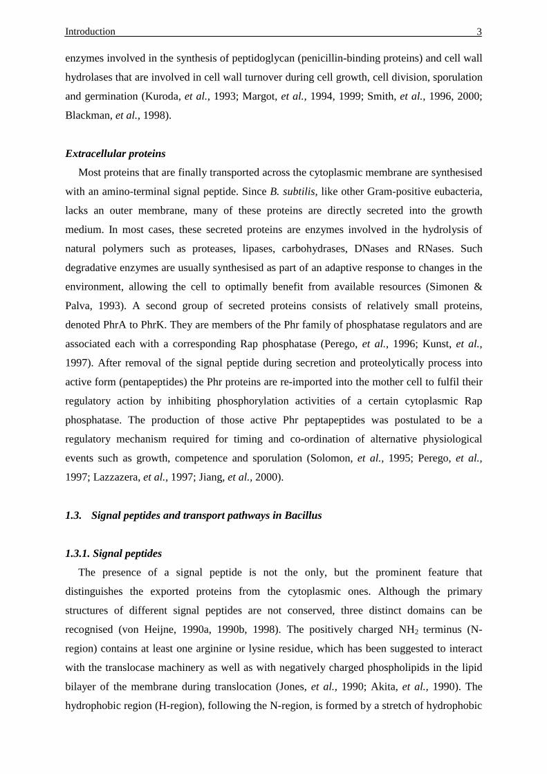
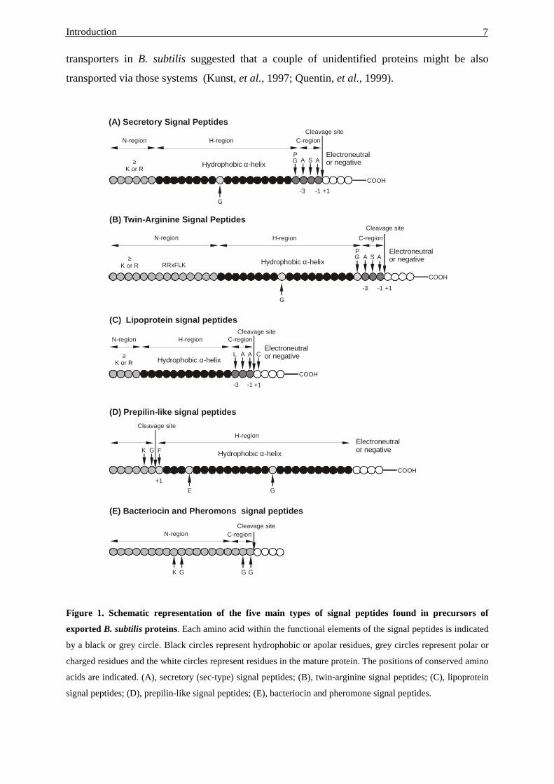
Figure 1. Schematic representation of the five main types of signal peptides found in precursors of
exported B. subtilis proteins. Each amino acid within the functional elements of the signal peptides is indicated
by a black or grey circle. Black circles represent hydrophobic or apolar residues, grey circles represent polar or
charged residues and the white circles represent residues in the mature protein. The positions of conserved amino
acids are indicated. (A), secretory (sec-type) signal peptides; (B), twin-arginine signal peptides; (C), lipoprotein
signal peptides; (D), prepilin-like signal peptides; (E), bacteriocin and pheromone signal peptides.
F
Cleavage site
N-region
H-region
+1
K G
COOH
E G
(D) Prepilin-like signal peptides
Hydrophobic -helixα
COOH
L AA
Cleavage site
≥K or R
N-region H-region C-region
-3 -1 +1
C
(C) Lipoprotein signal peptides
Hydrophobic -helixα
COOH
G
GP
A AS
Cleavage site
≥K or R RRxFLK
N-region H-region C-region
-3 -1 +1
(B) Twin-Arginine Signal Peptides
Hydrophobic -helixα
COOH
G
GP
A AS
Cleavage site
≥K or R
N-region H-region C-region
-3 -1 +1
(A) Secretory Signal Peptides
Hydrophobic -helixαElectroneutralor negative
Electroneutralor negative
Electroneutralor negative
Electroneutralor negative
Cleavage siteC-region
(E) Bacteriocin and Pheromons signal peptides
K G G G
Introduction 8
1.3.2. Transport pathways
In B. subtilis, most proteins seem to be exported to or inserted into the cytoplasmic
membrane via the Sec pathway, but apparently several alternative export pathways exist.
First, the recently identified twin-arginine translocation (Tat) pathway seems to be present in
B. subtilis, as judged from the identification of signal peptides with the RR-motif and
conserved components of this pathway. Second, the assembly of extracellular prepilin-like
structures depends on components which are, most likely, not involved in Sec-dependent
protein secretion. Finally, some small prepeptides contain signal peptides lacking a
hydrophobic domain. These peptides are transported across the membrane and cleaved by
ABC transporters. This report will focus on the first two pathways relating to our main topics:
“Type I SPases of B. amyloliquefaciens and their role in proteins secretion”.
1.3.2.1. Twin-Arginine translocation pathway (Tat pathway)
Protein secretion via this pathway was shown to be independent of Sec components in E.
coli and plant chloroplasts. Possibly, this pathway has evolved specifically for the export of
folded preproteins (Dalbey & Robinson, 1999; Berks, et al., 2000). The Tat pathway was first
discovered in chloroplasts, in which it is involved in ∆pH-dependent protein import into the
thylakoid lumen (Robinson, et al., 1994; Chaddock, et al., 1995; Settles, et al., 1997). For the
chloroplast system it was shown that, in contrast to Sec-dependent translocation, via this
pathway proteins can be translocated in a folded conformation (Clark & Theg, 1997; Hynds,
et al., 1998). Furthermore, it was demonstrated that two adjacent arginines combined with a
hydrophobic determinant (preferably leucine) at position +2 or +3, relative to the twin
arginines, are needed in the N-domain of signal peptides in order to direct precursors into this
pathway (Brink, et al., 1997, 1998; Cristobal, et al., 1999). Although the exact mechanism of
protein export via the Tat pathway has not been yet unravelled, five components of the Tat
pathway of E. coli have been identified. These are TatA (a putative membrane-bound
receptor, homologous to the maize Hcf106 protein) (Settles, et al., 1997), TatB (a TatA
paralogue) (Sargent, et al., 1998), TatC (the putative translocase), TatD (a predicted soluble
protein) and TatE (a TatA paralogue). Interestingly, B. subtilis contains three homologues of
TatA/B/E (encoded by the ydiI, yczB and ynzA genes), two homologues of TatC (encoded by
the ydiJ and ycbT genes) and another TatD homologue which is encoded by the yabD gene
(Kunst, et al., 1997).
Introduction 9
1.3.2.2. General secretory pathway (Sec pathway)
The Sec pathway is responsible for the transport of most proteins in B. subtilis. The various
components of the Sec-dependent secretion machinery can be divided into 5 groups: cytosolic
chaperones, the translocation machinery (SecA, SecY, SecE, SecG, and SecDF-YajC),
SPases, SPPases and folding factors that function at the trans side of the membrane (Figure
2). Moreover, the Sec pathway could be separated into three distinct, but sequential and
interdependent stages.
The stage I of protein export in E. coli requires two secretion-specific chaperones: SecB
and a ribonucleoprotein complex forming the signal recognition particle (SRP). SRP is
composed of an Ffh protein and a 4.5S RNA species. SRP and SecB both recognize subsets of
secreted preproteins, the SRP are most important for the targeting of membrane proteins,
which have multiple-membrane-spanning domains (de Gier, et al., 1998; Valent, et al., 1998).
In B. subtilis the Ffh protein and a small cytoplasmic RNA (functionally related to eukaryotic
7S RNA and E. coli 4.5S RNA) have been identified (Nakamura, et al., 1994). SRP of B.
subtilis seems to contain an additional component, HBsu (an Histone-like protein), which is
not present in SRP of E. coli (Nakamura, et al.,1999). The SecB homologue is not present in
B. subtilis, but a SecB analogue seems to exist. A candidate for a SecB analogue in B. subtilis
is the CsaA protein (Müller, et al., 1992, 2000a, b).
Stage II of protein transport in the Sec-pathway of B. subtilis involves the translocase
complex in the membrane, which consists of at least 6 subunits: SecA (the translocation
motor, an ATPase), SecE, SecDF, SecY, SecG and YajC. From electron-microscopy studies
in E. coli, it has been estimated that up to three SecYE dimers can assemble in a
quasipentagonal ring-shaped structure built around a putative pore (Meyer, et al., 1999). The
SecA ATPase, “translocation motor”, is essential and unique to bacteria (Pohlschroder, et al.,
1997). SecA is a large, elongated, dimeric molecule comprising two primary domains: the
ATPase (amino-terminal) domain and the dimerisation (carboxy-terminal) domain. It was
shown in E. coli that the carboxy-terminal domain allows SecA to bind to SecYEG, leading to
the functional translocase core (Economou & Wickner, 1994; Economou, et al., 1995; Duong
et al., 1997). In B. subtilis, SecDF is a natural Siamese Twin protein (Bolhuis, et al., 1998).
SecDF was shown not to be essential for the cell viability, but to optimise and to maintain a
high capacity for protein secretion (Bolhuis, et al., 1998). In E.coli, SecD and SecF form a
heterometric subcomplex with YajC (Duong, et al., 1997). Disruption of the yajC gene of E.
coli did not have a clear effect on protein export, but overproduction of YajC suppresses the
dominant negative phenotype of secY-d1 mutation, an internal inframe deletion in the secY
gene (Taura, et al., 1994). Finally, the energy for the translocase machinery comes from
Introduction 10
chemical (ATP) as well as electrochemical (proton motive force, PMF) souces; ATP is
essential, and PMF enhances translocation rates (Duong, et al., 1997; Economou, 1998).
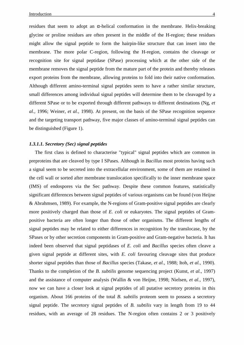
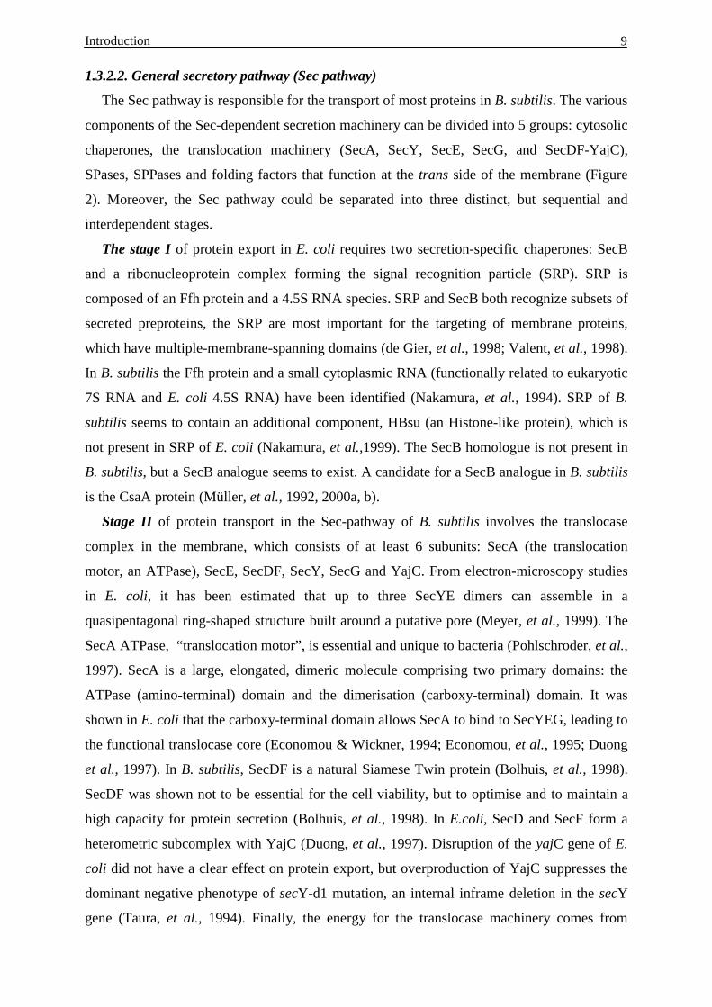
Figure 2. Schematic representation of components of the secretory machinery of B. subtilis. The secretory
proteins are synthesized as precursors with an N-terminal signal peptide. Cytoplasmic chaperones, such as the
SRP complex, FtsY, and Csa play a role in keeping these precursors in a translocation-competent form and
targeting them to the translocase in the membrane. The B. subtilis translocase are contains SecA, SecY, SecE,
SecG, and SecDF. Shortly after translocation, pre-proteins are processed either by one of the type I SPases
(SipS, SipT, SipU, SipV, SipW) in case of secretory proteins or by type II SPases (Lsp) in case of lipoproteins.
SppA and TepA are thought to be involved in the degradation of cleaved signal peptides. PrsA and/or DdbB/C
take part in the folding of the mature protein. The protein is finally released into the medium after passing the
cell wall.
Stage III involves the action of signal peptidase(s) (or leader peptidase(s)) and the
extracytoplasmic folding catalysis (PrsA, BdbA/B/C, metal ion, etc.). Five distinct
chromosomally encoded type I signal peptidases (SipS, SipT, SipU, SipV, and SipW), and
SecA
SecDF SecE SecY SecG
PrsA
BdbB/CLspSppA
ATP
ADP
CELL WALL
MEMBRANE
MEDIUM
SPas
e
SRP
Ribosome
N
C
FtsYSP
CsaA
TepACYTOPLASM
Introduction 11
one type II signal peptidase (LspA), have been identified in B. subtilis (van Dijl, et al., 1992;
Tjalsma, et al., 1997; Kunst, et al., 1997). In contrast to B. subtilis, several other eubacteria,
archaea and yeasts have only one type I signal peptidase (Dalbey & Wickner, 1985; Bult, et
al., 1996; Goffeau, et al., 1997; Smith, et al., 1997; Dalbey, et al., 1997). However, it seems
that most eukaryotic species contain two paralogous type I-SPases (Dalbey, et al., 1997). The
multiple type I-SPases have meanwhile been observed in many other bacteria and archaea,
such as Archaeoglobus fulgidus (Klenk, et al., 1997), Streptomyces lividans (Parro, et al.,
1999), Bradyrhizobium japonicum (Bairl & Müller, 1998) and Bacillus amyloliquefaciens
(Hoang & Hofemeister, 1995). It was shown in E. coli, S. serevisiae and B. subtilis that type
I-SPases are essential for cell viability (Dalbey & Wichne,r 1985; Böhni, et al., 1988; Dalbey
& von Heijne, 1992; Tjalsma, et al., 1998). However in B. subtilis, only SipS and SipT seem
to be of major importance for processing of preproteins as well as for cell viability. SipU,
SipV, SipW seem to play a minor role in protein secretion (Tjalsma, et al., 1998). In addition,
some B. subtilis strains have an extra type I-SPase (SipP), which resides on plasmids. It was
shown that SipP can functionally replace the major SPases SipS and SipT (Tjalsma, et al.,
1999). SipW apparently belongs to the subfamily of eukaryotic type (ER-type) SPases, while
all other type I-SPases of B. subtilis belong to the prokaryotic type (P-type). X-ray
crystallography studies of LepB of E. coli by Paetzel et al. (1998) proved that P-type SPases
use serine-lysine catalytic dyad. The (eukaryotic) ER-type SPases seem to apply a Ser-His-
Asp catalytic triad or Ser-His catalytic dyad, which is supported by site-directed mutant
experiments and by the fact that the catalytic Lys residue of P-type SPases could replaced by
His in ER-type SPases (Dalbey, et al., 1997).
Several extracellular folding catalysts (foldases) mediate folding of export proteins at the
trans side of the membrane. In B. subtilis, a lipoprotein PrsA (a PPIase), which catalyses the
cis-trans isomerization of peptidyl-prolyl bonds, has been identified. PrsA is essential for
viability and strains containing mutant PrsA were shown to be defect in secretion of proteins.
This is probably due to the slowdown of folding, which might result in increased sensitivity of
these exoproteins to proteolysis (Kontinen, et al., 1991, 1993). Another foldase which has
been identified in B. subtilis is thiol-disulfide oxidoreductase, which is responsible for the
formation of disulfied bonds. Three genes (bdbA, bdbB, bdbC) coding for proteins with
similarity to thiol-disulfide oxidoreductases have been found in the B. subtilis genome (Kunst,
et al., 1997; Bolhuis, et al., 1999). In addition, metal ions can act as folding factors. For
example, Ca2+ and Fe3+ can act as folding catalysts for levansucrase in B. subtilis.
Introduction 12
1.4. Protein traffic in sporulation
A large number of the Gram-positive bacilli, including B. subtilis, have adopted
sporulation as a means of survival when environmental conditions are less than optimal for
growth, e.g. after nutrient starvation, drought, extreme cold or heat (Stragier & Losick, 1996).
At the beginning of the sporulation process, a septum is formed that divides the cell into two
unequally sized compartments. Subsequently, the larger compartment (the mother cell)
engulfs the smaller compartment (the forespore), which ultimately becomes the spore. The
forespore is surrounded by two membranes. The inter-membrane space (IMS) is the assembly
site of two specialised peptidoglycan layers, called tile germ cell wall and the cortex. Finally
about six different spore coat layers are formed (Henriques & Moran, 2000). Consequently,
proteins residing in the germ cell wall or in the cortex must be sorted within the IMS towards
the forespore and the mother cell.
One of the processes that require protein transport during sporulation is the communication
between the mother cell and the forespore. Several proteins involved in stage II of
sporulation, such as SpoIID, SpoIIP, SpoIIQ and SpoIIR (Kunst, et al., 1997), contain a
putative signal peptide. The SpoIIR protein, synthesised in the forespore prior to engulfment,
was shown to be exported by the forespore and to interact with membrane proteins of the
mother cell. SpoIIR directly or indirectly activates the receptor/protease SpoIIGA, which is
required for pro-δE processing (Hofmeister, et al., 1995, 1998). SpoIID is homologous to
LytB, the modifier protein that enhances the activity of the major vegetative amidase LytC
(Lazarevic, et al., 1992; Kuroda, et al., 1993). This suggested that SpoIID might play a role in
activation of one or more autolysins, which are required for hydrolysis of the asymmetric
septum permitting prespore engulfment (Illing & Errington, 1991). The lysostaphin-like
SpoIIQ also has a role in prespore engulfment, although apparently not in septum hydrolysis,
because a spoIIQ mutant is blocked after the septum has disappeared (Londono-Vallejo, et al.,
1997). The SpoIIP protein is proposed to be involved in dissolution of the peptidoglycan
located in the sporulation septum. Disruption of spoIIP prevents complete degradation of the
septal cell wall and leads to bulging of the forespore into the mother cell without further
progression to engulfment (Frandsen & Stragier, 1995).
Other processes in sporulation that require transport of proteins, are the biogenesis of the
germ wall and spore-cortex in the IMS of the forespore and the degradation of the spore
peptidoglycan during germination. CwlD and DacB (also known as PBP5*) (Sekiguchi, et al.,
1995; Popham, et al., 1995, 1999) are two export proteins that were reported to be involved in
cortex synthesis. The germination-specific amidase SleB was found to be localized on the
exterior side of the cortex in spores, while it is synthesised in the forespore compartment
Introduction 13
(Moriyama, et al., 1996; Boland, et al., 2000). The fact that pre-SleB has to be transported
across the forespore inner membrane and processed into its mature form to reach the IMS
implies a functional protein translocation machinery and at least one of the type I SPases to be
present in the forespore inner membrane. The recent finding that TasA (for translocated
antibacterial spore-associated protein) with a broad spectrum of antibacterial activities is
transported to B. subtilis endospores provides another example of spore-specific protein
sorting. TasA is thought to confer a competitive advantage to the spore during the onset of
sporulation and later, during germination, by inhibiting the growth of competing organisms
(Stöver & Driks, 1999). In addition, TasA has been suggested to be required for proper spore
coat assembly, and recent studies showed that SipW (signal peptidase) is specifically required
for this process (Serano, et al., 1999; Stöver & Driks, 1999; Tjalsma, et al., 1999).
1.5. The aims of this study
Several Bacillus species are in use for industrial production of enzymes, of fine
biochemicals, antibiotics, insecticides and also have been used in several traditional food
fermentation processes (Harwood, 1992; Priest, 1993; Bron, et al., 1999). Because of
common apathogenicity (the GRAS status), high secretion capacity and the good knowledge
about their fermentation technology, Bacillus species have been regarded as attractive
production hosts, especially for the secretion of endogenous and heterologous proteins.
Among those species, B. amyloliquefaciens is well known for its high capacity of
extracellular enzyme production. In distinction from B. subtilis, B. amyloliquefaciens was
named for the first time in 1943 by Fukomoto (Fukomoto, 1943) but was only recently
recognised as an independent species (Priest, et al., 1987). Despite the fact that B.
amyloliquefaciens strains were widely used in industries, their usage is complicated due to
difficulties in the genetic manipulation techniques and also due to the few studies concerning
cell growth and physiology.
As reviewed, B. subtilis is known to contain five type I signal peptidases (SPases) and this
multiplicity of SPases is proposed to correlate with high export capacities as well as highly
specialised protein transportation during vegetative growth and cell differentiation leading to
sporulation and germination (Kunst, et al., 1997, Tjalsma, et al., 1997, 2000b). The export
capacity of B. amyloliquefaciens strains is even 10 times higher as documented by its use for
production of enzymes for large-scale fermentation (Ingle & Boyer, 1976; Vehmaanpera, et
al., 1991). In continuation of initial studies (Hoang & Hofemeister, 1995) and based on the
constitution of the genome of B. subtilis for multiple type I SPases, we decided to search for
and to specify the peculiarities of sip(Ba) genes in B. amyloliquefaciens and to investigate the

Introduction 14
specificity of Sip(Ba) enzymes with respect to their functions and subcellular localisation.
The specific functions of each of the SPases of B. amyloliquefaciens was studied by
construction of gene-specific (sip-) mutants, characterisation of the mutant phenotype,
heterologous expression in E. coli and complementation of LepB, the type I-SPase of E. coli
by wildtype and hybrid SPase enzymes. In additional, we decided to clone a gene encoding a
new extracellular protein, of which the transport was thought to be affected in the sipT
mutant. The chitin-binding properties of the protein will be characterised and its export will
be investigated.
Materials and Methods 15
2. Materials and Methods
2.1. Enzymes and Chemicals
Amersham-Pharmacia, Braunschweig ECL random primer and labelling kit, Hybond-N+,
Hyperfilm ECL, SureClone ligation kit, Restriction
enzymes, 35S-Methionine in vivo labelling grade,
Megaprimer DNA labelling systems, (α-32P)dCTP.
Roche, Mannheim Agarose, PCR-nucleotide mix, Expand long
template PCR-kit, restriction enzymes, Taq-DNA
polymerase, T4-DNA ligase, T4-DNA polymerase,
protease-inhibitor set.
Carl Roth, Karlsruhe Ampicillin, BSA, Chloramphenicol, EDTA, Acetic
acid, Ethanol, Ethidiumbromid, Fructose, Glucose,
Glycerin, HCl, IPTG, MgCl2, Sodium acetate, NaCl,
nButanol, Phenol, Phenol-Chloroform, Proteinase K,
Sucrose, SDS, Tris-HCl, X-Gal, rotiphorese Gel30
(30% acrylamid, 0.8% bisacrylamid solutions).
Difco Laboratories, Augsburg Agar-Agar, Trypton broth, Yeast extract.
Merck, Darmstadt KH2PO4, K2HPO4.
Millipore, Eschborn Nitrocellulose (0,025 µm) .
New England-Biolabs, Schwalbach/Ts. Restriction enzymes, Shrimp alkali phosphatase.
Qiagen, Hilden Plasmid Midi kit (50), Plasmid Maxi kit (10),
QIAEx gel extraction kit, QIAquick PCR
purification kit, QIAGEN Genomic-tip 100/G and
500/G, Rneasy Mini kit, Ni-NTA superflow (25),
His Antibody (100).
Promega, pGEM-T vector systems.
Sartorius, Göttingen Sterilfilter (0.2 µm).
Sigma-Aldrich, Steinheim ATP, MnSO4, Papain, Pepsin, PMSF, amino acids.
Serva, Heidelberg Succinat, Dextransulfat, Dimethylsulfoxid,
Lysozyme, RNase, Triton X-100, Tween20.
Winthorp, Dublin, Ireland Kanamycin.
Materials and Methods 16
2.2. Strains and growth conditions
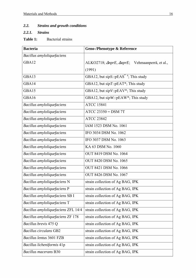
2.2.1. Strains
Table 1: Bacterial strains Bacteria Geno-/Phenotype & Reference
Bacillus amyloliquefaciens
GBA12
ALKO2718; ∆nprE, ∆aprE; Vehmaanperrä, et al.,
(1991)
GBA13 GBA12, but sipS::pEAS* a; This study
GBA14 GBA12, but sipT::pEAT*; This study
GBA15 GBA12, but sipV::pEAV*; This study
GBA16 GBA12, but sipW::pEAW*; This study
Bacillus amyloliquefaciens ATCC 15841
Bacillus amyloliquefaciens ATCC 23350 = DSM 7T
Bacillus amyloliquefaciens ATCC 23842
Bacillus amyloliquefaciens IAM 1523 DSM No. 1061
Bacillus amyloliquefaciens IFO 3034 DSM No. 1062
Bacillus amyloliquefaciens IFO 3037 DSM No. 1063
Bacillus amyloliquefaciens KA 63 DSM No. 1060
Bacillus amyloliquefaciens OUT 8419 DSM No. 1064
Bacillus amyloliquefaciens OUT 8420 DSM No. 1065
Bacillus amyloliquefaciens OUT 8421 DSM No. 1066
Bacillus amyloliquefaciens OUT 8426 DSM No. 1067
Bacillus amyloliquefaciens N strain collection of Ag BAG, IPK
Bacillus amyloliquefaciens P strain collection of Ag BAG, IPK
Bacillus amyloliquefaciens SB I strain collection of Ag BAG, IPK
Bacillus amyloliquefaciens T strain collection of Ag BAG, IPK
Bacillus amyloliquefaciens ZFL 14/4 strain collection of Ag BAG, IPK
Bacillus amyloliquefaciens ZF 178 strain collection of Ag BAG, IPK
Bacillus brevis 475 Q strain collection of Ag BAG, IPK
Bacillus circulans GB2 strain collection of Ag BAG, IPK
Bacillus lentus 3601 FZB strain collection of Ag BAG, IPK
Bacillus licheniformis 41p strain collection of Ag BAG, IPK
Bacillus macerans B30 strain collection of Ag BAG, IPK

Materials and Methods 17
Bacillus megaterium PV361 strain collection of Ag BAG, IPK
Bacillus polymyxa ATCC 842
Bacillus sphaericus ATCC 14577
Bacillus stearothermophilus DSM No. 22 T = ATCC 12980
Bacillus subtilis GSB26 arol906 metB6 sacA321 str6 amyE.
Derivative of QB1133; Steinmetz, et al., (1976) Bacillus subtilis 168 ATCC 6051
Bacillus thuringiensis 2046 strain collection of Ag BAG, IPK
Escherichia coli DH5α F’, φ80d/lacZ∆M15, recA1, endA1, gyrA96, thi-1,
hsdR17(rK-, mK+), supE44, relA1, deoR, ∆(lacZYA-
argF) U169; Hanahan (1983)
Escherichia coli XL1-Blue recA1, endA1, gyrA96, thi-1, hsdR17, supE44, relA1,
lac, [F proAB, lacIqZ∆M15, Tn10(tetR)]; Stratagene
Escherichia coli M15[pREP4] Nals, Strs, Rifs, Thi-, Lac-, Ara+, Gal+, Mtl-, F-, RecA+,
Uvr+, Lon+; QIAGEN
Escherichia coli IT41 W3110, Lep-9ts; Tcr; Inada, et al., (1989)
Thermoactinomyces vulgaris 94-2A Klingenberg, et al., (1979)
2.2.2. Nutrient media
All the media listed here were sterilized for 20 min at 1atm/121°C. If it is not indicated
otherwise, all the media were prepared with deionized water and the solid medium was
prepared with the same ingredients as liquid medium, but with addition of agar–agar (1.5 %).
2.2.2.1. DM3 - Agar
- for regeneration of protoplast of Bacillus Na-Succinate (2 M) 15 % Saccharose (2 M) 5 % K2HPO4 / KH2PO4 10 % Casamino acids 0.25 % Yeast extract 0.25 % Glucose 1 % Needed amino acids (2 mg/ml) 2.5 % Agar solution (2%) + soluble starch (2%) 50 %
Materials and Methods 18
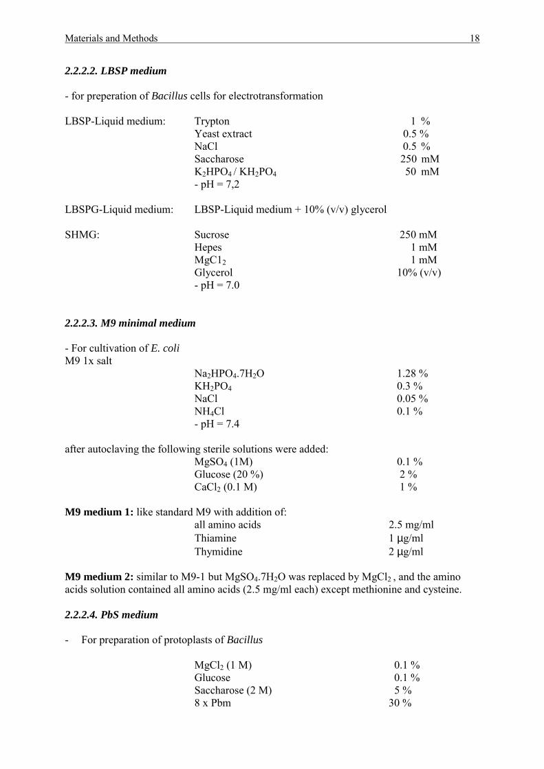
2.2.2.2. LBSP medium - for preperation of Bacillus cells for electrotransformation LBSP-Liquid medium: Trypton 1 % Yeast extract 0.5 % NaCl 0.5 % Saccharose 250 mM K2HPO4 / KH2PO4 50 mM - pH = 7,2 LBSPG-Liquid medium: LBSP-Liquid medium + 10% (v/v) glycerol SHMG: Sucrose 250 mM
Hepes 1 mM MgC12 1 mM Glycerol 10% (v/v) - pH = 7.0
2.2.2.3. M9 minimal medium - For cultivation of E. coli M9 1x salt
Na2HPO4.7H2O 1.28 % KH2PO4 0.3 % NaCl 0.05 % NH4Cl 0.1 %
- pH = 7.4 after autoclaving the following sterile solutions were added: MgSO4 (1M) 0.1 % Glucose (20 %) 2 % CaCl2 (0.1 M) 1 % M9 medium 1: like standard M9 with addition of:
all amino acids 2.5 mg/ml Thiamine 1 µg/ml Thymidine 2 µg/ml
M9 medium 2: similar to M9-1 but MgSO4.7H2O was replaced by MgCl2 , and the amino acids solution contained all amino acids (2.5 mg/ml each) except methionine and cysteine. 2.2.2.4. PbS medium - For preparation of protoplasts of Bacillus
MgCl2 (1 M) 0.1 % Glucose 0.1 % Saccharose (2 M) 5 % 8 x Pbm 30 %
Materials and Methods 19
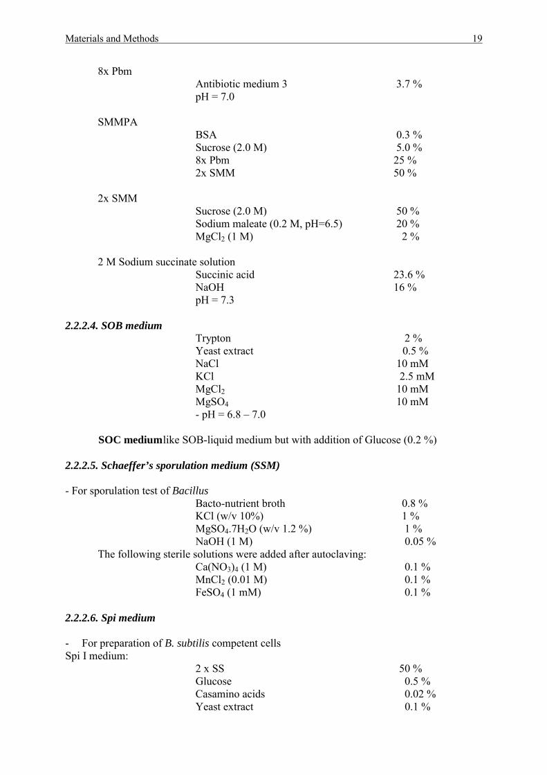
8x Pbm Antibiotic medium 3 3.7 % pH = 7.0
SMMPA
BSA 0.3 % Sucrose (2.0 M) 5.0 % 8x Pbm 25 % 2x SMM 50 %
2x SMM
Sucrose (2.0 M) 50 % Sodium maleate (0.2 M, pH=6.5) 20 % MgCl2 (1 M) 2 %
2 M Sodium succinate solution
Succinic acid 23.6 % NaOH 16 % pH = 7.3
2.2.2.4. SOB medium
Trypton 2 % Yeast extract 0.5 % NaCl 10 mM KCl 2.5 mM MgCl2 10 mM MgSO4 10 mM - pH = 6.8 – 7.0 SOC medium like SOB-liquid medium but with addition of Glucose (0.2 %) 2.2.2.5. Schaeffer’s sporulation medium (SSM) - For sporulation test of Bacillus Bacto-nutrient broth 0.8 % KCl (w/v 10%) 1 % MgSO4.7H2O (w/v 1.2 %) 1 % NaOH (1 M) 0.05 % The following sterile solutions were added after autoclaving: Ca(NO3)4 (1 M) 0.1 % MnCl2 (0.01 M) 0.1 % FeSO4 (1 mM) 0.1 % 2.2.2.6. Spi medium - For preparation of B. subtilis competent cells Spi I medium:
2 x SS 50 % Glucose 0.5 % Casamino acids 0.02 % Yeast extract 0.1 %
Materials and Methods 20
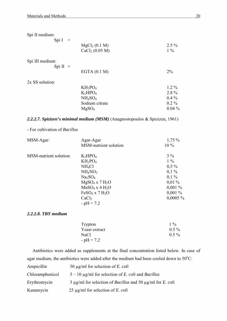
Spi II medium:
Spi I + MgCl2 (0.1 M) 2.5 % CaCl2 (0.05 M) 1 %
Spi III medium:
Spi II + EGTA (0.1 M) 2%
2x SS solution:
KH2PO4 1.2 % K2HPO4 2.8 % NH4SO4 0.4 % Sodium citrate 0.2 % MgSO4 0.04 %
2.2.2.7. Spizizen’s minimal medium (MSM) (Anagnostopoulos & Spizizen, 1961) - For cultivation of Bacillus MSM-Agar: Agar-Agar 1,75 % MSM-nutrient solution 10 % MSM-nutrient solution: K2HPO4 3 % KH2PO4 1 % NH4Cl 0,5 % NH4NO3 0,1 % Na2SO4 0,1 % MgSO4 x 7 H2O 0,01 % MnSO4 x 4 H2O 0,001 % FeSO4 x 7 H2O 0,001 % CaCl2 0,0005 % - pH = 7.2 2.2.2.8. TBY medium
Trypton 1 % Yeast extract 0.5 % NaCl 0.5 % - pH = 7.2
Antibiotics were added as supplements at the final concentration listed below. In case of
agar medium, the antibiotics were added after the medium had been cooled down to 50oC:
Ampicillin 50 µg/ml for selection of E. coli
Chloramphenicol 5 – 10 µg/ml for selection of E. coli and Bacillus
Erythromycin 3 µg/ml for selection of Bacillus and 50 µg/ml for E. coli
Kanamycin 25 µg/ml for selection of E. coli
Materials and Methods 21
200-700 µg/ml in DM3-agar for selection of Bacillus
7 µg/ml in all other media for selection of Bacillus
For blue-white selection of Lac-positive colonies, the respective agar media were
supplemented with 40 µg/ml X-Gal and 40 µg/ml IPTG.
2.2.3. Swarming plate assay
The swarming experiments were done according to Blackman et al., (1998). Swarming
motility of wild type and mutants strains was measured using TBY or MSM soft agar (0.3%)
plates. Samples (1 µl) from overnight (30oC) liquid cultures were spotted onto swarm plates
and incubated at 37oC (TBY agar for 18-22 h, MSM agar for 44-48 h) or 25oC (nutrient agar
for 44-48 h, minimal agar for 68-72 h). The extent of swarming motility was measured as
percentage of the diameter of growth colonies relative to the wild type strain control.
2.2.4. Na3N induced cell autolysis assay
Azide induced cell autolysis experiment was carried out as described by Blackman et al.,
(1998). Cultures of wild type and mutant strains of B. amyloliquefaciens were grown to the
mid-exponential phase (OD600 0.5-0.6) in TBY medium. After addition of 0.05 M sodium
azide, lysis of cells was followed spectrophotometrically while continuing incubation at 37oC
and 200 rpm.
2.2.5. Sporulation test
The frequency of sporulation was estimated by the heat resistance test according to
Nicholsen & Setlow (1990). Cultures of wild type and mutant strains of B. amyloliquefaciens
were grown in the Schaeffer’s sporulation medium (SSM). The samples were taken from the
cultures after 12, 24, 36 h and diluted serially 10-fold in 10 mM potassium phosphate buffer
(pH 7.4) containing 50 mM KCl and 1mM MgSO4. 0.2 ml aliquots of the dilutions were
plated on TBY agar plates before and after heat treatment at 80oC for 10 min. The spore
frequency was determined according to the proportion of the population which survived the
heat treatment by counting colonies the next day.
2.2.6. Phase contrast and electron microscopy
Microscopical pictures of bacterial cultures were made with phase contrast microscope
Nikon T120.
Materials and Methods 22
The electron microscopy picture were prepared using Zeiss CEM 920A transmission
electron microscope. For primary fixation and embedding, Bacillus amyliquefeciens cells
were kept in 50 mM cacodylate buffer (pH 7.2), containing 0.5% (v/v) glutaraldehyde and
2.0% (v/v) formaldehyde for 1 h at room temperature. After washing samples were kept for
the secondary fixation 1 h in a solution of 1% (w/v) OsO4 in 50 mM cacodylate buffer. Prior
to dehydration the cells were washed and transferred into 1,5% agar. The dehydration of
1mm3 agar blocks was done stepwise by increasing the concentration of ethanol. The steps
were performed as follows: 30% (v/v), 50% (v/v), 60% (v/v), 75% (v/v) and 90% (v/v)
ethanol for 60 min each, 100% (v/v) ethanol two times for 1 h. After 1 h dehydration with
propylene oxide the samples were infiltrated subsequently with Spurr (Plano GmbH,
Marburg, Germany) as follows: 33% (v/v), 50% (v/v) and 66% (v/v) Spurr resin in propylene
oxide for 2 h each and then 100% (v/v) Spurr overnight. Samples were transferred into
embedding molds, kept there for 6 h in fresh resin and polymerised at 70 °C for 24 h. Thin
sections with a thickness of approximately 70 nm were cut with a diamond knife and
contrasted with a saturated methanolic solution of uranyl acetate and lead citrate prior to
examination in a Zeiss CEM 920A transmission electron microscope at 80 kV.
2.3. Molecular biological methods
2.3.1. Vectors
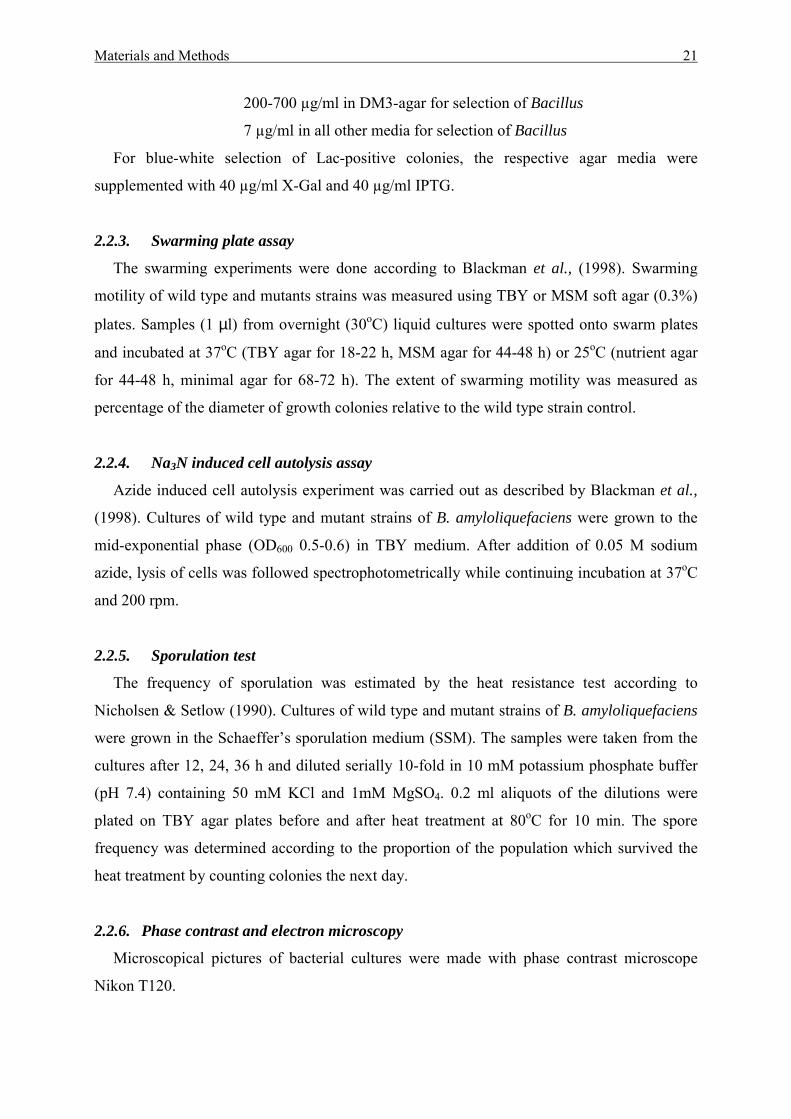
pDG148
Vector pDG148 possess the replicon of pBR322 and the β-lactamase gene ampR for the
replication and ampicilline selection in E. coli (Stragier, et al., 1988). Moreover as a shuttle-
vector, pDG148 possess the replicon from pUB110 for multiplication in B. subtilis
(McKenzie, et al., 1987) and also the phlR and kanR genes of pUB110 which permit a
selection by phleomycin and/or kanamycin. The presence of the Pspac-promoter with
associated Lac-operator and the lacI encoding Lac-repressors from E. coli under control of the
penicillinase promoter Ppen of B. licheniformis allowed the IPTG-induction expression of a
promoterless genes. The multiple cloning site (MCS) HindIII-SalI-SphI allowed to clone
interested genes into the vector under the control of the Pspac-promoter (Figure 3).
Materials and Methods 23
Figure 3. Physical map of Shuttle-Vector pDG148.
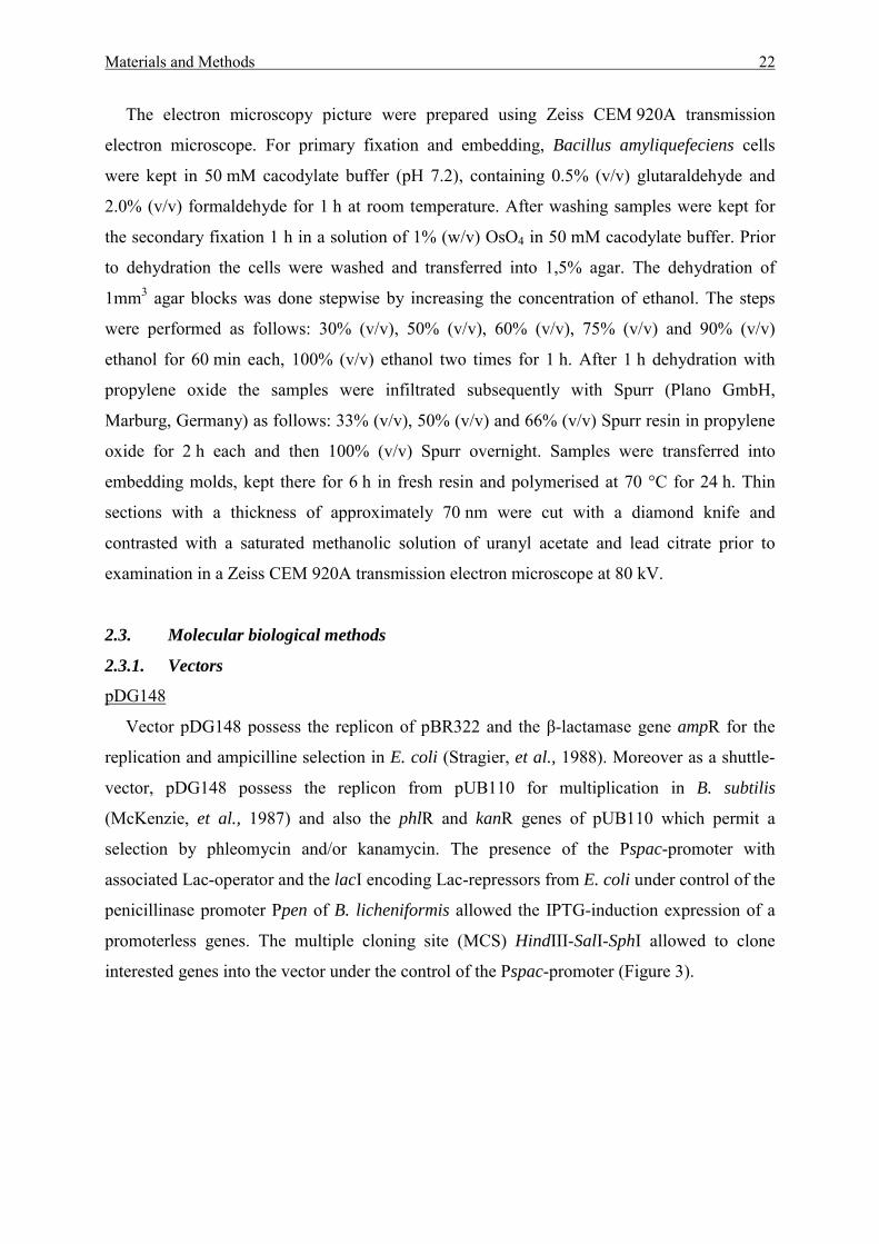
pE194ts
The thermo-sensitive vector pE194ts was used for construction of integrational gene
disruption mutants in Bacillus species that lack natural transformation competence. The
pE194ts (originally Staphylococcus) replicon is unable to sustain autonomous replication in
Bacillus at temperature above 37oC (Youngman, 1990). The pE194ts plasmid contains the
eryR gene that allowed for erythromycin selection. Moreover, the pE194ts could be easily
accomplished with vector contained ColE1-derived replicon such as pUC18 at the PstI site to
form a shuttle-plasmid which can work both in E. coli and in Bacillus.
Figure 4. Physical map of the temperature-sensitive vector pE194ts.
Eco RI (1)
kanR pDG1488274 bp
Pspac
Ppen
ori pBR322
ori pUB110
lacI
ampR
phlRBam HI (1679)
Hind III (289)Sal I (311)Sph I (323)
Eco RI (3976)
Eco RV (142)
Eco RV (1370)
pE194ts3728 bp
eryRrep(ts)
ClaI (1941)
PstI (232)
Materials and Methods 24
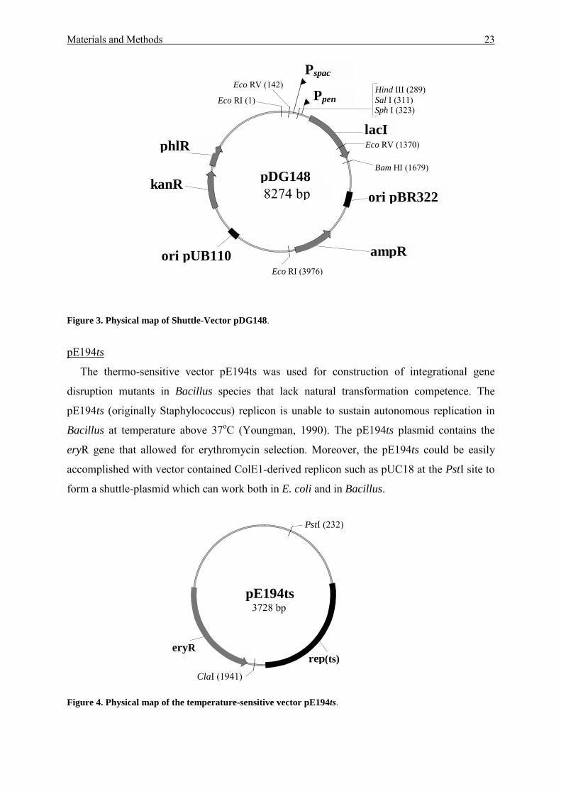
pGEM-T
The pGEM-T vector system (Promega) was used for the cloning of PCR products. The
vector was provided with added 3’ terminal thymidine to both ends of the EcoRV-digested
pGEM-5Zf. These single 3’-T overhangs at the insertion site allowed the efficient ligation of
PCR products as several thermostable polymerases often add a single deoxyadenosine to the
5’-end of the amplified products.
The pGEM-T vector contains T7 and SP6 promoters flangking a multiple cloning region
within the α-peptide coding region of the enzyme β-galactosidase. This allows recombinant
clones to be directly identified by blue-white screening on indicator plates (with IPTG/X-Gal
addition). The presence of the ampR gene coding for β-lactamase permits a selection by
ampicilline.
Figure 5. Physical map of pGEM-T vector for cloning of PCR products.
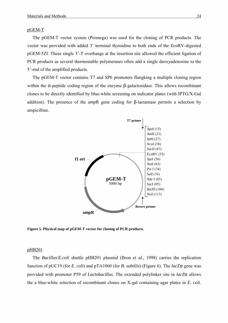
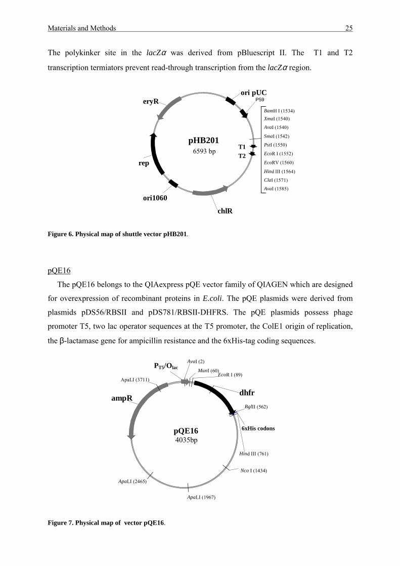
pHB201
The Bacillus/E.coli shuttle pHB201 plasmid (Bron et al., 1998) carries the replication
function of pUC19 (for E. coli) and pTA1060 (for B. subtilis) (Figure 6). The lacZα gene was
provided with promoter P59 of Lactobacillus. The extended polylinker site in lacZα allows
the a blue-white selection of recombinant clones on X-gal containing agar plates in E. coli.
pGEM-T3000 bp
ampR
f1 ori
NcoI (38)
Pst I (74)
AatII (21)ApaI (15)
BstXI (104)
Nde I (83)
NotI (63)
NsiI (113)
SacI (95)
SacII (47)
SalI (76)
SpeI (56)
SphI (27)
EcoRV (52)
T7 primer
Revers primer
Materials and Methods 25
The polykinker site in the lacZα was derived from pBluescript II. The T1 and T2
transcription termiators prevent read-through transcription from the lacZα region.
Figure 6. Physical map of shuttle vector pHB201.
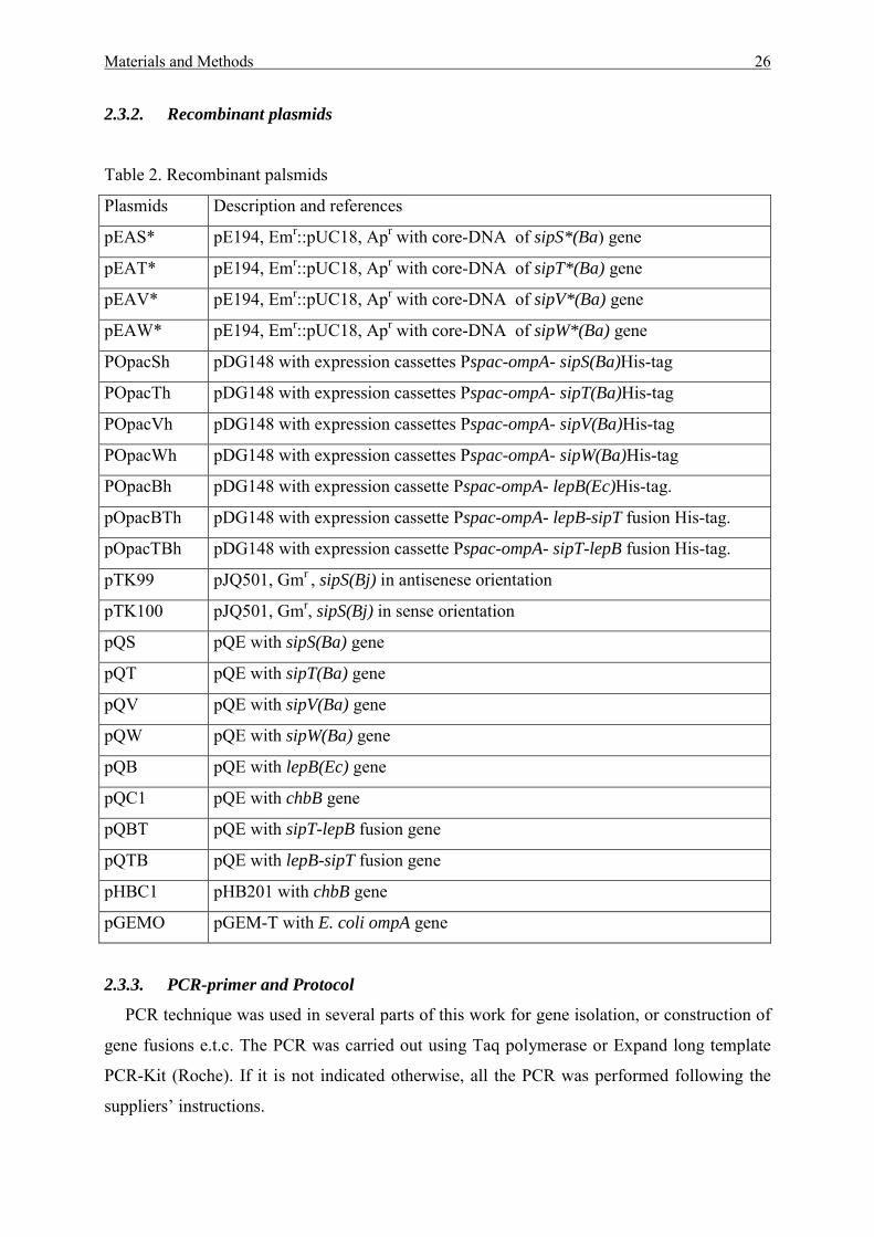
pQE16
The pQE16 belongs to the QIAexpress pQE vector family of QIAGEN which are designed
for overexpression of recombinant proteins in E.coli. The pQE plasmids were derived from
plasmids pDS56/RBSII and pDS781/RBSII-DHFRS. The pQE plasmids possess phage
promoter T5, two lac operator sequences at the T5 promoter, the ColE1 origin of replication,
the β-lactamase gene for ampicillin resistance and the 6xHis-tag coding sequences.
Figure 7. Physical map of vector pQE16.
pHB2016593 bp T1
T2
chlR
rep
eryR P59ori pUC
ori1060
BamH I (1534)
ClaI (1571)
EcoR I (1552)
EcoRV (1560)
Hind III (1564)
PstI (1550)
SmaI (1542)
XmaI (1540)
AvaI (1540)
AvaI (1585)
pQE164035bp
dhframpR
6xHis codons
AvaI (2)
Hind III (761)
MunI (60)
Nco I (1434)
EcoR I (89)
BglII (562)
ApaLI (1967)
ApaLI (2465)
ApaLI (3711)
PT5/Olac
Materials and Methods 26
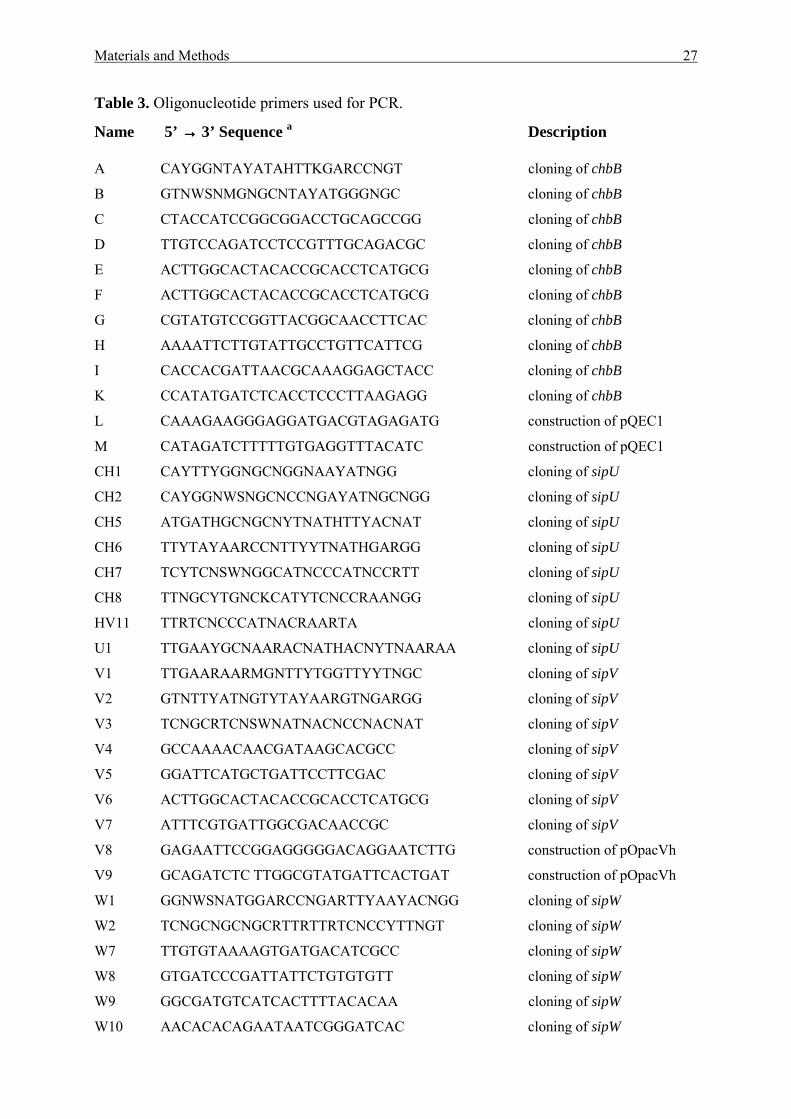
2.3.2. Recombinant plasmids
Table 2. Recombinant palsmids
Plasmids Description and references
pEAS* pE194, Emr::pUC18, Apr with core-DNA of sipS*(Ba) gene
pEAT* pE194, Emr::pUC18, Apr with core-DNA of sipT*(Ba) gene
pEAV* pE194, Emr::pUC18, Apr with core-DNA of sipV*(Ba) gene
pEAW* pE194, Emr::pUC18, Apr with core-DNA of sipW*(Ba) gene
POpacSh pDG148 with expression cassettes Pspac-ompA- sipS(Ba)His-tag
POpacTh pDG148 with expression cassettes Pspac-ompA- sipT(Ba)His-tag
POpacVh pDG148 with expression cassettes Pspac-ompA- sipV(Ba)His-tag
POpacWh pDG148 with expression cassettes Pspac-ompA- sipW(Ba)His-tag
POpacBh pDG148 with expression cassette Pspac-ompA- lepB(Ec)His-tag.
pOpacBTh pDG148 with expression cassette Pspac-ompA- lepB-sipT fusion His-tag.
pOpacTBh pDG148 with expression cassette Pspac-ompA- sipT-lepB fusion His-tag.
pTK99 pJQ501, Gmr , sipS(Bj) in antisenese orientation
pTK100 pJQ501, Gmr, sipS(Bj) in sense orientation
pQS pQE with sipS(Ba) gene
pQT pQE with sipT(Ba) gene
pQV pQE with sipV(Ba) gene
pQW pQE with sipW(Ba) gene
pQB pQE with lepB(Ec) gene
pQC1 pQE with chbB gene
pQBT pQE with sipT-lepB fusion gene
pQTB pQE with lepB-sipT fusion gene
pHBC1 pHB201 with chbB gene
pGEMO pGEM-T with E. coli ompA gene
2.3.3. PCR-primer and Protocol
PCR technique was used in several parts of this work for gene isolation, or construction of
gene fusions e.t.c. The PCR was carried out using Taq polymerase or Expand long template
PCR-Kit (Roche). If it is not indicated otherwise, all the PCR was performed following the
suppliers’ instructions.
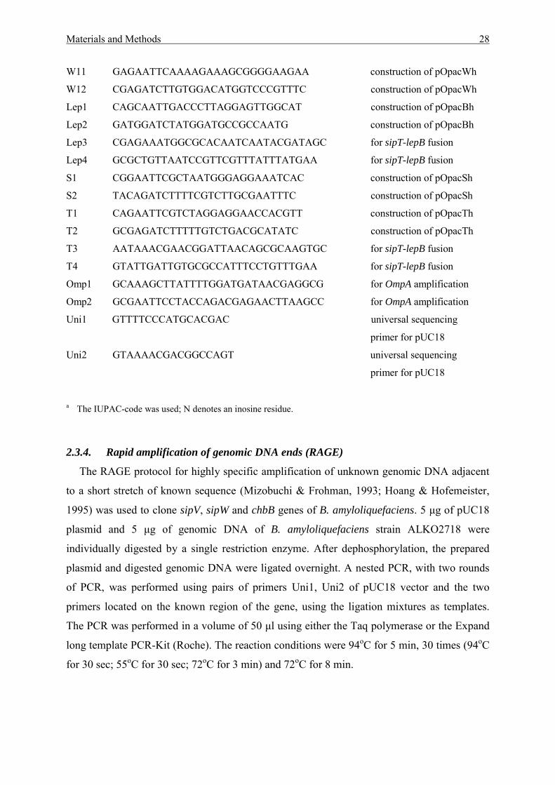
Materials and Methods 27
Table 3. Oligonucleotide primers used for PCR.
Name 5’ →→→→ 3’ Sequence a Description
A CAYGGNTAYATAHTTKGARCCNGT cloning of chbB
B GTNWSNMGNGCNTAYATGGGNGC cloning of chbB
C CTACCATCCGGCGGACCTGCAGCCGG cloning of chbB
D TTGTCCAGATCCTCCGTTTGCAGACGC cloning of chbB
E ACTTGGCACTACACCGCACCTCATGCG cloning of chbB
F ACTTGGCACTACACCGCACCTCATGCG cloning of chbB
G CGTATGTCCGGTTACGGCAACCTTCAC cloning of chbB
H AAAATTCTTGTATTGCCTGTTCATTCG cloning of chbB
I CACCACGATTAACGCAAAGGAGCTACC cloning of chbB
K CCATATGATCTCACCTCCCTTAAGAGG cloning of chbB
L CAAAGAAGGGAGGATGACGTAGAGATG construction of pQEC1
M CATAGATCTTTTTGTGAGGTTTACATC construction of pQEC1
CH1 CAYTTYGGNGCNGGNAAYATNGG cloning of sipU
CH2 CAYGGNWSNGCNCCNGAYATNGCNGG cloning of sipU
CH5 ATGATHGCNGCNYTNATHTTYACNAT cloning of sipU
CH6 TTYTAYAARCCNTTYYTNATHGARGG cloning of sipU
CH7 TCYTCNSWNGGCATNCCCATNCCRTT cloning of sipU
CH8 TTNGCYTGNCKCATYTCNCCRAANGG cloning of sipU
HV11 TTRTCNCCCATNACRAARTA cloning of sipU
U1 TTGAAYGCNAARACNATHACNYTNAARAA cloning of sipU
V1 TTGAARAARMGNTTYTGGTTYYTNGC cloning of sipV
V2 GTNTTYATNGTYTAYAARGTNGARGG cloning of sipV
V3 TCNGCRTCNSWNATNACNCCNACNAT cloning of sipV
V4 GCCAAAACAACGATAAGCACGCC cloning of sipV
V5 GGATTCATGCTGATTCCTTCGAC cloning of sipV
V6 ACTTGGCACTACACCGCACCTCATGCG cloning of sipV
V7 ATTTCGTGATTGGCGACAACCGC cloning of sipV
V8 GAGAATTCCGGAGGGGGACAGGAATCTTG construction of pOpacVh
V9 GCAGATCTC TTGGCGTATGATTCACTGAT construction of pOpacVh
W1 GGNWSNATGGARCCNGARTTYAAYACNGG cloning of sipW
W2 TCNGCNGCNGCRTTRTTRTCNCCYTTNGT cloning of sipW
W7 TTGTGTAAAAGTGATGACATCGCC cloning of sipW
W8 GTGATCCCGATTATTCTGTGTGTT cloning of sipW
W9 GGCGATGTCATCACTTTTACACAA cloning of sipW
W10 AACACACAGAATAATCGGGATCAC cloning of sipW
Materials and Methods 28
W11 GAGAATTCAAAAGAAAGCGGGGAAGAA construction of pOpacWh
W12 CGAGATCTTGTGGACATGGTCCCGTTTC construction of pOpacWh
Lep1 CAGCAATTGACCCTTAGGAGTTGGCAT construction of pOpacBh
Lep2 GATGGATCTATGGATGCCGCCAATG construction of pOpacBh
Lep3 CGAGAAATGGCGCACAATCAATACGATAGC for sipT-lepB fusion
Lep4 GCGCTGTTAATCCGTTCGTTTATTTATGAA for sipT-lepB fusion
S1 CGGAATTCGCTAATGGGAGGAAATCAC construction of pOpacSh
S2 TACAGATCTTTTCGTCTTGCGAATTTC construction of pOpacSh
T1 CAGAATTCGTCTAGGAGGAACCACGTT construction of pOpacTh
T2 GCGAGATCTTTTTGTCTGACGCATATC construction of pOpacTh
T3 AATAAACGAACGGATTAACAGCGCAAGTGC for sipT-lepB fusion
T4 GTATTGATTGTGCGCCATTTCCTGTTTGAA for sipT-lepB fusion
Omp1 GCAAAGCTTATTTTGGATGATAACGAGGCG for OmpA amplification
Omp2 GCGAATTCCTACCAGACGAGAACTTAAGCC for OmpA amplification
Uni1 GTTTTCCCATGCACGAC universal sequencing
primer for pUC18
Uni2 GTAAAACGACGGCCAGT universal sequencing
primer for pUC18
a The IUPAC-code was used; N denotes an inosine residue.
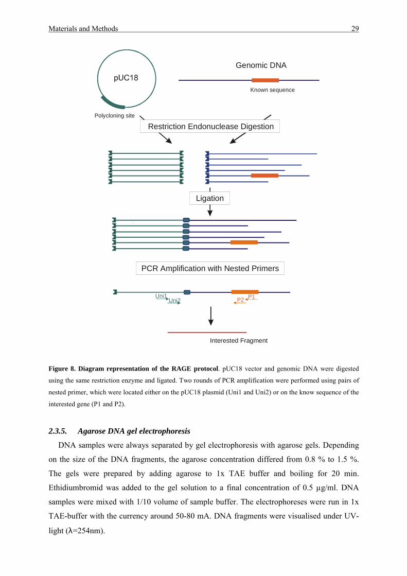
2.3.4. Rapid amplification of genomic DNA ends (RAGE)
The RAGE protocol for highly specific amplification of unknown genomic DNA adjacent
to a short stretch of known sequence (Mizobuchi & Frohman, 1993; Hoang & Hofemeister,
1995) was used to clone sipV, sipW and chbB genes of B. amyloliquefaciens. 5 µg of pUC18
plasmid and 5 µg of genomic DNA of B. amyloliquefaciens strain ALKO2718 were
individually digested by a single restriction enzyme. After dephosphorylation, the prepared
plasmid and digested genomic DNA were ligated overnight. A nested PCR, with two rounds
of PCR, was performed using pairs of primers Uni1, Uni2 of pUC18 vector and the two
primers located on the known region of the gene, using the ligation mixtures as templates.
The PCR was performed in a volume of 50 µl using either the Taq polymerase or the Expand
long template PCR-Kit (Roche). The reaction conditions were 94oC for 5 min, 30 times (94oC
for 30 sec; 55oC for 30 sec; 72oC for 3 min) and 72oC for 8 min.
Materials and Methods 29
Figure 8. Diagram representation of the RAGE protocol. pUC18 vector and genomic DNA were digested
using the same restriction enzyme and ligated. Two rounds of PCR amplification were performed using pairs of
nested primer, which were located either on the pUC18 plasmid (Uni1 and Uni2) or on the know sequence of the
interested gene (P1 and P2).
2.3.5. Agarose DNA gel electrophoresis
DNA samples were always separated by gel electrophoresis with agarose gels. Depending
on the size of the DNA fragments, the agarose concentration differed from 0.8 % to 1.5 %.
The gels were prepared by adding agarose to 1x TAE buffer and boiling for 20 min.
Ethidiumbromid was added to the gel solution to a final concentration of 0.5 µg/ml. DNA
samples were mixed with 1/10 volume of sample buffer. The electrophoreses were run in 1x
TAE-buffer with the currency around 50-80 mA. DNA fragments were visualised under UV-
light (λ=254nm).
Genomic DNA
Polycloning site
Known sequence
Restriction Endonuclease Digestion
Ligation
PCR Amplification with Nested Primers
Uni1Uni2 P1P2
Interested Fragment
Materials and Methods 30
TAE-Buffer:
Tris-acetate 0.04 M EDTA 0.001 M - pH = 8.0 Sample buffer "Helsinki": Glycerol 50 % Tris-HCl 10 mM SDS 0.05 % Bromophenol blue 0.2 % - pH = 8.0 TE-Puffer: Tris-HCl 10 mM EDTA 1 mM - pH = 8.0
2.3.6. Isolation of chromosomal DNA
The mini preparations of chromosomal DNA were carried out following a standard
procedure (Harwood, et al., 1990). 5 ml overnight culture (TBY medium containing
appropriate antibiotics) was grown in a 20 ml flask at 37oC in a waterbath shaker (200
rev/min). The culture was diluted 50-fold in 5 ml fresh medium and continued growth at 37oC
for 2 to 3 h, until the OD650 was 0.8. Cells were harvested by centrifugation for 10 min at
9000 g (4oC). The cell pellet was resuspended in 1.5 ml precooled (0oC) buffer 1 and re-
collected by centrifugation. The cell pellet was again resuspended in 0.7 ml lysis buffer,
containing 8 mg/ml of lysozyme, and mixed by vortexing. After incubation for 10 min at 0oC
and 10 min at 37oC, 25 µl Sarkosyl 30% and 5 µl proteinase K (10 mg/ml) were added. The
cell suspension was mixed by vortexing and incubated for 30 min at 70oC. After vortexing for
1 min at maximum speed, the lysate was subjected to 3 times phenol extraction by adding 700
µl phenol, vortexing gently for 1 min and centrifugation to separate phases in a microfuge for
15 min at full speed. The upper (water) phase was transferred with a 1-ml micropipet tip into
a fresh microtube. After addition of 5µl of RNase (10 mg/ml) and incubation for 15 min at
37oC, the lysate was extracted one time by phenol:chloroform (1:1) and one time by 600 µl
chloroform:isoamyl alcohol (24:1). The upper phase was collected. The DNA was
precipitated by adding 2.5 volume ice-cold ethanol and 1/10 volume sodium acetate (3M, pH
4.8), after keeping at –20oC for 20 min and centrifugation (14000 rpm) for 30 min at 4oC. The
pellet was washed two times with 70 % ethanol and dried by leaving the tubes open for 15
min. The DNA was dissolved in 100 µl of TE.
Materials and Methods 31
Buffer 1: Tris-HCl 10 mM NaCl 150 mM EDTA 10 mM - pH = 8.0
Lysis Buffer: Tris-HCl 20 mM NaCl 50 mM EDTA 10 mM Lysozym 8 mg/ml - pH = 8.0
QIAGEN genomic-tips 100/G and 500/G were used to isolate large amounts of
chromosomal DNA following the supplier’s instruction.
2.3.7. Isolation of plasmid DNA
Qiagen Plasmid Midi and Maxi Kits were used to prepare plasmid DNA for sequencing
and to prepare more than 10 µg DNA. In case plasmids isolation from Bacillus 8 mg/ml
lysozyme was added to buffer P1 and the incubation step was prolonged up to 30 min at RT
after buffer P2 was added.
The minipreparation of Plasmid-DNA of E. coli and Bacillus was done following the
method described by Birnboim & Doly (1979). 5 ml cultures in TBY or NBY were incubated
overnight at 37°C. Depending on the copy number of the plasmids 1-5 ml cell cultures were
collected by centrifugation for 5 min at 6000 rpm RT. The cell pellets were resuspended in
200 µl buffer P1 and incubated at 37°C for 10 min. For Bacillus cells, buffer P1 was supplied
with 8 mg/ml lysozyme and the incubation was extended up to 30 min. 200 µl buffer P2 were
added and the probes were incubated on ice for 10 min, then for another 10 min after adding
200 µl buffer P3. The lysates were subjected to two times phenol-chloroform (1:1) extraction
by adding 500 µl phenol-chloroform, vortexing and centrifugation at 14000 rpm for 15 min.
The upper phase was collected and transferred to a new tube. Two volumes of ice-cold
ethanol were added to precipitate the DNA. The DNA-pellet was recovered by centrifugation
at 14000 rpm 4oC for 30 min. The pellet was washes with ice-cold 70% ethanol, dried by
vacuum centrifugation and dissolved in 50 µl TE.
Buffer P1: Tris-HCl 50 mM EDTA 10 mM RNase A 100 µg/ml - pH = 8.0
Materials and Methods 32
Buffer P2: NaOH 0.2 N SDS 1 % Buffer P3: Sodium acetate 3 M - pH = 5.5
2.3.8. Extraction of DNA from agarose gels
DNA fragments were isolated from agarose gels after electrophoretic separation using
QIAquick or QIAEX II gel extraction kit following the supplier’s instructions. The latter was
used when the DNA fragment was larger than 5 kb.
2.3.9. Restriction digestion and ligation of DNA
The DNAs were cleaved with various restriction enzymes following the supplier’s
instructions. Ligation was performed with T4 DNA ligase (Sambrook, et al., 1989).
2.3.10. Methylation of restriction site
Transformation of plasmid DNA into B. amyloliquefaciens spec. which harboured the
restriction endonuclease enzyme BamHI, required the methylation of the BamHI restriction
site to protect the DNA from BamHI digestion. The BamHI methylase catalyzes the transfer
of a methyl group from S-adenosylmethionine to the nucleotide N4 (cytosine) of the
recognition sequence of BamHI (GGATCC). 5-10 units of the BamHI methylase were used to
methylate 1 µg DNA in 1x BamHI methylase buffer containing 80 µM S-adenosylmethionine.
Incubation was carried for 1 h at 37oC, followed by 15 min at 65oC for enzyme inactivation.
2.3.11. Filling of DNA ends
The “sticky ends” of DNA fragments, which were not suitable for ligation to other DNA
molecules, were “filled” using T4 DNA-polymerase. T4 DNA-polymerase has 3’-5’
exonuclease activity besides of 5’-3’ DNA-dependent polymerase activities. The DNAs were
incubated with T4 DNA-polymerase in a solution containing 1 mM dNTPs and 1x T4-
polymerase-buffer for 30 min at 12°C and then for 30 min at 37°C. The enzyme was
inactivated by incubation for 20 min at 70°C.
Materials and Methods 33
2.3.12. Dephosphorylation of linearized plasmid DNA
The vectors were linearized by restriction endonuclease and afterwards always
dephosphorylated by Shrimp alkaline phosphatase in order to prevent religation of the
linearized vector. One unit of shrimp alkaline phosphatase was used for dephosphorylation of
1 µg linearized vector in dephosphorylation buffer (50 mM TrisHCl, 0.1 mM EDTA pH 8.5)
at 37oC for 60 min. The shrimp alkaline phosphatase was inactivated by heating at 65°C for
15 min.
2.3.13. Southernblot hybridization procedures
DNA transfer and fixation
For hybridization the chromosomal or plasmid DNA were digested by restriction enzymes
and separated by electrophoresis on agarose gels (0.8%). After electrophoresis, the agarose
gel was placed in 0.25N HCl for 15min. The gel was rinsed two times with distilled water,
then placed in denaturation buffer for 30 min at room temprature with shaking. The gel was
again rinsed with distilled water and placed in neutralization buffer for 30 min with shaking.
The DNA was transferred onto a nylon membrane Hybond N+ using a Vacuum-Blotter
machine (Pharmacia). The Hybond N+ membrane was irrigated for 5 min in H2O and
subsequently for 10 min in transfer buffer. In the vacuum plate, the gel was placed on top of
the membrane and was then overlayed with transfer buffer. The transfer was carried out for 90
min. After transfer, the membrane was washed for 5 min in 2x SSC, air-dried and fixed for 2
h at 80°C. The dried blot was stored at RT or used immediately for a hybridization.
Denaturation buffer: NaOH 0,5 M NaCl 1,5 M Neutralization buffer: Tris-HCl 0,5 M NaCl 1,5 M - pH = 7,5 20x SSC: NaCl 3,0 M Sodium citrate 0,3 M Transfer solution: 10x SSC DNA hybridization
DNA hybridizations were done using either the ECL system (Amersham-Pharmacia) or the
Megaprimer DNA labelling system for 32P radioactive labelling. Hybridization was carried
out according to the supplier’s protocols.
Materials and Methods 34
2.3.14. DNA sequencing and computer analysis DNA sequencing was performed by an automated system (A.L.F. express, Pharmacia), using
the recommended primers for the pGEM-T vector and the AutoRead sequencing kit
(Pharmacia). Sequencing of the pQE constructs was done using the primers recommended by
Qiagen. Sequence analysis was performed with the PC/GENE software system of
IntelliGenetics, Inc. (Mountain View, Calif.) and DNASTAR software of Lasergene Inc.
(Madison, WI 53715, USA). The BLAST software (National Center for Biotechnology
Information, Bethesda, Md.) was used for online database scanning. Phenetic and cladistic
analyses of the amino acid alignment were performed in PAUP* 4.0b8 (Swofford, 2001).
Mean character differences were used to calculate pairwise distances, which were clustered
with the Neighbor-Joining algorithm. Fitch parsimony analysis was conducted with
ACCTRAN character optimization, gaps treated as missing data and the heuristic search
algorithm with 100 random sequence additions. To test the statistical support of the branches
in phenetic and cladistic analyses, bootstrap resamples were conducted with 5000 and 500
replicates, respectively. Parsimony analysis resulted in two equally parsimonious trees of
1388 steps length (CI 0.7177, RI 0.6631), which are compatible with the tree topology
obtained by the Neighbor-Joining analysis.
2.3.15. Transformation of plasmid DNA into E. coli
CaCl2 – method
E. coli competent cells were prepared with the CaCl2-method (Sambrook, et al., 1989). The
competent cells can be stored at - 80oC and used for up to 3 months. 2 ml of an overnight
TBY culture was inoculated from a fresh colony. The culture was diluted 100 fold in fresh
TBY and inoculated for about 3 h at 37oC, 200 rpm in a rotary shaker until the OD600 is 0.4 –
0.5. The culture was kept on ice for 30 min and pelleted by centrifugation at 4000 rpm for 10
min at 4oC in a Sorvall GS3 rotor. The cells were washed twice with 20 ml ice-cold 0.1 M
CaCl2 solution, then gently resuspended in 4 ml ice-cold 0.1 M CaCl2 15% Glycerol. 100 µl
of each suspension of competent cells was transferred to a sterile pre-chilled microfuge tube.
The competent cells were directly used or stored at – 80oC.
For transformation, plasmid DNA was mixed with 100 µl competent cells and incubated
on ice for 20–40 min. The tubes were placed at 42oC in a water bath for 90 sec, and on ice for
1 min. 400 µl SOC medium was added and incubated at 37oC for 30 – 60 min. 150 µl of
transformation culture was plated onto a agar TBY plate containing appropriate antibiotic(s).
Materials and Methods 35
Electrotransformation method
The electrotransformation was used in order to achieve high-efficiency transformation of
plasmid DNA in E. coli. 200 ml TBY culture was inoculated from a single fresh colony with
vigorous shaking (250 rpm) at 37oC until the OD600 reached 0.5 – 0.7. After chilling on ice for
15 min, the cells were collected by centrifugation 15 min 5000 g. The pellets were washed 2
times with 100 ml ice-cold sterile de-ionised water and resuspended in 4 ml ice-cold 10%
glycerol. 100 µl of each cell suspension was transferred into a pre-chilled tubes. The cells can
be directly used or stored at –80oC.
1 – 5 µl of solution containing 0.05-0.5 µg plasmid DNA were added to 100 µl fresh or
thawed cells. The cells were mixed by pipeting and then placed on ice for 15 min. The DNA-
cell mix was transferred into a pre-chilled cuvette. Electrotransformation was performed with
2500 V, 25 µF, and 200 Ohms. The transformed cells were diluted 10 fold with ice-cold SOC
medium and incubated for 30 – 60 min at 37oC. 150 µl of transformation culture was plated
onto a agar plate containing appropriate antibiotic(s) and incubated overnight at 37oC.
2.3.16. Transformation of plasmid DNA into Bacillus
Transformation of competent B. subtilis cells
Competent cells of B. subtilis were prepared as described by Cutting and Vander Horn
(1990). 2 ml Spi I culture were inoculated overnight from a fresh colony at 30oC, with 200
rpm shaking. The following morning the culture was diluted with 18 ml fresh Spi I and
continued incubation at 37oC with vigorous shaking (180 rpm). When the rate of cell growth
reached stationary phase (i.e. no significant change in cell density over 20-30 min), the culture
was mixed with 180 ml of prewarmed Spi II and continued incubation at 37oC for additional
90 min. The culture was kept on ice for 10 min and centrifugated (8000g) for 10 min at 4oC.
18 ml supernatant was saved and the remain was carefully decanted. The cell pellet was
gently resuspended in 18 ml of the “saved” supernatant with addition of 2 ml of sterile
glycerol. Aliquots of competent cells (0.2, 0.5 or 1.0 ml) were transferred into sterile tubes,
frizzed rapidly in liquid nitrogen or in a dry-ice/ethanol bath and stored at –80oC.
The competent cells were thawed rapidly by immersing frozen tubes in a 37oC water bath.
200 µl of Spi III was added to 200 µl thawed cells, mixed gently with 10 µl DNA solution
(0.5 – 5 µg) and the mixture was incubated at 30oC for 90 min or 37oC for 1 h with shaking
(150 rpm). The transformation mixture was appropriately diluted and plated onto TBY agar
plates with selective antibiotic(s).
Materials and Methods 36
Electrotransformation of Bacillus
Electrotransformation of Bacillus was performed as described by Vehmaanperä (1989). 2
ml of TBY culture were inoculated overnight from a single fresh colony. The overnight
culture was diluted 50 fold with 100 ml LBSP medium and incubated at 37oC to late
exponential phase (OD600 ~1.0) with vigorous shaking (200 rpm). After leaving on ice for 10
min, the cells were harvested by centrifugation (10000 g) for 5 min at 4oC using a pre-cooled
rotor and tubes. The cell pellet was washed three times with 100 ml of ice-cold SHMG and
resuspended in 3 ml of ice-cold SHMG. 0.1 ml aliquots of cell suspension were transferred
into microfuge tubes and stored at –80oC. The cells remain transformable for at least several
months.
100 µl portion of frozen cells were thawed quickly in a 37oC waterbath. 1-10 µl solution,
containing 0.01-1 µg of DNA in SHMG, was added to the cell suspension. The mixture was
transferred into a 2 mm pre-cooled electroporation cuvette and kept on ice for 30 min. The
electroporation was carried out with a single pulse (2500 V = 12.5 kV/cm, 25 µF for B.
subtilis; 1500 V = 7.5 kV/cm, 25 µF for B. amyloliquefaciens). The cell suspension was
immediately diluted 10 fold with LBSPG medium and incubated for 60 min with vigorous
aeration at 37oC (or 90 min at 30oC) to allow expression of antibiotic resistance markers. 150
µl of transformed cells were plated on a TBY agar plate containing appropriate antibiotic(s)
and overnight incubated.
Bacillus protoplast transformation
The protoplast transformation of Bacillus was done following the method of Chang &
Cohen (1979). A 5 ml of TBY culture was inoculated from a single colony for 5 h. The
culture was diluted 102, 104, 106 fold in TBY and inoculated overnight at 37oC in a waterbath
shaker (200 rpm). The overnight culture was diluted 20-fold into 10 ml of fresh PbS medium
and grown at 37oC in a waterbath shaker (200 rpm) until the OD470 reach 1.7. The cells were
harvested by centrifugation in glass tubes at 3500 rpm/RT for 10 min with a table centrifuge
machine (HERMLE). After removed the supernatant, the cell pellet was resuspended in 10 ml
of SMMPA containing 50 mg lysozyme. The cell suspension was incubated at 37oC in a
waterbath shaker (90 rpm) and examined under the microscope after 30 min and thereafter in
15 min intervals. The incubation was continued until 99% of the cells are apparently
converted into protoplasts (globular appearance). The protoplasts were harvested by
centrifugation 4000 rpm at RT. After 2 times of gentle washing with 7 ml of SMMPA, the
pellet was resuspended in 2.5 ml SMMPA.
Materials and Methods 37
1-10 µl of DNA (0.2 µg) were added into 0.5 ml protoplast suspension, gently mixed with
1.5 ml of PEG solution (40 % of PEG 6000 in 1x SMM solution) in a microtube, and placed
for 2 min at RT. 5 ml of SMMPA was added to this mixture. The protoplasts were harvest by
centrifugation (7 min, 4000 rpm). After complete removal of the supernatant, 1 ml of
SMMPA were added to the pellet. The suspension was incubated for 60-90 min at 37oC in a
waterbath shaker (100 rev/min) in order to allow the expression of antibiotic resistance
markers (the protoplasts become sufficiently resuspended through the shaking action of the
waterbath). The protoplasts were appropriately diluted in 1 x SSM and 0.1 ml of the dilution
was plated onto a DM3 agar plate.
2.3.17. Pulse–chase protein labelling and immunoprecipitation
Pulse chase labelling
The method described by Edens (1982) was modified and used for pulse chase in vivo
labelling of proteins in E.coli. An overnight culture was inoculated from a single colony in
M9-1 medium at 30oC. The pre-culture was diluted 20 fold to start a 20 ml culture with M9-1
medium and grown at 30oC until OD600 is 0.5. The cells were collected by centrifugation
(5000g) for 5 min, washed two times with M9-2 medium and resuspended in 10 ml M9-2
medium. The cultures were grown for 30 min at 30oC for methionine starvation. Aliquots of
0.5 ml culture were used for the pulse-chase experiment at either 30oC or 45oC. The pulse was
done by adding “hot” 35S-methionine into the culture to a final concentration of 50µCi / ml.
After 1 min of labelling, “cold” non-radioactive methionine and cysteine 2.5 mg/ml were
added. The samples were collected after 0, 1, 5, 10, 20 min. 0.5 ml ice-cold TCA 20% was
added to each sample and subsequently left on ice for 30 min. The pellets were collected by
centrifugation, washed three times with aceton and air-dried. The pellets were resuspended in
100 µl TES buffer and boiled for 5 min. For Bacillus, the M9 minimal medium was replaced
with S7 medium (van Dijl et al., 1991).
Immunoprecipitation
100 µl STD in 10 mM Tris-HCl pH 7.0 and 5 µl antiserum were added to the 100 µl
sample (from upper part), then incubated for 16 h at 4oC. 200 µl of STDS in 10 mM Tris-HCl
pH 8.5 were added together with 50 µl of a 10% (v/v) suspension of protein A-sepharose
(Pharmacia) in STDS and incubated for 30 min at room temperature. The mixture was layered
over 300 µl 10% sucrose in STDS / 10 mM Tris-HCl pH 8.2 and centrifuged to collect the A-
sepharose beads. The pellets were washed 3 times with STDS in 10 mM Tris-HCl pH 8.2.
The pellets were suspended in 50 µl sample buffer and heated for 5 min at 100oC.
Materials and Methods 38
SDS-PAGE and fluorography
The samples were loaded and separated onto 12% SDS-PAGE. Fluorography was
performed as described by Skinner and Griswold (1983).
STD buffer: 0.9% NaCl, 1% Triton X-100, 0.5% sodium deoxycholate.
STDS buffer: 0.9% NaCl, 1% Triton X-100, 0.5% sodium deoxycholate and 0.5% SDS.
TES buffer: 50 mM Tris/HCl, 2 mM EDTA, 2% SDS, pH 8.0
2.4. Biochemical methods
2.4.1. Protein determination
The protein concentrations of the samples were determined using the methods established
by Lowry et al., (1951) or Bradford (1976).
2.4.2. Purification of ChbB protein
Purification of the His-tag protein
The plasmid pQEC1 was transformed into E. coli M15 (pREP4), and overexpression was
achieved following the procedure outlined in the QIAexpressionist handbook (Qiagen). The
His-tag fusion protein was accumulated in the periplasm and was thus available by osmotic
shock treatment. The washed cells were suspended in 30 mM Tris/HCl, pH 8.0, 20 % sucrose,
incubated on ice for 10 min, sedimented (8,000 x g, 15 min, 4°C), resuspended in 5 mM of
ice-cold MgSO4 for 10 min, and centrifuged for 15 min at 4°C. The supernatant was collected,
equilibrated to 50 mM Na2HPO4, pH 8.0, 300 mM NaCl, 10 mM imidazole, and applied to an
Ni-NTA column (Qiagen). After five washes, corresponding to the volume of the column, in
the same buffer supplemented with 40 mM imidazole, proteins were released by increasing
the imidazole concentration. The fusion protein was found to be released in the presence of
200 mM imidazole. The purity of the protein was analyzed by SDS-PAGE and by
immunodetection using anti-ChbB antibodies.
Purification of ChbB from B. amyloliquefaciens culture supernatant
The strain was streaked on LB agar medium and incubated at 37°C overnight. Cells were
collected from 10 plates and transferred to 200 ml of MSM containing 1 % of ground crab
shell chitin on a rotary shaker (200 rpm for 4 h at 37°C). After 30-fold dilution (final volume
6 l), cultivation was continued for 36 h. The bacteria and the insoluble substrate were released
from the culture by centrifugation (8,000 x g, 30 min). The supernatant was cooled to 4°C,
Materials and Methods 39
and (NH4)2SO4 (70 % saturation) was added. After stirring for 2 h, the precipitate was
obtained by centrifugation (18,000 x g, 30 min, 4°C), suspended in 50 ml of 20 mM Tris/HCl
buffer (pH 9.0) and applied to a DEAE column (HR10/10, equilibrated with 20 mM Tris/HCl,
pH 9.0). The eluate comprising the unbound proteins was dialysed overnight using 20 mM
citrate/phosphate (pH 5.2) and applied to a MonoS column equilibrated to pH 5.2. Proteins
were eluted in the same buffer with continuously increasing concentrations of NaCl (from 0 -
0.5 M) at flow rate of 0.6 ml min-1. The fractions containing ChbB protein were collected and
equilibrated to pH 8.0, 1 M (NH4)2SO4, then applied to phenyl superose (HR 5/5) at a flow
rate of 0.6 ml min-1.
2.4.3. SDS-PAGE
The SDS polyacrylamide gel electrophoresis was performed according to Laemmli (1970).
2.4.4. Western blot
Proteins were separated by SDS-PAGE and blotted onto nitrocellulose (Satorius) or nylon
membranes (Fluorotrans, Pall). The membrane was treated with blocking buffer (1 % BSA,
150 mM NaCl, 10 mM Tris/HCl, pH 7.5) for 1 h, and incubated in the same buffer in the
presence of antiserum (dilution 1:1000) for 2 h. After two washes with TBS (20 mM
Tris/HCl, pH 7.5, 500 mM NaCl containing 0.05 % (v/v) Tween 20 and 0.2 % (v/v) Triton X-
100) and one wash with TBS only, the membrane was treated with alkaline phosphatase-
conjugated goat anti-rabbit secondary antibodies (diluted 1:10,000) for 2 h. Colour
development was performed in TBS-buffer after addition of 0.33 mg/ml NBT (Nitro blue
tetrazolium chloride) and 0.165 mg/ml BCIP (5-bromo-4-chloro-3-indolyl phosphate)
(Sigma).
2.4.5. Determination of amino-terminal amino acids
For N-terminal amino acid sequencing, proteins were separated by 12.5 % SDS-PAGE and
transferred to a PVDF membrane (Immobilon P, Millipore). The interested band was
subjected to Edman degradation using a model LF 3400 gas-phase sequencer (Beckman),
followed by HPLC of the phenyl thiohydantoin amino acids.
2.4.6. Nuclease detection after SDS-PAGE
The method of Rosenthal (1977) was used to detect nuclease activity bands by
enzymography after SDS-PAGE. Proteins were separated in a 12% SDS-PAGE using gels
Materials and Methods 40
contain 10 µg/ml calf thymus DNA. Following electrophoresis, the gel was rinsed with water
and placed in 300 ml of 0.04 M Tris-HCI, pH 7.6, 2 mM MgCl2, and 0.02% sodium azide (to
prevent microbial growth). After gentle shaking at room temperature for 1 h, the gel was
again rinsed with water and overnight incubated in the aforementioned buffer at 37oC. The
next morning, the gel was transferred to fresh buffer containing 2 mM CaCl2 (CaCl2 cannot be
added at earlier times because it precipitates with SDS in the gel). Incubation was continued
at 37oC. Within several hours, ethidium bromide was added to the incubation buffer to yield
approximately 1 µg/ml. After 30 min the gel was transferred to fresh buffer without ethidium
bromide. The presence of ethidium bromide in the gel did not interfere with nuclease action in
the course of further incubation. The gels were periodically placed on a long-wave UV light
box and were photographed.
2.4.7. Preparation of cell-wall-binding protein (CWBP)
The cell wall binding proteins of vegetative B. amyloliquefacines cells were prepared
according to Blackman et al. (1998). 1 litre cultures of B. amyloliquefaciens wild type or
mutant strains (OD600 1.0) were centrifuged (11000g, 4oC, 10 min) in order to harvest the
cells. The cells were washed once with ice-cold 50 mM Tris/HCl, 0.5 mM PMSF (pH 7.5),
and resuspended in 30 ml of the same buffer. The cells were broken by ultrasonication,
subsequently centrifuged at 5000 g for 10 min to remove unbroken cells, then at 27000 g for
15 min at 4oC. The pellets were washed twice with ice-cold 50 mM Tris/HCl (pH 7.5), once
with ice-cold 100 mM NaCl, 0.5 mM PMSF to remove any loosely associated proteins, then
again two times with ice-cold 50 mM Tris/HCl, 0.5 mM PMSF (pH 7.5). Cell wall proteins
were extracted (100oC, 3 min) with SDS sample buffer (62.5 mM Tris/HCl pH 6.8, 1mM
EDTA, 1%(w/v) SDS, 5% mercaptoethanol, 0.0025% bromophenolblue, 10% (v/v) glycerol).
2.4.8. Preparation of purified cell-wall from B. amyloliquefaciens
For preparation of purified cell-wall material, the bacteria was grown, collected and broken
in the same way as described for preparation of CWBP. The crude cell wall fraction was
resuspended in a minimal volume of water at 0oC and poured with stirring into boiling 4%
(w/v) sodium dodecyl sulphate (SDS) to a final concentration of about 200 mg (wet wt) per
ml, then incubated at 100oC for 15 min. The suspension was cooled to RT and centrifuged to
sediment the cell wall at 35000g for 30 min at 20oC. It is essential that, in all steps where SDS
may be present, low temperature is avoided to prevent irreversible precipitation of some of the
detergent in the cell wall pellet. The cell wall material was washed again two times with 4%
SDS at 100oC for 15min, repeatedly washed by resuspension and centrifugation with 0.9%
Materials and Methods 41
NaCl, and then with water until no further SDS can be removed. The cell walls were dried by
lyophilization and checked for being free of proteins and nucleic acids.
2.4.9. Autolysin activities detection after SDS-PAGE
The renaturing gel electrophoresis method to detect autolysin activity bands on SDS-
PAGE was employed as previously described by Forster (1992). SDS-PAGE (11% w/v
acrylamide) containing 0.05% (w/v) of purified B. amyloliquefaciens vegetative cell wall
material were used for the detection of lytic activity. Following electrophoresis, gels were
soaked for 30 min in 250 ml of distilled water at room temperature with gentle agitation. The
gels were then transferred to 250 ml of renaturation solution, gently agitated for 30 min at RT,
and then transferred to 250 ml of the same solution and incubated for 16 h at 37oC. After
incubation, the gels were rinsed in distilled water, stained in 0.1 % methylene blue in 0.01%
KOH for 3 h, and destained in distilled water. Autolytic activity appeared as clearing zones in
the blue background. Molecular masses were determined by comparison to standard proteins
(Sigma) of known sizes which were run on the same gel and stained with Coomassie blue.
The results are shown as representative photographs of gels.
Renaturation solution: 0.1% Triton X-100, 10 mM MgCl2 and 25 mM Tris-HCI [pH 7.5]
2.4.10. Assay for ChbB protein binding properties
5 µg of the His-tag ChbB protein purified from E. coli or the purified ChbB protein
secreted by B. amyloliquefaciens were mixed with 2 mg of substrate in 50 µl of the indicated
buffer (see below) containing 1 % BSA and shaken for 16 h at 4°C or for 3 h at room
temperature. The samples were centrifuged at 10,000 x g for 5 min, then each supernatant was
collected. The pellet was washed twice in the same buffer and resuspended with 50 µl of
buffer. Each sample (supernatant or resuspended pellet) was mixed with loading buffer,
heated for 10 min at 100°C, and analysed using 12 % SDS-PAGE. The relative quantities of
the protein were estimated after Coomassie staining or immunodetection (Zeltins &
Schrempf, 1995) by scanning of corresponding bands and subsequent analysis using Scion
Image software (Scion crop., Maryland 21701, USA). In order to study the effect of the pH,
crab shell powder was mixed with ChbB protein in aliquots of various buffers adjusted to
different pHs. Citrate-phosphate buffer (10 mM) was used for tests at pH 3 – pH 7, Tris/HCl
buffer (10 mM) for pH 7 – pH 10. The effect of salt was tested after the addition of NaCl (0.5
M or 1 M) to citrate-phosphate buffer (pH 7, 10 mM, 1 % BSA) containing the chitinous
sample. Various substrates, i.e. α-chitin (crab shells), β-chitin from Sepia and from
Materials and Methods 42
Siboglinum fjordicum, chitosan, β-glucan (yeast), β-glucan (barley), cellulose from cotton
linters, and xylan (oat spells), were used to study the binding specificity of the ChbB protein
in citrate-phosphate buffer (pH 7, 10 mM, 1 % BSA).
2.4.11. Test for chitinolytic activity
Cultures were grown in Spizizen minimal medium with 0.5 % colloidal chitin and 0.2 %
yeast extract at 37°C for 24 h. The cell-free supernatant was used for enzyme testing. The test
was performed using carboxymethyl chitin-Remazol Brilliant Violet (No. 04106, Loewe
Biochimica GmbH) by mixing 0.1 ml of citrate-phosphate buffer (0.2 M, pH 6.0), 0.1 ml of
the substrate (0.2 % in water) with 0.2 ml of respective culture supernatant or enzyme sample.
The incubation was performed for 12 h at 37°C. The reaction was stopped by adding 0.1 ml of
1 N HCl, and absorption was estimated at 600 nm. The plate assay was performed by striking
cells on TBY agar containing 2 % of colloidal chitin. The plates were incubated for 48 h at
37°C and rinsed with KJ/J2 reagent. Chitin hydrolysis was recorded by halos in zones of
colony growth.

Results 43
3. Results
3.1. Cloning of sipV(Ba) and sipW(Ba) genes of B. amyloliquefaciens
Using a PCR-based cloning strategy with oligonucleotide primers that were designed
according to amino acid sequence of B. subtilis SipS, we previously succeed in cloning two
different type I SPase (sip) genes in B. amyloliquefaciens. The genes were named sipS1 and
sipS2 (Hoang & Hofemeister, 1995), but have also been known as sipS(Ba) and sipT(Ba)
following the names of their B. subtilis counterparts (Kunst, et al., 1997; Tjalsma, et al.,
1997), respectively. Surprisingly, after completion of the B. subtilis 168 genome sequencing
project, three additional sip genes, sipU, sipV and sipW were found (Kunst, et al., 1997). The
proposed but not yet understood function of these multiple SPases induced us to search for
their likely counterparts in B. amyloliquefaciens in order to prove their general existence as
well as to search their (specific) functions in other closely related Bacillus species. As
previously indicated (Meijer, et al., 1995), the amino acid sequence alignment of type I
SPases of B. subtilis and B. amyloliquefaciens (see Figure 16 & 17), demonstrated conserved
regions and extensive sequence similarities among the group of type I SPases of B. subtilis.
The similarity of the latter was significantly higher than that of different SPases of the same
organism, as B. subtilis SipS was found closest homology with B. amyloliquefaciens SipS.
This allowed us to use the amino acid sequences of the various B. subtilis SPases to design
degenerated oligo primers for PCR amplification of a similar gene candidate in B.
amyloliquefaciens.
3.1.1. Cloning of a sipV gene homologue
The regions MKKRFWFLA, VFIDYKVEG, and IVGVISDAE were chosen from the
amino acid sequence of B. subtilis SipV to prepare the primers V1, V2, and V3 (Table 3).
PCR reactions with genomic DNA of the strain ATCC 23843 yielded two PCR products of
about 500 bps and 400 bps (see Figure 9). The fragments were cloned into pUC18 using the
sure cloning kit (see Materials and methods). Sequencing data confirmed that both fragments
overlap in one DNA region. The sequences were then used to make a BLAST search at the
NCBI server (http://www.ncbi.nlm.nih.gov/BLAST), and the resulting amino acid sequence
showed about 70% similarity to SipV of B. subtilis. The 500 bp sipV(Ba) DNA fragment was
used as probe for Southern hybridization of either PstI- or EcoRI-digested chromosomal DNA
of the strain ATCC 23843. Only one single band gave a positive signal (data not shown)
Results 44
indicating specificity to only one sipV-like gene [sipV(Ba)] in B. amyloliquefaciens genomic
DNA.
In order to isolate the full length of the sipV(Ba) gene as well as flanking DNA regions, we
decided to use the RAGE (rapid amplification of genomic DNA ends) protocol, as outlined in
Figure 9C. PstI- as well as EcoRI-digested chromosomal DNA of B. amyloliquefaciens was
ligated to either PstI- or EcoRI-cut vector DNA (pUC18) in order to get a suitable large
template for PCR amplification of the whole gene region (Figure 9C). Two distinct PCR
fragments were obtained, cloned and sequenced. The sequences were analysed and confirmed
to overlap with the core DNA of the proposed sipV(Ba) gene region. The BLAST search
using the nucleotide sequence of the amplified DNA fragment revealed the presence of three
ORFs, and the deduced proteins to have 70, 77, and 67 percent of identity with B. subtilis
proteins encoded by genes yhjE-sipV-yhjG, respectively (Figure 9C). Thus, flanking ORFs
indicated that the B. amyloliquefaciens sipV(Ba) gene occupies a similar genomic position as
compared to sipV(Bs) of B. subtilis (Kunst, et al., 1997). The sequence was submitted to the
GenBank database (http://www.ncbi.nlm.nih.gov/) and given the accession number
AF084950.
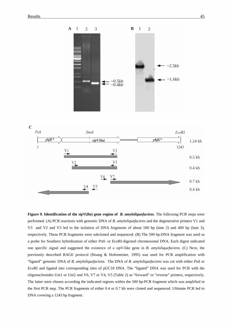
Results 45
Figure 9. Identification of the sipV(Ba) gene region of B. amyloliquefaciens. The following PCR steps were
performed. (A) PCR reactions with genomic DNA of B. amyloliquefaciens and the degenerative primers V1 and
V3 and V2 and V3 led to the isolation of DNA fragments of about 500 bp (lane 2) and 400 bp (lane 3),
respectively. These PCR fragments were subcloned and sequenced. (B) The 500 bp-DNA fragment was used as
a probe for Southern hybridisation of either PstI- or EcoRI-digested chromosomal DNA. Each digest indicated
one specific signal and suggested the existence of a sipV-like gene in B. amyloliquefaciens. (C) Next, the
previously described RAGE protocol (Hoang & Hofemeister, 1995) was used for PCR amplification with
“ligated” genomic DNA of B. amyloliquefaciens. The DNA of B. amyloliquefaciens was cut with either PstI or
EcoRI and ligated into corresponding sites of pUC18 DNA. The “ligated” DNA was used for PCR with the
oligonucleotides Uni1 or Uni2 and V6, V7 or V4, V5 (Table 2) as “forward” or “reverse” primers, respectively.
The latter were chosen according the indicated regions within the 500 bp-PCR fragment which was amplified in
the first PCR step. The PCR fragments of either 0.4 or 0.7 kb were cloned and sequenced. Ultimate PCR led to
DNA covering a 1243 bp fragment.
~0.5kb~0.4kb
A B
~2.5kb
~1.6kb
1 2 3 1 2
0.4 kb
0.5 kb
1.24 kb
0.7 kb
0.4 kb
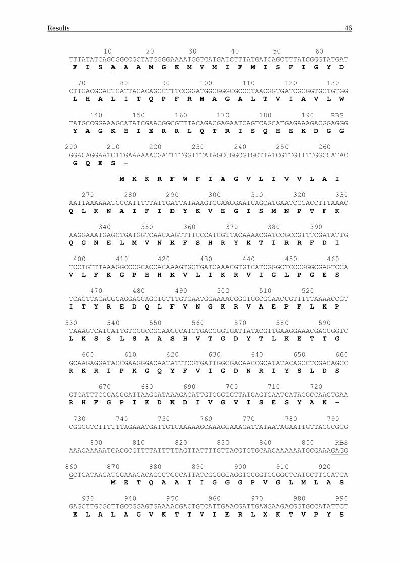
Results 46
10 20 30 40 50 60TTTATATCAGCGGCCGCTATGGGGAAAATGGTCATGATCTTTATGATCAGCTTTATCGGGTATGATF I S A A A M G K M V M I F M I S F I G Y D
70 80 90 100 110 120 130CTTCACGCACTCATTACACAGCCTTTCCGGATGGCGGGCGCCCTAACGGTGATCGCGGTGCTGTGGL H A L I T Q P F R M A G A L T V I A V L W
140 150 160 170 180 190 RBSTATGCCGGAAAGCATATCGAACGGCGTTTACAGACGAGAATCAGTCAGCATGAGAAAGACGGAGGGY A G K H I E R R L Q T R I S Q H E K D G G
200 210 220 230 240 250 260GGACAGGAATCTTGAAAAAACGATTTTGGTTTATAGCCGGCGTGCTTATCGTTGTTTTGGCCATACG Q E S -
M K K R F W F I A G V L I V V L A I
270 280 290 300 310 320 330AATTAAAAAATGCCATTTTTATTGATTATAAAGTCGAAGGAATCAGCATGAATCCGACCTTTAAACQ L K N A I F I D Y K V E G I S M N P T F K
340 350 360 370 380 390AAGGAAATGAGCTGATGGTCAACAAGTTTTCCCATCGTTACAAAACGATCCGCCGTTTCGATATTGQ G N E L M V N K F S H R Y K T I R R F D I
400 410 420 430 440 450 460TCCTGTTTAAAGGCCCGCACCACAAAGTGCTGATCAAACGTGTCATCGGGCTCCCGGGCGAGTCCAV L F K G P H H K V L I K R V I G L P G E S
470 480 490 500 510 520TCACTTACAGGGAGGACCAGCTGTTTGTGAATGGAAAACGGGTGGCGGAACCGTTTTTAAAACCGTI T Y R E D Q L F V N G K R V A E P F L K P
530 540 550 560 570 580 590TAAAGTCATCATTGTCCGCCGCAAGCCATGTGACCGGTGATTATACGTTGAAGGAAACGACCGGTCL K S S L S A A S H V T G D Y T L K E T T G
600 610 620 630 640 650 660GCAAGAGGATACCGAAGGGACAATATTTCGTGATTGGCGACAACCGCATATACAGCCTCGACAGCCR K R I P K G Q Y F V I G D N R I Y S L D S
670 680 690 700 710 720GTCATTTCGGACCGATTAAGGATAAAGACATTGTCGGTGTTATCAGTGAATCATACGCCAAGTGAAR H F G P I K D K D I V G V I S E S Y A K -
730 740 750 760 770 780 790CGGCGTCTTTTTTAGAAATGATTGTCAAAAAGCAAAGGAAAGATTATAATAGAATTGTTACGCGCG
800 810 820 830 840 850 RBSAAACAAAAATCACGCGTTTTATTTTTAGTTATTTTGTTACGTGTGCAACAAAAAATGCGAAAGAGG
860 870 880 890 900 910 920GCTGATAAGATGGAAACACAGGCTGCCATTATCGGGGGAGGTCCGGTCGGGCTCATGCTTGCATCA
M E T Q A A I I G G G P V G L M L A S
930 940 950 960 970 980 990GAGCTTGCGCTTGCCGGAGTGAAAACGACTGTCATTGAACGATTGAWGAAGACGGTGCCATATTCTE L A L A G V K T T V I E R L X K T V P Y S
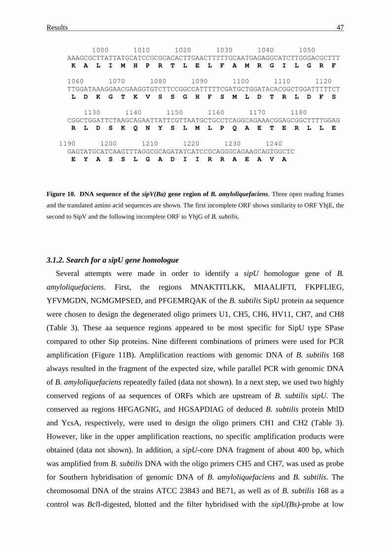
Results 47
1000 1010 1020 1030 1040 1050AAAGCGCTTATTATGCATCCGCGCACACTTGAACTTTTTGCAATGAGAGGCATCTTGGGACGCTTTK A L I M H P R T L E L F A M R G I L G R F
1060 1070 1080 1090 1100 1110 1120TTGGATAAAGGAACGAAGGTGTCTTCCGGCCATTTTTCGATGCTGGATACACGGCTGGATTTTTCTL D K G T K V S S G H F S M L D T R L D F S
1130 1140 1150 1160 1170 1180CGGCTGGATTCTAAGCAGAATTATTCGTTAATGCTGCCTCAGGCAGAAACGGAGCGGCTTTTGGAGR L D S K Q N Y S L M L P Q A E T E R L L E
1190 1200 1210 1220 1230 1240GAGTATGCATCAAGTTTAGGCGCAGATATCATCCGCAGGGCAGAAGCAGTGGCTCE Y A S S L G A D I I R R A E A V A
Figure 10. DNA sequence of the sipV(Ba) gene region of B. amyloliquefaciens. Three open reading frames
and the translated amino acid sequences are shown. The first incomplete ORF shows similarity to ORF YhjE, the
second to SipV and the following incomplete ORF to YhjG of B. subtilis.
3.1.2. Search for a sipU gene homologue
Several attempts were made in order to identify a sipU homologue gene of B.
amyloliquefaciens. First, the regions MNAKTITLKK, MIAALIFTI, FKPFLIEG,
YFVMGDN, NGMGMPSED, and PFGEMRQAK of the B. subtilis SipU protein aa sequence
were chosen to design the degenerated oligo primers U1, CH5, CH6, HV11, CH7, and CH8
(Table 3). These aa sequence regions appeared to be most specific for SipU type SPase
compared to other Sip proteins. Nine different combinations of primers were used for PCR
amplification (Figure 11B). Amplification reactions with genomic DNA of B. subtilis 168
always resulted in the fragment of the expected size, while parallel PCR with genomic DNA
of B. amyloliquefaciens repeatedly failed (data not shown). In a next step, we used two highly
conserved regions of aa sequences of ORFs which are upstream of B. subtilis sipU. The
conserved aa regions HFGAGNIG, and HGSAPDIAG of deduced B. subtilis protein MtlD
and YcsA, respectively, were used to design the oligo primers CH1 and CH2 (Table 3).
However, like in the upper amplification reactions, no specific amplification products were
obtained (data not shown). In addition, a sipU-core DNA fragment of about 400 bp, which
was amplified from B. subtilis DNA with the oligo primers CH5 and CH7, was used as probe
for Southern hybridisation of genomic DNA of B. amyloliquefaciens and B. subtilis. The
chromosomal DNA of the strains ATCC 23843 and BE71, as well as of B. subtilis 168 as a
control was BclI-digested, blotted and the filter hybridised with the sipU(Bs)-probe at low
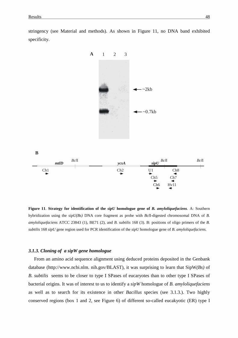
Results 48
stringency (see Material and methods). As shown in Figure 11, no DNA band exhibited
specificity.
Figure 11. Strategy for identification of the sipU homologue gene of B. amyloliquefaciens. A: Southern
hybridization using the sipU(Bs) DNA core fragment as probe with BclI-digested chromosomal DNA of B.
amyloliquefaciens ATCC 23843 (1), BE71 (2), and B. subtilis 168 (3). B: positions of oligo primers of the B.
subtilis 168 sipU gene region used for PCR identification of the sipU homologue gene of B. amyloliquefaciens.
3.1.3. Cloning of a sipW gene homologue
From an amino acid sequence alignment using deduced proteins deposited in the Genbank
database (http://www.ncbi.nlm. nih.gov/BLAST), it was surprising to learn that SipW(Bs) of
B. subtilis seems to be closer to type I SPases of eucaryotes than to other type I SPases of
bacterial origins. It was of interest to us to identify a sipW homologue of B. amyloliquefaciens
as well as to search for its existence in other Bacillus species (see 3.1.3.). Two highly
conserved regions (box 1 and 2, see Figure 6) of different so-called eucakyotic (ER) type I
BclI
Ch8
B
U1
BclIBclI
Ch1 Ch2
Ch7
Hv11
Ch5
Ch6
mtlD ycsA sipU
~0.7kb
~2kb
21A 3
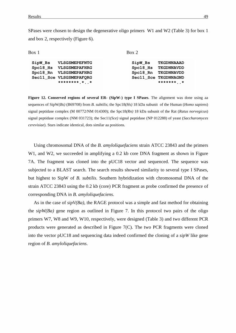
Results 49
SPases were chosen to design the degenerative oligo primers W1 and W2 (Table 3) for box 1
and box 2, respectively (Figure 6).
Box 1 Box 2
Figure 12. Conserved regions of several ER- (SipW-) type I SPases. The alignment was done using aa
sequences of SipW(Bs) (B69708) from B. subtilis; the Spc18(Hs) 18 kDa subunit of the Human (Homo sapiens)
signal peptidase complex (M 00772/NM 014300); the Spc18(Rn) 18 kDa subunit of the Rat (Ratus norvegicus)
signal peptidase complex (NM 031723); the Sec11(Sce) signal peptidase (NP 012288) of yeast (Saccharomyces
cerevisiae). Stars indicate identical, dots similar aa positions.
Using chromosomal DNA of the B. amyloliquefaciens strain ATCC 23843 and the primers
W1, and W2, we succeeded in amplifying a 0.2 kb core DNA fragment as shown in Figure
7A. The fragment was cloned into the pUC18 vector and sequenced. The sequence was
subjected to a BLAST search. The search results showed similarity to several type I SPases,
but highest to SipW of B. subtilis. Southern hybridization with chromosomal DNA of the
strain ATCC 23843 using the 0.2 kb (core) PCR fragment as probe confirmed the presence of
corresponding DNA in B. amyloliquefaciens.
As in the case of sipV(Ba), the RAGE protocol was a simple and fast method for obtaining
the sipW(Ba) gene region as outlined in Figure 7. In this protocol two pairs of the oligo
primers W7, W8 and W9, W10, respectively, were designed (Table 3) and two different PCR
products were generated as described in Figure 7(C). The two PCR fragments were cloned
into the vector pUC18 and sequencing data indeed confirmed the cloning of a sipW like gene
region of B. amyloliquefaciens.
SipW_Bs VLSGSMEPEFNTGSpc18_Hs VLSGSMEPAFHRGSpc18_Rn VLSGSMEPAFHRGSec11_Sce VLSGSMEPAFQRG
********.*..*
SipW_Bs TKGDNNAAADSpc18_Hs TKGDNNAVDDSpc18_Rn TKGDNNAVDDSec11_Sce TKGDNNAGND
*******..*
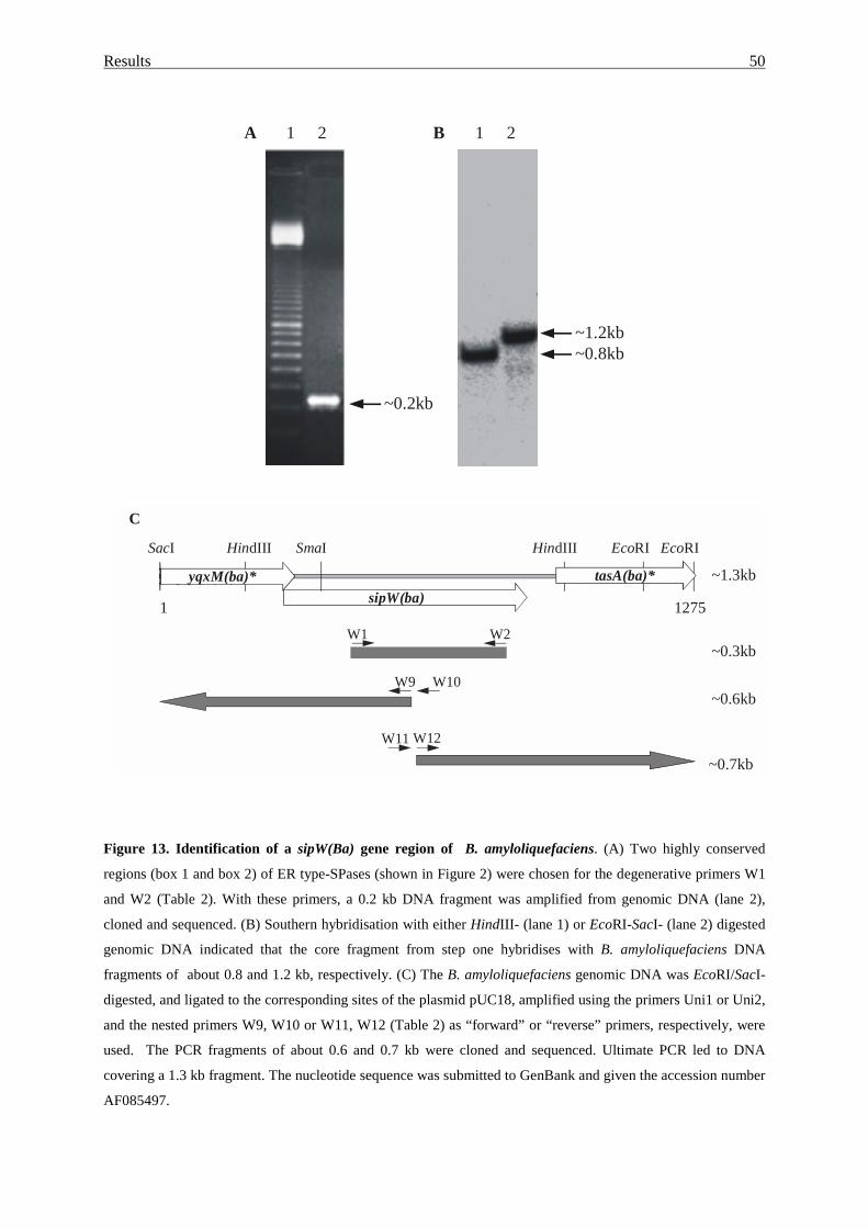
Results 50
Figure 13. Identification of a sipW(Ba) gene region of B. amyloliquefaciens. (A) Two highly conserved
regions (box 1 and box 2) of ER type-SPases (shown in Figure 2) were chosen for the degenerative primers W1
and W2 (Table 2). With these primers, a 0.2 kb DNA fragment was amplified from genomic DNA (lane 2),
cloned and sequenced. (B) Southern hybridisation with either HindIII- (lane 1) or EcoRI-SacI- (lane 2) digested
genomic DNA indicated that the core fragment from step one hybridises with B. amyloliquefaciens DNA
fragments of about 0.8 and 1.2 kb, respectively. (C) The B. amyloliquefaciens genomic DNA was EcoRI/SacI-
digested, and ligated to the corresponding sites of the plasmid pUC18, amplified using the primers Uni1 or Uni2,
and the nested primers W9, W10 or W11, W12 (Table 2) as “forward” or “reverse” primers, respectively, were
used. The PCR fragments of about 0.6 and 0.7 kb were cloned and sequenced. Ultimate PCR led to DNA
covering a 1.3 kb fragment. The nucleotide sequence was submitted to GenBank and given the accession number
AF085497.
yqxM(ba)*
W1 W2
~0.7kb
~0.6kb
C
HindIIISacI SmaI HindIII EcoRI EcoRI
yqxM(ba)*sipW(ba)
tasA(ba)*
W9 W10
W11 W12
~0.3kb
~1.3kb
1 1275
21 21A B
~0.2kb
~1.2kb~0.8kb
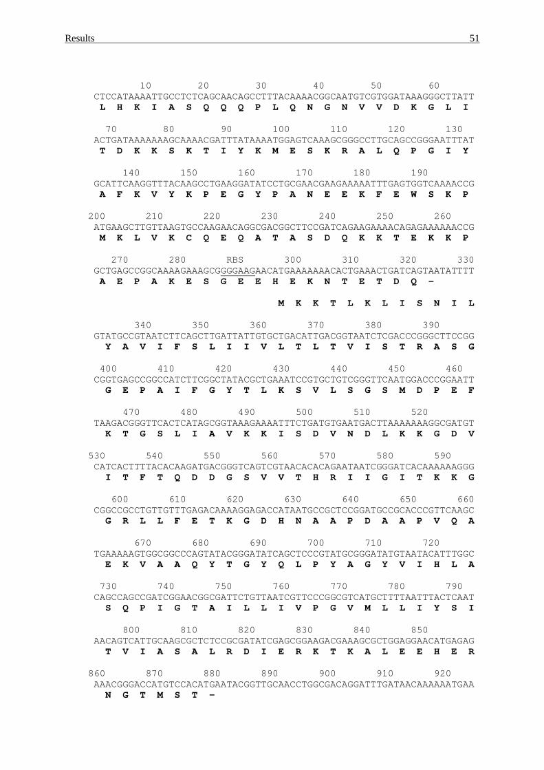
Results 51
10 20 30 40 50 60CTCCATAAAATTGCCTCTCAGCAACAGCCTTTACAAAACGGCAATGTCGTGGATAAAGGGCTTATTL H K I A S Q Q Q P L Q N G N V V D K G L I
70 80 90 100 110 120 130ACTGATAAAAAAAGCAAAACGATTTATAAAATGGAGTCAAAGCGGGCCTTGCAGCCGGGAATTTATT D K K S K T I Y K M E S K R A L Q P G I Y
140 150 160 170 180 190GCATTCAAGGTTTACAAGCCTGAAGGATATCCTGCGAACGAAGAAAAATTTGAGTGGTCAAAACCGA F K V Y K P E G Y P A N E E K F E W S K P
200 210 220 230 240 250 260ATGAAGCTTGTTAAGTGCCAAGAACAGGCGACGGCTTCCGATCAGAAGAAAACAGAGAAAAAACCGM K L V K C Q E Q A T A S D Q K K T E K K P
270 280 RBS 300 310 320 330GCTGAGCCGGCAAAAGAAAGCGGGGAAGAACATGAAAAAAACACTGAAACTGATCAGTAATATTTTA E P A K E S G E E H E K N T E T D Q -
M K K T L K L I S N I L
340 350 360 370 380 390GTATGCCGTAATCTTCAGCTTGATTATTGTGCTGACATTGACGGTAATCTCGACCCGGGCTTCCGGY A V I F S L I I V L T L T V I S T R A S G
400 410 420 430 440 450 460CGGTGAGCCGGCCATCTTCGGCTATACGCTGAAATCCGTGCTGTCGGGTTCAATGGACCCGGAATTG E P A I F G Y T L K S V L S G S M D P E F
470 480 490 500 510 520TAAGACGGGTTCACTCATAGCGGTAAAGAAAATTTCTGATGTGAATGACTTAAAAAAAGGCGATGTK T G S L I A V K K I S D V N D L K K G D V
530 540 550 560 570 580 590CATCACTTTTACACAAGATGACGGGTCAGTCGTAACACACAGAATAATCGGGATCACAAAAAAGGGI T F T Q D D G S V V T H R I I G I T K K G
600 610 620 630 640 650 660CGGCCGCCTGTTGTTTGAGACAAAAGGAGACCATAATGCCGCTCCGGATGCCGCACCCGTTCAAGCG R L L F E T K G D H N A A P D A A P V Q A
670 680 690 700 710 720TGAAAAAGTGGCGGCCCAGTATACGGGATATCAGCTCCCGTATGCGGGATATGTAATACATTTGGCE K V A A Q Y T G Y Q L P Y A G Y V I H L A
730 740 750 760 770 780 790CAGCCAGCCGATCGGAACGGCGATTCTGTTAATCGTTCCCGGCGTCATGCTTTTAATTTACTCAATS Q P I G T A I L L I V P G V M L L I Y S I
800 810 820 830 840 850AACAGTCATTGCAAGCGCTCTCCGCGATATCGAGCGGAAGACGAAAGCGCTGGAGGAACATGAGAGT V I A S A L R D I E R K T K A L E E H E R
860 870 880 890 900 910 920AAACGGGACCATGTCCACATGAATACGGTTGCAACCTGGCGACAGGATTTGATAACAAAAAATGAAN G T M S T -
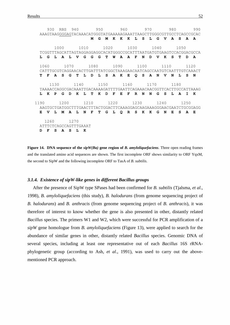
Results 52
930 RBS 940 950 960 970 980 990AAAGTAAGGGGAGTACAAACATGGGTATGAAAAAGAAATTAAGCTTGGGCGTTGCCTCAGCCGCAC
M G M K K K L S L G V A S A A
1000 1010 1020 1030 1040 1050TCGGTTTAGCATTAGTAGGAGGAGGCACATGGGCCGCATTTAATGATGTGAAGTCCACGGACGCCAL G L A L V G G G T W A A F N D V K S T D A
1060 1070 1080 1090 1100 1110 1120CATTTGCGTCAGGAACACTTGATTTATCGGCTAAAGAACAATCAGCCAATGTCAATTTGTCAAACTT F A S G T L D L S A K E Q S A N V N L S N
1130 1140 1150 1160 1170 1180TAAAACCAGGCGACAAATTGACAAAAGATTTTGAATTCAGAAACAACGGTTCACTTGCCATTAAAGL K P G D K L T K D F E F R N N G S L A I K
1190 1200 1210 1220 1230 1240 1250AAGTGCTGATGGCTTTGAACTTTACTGGACTTCAAAGGAGCAAGAAAGGGAACGAATCTGCGGAGGE V L M A L N F T G L Q R S K K G N E S A E
1260 1270ATTTCTCAGCCAGTTTGAAATD F S A S L K
Figure 14. DNA sequence of the sipW(Ba) gene region of B. amyloliquefaciens. Three open reading frames
and the translated amino acid sequences are shown. The first incomplete ORF shows similarity to ORF YqxM,
the second to SipW and the following incomplete ORF to TasA of B. subtilis.
3.1.4. Existence of sipW-like genes in different Bacillus groups
After the presence of SipW type SPases had been confirmed for B. subtilis (Tjalsma, et al.,
1998), B. amyloliquefaciens (this study), B. halodurans (from genome sequencing project of
B. halodurans) and B. anthracis (from genome sequencing project of B. anthracis), it was
therefore of interest to know whether the gene is also presented in other, distantly related
Bacillus species. The primers W1 and W2, which were successful for PCR amplification of a
sipW gene homologue from B. amyloliquefaciens (Figure 13), were applied to search for the
abundance of similar genes in other, distantly related Bacillus species. Genomic DNA of
several species, including at least one representative out of each Bacillus 16S rRNA-
phylogenetic group (according to Ash, et al., 1991), was used to carry out the above-
mentioned PCR approach.
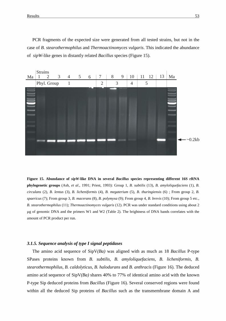
Results 53
PCR fragments of the expected size were generated from all tested strains, but not in the
case of B. stearothermophilus and Thermoactinomyces vulgaris. This indicated the abundance
of sipW-like genes in distantly related Bacillus species (Figure 15).
Figure 15. Abundance of sipW-like DNA in several Bacillus species representing different 16S rRNA
phylogenetic groups (Ash, et al., 1991; Priest, 1993): Group 1, B. subtilis (13), B. amyloliquefaciens (1), B.
circulans (2), B. lentus (3), B. licheniformis (4), B. megaterium (5), B. thuringiensis (6) ; From group 2, B.
spaericus (7); From group 3, B. macerans (8), B. polymyxa (9); From group 4, B. brevis (10); From group 5 etc.,
B. stearothermophilus (11); Thermoactinomyces vulgaris (12). PCR was under standard conditions using about 2
µg of genomic DNA and the primers W1 and W2 (Table 2). The brightness of DNA bands correlates with the
amount of PCR product per run.
3.1.5. Sequence analysis of type I signal peptidases
The amino acid sequence of SipV(Ba) was aligned with as much as 18 Bacillus P-type
SPases proteins known from B. subtilis, B. amyloliquefaciens, B. licheniformis, B.
stearothermophilus, B. caldolyticus, B. halodurans and B. anthracis (Figure 16). The deduced
amino acid sequence of SipV(Ba) shares 40% to 77% of identical amino acid with the known
P-type Sip deduced proteins from Bacillus (Figure 16). Several conserved regions were found
within all the deduced Sip proteins of Bacillus such as the transmembrane domain A and
~0.2kb
1 2 3 4 5 6 7 8 9 10 11 12 13Ma MaPhyl. Group 1 2 3 4 5
Strains
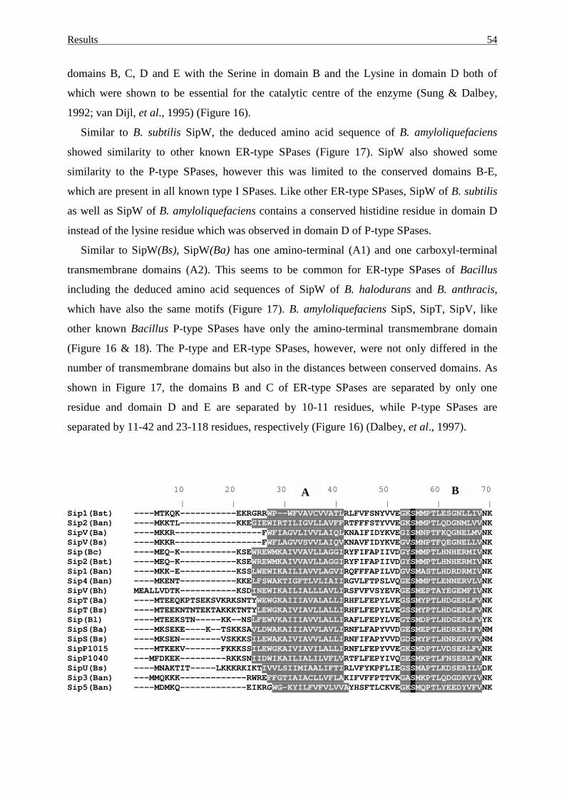
Results 54
domains B, C, D and E with the Serine in domain B and the Lysine in domain D both of
which were shown to be essential for the catalytic centre of the enzyme (Sung & Dalbey,
1992; van Dijl, et al., 1995) (Figure 16).
Similar to B. subtilis SipW, the deduced amino acid sequence of B. amyloliquefaciens
showed similarity to other known ER-type SPases (Figure 17). SipW also showed some
similarity to the P-type SPases, however this was limited to the conserved domains B-E,
which are present in all known type I SPases. Like other ER-type SPases, SipW of B. subtilis
as well as SipW of B. amyloliquefaciens contains a conserved histidine residue in domain D
instead of the lysine residue which was observed in domain D of P-type SPases.
Similar to SipW(Bs), SipW(Ba) has one amino-terminal (A1) and one carboxyl-terminal
transmembrane domains (A2). This seems to be common for ER-type SPases of Bacillus
including the deduced amino acid sequences of SipW of B. halodurans and B. anthracis,
which have also the same motifs (Figure 17). B. amyloliquefaciens SipS, SipT, SipV, like
other known Bacillus P-type SPases have only the amino-terminal transmembrane domain
(Figure 16 & 18). The P-type and ER-type SPases, however, were not only differed in the
number of transmembrane domains but also in the distances between conserved domains. As
shown in Figure 17, the domains B and C of ER-type SPases are separated by only one
residue and domain D and E are separated by 10-11 residues, while P-type SPases are
separated by 11-42 and 23-118 residues, respectively (Figure 16) (Dalbey, et al., 1997).
10 20 30 40 50 60 70
| | | | | | |Sip1(Bst) ----MTKQK-----------EKRGRRWP--WFVAVCVVATLRLFVFSNYVVEGKSMMPTLESGNLLIVNKSip2(Ban) ----MKKTL-----------KKEGIEWIRTILIGVLLAVFFRTFFFSTYVVEGKSMMPTLQDGNMLVVNKSipV(Ba) ----MKKR-----------------FWFIAGVLIVVLAIQLKNAIFIDYKVEGISMNPTFKQGNELMVNKSipV(Bs) ----MKKR-----------------FWFLAGVVSVVLAIQVKNAVFIDYKVEGVSMNPTFQEGNELLVNKSip(Bc) ----MEQ-K-----------KSEWREWMKAIVVAVLLAGGIRYFIFAPIIVDGYSMMPTLHNHERMIVNKSip2(Bst) ----MEQ-K-----------KSEWREWMKAIVVAVLLAGGIRYFIFAPIIVDGYSMMPTLHNHERMIVNKSip1(Ban) ----MKK-E-----------KSSLWEWIKAILIAVVLAGVIRQFFFAPILVDGVSMASTLHDRDRMIVNKSip4(Ban) ----MKENT-----------KKELFSWAKTIGFTLVLIAIIRGVLFTPSLVQGESMMPTLENNERVLVNKSipV(Bh) MEALLVDTK-----------KSDINEWIKAILIALLLAVLIRSFVFVSYEVRGESMEPTAYEGEMFIVNKSipT(Ba) ----MTEEQKPTSEKSVKRKSNTYWEWGKAIIIAVALALLIRHFLFEPYLVEGSSMYPTLHDGERLFVNKSipT(Bs) ----MTEEKNTNTEKTAKKKTNTYLEWGKAIVIAVLLALLIRHFLFEPYLVEGSSMYPTLHDGERLFVNKSip(Bl) ----MTEEKSTN-----KK--NSLFEWVKAIIIAVVLALLIRAFLFEPYLVEGTSMDPTLHDGERLFVYKSipS(Ba) ----MKSEKE----K--TSKKSAVLDWAKAIIIAVVLAVLIRNFLFAPYVVDGESMEPTLHDRERIFVNMSipS(Bs) ----MKSEN--------VSKKKSILEWAKAIVIAVVLALLIRNFIFAPYVVDGDSMYPTLHNRERVFVNMSipP1015 ----MTKEKV-------FKKKSSILEWGKAIVIAVILALLIRNFLFEPYVVEGKSMDPTLVDSERLFVNKSipP1040 ---MFDKEK---------RKKSNIIDWIKAILIALILVFLVRTFLFEPYIVQGESMKPTLFNSERLFVNKSipU(Bs) ----MNAKTIT-----LKKKRKIKTIVVLSIIMIAALIFTIRLVFYKPFLIEGSSMAPTLKDSERILVDKSip3(Ban) ---MMQKKK-------------RWREFFGTIAIACLLVFLAKIFVFFPTTVKGASMKPTLQDGDKVIVNKSip5(Ban) ----MDMKQ-------------EIKRGWG-KYILFVFVLVVAYHSFTLCKVEGKSMQPTLYEEDYVFVNK
A B
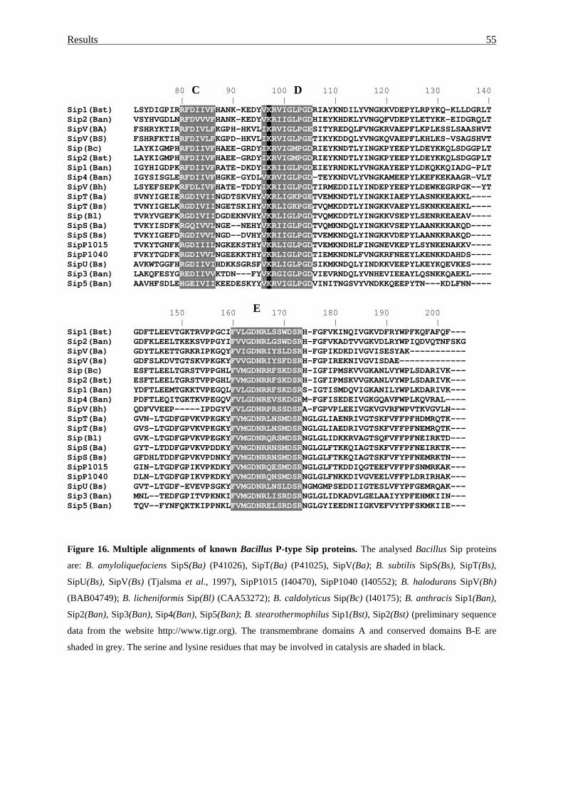
Results 55
E
80 90 100 110 120 130 140| | | | | | |
Sip1(Bst) LSYDIGPIRRFDIIVFHANK-KEDYVKRVIGLPGDRIAYKNDILYVNGKKVDEPYLRPYKQ-KLLDGRLTSip2(Ban) VSYHVGDLNRFDVVVFHANK-KEDYVKRIIGLPGDHIEYKHDKLYVNGQFVDEPYLETYKK-EIDGRQLTSipV(BA) FSHRYKTIRRFDIVLFKGPH-HKVLIKRVIGLPGESITYREDQLFVNGKRVAEPFLKPLKSSLSAASHVTSipV(BS) FSHRFKTIHRFDIVLFKGPD-HKVLIKRVIGLPGETIKYKDDQLYVNGKQVAEPFLKHLKS-VSAGSHVTSip(Bc) LAYKIGMPHRFDIIVFHAEE-GRDYIKRVIGMPGDRIEYKNDTLYINGKPYEEPYLDEYKKQLSDGGPLTSip2(Bst) LAYKIGMPHRFDIIVFHAEE-GRDYIKRVIGMPGDRIEYKNDTLYINGKPYEEPYLDEYKKQLSDGGPLTSip1(Ban) IGYHIGDPKRFDIIVFRATE-DKDYIKRIIGLPGDEIEYRNDKLYVNGKAYEEPYLDKQKKQIADG-PLTSip4(Ban) IGYSISGLERFDIIVFHGKE-GYDLVKRVIGLPGD-TEYKNDVLYVNGKAMEEPYLKEFKEKAAGR-VLTSipV(Bh) LSYEFSEPKRFDLIVFHATE-TDDYIKRIIGLPGDTIRMEDDILYINDEPYEEPYLDEWKEGRPGK--YTSipT(Ba) SVNYIGEIERGDIVIINGDTSKVHYVKRLIGKPGETVEMKNDTLYINGKKIAEPYLASNKKEAKKL----SipT(Bs) TVNYIGELKRGDIVIINGETSKIHYVKRLIGKPGETVQMKDDTLYINGKKVAEPYLSKNKKEAEKL----Sip(Bl) TVRYVGEFKRGDIVIIDGDEKNVHYVKRLIGLPGDTVQMKDDTLYINGKKVSEPYLSENRKEAEAV----SipS(Ba) TVKYISDFKRGQIVVLNGE--NEHYVKRIIGLPGDTVQMKNDQLYINGKKVSEPYLAANKKKAKQD----SipS(Bs) TVKYIGEFDRGDIVVLNGD--DVHYVKRIIGLPGDTVEMKNDQLYINGKKVDEPYLAANKKRAKQD----SipP1015 TVKYTGNFKRGDIIILNGKEKSTHYVKRLIGLPGDTVEMKNDHLFINGNEVKEPYLSYNKENAKKV----SipP1040 FVKYTGDFKRGDIVVLNGEEKKTHYVKRLIGLPGDTIEMKNDNLFVNGKRFNEEYLKENKKDAHDS----SipU(Bs) AVKWTGGFHRGDIIVIHDKKSGRSFVKRLIGLPGDSIKMKNDQLYINDKKVEEPYLKEYKQEVKES----Sip3(Ban) LAKQFESYGREDIIVVKTDN---FYVKRGIGLPGDVIEVRNDQLYVNHEVIEEAYLQSNKKQAEKL----Sip5(Ban) AAVHFSDLEHGEIVIIKEEDESKYYVKRVIGLPGDVINITNGSVYVNDKKQEEPYTN---KDLFNN----
150 160 170 180 190 200| | | | | |
Sip1(Bst) GDFTLEEVTGKTRVPPGCIFVLGDNRLSSWDSRH-FGFVKINQIVGKVDFRYWPFKQFAFQF---Sip2(Ban) GDFKLEELTKEKSVPPGYIFVVGDNRLGSWDSRH-FGFVKADTVVGKVDLRYWPIQDVQTNFSKGSipV(Ba) GDYTLKETTGRKRIPKGQYFVIGDNRIYSLDSRH-FGPIKDKDIVGVISESYAK-----------SipV(Bs) GDFSLKDVTGTSKVPKGKYFVVGDNRIYSFDSRH-FGPIREKNIVGVISDAE-------------Sip(Bc) ESFTLEELTGRSTVPPGHLFVMGDNRRFSKDSRH-IGFIPMSKVVGKANLVYWPLSDARIVK---Sip2(Bst) ESFTLEELTGRSTVPPGHLFVMGDNRRFSKDSRH-IGFIPMSKVVGKANLVYWPLSDARIVK---Sip1(Ban) YDFTLEEMTGKKTVPEGQLFVLGDNRRFSKDSRS-IGTISMDQVIGKANILYWPLKDARIVK---Sip4(Ban) PDFTLEQITGKTKVPEGQVFVLGDNREVSKDGRM-FGFISEDEIVGKGQAVFWPLKQVRAL----SipV(Bh) QDFVVEEP-----IPDGYVFVLGDNRPRSSDSRA-FGPVPLEEIVGKVGVRFWPVTKVGVLN---SipT(Ba) GVN-LTGDFGPVKVPKGKYFVMGDNRLNSMDSRNGLGLIAENRIVGTSKFVFFPFHDMRQTK---SipT(Bs) GVS-LTGDFGPVKVPKGKYFVMGDNRLNSMDSRNGLGLIAEDRIVGTSKFVFFPFNEMRQTK---Sip(Bl) GVK-LTGDFGPVKVPEGKYFVMGDNRQRSMDSRNGLGLIDKKRVAGTSQFVFFPFNEIRKTD---SipS(Ba) GYT-LTDDFGPVKVPDDKYFVMGDNRRNSMDSRNGLGLFTKKQIAGTSKFVFFPFNEIRKTK---SipS(Bs) GFDHLTDDFGPVKVPDNKYFVMGDNRRNSMDSRNGLGLFTKKQIAGTSKFVFYPFNEMRKTN---SipP1015 GIN-LTGDFGPIKVPKDKYFVMGDNRQESMDSRNGLGLFTKDDIQGTEEFVFFPFSNMRKAK---SipP1040 DLN-LTGDFGPIKVPKDKYFVMGDNRQNSMDSRNGLGLFNKKDIVGVEELVFFPLDRIRHAK---SipU(Bs) GVT-LTGDF-EVEVPSGKYFVMGDNRLNSLDSRNGMGMPSEDDIIGTESLVFYPFGEMRQAK---Sip3(Ban) MNL--TEDFGPITVPKNKIFVMGDNRLISRDSRNGLGLIDKADVLGELAAIYYPFEHMKIIN---Sip5(Ban) TQV--FYNFQKTKIPPNKLFVMGDNRELSRDSRNGLGYIEEDNIIGKVEFVYYPFSKMKIIE---
Figure 16. Multiple alignments of known Bacillus P-type Sip proteins. The analysed Bacillus Sip proteins
are: B. amyloliquefaciens SipS(Ba) (P41026), SipT(Ba) (P41025), SipV(Ba); B. subtilis SipS(Bs), SipT(Bs),
SipU(Bs), SipV(Bs) (Tjalsma et al., 1997), SipP1015 (I40470), SipP1040 (I40552); B. halodurans SipV(Bh)
(BAB04749); B. licheniformis Sip(Bl) (CAA53272); B. caldolyticus Sip(Bc) (I40175); B. anthracis Sip1(Ban),
Sip2(Ban), Sip3(Ban), Sip4(Ban), Sip5(Ban); B. stearothermophilus Sip1(Bst), Sip2(Bst) (preliminary sequence
data from the website http://www.tigr.org). The transmembrane domains A and conserved domains B-E are
shaded in grey. The serine and lysine residues that may be involved in catalysis are shaded in black.
C D
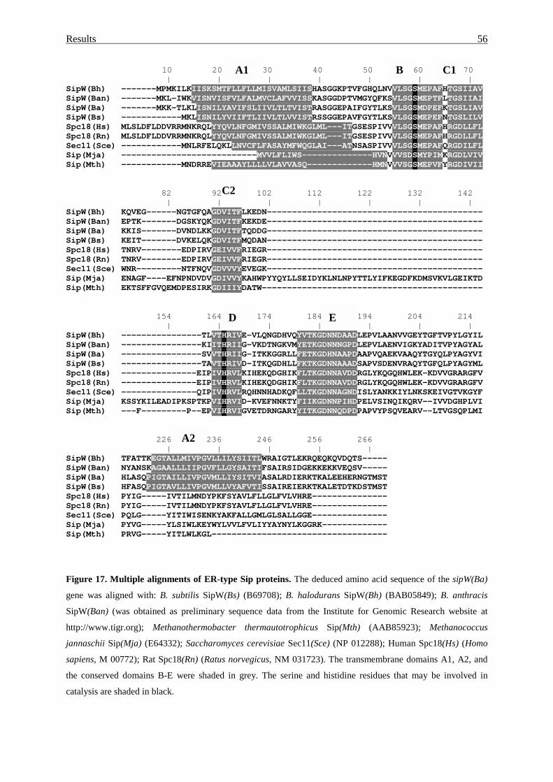
Results 56
B C1
C2
D E
A2
10 20 30 40 50 60 70| | | | | | |
SipW(Bh) -------MPMKILKIISKSMTFLLFLLMISVAMLSIISHASGGKPTVFGHQLNVVLSGSMEPAFHTGSIIAVSipW(Ban) -------MKL-IWKVISNVISFVLFALMVCLAFVVISSKASGGDPTVMGYQFKSVLSGSMEPTFLTGSIIAISipW(Ba) -------MKK-TLKLISNILYAVIFSLIIVLTLTVISTRASGGEPAIFGYTLKSVLSGSMDPEFKTGSLIAVSipW(Bs) ------------MKLISNILYVIIFTLIIVLTLVVISTRSSGGEPAVFGYTLKSVLSGSMEPEFNTGSLILVSpc18(Hs) MLSLDFLDDVRRMNKRQLYYQVLNFGMIVSSALMIWKGLML---ITGSESPIVVVLSGSMEPAFHRGDLLFLSpc18(Rn) MLSLDFLDDVRRMNKRQLYYQVLNFGMIVSSALMIWKGLML---ITGSESPIVVVLSGSMEPAFHRGDLLFLSec11(Sce) ------------MNLRFELQKLLNVCFLFASAYMFWQGLAI---ATNSASPIVVVLSGSMEPAFQRGDILFLSip(Mja) ---------------------------MVVLFLIWS--------------HVNVVVSDSMYPIMKRGDLVIVSip(Mth) ------------MNDRREVIEAAAYLLLLVLAVVASQ-------------HMNVVVSGSMEPVFYRGDIVII
82 92 102 112 122 132 142| | | | | | |
SipW(Bh) KQVEG------NGTGFQAGDVITFLKEDN-------------------------------------------SipW(Ban) EPTK-------DGSKYQKGDVITFKEKDE-------------------------------------------SipW(Ba) KKIS-------DVNDLKKGDVITFTQDDG-------------------------------------------SipW(Bs) KEIT-------DVKELQKGDVITFMQDAN-------------------------------------------Spc18(Hs) TNRV--------EDPIRVGEIVVFRIEGR-------------------------------------------Spc18(Rn) TNRV--------EDPIRVGEIVVFRIEGR-------------------------------------------Sec11(Sce) WNR---------NTFNQVGDVVVYEVEGK-------------------------------------------Sip(Mja) ENAGF----EFNPNDVDVGDIVVYKAHWPYYQYLLSEIDYKLNLNPYTTLYIFKEGDFKDMSVKVLGEIKTDSip(Mth) EKTSFFGVQEMDPESIRKGDIIIYDATW--------------------------------------------
154 164 174 184 194 204 214| | | | | | |
SipW(Bh) ----------------TLVTHRIVE-VLQNGDHVQYVTKGDNNDAADLEPVLAANVVGEYTGFTVPYLGYILSipW(Ban) ----------------KIITHRIIG-VKDTNGKVMYETKGDNNNGPDLEPVLAENVIGKYADITVPYAGYALSipW(Ba) ----------------SVVTHRIIG-ITKKGGRLLFETKGDHNAAPDAAPVQAEKVAAQYTGYQLPYAGYVISipW(Bs) ----------------TAVTHRIVD-ITKQGDHLLFKTKGDNNAAADSAPVSDENVRAQYTGFQLPYAGYMLSpc18(Hs) ---------------EIPIVHRVLKIHEKQDGHIKFLTKGDNNAVDDRGLYKQGQHWLEK-KDVVGRARGFVSpc18(Rn) ---------------EIPIVHRVLKIHEKQDGHIKFLTKGDNNAVDDRGLYKQGQHWLEK-KDVVGRARGFVSec11(Sce) ---------------QIPIVHRVLRQHNNHADKQFLLTKGDNNAGNDISLYANKKIYLNKSKEIVGTVKGYFSip(Mja) KSSYKILEADIPKSPTKPVIHRVID-KVEFNNKTYFIIKGDNNPIHDPELVSINQIKQRV--IVVDGHPLVISip(Mth) ---F---------P--EPVIHRVIGVETDRNGARYYITKGDNNQDPDPAPVYPSQVEARV--LTVGSQPLMI
226 236 246 256 266| | | | |
SipW(Bh) TFATTKEGTALLMIVPGVLLILYSIITLWRAIGTLEKRQEQKQVDQTS-----SipW(Ban) NYANSKAGAALLLIIPGVFLLGYSAITIFSAIRSIDGEKKEKKVEQSV-----SipW(Ba) HLASQPIGTAILLIVPGVMLLIYSITVIASALRDIERKTKALEEHERNGTMSTSipW(Bs) HFASQPIGTAVLLIVPGVMLLVYAFVTISSAIREIERKTKALETDTKDSTMSTSpc18(Hs) PYIG-----IVTILMNDYPKFSYAVLFLLGLFVLVHRE---------------Spc18(Rn) PYIG-----IVTILMNDYPKFSYAVLFLLGLFVLVHRE---------------Sec11(Sce) PQLG-----YITIWISENKYAKFALLGMLGLSALLGGE---------------Sip(Mja) PYVG-----YLSIWLKEYWYLVVLFVLIYYAYNYLKGGRK-------------Sip(Mth) PRVG-----YITLWLKGL-----------------------------------
Figure 17. Multiple alignments of ER-type Sip proteins. The deduced amino acid sequence of the sipW(Ba)
gene was aligned with: B. subtilis SipW(Bs) (B69708); B. halodurans SipW(Bh) (BAB05849); B. anthracis
SipW(Ban) (was obtained as preliminary sequence data from the Institute for Genomic Research website at
http://www.tigr.org); Methanothermobacter thermautotrophicus Sip(Mth) (AAB85923); Methanococcus
jannaschii Sip(Mja) (E64332); Saccharomyces cerevisiae Sec11(Sce) (NP 012288); Human Spc18(Hs) (Homo
sapiens, M 00772); Rat Spc18(Rn) (Ratus norvegicus, NM 031723). The transmembrane domains A1, A2, and
the conserved domains B-E were shaded in grey. The serine and histidine residues that may be involved in
catalysis are shaded in black.
A1
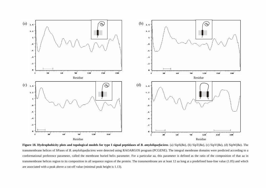
Residue Residue
Residue
Residue Residue
Figure 18. Hydrophobicity plots and topological models for type I signal peptidases of B. amyloliquefaciens. (a) SipS(Ba), (b) SipT(Ba), (c) SipV(Ba), (d) SipW(Ba). The
transmembrane helices of SPases of B. amyloliquefaciens were detected using RAOARGOS program (PCGENE). The integral membrane domains were predicted according to a
conformational preference parameter, called the membrane buried helix parameter. For a particular aa, this parameter is defined as the ratio of the composition of that aa in
transmembrane helices region to its composition in all sequence region of the protein. The transmembrane are at least 12 aa long at a predefined base-line value (1.05) and which
are associated with a peak above a cut-off value (minimal peak height is 1.13).
(a) (b)
(c) (d)
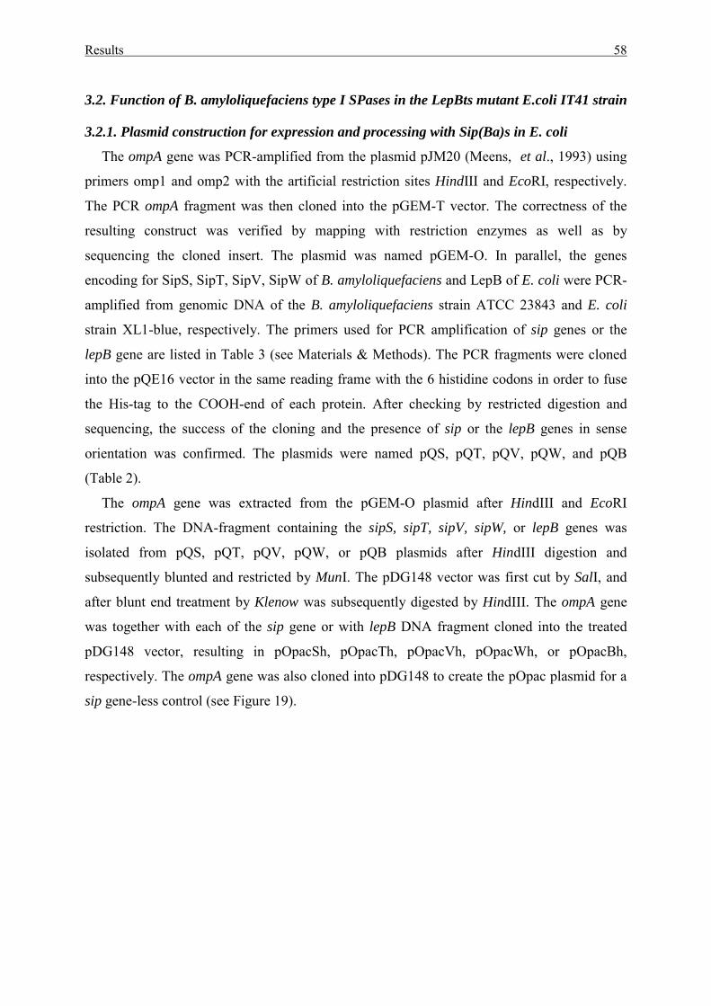
Results 58
3.2. Function of B. amyloliquefaciens type I SPases in the LepBts mutant E.coli IT41 strain
3.2.1. Plasmid construction for expression and processing with Sip(Ba)s in E. coli
The ompA gene was PCR-amplified from the plasmid pJM20 (Meens, et al., 1993) using
primers omp1 and omp2 with the artificial restriction sites HindIII and EcoRI, respectively.
The PCR ompA fragment was then cloned into the pGEM-T vector. The correctness of the
resulting construct was verified by mapping with restriction enzymes as well as by
sequencing the cloned insert. The plasmid was named pGEM-O. In parallel, the genes
encoding for SipS, SipT, SipV, SipW of B. amyloliquefaciens and LepB of E. coli were PCR-
amplified from genomic DNA of the B. amyloliquefaciens strain ATCC 23843 and E. coli
strain XL1-blue, respectively. The primers used for PCR amplification of sip genes or the
lepB gene are listed in Table 3 (see Materials & Methods). The PCR fragments were cloned
into the pQE16 vector in the same reading frame with the 6 histidine codons in order to fuse
the His-tag to the COOH-end of each protein. After checking by restricted digestion and
sequencing, the success of the cloning and the presence of sip or the lepB genes in sense
orientation was confirmed. The plasmids were named pQS, pQT, pQV, pQW, and pQB
(Table 2).
The ompA gene was extracted from the pGEM-O plasmid after HindIII and EcoRI
restriction. The DNA-fragment containing the sipS, sipT, sipV, sipW, or lepB genes was
isolated from pQS, pQT, pQV, pQW, or pQB plasmids after HindIII digestion and
subsequently blunted and restricted by MunI. The pDG148 vector was first cut by SalI, and
after blunt end treatment by Klenow was subsequently digested by HindIII. The ompA gene
was together with each of the sip gene or with lepB DNA fragment cloned into the treated
pDG148 vector, resulting in pOpacSh, pOpacTh, pOpacVh, pOpacWh, or pOpacBh,
respectively. The ompA gene was also cloned into pDG148 to create the pOpac plasmid for a
sip gene-less control (see Figure 19).
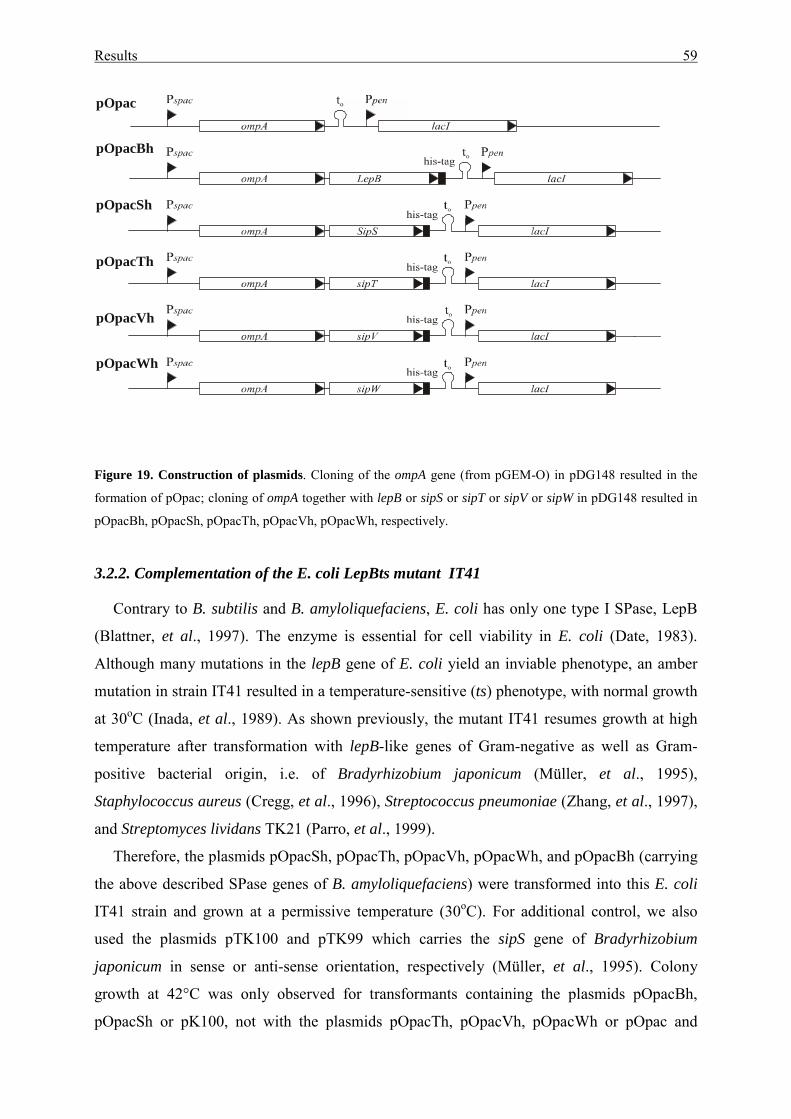
Results 59
Figure 19. Construction of plasmids. Cloning of the ompA gene (from pGEM-O) in pDG148 resulted in the
formation of pOpac; cloning of ompA together with lepB or sipS or sipT or sipV or sipW in pDG148 resulted in
pOpacBh, pOpacSh, pOpacTh, pOpacVh, pOpacWh, respectively.
3.2.2. Complementation of the E. coli LepBts mutant IT41
Contrary to B. subtilis and B. amyloliquefaciens, E. coli has only one type I SPase, LepB
(Blattner, et al., 1997). The enzyme is essential for cell viability in E. coli (Date, 1983).
Although many mutations in the lepB gene of E. coli yield an inviable phenotype, an amber
mutation in strain IT41 resulted in a temperature-sensitive (ts) phenotype, with normal growth
at 30oC (Inada, et al., 1989). As shown previously, the mutant IT41 resumes growth at high
temperature after transformation with lepB-like genes of Gram-negative as well as Gram-
positive bacterial origin, i.e. of Bradyrhizobium japonicum (Müller, et al., 1995),
Staphylococcus aureus (Cregg, et al., 1996), Streptococcus pneumoniae (Zhang, et al., 1997),
and Streptomyces lividans TK21 (Parro, et al., 1999).
Therefore, the plasmids pOpacSh, pOpacTh, pOpacVh, pOpacWh, and pOpacBh (carrying
the above described SPase genes of B. amyloliquefaciens) were transformed into this E. coli
IT41 strain and grown at a permissive temperature (30oC). For additional control, we also
used the plasmids pTK100 and pTK99 which carries the sipS gene of Bradyrhizobium
japonicum in sense or anti-sense orientation, respectively (Müller, et al., 1995). Colony
growth at 42°C was only observed for transformants containing the plasmids pOpacBh,
pOpacSh or pK100, not with the plasmids pOpacTh, pOpacVh, pOpacWh or pOpac and
pOpac
pOpacBh
pOpacSh
pOpacTh
pOpacVh
pOpacWh
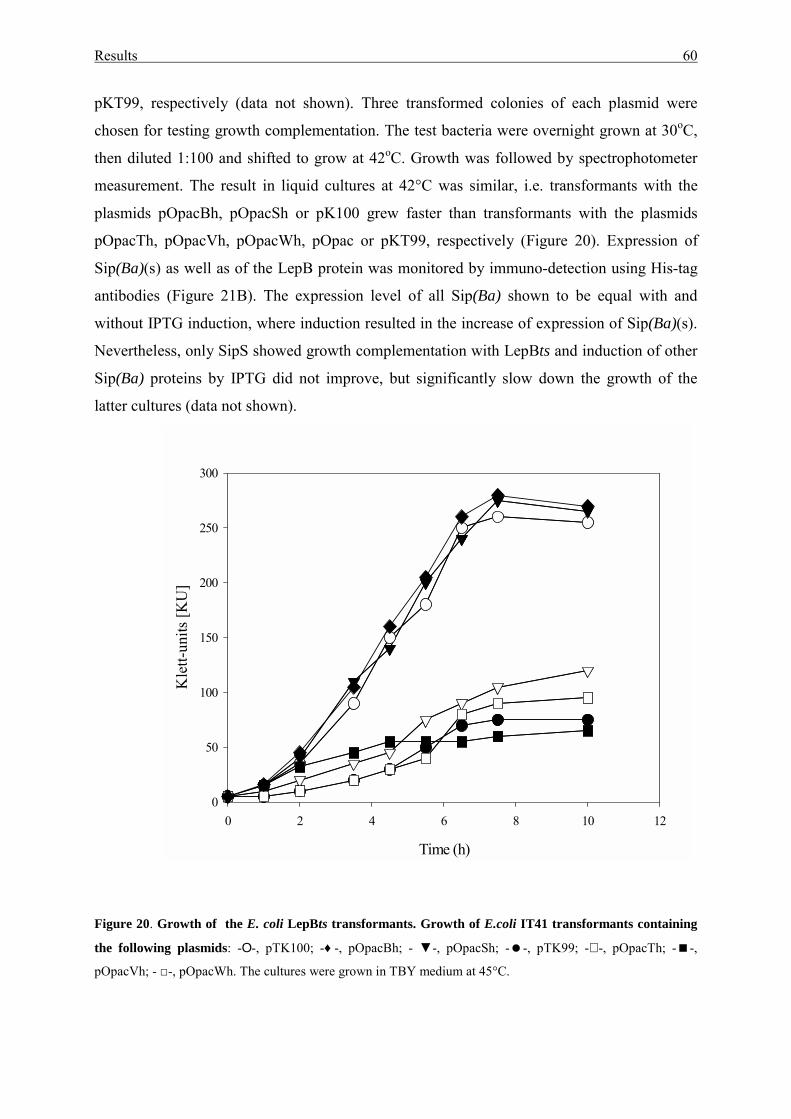
Results 60
pKT99, respectively (data not shown). Three transformed colonies of each plasmid were
chosen for testing growth complementation. The test bacteria were overnight grown at 30oC,
then diluted 1:100 and shifted to grow at 42oC. Growth was followed by spectrophotometer
measurement. The result in liquid cultures at 42°C was similar, i.e. transformants with the
plasmids pOpacBh, pOpacSh or pK100 grew faster than transformants with the plasmids
pOpacTh, pOpacVh, pOpacWh, pOpac or pKT99, respectively (Figure 20). Expression of
Sip(Ba)(s) as well as of the LepB protein was monitored by immuno-detection using His-tag
antibodies (Figure 21B). The expression level of all Sip(Ba) shown to be equal with and
without IPTG induction, where induction resulted in the increase of expression of Sip(Ba)(s).
Nevertheless, only SipS showed growth complementation with LepBts and induction of other
Sip(Ba) proteins by IPTG did not improve, but significantly slow down the growth of the
latter cultures (data not shown).
Figure 20. Growth of the E. coli LepBts transformants. Growth of E.coli IT41 transformants containing
the following plasmids: -Ο-, pTK100; -♦ -, pOpacBh; - ▼-, pOpacSh; -�-, pTK99; -∇ -, pOpacTh; -�-,
pOpacVh; - □-, pOpacWh. The cultures were grown in TBY medium at 45°C.
Time (h)
0 2 4 6 8 10 12
Kle
tt-un
its [K
U]
0
50
100
150
200
250
300
Results 61
3.2.3. Processing studies with pre-OmpA in LepBts E. coli IT41
Processing of the pre-OmpA precursor in the transformed LepBts E. coli was done in order
to verify in more detail the complementation of LepBts after Sip(Ba) protein expressions. The
E. coli IT41 strains, which carried the plasmids pOpac, or pOpacSh, or pOpacTh, or
pOpacVh, or pOpacWh, or pOpacBh, were used for the pulse chase radioactive 35S labelling
at either 30oC or 42oC. After immunoprecipitation by OmpA antiserum, the samples were
separated by SDS-PAGE and autoradiograms were obtained by the Phospho-Imager system
(see Material & Methods). The results are shown in Figure 21A. While the processing of pre-
OmpA was enhanced in cells with expression of either B. amyloliquefaciens SipS(Ba),
SipT(Ba), or of LepB, the two other B. amyloliquefaciens Sip proteins SipV(Ba) and
SipW(Ba) did not significantly improve processing of pre-OmpA compared to the mutant
control culture. The two latter cases could be explained either by the limited activities of the
enzymes or by the improper membrane integration of the two proteins, resulting in
degradation as shown on the Western blot (see Figure 21B) for Sip(Ba) protein expression
using His-tag antiserum. No such degradation forms were visible with SipS(Ba) and SipT(Ba)
in E. coli.
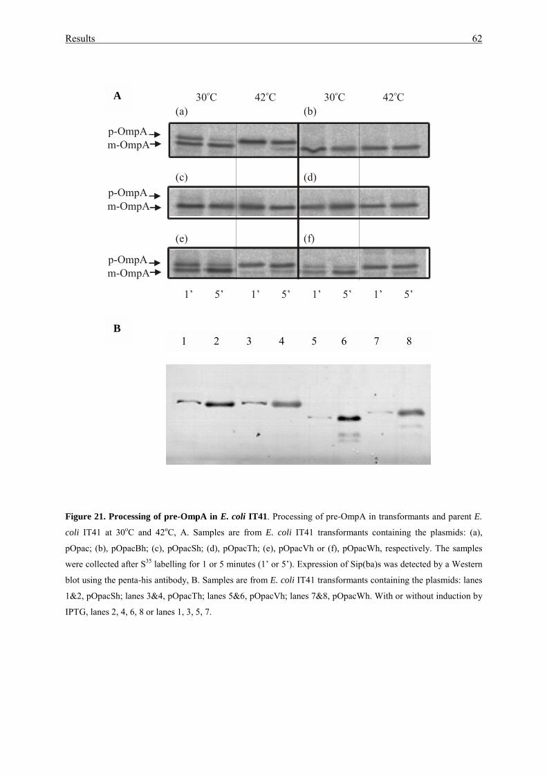
Results 62
Figure 21. Processing of pre-OmpA in E. coli IT41. Processing of pre-OmpA in transformants and parent E.
coli IT41 at 30oC and 42oC, A. Samples are from E. coli IT41 transformants containing the plasmids: (a),
pOpac; (b), pOpacBh; (c), pOpacSh; (d), pOpacTh; (e), pOpacVh or (f), pOpacWh, respectively. The samples
were collected after S35 labelling for 1 or 5 minutes (1’ or 5’). Expression of Sip(ba)s was detected by a Western
blot using the penta-his antibody, B. Samples are from E. coli IT41 transformants containing the plasmids: lanes
1&2, pOpacSh; lanes 3&4, pOpacTh; lanes 5&6, pOpacVh; lanes 7&8, pOpacWh. With or without induction by
IPTG, lanes 2, 4, 6, 8 or lanes 1, 3, 5, 7.
(a) (b)
(d)(c)
(f)(e)
1’ 5’ 1’ 5’ 1’ 5’ 1’ 5’
30 Co 42 Co 30 Co 42 Co
p-OmpAm-OmpA
p-OmpAm-OmpA
p-OmpAm-OmpA
A
B 1 2 3 4 5 6 7 8
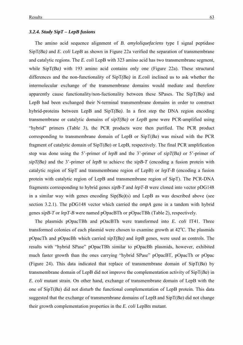
Results 63
3.2.4. Study SipT – LepB fusions
The amino acid sequence alignment of B. amyloliquefaciens type I signal peptidase
SipT(Ba) and E. coli LepB as shown in Figure 22a verified the separation of transmembrane
and catalytic regions. The E. coli LepB with 323 amino acid has two transmembrane segment,
while SipT(Ba) with 193 amino acid contains only one (Figure 22a). These structural
differences and the non-functionality of SipT(Ba) in E.coli inclined us to ask whether the
intermolecular exchange of the transmembrane domains would mediate and therefore
apparently cause functionality/non-fuctionality between these SPases. The SipT(Ba) and
LepB had been exchanged their N-terminal transmembrane domains in order to construct
hybrid-proteins between LepB and SipT(Ba). In a first step the DNA region encoding
transmembrane or catalytic domains of sipT(Ba) or LepB gene were PCR-amplified using
“hybrid” primers (Table 3), the PCR products were then purified. The PCR product
corresponding to transmembrane domain of LepB or SipT(Ba) was mixed with the PCR
fragment of catalytic domain of SipT(Ba) or LepB, respectively. The final PCR amplification
step was done using the 5’-primer of lepB and the 3’-primer of sipT(Ba) or 5’-primer of
sipT(Ba) and the 3’-primer of lepB to achieve the sipB-T (encoding a fusion protein with
catalytic region of SipT and transmembrane region of LepB) or lepT-B (encoding a fusion
protein with catalytic region of LepB and transmembrane region of SipT). The PCR-DNA
fragments corresponding to hybrid genes sipB-T and lepT-B were cloned into vector pDG148
in a similar way with genes encoding Sip(Ba)(s) and LepB as was described above (see
section 3.2.1). The pDG148 vector which carried the ompA gene in a tandem with hybrid
genes sipB-T or lepT-B were named pOpacBTh or pOpacTBh (Table 2), respectively.
The plasmids pOpacTBh and pOacBTh were transformed into E. coli IT41. Three
transformed colonies of each plasmid were chosen to examine growth at 42oC. The plasmids
pOpacTh and pOpacBh which carried sipT(Ba) and lepB genes, were used as controls. The
results with “hybrid SPase” pOpacTBh similar to pOpacBh plasmids, however, exhibited
much faster growth than the ones carrying “hybrid SPase” pOpacBT, pOpacTh or pOpac
(Figure 24). This data indicated that replace of transmembrane domain of SipT(Ba) by
transmembrane domain of LepB did not improve the complementation activity of SipT(Ba) in
E. coli mutant strain. On other hand, exchange of transmembrane domain of LepB with the
one of SipT(Ba) did not disturb the functional complementation of LepB protein. This data
suggested that the exchange of transmembrane domains of LepB and SipT(Ba) did not change
their growth complementation properties in the E. coli LepBts mutant.
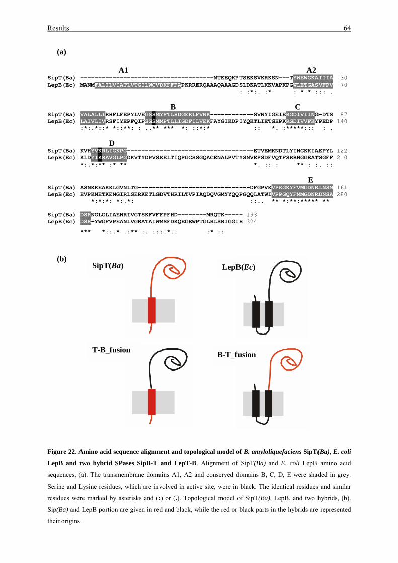
Results 64
(a)
A1 A2SipT(Ba) -------------------------------------MTEEQKPTSEKSVKRKSN---TYWEWGKAIIIA 30LepB(Ec) MANMFALILVIATLVTGILWCVDKFFFAPKRRERQAAAQAAAGDSLDKATLKKVAPKPGWLETGASVFPV 70
: :*:. :* : * * ::: .
B CSipT(Ba) VALALLIRHFLFEPYLVEGSSMYPTLHDGERLFVNK------------SVNYIGEIERGDIVIING-DTS 87LepB(Ec) LAIVLIVRSFIYEPFQIPSGSMMPTLLIGDFILVEKFAYGIKDPIYQKTLIETGHPKRGDIVVFKYPEDP 140
:*:.*::* *::**: : ..** *** *: ::*:* :: *. :*****::: : .
DSipT(Ba) KVHYVKRLIGKPG-----------------------------------ETVEMKNDTLYINGKKIAEPYL 122LepB(Ec) KLDYIKRAVGLPGDKVTYDPVSKELTIQPGCSSGQACENALPVTYSNVEPSDFVQTFSRRNGGEATSGFF 210
*:.*:** :* ** *. :: : ** : :. ::
ESipT(Ba) ASNKKEAKKLGVNLTG-------------------------------DFGPVKVPKGKYFVMGDNRLNSM 161LepB(Ec) EVPKNETKENGIRLSERKETLGDVTHRILTVPIAQDQVGMYYQQPGQQLATWIVPPGQYFMMGDNRDNSA 280
*:*:*: *:.*: ::.. ** *:**:***** **
SipT(Ba) DSRNGLGLIAENRIVGTSKFVFFPFHD--------MRQTK----- 193LepB(Ec) DSR-YWGFVPEANLVGRATAIWMSFDKQEGEWPTGLRLSRIGGIH 324
*** *::.* .:** :. :::.*.. :* ::
(b)
Figure 22. Amino acid sequence alignment and topological model of B. amyloliquefaciens SipT(Ba), E. coli
LepB and two hybrid SPases SipB-T and LepT-B. Alignment of SipT(Ba) and E. coli LepB amino acid
sequences, (a). The transmembrane domains A1, A2 and conserved domains B, C, D, E were shaded in grey.
Serine and Lysine residues, which are involved in active site, were in black. The identical residues and similar
residues were marked by asterisks and (:) or (.). Topological model of SipT(Ba), LepB, and two hybrids, (b).
Sip(Ba) and LepB portion are given in red and black, while the red or black parts in the hybrids are represented
their origins.
SipT(Ba) LepB(Ec)
T-B_fusion B-T_fusion
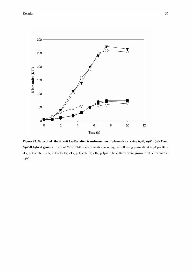
Results 65
Figure 23. Growth of the E. coli LepBts after transformation of plasmids carrying lepB, sipT, sipB-T and
lepT-B hybrid genes. Growth of E.coli IT41 transformants containing the following plasmids: -Ο-, pOpacBh; -
�-, pOpacTh; -∇ -, pOpacB-Th; -▼-, pOpacT-Bh; -�-, pOpac. The cultures were grown in TBY medium at
42°C.
Time (h)
0 2 4 6 8 10 12
Kle
tt-un
its (K
U)
0
50
100
150
200
250
300
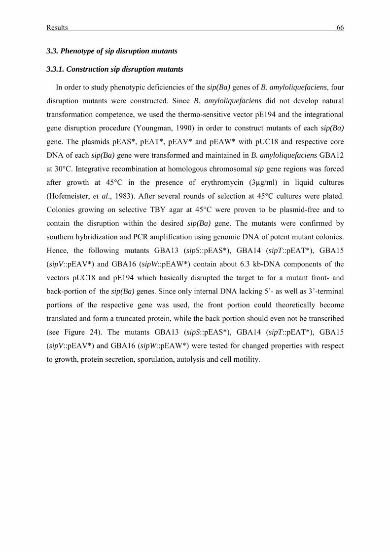
Results 66
3.3. Phenotype of sip disruption mutants 3.3.1. Construction sip disruption mutants
In order to study phenotypic deficiencies of the sip(Ba) genes of B. amyloliquefaciens, four
disruption mutants were constructed. Since B. amyloliquefaciens did not develop natural
transformation competence, we used the thermo-sensitive vector pE194 and the integrational
gene disruption procedure (Youngman, 1990) in order to construct mutants of each sip(Ba)
gene. The plasmids pEAS*, pEAT*, pEAV* and pEAW* with pUC18 and respective core
DNA of each sip(Ba) gene were transformed and maintained in B. amyloliquefaciens GBA12
at 30°C. Integrative recombination at homologous chromosomal sip gene regions was forced
after growth at 45°C in the presence of erythromycin (3µg/ml) in liquid cultures
(Hofemeister, et al., 1983). After several rounds of selection at 45°C cultures were plated.
Colonies growing on selective TBY agar at 45°C were proven to be plasmid-free and to
contain the disruption within the desired sip(Ba) gene. The mutants were confirmed by
southern hybridization and PCR amplification using genomic DNA of potent mutant colonies.
Hence, the following mutants GBA13 (sipS::pEAS*), GBA14 (sipT::pEAT*), GBA15
(sipV::pEAV*) and GBA16 (sipW::pEAW*) contain about 6.3 kb-DNA components of the
vectors pUC18 and pE194 which basically disrupted the target to for a mutant front- and
back-portion of the sip(Ba) genes. Since only internal DNA lacking 5’- as well as 3’-terminal
portions of the respective gene was used, the front portion could theoretically become
translated and form a truncated protein, while the back portion should even not be transcribed
(see Figure 24). The mutants GBA13 (sipS::pEAS*), GBA14 (sipT::pEAT*), GBA15
(sipV::pEAV*) and GBA16 (sipW::pEAW*) were tested for changed properties with respect
to growth, protein secretion, sporulation, autolysis and cell motility.
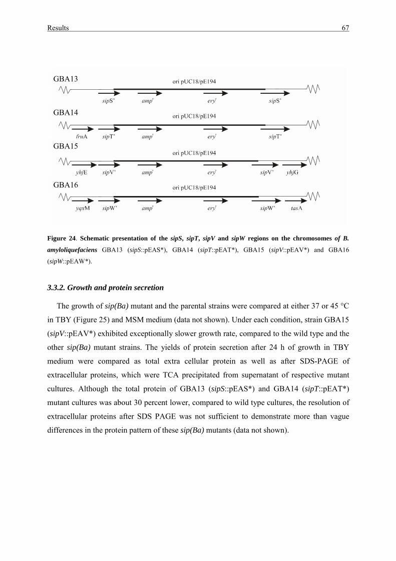
Results 67
Figure 24. Schematic presentation of the sipS, sipT, sipV and sipW regions on the chromosomes of B.
amyloliquefaciens GBA13 (sipS::pEAS*), GBA14 (sipT::pEAT*), GBA15 (sipV::pEAV*) and GBA16
(sipW::pEAW*).
3.3.2. Growth and protein secretion
The growth of sip(Ba) mutant and the parental strains were compared at either 37 or 45 °C
in TBY (Figure 25) and MSM medium (data not shown). Under each condition, strain GBA15
(sipV::pEAV*) exhibited exceptionally slower growth rate, compared to the wild type and the
other sip(Ba) mutant strains. The yields of protein secretion after 24 h of growth in TBY
medium were compared as total extra cellular protein as well as after SDS-PAGE of
extracellular proteins, which were TCA precipitated from supernatant of respective mutant
cultures. Although the total protein of GBA13 (sipS::pEAS*) and GBA14 (sipT::pEAT*)
mutant cultures was about 30 percent lower, compared to wild type cultures, the resolution of
extracellular proteins after SDS PAGE was not sufficient to demonstrate more than vague
differences in the protein pattern of these sip(Ba) mutants (data not shown).
GBA13
GBA14
GBA15
GBA16
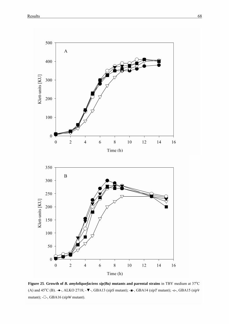
Results 68
Time (h)
0 2 4 6 8 10 12 14 16
Kle
tt-un
its [K
U]
0
100
200
300
400
500
Time (h)
0 2 4 6 8 10 12 14 16
Kle
tt-un
its [K
U]
0
50
100
150
200
250
300
350
Figure 25. Growth of B. amyloliquefaciens sip(Ba) mutants and parental strains in TBY medium at 37oC
(A) and 45oC (B). -●-, ALKO 2718; -▼-, GBA13 (sipS mutant); -■-, GBA14 (sipT mutant); -o-, GBA15 (sipV
mutant); -∇ -, GBA16 (sipW mutant).
A
B
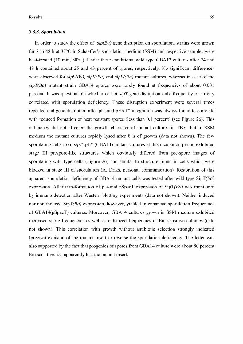
Results 69
3.3.3. Sporulation
In order to study the effect of sip(Ba) gene disruption on sporulation, strains were grown
for 8 to 48 h at 37°C in Schaeffer’s sporulation medium (SSM) and respective samples were
heat-treated (10 min, 80°C). Under these conditions, wild type GBA12 cultures after 24 and
48 h contained about 25 and 43 percent of spores, respectively. No significant differences
were observed for sipS(Ba), sipV(Ba) and sipW(Ba) mutant cultures, whereas in case of the
sipT(Ba) mutant strain GBA14 spores were rarely found at frequencies of about 0.001
percent. It was questionable whether or not sipT-gene disruption only frequently or strictly
correlated with sporulation deficiency. These disruption experiment were several times
repeated and gene disruption after plasmid pEAT* integration was always found to correlate
with reduced formation of heat resistant spores (less than 0.1 percent) (see Figure 26). This
deficiency did not affected the growth character of mutant cultures in TBY, but in SSM
medium the mutant cultures rapidly lysed after 8 h of growth (data not shown). The few
sporulating cells from sipT::pE* (GBA14) mutant cultures at this incubation period exhibited
stage III prespore-like structures which obviously differed from pre-spore images of
sporulating wild type cells (Figure 26) and similar to structure found in cells which were
blocked in stage III of sporulation (A. Driks, personal communication). Restoration of this
apparent sporulation deficiency of GBA14 mutant cells was tested after wild type SipT(Ba)
expression. After transformation of plasmid pSpacT expression of SipT(Ba) was monitored
by immuno-detection after Western blotting experiments (data not shown). Neither induced
nor non-induced SipT(Ba) expression, however, yielded in enhanced sporulation frequencies
of GBA14(pSpacT) cultures. Moreover, GBA14 cultures grown in SSM medium exhibited
increased spore frequencies as well as enhanced frequencies of Em sensitive colonies (data
not shown). This correlation with growth without antibiotic selection strongly indicated
(precise) excision of the mutant insert to reverse the sporulation deficiency. The letter was
also supported by the fact that progenies of spores from GBA14 culture were about 80 percent
Em sensitive, i.e. apparently lost the mutant insert.
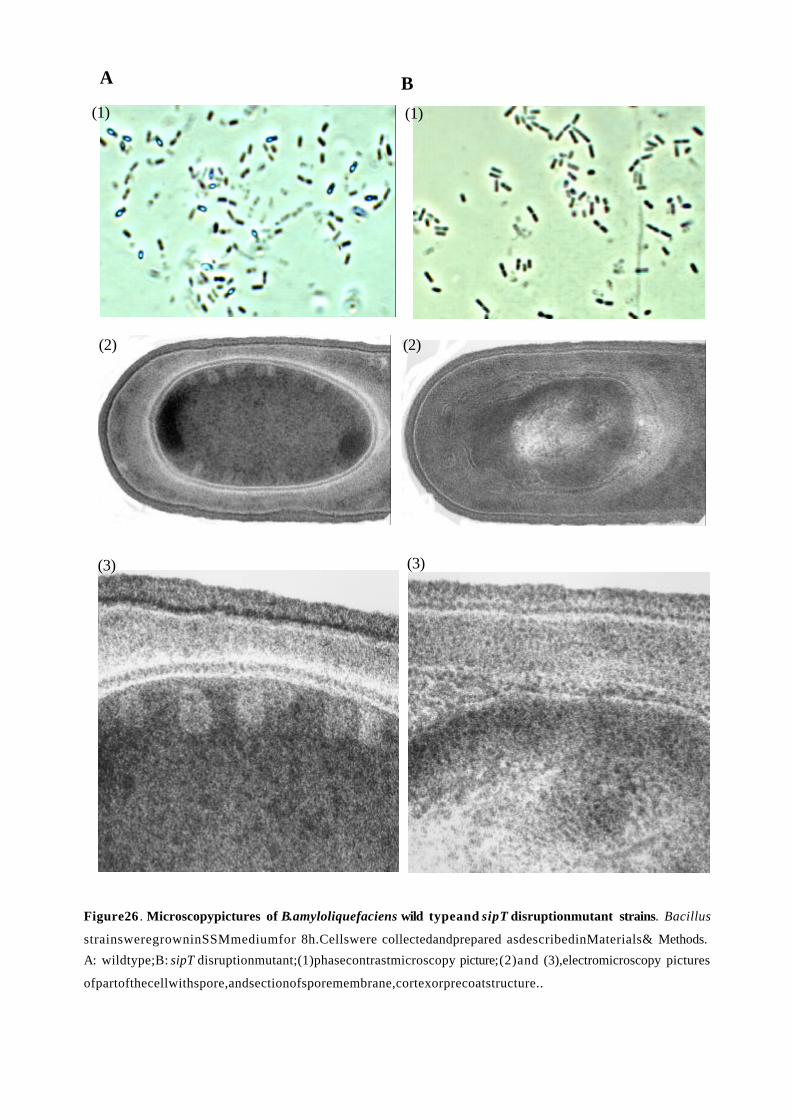
BA
(2)
(3)
(2)
(3)
(1)(1)
Figure26 Microscopypictures of wild typeand disruptionmutant strains. .
strainsweregrowninSSMmediumfor 8h.Cellswere collectedandprepared asdescribedinMaterials& Methods.
A: wildtype;B: disruptionmutant;(1)phasecontrastmicroscopy picture;(2)and (3),electromicroscopy pictures
ofpartofthecellwithspore,andsectionofsporemembrane,cortexorprecoatstructure..
B.amyloliquefaciens sipT Bacillus
sipT
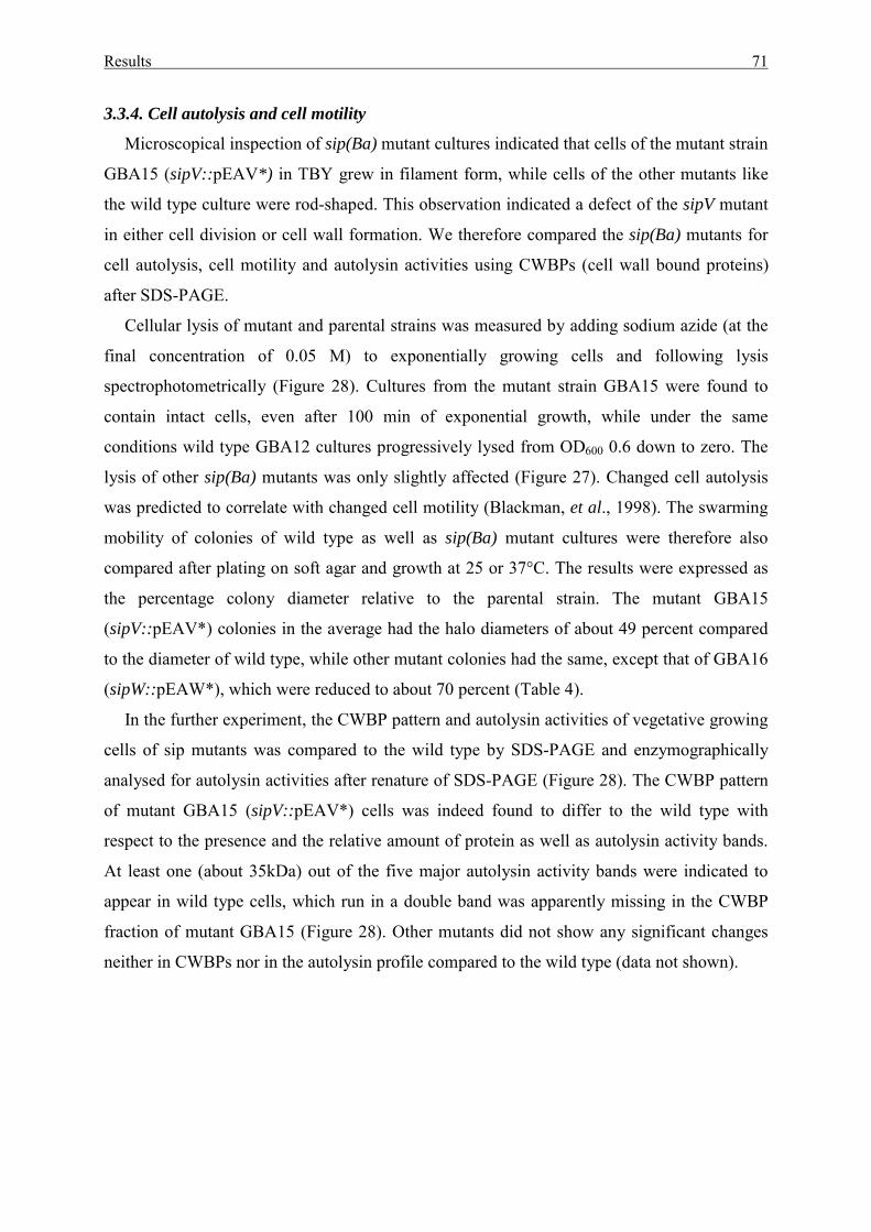
Results 71
3.3.4. Cell autolysis and cell motility
Microscopical inspection of sip(Ba) mutant cultures indicated that cells of the mutant strain
GBA15 (sipV::pEAV*) in TBY grew in filament form, while cells of the other mutants like
the wild type culture were rod-shaped. This observation indicated a defect of the sipV mutant
in either cell division or cell wall formation. We therefore compared the sip(Ba) mutants for
cell autolysis, cell motility and autolysin activities using CWBPs (cell wall bound proteins)
after SDS-PAGE.
Cellular lysis of mutant and parental strains was measured by adding sodium azide (at the
final concentration of 0.05 M) to exponentially growing cells and following lysis
spectrophotometrically (Figure 28). Cultures from the mutant strain GBA15 were found to
contain intact cells, even after 100 min of exponential growth, while under the same
conditions wild type GBA12 cultures progressively lysed from OD600 0.6 down to zero. The
lysis of other sip(Ba) mutants was only slightly affected (Figure 27). Changed cell autolysis
was predicted to correlate with changed cell motility (Blackman, et al., 1998). The swarming
mobility of colonies of wild type as well as sip(Ba) mutant cultures were therefore also
compared after plating on soft agar and growth at 25 or 37°C. The results were expressed as
the percentage colony diameter relative to the parental strain. The mutant GBA15
(sipV::pEAV*) colonies in the average had the halo diameters of about 49 percent compared
to the diameter of wild type, while other mutant colonies had the same, except that of GBA16
(sipW::pEAW*), which were reduced to about 70 percent (Table 4).
In the further experiment, the CWBP pattern and autolysin activities of vegetative growing
cells of sip mutants was compared to the wild type by SDS-PAGE and enzymographically
analysed for autolysin activities after renature of SDS-PAGE (Figure 28). The CWBP pattern
of mutant GBA15 (sipV::pEAV*) cells was indeed found to differ to the wild type with
respect to the presence and the relative amount of protein as well as autolysin activity bands.
At least one (about 35kDa) out of the five major autolysin activity bands were indicated to
appear in wild type cells, which run in a double band was apparently missing in the CWBP
fraction of mutant GBA15 (Figure 28). Other mutants did not show any significant changes
neither in CWBPs nor in the autolysin profile compared to the wild type (data not shown).
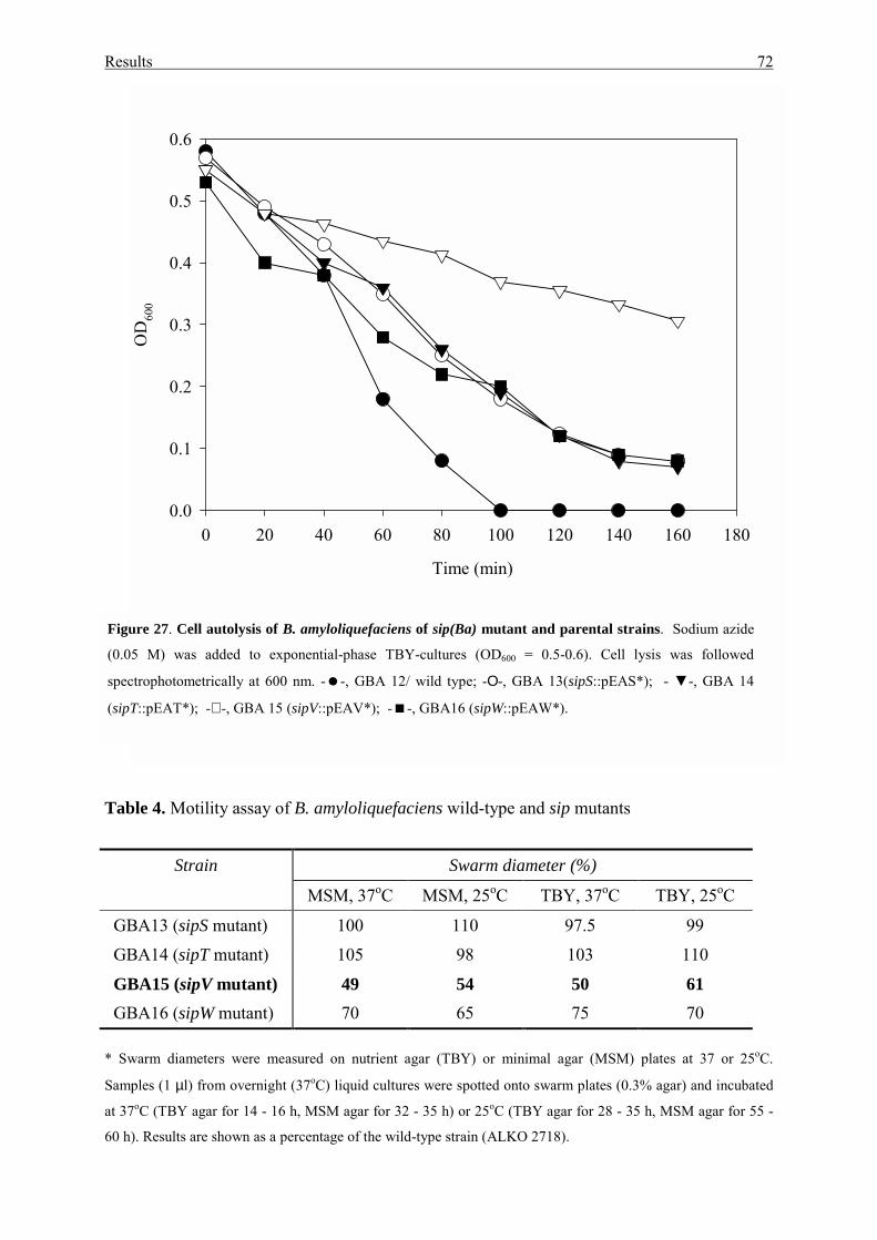
Results 72
Table 4. Motility assay of B. amyloliquefaciens wild-type and sip mutants
Strain Swarm diameter (%)
MSM, 37oC MSM, 25oC TBY, 37oC TBY, 25oC
GBA13 (sipS mutant) 100 110 97.5 99
GBA14 (sipT mutant) 105 98 103 110
GBA15 (sipV mutant) 49 54 50 61 GBA16 (sipW mutant) 70 65 75 70 * Swarm diameters were measured on nutrient agar (TBY) or minimal agar (MSM) plates at 37 or 25oC.
Samples (1 µl) from overnight (37oC) liquid cultures were spotted onto swarm plates (0.3% agar) and incubated
at 37oC (TBY agar for 14 - 16 h, MSM agar for 32 - 35 h) or 25oC (TBY agar for 28 - 35 h, MSM agar for 55 -
60 h). Results are shown as a percentage of the wild-type strain (ALKO 2718).
Time (min)
0 20 40 60 80 100 120 140 160 180
OD
600
0.0
0.1
0.2
0.3
0.4
0.5
0.6
Figure 27. Cell autolysis of B. amyloliquefaciens of sip(Ba) mutant and parental strains. Sodium azide
(0.05 M) was added to exponential-phase TBY-cultures (OD600 = 0.5-0.6). Cell lysis was followed
spectrophotometrically at 600 nm. -�-, GBA 12/ wild type; -Ο-, GBA 13(sipS::pEAS*); - ▼-, GBA 14
(sipT::pEAT*); -∇ -, GBA 15 (sipV::pEAV*); -�-, GBA16 (sipW::pEAW*).
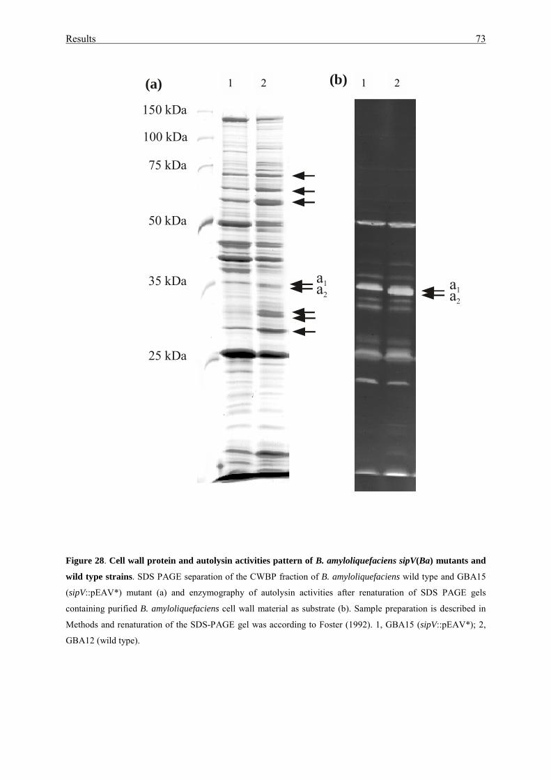
Results 73
Figure 28. Cell wall protein and autolysin activities pattern of B. amyloliquefaciens sipV(Ba) mutants and
wild type strains. SDS PAGE separation of the CWBP fraction of B. amyloliquefaciens wild type and GBA15
(sipV::pEAV*) mutant (a) and enzymography of autolysin activities after renaturation of SDS PAGE gels
containing purified B. amyloliquefaciens cell wall material as substrate (b). Sample preparation is described in
Methods and renaturation of the SDS-PAGE gel was according to Foster (1992). 1, GBA15 (sipV::pEAV*); 2,
GBA12 (wild type).
(a) (b)
a1a2a1a2
150 kDa
100 kDa
75 kDa
50 kDa
35 kDa
25 kDa
1 2 1 2
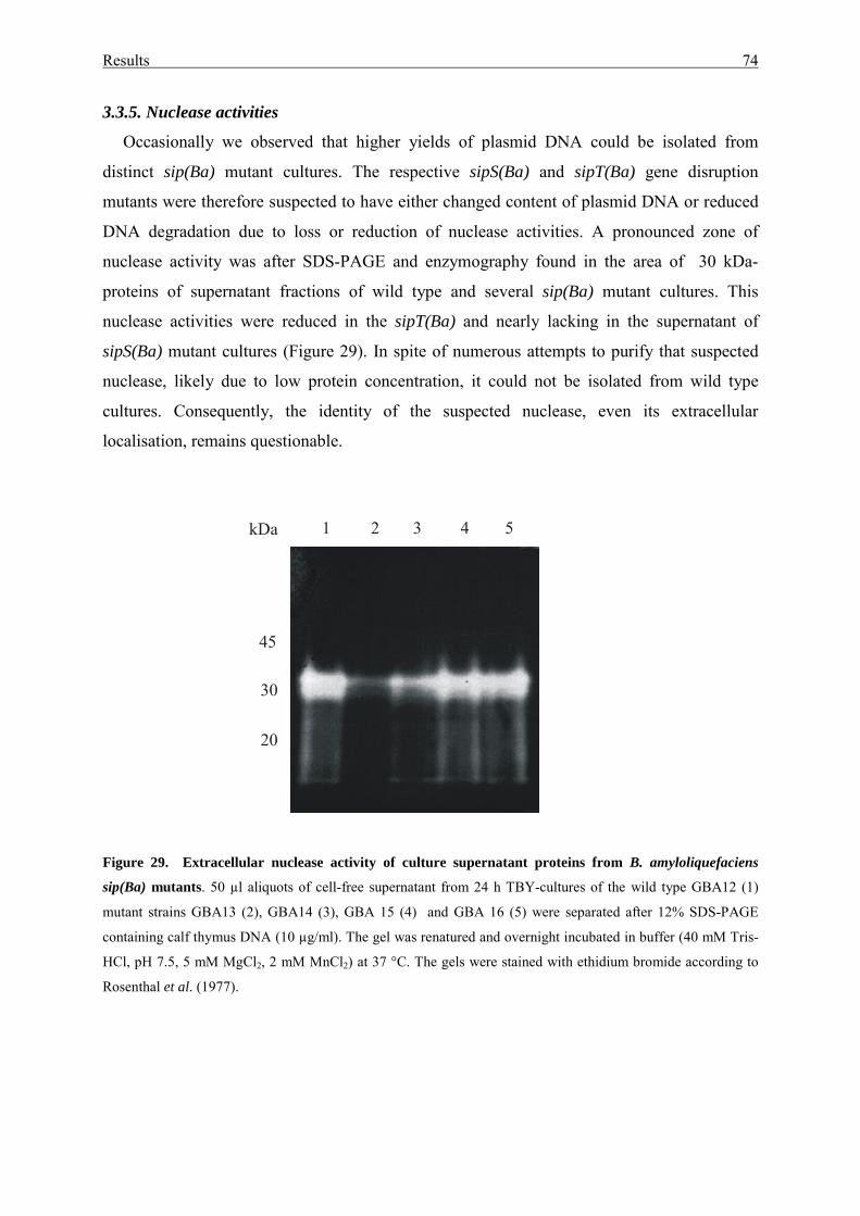
Results 74
3.3.5. Nuclease activities
Occasionally we observed that higher yields of plasmid DNA could be isolated from
distinct sip(Ba) mutant cultures. The respective sipS(Ba) and sipT(Ba) gene disruption
mutants were therefore suspected to have either changed content of plasmid DNA or reduced
DNA degradation due to loss or reduction of nuclease activities. A pronounced zone of
nuclease activity was after SDS-PAGE and enzymography found in the area of 30 kDa-
proteins of supernatant fractions of wild type and several sip(Ba) mutant cultures. This
nuclease activities were reduced in the sipT(Ba) and nearly lacking in the supernatant of
sipS(Ba) mutant cultures (Figure 29). In spite of numerous attempts to purify that suspected
nuclease, likely due to low protein concentration, it could not be isolated from wild type
cultures. Consequently, the identity of the suspected nuclease, even its extracellular
localisation, remains questionable.
Figure 29. Extracellular nuclease activity of culture supernatant proteins from B. amyloliquefaciens
sip(Ba) mutants. 50 µl aliquots of cell-free supernatant from 24 h TBY-cultures of the wild type GBA12 (1)
mutant strains GBA13 (2), GBA14 (3), GBA 15 (4) and GBA 16 (5) were separated after 12% SDS-PAGE
containing calf thymus DNA (10 µg/ml). The gel was renatured and overnight incubated in buffer (40 mM Tris-
HCl, pH 7.5, 5 mM MgCl2, 2 mM MnCl2) at 37 °C. The gels were stained with ethidium bromide according to
Rosenthal et al. (1977).
1 2 3 4 5
30
45
20
kDa
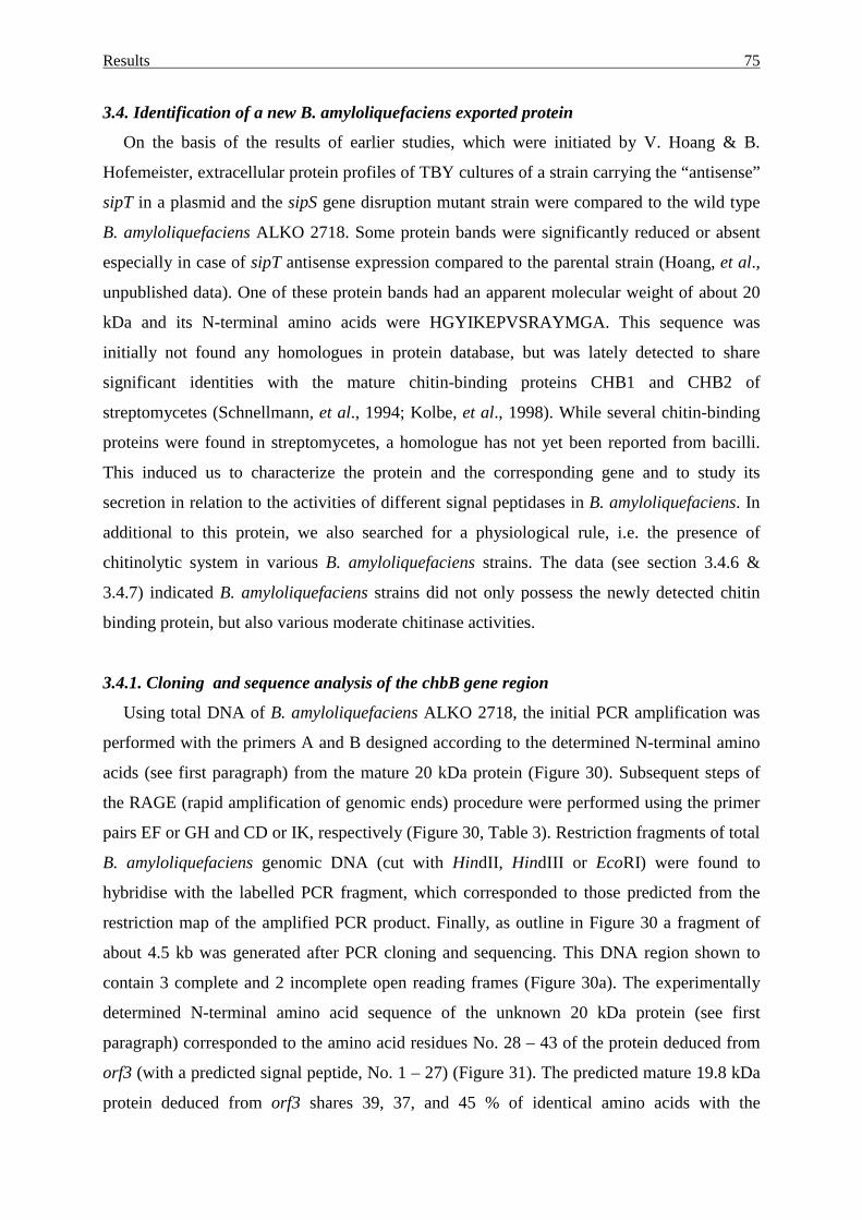
Results 75
3.4. Identification of a new B. amyloliquefaciens exported protein
On the basis of the results of earlier studies, which were initiated by V. Hoang & B.
Hofemeister, extracellular protein profiles of TBY cultures of a strain carrying the “antisense”
sipT in a plasmid and the sipS gene disruption mutant strain were compared to the wild type
B. amyloliquefaciens ALKO 2718. Some protein bands were significantly reduced or absent
especially in case of sipT antisense expression compared to the parental strain (Hoang, et al.,
unpublished data). One of these protein bands had an apparent molecular weight of about 20
kDa and its N-terminal amino acids were HGYIKEPVSRAYMGA. This sequence was
initially not found any homologues in protein database, but was lately detected to share
significant identities with the mature chitin-binding proteins CHB1 and CHB2 of
streptomycetes (Schnellmann, et al., 1994; Kolbe, et al., 1998). While several chitin-binding
proteins were found in streptomycetes, a homologue has not yet been reported from bacilli.
This induced us to characterize the protein and the corresponding gene and to study its
secretion in relation to the activities of different signal peptidases in B. amyloliquefaciens. In
additional to this protein, we also searched for a physiological rule, i.e. the presence of
chitinolytic system in various B. amyloliquefaciens strains. The data (see section 3.4.6 &
3.4.7) indicated B. amyloliquefaciens strains did not only possess the newly detected chitin
binding protein, but also various moderate chitinase activities.
3.4.1. Cloning and sequence analysis of the chbB gene region
Using total DNA of B. amyloliquefaciens ALKO 2718, the initial PCR amplification was
performed with the primers A and B designed according to the determined N-terminal amino
acids (see first paragraph) from the mature 20 kDa protein (Figure 30). Subsequent steps of
the RAGE (rapid amplification of genomic ends) procedure were performed using the primer
pairs EF or GH and CD or IK, respectively (Figure 30, Table 3). Restriction fragments of total
B. amyloliquefaciens genomic DNA (cut with HindII, HindIII or EcoRI) were found to
hybridise with the labelled PCR fragment, which corresponded to those predicted from the
restriction map of the amplified PCR product. Finally, as outline in Figure 30 a fragment of
about 4.5 kb was generated after PCR cloning and sequencing. This DNA region shown to
contain 3 complete and 2 incomplete open reading frames (Figure 30a). The experimentally
determined N-terminal amino acid sequence of the unknown 20 kDa protein (see first
paragraph) corresponded to the amino acid residues No. 28 – 43 of the protein deduced from
orf3 (with a predicted signal peptide, No. 1 – 27) (Figure 31). The predicted mature 19.8 kDa
protein deduced from orf3 shares 39, 37, and 45 % of identical amino acids with the
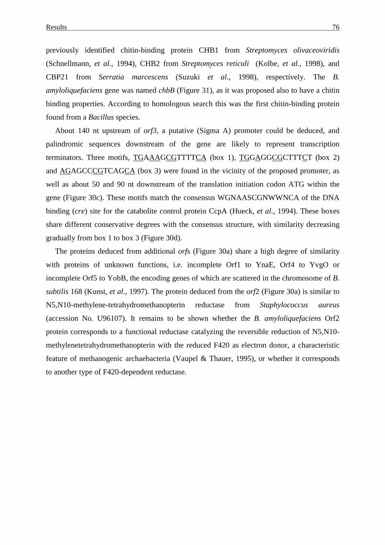
Results 76
previously identified chitin-binding protein CHB1 from Streptomyces olivaceoviridis
(Schnellmann, et al., 1994), CHB2 from Streptomyces reticuli (Kolbe, et al., 1998), and
CBP21 from Serratia marcescens (Suzuki et al., 1998), respectively. The B.
amyloliquefaciens gene was named chbB (Figure 31), as it was proposed also to have a chitin
binding properties. According to homologous search this was the first chitin-binding protein
found from a Bacillus species.
About 140 nt upstream of orf3, a putative (Sigma A) promoter could be deduced, and
palindromic sequences downstream of the gene are likely to represent transcription
terminators. Three motifs, TGAAAGCGTTTTCA (box 1), TGGAGGCGCTTTCT (box 2)
and AGAGCCCGTCAGCA (box 3) were found in the vicinity of the proposed promoter, as
well as about 50 and 90 nt downstream of the translation initiation codon ATG within the
gene (Figure 30c). These motifs match the consensus WGNAASCGNWWNCA of the DNA
binding (cre) site for the catabolite control protein CcpA (Hueck, et al., 1994). These boxes
share different conservative degrees with the consensus structure, with similarity decreasing
gradually from box 1 to box 3 (Figure 30d).
The proteins deduced from additional orfs (Figure 30a) share a high degree of similarity
with proteins of unknown functions, i.e. incomplete Orf1 to YnaE, Orf4 to YvgO or
incomplete Orf5 to YobB, the encoding genes of which are scattered in the chromosome of B.
subtilis 168 (Kunst, et al., 1997). The protein deduced from the orf2 (Figure 30a) is similar to
N5,N10-methylene-tetrahydromethanopterin reductase from Staphylococcus aureus
(accession No. U96107). It remains to be shown whether the B. amyloliquefaciens Orf2
protein corresponds to a functional reductase catalyzing the reversible reduction of N5,N10-
methylenetetrahydromethanopterin with the reduced F420 as electron donor, a characteristic
feature of methanogenic archaebacteria (Vaupel & Thauer, 1995), or whether it corresponds
to another type of F420-dependent reductase.
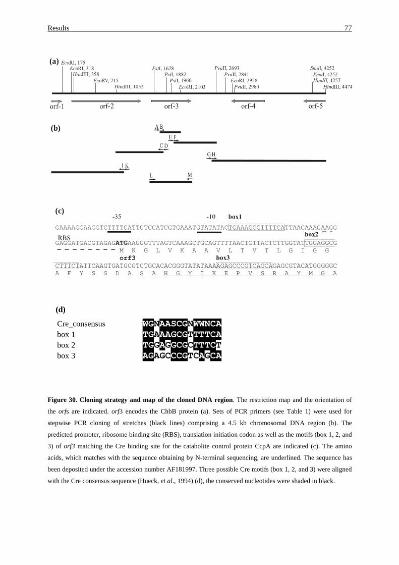
Results 77
(d)
Cre_consensus WGNAASCGNWWNCA box 1 TGAAAGCGTTTTCA box 2 TGGAGGCGCTTTCT box 3 AGAGCCCGTCAGCA
Figure 30. Cloning strategy and map of the cloned DNA region. The restriction map and the orientation of
the orfs are indicated. orf3 encodes the ChbB protein (a). Sets of PCR primers (see Table 1) were used for
stepwise PCR cloning of stretches (black lines) comprising a 4.5 kb chromosomal DNA region (b). The
predicted promoter, ribosome binding site (RBS), translation initiation codon as well as the motifs (box 1, 2, and
3) of orf3 matching the Cre binding site for the catabolite control protein CcpA are indicated (c). The amino
acids, which matches with the sequence obtaining by N-terminal sequencing, are underlined. The sequence has
been deposited under the accession number AF181997. Three possible Cre motifs (box 1, 2, and 3) were aligned
with the Cre consensus sequence (Hueck, et al., 1994) (d), the conserved nucleotides were shaded in black.
(a)
(b)
(c)
GAAAAGGAAGGTCTTTTCATTCTCCATCGTGAAATGTATATACTGAAAGCGTTTTCATTAACAAAGAAGG
GAGGATGACGTAGAG AAGGGTTTAGTCAAAGCTGCAGTTTTAACTGTTACTCTTGGTATTGGAGGCGM K G L V K A A V L T V T L G I G G
CTTTCTATTCAAGTGATGCGTCTGCACACGGGTATATAAAAGAGCCCGTCAGCAGAGCGTACATGGGGGCA F Y S S D A S A
ATG
orf3
H G Y I K E P V S R A Y M G A
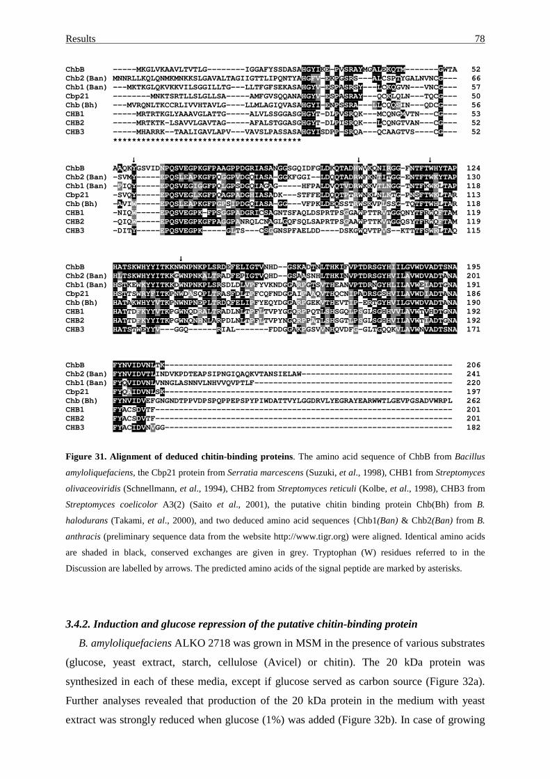
Results 78
ChbB -----MKGLVKAAVLTVTLG--------IGGAFYSSDASAHGYIKE-PVSRAYMGALEKQTM-------GWTA 52Chb2(Ban) MNNRLLKQLQNMKMNKKSLGAVALTAGIIGTTLIPQNTYAHGFV-EKPGSRS---ALCSPTYGALNVNCG--- 66Chb1(Ban) ---MKTKGLQKVKKVILSGGILLTG---LLTFGFSEKASAHGYV-ESPASRSY---LCKQGVN---VNCG--- 57Cbp21 --------MNKTSRTLLSLGLLSA-----AMFGVSQQANAHGYV-ESPASRAY---QCKLQLN---TQCG--- 50Chb(Bh) ---MVRQNLTKCCRLIVVHTAVLG----LLMLAGIQVASAHGYI-ENPSSRA---LLCQQSIN---QDCG--- 56CHB1 -----MRTRTKGLYAAAVGLATTG-----ALVLSSGGASGHGYT-DLPVSRQK---MCQNGMVTN---CG--- 53CHB2 -----MRTKTK-LSAVVLGAVTAG-----AFALSTGGASGHGYT-DLPISRQK---LCQNGTVAN---CG--- 52CHB3 -----MHARRK--TAALIGAVLAPV---VAVSLPASSASAHGYISDPP-SRQA---QCAAGTVS----CG--- 52
****************************************
↓↓↓↓ ↓↓↓↓ ↓↓↓↓ChbB AAQKYGSVIDNPQSVEGPKGFPAAGPPDGRIASANGGSGQIDFGLDKQTADHWVKQNIRGG-FNTFTWHYTAP 124Chb2(Ban) -SVMY-----EPQSLEAPKGFPQGGPVDGQIASA-GGKFGGI--LDQQTADRWFKNTITGG-ENTFTWKYTAP 130Chb1(Ban) -PIQY-----EPQSVEGIGGFPQLGPSDGQIAGAG-----HFPALDVQTVDRWKKVTLNGG-TNTFKWKLTAP 118Cbp21 -SVQY-----EPQSVEGLKGFPQAGPADGHIASADK---STFFELDQQTPTRWNKLNLKTG-PNSFTWKLTAR 113Chb(Bh) -AVIW-----EPQSLEAPKGFPGPSIPDGQIASA-GG---VFPKLDEQSSTRWSKVPLSSG-TQTFTWHLTAR 118CHB1 -NIQW-----EPQSVEGPK-FPSGGPADGRICSAGNTSFAQLDSPRTPSGGAWPTTRVTGGQNYTFRWQFTAM 119CHB2 -QIQW-----EPQSVEGPKGFPAGGPANRQLCNAGLGQFSQLSAPRTPSGAAWPTTKVTGGQSYTFRWQFTAM 119CHB3 -DITY-----EPQSVEGPK-----GLTS---CSGGNSPFAELDD----DSKGWQVTPVS--KTTTFSWRLTAQ 115
↓↓↓↓ChbB HATSKWHYYITKKNWNPNKPLSRDEFELIGTVNHD--GSKADTNLTHKIFVPTDRSGYHIILGVWDVADTSNA 195Chb2(Ban) HLTSKWHYYITKKGWNPNKALTRADFEPIGTVQHD--GSAASNHLTHKINVPTDRSGYHVILAVWDVADTANA 201Chb1(Ban) HSTKEWKYYITKKDWNPNKPLSRSDLDLVPFYVKNDGGARPGTSVTHEANVPTDRNGYHLILAVWEIADTGNA 191Cbp21 HSTTSWRYFITKPNWDASQPLTRASFDLTPFCQFNDGGAIPAAQVTHQCNIPADRSGSHVILAVWDIADTANA 186Chb(Bh) HATAKWHYYVTKPNWNPNEPLTRDQFELIPFYEQYDGGARPGEKVTHEVTIP-ERTGYHVILGVWDVADTANA 190CHB1 HATTDFKYYVTKPGWNQDRALTRADLNLTPFLTVPYGGQRPPQTLSHSGQLPSGLSGHHVVLAVWTVHDTGNA 192CHB2 HATTDFKYYITKPGWNQNHNLARPDLNLTPFLTVPYNGQRPPATLSHSGTLPSGLSGHHVILAVWTIADTGNA 192CHB3 HATSTWEYYV---GGQ------RIAL-------FDDGGAKPGSVVNHQVDFG-GLTGQQKVLAVWNVADTSNA 171
ChbB FYNVIDVNLTK------------------------------------------------------------- 206Chb2(Ban) FYNVIDVTLINDVKPDTEAPSIPNGIQAQKVTANSIELAW-------------------------------- 241Chb1(Ban) FYQVIDVNLVNNGLASNNVLNHVVQVPTLF------------------------------------------ 220Cbp21 FYQAIDVNLSK------------------------------------------------------------- 197Chb(Bh) FYNVIDVEFGNGNDTPPVDPSPQPPEPSPYPIWDATTVYLGGDRVLYEGRAYEARWWTLGEVPGSADVWRPL 262CHB1 FYACSDVTF--------------------------------------------------------------- 201CHB2 FYACSDVTF--------------------------------------------------------------- 201CHB3 FYACIDVNVGG------------------------------------------------------------- 182
Figure 31. Alignment of deduced chitin-binding proteins. The amino acid sequence of ChbB from Bacillus
amyloliquefaciens, the Cbp21 protein from Serratia marcescens (Suzuki, et al., 1998), CHB1 from Streptomyces
olivaceoviridis (Schnellmann, et al., 1994), CHB2 from Streptomyces reticuli (Kolbe, et al., 1998), CHB3 from
Streptomyces coelicolor A3(2) (Saito et al., 2001), the putative chitin binding protein Chb(Bh) from B.
halodurans (Takami, et al., 2000), and two deduced amino acid sequences {Chb1(Ban) & Chb2(Ban) from B.
anthracis (preliminary sequence data from the website http://www.tigr.org) were aligned. Identical amino acids
are shaded in black, conserved exchanges are given in grey. Tryptophan (W) residues referred to in the
Discussion are labelled by arrows. The predicted amino acids of the signal peptide are marked by asterisks.
3.4.2. Induction and glucose repression of the putative chitin-binding protein
B. amyloliquefaciens ALKO 2718 was grown in MSM in the presence of various substrates
(glucose, yeast extract, starch, cellulose (Avicel) or chitin). The 20 kDa protein was
synthesized in each of these media, except if glucose served as carbon source (Figure 32a).
Further analyses revealed that production of the 20 kDa protein in the medium with yeast
extract was strongly reduced when glucose (1%) was added (Figure 32b). In case of growing
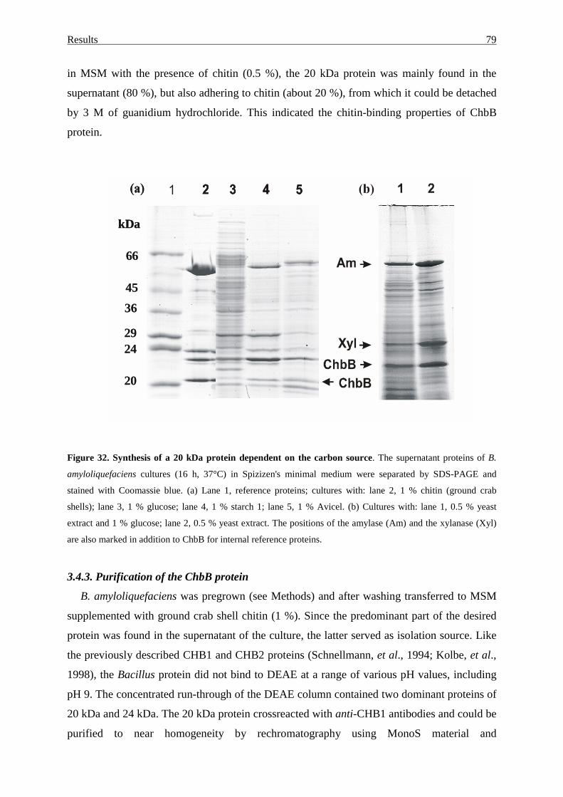
Results 79
in MSM with the presence of chitin (0.5 %), the 20 kDa protein was mainly found in the
supernatant (80 %), but also adhering to chitin (about 20 %), from which it could be detached
by 3 M of guanidium hydrochloride. This indicated the chitin-binding properties of ChbB
protein.
Figure 32. Synthesis of a 20 kDa protein dependent on the carbon source. The supernatant proteins of B.
amyloliquefaciens cultures (16 h, 37°C) in Spizizen's minimal medium were separated by SDS-PAGE and
stained with Coomassie blue. (a) Lane 1, reference proteins; cultures with: lane 2, 1 % chitin (ground crab
shells); lane 3, 1 % glucose; lane 4, 1 % starch 1; lane 5, 1 % Avicel. (b) Cultures with: lane 1, 0.5 % yeast
extract and 1 % glucose; lane 2, 0.5 % yeast extract. The positions of the amylase (Am) and the xylanase (Xyl)
are also marked in addition to ChbB for internal reference proteins.
3.4.3. Purification of the ChbB protein
B. amyloliquefaciens was pregrown (see Methods) and after washing transferred to MSM
supplemented with ground crab shell chitin (1 %). Since the predominant part of the desired
protein was found in the supernatant of the culture, the latter served as isolation source. Like
the previously described CHB1 and CHB2 proteins (Schnellmann, et al., 1994; Kolbe, et al.,
1998), the Bacillus protein did not bind to DEAE at a range of various pH values, including
pH 9. The concentrated run-through of the DEAE column contained two dominant proteins of
20 kDa and 24 kDa. The 20 kDa protein crossreacted with anti-CHB1 antibodies and could be
purified to near homogeneity by rechromatography using MonoS material and
kDa
66
45
36
2924
20
kDa
66
4536
2924
20
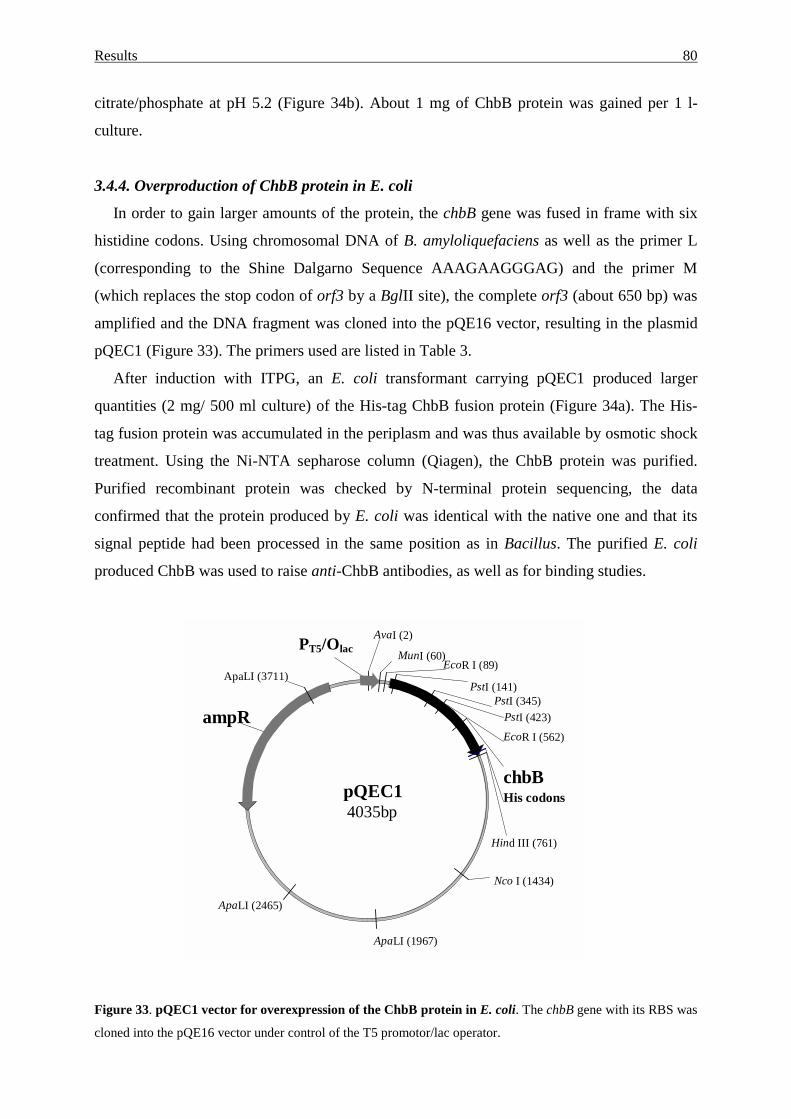
Results 80
citrate/phosphate at pH 5.2 (Figure 34b). About 1 mg of ChbB protein was gained per 1 l-
culture.
3.4.4. Overproduction of ChbB protein in E. coli
In order to gain larger amounts of the protein, the chbB gene was fused in frame with six
histidine codons. Using chromosomal DNA of B. amyloliquefaciens as well as the primer L
(corresponding to the Shine Dalgarno Sequence AAAGAAGGGAG) and the primer M
(which replaces the stop codon of orf3 by a BglII site), the complete orf3 (about 650 bp) was
amplified and the DNA fragment was cloned into the pQE16 vector, resulting in the plasmid
pQEC1 (Figure 33). The primers used are listed in Table 3.
After induction with ITPG, an E. coli transformant carrying pQEC1 produced larger
quantities (2 mg/ 500 ml culture) of the His-tag ChbB fusion protein (Figure 34a). The His-
tag fusion protein was accumulated in the periplasm and was thus available by osmotic shock
treatment. Using the Ni-NTA sepharose column (Qiagen), the ChbB protein was purified.
Purified recombinant protein was checked by N-terminal protein sequencing, the data
confirmed that the protein produced by E. coli was identical with the native one and that its
signal peptide had been processed in the same position as in Bacillus. The purified E. coli
produced ChbB was used to raise anti-ChbB antibodies, as well as for binding studies.
Figure 33. pQEC1 vector for overexpression of the ChbB protein in E. coli. The chbB gene with its RBS was
cloned into the pQE16 vector under control of the T5 promotor/lac operator.
pQEC14035bp
chbB
ampR
His codons
AvaI (2)
Hind III (761)
MunI (60)
Nco I (1434)
EcoR I (89)
EcoR I (562)
ApaLI (1967)
ApaLI (2465)
ApaLI (3711) PstI (141)
PstI (345) PstI (423)
PT5/Olac
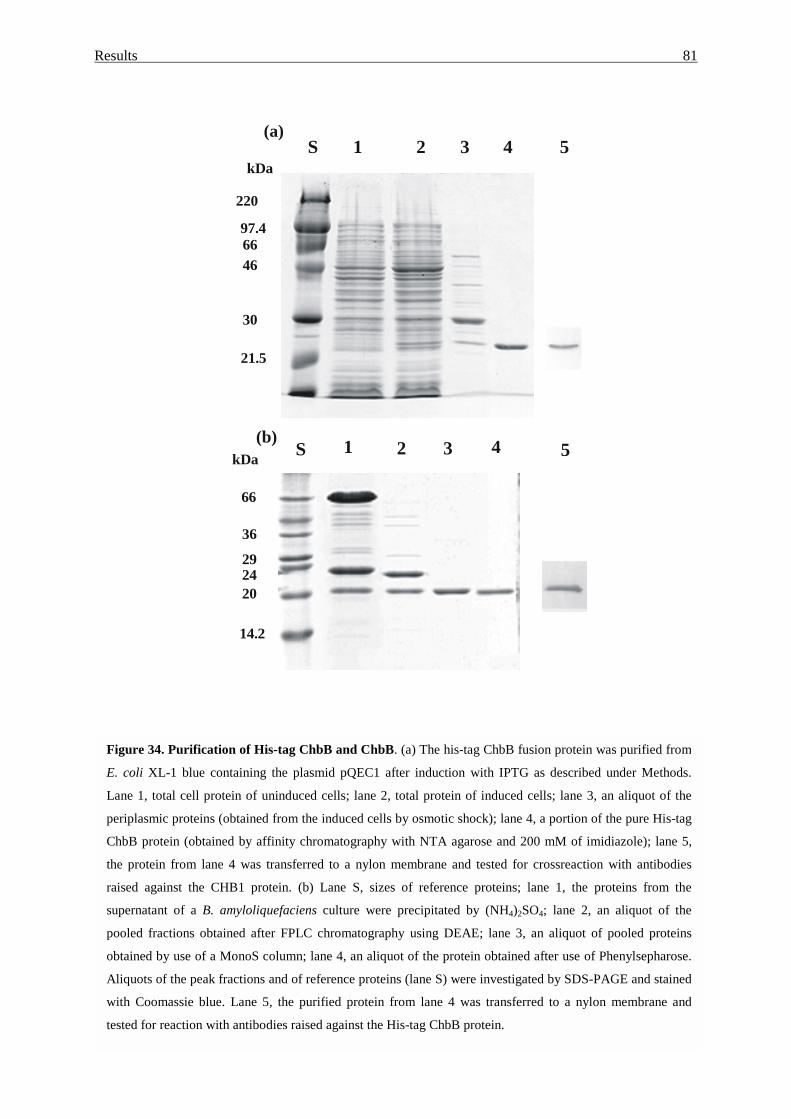
Results 81
1 2 3 4kDa S
66
36
292420
14.2
(b)
1 2 3 4kDa
220
97.46646
30
21.5
S 5(a)
5
Figure 34. Purification of His-tag ChbB and ChbB. (a) The his-tag ChbB fusion protein was purified from
E. coli XL-1 blue containing the plasmid pQEC1 after induction with IPTG as described under Methods.
Lane 1, total cell protein of uninduced cells; lane 2, total protein of induced cells; lane 3, an aliquot of the
periplasmic proteins (obtained from the induced cells by osmotic shock); lane 4, a portion of the pure His-tag
ChbB protein (obtained by affinity chromatography with NTA agarose and 200 mM of imidiazole); lane 5,
the protein from lane 4 was transferred to a nylon membrane and tested for crossreaction with antibodies
raised against the CHB1 protein. (b) Lane S, sizes of reference proteins; lane 1, the proteins from the
supernatant of a B. amyloliquefaciens culture were precipitated by (NH4)2SO4; lane 2, an aliquot of the
pooled fractions obtained after FPLC chromatography using DEAE; lane 3, an aliquot of pooled proteins
obtained by use of a MonoS column; lane 4, an aliquot of the protein obtained after use of Phenylsepharose.
Aliquots of the peak fractions and of reference proteins (lane S) were investigated by SDS-PAGE and stained
with Coomassie blue. Lane 5, the purified protein from lane 4 was transferred to a nylon membrane and
tested for reaction with antibodies raised against the His-tag ChbB protein.
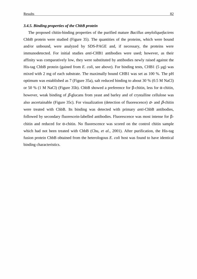
Results 82
3.4.5. Binding properties of the ChbB protein
The proposed chitin-binding properties of the purified mature Bacillus amyloliquefaciens
ChbB protein were studied (Figure 35). The quantities of the proteins, which were bound
and/or unbound, were analyzed by SDS-PAGE and, if necessary, the proteins were
immunodetected. For initial studies anti-CHB1 antibodies were used; however, as their
affinity was comparatively low, they were substituted by antibodies newly raised against the
His-tag ChbB protein (gained from E. coli, see above). For binding tests, CHB1 (5 µg) was
mixed with 2 mg of each substrate. The maximally bound CHB1 was set as 100 %. The pH
optimum was established as 7 (Figure 35a), salt reduced binding to about 30 % (0.5 M NaCl)
or 50 % (1 M NaCl) (Figure 35b). ChbB showed a preference for β-chitin, less for α-chitin,
however, weak binding of β-glucans from yeast and barley and of crystalline cellulose was
also ascertainable (Figure 35c). For visualization (detection of fluorescence) α- and β-chitin
were treated with ChbB. Its binding was detected with primary anti-ChbB antibodies,
followed by secondary fluorescein-labelled antibodies. Fluorescence was most intense for β-
chitin and reduced for α-chitin. No fluorescence was scored on the control chitin sample
which had not been treated with ChbB (Chu, et al., 2001). After purification, the His-tag
fusion protein ChbB obtained from the heterologous E. coli host was found to have identical
binding characteristics.
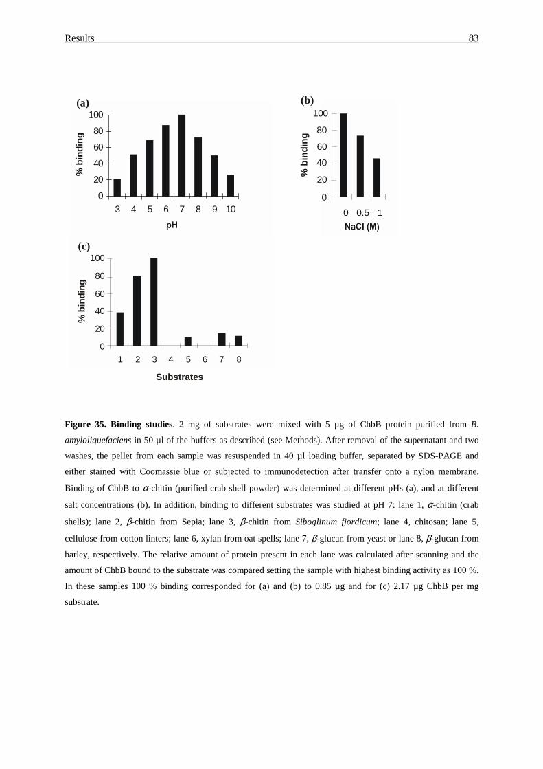
Results 83
Figure 35. Binding studies. 2 mg of substrates were mixed with 5 µg of ChbB protein purified from B.
amyloliquefaciens in 50 µl of the buffers as described (see Methods). After removal of the supernatant and two
washes, the pellet from each sample was resuspended in 40 µl loading buffer, separated by SDS-PAGE and
either stained with Coomassie blue or subjected to immunodetection after transfer onto a nylon membrane.
Binding of ChbB to α-chitin (purified crab shell powder) was determined at different pHs (a), and at different
salt concentrations (b). In addition, binding to different substrates was studied at pH 7: lane 1, α-chitin (crab
shells); lane 2, β-chitin from Sepia; lane 3, β-chitin from Siboglinum fjordicum; lane 4, chitosan; lane 5,
cellulose from cotton linters; lane 6, xylan from oat spells; lane 7, β-glucan from yeast or lane 8, β-glucan from
barley, respectively. The relative amount of protein present in each lane was calculated after scanning and the
amount of ChbB bound to the substrate was compared setting the sample with highest binding activity as 100 %.
In these samples 100 % binding corresponded for (a) and (b) to 0.85 µg and for (c) 2.17 µg ChbB per mg
substrate.
Substrates
0
20
40
60
80
100
1 2 3 4 5 6 7 8
% b
indi
ng
(c)
(b)
% b
indi
ng
0
20
40
60
80
100
0 0.5 1ConcentrationofNaCl(M)
%
100
80
60
40
20
00 10.5
0
20
40
60
80
100
3 4 5 6 7 8 9 10
pH
%
(a)
% b
indi
ng
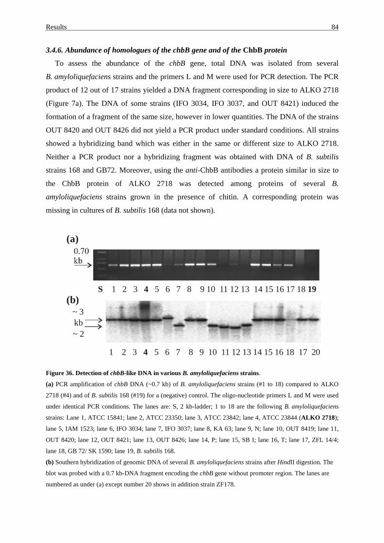
Results 84
3.4.6. Abundance of homologues of the chbB gene and of the ChbB protein
To assess the abundance of the chbB gene, total DNA was isolated from several
B. amyloliquefaciens strains and the primers L and M were used for PCR detection. The PCR
product of 12 out of 17 strains yielded a DNA fragment corresponding in size to ALKO 2718
(Figure 7a). The DNA of some strains (IFO 3034, IFO 3037, and OUT 8421) induced the
formation of a fragment of the same size, however in lower quantities. The DNA of the strains
OUT 8420 and OUT 8426 did not yield a PCR product under standard conditions. All strains
showed a hybridizing band which was either in the same or different size to ALKO 2718.
Neither a PCR product nor a hybridizing fragment was obtained with DNA of B. subtilis
strains 168 and GB72. Moreover, using the anti-ChbB antibodies a protein similar in size to
the ChbB protein of ALKO 2718 was detected among proteins of several B.
amyloliquefaciens strains grown in the presence of chitin. A corresponding protein was
missing in cultures of B. subtilis 168 (data not shown).
Figure 36. Detection of chbB-like DNA in various B. amyloliquefaciens strains.
(a) PCR amplification of chbB DNA (~0.7 kb) of B. amyloliquefaciens strains (#1 to 18) compared to ALKO
2718 (#4) and of B. subtilis 168 (#19) for a (negative) control. The oligo-nucleotide primers L and M were used
under identical PCR conditions. The lanes are: S, 2 kb-ladder; 1 to 18 are the following B. amyloliquefaciens
strains: Lane 1, ATCC 15841; lane 2, ATCC 23350; lane 3, ATCC 23842; lane 4, ATCC 23844 (ALKO 2718);
lane 5, IAM 1523; lane 6, IFO 3034; lane 7, IFO 3037; lane 8, KA 63; lane 9, N; lane 10, OUT 8419; lane 11,
OUT 8420; lane 12, OUT 8421; lane 13, OUT 8426; lane 14, P; lane 15, SB I; lane 16, T; lane 17, ZFL 14/4;
lane 18, GB 72/ SK 1590; lane 19, B. subtilis 168.
(b) Southern hybridization of genomic DNA of several B. amyloliquefaciens strains after HindII digestion. The
blot was probed with a 0.7 kb-DNA fragment encoding the chbB gene without promoter region. The lanes are
numbered as under (a) except number 20 shows in addition strain ZF178.
S 4 19 1 2 3 5 6 7 8 9 10 11 12 13 14 15 16 17 18
1 2 3 5 6 7 8 9 10 11 12 13 14 15 16 18 17 20 4
~ 3 ~ 2
(a)
(b)
kb
Results 85
3.4.7. Chitinolytic activity of B. amyloliquefaciens strains
Growing on chitin-containing plates, the B. amyloliquefaciens strains OUT 8419, OUT
8420, OUT 8421, OUT 8426, IFO 3034 and IFO 3037 degraded chitin (clear zones), like the
strain B. licheniformis 41p known to have chitinolytic activity. Using the dye Remazol
brilliant violet coupled with carboxymethyl-chitin, the relative chitinolytic activities were
compared. The strains ATCC 23842, KA63 and N showed only low activities, others
(including ALKO 2718 and ATCC 15841, ATCC 23350, IAM 1523, P, SBI, T, ZFL 14/4)
had moderate activities, while the strains IFO 3034, IFO 3037, OUT 8419, OUT 8420, OUT
8421 and OUT 8426 showed increased activities (Figure 33); these were however lower than
those of B. licheniformis 41p (Hofemeister et al., unpublished).
Figure 37. Detection of chitinolytic activity. Hydrolysis of carboxymethyl chitin-Remazol brilliant violet by an
aliquot of the culture supernatant of several B. amyloliquefaciens strains (#1 to 17) compared to B. licheniformis
(BL 41p) and to 0.007 units of Serratia marcescens chitinase (Sigma). Numbers 1 to 17 indicate
B. amyloliquefaciens strains: bar 1, ATCC 15841; bar 2, ATCC 23350; bar 3, ATCC 23842; bar 4, ATCC 23844
(ALKO 2718); bar 5, IAM 1523; bar 6, IFO 3034; bar 7, IFO 3037; bar 8, KA 63; bar 9, N; bar 10, OUT 8419;
bar 11, OUT 8420; bar 12, OUT 8421; bar 13, OUT 8426; bar 14, P; bar 15, SB I; bar 16, T; bar 17, ZFL 14/4.
Numbers 18 and 19 show the activities of B. subtilis 168 with either plasmid pHB201 (without the chbB gene)
and plasmid pHBC1 (with the chbB gene).
StrainsChi
tinas
e
BL 4
1p 1 2 3 4 5 6 7 8 9 10 11 12 13 14 15 16 17 18 19
Rel
. enz
yme
activ
ities
(OD
600)
0.00
0.02
0.04
0.06
0.08
0.10
0.12
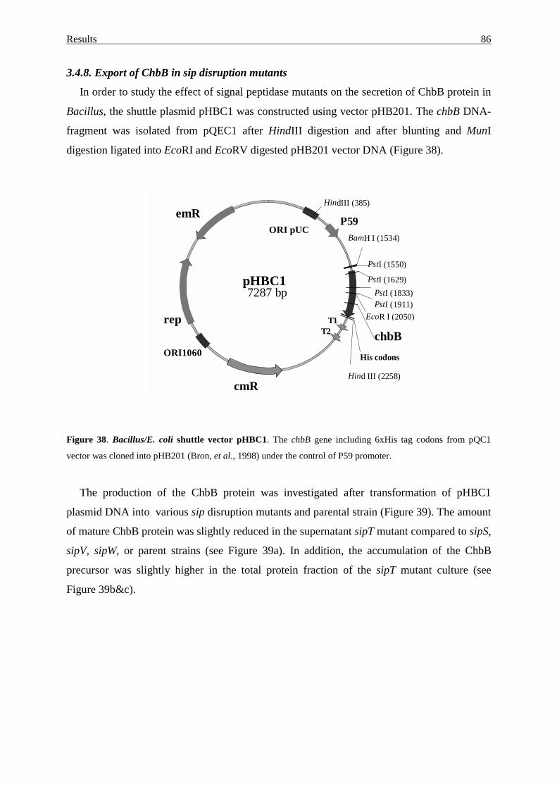
Results 86
3.4.8. Export of ChbB in sip disruption mutants
In order to study the effect of signal peptidase mutants on the secretion of ChbB protein in
Bacillus, the shuttle plasmid pHBC1 was constructed using vector pHB201. The chbB DNA-
fragment was isolated from pQEC1 after HindIII digestion and after blunting and MunI
digestion ligated into EcoRI and EcoRV digested pHB201 vector DNA (Figure 38).
Figure 38. Bacillus/E. coli shuttle vector pHBC1. The chbB gene including 6xHis tag codons from pQC1
vector was cloned into pHB201 (Bron, et al., 1998) under the control of P59 promoter.
The production of the ChbB protein was investigated after transformation of pHBC1
plasmid DNA into various sip disruption mutants and parental strain (Figure 39). The amount
of mature ChbB protein was slightly reduced in the supernatant sipT mutant compared to sipS,
sipV, sipW, or parent strains (see Figure 39a). In addition, the accumulation of the ChbB
precursor was slightly higher in the total protein fraction of the sipT mutant culture (see
Figure 39b&c).
pHBC17287 bp
chbB
emR
cmR
T1T2
rep
His codons
P59ORI pUC
ORI1060
BamH I (1534)
EcoR I (2050)
Hind III (2258)
HindIII (385)
PstI (1550)
PstI (1629)PstI (1833)PstI (1911)
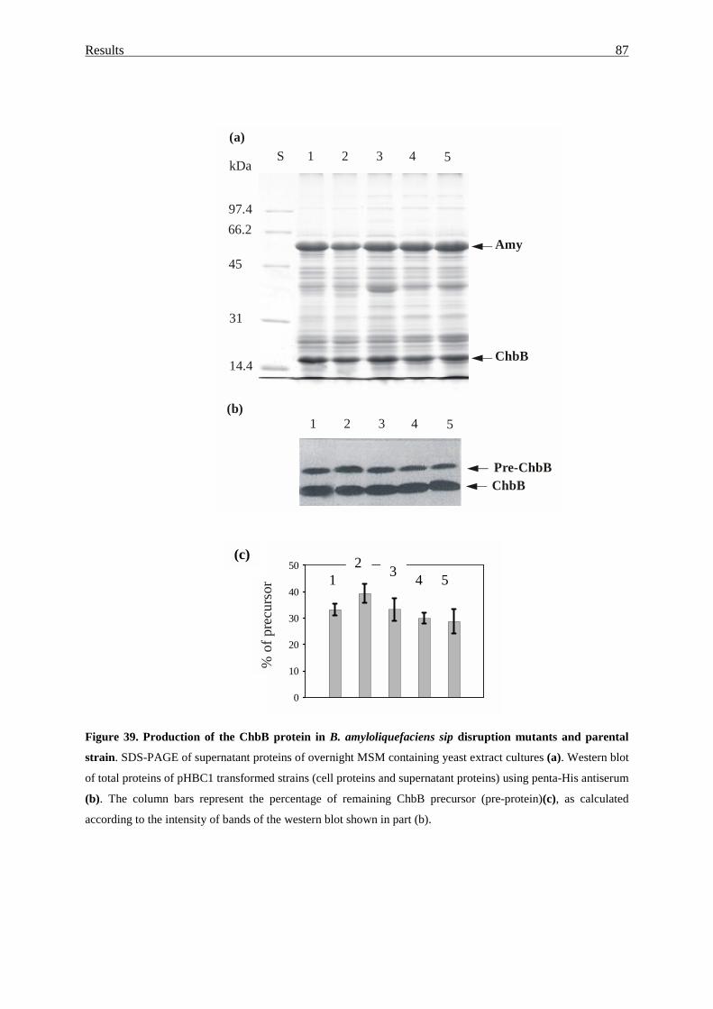
Results 87
Figure 39. Production of the ChbB protein in B. amyloliquefaciens sip disruption mutants and parental
strain. SDS-PAGE of supernatant proteins of overnight MSM containing yeast extract cultures (a). Western blot
of total proteins of pHBC1 transformed strains (cell proteins and supernatant proteins) using penta-His antiserum
(b). The column bars represent the percentage of remaining ChbB precursor (pre-protein)(c), as calculated
according to the intensity of bands of the western blot shown in part (b).
1S 2 3 4 5
97.4
kDa
66.2
45
31
14.4ChbB
Amy
(a)
1 2 3 4 5(b)
ChbBPre-ChbB
0
10
20
30
40
50(c)
% o
f pre
curs
or 12 3 4 5
Discussion
88
4. Discussion
4.1. General remarks
The main question behind the present studies was the mystery of the presence of multiple
signal peptidases in various eubacterial species, notable in bacilli (see the Introduction). As
prokaryotic genomes are extremely “economical”, i.e. are usually devoid of non essential
DNA or genes and the most Gram-negative bacteria indeed live with only one single type I
signal peptidase gene in their genome, it was reasonable to speculate about the “necessity” of
so many Sip- like proteins. This question may be addressed i) by isolation of the enzymes and
characterisation of enzymatic properties, ii) by construction and characterisation of gene
knockout mutants, iii) by genetic (functional) substitution of one type of one Sip by another
Sip-like protein, iv) by comparing the processing activity of a distinct Sip enzyme using a
special, likely specific export protein for a substrate, etc. In this studies we attempted to
analyse B. amyloliquefaciens in comparison to B. subtilis 168, where the genome project
convincingly proved the existence of two major (SipS, SipT) and three minor Sip proteins
(SipU, SipV, SipW) in B. subtilis 168 (Kunst, et al., 1997; van Roosmalen, et al., 2001). If
evolutionary constrains within the genus Bacillus caused the branching of species, these
should be also reflected on the genetic level, i.e. by changed, preserved or missing genes.
Especially protein export is one ostentatious property of B. amyloliquefaciens, as it is
basically about 10 to 20 times more efficient compared to B. subtilis (Ingle & Boyer, 1976;
Vehmaanpera, et al., 1991). The export apparatus of these two (closely related) species might
have been adapted, i.e. by changed bottleneck functions. We therefore proposed Sip proteins
to differ either in their capacity or processing specificity. Any change in the presence or in the
specificity of Sip proteins from B. subtilis to B. amyloliquefaciens could then elucidate the
selective value of those export components. However, this strategy was not without
complications mainly due to the inert genetic manageability of B. amyloliquefaciens in
comparison to B. subtilis. With the methods ad hands, the transformation efficiencies are
about 103-105 times lower and the nucleotide sequence diversity compared to B. subtilis did
not allow the use of DNA sequence information from B. subtilis for reverse genetical aims, as
for cloning of proposed genes. The latter affected mainly the characterisation of sip insertion
mutants with respect to the proposed target proteins as well as the stringency of the claimed
number of sip genes in B. amyloliquefaciens. The detailed analysis of a proposed new export
target protein of B. amyloliquefaciens, which seemed to have processing specificity towards a
distinct Sip proteins, i.e. a chitin-binding protein ChbB, was thus “a leap in the dark” which
Discussion
89
finally was disappointed as a processing target. On the other hand, however, it was welcome
as a new export protein of this species.
4.2. Cloning and similarity of Sip-like signal peptidases of B. amyloliquefaciens
In addition to sipS and sipT (Hoang & Hofemeister, 1995; Meijer, et al., 1995), here we
present evidences for the existence of two more sip-like genes in chromosomal DNA of B.
amyloliquefaciens strain ATCC 23843, which are sipV and sipW. As in case of sipS and sipT
genes, which were defined according to their sequence similarity and similar map positions
with their counterparts sipS and SipT of B. subtilis (Hoang & Hofemeister, 1995; Meijer, et
al., 1995; Tjalsma, et al., 1997), sipV and sipW of B. amyloliquefaciens were also classified
with respect to sequence similarity as well as the map position to their B. subtilis counterparts.
A sipU-like gene was not found. Although, the absence of a sipU gene could have been
overlooked due to sequence diversity, one of these two species might have gained or lost a
sipU-type gene after evolutionary constrains, likely after plasmid mediated transmission. This
hypothesis was supported by the presence of plasmids in certain B. subtilis (natto) strains
which carry the so-called sipP genes (Meijer, et al., 1995; Bron, et al., 1998). Consequently,
our data would suggest that B. amyloliquefaciens differs from B. subtilis with respect to the
number of sip-like genes, i.e. to contain in the genome sipS, sipT, sipV and sipW, but likely to
miss a sipU-like gene candidate.
Many bacteria contain paralogous type I Spases in multiplicity (isoforms). The presence of
multiple type I SPases seems to be a common feature of Gram-positive eubacteria. For
example, Staphylococcus aureus contains at least two type I SPases, whereas Streptomyces
lividans seems to contain four (Parro, et al., 1999). Data obtained from genome sequencing
projects of B. subtilis (Kunst et al., 1997), B. halodurans (Takami, et al., 2000) and B.
anthracis (preliminary data from the Institute for Genomic Research website at
http://www.tigr.org), provided a precise estimation of the number of genes encoding type I
SPases. While the B. subtilis genome harbours five sip genes (Kunst, et al., 1997), only two
are found in B. halodurans. The largest number of sip genes was found in B. antharacis,
where at least six genes encoding for type I SPases could be identified. These findings would
rise questions about the phylogenetic diversity and the mode of conservation of structural and
enzymatic properties among these SPase isoforms in bacilli. The alignment of as much as 18
Bacillus P-type SPases proteins from B. subtilis, B. amyloliquefaciens, B. licheniformis, B.
stearothermophilus, B. caldolyticus, B. halodurans and B. anthracis is shown in Figure 16.
For example, the amino acid sequence of SipV(Ba) shares 40% to 77% of identical amino
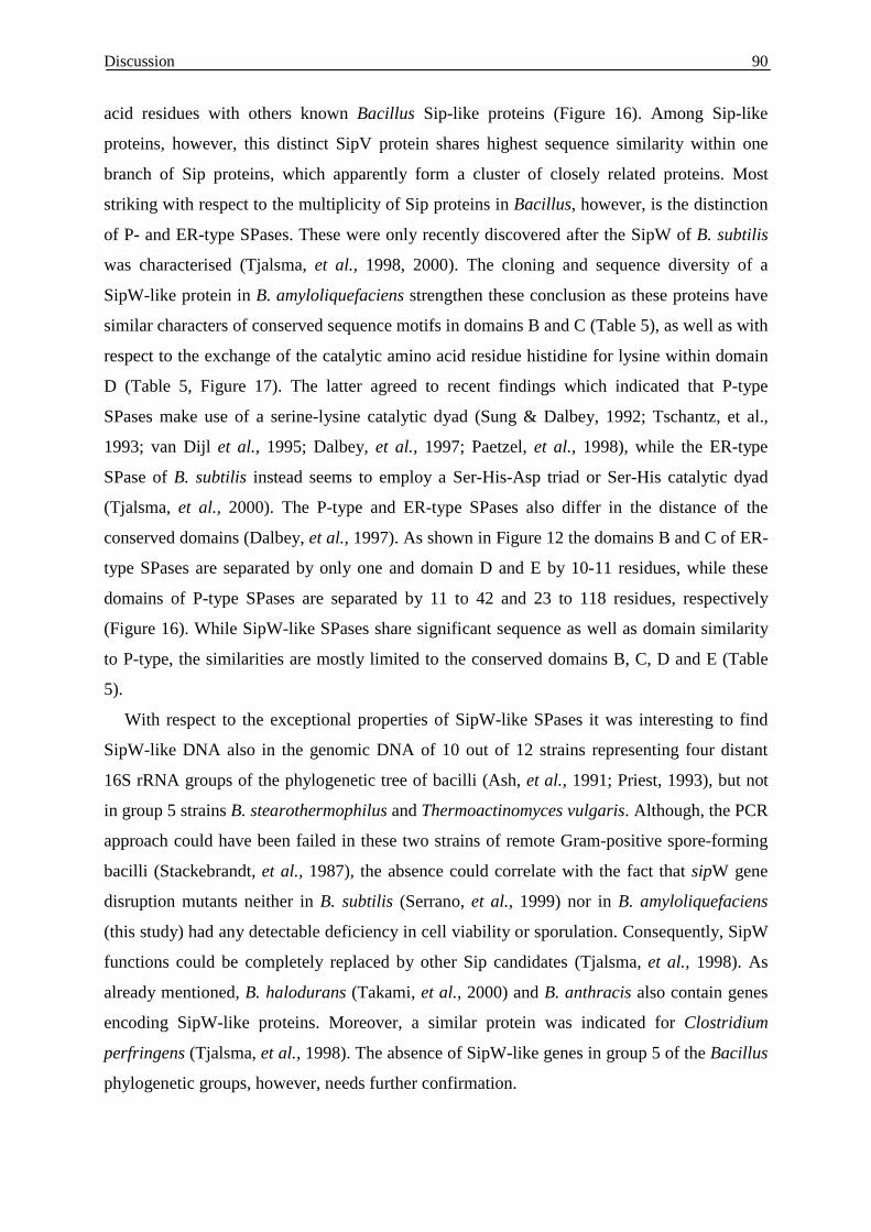
Discussion
90
acid residues with others known Bacillus Sip-like proteins (Figure 16). Among Sip-like
proteins, however, this distinct SipV protein shares highest sequence similarity within one
branch of Sip proteins, which apparently form a cluster of closely related proteins. Most
striking with respect to the multiplicity of Sip proteins in Bacillus, however, is the distinction
of P- and ER-type SPases. These were only recently discovered after the SipW of B. subtilis
was characterised (Tjalsma, et al., 1998, 2000). The cloning and sequence diversity of a
SipW-like protein in B. amyloliquefaciens strengthen these conclusion as these proteins have
similar characters of conserved sequence motifs in domains B and C (Table 5), as well as with
respect to the exchange of the catalytic amino acid residue histidine for lysine within domain
D (Table 5, Figure 17). The latter agreed to recent findings which indicated that P-type
SPases make use of a serine-lysine catalytic dyad (Sung & Dalbey, 1992; Tschantz, et al.,
1993; van Dijl et al., 1995; Dalbey, et al., 1997; Paetzel, et al., 1998), while the ER-type
SPase of B. subtilis instead seems to employ a Ser-His-Asp triad or Ser-His catalytic dyad
(Tjalsma, et al., 2000). The P-type and ER-type SPases also differ in the distance of the
conserved domains (Dalbey, et al., 1997). As shown in Figure 12 the domains B and C of ER-
type SPases are separated by only one and domain D and E by 10-11 residues, while these
domains of P-type SPases are separated by 11 to 42 and 23 to 118 residues, respectively
(Figure 16). While SipW-like SPases share significant sequence as well as domain similarity
to P-type, the similarities are mostly limited to the conserved domains B, C, D and E (Table
5).
With respect to the exceptional properties of SipW-like SPases it was interesting to find
SipW-like DNA also in the genomic DNA of 10 out of 12 strains representing four distant
16S rRNA groups of the phylogenetic tree of bacilli (Ash, et al., 1991; Priest, 1993), but not
in group 5 strains B. stearothermophilus and Thermoactinomyces vulgaris. Although, the PCR
approach could have been failed in these two strains of remote Gram-positive spore-forming
bacilli (Stackebrandt, et al., 1987), the absence could correlate with the fact that sipW gene
disruption mutants neither in B. subtilis (Serrano, et al., 1999) nor in B. amyloliquefaciens
(this study) had any detectable deficiency in cell viability or sporulation. Consequently, SipW
functions could be completely replaced by other Sip candidates (Tjalsma, et al., 1998). As
already mentioned, B. halodurans (Takami, et al., 2000) and B. anthracis also contain genes
encoding SipW-like proteins. Moreover, a similar protein was indicated for Clostridium
perfringens (Tjalsma, et al., 1998). The absence of SipW-like genes in group 5 of the Bacillus
phylogenetic groups, however, needs further confirmation.
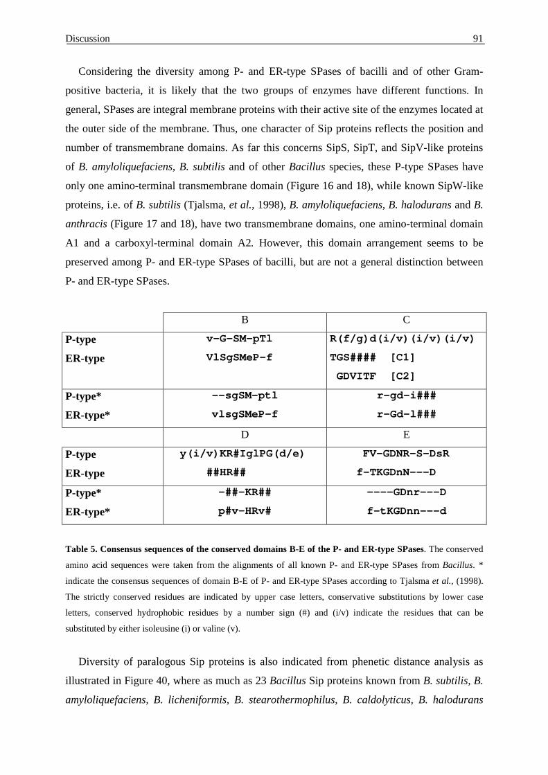
Discussion
91
Considering the diversity among P- and ER-type SPases of bacilli and of other Gram-
positive bacteria, it is likely that the two groups of enzymes have different functions. In
general, SPases are integral membrane proteins with their active site of the enzymes located at
the outer side of the membrane. Thus, one character of Sip proteins reflects the position and
number of transmembrane domains. As far this concerns SipS, SipT, and SipV-like proteins
of B. amyloliquefaciens, B. subtilis and of other Bacillus species, these P-type SPases have
only one amino-terminal transmembrane domain (Figure 16 and 18), while known SipW-like
proteins, i.e. of B. subtilis (Tjalsma, et al., 1998), B. amyloliquefaciens, B. halodurans and B.
anthracis (Figure 17 and 18), have two transmembrane domains, one amino-terminal domain
A1 and a carboxyl-terminal domain A2. However, this domain arrangement seems to be
preserved among P- and ER-type SPases of bacilli, but are not a general distinction between
P- and ER-type SPases.
B C
P-type
ER-type
v-G-SM-pTl
VlSgSMeP-f
R(f/g)d(i/v)(i/v)(i/v)
TGS#### [C1]
GDVITF [C2]
P-type*
ER-type*
--sgSM-ptl
vlsgSMeP-f
r-gd-i###
r-Gd-l###
D E
P-type
ER-type
y(i/v)KR#IglPG(d/e)
##HR##
FV-GDNR-S-DsR
f-TKGDnN---D
P-type*
ER-type*
-##-KR##
p#v-HRv#
----GDnr---D
f-tKGDnn---d
Table 5. Consensus sequences of the conserved domains B-E of the P- and ER-type SPases. The conserved
amino acid sequences were taken from the alignments of all known P- and ER-type SPases from Bacillus. *
indicate the consensus sequences of domain B-E of P- and ER-type SPases according to Tjalsma et al., (1998).
The strictly conserved residues are indicated by upper case letters, conservative substitutions by lower case
letters, conserved hydrophobic residues by a number sign (#) and (i/v) indicate the residues that can be
substituted by either isoleusine (i) or valine (v).
Diversity of paralogous Sip proteins is also indicated from phenetic distance analysis as
illustrated in Figure 40, where as much as 23 Bacillus Sip proteins known from B. subtilis, B.
amyloliquefaciens, B. licheniformis, B. stearothermophilus, B. caldolyticus, B. halodurans
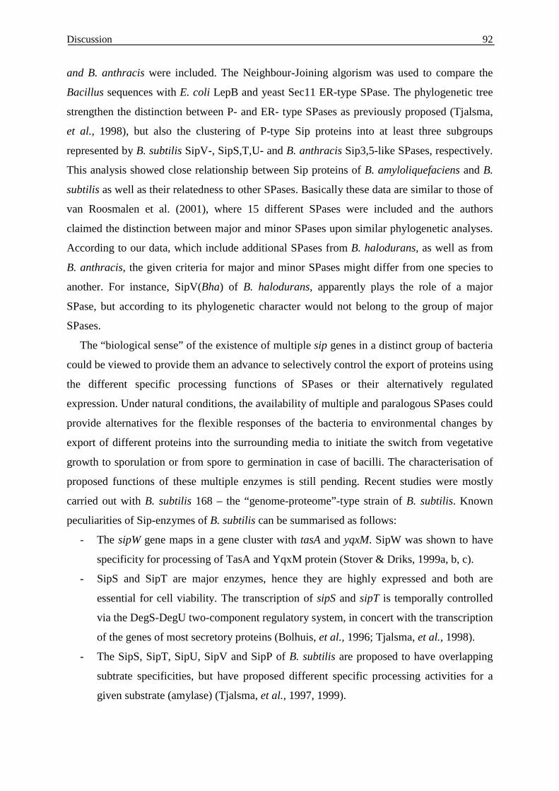
Discussion
92
and B. anthracis were included. The Neighbour-Joining algorism was used to compare the
Bacillus sequences with E. coli LepB and yeast Sec11 ER-type SPase. The phylogenetic tree
strengthen the distinction between P- and ER- type SPases as previously proposed (Tjalsma,
et al., 1998), but also the clustering of P-type Sip proteins into at least three subgroups
represented by B. subtilis SipV-, SipS,T,U- and B. anthracis Sip3,5-like SPases, respectively.
This analysis showed close relationship between Sip proteins of B. amyloliquefaciens and B.
subtilis as well as their relatedness to other SPases. Basically these data are similar to those of
van Roosmalen et al. (2001), where 15 different SPases were included and the authors
claimed the distinction between major and minor SPases upon similar phylogenetic analyses.
According to our data, which include additional SPases from B. halodurans, as well as from
B. anthracis, the given criteria for major and minor SPases might differ from one species to
another. For instance, SipV(Bha) of B. halodurans, apparently plays the role of a major
SPase, but according to its phylogenetic character would not belong to the group of major
SPases.
The “biological sense” of the existence of multiple sip genes in a distinct group of bacteria
could be viewed to provide them an advance to selectively control the export of proteins using
the different specific processing functions of SPases or their alternatively regulated
expression. Under natural conditions, the availability of multiple and paralogous SPases could
provide alternatives for the flexible responses of the bacteria to environmental changes by
export of different proteins into the surrounding media to initiate the switch from vegetative
growth to sporulation or from spore to germination in case of bacilli. The characterisation of
proposed functions of these multiple enzymes is still pending. Recent studies were mostly
carried out with B. subtilis 168 – the “genome-proteome”-type strain of B. subtilis. Known
peculiarities of Sip-enzymes of B. subtilis can be summarised as follows:
- The sipW gene maps in a gene cluster with tasA and yqxM. SipW was shown to have
specificity for processing of TasA and YqxM protein (Stover & Driks, 1999a, b, c).
- SipS and SipT are major enzymes, hence they are highly expressed and both are
essential for cell viability. The transcription of sipS and sipT is temporally controlled
via the DegS-DegU two-component regulatory system, in concert with the transcription
of the genes of most secretory proteins (Bolhuis, et al., 1996; Tjalsma, et al., 1998).
- The SipS, SipT, SipU, SipV and SipP of B. subtilis are proposed to have overlapping
subtrate specificities, but have proposed different specific processing activities for a
given substrate (amylase) (Tjalsma, et al., 1997, 1999).
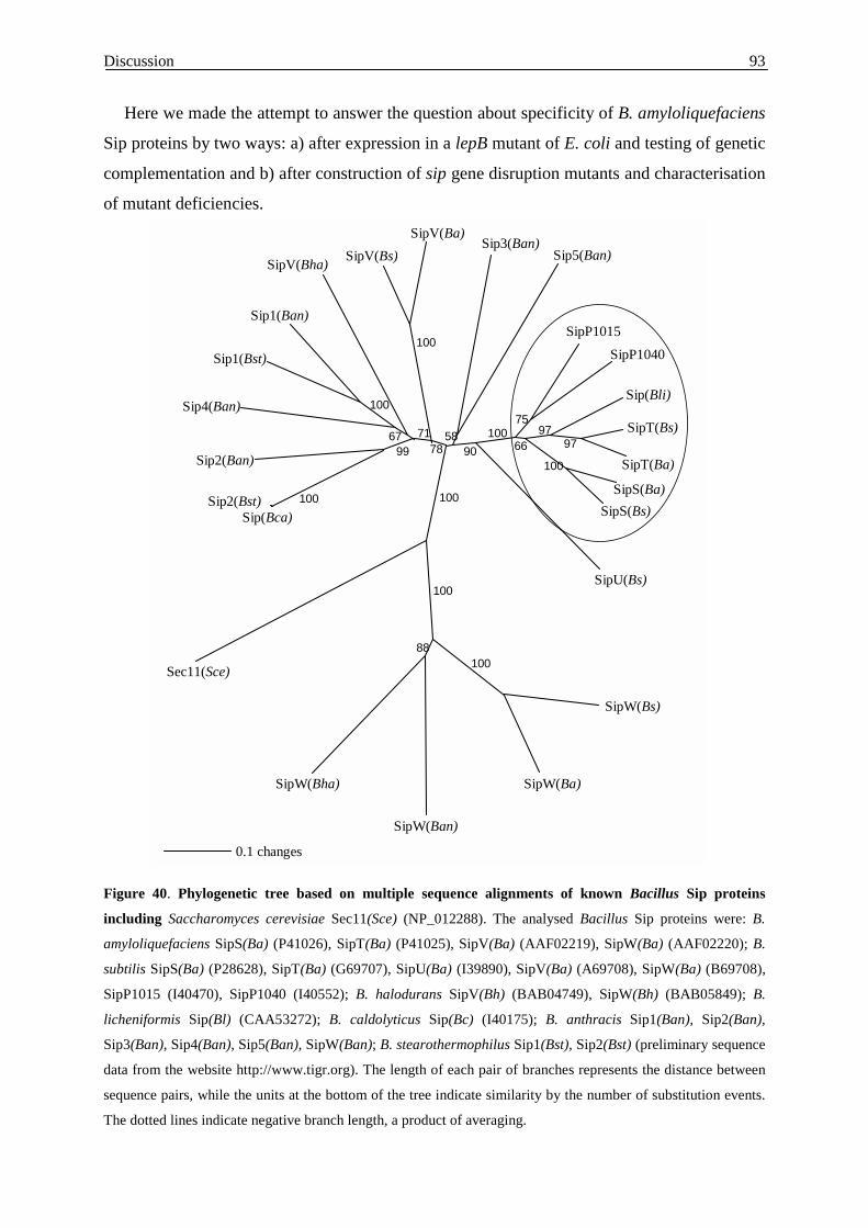
Discussion
93
Here we made the attempt to answer the question about specificity of B. amyloliquefaciens
Sip proteins by two ways: a) after expression in a lepB mutant of E. coli and testing of genetic
complementation and b) after construction of sip gene disruption mutants and characterisation
of mutant deficiencies.
Figure 40. Phylogenetic tree based on multiple sequence alignments of known Bacillus Sip proteins
including Saccharomyces cerevisiae Sec11(Sce) (NP_012288). The analysed Bacillus Sip proteins were: B.
amyloliquefaciens SipS(Ba) (P41026), SipT(Ba) (P41025), SipV(Ba) (AAF02219), SipW(Ba) (AAF02220); B.
subtilis SipS(Ba) (P28628), SipT(Ba) (G69707), SipU(Ba) (I39890), SipV(Ba) (A69708), SipW(Ba) (B69708),
SipP1015 (I40470), SipP1040 (I40552); B. halodurans SipV(Bh) (BAB04749), SipW(Bh) (BAB05849); B.
licheniformis Sip(Bl) (CAA53272); B. caldolyticus Sip(Bc) (I40175); B. anthracis Sip1(Ban), Sip2(Ban),
Sip3(Ban), Sip4(Ban), Sip5(Ban), SipW(Ban); B. stearothermophilus Sip1(Bst), Sip2(Bst) (preliminary sequence
data from the website http://www.tigr.org). The length of each pair of branches represents the distance between
sequence pairs, while the units at the bottom of the tree indicate similarity by the number of substitution events.
The dotted lines indicate negative branch length, a product of averaging.
SipW(Bs)
SipW(Ba)
SipW(Ban)
SipW(Bha)
Sec11(Sce)
Sip(Bca)Sip2(Bst)
Sip2(Ban)
Sip4(Ban)
Sip1(Bst)
Sip1(Ban)
SipV(Bha) SipV(Bs)SipV(Ba)
Sip3(Ban)Sip5(Ban)
SipP1015SipP1040
Sip(Bli)
SipT(Bs)
SipT(Ba)
SipS(Ba)SipS(Bs)
SipU(Bs)
0.1 changes
100
6799
100
71
100
78
100
100
10088
5890
10066
75
100
9797
Discussion
94
4.3. Functional complementation of an E. coli LepBts mutant
In conclusion of the data discussed in the previous section, one might assume that SPases
with close sequence similarity are likely to complement each other more efficiently than those
from another subgroup of the SPases. The difficulties of genetic manipulation in B.
amyloliquefaciens, especially its inaccessibility to construct multiple sip knockout mutants,
inclined us to address the question in E. coli, and to ask if B. amyloliquefaciens SPases
substitute the E. coli LepB functions with respect to growth and processing.
The E. coli strain IT41, which carried an TAG amber terminator codon mutant at
nucleotide position 115 in the lepB coding sequence (Inada, et al., 1988; Cregg, et al., 1996),
has been previously proved to be a useful tool to study functions of SPases from
Bradyrhizobium japonicum (Müller, et al., 1995), Staphylococcus aureus (Cregg, et al.,
1996), Streptococcus pneumoniae (Zhang, et al., 1997) and Streptomyces lividans TK21
(Parro et al., 1999). Therefore, in this studies each of the four B. amyloliquefaciens Sip
proteins were expressed in this E. coli LepBts mutant strain and tested for processing of pre-
OmpA and growth restoration at the non-permissive temperature. In summary, only SipS(Ba)
and SipT(Ba), but neither SipV(Ba) nor SipW(Ba), were active in processing pre-OmpA in E.
coli. In contrast, the growth of the LepBts mutant was only restored after SipS(Ba) but not
after SipT(Ba) expression. These likely reflects differences in processing specificity or
capacities of SipT(Ba) compared to SipS(Ba) in order to cope with growth-limiting LepB
processing functions. (Figure 20 and 21). In B. subtilis, SipS, SipT, as well as the plasmid
encoded SipP, were shown to functionally replace each other and to be the “major” SPase as
the cell need at least one of them to survive, while SipU, SipV and SipW seem to play
“minor” roles. The latter were proposed to have limited substrate specificity for a subset of
about 166 predicted export proteins (Tjalsma, et al., 2000). When expressed in E. coli, only
SipS, SipT and SipU were active in processing of the pre(A13i)-β -lactamase precursor, while
SipV was inactive (Tjalsma, et al., 1997). In turn, B. amylolioquefaciens SipV(Ba) as well as
SipW(Ba) were not active in processing of the pre-OmpA in E. coli. The lack of processing in
E. coli was not caused by differences in production levels of these Sip(Ba) proteins, but likely
correlated with enhanced degradation and inactivation, as indicated by additional protein
bands in SipV(Ba) and SipW(Ba) producing E. coli cells after SDS-PAGE and immuno-
detection (Figure 21B). These observation could reflect less efficient membrane insertion of
these Sip(Ba) proteins in E. coli, since soluble (truncated) forms of B. amyloliquefaciens
SipS(Ba), lacking their unique N-terminal membrane anchor, were also found to become
inactivated in E. coli by self-cleavage (van Roosmalen, et al., 2000). In contrast, SipT(Ba)
Discussion
95
was apparently quite stable and efficient in processing of pre-OmpA, but was nevertheless
unable to restore growth of LepBts E. coli cultures. This distinction was likely due to non-
efficiency in processing of some E. coli export proteins by SipT(Ba). Hence, SipT(Ba)
apparently did not support the processing of unknown but essential proteins, which were,
however, efficiently processed by LepB or by SipS(Ba).
The data indicated that heterologous function of Sip protein in E. coli can be affected by
two factors: a) the protein stability due to membrane insertion and b) the range of substrate
specificity of individual SPases. Therefore, we designed hybrid sip/lep-genes by exchanging
the transmembrane and catalytic regions of E. coli LepB and B. amyloliquefaciens SipT(Ba).
The schema of the two hybrids LepT-B and SipB-T are shown in Figure 22. After expression
in the E. coli LepBts mutant strain only the hybrid LepT-B was found to support growth at the
non-permissive temperature, while the hybrid SipB-T did not. This indicated that the
transmembrane portion of SipT(Ba), in fusion with the LepB catalytic domain, efficiently
restored the LepBts deficiency, but that the catalytic domain of SipT(Ba) in fusion with the
transmembrane domain of LepB did not. This can be explained by the fact that both the
transmembrane as well as the catalytic domain of SipT functioned, but the latter, however,
was not able to complement the whole range of LepB processing functions. This suggested
that complementation or non-complementation of LepBts by any B. amyloliquefaciens SPase
was most likely due to processing specificity and not or to an lesser extend due to inefficiency
of their membrane insertion. This promising “hybrid” studies did not solve the problem
completely, but indicated a genetic route to answer questions related to the limits of function
or specificity of individual B. amyloliquefaciens SPases.
4.4. Mutant studies
Without processing, i.e. after inactivation of essential SPase genes, export proteins remain
anchored at the membrane and could not finally target to their proper locations. Consequently,
E. coli mutants lacking LepB are non-viable (Date, 1983; Inada, et al., 1989). However, B.
subtilis mutants lacking as many as four SPases, i.e. SipT,-U,-V,-W, are viable, but heat
inactivation of SipSts in a SipT mutant background, i.e. the SipS-SipT double deficiency, had
lethal consequences (Bron, et al., 1998; Tjalsma, et al., 1998). All of the B. amyloliquefaciens
Sip(Ba) disruption mutants were viable, but some had impaired growth, sporulation and
changed cell division properties. Similar to B. subtilis (Bron, et al., 1998), inactivation of
either SipS or SipT decreased the total yields of export proteins compared to the wild type by
about 30 percent, as was the quantity of extracellular amylase also significantly enhanced in
Discussion
96
SipS(Ba) mutant cultures (data not shown). These two observation were also made with B.
subtilis mutants and the former explained to reflect the predominant character of SipS and
SipT enzymes (Tjalsm,a et al., 1997; Bron, et al., 1998). The other deficiencies of B.
amyloliquefaciens sip(Ba) disruption mutants have not been reviewed for B. subtilis.
However, strict correlation of a distinct mutant phenotype with that distinct sip::pEA*
integration event, as well as restoration of the mutant phenotype after spontaneous excision of
the insertion cassette from B. amyloliquefaciens sipT::pEAT* mutants, strongly indicated
gene disruption to correlate with that distinct deficiencies. The recent finding about SipW
activities in pre-TasA processing and transport into B. subtilis endospores provided a first
example of specification of a SPase for spore-specific protein sorting (Tjalsma, et al., 1998;
Serrano, et al., 1999; Stöver & Driks, 1999). In B. amyloliquefaciens, disruption of sipT(Ba)
but not of sipW(Ba) correlated with a drastic reduction of heat resistant spores. The rare cells
with spore structures after electronmicroscopical inspection exhibited forespore structures in
stage III development with apparent changes in cortex or coat structures (Figure 26). These
inhibition likely resulted from missing spore-specific export proteins. Sporulation-related
export protein is suggested, but yet nearly unknown (Stragier & Losick, 1996; Dricks, 1999).
In B. subtilis, sporulation deficiency was reported to correlate with sipT-sipV double deletion
and suggested to indicate processing specificity of these two SPases for sporulation-related
export proteins (Jiang, et al., 2000). In this concern, it should be considered that SipS as well
as SipT of B. subtilis are temporally controlled during transient growth in correlation with
export of the mass of Sec proteins (Tjalsma, et al., 1997) as well as in correlation with the
initiation of sporulation. In analogy, SipT(Ba) in B. amyloliquefaciens might exclusively cope
with processing of either a single or a couple of respective sporulation-specific export
proteins (Stragier & Losick, 1996) either by growth phase specific expression and/or
processing specificity. The same might be true for export of a not yet defined nuclease in B.
amyloliquefaciens, which was most affected by sipS(Ba), but to a lesser extend also by
sipT(Ba) gene disruption. The respective nuclease of B. amyloliquefaciens is not likely to be a
homologue of the 12 kDa-extracellular NucB of B. subtilis (van Sinderen, et al., 1995), as the
size of the protein was about 30 kDa and all attempt to amplify respective NucB-like DNA by
reverse genetics from B. amyloliquefaciens were unsuccessful (data not shown). Moreover,
impaired growth, inhibited cell autolysis and reduced cell motility specified sipV::pEAV*
mutants of B. amyloliquefaciens to have changed autolysin activities. Indeed, changed pattern
of CWBP’s as well as loss of at least one (major) 35-kDa autolysin correlated with the
SipV(Ba) deficiency. In B. subtilis, LytC (50 kDa amidase), LytD (90 kDa glucosaminidase)
Discussion
97
or a sigD controlled (minor) 49-kDa autolysins are shown to change cell wall turnover,
septation, cell lysis as well as swarming motility (Blackman, et al., 1998). Processing of these
unknown autolysins of B. amyloliquefaciens might correlate with the distinct SipV(Ba)
mutant phenotype, but other autolysins are not excluded, as B. subtilis was suspected to
contain several autolysins (Foster, 1992; Kunst, et al., 1997).
In summary, more detailed studies are required to explain the mystery of multiple Sip
proteins with more or less different characters in various Bacillus species. Genetic and
biochemical data ad hands already indicated that the multiple SPases of Bacillus might be an
prerequisite for adaptation of the secretion apparatus either due to their gene expression mode
(Bolhuis, et al., 1996; Tjalsma, et al., 1997) or their different processing specificity (Bron, et
al., 1998; Tjalsma, et al., 2000). Moreover, different localisation of SPases either in the cell-
or membrane-specific compartment of the mother cell or forespore seems to be possible
(Serrano, et al., 1999; Stöver & Driks, 1999; Tjalsma, et al., 2000). The presence of different
Sip proteins might be interpreted in the same way as the obvious multiplicity of two-
component signal transducer proteins in B. subtilis. In this case, 34 different candidates were
found (Kunst, et al., 1997) and each has regulatory functions for a distinct biosynthesis or
pathway. We are therefore inclined to propose that multiple SPases are likewise serving for
“specific” functions. Our mutant studies would need more precise definition of casual
relationship between the Sip deficiencies and the mutant phenotype, but already indicated a
specific and essential function of this distinct SPases in cell growth, cell division and spore
formation. Since B. amyloliquefaciens strains were only recently recognised as an
independent species (Stackebrandt, et al., 1987; Priest, 1993), the differences of B.
amyloliquefaciens Sip candidates with respect to the presence and specification of SPases in
comparison to B. subtilis homologues could indicate that these closely related species already
have slightly varied characters in their processing apparatus.
4.5. A new export protein of B. amyloliquefaciens
In conclusion of the data discussed in the previous sections, the presence of multiple
paralogous SPases could be interpreted as a particular important feature for flexible
adaptation of Bacillus and others Gram-positive bacteria in responses to environmental
changes. In B. subtilis, 166 proteins had been predicted to be secretory proteins. While up to
date only two proteins TasA and YqxM (Stöver & Driks, 1999a, b) were reported to be SipW
specific, there are likely more undiscovered proteins, of which export requires one or another
SPase. On the basis of earlier studies in B. amyloliquefaciens one distinct extracellular protein
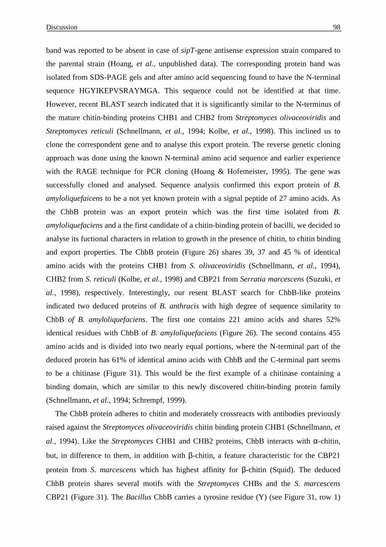
Discussion
98
band was reported to be absent in case of sipT-gene antisense expression strain compared to
the parental strain (Hoang, et al., unpublished data). The corresponding protein band was
isolated from SDS-PAGE gels and after amino acid sequencing found to have the N-terminal
sequence HGYIKEPVSRAYMGA. This sequence could not be identified at that time.
However, recent BLAST search indicated that it is significantly similar to the N-terminus of
the mature chitin-binding proteins CHB1 and CHB2 from Streptomyces olivaceoviridis and
Streptomyces reticuli (Schnellmann, et al., 1994; Kolbe, et al., 1998). This inclined us to
clone the correspondent gene and to analyse this export protein. The reverse genetic cloning
approach was done using the known N-terminal amino acid sequence and earlier experience
with the RAGE technique for PCR cloning (Hoang & Hofemeister, 1995). The gene was
successfully cloned and analysed. Sequence analysis confirmed this export protein of B.
amyloliquefaicens to be a not yet known protein with a signal peptide of 27 amino acids. As
the ChbB protein was an export protein which was the first time isolated from B.
amyloliquefaciens and a the first candidate of a chitin-binding protein of bacilli, we decided to
analyse its fuctional characters in relation to growth in the presence of chitin, to chitin binding
and export properties. The ChbB protein (Figure 26) shares 39, 37 and 45 % of identical
amino acids with the proteins CHB1 from S. olivaceoviridis (Schnellmann, et al., 1994),
CHB2 from S. reticuli (Kolbe, et al., 1998) and CBP21 from Serratia marcescens (Suzuki, et
al., 1998), respectively. Interestingly, our resent BLAST search for ChbB-like proteins
indicated two deduced proteins of B. anthracis with high degree of sequence similarity to
ChbB of B. amyloliquefaciens. The first one contains 221 amino acids and shares 52%
identical residues with ChbB of B. amyloliquefaciens (Figure 26). The second contains 455
amino acids and is divided into two nearly equal portions, where the N-terminal part of the
deduced protein has 61% of identical amino acids with ChbB and the C-terminal part seems
to be a chitinase (Figure 31). This would be the first example of a chitinase containing a
binding domain, which are similar to this newly discovered chitin-binding protein family
(Schnellmann, et al., 1994; Schrempf, 1999).
The ChbB protein adheres to chitin and moderately crossreacts with antibodies previously
raised against the Streptomyces olivaceoviridis chitin binding protein CHB1 (Schnellmann, et
al., 1994). Like the Streptomyces CHB1 and CHB2 proteins, ChbB interacts with α-chitin,
but, in difference to them, in addition with β-chitin, a feature characteristic for the CBP21
protein from S. marcescens which has highest affinity for β-chitin (Squid). The deduced
ChbB protein shares several motifs with the Streptomyces CHBs and the S. marcescens
CBP21 (Figure 31). The Bacillus ChbB carries a tyrosine residue (Y) (see Figure 31, row 1)
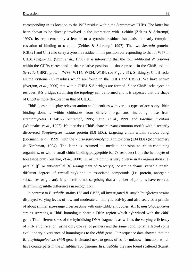
Discussion
99
corresponding in its location to the W57 residue within the Streptomyes CHBs. The latter has
been shown to be directly involved in the interaction with α-chitin (Zeltins & Schrempf,
1997). Its replacement by a leucine or a tyrosine residue also leads to nearly complete
cessation of binding to α-chitin (Zeltins & Schrempf, 1997). The two Serratia proteins
(CBP21 and Chi) also carry a tyrosine residue in this position corresponding to that of W57 in
CHB1 (Figure 31) (Shin, et al., 1996). It is interesting that the four additional W residues
within the CHBs correspond in their relative positions to those present in the ChbB and the
Serratia CBP21 protein (W99, W114, W134, W184, see Figure 31). Strikingly, ChbB lacks
all the cysteine (C) residues which are found in the CHBs and CBP21. We have shown
(Svergun, et al., 2000) that within CHB1 S-S bridges are formed. Since ChbB lacks cysteine
residues, S-S bridges stabilising the topology can be formed and it is expected that the shape
of ChbB is more flexible than that of CHB1.
ChbB does not display relevant amino acid identities with various types of accessory chitin
binding domains within chitinases from different organisms, including those from
streptomycetes (Blaak & Schrempf, 1995; Saito, et al., 1999) and Bacillus circulans
(Watanabe, et al., 1992). Neither does ChbB share relevant common motifs with a recently
discovered Streptomyces tendae protein (9.8 kDa), targeting chitin within various fungi
(Bormann, et al., 1999), with the Vibrio parahemolyticus chitovibrin (134 kDa) (Montgomery
& Kirchman, 1994). The latter is assumed to mediate adhesion to chitin-containing
organisms, or with a small chitin binding polypeptide (of 73 residues) from the hemocyte of
horseshoe crab (Suetake, et al., 2000). In nature chitin is very diverse in its organisation (i.e.
parallel [β] or anti-parallel [α] arrangement of N-acetylglucosamine chains, variable length,
different degrees of crystallinity) and its associated compounds (i.e. protein, anorganic
substances or glucan). It is therefore not surprising that a number of proteins have evolved
determining subtle differences in recognition.
In contrast to B. subtilis strains 168 and GB72, all investigated B. amyloliquefaciens strains
displayed varying levels of low and moderate chitinolytic activity and also secreted a protein
of about similar size-range crossreacting with anti-ChbB antibodies. All B. amyloliquefaciens
strains secreting a ChbB homologue share a DNA region which hybridised with the chbB
gene. The different sizes of the hybridising DNA fragments as well as the varying efficiency
of PCR amplification (using only one set of primers and the same conditions) reflected some
evolutionary divergence of homologues to the chbB gene. Our sequence data showed that the
B. amyloliquefaciens chbB gene is situated next to genes of so far unknown function, which
have counterparts in the B. subtilis 168 genome. In B. subtilis they are found scattered (Kunst,
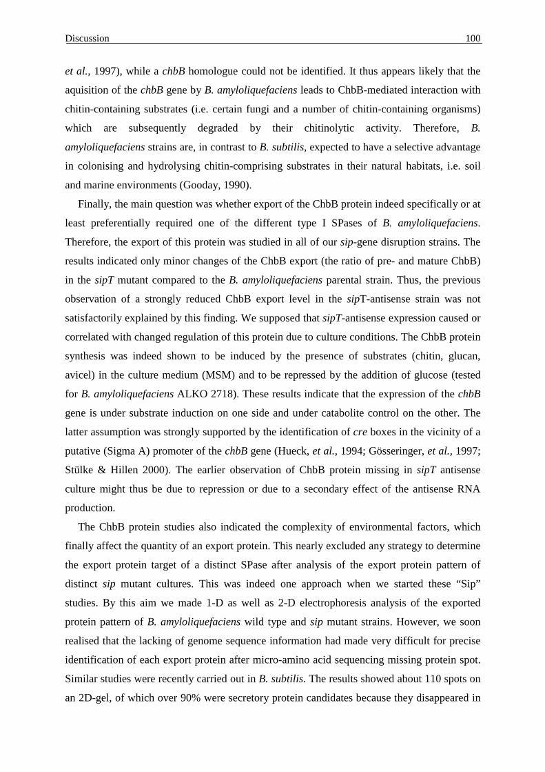
Discussion
100
et al., 1997), while a chbB homologue could not be identified. It thus appears likely that the
aquisition of the chbB gene by B. amyloliquefaciens leads to ChbB-mediated interaction with
chitin-containing substrates (i.e. certain fungi and a number of chitin-containing organisms)
which are subsequently degraded by their chitinolytic activity. Therefore, B.
amyloliquefaciens strains are, in contrast to B. subtilis, expected to have a selective advantage
in colonising and hydrolysing chitin-comprising substrates in their natural habitats, i.e. soil
and marine environments (Gooday, 1990).
Finally, the main question was whether export of the ChbB protein indeed specifically or at
least preferentially required one of the different type I SPases of B. amyloliquefaciens.
Therefore, the export of this protein was studied in all of our sip-gene disruption strains. The
results indicated only minor changes of the ChbB export (the ratio of pre- and mature ChbB)
in the sipT mutant compared to the B. amyloliquefaciens parental strain. Thus, the previous
observation of a strongly reduced ChbB export level in the sipT-antisense strain was not
satisfactorily explained by this finding. We supposed that sipT-antisense expression caused or
correlated with changed regulation of this protein due to culture conditions. The ChbB protein
synthesis was indeed shown to be induced by the presence of substrates (chitin, glucan,
avicel) in the culture medium (MSM) and to be repressed by the addition of glucose (tested
for B. amyloliquefaciens ALKO 2718). These results indicate that the expression of the chbB
gene is under substrate induction on one side and under catabolite control on the other. The
latter assumption was strongly supported by the identification of cre boxes in the vicinity of a
putative (Sigma A) promoter of the chbB gene (Hueck, et al., 1994; Gösseringer, et al., 1997;
Stülke & Hillen 2000). The earlier observation of ChbB protein missing in sipT antisense
culture might thus be due to repression or due to a secondary effect of the antisense RNA
production.
The ChbB protein studies also indicated the complexity of environmental factors, which
finally affect the quantity of an export protein. This nearly excluded any strategy to determine
the export protein target of a distinct SPase after analysis of the export protein pattern of
distinct sip mutant cultures. This was indeed one approach when we started these “Sip”
studies. By this aim we made 1-D as well as 2-D electrophoresis analysis of the exported
protein pattern of B. amyloliquefaciens wild type and sip mutant strains. However, we soon
realised that the lacking of genome sequence information had made very difficult for precise
identification of each export protein after micro-amino acid sequencing missing protein spot.
Similar studies were recently carried out in B. subtilis. The results showed about 110 spots on
an 2D-gel, of which over 90% were secretory protein candidates because they disappeared in

Discussion
101
the absence of SecA (Hirose, et al., 2000). Indeed, the genome and proteome analysis, which
are more advanced in B. subtilis 168, would make this species more preferable for studies and
rank B. amyloliquefaciens in a secondary position.
Nevertheless, it remains worth asking, which peculiarities of the export machinery or other
properties make B. amyloliquefaciens to be about 10 to 20 times more efficient in protein
secretion than B. subtilis, a question was already asked 25 years ago by Ingle and Boyer
(1976). This study was aimed towards an answer.
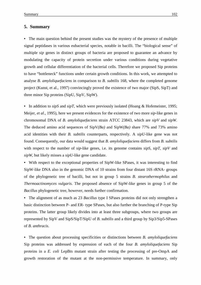
Summary
102
5. Summary
• The main question behind the present studies was the mystery of the presence of multiple
signal peptidases in various eubacterial species, notable in bacilli. The “biological sense” of
multiple sip genes in distinct groups of bacteria are proposed to guarantee an advance by
modulating the capacity of protein secretion under various conditions during vegetative
growth and cellular differentiation of the bacterial cells. Therefore we proposed Sip proteins
to have “bottleneck” functions under certain growth conditions. In this work, we attempted to
analyse B. amyloliquefaciens in comparison to B. subtilis 168, where the completed genome
project (Kunst, et al., 1997) convincingly proved the existence of two major (SipS, SipT) and
three minor Sip proteins (SipU, SipV, SipW).
• In addition to sipS and sipT, which were previously isolated (Hoang & Hofemeister, 1995;
Meijer, et al., 1995), here we present evidences for the existence of two more sip-like genes in
chromosomal DNA of B. amyloliquefaciens strain ATCC 23843, which are sipV and sipW.
The deduced amino acid sequences of SipV(Ba) and SipW(Ba) share 77% and 73% amino
acid identities with their B. subtilis counterparts, respectively. A sipU-like gene was not
found. Consequently, our data would suggest that B. amyloliquefaciens differs from B. subtilis
with respect to the number of sip-like genes, i.e. its genome contains sipS, sipT, sipV and
sipW, but likely misses a sipU-like gene candidate.
• With respect to the exceptional properties of SipW-like SPases, it was interesting to find
SipW-like DNA also in the genomic DNA of 10 strains from four distant 16S rRNA- groups
of the phylogenetic tree of bacilli, but not in group 5 strains B. stearothermophilus and
Thermoactinomyces vulgaris. The proposed absence of SipW-like genes in group 5 of the
Bacillus phylogenetic tree, however, needs further confirmation.
• The alignment of as much as 23 Bacillus type I SPases proteins did not only strengthen a
basic distinction between P- and ER- type SPases, but also further the branching of P-type Sip
proteins. The latter group likely divides into at least three subgroups, where two groups are
represented by SipV and SipS/SipT/SipU of B. subtilis and a third group by Sip3/Sip5-SPases
of B. anthracis.
• The question about processing specificities or distinctions between B. amyloliquefaciens
Sip proteins was addressed by expression of each of the four B. amyloliquefaciens Sip
proteins in a E. coli LepBts mutant strain after testing the processing of pre-OmpA and
growth restoration of the mutant at the non-permissive temperature. In summary, only
Summary
103
SipS(Ba) and SipT(Ba), but neither SipV(Ba) nor SipW(Ba) were active in processing pre-
OmpA in E. coli. In contrast, the growth of the LepBts mutant was only restored after
SipS(Ba) but not after SipT(Ba) expression. These likely reflects differences in processing
specificities, since different production levels of these Sip(Ba) proteins were excluded. The
lack of processing activities of SipV(Ba) and SipW(Ba) likely correlated with enhanced
degradation.
• The complementation data indicated either the N-terminal transmembrane or the C-
terminal catalytic portion of Sip proteins to limit processing functions in E. coli. This
hypothesis was tested after construction of sipT(Ba)-lepB hybrid genes. The hybrid studies
indicated that in case of SipT(Ba) lacking of processing capacity rather than membrane
insertion properties causes non-complementation.
• All of the B. amyloliquefaciens Sip(Ba) disruption mutants were viable, but some had
impaired growth, sporulation and changed cell division properties. The strict correlation of a
distinct mutant phenotype with that distinct sip::pEA* integration event as well as restoration
of the mutant phenotype after spontaneous excision of the insertion cassette from B.
amyloliquefaciens sipT::pEAT* mutants, strongly indicated gene disruption to correlate with
that distinct deficiencies.
• Disruption of sipT(Ba) resulted in a drastic reduction of heat resistant spores and apparent
changes in cortex or pre-coat structures.
• SipV deficiency of B. amyloliquefaciens correlated with impaired growth, inhibited cell
autolysis, reduced motillity specified and changed autolysin activities. The change of the
CWBP’s pattern correlated with the loss of at least one (major) 35-kDa autolysin, which
could cause that distinct SipV(Ba) mutant phenotype.
• A 30 kDa extracellular DNase was strongly affected in sipS disruption mutants, but to a
lesser extent also seen in sipT mutants. The respective nuclease of B. amyloliquefaciens was
not identified.
• In an effort to search in B. amyloliquefaciens for export proteins, which require the
function of a distinct SPases, a new export protein was identified. The correspondent gene
was cloned. Its sequence similarity indicated ChbB to be a homologue of chitin-binding
proteins CHB1, CHB2 and CBP21 from S. olivaceoviridis, S. reticuli or Serratia marcescens,
respectively. This ChbB protein of B. amyloliquefaciens is the first of its type isolated from
Bacillus. Binding studies indicated that ChbB protein targets β-chitin best, then α-chitin, but
barely any other polysaccharide.
Summary
104
• The export of ChbB was studied in the four sip-gene disruption strains. The results
indicated only minor changes of the ChbB export in the sipT mutant comparing to the parental
strain. However, this finding did not satisfactorily explain the previous observation, where
ChbB export was found to be strongly reduced in a sipT-antisense strain. Beside of a slight
“bottle neck” function of SipT(Ba), it is highlight that ChbB protein synthesis is affected by
substrate induction and catabolite repression.
• Since B. amyloliquefaciens strains were only recently ranked as an independent species
(Stackebrandt, et al., 1987; Priest, 1993), the differences of B. amyloliquefaciens Sip
candidates with respect to the presence and specificity of SPases in comparision to B. subtilis
homologues could indicate that these closely related species already have slightly varied
characters in their processing apparatus. More detailed studies are still required to explain the
mystery of multiple Sip proteins. However, in addition to processing specificity our results
suggest that SPases of Bacillus may also differ in their interaction with preprotein
translocases or their localisation within the membrane such as the septa of dividing cells or
the membrane of pre-spore compartments.
Zusammenfassung 105
Zusammenfassung
Den vorliegenden Untersuchungen liegt die Vermutung zu Grunde, dass multiple (paraloge)
Gene bei Prokaryoten im Prinzip nur dann vorkommen, wenn sie dem Wirt einen selektiven
Vorteil bieten, sich innerhalb einer taxonomischen Gruppe sowohl hinsichtlich Anzahl, als
auch Typus unterscheiden könnten und diese Unterschiede wiederum Aufschluss über die
Funktion isoformer Enzyme geben könnten.
Gegenstand der Arbeit sind Typ-I Signalpeptidase-Isoformen bei B. amyloliquefaciens. Die
Arbeit gliedert sich in drei Komplexe: 1. Nachweis von Signalpeptidase (sip-) Genen,
Konstruktion, die Charakterisierung von Gendisruptionsmutanten, 2. die funktionelle Analyse
von Sip-Isoformen und erster Sip-LepB-Hybridenzyme zur Komplementation einer E. coli
LepBts- Mutante bezüglich Processing und Wachstum und 3. die Isolierung und Analyse
eines vermutlich Sip- spezifischen Exportproteins, dessen N- terminale Aminosäuresequenz
den Ausgangspunkt für die Klonierung bot.
Der Nachweis von fünf paralogen sip- Genen (sipS, sipT, sipU, sipV und sipW) bei B. subtilis
beruht auf der 1997 abgeschlossenen Genomsequenzierung [Kunst et al. (1997) Nature 390,
pp249]. Bei B. amyloliquefaciens haben Hoang, V. & Hofemeister, J. [(1995) BBA 1269, 64-
68] durch reverse- genetische Methoden, den Nachweis von SipS und SipT erbracht. In
vorliegender Arbeit, ist die Isolierung zweier weiterer sip- Gene, sipV(Ba) und sipW(Ba), auf
ähnlicher Grundlage gelungen. Ein zu sipU- ähnliches Gen wurde nicht gefunden. Obwohl
die Nachweismethode einen endgültigen Beweis nicht geben kann, wird spekuliert, das
darin Unterschiede zu B. subtilis bezüglich der sip- Genausstattung erkennbar werden,
wobei die vorhandenen sip- Gene in einer zu B. subtilis übereinstimmenden (konservierten)
Genomposition vorgefunden wurden.
In einer vergleichende PCR-Studie, wird das Fehlen von SipW, einer zu eukaryotischen (ER-
Membran-) SPasen verwandten Sip- Isoform, bei 2 von 12 Bacillus- Stämmen, und zwar in
der (Extremophilen-) Gruppe V (bei B. stearothermophilus und Thermoactinomyces
vurlgaris), festgestellt. Nach diesen Ergebnisse variieren Art und Anzahl von Sip-
Isoformen bei Bacillus sowohl bei Gruppen mit phylogenetischer Distanz aber auch
zwischen nahe verwandten Arten.
Zusammenfassung 106
Eine phylogenetische Analyse der 23 bekannten Bacillus- Sip- Sequenzen unterstreicht die
bereits früher getroffene Unterscheidung von P- und ER- Typ SPasen und lässt eine weitere
Einteilung der P-Typ SPasen in SipV-, SipS-T-U- und Sip3-Sip5- ähnliche Untergruppen
sinnvoll erscheinen. Es wird postuliert, dass sich Sip- Proteine einer (phylogenetischen)
Gruppe effektiver, als Isoformen einer anderen Subgruppe funktionell ersetzen können.
Komplementationsstudien belegen, dass phylogenetisch manifeste Unterschiede
zwischen SipV und SipS-T-U SPasen auch funktionelle Konsequenzen haben, d.h. mit
Unterschieden hinsichtlich der Membraninsertion bei E. coli korrelieren. Darüber hinaus
sind aus einem Vergleich der „Ersetzbarkeit“ von E. coli Signalpeptidase LepB durch
SipS(Ba) bzw. SipT(Ba), auch Unterschiede zwischen SPasen einer phylogenetischen Gruppe
hinsichtlich Erkennen- und/oder Processing von Exportproteinen in E. coli abzuleiten. Durch
Testung erster SipT- LepB bzw. LepB- SipT- Hybrid-SPasen in E. coli wird für SipT(Ba) eine
im Vergleich zu LepB und SipS(Ba) eingeschränkte Processing- Spezifiät der katalytischen
Domäne vermutet.
Ausfallfunktionen von sip- Gendisruptionsmutanten von B. amyloliquefaciens
unterstreichen Unterschiede in der Funktion einzelner Sip- Isoformen bei B.
amyloliquefaciens, d.h. konkret für SipT(Ba) hinsichtlich des Exportes von Sporulations-
spezifischen Proteinen, für SipV(Ba) in Beziehung zur Lokalisierung Zellteilungs-beteiligter
Autolysine und für SipS(Ba) und SipT(Ba) bezüglich des Exportes einer (unbekannten) 35
kDa-Deoxyribonuklease. Das Fehlen vergleichbarer Ergebnisse bei B. subtilis könnte auf
Unterschieden in der Spezialisierung vorhandener Sip- Isoformen, der Eigenart der
konstruierten (Disruptions-) Mutanten oder dem Fehlen vergleichbarer Analysen
zurückzuführen sein. Die Mutationsergebnisse zeigen einen gangbaren Weg, um die noch
unbekannte Spezifität von Sip-Isoformen herauszufinden und nach Isolierung betroffener
Komponenten im Detail zu untersuchen.
Mit dem in dieser Arbeit als potentiell spezifisches Sip-Target isolierten Chitin-
Bindeprotein (ChbB) wird ein für B. amyloliquefaciens bisher unbekanntes
Exportprotein beschrieben, welches bei B. subtilis fehlt aber entgegen der Annahme, nicht
Sip-spezifisch exportiert wird.
Eine hervorstechende Besonderheit von B. amyloliquefaciens gegenüber B. subtilis ist eine
100 bis 1000fach höhere Exkretion von Amylase und (alkalische und neutrale) Protease, was

Zusammenfassung 107
zur Nutzung für die industrielle Enzymproduktion geführt hat. Dabei handelt es sich um nahe
verwandte Arten, wobei B. amyloliquefaciens noch vor einigen Jahren als Unterart von B.
subtilis betrachtet wurde, die Übereinstimmung homologer Proteine im Durchschnitt ca. 85 %
beträgt und die Transformierbarkeit von DNA (in B. subtilis) um ca. drei Zehnerpotenzen
verringert ist. Vorliegende Ergebnisse lassen vermuten, dass die veränderte Anzahl und
Spezifität der vorhandenen Signalpeptidasen (Sip- Proteine, sip- Gene) in einem noch
nicht durchschaubaren Zusammenhang zur unterschiedlichen Proteinexportleistung
dieser beiden Bacillus- Arten steht.
References 108
6. References Akita M., S. Sasaki, S. Matsuyama, S. Mizushima. 1990. SecA interacts with secretory proteins by
recognizing the positive charge at the amino terminus of the signal peptide in Escherichia coli. J. Biol.
Chem. 265:8164-8169.
Anagnostopoulos, C. & J. Spizizen. 1961. Requirements for transformation in Bacillus subtilis. J
Bacteriol. 81:741-746.
Archibald, A. R., Hancock, I. C. and Harwood, C. R. 1993. Cell wall structure, synthesis, and
turnover. In Bacillus subtilis and other Gram-positive Bacteria, pp. 381-410. Edited by J. A. Hoch
and R. Losick. Washington, DC: American Society for Microbiology.
Ash, C., J.A. Farrow, M. Dorsch, E. Stackebrandt and M.D. Collins. 1991. Comparative analysis
of Bacillus anthracis, Bacillus cereus, and related species on the basis of reverse transcriptase
sequencing of 16S rRNA. Int. J. Syst. Bacteriol. 41:343-346.
Bairl, A., & P. Müller. 1998. A second gene for type I signal peptidase in Bradyrhizobium
japonicum, sipF, is located near genes involved in RNA processing and cell division. Mol. Gen.
Genet. 260:346-356.
Banerjee, S., and J. N. Hansen. 1988. Structure and expression of a gene encoding the precursor of
subtilin, a small protein antibiotic. J. Biol. Chem. 263:9508-9514.
Beissinger, M., and J. Buchner. 1998. How chaperones fold proteins. J. Biol. Chem. 379:245-259.
Bersk, B. C. 1996. A common export pathway for proteins binding complex redox cofactors? Mol.
Microbiol. 22:393-404.
Berks, B. C., F. Sargent, and T. Palmer. 2000. The Tat protein export pathway. Mol. Microbiol.
35:260-274.
Birnboim, H.C., J. Doly. 1979. A rapid alkaline extraction procedure for screening recombinant
plasmid DNA. Nucleic Acid Res. 7:1513-1523.
Blaak, H. & H. Schrempf. 1995. Binding and substrate specificities of a Streptomyces olivaceoviridis
chitinase in comparison with its proteolytically processed form. Eur J Biochem 229:132-139.
Blackman, S. A., Smith, T. J. and Foster, S. J. 1998. The role of autolysins during vegetative
growth of Bacillus subtilis 168. Microbiology 144:73-82.
Blattner F.R., G. 3rd. Plunkett, C.A. Bloch, N.T. Perna, V. Burland, M. Riley, et al. 1997. The
complete genome sequence of Escherichia coli K-12. Science. 277:1453-1474.
Bohni P.C., R.J. Deshaies, R.W. Schekman. 1988. SEC11 is required for signal peptide processing
and yeast cell growth. J. Cell Biol. 106:1035-1042.
Boland, F. M., A. Atrih, H. Chirakkal, S. J. Foster, and A. Moir. 2000. Complete spore-cortex
hydrolysis during germination of Bacillus subtilis 168 requires SleB and YpeB. Microbiology 144:73-
82.
Bolhuis, A., A. Sorokin, V. Azevedo, S. D. Ehrlich, P. G. Braun, A. de Jong, G. Venema, S. Bron,
and J. M. van Dijl. 1996. Bacillus subtilis can modulate its capacity and specificity for protein
References 109
secretion by temporary controlled expression of the sipS gene for signal peptidase J. Mol. Microbiol.
22:605-618.
Bolhuis, A., C. P. Broekhuizen, A. Sorokin, M. L. van Roosmalen, G. Venema, S. Bron, W. J.
Quax, and 1. M. van Dijl. 1998. SecDF of Bacillus subtilis, a molecular Siamese twin required for
the efficient secretion of proteins. J. Biol. Chem. 273:21217-21224.
Bolhuis, A., G. Venema, W. J. Quax, S. Bron, R., and j. M. van Dijl. 1999. Functional analysis of
paralogous thiol-disulfide oxidoreductases in Bacillus subtilis. J. Biol. Chem. 274:24531-24538.
Bormann, C., D. Baier, I. Hörr, C. Raps, J. Berger, G. Jung & H. Schwarz. 1999.
Characterization of a novel, antifungal, chitin-binding protein from Streptomyces tendae Tü901 that
interferes with growth polarity. J Bacteriol 181:7421-7429.
Bradford, M.M. 1976. A rapid and sensitive method for the quantitation of microgram quantities of
protein utilizing the principle of protein-dye binding. Any. Biochem. 72:248-254.
Briggs, M., and L. M. Gierasch. 1986. Molecular mechanisms of protein secretion: the role of the
signal sequence. Adv. Protein Chem. 38:109-180.
Brink S., E.G. Bogsch, A. Mant, C. Robinson. 1997. Unusual characteristics of amino-terminal and
hydrophobic domains in nuclear-encoded thylakoid signal peptides. Eur. J. Biochem. 245:340-348.
Brink S., E.G. Bogsch, W.R. Edwards, P.J. Hynds, C. Robinson. 1998. Targeting of thylakoid
proteins by the delta pH-driven twin-arginine translocation pathway requires a specific signal in the
hydrophobic domain in conjunction with the twin-arginine motif. FEBS Lett. 434:425-430.
Bron S., R. Meima, J.M. van Dijl, A. Wipat & C. Harwood. 1999. Molecular biology and genetics
of Bacillus spp. In Manual of Industrial Microbiology and Biotechnology (Demain, A.L. & Davies
J.E., eds), pp. 392- 416. ASM Press, Washington, D.C.
Bron S., A. Bolhuis, H. Tjalsma, S. Holsappel, G. Venema and van J.M. Dijl. 1998. Protein
secretion and possible roles for multiple signal peptidases for precursor processing in Bacilli. J.
Biotechnol. 64: 3-13.
Brüser T., R. Deutsmann, and C. Dahl. 1998. Evidence against the double-arginine motif as the only
determinant for protein translocation by a novel Sec-independent pathway in Escherichia coli. FEMS
Microbiol. Lett. 164:329-336.
Bult C.J., O. White, G.J. Olsen, L. Zhou, R.D. Fleischmann, G.G. Sutton, et al. 1996. Complete
genome sequence of the methanogenic archaeon, Methanococcus jannaschii. Science. 273:1058-1073.
Chaddock A.M., A. Mant, I. Karnauchov, S. Brink, R.G. Herrmann, R.B. Klosgen, C. Robinson.
1995. A new type of signal peptide: central role of a twin-arginine motif in transfer signals for the
delta pH-dependent thylakoidal protein translocase. EMBO J. 14:2715-2722.
Chang S. and S.N. Cohen. 1979. High frequency transformation of Bacillus subtilis protoplasts by
plasmid DNA. Mol. Gen. Genet. 168:111-115.
Chung, Y. J., M. T. Steen, and J. N. Hansen, 1992. The subtilin gene of Bacillus subtilis ATCC
6633 is encoded in an operon that contains a homolog of the hemolysin B Transport protein. J.
Bacteriol. 174:1417-1422.
References 110
Chung, Y. S., and D. Dubnau. 1995. ComC is required for the processing and translocation of
ComGC, a pilin-like competence protein of Bacillus subtilis. Mol. Microbiol. 15:543-551.
Chung, Y. S., and D. Dubnau. 1998. All seven comG open reading frames are required for DNA
binding during transformation of competent Bacillus subtilis. J. Bacteriol. 180:41-45.
Chung, Y. S., F. Breidt, and D. Dubnau. 1998. Cell surface localilation and processing of the ComG
protein, required for DNA binding during transformation of Bacillus subtilis. Mol. Microbiol. 29:905-
913.
Clark, S.A., and S.M. Theg. 1997. A folded protein can be transported across the chloroplast
envelope and thylakoid membranes. Mol. Biol. Cell. 8:923-934.
Cregg, K.M., I. Wilding & M.T. Black. 1996. Molecular cloning and expression of the spsB gene
encoding an essential type I signal peptidase from Staphylococcus aureus. J. Bacteriol. 178:5712-
5718.
Cristobal, S., J. –W. de Gier, H. Nielsen, and G. von Heijne. 1999. Competition between Sec- and
Tat-dependent protein translocation in Escherichia coli. EMBO J. 18:2982-2990.
Cutting S. M. and P. B.Vander Horn. 1990. In: Harwood, C.R., Cutting, S.M. (Eds.). Molecular
Biological Methods for Bacillus. John Viley & Sons. Ltd. p 27-74.
Dalbey, R. E., and W. Wickner. 1985. Leader peptidase catalyzes the release of exported proteins
from the outer surface of the Escherichia coli plasma membrane. J. Biol. Chem. 260:15925-15931.
Dalbey, R. E., and G. von Heijne. 1992. Signal peptidases in prokaryotes and eukaryotes: a new
protease family. Trends Biochem. Sci. 17:474-478.
Dalbey, R. E., and C. Robinson. 1999. Protein translocation into and across the bacterial plasma
membrane and the plant thylaloid membrane. Trends Biochem. Sci. 24:17-22.
Dalbey, R. E., M. O. Lively, S. Bron, and J. M. van Dijl. 1997. The chemistry and enzymology of
the type I signal peptidases. Protein Sci. 6:1129-1138.
Date, T. 1983. Demonstration by a novel genetic technique that leader peptidase is an essential
enzyme of Escherichia coli. J. Bacteriol. 154:76-83.
de Gier, J. W., P. A. Scotti, A. Saaf, Q. A. Valent, A. Kuhn, J. Luirink, and G. von Heijne. 1998.
Differential use of the signal recognition particle translocase targeting pathway for inner membrane
protein assembly in Escherichia coli. Proc. Natl. Acad. Sci. USA 95:14646-146451.
Deshaies, R. J., F. Kepes, and P. C. Böhni. 1989. Genetic dissection of the early stages of protein
secretion in yeast. Trends Genet. 5:87-93.
Dricks, A. 1999. Bacillus subtilis spore coat. Microbiol. Mol. Biol. Rev. 63:1-20.
Dubnau, D. 1997. Binding and Transport of transforming DNA by Bacillus subtilis: the rote of type-
IV pilin-like proteins-a review. Gene 192:191-198.
Duong, F., J. Eichler, A. Price, M. R. Leonard, and W. Wickner. 1997. Biogenesis of the Gram-
negative bacterial envelope. Cell 91:567-573.
Economou, A. 1998. Bacterial pre-protein translocase mechanism and conformational dynamic of a
processive enzyme. Mol. Microbiol. 27:511-518.
References 111
Economou, A., and W. Wickner. 1994. SecA promotes pre-protein translocation by undergoing
ATP-driven cycles of membrane insertion and deinsertion. Cell 78:835-843.
Economou, A., J. A. Pogliano, J. Beckwith, D. B. Oliver, and W. Wickner. 1995. SecA membrane
cycling at SecYEG is driven by distinct ATP binding and hydrolysis events and is regulated by SecD
and SecF. Cell 83:1171-1181.
Edens L., L. Heslinga, R. Klok, A.M. Ledeboer, J. Maat, M.Y. Toonen, C. Visser & T. Verrips.
1982. Cloning of cDNA encoding the sweet-tasting plant protein thaumatin and its expression in
Escherichia coli. Gene 18:1-12.
Ewalt, K. L., J. P. Hendrick, W. A. Houry, and F. U. Hartl. 1997. In vivo observation of
polypeptide flux through the bacterial chaperonin System. Cell 90:491-500.
Foster, S.J. 1992. Analysis of the autolysins of Bacillus subtilis 168 during vegetative growth and
differentiation by using renaturing polyacrylamide gel electrophoresis. J. Bacteriol. 174:464-470.
Frandsen, N., and P. Stragier. 1995. Identification and characterization of the Bacillus subtilis
spoIIP locus. J. Bacteriol. 177:716-722.
Franke, C. M., J. Tiemersma, G. Venema, and J. Kok. 1999. Membrane topology of the lactococcal
bacteriocin ATP-binding cassette transporter protein LcnC. Involvement of LcnC in lactococcin a
maturation. J. Biol. Chem. 274:8484-8490.
Fukomoto, J. 1943. Studies on the production of bacterial amylase. I. Isolation of bacteria secreting
potent amylase and their distribution. J. Agric. Chem. Soc. Jpn. 19:487-503. (In Japanese)
Goffeau A., J. Park, I.T. Paulsen, J.L. Jonniaux, T. Dinh, P. Mordant, M.H. Jr. Saier. 1997.
Multidrug-resistant transport proteins in yeast: complete inventory and phylogenetic characterization
of yeast open reading frames with the major facilitator superfamily. Yeast. 13:43-54.
Gooday W. G. 1990. The ecology of chitin degradation. In Advances in microbial ecology, pp. 387-
430. Edited by K.C. Marshall. Plenum Press, New York.
Gösseringer R., E. Kuster, A. Galinier, J. Deutscher & W. Hillen. 1997. Cooperative and non-
cooperative DNA binding modes of catabolite control protein CcpA from Bacillus megaterium result
from sensing two different signals. J Mol Biol 266:665-676.
Hanahan D. 1983. Studies on transformation of Escherichia coli with plasmids. J. Mol. Biol.
166:557-580.
Hartl, F. U. 1998. Highlight: protein folding in vivo. J. Biol. Chem. 379:235.
Harwood C. R. 1992. Bacillus subtilis and its relatives: molecular biological and industrial
workhorses. Trends Biotechnology. 10:247-256.
Havarstein, L. S., D. B. Diep, and I. F. Nes. 1995. A family of bacteriocin ABC transporters carry
out proteolytic processing of their substrates concomitant with export. Mol. Microbiol. 16:229-240.
Henriques A.O., and C.P. Jr. Moran. 2000. Structure and assembly of the bacterial endospore coat.
Methods. 20:95-110. Review.
References 112
Hirose I., K. Sano, I. Shioda, M. Kumano, K. Nakamura & K. Yamane. 2000. Proteome analysis
of Bacillus subtilis extracellular proteins: a two-dimensional protein electrophoretic study.
Microbiology. 146:65-75.
Hoang, V., and J. Hofemeister. 1995. Bacillus amyloliquefaciens possesses a second type I signal
peptidase with extensive sequence similarity to other Bacillus SPases. Biochim. BioPhys. Acta. 1269:
64-68.
Hofmeister, A. 1998. Activation of the proprotein transcription factor prosigma E is associatted with
its progression through three patterns of subcellular localization during sporulation in Bacillus subtilis.
J. Bacteriol. 180: 2426-2433.
Hofmeister, A., A. Londono-Vallejo, E. Harry, P. Stragier., and R. Losick. 1995. Extracellular
signal protein triggering the proteolytic activation of a developmental transcription factor in Bacillus
subtilis. Cell 83:219-226.
Hofemeister, J., M. Israelis-Reches & D. Dubnau. 1983. Integration of plasmid pE194 at multiple
sites on the Bacillus subtilis chromosome. Mol. Gen. Genet. 189:58-68.
Hueck C.J., W. Hillen & M.H.Jr. Saier. 1994. Analysis of a cis-active sequence mediating
catabolite repression in gram-positive bacteria. Res Microbiol 145:503-518.
Hynds P.J., D. Robinson, C. Robinson. 1998. The sec-independent twin-arginine translocation
system can transport both tightly folded and malfolded proteins across the thylakoid membrane. J.
Biol. Chem. 273:34868-34874.
Illing, N., and J. Errington. 1991. Genetic regulation of morphogenesis in Bacillus subtilis: roles of
σE and σF in prespore engulfment. J. Bacteriol. 173:3159-3169.
Inada T., D.L. Court, K. Ito and Y. Nakamura. 1989. Conditionally lethal amber mutations in the
leader peptidase gene of Escherichia coli. J. Bacteriol. 171:585-587.
Ingle M.B. and E.W. Boyer. 1976. Production of industrial enzymes by Bacillus species. In
Microbiology (Schlessinger D. eds). pp. 420-426. American Society for Microbiology, Washington,
D.C. 1976.
Itoh Y., K. Kanoh, K. Nakamura, K. Takase, K. Yamane. 1990. Artificial insertion of peptides
between signal peptide and mature protein: effect on secretion and processing of hybrid thermostable
alpha-amylases in Bacillus subtilis and Escherichia coli cells. J. Gen. Microbiol. 136:1551-1558.
Jiang, M., R. Grau and M. Perego. 2000. Differential processing of propeptide inhibitors of Rap
phosphatases in Bacillus subtilis. J. Bacteriol. 182:303-310.
Jones J. D., C. J. McKnight, L. M. Gierasch 1990. Biophysical studies of signal peptides:
implications for signal sequence functions and the involvement of lipid in protein export. J. Bioenerg
Biomembr. 22:213-32.
Klenk, H.P., Clayton, R.A., Tomb, J.F., White, O., Nelson, K.E., et al. 1997. The complete genome
sequence of the hyperthermophilic, sulphate-reducing archaeon Archaeoglobus fulgidus. Nature 390:
364-370.
References 113
Klingenberg P., U. Behnke & A. Leuchtenberger. 1985. Nutzung phänotypischer Merkmale für die
Selektion leistungsgesteigerter Stämme des Thermitase-Produzenten Thermoactinomyces vulgaris.
Nahrung 29:969-978.
Kolbe, S., S. Fischer, A. Becirevic, P. Hinz & H. Schrempf. 1998. The Streptomyces reticuli α-
chitin-binding protein CHB2 and its gene. Microbiology 144:1291-1297.
Kontinen V.P., P. Saris, M. Sarvas. 1991. A gene (prsA) of Bacillus subtilis involved in a novel, late
stage of protein export. Mol. Microbiol. 5:1273-83.
Kontinen V.P., and M. Sarvas. 1993. The PrsA lipoprotein is essential for protein secretion in
Bacillus subtilis and sets a limit for high-level secretion. Mol. Microbiol. 8:727-737.
Kunst, F., N. Ogasawara, I. Moszer, A. M. Albertini, G. Alloni, et al. 1997, The complete genome
sequence of the Gram-positive bacterium Bacillus subtilis. Nature 390:249-256.
Kuroda, A., Asami, Y. and Sekiguchi, J. 1993. Molecular cloning of a sporulation-specific cell wall
hydrolase gene of Bacillus subtilis. J. Bacteriol. 175:6260-6268.
Lazarevic, V., Margot, P., Soldo, B. and Karamata, D. 1992. Sequencing and analysis of the
Bacillus subtilis lytRABC divergon: a regulatory unit encompassing the structural genes of the N-
acetylmuramoyl-L-alanine amidase and ist modifier. J. Gen. Microbiol. 138:1949-1969.
Lazzazera, B. A., J. M. Solomon, and A. D. Grossman. 1997. An exported peptide functions
intracellularly to contribute to cell density signaling in Bacillus subtilis. Cell 13:917-925.
Laemmli, U. K. 1970. Cleavage of structural proteins during the assembly of the head of
bacteriophage T4. Nature 227:680-685.
Londono-Vallejo, J. –A., C. Frehel, and P. Stragier. 1997. spoIIQ, a forespore-expressed gene
required for engulfment in Bacillus subtilis. Mol. Microbiol. 24:29-39.
Lory, S. 1994. Leader peptidase of type IV prepilins and related proteins. p. 17-29. in G. von Heijne
(ed.), Signal peptidases. R. G. Landes Company, Austin, Tex.
Lowry, O.H., N.J. Rosebrough, A.L. Farr and R.J. Randall. 1951. Protein measurement with th
Folin phenol reagent. J. Biol. Chem. 193:265-275.
Magnuson, R., J. Solomon, and A. D. Grossman. 1994. Biochemical and genetic characterization of
a competence pheromone from Bacillus subtilis. Cell 77:207-216.
Margot, P., Mauel, C. and Karamata, D. 1994. The gene of the N-acetylglucosaminidase, a Bacillus
subtilis 168 cell wall hydrolase not involved in vegetative cell autolysis. Mol. Microbiol. 12:535-545.
Margot, P., and D. Karamata. 1996. The wprA gene of Bacillus subtilis 168, expressed during
exponential growth, encodes a cell-wall-associated protease. Microbiology 142:3437-3444.
Margot, P., M. Pagni, and D. Karamata. 1999. Bacillus subtilis 168 lytF encodes a γ-D-glutamate-
meso-diaminopimelate muropeptidase expressed by the alternative vegetative sigma factor, σD.
Microbiology 141:2379-2389.
McKenzie T., T. Hoshino, T. Tanaka, N. Sueoka. 1987. Correction. A revision of the nucleotide
sequence and functional map of pUB110. Plasmid. 17:83-85.
References 114
Meens J., M. Herbort, M. Klein and R. Freudl. 1997. Use of the Pre-Pro part of Staphylococcus
hyicus lipase as a carrier for secretion of Eschrichia coli outer membrane protein A (OmpA) prevents
proteolytic degradation of OmpA by cell-associated protease(s) in two different Gram-positive
bacteria. Appl Environ Microbiol. 63:2814-2820.
Meijer, W.J.J., A. de Jong, G. Bea, A. Wisman, H. Tjalsma, G. Venema, Bron, S. and J.M. van
Dijl. 1995. The endogenous Bacillus subtilis (natto) plasmids pTA1015 and pTA1040 contain signal
peptidase- encoding genes: identification of a new structural module on cryptic plasmids. Mol.
Microbiol. 17:621-631.
Merchante, R., H. M. Pooley, and D. Karamata. 1995. A periplasm in Bacillus subtilis. J. Bacteriol.
177:6176-6183.
Meyer, T.H., J.F. Menetret, R. Breitling, K.R. Miller, C.W. Akey and T.A. Rapoport. 1999. The
bacterial SecY/E translocation complex forms channel-like structures similar to those of the eukaryotic
Sec61p complex. J. Mol. Biol. 285:1789-1800.
Mizobuchi M. and L.A. Frohman. 1993. Rapid amplification of genomic DNA ends. Biotechniques.
15:214-216.
Montgomery M.T. & D.L. Kirchman. 1994. Induction of chitin-binding proteins during the specific
attachment of the marine bacterium Vibrio harveyi to chitin. Appl Environ Microbiol 60:4284-4288.
Moriyama, R., H. Fukuoka, S. Miyata, S. Kudoh, A. Hattori, S. Kozuka, Y. Yasuda, K.
Tochikubo, and S. Makino. 1999. Expression of a germinationspecific amidase, SleB, of bacilli in
the forespore compartment of sporulating cells and its localization on the exterior side of the cortex in
dormant spores. J. Bacteriol. 181:2373-2378.
Msadek, T., V. Dartois, F. Kunst, ML. Herbaud, F. Denizot, G. Rapoport. 1998. ClpP of Bacillus
subtilis is required for competence development, motility, degradative enzyme synthesis, growth at
high temperature and sporulation. Mol. Microbiol. 27:899-914.
Müller, P., K. Ahrens, T. Keller and A. Klaucke. 1995. A TnphoA insertion within the
Bradyrhizobium japonicum sipS gene, homologous to prokaryotic signal peptidases, results in
extensive changes in the expression of PBM-specific nodulins of infected soybean (Glycine max) cells.
Mol. Microbiol. 18:831-840.
Müller, J., F. Walter, J. M. van Dijl, and D. Behnke. 1992. Suppression of the growth and export
defects of an Escherichia coli secA(Ts) mutant by a gene cloned from Bacillus subtilis. Mol. Gen.
Genet. 235:89-96.
Müller, J., S. Bron, G. Venema, and J. M. van Dijl. 2000. Chaperone-like activities of the CsaA
protein of Bacillus subtilis. Microbiology 146:77-88.
Müller, J., J. Ozegowski, S. Vettermann, J. Swaving, K. H. van Wely, and A. J. M. Driessen.
2000. Interaction of Bacillus subtilis CsaA with SecA and precursor proteins. Biochem. J. 348:367-
373.
References 115
Nakamura K, Yahagi S, Yamazaki T, Yamane K. 1999. Bacillus subtilis histone-like protein,
HBsu, is an integral component of a SRP-like particle that can bind the Alu domain of small
cytoplasmic RNA. J. Biol. Chem. 274:13569-13576.
Nakamura K., M. Nishiguchi, K. Honda, K. Yamane. 1994. The Bacillus subtilis SRP54
homologue, Ffh, has an intrinsic GTPase activity and forms a ribonucleoprotein complex with small
cytoplasmic RNA in vivo. Biochem Biophys Res Commun. 199: 1394-1399.
Nakano, M. M., and P. Zuber. 1990. Molecular biology of antibiotic production in Bacillus. Crit.
Rev. Biotechnol. 10:223-240.
Navarre, W. W., and O. Schneewind. 1994. Proteolytic cleavage and cell wall anchoring at the
LPXTG motif of surface proteins in Gram-positive bacteria Mol. Microbiol. 14:115-121.
Navarre, W. W., S. Daefler, and O. Schneewind. 1996. Cell Wall sorting of lipoproteins in
Staphylococcus aureus. J. Bacteriol. 178:441-446.
Netzer, W. J., und F. U. Hartl. 1998. Protein folding in the cytosol: chaperonin-dependent and -
independent mechanisms. Trends Biochem. Sci. 23:68-73.
Ng DT, Brown JD, Walter P. 1996. Signal sequences specify the targeting route to the endoplasmic
reticulum membrane. J. Cell Biol. 134:269-278.
Nicholsen W.L. and P. Setlow. 1990. Sporulation, germination and outgrowth. In Molecular
Biological Methods for Bacillus (Harwood, C.R. & Cutting, S.M., eds), pp. 391-450. John Viley &
Sons. Ltd.
Nielsen H., J. Engelbrecht, S. Brunak, G. von Heijne. 1997. A neural network method for
identification of prokaryotic and eukaryotic signal peptides and prediction of their cleavage sites. Int.
J. Neural. Syst. 8:581-599.
Paetzel M., R.E. Dalbey, N.C. Strynadka. 1998. Crystal structure of a bacterial signal peptidase in
complex with a beta-lactam inhibitor. Nature. 396:186-190.
Paik, S. H., A. Chakicheria, and J. N. Hansen. 1998. Identification and characterization of the
structural and transporter genes for, and the chemical and biological properties of, sublancin 168, a
novel lantibiotic produced by Bacillus subtilis 168. J. Biol. Chem. 273:23134-23142.
Parro, V., S. Schacht, J. Anne & R.P. Mellado. 1999. Four genes encoding different type I signal
peptidases are organized in a cluster in Streptomyces lividans TK21. Microbiology 145:2255-2263.
Perego, M., and J. A. Hoch. 1996. Cell-cell communication regulates the effects of protein aspartate
phosphatases on the phosphorelay controlling development in Bacillus subtilis. Proc. Natl. Acad. Sci.
USA 93:1549-1553.
Perego, M. 1997. A peptide export-import control circuit modulating bacterial development regulates
protein phosphatases of the phosphorelay. Proc. Natl. Acad. Sci. USA 94:8612-8617.
Pohlschröder, M., W. A. Prinz, E. Hartmann, and J. Becwith. 1997. Protein translocation in the
three domains of life: variations on a theme. Cell 91:563-566.
References 116
Popham, D. L., B. Iliades-AGuiar, and P. Setlow. 1995. The Bacillus subtilis dacB gene, encoding
penicillin-binding protein 5*, is part of a three-gene operon required for proper spore cortex synthesis
and spore core dehydration. J. Bacteriol. 177:4721-4729.
Popham, D. L., J. Meado-Parton, C. E. Costello, and P. Setlow. 1999. Spore peptidoglycan
structure in a cwlD dacB double mutant of Bacillus subtilis. J. Bacteriol. 181:6205-6209.
Pooley, H. M., R. Merchante, and D. Karamata. 1996. Overall protein content and induced enzyme
components of the periplasm of Bacillus subtilis. Microb. Drug Resist. 2:9-15.
Pragai, Z., H. Tjalsma, A. Bolhuis, J. M. van Dijl, G. Venema. und S. Bron. 1997. The signal
peptidase II (lsp) gene of Bacillus subtilis. Microbiology 143:1327-1333.
Priest, F.G. 1993. Systematic and ecology of Bacillus. In Bacillus subtilis and Other Gram-Positive
Bacteria (Sonenshein, A.L., Hoch, J.A. & Losick, R., eds). pp. 3-16. American Society for
Microbiology, Washington, D.C.
Pugsley, A. P. 1993. The complete general secrotory pathway in gram-negative bacteria. Microbiol.
Rev. 57:50-108.
Quentin, Y., G. Fichant, and F. Denizot. 1999. Inventory, assembly and analysis of Bacillus subtilis
ABC transport systems. J. Mol. Biol. 287:467-484.
Riezman, H. 1997. The ins and outs of protein translocation. Science 278:1728-1729.
Robinson, C., D. Cai, A. Hulford, I.W. Brock, D. Michl, L. Hazell, I. Schmidt, R.G. Herrmann,
R.B. Klosgen. 1994. The presequence of a chimeric construct dictates which of two mechanisms are
utilized for translocation across the thylakoid membrane: evidence for the existence of two distinct
translocation systems. EMBO J. 13:279-285.
Robinson, C., and A. Mant. 1997. Targeting of proteins into and across the thylakoid membrane.
Trends in Plant Science 2: 431-437.
Rosenthal, A.L. & Lacks, S.A. 1977. Nuclease detection in SDS-polyacrylamide gel electrophoresis.
Analytical Biochem. 80:76-90.
Sahl, H. G., R. W. Jack, and G. Bierbaum. 1995. Biosynthesis and biological activities of
lantibiotics with unique post-translational modifications. Eur. J. Biochem. 230:827-853.
Saito, J., A. Kita, Y. Higuchi, Y. Nagata, A. Ando & K. Miki. 1999. Crystal structure of chitosanase
from Bacillus circulans MH-K1 at 1.6-A resolution and its substrate recognition mechanism. J Biol
Chem 274:30818-30825.
Sambrook J., E.J. Fritsch and T. Maniatis. 1989. Molecular Cloning: a Laboratory Manual, Cold
Spring Harbor Laboratory Press, Cold Spring Harbor, New York.
Sargent F, Bogsch EG, Stanley NR, Wexler M, Robinson C, Berks BC, Palmer T. 1998.
Overlapping functions of components of a bacterial Sec-independent protein export pathway. EMBO
J. 17:3640-3650.
Schatz, G., and B. Dobberstein. 1996. Common principles of protein translocation across
membranes. Science 271:1519-1526.
References 117
Schneewind, O., A. Fowler, and K. F. Faull. 1995. Structure of the cell wall anchor of surface
proteins in Staphylococcus aureus. Science 268:103-106.
Schneewind, O., D. Mihaylova-Petkov, and P. Model. 1993. cell wall sorting signals in surface
proteins of Gram-positive bacteria. EMBO J. 12:4803-4811.
Schneewind, O., P. Model, and V. A. Fischetti. 1992. Sorting of protein A to the staphylococcal cell
wall. Cell 70:267-281.
Schnellmann, J., A. Zeltins, H. Blaak & H. Schrempf. 1994. The novel lectin-like protein CHB1 is
encoded by a chitin-inducible Streptomyces olivaceoviridis gene and binds specifically to crystalline
α-chitin of fungi and other organisms. Mol Microbiol 13:807-819.
Schrempf, H. 1999. Chitin-binding proteins in streptomycetes. In Chitin and Chitinases, pp. 99-108.
Edited by P. Jollès & R. A. A. Muzzarelli. Birkhäuser Verlag.
Sekiguchi, J., K. Akeo, H. Yamamoto, F. K. Khasanov, J. C. Alonso, A. Kuroda. 1995. Nucleotide
sequence and regulation of a new putative cell wall hydrolase gene, cwlD, which affects germination
in Bacillus subtilis. J. Bacteriol. 177:5582-5589.
Serrano, M., R. Zilhao, E. Ricca, A. J. Ozin, C. P. Moran Ir, and A. O. Henriques. 1999. A
Bacillus subtilis secreted protein with a role in endospore coat assembly and function. J. Bacteriol.
181:3632-3643.
Settles A.M., A. Yonetani, A. Baron, D.R. Bush, K. Cline, R. Martienssen. 1997. Sec-independent
protein translocation by the maize Hcf106 protein. Science. 278:1467-1470.
Simonen, M., and I, Palva. 1993. Protein secretion in Bacillus species. Microbiol. Rev. 57:109-137.
Smith, T. J., Blackman, S. A. and Foster, S. J. 1996. Peptidoglycan hydrolases of Bacillus subtilis
168. Microb. Drug Resist 2:113-118.
Smith, T. J., Blackman, S. A. and Foster, S. J. 2000. Autolysins of Bacillus subtilis: multiple
enzymes with multiple functions. Microbiology 146:249-262.
Smith D.R., L.A. Doucette-Stamm, C. Deloughery, H. Lee, J. Dubois, et al. 1997. Complete
genome sequence of Methanobacterium thermoautotrophicum deltaH: functional analysis and
comparative genomics. J. Bacteriol. 179:7135-7155.
Solomon, J. M., R. Magnuson, A. Srivastava, and A. D. Grossman. 1995. Convergent sensing
pathways mediate response to two extracellular competence factors in Bacillus subtilis. Genes Dev.
9:547-558.
Stackebrandt, E., W. Ludwig, M. Weizenegger, S. Dorn, T. McGill, G.E. Fox, C.R. Woese, W.
Schubert & K.-H. Schleifer. 1987. Comparative 16S rRNA oligonucleotide analyses and murein
types of round–spore-forming bacilli and non-spore-forming relatives. J. Gen. Microbiology
133:2523-2529.
Stein, T., S. Borchert, B. Conrad, J. Feesche, B. Hofemeister, K. –D. Entian and J. Hofemeister.
2001. Two ericins originate from the lantibiotic gene cluster of Bacillus subtilis A1/3. J. Bacteriol. (in
submitting).
References 118
Steinmetz, M., F. Kunst and R. Dedonder. 1976. Mapping of mutations affecting synthesis of
extracellular enzymes in Bacillus. Identity of sacU, amyB and pap mutations. Mol. Gen. Genet.
148:281-285.
Stöver, A. G., and A. Driks. 1999a. Secretion, localization, and antibacterial activity of TasA, a
Bacillus subtilis spore-associated protein. J. Bacteriol. 181:1664-1672.
Stöver, A. G., and A. Driks. 1999b. Regulation of synthesis of the Bacillus subtilis transition-phase,
spore-associated antibacterial protein TasA. J Bacteriol. 181:5476-5481.
Stöver, A. G., and A. Driks. 1999c. Control of synthesis and secretion of the Bacillus subtilis protein
YqxM. J Bacteriol. 181:7065-7069.
Stragier, P., and R. Losick. 1996. Molecular genetics of sporulation in Bacillus subtilis. Annu. Rev.
Genet. 30:297-341.
Stragier P., C. Bonamy and C. Karmazyn-Campelli. 1988. Processing of a sporulation sigma factor
in Bacillus subtilis: How morphological structure could control gene expression. Cell 52:697-704.
Stülke J. & W. Hillen. 2000. Regulation of carbon catabolism in Bacillus species. Annu Rev
Microbiol 54:849-880.
Suetake T., S. Tsuda, S. Kawabata, K. Miura, S. Iwanaga, K. Hikichi, K. Nitta & K. Kawano.
2000. Chitin-binding proteins in invertebrates and plants comprise a common chitin-binding structural
motif. J Biol Chem. 275:17929-17932.
Sung M. and R.E. Dalbey. 1992. Identification of potential active-site residues in the Escherichia
coli leader peptidase. J. Biol Chem. 267:13154-13159.
Suzuki, K., M. Suzuki, M. Taiyoji, N. Nikaidou & T. Watanabe. 1998. Chitin binding protein
(CBP21) in the culture supernatant of Serratia marcescens 2170. Biosci Biotechnol Biochem 62:128-
135.
Svergun D.I., A. Becirevic, H. Schrempf, M.H. Koch and G. Gruber. 2000. Solution structure and
conformational changes of the Streptomyces chitin-binding protein (CHB1). Biochemistry. 39:10677-
10683.
Swofford, D. L. 2001. PAUP*. Phylogenetic analysis using parsimony (*and other methods). Version
4. Sinauer Associates, Sunderland, Massachusetts.
Takami H, K. Nakasone, Y. Takaki, G. Maeno, R. Sasaki, N. Masui, F. Fuji, et al. 2000.
Complete genome sequence of the alkaliphilic bacterium Bacillus halodurans and genomic sequence
comparison with Bacillus subtilis. Nucleic Acids Res. 28:4317-4331.
Takase K., H. Mizuno, K. Yamane. 1988. NH2-terminal processing of Bacillus subtilis alpha-
amylase. J. Biol. Chem. 263:11548-11553.
Taura T., Y. Akiyama, K. Ito. 1994. Genetic analysis of SecY: additional export-defective mutations
and factors affecting their phenotypes. Mol. Gen. Genet. 243:261-269.
Tjalsma, H., M. A. Noback, S. Bron, G. Venema, K. Yamane, and J. M. van Dijl. 1997. Bacillus
subtilis contains four closely related type I signal peptidase with overlapping substrate specificities:
constitutive and temporally controlled expression of different genes. J. Biol. Chem. 272:25983-25992.
References 119
Tjalsma, H., A. Bolhuis, M. L van Roosmalen, T. Wiegert, W. Schumann, C. P. Broekhuizen, W.
Quax, G. Venema, S. Bron, and J. M. van Dijl. 1998. Functional analysis of the secretory precursor
processing machinery of Bacillus subtilis: identification of an eubacterial homolog of archaeal and
eukaryotic signal peptidases. Genes Dev. 12:2318-2331.
Tjalsma, H., V. P. Kontinen, Z. Pragai, H. Wu, R. Meima, G. Venema, S. Bron, M. Sarvas, and
J. M. van Dijl. 1999a. The role of lipoprotein processing by signal peptidase II in the Gram-positive
eubacterium Bacillus subtilis signal peptidase II is required for the efficient secretion of α-amylase, a
non-lipoprotein. J. Biol. Chem. 274:1698-1707.
Tjalsma, H., J. van den Dolder, W. j. J. Meijer, G. Venema, S. Bron, and J.M. van Dijl. 1999b.
The plasmid-encoded type I signal peptidase SipP can functionally replace the major signal peptidases
SipS and SipT of Bacillus subtilis. J. Bacteriol. 181:2448-2454.
Tjalsma, H., A. G. Stöver, A. Driks, G. Venema, S. Bron, and J. M. van Dijl. 2000a. Conserved
serine and histidine residues are critical for activity of the ER-type signal peptidase SipW of Bacillus
subtilis. J. Biol. Chem. 275:25102-25108.
Tjalsma, H., A. Bolhuis, J. Jongbloed, S. Bron, and J. M. van Dijl. 2000. Signal peptide-dependent
protein transport in Bacillus subtilis: a genome-base survey of the secretome. Mirobiol. Mol. Biol.
Rev. 64:515-547.
Tschantz, W. R., M. Sung, V. M. Delgado-Partin, and R. E. Dalbey. 1993. A serine and a lysine
residue implicated in the catalytic mechanism of the Escherichia coli leader peptidase. J. Biol. Chem.
268:27349-27354.
Valent, Q. A., P. A. Scotti, S. High, J. W. de Gier, G. von Heijne, G. Lentzen, W. Wintermeyer,
B. Oudega, and J. Luirink. 1998. The Escherichia coli SRP and SecB targeting pathways converge
at the translocon. EMBO J. 17:2504-2512.
van Dijl, J. M., A. de Jong, G. Venema, and S. Bron. 1995. Identification of the Potential active site
of the signal peptidase SipS of Bacilltis subtills: structural and functional similarities with LexA-like
proteases. J. Biol. Chem. 270:3611-3618.
van Dijl, J, M., A. de Jong, J. Vehmannperä, G. Venema, and S. Bron. 1992. Signal peptidase I of
Bacillus subtilis: patterns of conserved amino acids in prokaryotic and eukaryotic type I Signal
peptides. EMBO J. 11:2819-2828.
van Roosmalen, M.L., J.D.H. Jongbloed, A. Kuipers, G. Venema, S. Bron and J.M. van Dijl.
2000. A truncated soluble Bacillus signal peptidase produced in Escherichia coli is subject to self-
cleavage at its active site. J. Bacteriol. 182:5765-5770.
van Roosmalen, M.L., J.D.H. Jongbloed, J.-Y. F. Dubois, G. Venema, S. Bron & J.M. van Dijl.
2001. Distinction between major and minor Bacillus signal peptidases based on phylogenetic and
structural criteria. J. Biol. Chem. 276:25230-25235.
van Sinderen, D., R. Kiewiet, and G. Venema. 1995. Differential expression of two closely ralated
deoxyribonuclease genes, nucA and nucB, in Bacillus subtilis. Mol. Microbiol. 15:213-223.
References 120
Vaupel M. & R.K. Thauer. 1995. Coenzyme F420-dependent N5,N10-
methylenetetrahydromethanopterin reductase (Mer) from Methanobacterium thermoautotrophicum
strain Marburg. Cloning, sequencing, transcriptional analysis, and functional expression in
Escherichia coli of the mer gene. Eur J Biochem 231:773-778.
Vehmaanperä J. 1989. Transformation of Bacillus amyloliquefaciens by electroporation. FEMS
Microbiol. Lett. 61:165-170.
Vehmaanperä J., G. Steinborn & J. Hofemeister. 1991. Genetic manipulation of Bacillus
amyloliquefaciens. J. Biotechnol. 19:221-240.
von Heijne, G. 1989. The structure of signal peptides from bacterial lipoproteins. Protein Eng. 2:531-
534.
von Heijne, G. 1990a. Protein targeting Signals. Curr. Opin. Cell Biol. 2:604-608.
von Heijne, G. 1990b. The signal peptide. J. Membr. Biol. 115:195-201.
von Heijne, G. 1998. Life and death of the Signal peptide. Nature 396:111-113.
von Heijne, G., and L. Abrahmsen. 1989. Species-specific variation in signal peptide design.
Implications for protein secretion in foreign hosts. FEBS Lett. 27:439-446.
Wallin E., and G. von Heijne. 1998. Genome-wide analysis of integral membrane proteins from
eubacterial, archaean, and eukaryotic organisms. Protein Sci. 7:1029-1038.
Watanabe T., W. Oyanagi, K. Suzuki, K. Ohnishi & H. Tanaka. 1992. Structure of the gene
encoding chitinase D of Bacillus circulans WL-12 and possible homology of the enzyme to other
prokaryotic chitinases and class III plant chitinases. J Bacteriol 174:408-414.
Weiner, J. H., P. T. Bilous, G. M. Shaw, S. P. Lubitz, L. Frost, G. H. Thomas, J. A. Cole, R. J.
Turner. 1998. A novel and ubiquitous system for membrane targeting and secretion of cofactor-
containing proteins. Cell 93:93-101.
Wexler, M., E. Bogsch, R. B. Klösgen, T. Palmer, C. Robinson, and B. C. Berks. 1998. Targeting
signals for a bacterial Sec-independent export system direct plant thylakoid import by the ∆pH
pathway. FEBS Lett. 431:339-342.
Youngman P. 1990. In: Harwood, C.R., Cutting, S.M. (Eds.). Molecular Biological Methods for
Bacillus. John Viley & Sons. Ltd. p 221-266.
Zeltins, A. and H. Schrempf. 1995. Visualization of α-chitin with a specific chitin-binding protein
(CHB1) from Streptomyces olivaceoviridis. Anal Biochem 231:287-294.
Zeltins, A. & H. Schrempf. 1997. Specific interaction of the Streptomyces chitin-binding protein
CHB1 with α-chitin: the role of individual tryptophan residues. Eur J Biochem 246:557-564.
Zhang, Y.B., Greenberg, B. & S.A. Lacks. 1997. Analysis of a Streptococcus pneumoniae gene
encoding signal peptidase I and overproduction of the enzyme. Gene 194:249-255.
Zheng, G., L. Z. Yan, J. C. Wederas, and P. J. Zuber. 1999. Genes of the sbo-alb locus of Bacillus
subtitis are required for production of the antilisterial bacteriocin subtilosin. J. Bacteriol. 181:7346-
7355.

References 121
Zuber, P., M. M. Nakano, and M. A. Marahiel. 1993. Peptide antibiotics. In Bacillus subtilis and
other Gram-positive Bacteria, pp. 381-410. Edited by J. A. Hoch and R. Losick. Washington, DC:
American Society for Microbiology.

Acknowledgments
I would like particularly to express my gratitude to PD. Dr. Jürgen Hofemeister, head of the
Bacteria genetic group (BAG), for giving me the opportunity to work in his group, for his
careful supervision, helpful and stimulating discussions, for critical reading of the manuscript
and for his unending personal support during my study.
I am sincerely grateful to Prof. Dr. Hildgund Schrempf, head of AGM group at Osnabrück
University, for providing me an opportunity to work in her group for a short but sucessful
time and for her support in critical reading of this manuscript.
I wish to thank all my colleagues in the BAG group, Mrs. Brigitte Hofemeister for her kindly
help and teaching me some techniques; Silke Gorgulla for her kind help to finish some
experiments; Dr. Viet Hoang for his helpful advice and discussion; Mrs. Susanne König for
DNA sequencing; Dr. Angelika Lehr, Dr. Gerhard Steinborn, Dr. Barbara Adler, Mrs.
Ingelore Dommes, Mrs. Rotraud Losse for their kind helps.
I would like to thank Dr. Christian Horstmann for N-terminal protein analysis, Dr. Renate
Manteuffel for preparation of ChbB antibodies, Dr. Michael Melzer for preparing the
electromicroscopy pictures and Dr. Frank Blattner for his professional advice in phylogenetic
analyses.
I also would like to thank Dr. Peter Müller for providing plasmids pKT99 and pKT100; Prof.
Dr. Roland Freudl for providing the anti-OmpA- antibodies.
I am grateful to members of the AGM group at Osnabrück University: Goran Biukovic and
Ardina Bećirević for helping me with FPLC and binding studies of the ChbB protein and
Martina Lemme for proofreading some parts of the manuscript.
Finally, I wish to thank my family, my wife Binh and my son Bao Khanh for their never
ending support and encouragement.

List of publications Hoang Ha Chu, Viet Hoang, Jürgen Hofemeister and Hilgund Schrempf. 2001. A Bacillus
amyloliquefaciens ChbB protein bind β- and α-chitin and has homologues in related strains. Microbiology. 147:1793-1803.
Hoang Ha Chu, Viet Hoang, Peter Kreutzmann, Brigitte Hofemeister, Michael Melzer and
Jürgen Hofemeister. 2001. Identification and properties of type I-signal peptidase of Bacillus amyloliquefaciens. Eur. J. Biochem. (accepted)
Curriculum Vitae Name: Chu, Hoang Ha
Sex: Male
Date of birth: April 17, 1969
Place of birth: Hanoi, Vietnam
Marital state: Married, one child
Nationality: Vietnamese
Professional career
June, 1986: Graduated from the Viet Duc high school, Hanoi, Vietnam.
1987-1992: Studying Biochemistry at Faculty of Biology, Hanoi National
University, Vietnam.
7/1992-5/1997 Assistant researcher at Plant Cell Technology Group, Institute of
Biotechnology (IBT), National Centre for Natural Sciences and
Technology (NCST), Vietnam.
6/1997-6/2001 PhD student at Group of Bacteria Genetics, Department of Molecular
Genetics Institut für Pflanzengenetik und Kulturpflanzenforschung
(IPK), Gatersleben, Germany.
Supervisor: PD. Dr. J. Hofemeister
7/2001-8/2001 Visiting scientist at AGM lab (Angewandt Genetik für Mikrobiologie),
Department of Biology/Chemistry, Osnabrück University, Germany.
Statement
I declare that I wrote this thesis myself. I did not use other auxiliary material than indicated.
Other work is always cited.
I have not tried to get a Ph.D. degree before.
Gatersleben, August, 2001.
Chu, Hoang Ha