jdn22 thematic school - cea/cea
TRANSCRIPT


JDN22 Thematic school
« Crystallography and Neutrons»
September 21st
– 24th
Sunday, September 21st
3 pm registration
3.45 – 4 pm Welcome (J.M. Kiat)
4 – 5.30 pm Bases of Crystallography (O. Perez, Caen)
5.30 – 6 pm coffee break
6 – 7.30 pm Bases of Diffraction (P. Becker, ECP)
7.30 – 8.30 pm dinner
8.30 – 10 pm Neutrons: production and sources (P. Courtois, Grenoble)
Monday, September 22nd
7.30 – 9 am breakfast
9 – 11 am Diffraction – Nuclear structures (V. Petricek, Prag)
11 – 11.30 am coffee break
11.30 am – 12.30 pm Instrumental aspects (N. Qureshi, Grenoble)
12.30 – 2 pm lunch
2 – 3.45 pm Practicals – Nuclear structures (V. Petricek, Prag)
3.45 – 4.15 pm coffee break
4.15 – 5.15 pm Introduction to reflectometry (E. Kentzinger, Jülich)
5.15 – 6.15 pm Textures (V. Klosek, Saclay)
7.30 – 8.30 pm dinner
8.30 – 10 pm Poster session

Tuesday, September 23rd
7.30 – 9 am breakfast
9 – 11 am Diffraction – Magnetic structures (J. Rodriguez- Carvajal, Grenoble)
11 – 11.30 am coffee break
11.30 am – 12.30 pm Polarised Neutrons, Extreme conditions (A. Goukassov, Saclay)
12.30 – 2 pm lunch
2 – 3.45 pm Practicals – Magnetic structures (J. Rodriguez-Carvajal, Grenoble)
3.45 – 4.15 pm coffee break
4.15 – 5.15 pm Diffuse scattering (I. Mirebeau, Saclay)
5.15 – 6.15 pm INS and other spectroscopies (S. Petit, Saclay)
8.30 pm - buffet
Wednesday, September 24nd
7.30 – 9 am breakfast
9 am – 12.20 pm Session on Crystallography
9 – 9.45 am Introduction (B. Toudic, Univ. Rennes)
9.45 – 10.05 am « Magnetic order in frustated Ising-like chain compound Sr3NiIrO6»
E. Lefrançois (ILL)
10.05 – 10.25 am « From disorder to order: the case of the highly ion-conducting
Li7-3x � 2x GaxLa3Zr2O12 garnet and its protonated analogue »
W. Manalastas Jr. (CIC Energigune, Miñano, Spain)
10.30 – 11 am coffee break
11 am – 11.20 pm SFN Prize
11.20 am – 11.55 am « Complémentarité X / Neutrons: Affinement d’un Modèle Commun»
M. Deutsch (LLB), SFN prize winner
11.55 am – 12.30 pm « Des microémulsions sans tensio-actifs ? »,
T. Zemb (ICSM, Marcoule), President of the jury
12.30 – 2 pm lunch

12.45 pm Departure - free shuttle to La Rochelle

JDN22
Rencontres Rossat - Mignod
September 24st
– 26th
Tuesday, September 23
rd
6 - 9 pm registration
9 pm - buffet – welcome party
Wednesday, September 24nd
7.30 – 9 am breakfast
9 am – 12.20 pm Session « Crystallography »
9 – 9.45 am Introduction (B. Toudic, Univ. Rennes)
9.45 – 10.05 am « Magnetic order in frustated Ising-like chain compound Sr3NiIrO6»
E. Lefrançois (ILL)
10.05 – 10.25 am « From disorder to order: the case of the highly ion-conducting
Li7-3x � 2x GaxLa3Zr2O12 garnet and its protonated analogue »
W. Manalastas Jr. (CIC Energigune, Miñano, Spain)
10.30 – 11 am coffee break
11 am – 11.20 pm SFN Prize – Mot du president de la SFN
11.20 am – 11.55 am « Complémentarité X / Neutrons: Affinement d’un Modèle Commu»,
M. Deutsch (LLB), SFN prize winner
11.55 am – 12.30 pm « Des microémulsions sans tensio-actifs ? »,
T. Zemb (ICSM, Marcoule), President of the jury
12.30 – 2 pm lunch
2 – 3.30 pm News from LLB/ILL/ESS
2 – 2.30 pm D. Argyriou (ESS)
2.30 – 3 pm J-P. Visticot (LLB)
3 - 3.30 pm C. Simon (ILL)
3.30 – 4 am coffee break

4 – 6.05 pm Session « Soft Matter and Biology »
4 – 4.45 pm Introduction, S. Teixera (ILL)
4.45 – 5.05 pm « Membrane functionalisation using Polyrotaxanes with modified
cyclodextrins», T. Charitat (ICS, Strasbourg)
5.05 – 5.25 pm « Solvent contribution to the stability of a physical gel characterised
by quasi-elastic neutron scattering », M. Plazanet (Univ. Grenoble)
5.25 – 5.45 pm « Dispersion of modified silica nanoparticles in a PEA matrix following
an original aqueous route », C. Schmitt (Univ. Montpellier)
5.45 – 6.05 pm « Un modèle classique de collisions pour décrire la diffusion de roues
dentées nanométriques », P. Fouquet (ILL)
6.15 – 7.30 pm Poster session
7.30 – 8.30 pm dinner
Thursday, September 25nd
7.30 – 9 am breakfast
9 am – 12.20 pm Session « Magnetism »
9 – 9.45 am Introduction, R. Lescouezec (UPMC, Paris)
9.45 – 10.05 am « Inelastic neutron scattering reveals magnetic excitations in High-
spin and Low-spin phases of [MnIII
(pyrol)3tren] », K. Ridier (LLB)
10.05 – 10.25 am « Small Angle Neutron Scattering studies of magnetic Prussian blue
analogue nanoparticles CsNi[Cr(CN)6] », G. Chaboussant (LLB)
10.30 – 11 am coffee break
11 – 11.20 pm « Spiral magnetic structure in the iron diarsenate LiFeAs2O7: a
neutron diffraction study », G. Rousse (IMPMC, Paris)
11.20 – 11.40 am « Effets de dilution sur les propriétés magnétiques du système 1D
frustré CaCr2-x ScxO4 », M. Songvilay (LLB)
11.40 am – 12 pm « Inversion de chiralité des excitations magnétiques dans les
matériaux multiferroiques », S. Holbein (ILL)
12 – 12.20 pm « Magnetic and structural properties of alpha- and gamma-CoV2O6 »,
M. Lenertz (LLB)
12.30 – 2 pm lunch

2 – 5 pm Session « Instrumentation »
2 – 3 pm Panorama des sources actuelles de neutrons et discussion
C. Alba-Simionesco (LLB), C. Simon (ILL)
3 – 3.20 pm « Polarized single crystal magnetism diffraction at ESS »,
X. Fabrèges (LLB)
3.30 – 4 am coffee break
4 – 4.20 pm « Le spectromètre IN16B », B. Frick (ILL)
4.20 – 4.40 pm « Le spectromètre Fa# », S. Rodrigues (LLB)
4.40 – 5 pm « FAULTS: an accessible program for refining powder diffraction
patterns of layered structures », M. Reynaud (CIC Energigune, Miñano,
Spain)
6.15 – 7.30 pm SFN General Assembly
7.30 – 8.30 pm dinner
8.30 pm - JDN's party
Friday, September 25nd
7.30 – 9 am breakfast
9 – 11.35 am Session « Material Sciences »
9 – 9.45 am Introduction, V. Paul-Boncour (Thiais)
9.45 – 10.05 am « NaFe1-xMnxPO4 (0<x<1) as positive electrode materials for Na-ion
batteries», M. Giner (CIC Energigune, Miñano, Spain)
10.05 – 10.35 am coffee break
10.35 – 10.55 am « Discovering advanced high potential materials for high energy
density Li-ion batteries », M. Freire (CRISMAT, Caen)
10.55 – 11.15 pm « Effect of mechanical fatigue on structural properties of cryogenic
steels », S. Hany (Univ. Lille)
11.15 – 11.35 am « Lattice dynamics of the ionic superconductor Li4C60: Inelastic
neutron scattering and PALD investigations », S. Rols (ILL)
11.35 – 11.45 pm Closing session
11.45 am – 12.45 pm lunch
12.45 pm Departure - free shuttle to La Rochelle

Oral
presentations
- abstracts -

Crystallography
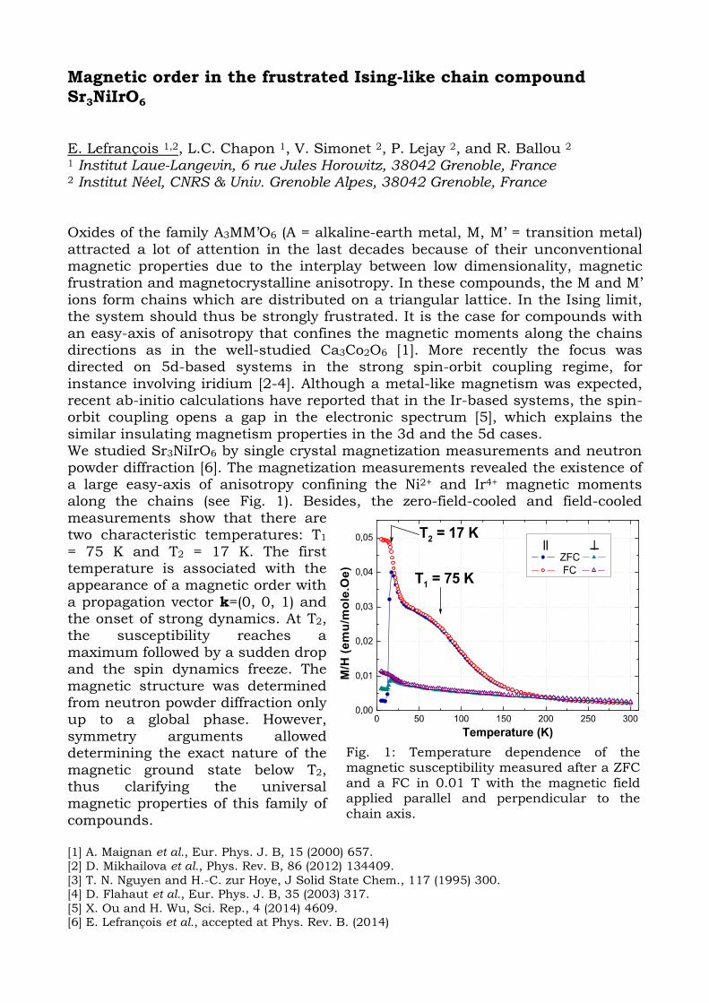
Magnetic order in the frustrated Ising-like chain compound Sr3NiIrO6 E. Lefrançois 1,2, L.C. Chapon 1, V. Simonet 2, P. Lejay 2, and R. Ballou 2 1 Institut Laue-Langevin, 6 rue Jules Horowitz, 38042 Grenoble, France 2 Institut Néel, CNRS & Univ. Grenoble Alpes, 38042 Grenoble, France Oxides of the family A3MM’O6 (A = alkaline-earth metal, M, M’ = transition metal) attracted a lot of attention in the last decades because of their unconventional magnetic properties due to the interplay between low dimensionality, magnetic frustration and magnetocrystalline anisotropy. In these compounds, the M and M’ ions form chains which are distributed on a triangular lattice. In the Ising limit, the system should thus be strongly frustrated. It is the case for compounds with an easy-axis of anisotropy that confines the magnetic moments along the chains directions as in the well-studied Ca3Co2O6 [1]. More recently the focus was directed on 5d-based systems in the strong spin-orbit coupling regime, for instance involving iridium [2-4]. Although a metal-like magnetism was expected, recent ab-initio calculations have reported that in the Ir-based systems, the spin-orbit coupling opens a gap in the electronic spectrum [5], which explains the similar insulating magnetism properties in the 3d and the 5d cases. We studied Sr3NiIrO6 by single crystal magnetization measurements and neutron powder diffraction [6]. The magnetization measurements revealed the existence of a large easy-axis of anisotropy confining the Ni2+ and Ir4+ magnetic moments along the chains (see Fig. 1). Besides, the zero-field-cooled and field-cooled measurements show that there are two characteristic temperatures: T1 = 75 K and T2 = 17 K. The first temperature is associated with the appearance of a magnetic order with a propagation vector k=(0, 0, 1) and the onset of strong dynamics. At T2, the susceptibility reaches a maximum followed by a sudden drop and the spin dynamics freeze. The magnetic structure was determined from neutron powder diffraction only up to a global phase. However, symmetry arguments allowed determining the exact nature of the magnetic ground state below T2, thus clarifying the universal magnetic properties of this family of compounds. [1] A. Maignan et al., Eur. Phys. J. B, 15 (2000) 657. [2] D. Mikhailova et al., Phys. Rev. B, 86 (2012) 134409.
[3] T. N. Nguyen and H.-C. zur Hoye, J Solid State Chem., 117 (1995) 300. [4] D. Flahaut et al., Eur. Phys. J. B, 35 (2003) 317.
[5] X. Ou and H. Wu, Sci. Rep., 4 (2014) 4609. [6] E. Lefrançois et al., accepted at Phys. Rev. B. (2014)
Fig. 1: Temperature dependence of the magnetic susceptibility measured after a ZFC and a FC in 0.01 T with the magnetic field applied parallel and perpendicular to the chain axis.

From disorder to order: The case of the highly Li-ion-conducting Li7-3x□2xGaxLa3Zr2O12 garnet and its protonated analogue ¶ ¶ W. Manalastas Jr1, A. Aguadero2, R. Jalem3,4, C. Bernuy-López1, J.M. López del Amo1, F. Aguesse1, and J. Kilner1,2 1 CIC EnergiGUNE, Albert Einstein 48, 01510 Miñano, Spain 2 Department of Materials, Imperial College London, SW7 2AZ UK 3 Nagoya Institute of Technology, Gokiso, Showa, Nagoya, Aichi 466-8555, Japan 4 Unit of Elements Strategy Initiative for Catalysts & Batteries (ESICB), Kyoto University, Katsura, Saikyo-ku, Kyoto, 615-8520,Japan 
Rechargeable Li-based batteries are promising candidates for powering electric vehicles and stationary applications, but they are hampered by organic electrolyte limitations. Phasing out organic electrolytes for highly Li-ion-conducting ceramics can enable designs with Li-metal anodes, high-voltage cathode materials (and thus high energy/power densities), low manufacturing cost, nontoxicity, operational safety, and modular miniaturization or scale-up.
The nominal Li7La3Zr2O12 (LLZO) garnet presents an O2- anion network that
allows for the following cationic sites per formula unit: 3 tetrahedra and 6 linking octahedra for Li, 3 dodecahedra for La and 2 octahedra for Zr (Figure a). The predicted cubic-phase displays Li-disorder between interconnected tetrahedral 24d and octahedral 48g/96h site occupancies, which facilitates high ionic conductivity via a concerted migration mechanism.[1] Experimentally however, the pure compound crystallizes in a Li-ordered tetragonal phase effecting only 10-
2 mS/cm. Aliovalent substitution strategies can induce more lithium vacancies and shift the thermodynamic preference to a cubic phase, as demonstrated with Nb [2] or Ta [3] which replace Zr atoms and do not block the lithium pathway.
Attempts with Ga [4,5] have also been reported but with limited success. In
our lab, we recently found a cubic-phase Ga-substituted Li7La3Zr2O12 capable of 1.3 mS/cm conductivity by processing under controlled-atmosphere conditions and unprecedented low doping levels (Figure b).[6] We elucidate for the first time using a combination of neutron/x-ray diffraction, solid-state MAS-NMR and MD-simulation data the lithium disorder when minute aliovalent dopants reside on the Li pathway, and how proton-contamination causes detrimental effects.
[1] R. Jalem, et al., Chem. Mater., 25 (2013), 425 [2] S. Ohta, et al. J. Power Sources, 238 (2013) 5, 53-56; [3] Y. Li, et al., J. Mater. Chem., 22 (2012) 15357-15361 [4] MA Howard, et al. Dalton Trans., 41 (2012) 12048; [5] H. Shinawi and J. Janek. J. Power Sources, 225 (2013), 13-19 [6] Bernuy-Lopez, et al., Chem. Mater., 26 (2014), pp. 3610-3617

Soft Matter and
Biology
Matière molle & Biologie ¶ ¶ S. C. M. Teixeira 1,2
1 Keele University – EPSAM, Keele, Staffordshire ST5 5BG, UK. 2 Institut Laue Langevin – 71 Av. des Martyrs, CS20156, F-38042 Grenoble, France. ¶ ¶ La conception de nouvelles applications et développements fait face à des lacunes conséquentes en ce qui concerne l’élément le plus présent dans l’eau. D’ailleurs la sensibilité aux atomes d’hydrogène des techniques à haute résolution devient critique pour les macromolécules qu’on trouve en matière molle ou biologiques. La fonction et propriétés moléculaires, traduites par la combinaison de structure, dynamique et environnent, ne peuvent portant pas être gérées sans saisir la contribution de l’hydrogène [1]. En revanche, les neutrons sont très sensibles à l’hydrogène, aussi bien qu’à son isotope deutérium. Cela permet d’identifier et positionner ces atomes, de même que mettre en évidence les domaines spécifiques d’une structure complexe. En autre, les nouvelles sources de neutrons promettent d’agrandir la portée des études de neutrons [2], avec des résolutions spatiales et temporales encore plus puissantes. La nature non ionisante des neutrons leurs rends aussi spéciales dans les cadres d’un suivi des propriétés des échantillons ou de la validation des techniques engagés par des dégâts d’irradiation [3]. Ma présentation réunis des exemples récents en matière molle et Biologie structurale, dans des domaines spécialement exigeants. J’aborderais des recherches scientifiques dans les sciences de la nourriture [4-5], où le design d’une nouvelle recette doit prendre en compte la production, consommation, stockage, transport, durabilité et même la viabilité économique des molécules. Finalement je exposerai un exemple où les dégâts d’irradiation avec des rayons X ont, pendant longtemps, caché comme les organismes aérobies utilisent le pouvoir oxydative de l’oxygène moléculaire dans l’atmosphère [6].
¶ [1] E. Głowacki, M. Irimia-Vladu, S. Bauerb, and N. Sariciftcia, J. Mater. Chem. B1 (2013), 3742-
3753. [2] S. Teixeira, J. Zaccai, J. Ankner, M. Bellissent-Funel, R. Bewley, M. Blakeley, P. Callow, L. Coates, R. Dahint, R. Dalgliesh et al., Chem. Phys. 345(2-3) (2008), 133-151.
[3] T. Beitlich, K. Kühnel, C. Schulze-Briese, R. Shoeman, I. Schlichting, J. Synchrotron Rad. 14 (2010), 11-23. [4] S. Teixeira, Neutron News 23(2) (2012), 19-21. [5] C. Holt, S. Lenton, T. Nylander, E. Sørensen, S. Teixeira, J. Struct. Biology 185(3) (2014), 383-96. [6] C. Casadei, A. Gumiero, C. Metcalfe, E. Murphy, J. Basran, M. Concilio, S. Teixeira, T. Schrader, A. Fielding, A. Ostermann, M. Blakeley, E. Raven, P. Moody, Science 345(6193) (2014), 193-197.

Membrane Functionalization Using Polyrotaxanes with Modified Cyclodextrins ¶ ¶ T. Charitat 1, M. Bauer1,2 J. Daillant2,3, C. Fajolles2, G. Fragneto4, P. Kékicheff1, C. Marques1 1 Institut Charles Sadron, 23 rue du Loess, BP 84047, F-67034 Strasbourg Cedex 7 2 CEA/ IRAMIS/SIS2M/LIONS, UMR 3299 CEA/CNRS, CEA-Saclay bât. 125, F-91191 Gif-sur-Yvette Cedex 3 Synchrotron SOLEIL, BP 48, F-91192 Gif-sur-Yvette Cedex 4 Institut Laue-Langevin, 6 Rue Jules Horowitz, BP 156, F-38042 Grenoble
¶ 
Dans le cadre d'une collaboration entre des équipes de Chimistes et de Physiciens, nous avons réalisé la conception et à la caractérisation d’une nouvelle famille de ligands pour la fonctionnalisation des membranes lipidiques, appelé “Sliding Tethered Ligands” (STLs) [1]. Ces molécules, basées sur des complexes topologiques entre des polymères et des cyclodextrines amphiphiles modifiées par
un groupement cholestérol(CD), peuvent être insérées dans les membranes phospholipidiques. Nous avons étudié les propriétés d’insertion membranaire de ces dérivés de CD amphiphiles en utilisant différentes techniques, dont la réflectivité de neutrons, ce qui a permis de démontrer que les cavités des CD restaient accessibles lors de l’insertion dans des membranes modèles lipidiques [2,3]. Nous avons alors développé une voie de synthèse pour assembler les nouveaux ligands STL à partir des polyrotaxanes composés d'un très faible nombre de Cds enfilées sur une chaine de polyéthylène glycol (PEG). En utilisant de nouvelles méthodes in situ les polyrotaxanes sont bouchonnés avec des ligands d’adamantane, qui peuvent être reconnus par des récepteurs de a-CD. En utilisant la réflectivité de neutrons, nous avons alors démontrer que les STLs sont très bien ancrés dans des membranes modèles phospholipidiques (DPPC). Pour des densités de polymère suffisamment élevées les STLs forment des brosses de polymères qui suivent les lois d’échelle prédite par la théorie du champ moyen [4]. Finalement, des expériences de machine de force (SFA), nous ont permis de montrer que des membranes modèles modifiées avec des STLs et des récepteurs de cholestérol donnaient lieu à des profils d’interaction typiques entre des ligands et des récepteurs [5]. Références :
[1] M. Bauer, PhD thesis from Université de Strasbourg and Université de Ratisbonne (Regensburg), Prix de Thèse de l'Université de Strasbourg. [2] M. Bauer, C. Fajolles, T. Charitat, H. Wacklin, and J. Daillant, The Journal of Physical Chemistry B, 115 (2011) 15263-15270. [3] M. Bauer, T. Charitat, C. Fajolles, G. Fragneto, and J. Daillant, Soft Matter, 8 (2012) 942-953. [4] M. Bauer, M. Bernhardt, T. Charitat, P. Kekicheff, C. Fajolles, G. Fragneto, C. Marques and J. Daillant, Soft Matter 9 (2013) 1700-1710. [5] M. Bauer, P. Kékicheff, C. Fajolles, T. Charitat, J. Daillant and C. Marques, submitted for
publication.
Solvent contribution to the stability of a physical gel characterised by quasi-elastic neutron scattering
¶ ¶ I. Morfin1, S. Spagnoli1, M. A. Gonzalez2 et M. Plazanet1,3
1 Laboratoire Interdisciplinaire de Physique, Univ. Grenoble 1 et CNRS, Grenoble, France 2 Institut Laue Langevin, Grenoble, France
3 Dipartimento di Fisica, Univ. di Perugia, Italy ¶ ¶ Physicals gels formed by low molecular weight organic gelators (LMMOG) are composed of a rigid network formed by the gelators, in which is trapped a large quantity of solvent. Gathering properties of both liquids and solids, they find applications as functionalised nanomaterials in diverse domains, such as temperature sensors or drug delivery [1]. However, applications suffer several limitations, due to the metastability of the gels that may collapse after some time, and to the difficulty to predict gelation ability of new molecules [2]. The subtle interplay of the different forces exerted between the solvent and gelators, involving hydrogen bonds, hydrophobic forces, π- π interactions or dipolar forces, enable the gels to reversibly assemble in a restricted temperature range in a complex structure that depends on the solvent. Because of the instrinsic complexity, at the molecular level, of the structural organisation and the dynamics, these materials are quantitatively studied with difficulty. In this context, we undertook the study of the microscopic dynamics in a model physical molecular gel, with the goal of characterising the molecular interactions and understanding the formation and stability of the gel phase. The dynamics of a physical gel, namely the Low Molecular Weight Organic Gelator Methyl-4,6-O-benzylidene-α-D-mannopyranoside (α-manno) [3] in water and toluene are probed by Neutron Scattering [4]. The α-manno is an amphiphilic gelator that adopts different organisation in water or toluene. We were able to determine, on a timescale of about 600 ps, the number of solvent molecules that are immobilised by the rigid network formed by the gelators. We could determine that about 10 toluene molecules per gelator participate to the network which is formed by hydrogen bounding between the gelators' sugar moieties. Differently, no water molecule is seen to be immobilised in the water-based gel, in which the rigid network holds mainly thanks to the staking of the gelators’ aromatic groups. These results are in agreement with NMR studies of similar systems, and with observation of macroscopic behaviour. This study shows that the combination of NMR and Neutron Scattering may lead to a complete characterisation of the solvent behaviour confined in a molecular gel.
[1] N. Sangeetha and U. Maitra, Chem. Soc. Rev., 34 (2005), 821-836. [2] R.G. Weiss, Langmuir 25 (2009), 8369-8369. [3] O. Gronwald et S. Shinkai, Chem. Eur. J 7 (2001), 4329-4334. [4] I. Morfin, S. Spagnoli, M.A. Gonzalez and M. Plazanet, to be submitted (Tetrahedron).

Dispersion of modified silica nanoparticles in a PEA matrix following an original aqueous route ¶ ¶ C. Schmitt1,2, A.C. Genix1, J. Alauzun2, G. Guerrero2, P.H. Mutin2, J. Oberdisse1 1 Laboratoire C.Coulomb, Université de Montpellier 2, F-34 095 Montpellier, France 2 Institut C. Gerhardt, Université de Montpellier 2, F-34 095 Montpellier, France ¶ ¶ The dispersion of inorganic nanoparticles (NPs) in polymer matrices leads to a modification of mechanical properties, e.g. like in car tires reinforced with carbon black and silica NPs. In the past years, we have progressed in the microscopic understanding of this phenomenon in different systems, in particular using scattering techniques in model systems [1], but also scattering, electron microscopy and modeling in simplified industrial systems. [2-3] Grafting organic molecules on NPs is a method of choice to modify the interactions between NPs in solution and between NPs and polymer chains in the nanocomposite. In the case of silica NPs, this is usually done in organic medium using organosilane molecules. In this work, aqueous suspension of silica NPs covered by an alumina layer were grafted by water stable phosphonic acids: H3C(CH2)2PO(OH)2, H3C(CH2)4PO(OH)2, H3C(CH2)2PO(OH)2, H(OCH2CH2)3PO(OH)2. [4-5] The dispersion of the NPs in solution was studied using SAXS and DLS. The NPs were incorporated in a hydrophobic polyethylacrylate (PEA) matrix using an original aqueous way, by mixing a PEA latex with the aqueous suspension of NPs. We could show by SANS studies that one can influence the structure of the final nanocomposites by changing the nature and amount of grafted molecules (as illustrated by the curves below). ¶ [1] A.C. Genix et al, Macromolecules, 45 (2012), 1663-1675. [2] G. Baeza et al, Macromolecules, 46 (2013), 317-329. [3] G. Baeza et al, Macromolecules, 46 (2013), 6621-6633. [4] P.H. Mutin et al, Journal of Material Chemistry, 15 (2005), 3761-3768. [5] S. Lassiaz et al, Journal of Materials Chemistry, 21 (2011), 8199-8205.
100
101
102
103
104
10-2 10-1
raw NP (solution in water)raw NPs0.5 P/nm²1 P/nm²1.5 P/nm²
I/phi
q (Å -1)

Un modèle classique de collisions décrive la diffusion de roues dentées nanométriques 
¶ P. Fouquet1, I. Calvo-Almazan2, E. Bahn1, S. Miret-Artes3, M. Telling4, M. M. Koza1, M. Maccarini1 et M. Zbiri1 1Institut Laue-Langevin, CS 20156, F-38042 Grenoble Cedex 9, France; 2Universidad de Zaragoza, Ciudad Universitaria, E-50009 Zaragoza, Spain; 3Instituto de Fisica Fundamental, CSIC, Serrano 123, E-28006 Madrid, Spain; 4ISIS Facility, Rutherford Appleton Laboratory, Chilton, OX11 0QX, UK ¶ ¶ Friction, frottement et diffusion sont des phénomènes fondamentaux en mécanique qui sont déterminés par des processus en échelle moléculaire comme Bowden et Tabor ont démontré il y a 75 ans [1]. Malheureusement ces processus moléculaires sont difficilement obtenus en expérience en raison de leur échelle spatiale nanométrique et leur échelle temporelle de nanosecondes. Nous avons réussi d’établir la dissipation d’énergie intermoléculaire ainsi que la friction avec la surface pour un système prototypique, la roue dentée nanométrique benzène. Les molécules étant adsorbés sur une surface de graphite, nous avons utilisé une large gamme de spectromètres neutroniques ainsi que la diffraction neutronique et des simulations de la dynamique moléculaire, ce que nous a permis de tester des modèles théoriques variés. Les résultats nous ont menés vers un modèle d’une simplicité surprenante : La friction intermoléculaire peut-être expliquée entièrement par un modèle de disques rugueux a parois durs, que nous avons dérivé a base du modèle de sphères durs rugueux [2]. Le modèle couvre la diffusion a basse couverture (< 0.5 monocouches) mais ne fonctionne plus quand on approche une monocouche car collisions multiples et effets de cluster dominent dans ce domaine. ¶ [1] Bowden, F.P. and Tabor, D. « The area of contact between stationary and between moving surfaces » Proc. R. Soc. Lond. A 169, 391-413 (1939). [2] Chandler, D.. « Translational and rotational diffusion in liquids. I. translational single-particle correlation functions » J. Chem. Phys. 60, 3500 (1974).

Magnetism
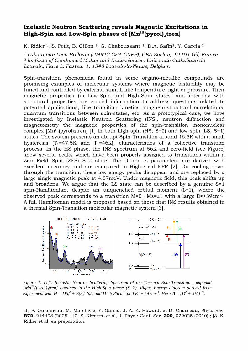
Inelastic Neutron Scattering reveals Magnetic Excitations in High-Spin and Low-Spin phases of [MnIII(pyrol)3tren] K. Ridier 1, S. Petit, B. Gillon 1, G. Chaboussant 1, D.A. Safin2, Y. Garcia 2
1 Laboratoire Léon Brillouin (UMR12 CEA-CNRS), CEA Saclay, 91191 Gif, France 2 Institute of Condensed Matter and Nanosciences, Université Catholique de Louvain, Place L. Pasteur 1, 1348 Louvain-la-Neuve, Belgium Spin-transition phenomena found in some organo-metallic compounds are promising examples of molecular systems where magnetic bistability may be tuned and controlled by external stimuli like temperature, light or pressure. Their magnetic properties (in Low-Spin and High-Spin states) and interplay with structural properties are crucial information to address questions related to potential applications, like transition kinetics, magneto-structural correlations, quantum transitions between spin-states, etc. As a prototypical case, we have investigated by Inelastic Neutron Scattering (INS), neutron diffraction and magnetometry the magnetic properties of the spin-transition mononuclear complex [MnIII(pyrol)3tren] [1] in both high-spin (HS, S=2) and low-spin (LS, S=1) states. The system presents an abrupt Spin-Transition around 46.5K with a small
hysteresis (T↑=47.5K and T↓=46K), characteristics of a collective transition process. In the HS phase, the INS spectrum at 56K and zero-field (see Figure) show several peaks which have been properly assigned to transitions within a Zero-Field Split (ZFS) S=2 state. The D and E parameters are derived with excellent accuracy and are compared to High-Field EPR [2]. On cooling down through the transition, these low-energy peaks disappear and are replaced by a large single magnetic peak at 4.87meV. Under magnetic field, this peak shifts up and broadens. We argue that the LS state can be described by a genuine S=1 spin-Hamiltonian, despite an unquenched orbital moment (L=1), where the
observed peak corresponds to a transition M=0→Ms=±1 with a large D≈+39cm-1. A full Hamiltonian model is proposed based on these first INS results obtained in a thermal Spin-Transition molecular magnetic system [3]. [1] P. Guionneau, M. Marchivie, Y. Garcia, J. A. K. Howard, et D. Chasseau, Phys. Rev. B72, 214408 (2005) ; [2] S. Kimura, et al, J. Phys.: Conf. Ser. 200, 022025 (2010) ; [3] K. Ridier et al, en préparation.
Figure 1: Left: Inelastic Neutron Scattering Spectrum of the Thermal Spin-Transition compound [Mn3+(pyrol)3tren] obtained in the High-Spin phase (S=2). Right: Energy diagram derived from experiment with H = DSz
2 + E(Sx2-Sy
2) and D≈-5.85cm-1 and E≈+0.47cm-1. Here ∆ = [D 2 + 3E2] 1/2.

Small Angle Neutron Scattering (SANS) studies of magnetic Prussian-Blue analogue nanoparticles CsNi[Cr(CN)6] K. Ridier 1, B. Gillon 1, G. Chaboussant 1, S. Mazérat 2, L. Catala 2, et T. Mallah 2
1 Laboratoire Léon Brillouin (UMR12 CEA-CNRS), CEA Saclay, 91191 Gif sur Yvette, France 2 Institut de Chimie Moléculaire et des Matériaux d’Orsay, CNRS-Université Paris Sud 11, 91405 Orsay, France
Small-Angle Neutron Scattering (SANS) is a technique able to probe the morphology and spatial organization of nano-objects, dispersed in solution or embedded in solid matrices, from a few nanometers up to the µm range. One major asset of this technique is its sensitivity to the chemical contrast between the nano-objects and the medium. Moreover, neutrons are sensitive to the magnetic induction so that SANS enables to probe not only the magnetization of the particles but also the induced stray fields. We performed SANS and neutron diffraction studies on coordination nanoparticles (CNP’s) based on cyanide-bridged networks embedded in CTA or PVP polymer matrices. These magnetic nanoparticles, of the Prussian-Blue analogue form CsxNi[Cr(CN)6], present magnetic properties which are size and concentration dependent [1,2]. After an initial study of the structural properties through the determination of the geometrical form factors and structure factors as a function of concentration and matrix type, we investigate the super-paramagnetic behaviour below ca. 90K under magnetic field [3]. As shown in the Figure below, the ZFC/FC difference is proportional to the spherical magnetic form factor FM(q) of the particles and to the internal magnetization M(T) described by a Langevin expression. The present results evidence the possibility, thanks to SANS, to describe the individual spatial magnetization process of nanoparticles. [1] L. Catala et al, Inorg. Chem., 48, 3360 2009 [2] L. Catala et al, Angew. Chem., 121, 189, 2009 [3] K. Ridier et al, en préparation.
0,01 0,02 0,03 0,04 0,05 0,06 0,07
0
100
200
300
400
500
600
700
800
0 20 40 60 80 1000
500
10005K15K
30K
60K
100K
15nm CTA (PAXY)
∆I(q
) =
I(q)
ZF
C -
I(q)
FC
(a.u
.)
Q (in Å-1)
∆I(q) ~ F2
M(Q) M(T)
FM(Q) = sphere R=6nm
Par
ticle
Mag
. M(T
)
Temperature (K)
Figure 1 : ∆0(Q) = I(Q)ZFC(0.01T) – I(Q)FC(1T) for H//ki and temperatures between 5K and 100K. Solid lines are calculated magnetic form factor with R=6 nm. The scalar pre-factor related to the
absolute magnetization M(T) of the particles is shown in the inset.

Spiral magnetic structure in the iron di-arsenate LiFeAs2O7: a neutron diffraction study
Gwenaëlle Rousse,1,2,3* Juan Rodríguez-Carvajal,4 Calin Wurm 5 and
Christian Masquelier 3,5 1 Institut de Minéralogie et de Physique des Milieux Condensés (IMPMC), UMR 7590 CNRS-
Université Pierre et Marie Curie UPMC Univ Paris 06, Case courrier 115, 4 Place Jussieu, 75252
Paris Cedex 05, France 2 Collège de France, 11 place Marcelin Berthelot, 75231 Paris Cedex 05, France
3 Réseau sur le Stockage Electrochimique de l’Energie (RS2E), FR CNRS 3459, France 4 Institut Laue-Langevin (ILL), Diffraction Group, 6, rue Jules Horowitz, BP 156 –
38042 Grenoble Cedex 9, France 5 Laboratoire de Réactivité et Chimie des Solides, CNRS UMR 6007, Université de Picardie Jules
Verne, 33 rue Saint-Leu, 80039 Amiens, France
The magnetic structure of LiFeAs2O7 (monoclinic, space group C2) has been solved using neutron
powder diffraction. This compound presents an antiferromagnetic behavior characterized by a long-
range ordering observed in the neutron diffraction patterns below the Néel temperature (TN = 35 K).
The magnetic structure is found to be incommensurate with respect to the nuclear structure, the
magnetic peaks being indexed with a propagation vector k= (0.709, 0, 0.155). The magnetic
moments form a general spiral (helical-cycloidal) arrangement with a constant magnetic moment of
4.21 µB. The magnetic structure is discussed in terms of super-super exchange interactions
involving two oxygen atoms belonging to an AsO4 tetrahedron, and compared with the magnetic
structure of the di-phosphate analogue LiFeP2O7. The presence of triangular super-super exchange
paths is believed to be at the origin of this incommensurate magnetic structure. The potential of
LiFeAs2O7 as a possible multiferroic material is discussed.
Reference : Rousse, G.; Rodríguez-Carvajal, J.; Wurm, C.; Masquelier, C. Phys. Rev. B 2013, 88, 214433.

Effets de dilution sur les propriétés magnétiques du système 1D frustré CaCr2-xScxO4
M. Songvilay1, S. Petit1, J.P. Castellan1, G. André1, E. Elkaim3, C. Martin2, V. Hardy2 et F. Damay1 1 Laboratoire Léon Brillouin, CEA-CNRS UMR 12, 91191 Gif-Sur-Yvette Cedex, France 2 Laboratoire CRISMAT, CNRS UMR 6508, 6 bvd Maréchal Juin, 14050 Caen Cedex, France 3 Synchrotron Soleil, Saint-Aubin BP 48, 91192 Gif-Sur-Yvette Cedex, France
β-CaCr2O4 est un composé dans lequel les ions magnétiques de chrome (S = 3/2) forment des échelles triangulaires parallèles. Cet arrangement allie à la fois frustration magnétique et basse dimensionnalité, de par sa topologie en échelle zigzag. Ainsi, pour T < 21 K, le système s’ordonne, avec une structure
incommensurable de vecteur de propagation k = (0 0 q) (q ≈ 0.47) [2] ; pour T < 150 K, des excitations 1D sont observées en diffusion inélastique des neutrons [1].
Une série de composés substitués CaCr2-xScxO4 (0 ≤ x ≤ 1) a été étudiée par diffusion de neutrons et mesures de magnétométrie, pour étudier l’impact de la dilution des échelles magnétiques sur l’ordre magnétique et leur caractère 1D.
Dans l’intervalle 0 ≤ x ≤ 0.3, pour lesquels un ordre magnétique à longue distance est toujours présent, les excitations non-classiques 1D persistent au-délà de TN, avec une fréquence caractéristique dépendante de T et de x, comme observé dans d’autres systèmes 1D [3]. Ces excitations disparaissent pour x = 0.5, remplacées par un signal quasi-élastique s’étendant jusqu’à 8 meV. Ces résultats sont interprétés en terme de confinement progressif des excitations 1D, la dilution magnétique ayant pour effet de raccourcir les chaînes de spins, jusqu’à un seuil où le système se décompose en agrégats de taille finie et non-corrélés. La longueur de corrélation entre les spins mesurée expérimentalement est proche de la valeur théorique pour une chaîne de type J1-J2 et montre le rôle crucial que joue J2 dans la propagation des excitations 1D.
[1] F. Damay, C. Martin, V. Hardy, A. Maignan, C. Stock and S. Petit, Phys. Rev. B, 84 (2011). [2] F. Damay, C. Martin, V. Hardy, A. Maignan, G. André, K. Knight, S.R. Giblin and L. Chapon, Phys. Rev. B, 81 (2010). [3] I.A. Zaliznyak, L.-P. Regnault, D. Petitgrand, Phys. Rev. B, 50 (1994)
Ca!
Cr(1)!
Cr(2)!
O!
a) b) Fig.1. a) Structure
cristallographique de β-CaCr2O4 : projection dans le plan ab (en haut) et ac (en bas) des échelles zigzag. b) Diagramme de phase (x, T) de la série CaCr2-xScxO4. Inserts : spectre de diffusion inélastique des neutrons sur poudre pour x = 0.3 et x = 0.5 à 90K.

Investigations de chiralité des excitations magnétiques dans les matériaux multiferroïques ¶ ¶ S. Holbein1,2, P. Steffens1, L. Chapon1, D. Senff2, T. Finger2, J. Ollivier1, Y. Sidis3, P. Becker4, L. Bohatý4, and M. Braden2 1Institut Laue-Langevin, Grenoble, France 2II. Physikalisches Institut, Universität zu Köln, Germany 3Laboratoire Léon Brillouin, Saclay, France 4Institut für Kristallographie, Universität zu Köln, Germany ¶ ¶ We have studied multiferroic materials in which the strong magnetoelectric coupling results in new collective excitations of mixed magnon-phonon character, whose existence has been demonstrated by different experimental approaches [1-3]. While Infrared spectroscopy senses the phonon contribution of these modes, inelastic neutron scattering determines the magnetic part. The intrinsic nature of these electromagnon excitations remains matter of controversy in the REMnO3 family [4]. It seems likely that there are different origins of electromagnon modes, and that the mechanism of the static magnetoelectric coupling only explains a small part of these excitations. We investigated the magnetic excitation spectrum of two of the key multiferroic materials TbMnO3 and MnWO4 by inelastic neutron scattering. With the application of electric fields and the use of spherical neutron polarimetry it is possible to distinguish the different magnon branches and to follow their chirality across the Brillouin zone. In MnWO4 we find good agreement of our neutron results with infrared studies and a detailed analysis of the polarization dependence of the signal allows us to establish an electromagnon character for one mode. In TbMnO3 we have performed similar studies. In this system we have in particular investigated the influence of the magnetic field which changes the magnetic structure to a commensurate one [5]. We compare and discuss the magnetic excitation spectrum for different phases.
¶ [1] T. Kimura et al., Nature, 426, 55 (2003). [2] A. Pimenov et al., Nature Physics., 2, 97 (2006). [3] D. Senff et al., Physical Review Letters, 98, 137206 (2007). [4] R. Valdés Aguilar et al., Physical Review Letters, 102, 047203 (2009). [5] D. Senff et al., Physical Review B, 77, 174419 (2008).

Magnetic and structural properties of α and γ CoV2O6 low dimensional oxides
Marc Lenertz3,1, Silviu Colis1, Aziz Dinia1, Olivier Mentré2, Gilles André3, Florence Porcher3 et Emmanuelle Suard4
[email protected] 1 Institut de Physique et Chimie des Matériaux de Strasbourg (IPCMS),
UMR 7504 CNRS et Université de Strasbourg (UDS-ECPM), 23 rue du Loess, BP 43, F-67034 Strasbourg Cedex 2, France
2 Université Lille Nord de France, UMR 8181 CNRS, Unité de Catalyse et de Chimie du Solide (UCCS USTL), F-59655 Villeneuve d’Ascq, France
3 CEA, Centre de Saclay, DSM/IRAMIS, Laboratoire Léon Brillouin (LLB), F-91191 Gif-sur-Yvette, France 4 Institut Max von Laue-Paul Langevin (ILL), 6 rue Jules Horowitz, BP 156, F-38042 Grenoble Cedex 9, France
Keywords: CoV2O6, magnetization steps, magnetic structure Low dimensional oxides are of increasing interest because of their strong anisotropy or their
peculiar magnetic behavior. CoV2O6 is such an oxide which exists in two allotropic phases, generally called α and γ, showing monoclinic (C2/m) and triclinic (P-1) structures, respectively [1]. They are however close from the structural point of view: their crystalline structures are constituted of parallel 1D Co chains organized in magnetic planes that are separated by nonmagnetic vanadium oxide sheets. Both are antiferromagnetic in the ground state and the magnetization curves recorded below their respective Néel temperature (15 and 7 K for α and γ-CoV2O6 respectively) show stepped variations with sharp field-induced magnetic transitions and a magnetization plateau at one-third of the saturation magnetization (i.e. figure). The ground state and field induced magnetic structures of both α and γ CoV2O6 were studied by neutron powder diffraction (PND). The refinement of the different magnetic structure confirmed the antiferro- (AF), ferri- (Fi) or ferromagnetic (F) character at the different magnetization steps. It is also to note that the intra-chains magnetic order is always ferromagnetic as expected for 1D chains.
The monoclinic α phase exhibit collinear magnetic structures with the magnetic moments direction close to the c direction (i.e. perpendicular to the magnetic chains). The high magnetic moment of 4.5 µB per Co2+ atom observed by SQUID and by PND measurement also suggests a high orbital moment. The triclinic γ one shows some differences as the easy magnetization axis laying along the magnetic chains and the lower magnetic moment (~3.2 µB/Co2+ at 7 T). The AF ground state needs also two propagation vectors to be correctly described, which leads to a commensurate modulated structure with the magnetic moment mostly along the chains direction. Thos differences can be attributed to the lower symmetry and the lower interaction in γ-CoV2O6.
0 1 2 3 4 50
1
2
3
4
5
0,0 0,5 1,0 1,5 2,00
1
2
3γ-CoV2O6 at 1.8 K
Fi
F
AF
m (
µ b/C
o2+)
µ0H (T)
α-CoV2O6 at 5 K
F
Fi
µ0H (T)
AF
[1] M. Lenertz, J. Alaria, D. Stoeffler, S. Colis, and A. Dinia, “Magnetic Properties of Low-Dimensional α and γ CoV2O6,” J. Phys. Chem. C, vol. 115, no. 34, pp. 17190–17196, 2011.

Instrumentation

FAULTS, an accessible program for refining powder diffraction patterns of layered structures M. Casas-Cabanas1, J. Rikarte1, M. Reynaud1, J. Rodríguez-Carvajal2
1 CIC Energigune, Parque tecnológico de Álava, Calle Albert Einstein 48, 01510 Miñano (Álava), Spain. 2 Institut Laue Langevin, 6 rue Jules Horowitz, BP 156, 38042 Grenoble cedex 9, France.
Layered systems include a large number of mineral families and synthetic compounds of great technological importance with, for instance, applications in the field of electrochemical energy storage. Their physical-chemical properties being directly related to their structural features, the microstructural characterization of these materials is of high importance and includes the determination of different kinds of defects, their amount and their locations.
So far, a widely used tool to interpret the diffraction data of one-
dimensionally disordered systems was the program DIFFaX [1], which permits to simulate X-ray and Neutron powder diffraction patterns. In order to overtake the limitations of simple simulations, we have developed the FAULTS program [2], based on the DIFFaX code, which enables to refine experimental XRD and NPD patterns of crystal systems with any type of planar defects, such as twins and stacking faults. An improved version of this program, more performant and with additional features is now available within the FullProf suite of programs [3].
FAULTS can read experimental XRD and NPD data from many different
formats. Refinable parameters are provided by the user in a free-format input data file, in which the structure is described in terms of layers of atoms which are interconnected via stacking operations that occur with a certain probability. Refinements of all the parameters involved in the calculation of the diffracted intensities is carried out using the Levenberg-Marquard optimization algorithm, implemented in the Crystallographic Fortran Modules Library (CrysFML) [4].
Another major feature of FAULTS with respect to DIFFaX is the implementation of a more adequate isotropic size broadening treatment which takes into account the Gaussian (HG) and Lorentzian (HL) contributions to the FWHM in addition to the consideration of a finite number of layers per crystallite already present in DIFFaX. As these are refinable parameters, this treatment allows a successful description of the separate contributions to line broadening of instrumental features, the finite crystallite size and planar defects.
Our presentation will show the structure and operation of the program
FAULTS, and some examples will be given. [1] M.M. Treacy, J.M. Newsam, M.W. Deem, Proc. R. Soc. London Ser. A, 433 (1991) pp. 499-520.
[2] M. Casas-Cabanas, J. Rodríguez-Carvajal, M.R. Palacín, Z. Kristallogr. Suppl., 23 (2006) pp. 243-248. [3] J. Rodríguez-Carvajal, FullProf Suite. Available at http://www.ill.eu/sites/fullprof/index.html. [4] J. Rodríguez-Carvajal and J. González-Platas, IUCr CompComm Newsletter, 1 (2003) pp. 50-58.
Available at http://www.iucr.org/iucr-top/comm/ccom/newsletters/.

Material sciences

NaFe1-xMnxPO4 (0≤x≤1) as positive electrode materials for Na-ion batteries. M. Giner1, M. Casas-Cabanas1 et T. Rojo1
1 CIC ENERGIGUNE, Parque Tecnológico de Álava, Albert Einstein 48, ED. CIC, 01510, Miñano, Spain.
Na-ion batteries represent an interesting alternative to Li-ion batteries owing to the low cost and unlimited natural abundance of sodium [1,2].
Olivine transition metal phosphates isostructural to the well-known
LiFePO4[3] can be good materials for its use as positive electrodes for Na-ion batteries. Indeed, 1 Na per formula unit can be reversibly deintercalated at a potential of about 3V vs. Na/Na+ delivering a theoretical capacity of 154 mAh/g. However, materials working at higher voltage are highly desirable to increase the energy density. Following this approach, mixed Na[Fe,Mn]PO4 with olivine structure appears to be very promising.
In this work, we describe the synthesis and characterisation of the solid
solution NaMnxFe1-xPO4 prepared from their lithium counterparts (LiMnxFe1-xPO4). We have analysed the structural evolution of these materials with varying Mn content and the electrochemical properties of carbon coated NaMnxFe1-xPO4 (0≤x≤1) phosphate materials have been characterized in Na half-cells by galvanostatic cycling under various conditions. It has been seen than depending on the amount of manganese, a different electrochemical response can be observed. ¶ [1]B. L. Ellis, W. R. M. Makahnouk, Y. Makimura, K. Toghill and L. F. Nazar, Nat. Mater. (2007), 6, 749. [2]V. Palomares, P. Serras, I. Villaluenga, K. B. Hueso, J. Carretero-Gonzalez and T. Rojo, Energy Environ. Sci., (2012), 5, 5884. [3]M. Casas-Cabanas, V. Roddatis, D. Saurel, P. Kubiak, J. Carretero-Gonzalez, V. Palomares, P. Serras and T. Rojo, J. Mater. Chem., (2012), 22, 17421.

Discovering Advanced high potential Materials for high Energy density Li ion batteries
M. Freirea, C. Jordyb, D. Chateignera, O.I. Lebedeva, A. Maignana and V. Pralonga*
aLaboratoire de Cristallographie et Sciences des Matériaux CRISMAT, ENSICAEN, Université de Caen, CNRS, 6 Bd Maréchal Juin, F-14050 Caen 4, France
bSaft, Direction de la Recherche, 111/113 Bd Alfred Daney, 33074 Bordeaux, France. Due to their low weight and high energy densities and long cycle life, the development of rechargeable Li-ions batteries is a major scientific challenge. For many applications requiring energy storage, the need to increase the energy density of storage devices is of first importance, e.g. in computers, cell phones, electric vehicles, aeronautics, space, etc. This is the reason why the battery scientific community is studying intensively and worldwide new high capacity cathodes materials. A new composition mainly based on manganese and lithium offers promising results: it has been demonstrated in recent years that Li-rich Mn-based materials are excellent substitutes for the conventional LiCoO2 cathode [1-4]. After a composition screening, a new material family has been discovered: the Li-Mn-O system has been investigated as potential new material for Li-ion batteries. In this presentation, the first results will be presented in terms of cycling performances but also the chemical characterizations (X-ray diffraction, microscopy, thermogravimetric analysis, redox titration) performed in the laboratory to better understand and improve the performances of this new cathode material will be detailed.
[1] B.L. Ellis, et al., Chem. Mater., 2010, 22, 691 [2] T. Ohzuku, et al., Chem. Lett., 2001, 642H [3] Koga, et al., J. Electrochem. Soc., 2013, 160(6), A786 [4] W.B. Luo et al., Electrochim. Acta, 2009, 54, 4655

Effect of mechanical fatigue at low-temperature on structural properties of cryogenic steels. Sara Hany 1,2,3,4,6, Wadad Daaboul2,3, Benoit Duponchel1,2,5, Christophe Poupin1,2,3, Antoine Abou Kais 1,2,3, Mariana Milochova4, Eugène Bychkov1,2,4, Edmond Abi Aad1,2,3,6 1 Université Lille Nord de France, 59000 Lille, France 2 Université du Littoral Côte D’Opale, 59000 Lille, France 3 UCEIV, EA 4492, ULCO, 145 Avenue Maurice Schumann, 59140 Dunkerque, France 4 LPCA, EA 4493, ULCO, 189A Avenue Maurice Schumann, 59140 Dunkerque, France 5 UDSMM, EA 4476, ULCO, 145 Avenue Maurice Schumann, 59140 Dunkerque, France 6 INNOCOLD, Institut Technologique du Froid, 145 Avenue Maurice Schumann, 59140 Dunkerque, France The growing needs for energy combined with the will to reduce CO2 emissions lead to the increase of the demand for liquefied natural gas (LNG), which will be stably available for many decades to come. Cryogenic materials used for the manufacture of the inner tanks for LNG storage are composed of 9% Ni steel plates with excellent low-temperature toughness, great ductility and crack resistance for a high safety level [1]. To guarantee the reliability and safe operation of large-scale metallic structures exposed to cryogenic temperatures for extended periods, it is important to evaluate structural properties of the metal base as well as the welded zone in their own operating conditions in order to prevent damages that could occur [2]. The effects of cyclic fatigue at very low temperature on structural properties of 9% Ni steel were investigated. The structural studies of the base metal 9% Ni and the corresponding welded joint, before and after mechanical stress at low temperature, was determined by means of several techniques such as: X-ray Diffraction at low temperature, High Energy Synchrotron X-ray diffraction, 57Fe Mossbauer Spectroscopy, Small Angle X-rays Scattering… Further structural details and phase transformations during accelerated mechanical aging at low temperature will be reported and discussed in this report. [1] F. Lebel, E. Abi-Aad, B. Duponchel, I. P. Serre, S. Ringot, P. Langry, and A. Aboukaïs, “Thermal ageing process at laboratory scale to evaluate the lifetime of Liquefied Natural Gas storage and loading/unloading materials,” Mater. Des., vol. 44, pp. 283–290, Feb. 2013.
[2] S. Wei and S. Lu, “Effects of multiple normalizing processes on the microstructure and mechanical properties of low carbon steel weld metal with and without Nb,” Mater. Des., vol. 35,
pp. 43–54, Mar. 2012.

Lattice dynamics of the ionic superconductor Li4C60. Inelastic neutron scattering and PALD investigations. ¶ ¶ S. Rols 1, D. Pontiroli 2, M. Aramini 2, M Gaboardi 2, M. Mazzani 2, M. Riccò 2, C. Cavallari 1,2, E. Suard 1, D. Richard 1 et M. R. Johnson 1 1 Institut Laue Langevin, Grenoble, France 2 Département de Physique, Université de Parme, Parme, Italie ¶ ¶ La phase polymère de C60 obtenue, à partir d’un dopage avec des ions alcalins lithium, présente une structure bidimensionnelle unique avec une alternance de liaisons covalentes de type simple et ponts [2+2] entre les fullerènes, suivant deux directions perpendiculaires dans le plan. Les ions alcalins diffusent dans cette structure, engendrant une conductivité ionique de 10-2 S/cm à température ambiante [1]. Nous présentons dans cette contribution une étude de la dynamique du réseau et identifions son rôle dans la mobilité du sous-réseau alcalin. Les mesures de diffusion inélastique des neutrons sont interprétées grâce à des calculs DFT utilisant la méthode PALD [2].
¶ [1] M. Ricco et al., Phys. Rev. Lett., 102 (2009) 145901. [2] M.M. Koza et al. Phys. Rev. B, 84 (2011) 014306.

Posters
- abstracts-

How To Explain Efficiency of PEGylated Blood Substitutes? A SANS Study of Hb/PEG Conformation
Clémence Le Coeur1, Sophie Combet1, Géraldine Carrot1, Peter Busch2, José Teixeira1, and
Stéphane Longeville1
1Laboratoire Léon-Brillouin, UMR 12 CEA-CNRS, CEA-Saclay, 91191 Gif-sur-Yvette CEDEX, France, 2Forschungszentrum Jülich GmbH, Jülich Centre for Neutron Science (JCNS) at Heinz
Maier-Leibnitz Zentrum (MLZ), Lichtenbergst. 1, 85747 Garching, Germany. [email protected]
Cell-free hemoglobin (Hb)-based oxygen carriers have long been proposed as blood substitutes but their clinical use remains tricky due to problems of inefficiency and/or toxicity. Conjugation of Hb with the biocompatible polymer poly(ethylene glycol) (PEG) has improved their performance. However, physiological data have suggested a polymer Mw threshold of about 10 kDa, beyond which PEGylation no longer improves efficiency and nontoxicity of Hb/PEG conjugates. We used small angle neutron scattering and contrast variation, which are the only techniques able to probe separately the conformation of PEG chains and Hb protein within the Hb/PEG complex. Our study enabled us to investigate the role of PEG chain conformation in diPEGylated Hb conjugates, as a function of the polymer Mw. We found out that the low tridimensional structure of Hb tetramer is not modified by the polymer grafting. Also, there is no significant change of the gaussian conformation of free and grafted PEG below about 10 kD. However, beyond this threshold, the radius of gyration of grafted PEG is smaller than that of the free polymer. Therefore, we suggest that, below a polymer Mw threshold of about 10 kDa, the complex with two gaussian PEG chains grafted on Hb may be described by the “dumbbell” model. In contrast, beyond that threshold, PEG molecules are more compacted and may be wrapped on the surface of the protein, spreading a protective “shielding” effect over a larger fraction of the protein, according to the “shroud” model. The proposed models are in good agreement with the physiological data reported in the literature.

The future is wide and bright: News on the ILL wide angle neutron spin echo spectrometers
Peter FOUQUET1, David BAZZOLI1, Thierry BIGAULT1, Pierre COURTOIS, 1 Orsolya
CZAKKEL1, Georg EHLERS2, Peter FALUS1, Bela FARAGO1, Benjamin GIROUD1, Ferenc MEZEI3, Catherine PAPPAS4, Gilles PASTRELLO1, Ian SUTTON1,3, Eric THAVERON1 and
Frederic THOMAS1
1Institut Laue-Langevin, 6 rue Jules Horowitz, 38042 Grenoble Cedex 9, France 2Spallation Neutron Source, ORNL, Oak Ridge, TN 37831-6477, U.S.A.
3European Spallation Source, Box 176, 22100 Lund, Sweden 4TU Delft, Postbus 5, 2600 AA Delft, The Netherlands
The ILL neutron spin echo spectrometer for high Q studies, IN11, has received a new neutron guide and polarizer in the course of the recent H14 guide project at ILL. We will look back on an exciting first year of re-commissioning and user experiments with a strongly improved instrument. The neutron flux of IN11 has increased by a factor of 3 and the coil system has been re-aligned. We will display examples of incoherent and magnetic scattering experiments, which showcase the improved performance. In parallel we are now in the hot construction phase of our next generation wide angle spin echo spectrometer, WASP [1-3]. As the installation of the neutron guides is progressing and the magnetic coil design is finalized, we will present the project status and discuss the projected performance and the interest for our user base. [1] P. Fouquet, G. Ehlers, B. Farago, C. Pappas and F. Mezei, J. Neutron Res. 15, 39 (2007). [2] P. Fouquet, B. Farago, K.H. Andersen, P.M. Bentley, G. Pastrello, I. Sutton, E. Thaveron, F. Thomas, E. Moskvin and C. Pappas, Rev. Sci. Instrum. 80, 095105 (2009). [3] P. M. Bentley, P. Fouquet, M. Böhm, I. Sutton, C. D. Dewhurst and K. H. Andersen, J. Appl. Cryst. 44, 483 (2011).
E-mail for corresponding author: [email protected]

Investigation of magnetic excitations in the refere nce multiferroic materials TbMnO 3 and MnWO 4 by inelastic neutron scattering S. Holbein1,2, P. Steffens1, L. Chapon1, D. Senff2, T. Finger2, C. Toelzer2, A. Komarek2,3, J. Ollivier1, Y. Sidis4, P. Becker5, L. Bohatý5, and M. Braden2 1Institut Laue-Langevin, Grenoble, France 2II. Physikalisches Institut, Universität zu Köln, Germany 3Max-Planck-Institute for Chemical Physics of Solids, Dresden, Germany 4Laboratoire Léon Brillouin, Saclay, France 5Institut für Kristallographie, Universität zu Köln, Germany

Pb2(YbNb)O6-PbTiO3 Structural Features
¶ ¶ C. Cochard1,2, N. Guiblin1, J.M. Kiat1,3, F. Porchet3, L. Clopez Conesa4, S. Estradé4, F. Peiro4, Y. Waltier5, O. Guedes2 et P.-E. Janolin1
1 Laboratoire Structures, Propriétés et Modélisation des Solides, UMR CNRS-Ecole Centrale Paris, Châtenay-Malabry, France
2 Schlumberger, Clamart, France
3 Laboratoire Léon Brillouin, UMR12 CEA/CNRS , CEA Saclay, 91191 Gif-sur-Yvette, France 4 Laboratory of Electron Nanoscopies (LENS), Electronics Department, University of Barcelona, Marti I Franquès 1, 08028 Barcelona, Spain 5 ESRF The European Synchrotron, 76 av. des Martyrs, 38043 Grenoble, France ¶ ¶ Piezoelectricity has attracted a considerable amount of work due to the variety of possible applications such as accelerometers, gyroscopes or transducers. Solid solutions of relaxors and classical ferroelectrics are know to exhibit enhanced piezoelectric properties close to the morphotropic phase boundary (MPB) compared to the pure materials counterpart. Therefore, materials such as Pb(Mg1/3Nb2/3)O3-PbTiO3 or Pb(Zn1/3Nb2/3)O3-PbTiO3 have been extensively studied over the last 50 years. On the contrary, Pb(Yb1/2Nb1/2)O3-PbTiO3 (PYN-PT) which is long known to have good piezoelectric properties (d33=510pC/N) and a relatively high Curie temperature (~370°C at the MPB) has not been examined so extensively probably because of the scarcity of single crystals. In this work, we will present experimental data obtained on PYN-PT of different compositions reflecting the structural complexity encountered in this phase diagram. These data were obtained by X-ray diffraction, neutron scattering and synchrotron diffraction as a function of temperature.

Vector magnet for polarized neutron analysis in 4-circle geometry
1Marc LENERTZ, 1Alexandre M. BATAILLE, 1Thomas ROBILLARD, 1Frédéric OTT, 2Karine DUMESNIL and 1Arsen GOUKASSOV
1 Laboratoire Léon Brillouin (LLB) DSM/IRAMIS, CEA Saclay, 91191 Gif-sur-Yvette, France
2 Institut Jean Lamour (IJL), UMR 7198 CNRS - Université de Lorraine, 54506 Vandoeuvre les Nancy, France
Polarized neutrons scattering analysis is already available on the 6T2 single crystal diffractometer at the LLB (Laboratoire Léon Brillouin) facility. Standard longitudinal half-polarized diffraction or diffraction with polarization analysis is available with a vertical magnetic field nearly perpendicular to the scattering vector. In order to improve the capability of the diffractometer a vector magnet has been designed to allow diffraction with polarization analysis in 4-circle geometry.
The vector magnet is composed of a Helmholtz magnet that produces a ‘vertical’ field along z, while three other window coils are used to generate the magnetic field in the xy plan. In such a way it is possible to generate a vector field up to 25 G in any direction at the sample position. This allowed us to fix the magnets on any of the rotation circles (χ or ω circle in our case) while the field can be applied parallel or perpendicular to the scattering vector. Such a configuration largely increases the accessible reciprocal space and hence the number of Bragg reflexions that can be measured. Furthermore, the use of four magnets for the generation of a xyz vector field lead, for the field generation, to one degree of freedom. This later can be used for the control of the stray field used as guide to keep the neutron’s polarisation.
This setup is interesting in order to study complex magnetic structures, particularly in anisotropic systems. For example, Eu (110) thin films present a symmetry breaking due clamping effects and lead to an imbalance of the magnetic domains populations [1]. It could for example bring to light if the symmetry breaking also leads to an unbalanced chirality in the magnetic helices composing the magnetic structure.
Figure: Picture of the vector magnet.
[1] S. Soriano, K. Dumesnil, C. Dufour, M. Hennion, J. A. Borchers, and P. Mangin, “Clamping effect on the magnetic behavior of europium epitaxial thin films,” J. Appl. Phys., vol. 97, no. 10, p. 10K111, May 2005.

Porosity of extruded cereals studied by neutron tomography
Claire Madeleine-Perdrillat1, Hassina Bilheux2, Dominique Champion3, Camille Loupiac1,3
1Laboratoire Léon Brillouin, CEA-Saclay, Gif-sur-Yvette, France [email protected]
2Chemical and Engineering Materials Division, Oak Ridge National Laboratory, Oak Ridge, TN, USA
3Equipe PAPC (Procédés Alimentaires et PhysicoChimie),UMR PAM (Procédés Alimentaires et Microbiologiques, AgroSup Dijon, Université de Bourgogne, Dijon, France
[email protected] The context of this research was to investigate the three-dimensional microstructure of extruded cereals. Porous media are characterized by a coupling between the solid matrix and the fluid presents in their porous network. In fact, the hydration of food porous materials provokes volume deformation of the entire object but also modifies the porous distribution. The assessment of the porosity appears to be essential because mechanical properties of cereals products strongly depend on their structure. Several techniques have been used to study microstructures of food. These include microscopy1,2, magnetic resonance imaging3, computer vision technique4,5, porosimetry6,7 and most recently X-ray computed tomography8-10. The advent of powerful noninvasive techniques such as X-ray and neutron microtomography has enabled better to quantify the micro-structure (wall thickness, cell size distribution or tortuosity of the porous network) because structure are precisely located in space. Unlike magnetic resonance imaging, neutron imaging allows the ability to image low-moisture materials. In fact this technique is a particularly powerful tool for measuring water diffusion, due to its transparency to heavy elements and high sensitivity to the hydrogen in water. Neutron tomography can give the 3-D distribution of hydrogen atoms in porous materiel and the impact of water on the porous network, which should lead us to a better theoretical understanding of the relationship between the porous structure of a material and its mechanical behavior. We studied the impact of the water content (during extrusion and during storage) on the microporosity of extruded cereals with a polychromatic neutrons beam. The detection system consists of a new type of neutron imaging detector allowing very high spatial resolution (10 µm): the MicroChannel Plate (MCP). A tomographic scan is accomplished by rotating the specimen between a fixed neutron source and detector, around the axis perpendicular to the neutron beam, while collecting radiographs (2-D slices) of the specimen at small angular increments in the range of 0-180°. The series of 2-D slices are then reconstructed into a 3-D image. References (1) Kaláb, M.; Allan-Wojtas, P.; Miller, S. S. Trends in Food Science & Technology 1995, 6, 177. (2) Blonk, J. C. G.; van Aalst, H. Food Research International 1993, 26, 297. (3) Maas, J. L.; Millard, M. M.; Line, M. J.; Galletta, G. J. HortScience 1993, 28, 590. (4) Hullberg, A.; Ballerini, L. Meat Science 2003, 65, 1231. (5) Du, C.-J.; Sun, D.-W. Meat Science 2006, 72, 294. (6) Rahman, M. S.; Al-Amri, O. S.; Al-Bulushi, I. M. Journal of Food Engineering 2002, 53, 301. (7) Kassama, L. S.; Ngadi, M. O. Journal of Food Engineering 2005, 66, 369. (8) Trater, A. M.; Alavi, S.; Rizvi, S. S. H. Food Research International 2005, 38, 709. (9) Léonard, A.; Blacher, S.; Nimmol, C.; Devahastin, S. Journal of Food Engineering 2008, 85, 154. (10) Falcone, P. M.; Baiano, A.; Zanini, F.; Mancini, L.; Tromba, G.; Montanari, F.; Del Nobile, M. A. Journal of Food Science 2004, 69, FEP38.

Magnetic anisotropy tensors of Co2+ molecular complexes determined by polarized neutron diffraction
K. Ridiera, B. Gillona, G. Chaboussanta, D. Luneaub, A. Bortab, O. Iascob, A. Gukasova, Y. Chibac, H. Sakiyamac, M. Mikuriyad and M. Handae
a Laboratoire Léon Brillouin (UMR12 CEA-CNRS), CEA Saclay, France
b Laboratoire des Multimatériaux et Interfaces (UMR5615), Univ. Lyon-1, France c Faculty of Science, Yamagata University, Japan d School of Science and Technology, Kwansei Gakuin University, Japan e Shimane University, Japan
Research in molecular magnetism has motivated a growing interest for the study of magnetic anisotropy in molecular complexes since a strong axial anisotropy is required for potential applications like high-density data storage. For this purpose, we used Polarized Neutron Diffraction (PND) combined with the local site susceptibility tensor approach. PND is an invaluable experimental technique to address the magnetic properties at the atomic scale and therefore to characterize magnetic anisotropy in molecular paramagnets. Magnetic studies on High-Spin Co2+ complexes are of great interest because of their characteristic strong magnetic anisotropy caused by the existence of a significant orbital angular momentum in the ground state and consequently a strong spin-orbit coupling. We report the PND studies performed on two six-coordinated Co2+ molecular complexes: a mononuclear complex [Co(DMF)6](BPh4)2 [1] and a dinuclear complex [Co2(sym-hmp)2](BPh4)2 [2]. The local site susceptibility tensor approach, used to interpret the PND data for highly anisotropic compounds, enables to describe the magnetic response of individual atoms of a paramagnet to an external field and then to determine the local susceptibility tensor of each magnetic atom in the unit cell [3]. One can thus address the magneto-structural relationships, i.e. the correlations between the magnetic anisotropy tensor and the local structure around the Co2+ ions. In the present study, we have directly extracted the magnetic anisotropy tensors on the Co2+ ions through PND data and compared them to the structural properties. In the mononuclear complex case, we find that the magnetic anisotropy is clearly related to the existence of a trigonal elongation axis of the coordination octahedron of the Co ion [4]. For the dinuclear complex, the antiferromagnetic exchange coupling between the Co ions competes with the local coordination structure and the magnetic anisotropy can only be explained considering the interplay between these two contributions. [1] K. Abe, Y. Chiba, D. Yoshioka, R. Yamaguchi, M. Mikuriya, and H. Sakiyama, X-ray Structure Analysis Online, 2012, 28, 65-66. [2] A. Borta, B. Gillon, A. Gukasov, A. Cousson, D. Luneau, E. Jeanneau, I. Ciumacov, H. Sakiyama, K. Tone, and M. Mikuriya, Phys. Rev. B, 2011, 83, 184429. [3] A. Gukasov and P. J. Brown, J. Phys. Condens. Matter, 2002, 14, 8831. [4] K. Ridier et al. in preparation.

Dénaturation thermique de protéines du muscle de bœuf: spectroscopies et imagerie de neutrons ¶ ¶ Scussat S.1,2, Bou-Maroun E.1, Fant C.3, Cayot P.1, Loupiac C.1,2 1 Équipe PAPC, UMR PAM (Université de Bourgogne – AgroSup Dijon), 1, Esplanade Erasme, 21000 Dijon. 2 Laboratoire Léon Brillouin (LLB), UMR 12 (CEA – CNRS), CEA Saclay, Bât. 563, 91191 Gif-sur-Yvette Cedex. 3 Groupe EMN 3A, AgroSup Dijon, 26, boulevard Docteur – Petitjean, BP 87999, 21079 – Dijon cedex. ¶ ¶ Résumé: Open Food System est un projet d’innovation industriel qui a comme objectif le développement d’appareils d’électroménagers « intelligents », capables de piloter la cuisson des viandes et poissons sans intervention humaine. Ma thèse s’inscrit dans ce projet avec l’objectif de suivre l’évolution de la cuisson à l’échelle moléculaire, en décrivant les réactions biochimiques qui se produisent au niveau des constituants du muscle et en les associant aux caractéristiques macroscopiques (texture, couleur, …) qui sont plus classiquement étudiées en science des aliments. Nous étudions la dénaturation thermique de protéines d’origine musculaire par spectroscopie, en observant les modifications conformationelles qu’elles subissent après l’application de plusieurs barèmes temps/températures. Nous avons mesuré les signatures spectroscopiques (infra-rouge et fluorescence) de ces changements conformationnels (échelle moléculaire), selon le degré de cuisson appliqué (peu cuit, moyennement cuit, trop cuit). L’analyse statistique des données obtenues par spectroscopie nous a montré que les échantillons cuits à faible température apparaissent être bien séparés d’échantillons cuits à température moyenne et élevée. Afin d’apporter des informations structurales à une autre échelle de taille (macroscopique > 100 µm), nous avons aussi utilisé l’imagerie neutronique permettant de suivre l’évolution de la morphologie de l’échantillon due à la contraction des myofibrilles et à la migration de l’eau sur et dans la matrice, après les cuissons. ¶ Mots-clés: muscle, protéines, température, spectroscopies, imagerie neutronique Remerciements: Open Food System est un projet de recherche labellisé par Vitagora, Cap Digital, Imaginove, Aquimer, Microtechniques et Agrimip, financé par l’Etat français et la Région Franche-Comté dans le cadre du Programme d’Investissements d’Avenir géré par Bpifrance. www.openfoodsystem.fr

Liste des participants à l’école
Name Affiliation email Rousse Gwenaëlle IPMCP, Paris [email protected] Desmedt Arnaud ISM, Bordeaux [email protected] Grellier Mary LCC, Toulouse [email protected] Damay Françoise LLB, CEA/Saclay [email protected] Fronc Marek Slovaquie [email protected] Gauthier Romain IMN, Nantes [email protected] Hany Sara Université de Lille [email protected] Songvilay Manila LLB, CEA/Saclay [email protected] Demchenko Anna IPCMS, Strasbourg [email protected] Simone LLB, CEA/Saclay [email protected] Loupiac Camille PAM, Dijon [email protected] Freire Mélanie CRISMAT, Caen [email protected] Petuya Claire Bordeaux [email protected] Giner Morgane CIC, Espagne [email protected] Lenertz Marc LLB, CEA/Saclay [email protected] Foucher Damien ILV, Versailles - Saint Quentin [email protected] Mettus Denis Luxembourg [email protected] Manalatas William CIC, Espagne [email protected] Sirotkin Serguey ISM, Bordeaux [email protected]
Pagnier Thierry LEPMI, Grenoble [email protected]
Owen Lewis Cambridge, Royaume-Uni [email protected] Guégan Frédéric ENS, Lyon [email protected] Lainé Fabien CRISMAT, Caen [email protected] Herisson de Beauvoir Thomas ICMCB, Bordeaux [email protected] Shen Lingjia Birmingham, Royaume-Uni [email protected]
Liste des intervenants à l’école
Nom Affiliation Affiliation Perez Olivier CRISMAT, Caen [email protected] Courtois Pierre ILL, Grenoble [email protected] Petricek Vaclav Prague, République Tchèque [email protected] Qureshi Navid ILL, Grenoble [email protected] Kentzinger Emmanuel Jülich, Allemagne [email protected] Klosek Vincent LLB, CEA/Saclay [email protected] Rodriguez-Carvajal Juan ILL, Grenoble [email protected] Goukassov Arsen LLB, CEA/Saclay [email protected] Mirebeau Isabelle LLB, CEA/Saclay [email protected] Petit Sylvain LLB, CEA/Saclay [email protected] Toudic Bertrand Université de Rennes [email protected] Becker Pierre SPMS, ECP, Chatenay-Malabry [email protected]

Liste des participants aux Rencontres
Nom Affiliation Affiliation Rousse Gwenaëlle IPMCP, Paris [email protected]
Cousin Fabrice LLB, CEA/Saclay [email protected]
Desmedt Arnaud ISM, Bordeaux [email protected]
Frick, Bernhardt ILL, Grenoble [email protected]
Chaboussant Gregory LLB, CEA/Saclay [email protected]
Fouquet Peter ILL, Grenoble [email protected]
Vettier Christian ESS Lund, Suède [email protected]
Rodrigues Sylvain LLB, CEA/Saclay [email protected]
Hany Sara Université de Lille [email protected]
Songvilay Manila LLB, CEA/Saclay [email protected]
Demchenko Anna IPCMS, Strasbourg [email protected] Simone LLB, CEA/Saclay [email protected]
Loupiac Camille PAM, Dijon [email protected]
Freire Mélanie CRISMAT, Caen [email protected]
Petuya Claire Bordeaux [email protected]
Giner Morgane CIC, Espagne [email protected]
Reynaud Marine CIC, Espagne [email protected]
Lenertz Marc LLB, CEA/Saclay [email protected]
Ridier Karl LLB, CEA/Saclay [email protected] Foucher Damien ILV, Versailles - Saint Quentin [email protected]
Mettus Denis Luxembourg [email protected]
Manalatas William CIC, Espagne [email protected] Serguey ISM, Bordeaux [email protected]
Charitat Thierry Institut Ch. Sadron, Strasbourg [email protected]
Holbein Simon ILL, Grenoble [email protected]
Plazanet Marie LIPhy, Grenoble [email protected]
Schmitt Céline Université de Montpellier [email protected]
Lefrançois Emilie ILL, Grenoble [email protected]
Madeleine-Perdrillat Claire LLB, CEA/Saclay [email protected]
Fabrèges Xavier LLB, CEA/Saclay [email protected]
Deutsch Maxime LLB, CEA/Saclay [email protected]
Combet Jérome Institut Ch. Sadron, Strasbourg [email protected]
Rols Stéphane ILL, Grenoble [email protected]
Chiang Hsin Yin Münich, Allemagne [email protected]
Lavie Pascal LLB, CEA/Saclay [email protected]
Rodriguez-Carvajal Juan ILL, Grenoble [email protected]
Combet Sophie LLB, CEA/Saclay [email protected]

Liste des intervenants aux Rencontres
Nom Affiliation Affiliation Toudic Bertrand Université de Rennes [email protected]
Zemb Thomas ICSM, CEA/Marcoule [email protected]
Visticot Jean-Paul LLB, CEA/Saclay [email protected]
Simon Charles ILL, Grenoble [email protected]
Argyriou Dimitri ESS Lund, Suède [email protected]
Teixera Susana ILL, Grenoble [email protected]
Lescouëzec Rodrigue UPMC, Paris [email protected]
Alba-Simionesco Christiane LLB, CEA/Saclay [email protected]
Paul-Boncour Valérie CMTR, Thiais [email protected]
Liste des organisateurs
Nom Affiliation Affiliation Kiat Jean-Michel SPMS, ECP, Chatenay-Malabry [email protected]
Gillon Béatrice LLB, CEA/Saclay [email protected]
Porcher Florence LLB, CEA/Saclay [email protected]
Fernandez-Diaz Maria-Térésa ILL, Grenoble [email protected]
Janolin Pierre-Eymeric SPMS, ECP, Chatenay-Malabry [email protected]
Jegou Pascale LLB, CEA/Saclay [email protected]
Sineau Olivier LLB, CEA/Saclay [email protected]