jessica garvey pt, csrs rehab supervisor chartercare … · peripheral: semi-circular canals,...
TRANSCRIPT

Jessica Garvey PT, CSRS Rehab Supervisor
CharterCARE Home Health

Describe the Assessment of the Central Vestibular System
Identify Differential Diagnosis for Central Vestibular
Impairment Discuss Treatment Recommendations for the Central
Vestibular Rehabilitation Patient
Anatomy of the Vestibular System Central Vestibular Testing with Demo Review Pathology and Common Central Vestibular
Diseases Basic Treatment Principles
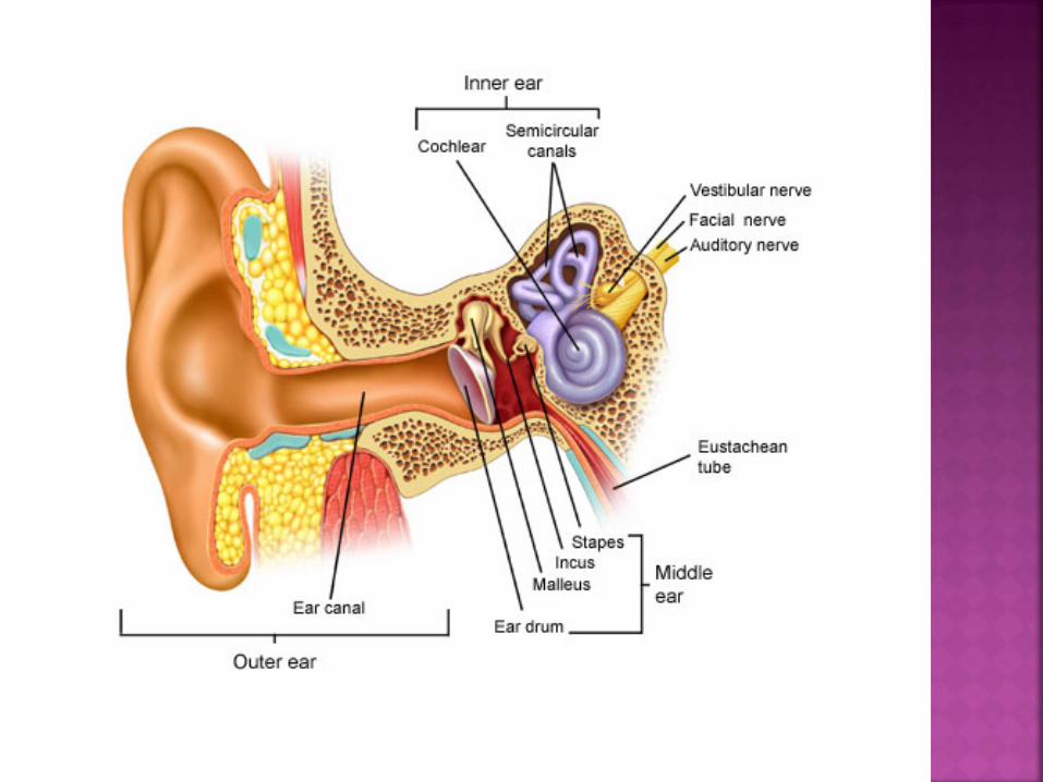

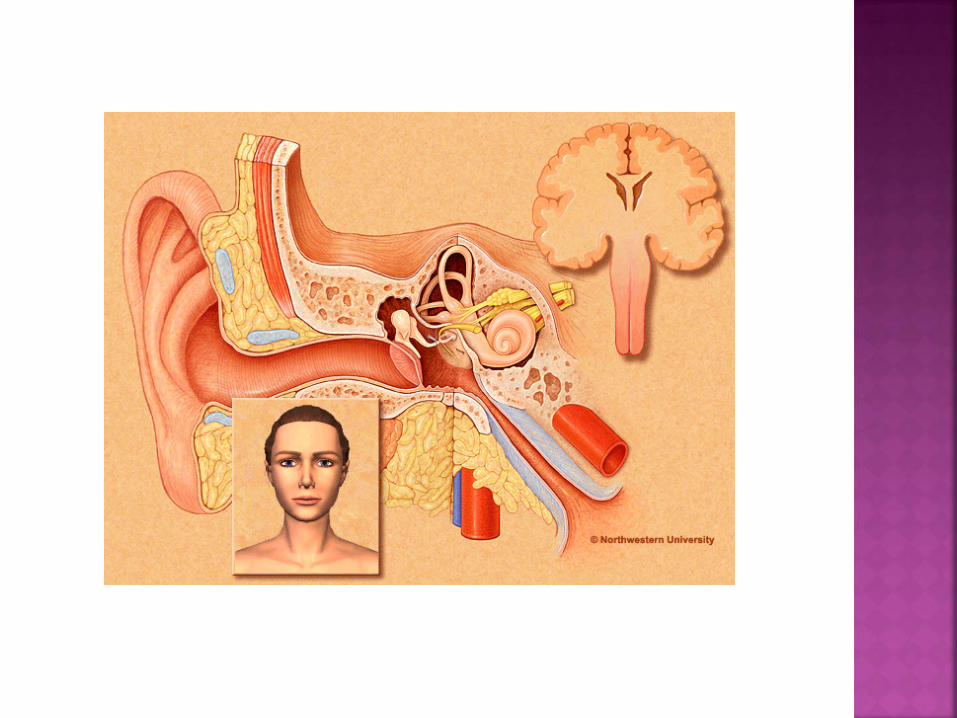
Labyrinth: Organs of Balance Located in the inner ear Outer Shell: Bony Labyrinth Inner Contents: Membranous Labyrinth
Cavities are filled with perilymphatic fluid High Na: K ratio (similar to CSFluid) Houses the auditory and vestibular sensory organs
Membranous Labyrinth - Housed within the cavities and chambers of the bony
labyrinth - Canals and chambers filled with endolymphatic fluid High
K:Na ratio
- Ionic composition necessary for vestibular and auditory hair cells to function properly (Important to understand with dizziness related to dehydration)

Contains 5 Structures that detect head motion, coordinate eye movement with head rotation and detect angular and linear acceleration. 3 Semicircular canals Anterior Posterior Horizontal 2 Otholithic Organs Saccule Utricle
All work together to keep eyes focused on objects in
motion to move safely. 70 % of Balance when moving

The peripheral vestibular system is connected to the central vestibular system by the VIII Cranial Nerve.
Central Vestibular System Consists of VIII cranial Nerve Root Entry Zone into Brain
Stem (auditory and vestibular nerve come together) Vestibular Nuclear Complex (in Pons and Medulla) Cerebellum
4 major Nuclei, 7+ minor nuclei Important anatomical connections:
Superior and Medial Nuclei: Regulate the vestibular-oculo reflex (VOR) VOR is the key motor output of the vestibular system How we are able to keep eye focused on an object when we turn our
head fast or slow Medial Nuclei: Coordinate head and eye movement (regulate eye movement)
Lateral Nuclei: Regulates the vestibulo-spinal reflex (VSR) VSR is the ability to work with the brain to maintain upright posture
against gravity

This is the major computer of the vestibular system
Processes all information for ALL systems of balance Receives information from the vestibular nuclear complex as
well as the cerebellum’s own input Inhibitory control of the vestibular systems Adjusts VOR response and processes otoliths input in relation to
gravity (can inhibit info coming in to tone it down) Sends out signals to motor cortex for appropriate motor
output
Temporal-Parietal Lobes Related to visuo-vestibular system Coordination of eye/head movement with vestibular system
Important for gaze stabilization with head moving or stationary.
Hippocampus Important for erect posture Postural control center Gait Control Maintains proper upright posture/vestibular input with utricle and
saccule from the peripheral vestibular system.

Coordinates head and eye movement Gaze Stabilization
Ability to maintain focus on target with head moving Peripheral Vestibular System – VOR: head moving and vestibular
system intact Ability to track a moving target while head is still Central vestibular system: head is still
Head and Posture Stabilization Ability to select compensatory body movements to maintain head
stability and upright posture Vestibulo-spinal Reflex (VSR)

All 4 sensory systems send information to the Cerebellum. Vestibular Visual Somatosensory Musculoskeletal
The Cerebellum filters, adapts and fine tunes the sensory
output response for postural control and coordination of eye movement.

Oculomotor and Somatosensory systems serve as our primary EXTERNAL references for the CNS regarding visual and support surfaces.
The Vestibular system functions as our primary INTERNAL reference to inform the CNS about any head movement and body alignment.

Primary Processing unit Very Plastic and Adaptable (Unless sustains a lesion) The ability of the cerebellum to recalibrate the neural
firing from a malfunctioning vestibular system
BASIS FOR VESTIBULAR REHABILITATION

Major output of the vestibular system is eye movement for GAZE STABELIZATION while there is movement occuring.
Vestibular system and Visual system work together to maintain the stability of images on the fovea while the head and body are in motion and to maintain postural control during motion.

In order to maintain focus on an object you need: Vestibular system Musculoskeletal strength of ocular muscles Coordination of muscles Integration of Vestibular and ocular systems

Helps us to separate dysfunctions of the peripheral and central vestibular system

Test in room light with glasses off Tests are performed 18 to 24 inches away from patient Use small objects patient can see, but large enough to track. Tests:
Spontaneous Nystagmus (Central) Smooth Pursuit (Central) Gaze-evoked nystagmus (Central) Saccades (Central) Vergence and Divergence (Central) Ocular ROM/Mobility/Motility Testing (Central) VOR (Peripheral) Head Thrust (Peripheral) Dynamic Visual Acuity (Peripheral)
Involuntary rhythmical movement of the eyes Normal or Pathological Nystagmus has a slow phase that is generated by the
vestibular system Fast phase is generated by the CNS system (saccadic) Named by the direction of the fast phase Linear: up/down/left/right Torsional: clockwise or counterclockwise

http://www.youtube.com/watch?feature=player_detailpage&v=hby75sZSAvg

We perform oculomotor tests to help us determine peripheral or central vestibular involvement and to assess for the patients ability to stabilize their gaze with the head or body movement.

Central Test Ask patient to look straight ahead and observe for
nystagmus. You may have to hold your hand on the patient’s chin or top of head to stabilize.
Central Lesion: Nystagmus frequently purely linear or
torsional and may change direction with gaze. Peripheral Lesion: Nystagmus frequently mixed, and does
not change direction with gaze. Can be suppressed with fixation and generally resolves in a few days.

Is the ability to track an object,
maintaining focus on the fovea with eye movement only (no head movement). If tracking is saccadic in nature then the pursuit is considered abnormal. More sensitive to slower velocities. The test is most sensitive to age-related eye disease.
Smooth pursuit is a voluntary
function and therefore under central control. It requires attention to the moving target.

Central Test Smooth Pursuits: Assess through full field using “H”
pattern. Test within a 30 degree range. Beware of end range nystagmus. Saccadic or asymmetrical movements during smooth pursuits is
considered abnormal.
Gaze Evoked Nystagmus: While performing smooth pursuits, hold at random points and observe for nystagmus. Hold position for full 10 seconds before moving target.
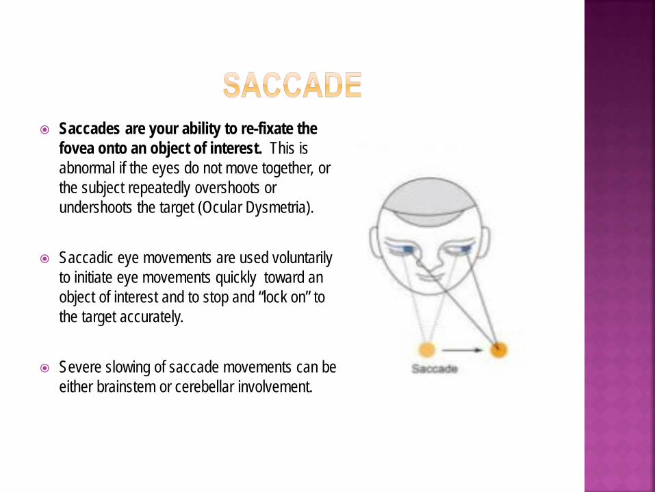
Saccades are your ability to re-fixate the fovea onto an object of interest. This is abnormal if the eyes do not move together, or the subject repeatedly overshoots or undershoots the target (Ocular Dysmetria).
Saccadic eye movements are used voluntarily to initiate eye movements quickly toward an object of interest and to stop and “lock on” to the target accurately.
Severe slowing of saccade movements can be
either brainstem or cerebellar involvement.

Central Test Saccades: Hold finger/object 18 inches from the
patient and 15 degrees to the side. Ask the patient to look at the targets on your command. Patient should be able to lock on the target with one or less corrections. Use random timing Perform in both horizontal and vertical planes Do not cross mid-line

Central Test Ask the patient to follow your finger or a specific target to test full
vertical and horizontal eye movements. Tests for: ability to produce full ROM, conjugate eye movement.
This evaluates muscular strength and coordinative abilities for conjugate eye movement.
Positive Test: If eyes not moving together. Eyes not moving
at same speed. May notice saccadic intrusions (eyes move with a series of jumps)
Note: May be positive due to eye related age changes.


• Progressive Neurological Disorders o Dementia o MS o Parkinson’s/Basal Ganglia Disease o Cerebellar Degeneration
• Ischemic Diseases and Other Brain Disorders
o CVA * Small Vessel Disease o TIA * Migraine o Seizure o TBI o Tumors

• To establish an effective treatment plan, the origin of the disorder needs to be determined.
o Peripheral: Semi-circular canals, otoliths or external
portion of 8th cranial nerve o Central: nerve root of 8th cranial nerve (Internal Portion),
vestibular nuclei or cerebellum o Both: Peripheral and Central Both Central and Peripheral Disorders can be treated
and improved, but goals and treatment approach will be different

Symptoms Peripheral Central
Imbalance Mild to Moderate Severe
Nausea/Vomiting Severe Variable, may be minimal
Auditory Common Rare
Neurologic Rare Common
Compensation Rapid Slow
Outcome Likely return to PLOF Questionable return to PLOF
• “Dizziness” not sure where you are in space (not room spinning dizziness) • Frequent falls • Decreased sensory integration • Ataxic gait • Positive Spontaneous Nystagmus • Poor Smooth Pursuit, Ocular ROM and Saccades • Disequilibrium • Decreased righting reactions

• Light-headedness: o Feeling of fainting is about to occur (Blood Pressure)
• Vertigo: o Perception of movement (Either self or surroundings)
• Oscillopsia: o Experience that objects known to be stationary are in motion
• Disequilibrium: o Unsteadiness, imbalance or loss of equilibrium. Sensation of
not knowing where one’s body is in relation to the vertical or horizontal planes.

4 Categories 1. Time Frame of Sx
Duration How long does is last Exacerbation of the Intensity Peripheral comes and goes and Central all the time
2.Circumstance surrounding the onset of sx Spontaneous (Central) Provoked by head or visual movement, visual complexity or visual patterns (Peripheral)
3.Characteristics of the Sx What does the patient mean when they say are dizzy? (room spinning (Subjective/Objective,
Lightheadness, unexplained falls or imbalance) Are sx accompanied by nausea, vomiting, headaches, heart palpitations, panic or drop attacks or
stroke sx? 4.Status of their hearing by their perception
Unilateral or bilateral hearing loss Slowly progressive and is one ear worse than other? Do they have sudden changes in hearing of fluctuations? Are they experiencing tinnitus or fullness?
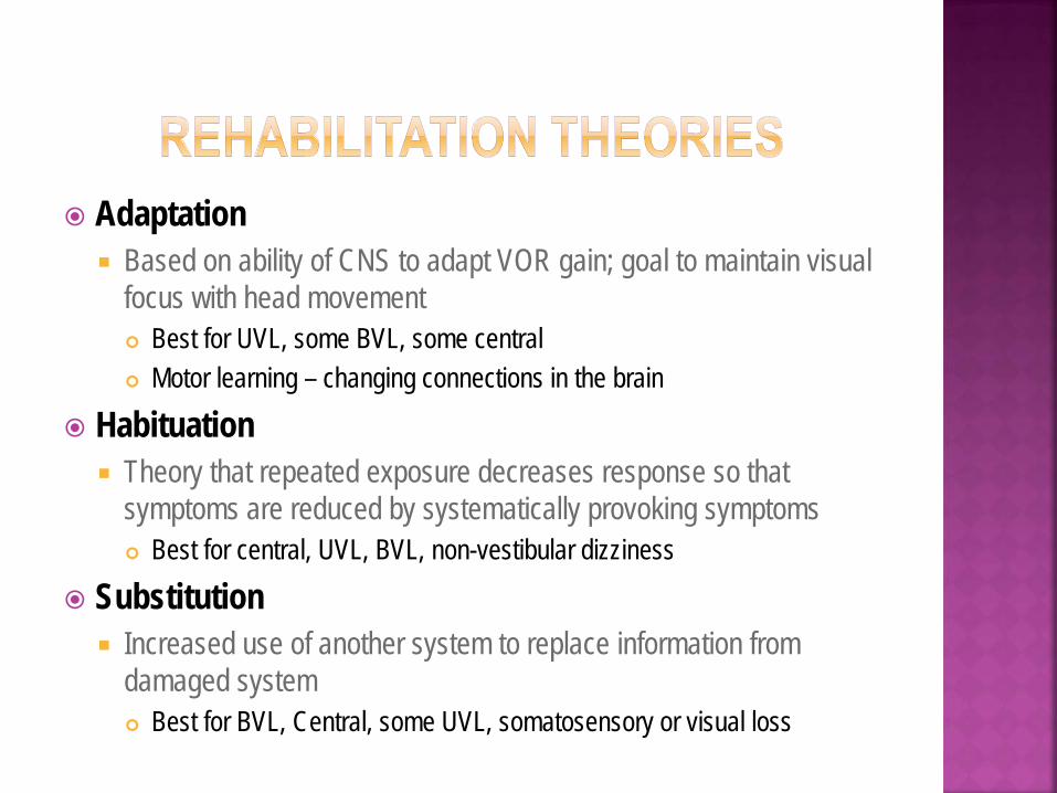
Adaptation Based on ability of CNS to adapt VOR gain; goal to maintain visual
focus with head movement Best for UVL, some BVL, some central Motor learning – changing connections in the brain
Habituation Theory that repeated exposure decreases response so that
symptoms are reduced by systematically provoking symptoms Best for central, UVL, BVL, non-vestibular dizziness
Substitution Increased use of another system to replace information from
damaged system Best for BVL, Central, some UVL, somatosensory or visual loss

Adaptation is a dynamic process, not static Adaptation is the gradual change in behavior that
results from experiencing demands that exceed the current state of the system.
Adaptation needs an error and an after-effect (when opening a light door after opening a heavy door is learned, there is a gradual reduction of error) Motor planning – the error occurs when the activity did not
go correctly, after effect is how the system will change what you will do next time.
Adaptation requires practice/experience and the ability to store the information

Motor Learning Process Always over-responds then gradually reduces in
magnitude Fewer muscles or torque are generated for the task Perceived task becomes easier More efficient (less force) Less anxiety, stress, and fear Need to give the patient the opportunity to fail, once the
patient is at the point of failure, then they can learn to adapt.

Gaze Stabilization VOR target still VOR target moving (head moving opposite the
target) VOR cancellation Saccades Smooth Pursuit Dynamic Visual Acuity (DVA)

Adaptation Exercises Gaze stabilization and VOR exercises for
vestibular disorders Postural strategies that produce “errors” Progression of activities is achieved by
performing the exercises in situations with sensory conflict

Target must always remain in focus Therapist needs to be able to see patient’s eyes
Must use stimulus (activity) sufficient to create error signal, but not overstimulate Therapist needs to know what error signals are for
that patient
Allow patient to rest long enough for symptoms to subside before next repetition
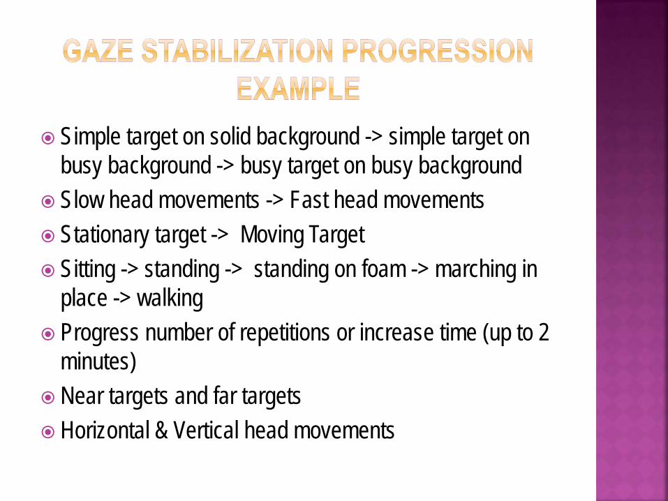
Simple target on solid background -> simple target on busy background -> busy target on busy background
Slow head movements -> Fast head movements Stationary target -> Moving Target Sitting -> standing -> standing on foam -> marching in
place -> walking Progress number of repetitions or increase time (up to 2
minutes) Near targets and far targets Horizontal & Vertical head movements

Another mechanism of recovery is referred to as Habituation Habituation refers to the ability of the
cerebellum to reduce the subjective symptoms produced by specific head or body movements You need to encourage the patient to perform
activities that make them feel dizzy or “bad” Repeated exposure of the movement that
produces symptoms until it is no longer perceived as noxious by the cerebellum
Motion Sensitivity Quotient (MSQ) – best tool, helps to guide treatment

Habituation Exercises Involves repetitive motions that reproduce symptoms of
vertigo or “dizziness” MSQ – tool used to determine motion intolerance 16 item test Patients rate severity of symptoms on 0-5 while therapist
records how long symptoms are present Begin with a mildly provoking position and advance to
more provocative positions as response to stimulus declines
Advanced by involving sensory conflict situations

Habituation Exercises Rocking chair Cawthorne – Cooksey MSQ Brandt-Daroff Settling – giving the somatosensory system added input,
while having patient close their eyes and count out loud backwards from 100 by 1’s, 2’s or 3’s. Must be challenging, this assists in recalibration of vestibular and somatosensory input.

VOR exercises – as stated previous Spinning in a chair w/ focusing on predefined targets (can
alter by changing speeds) Walking with head turns vertically and horizontally
For example in ALF have patient read door numbers or “count the puppies” in a painting while walking
Walking turns – Walking completing a 360 degree turn and continuing to walk in the same direction. Normal is to be able to complete in 3 steps.
Optokinetic activities Walking in figure 8 pattern, performing a ball throw (progress to ball
throw while walking) Altering visual background (disco ball, outdoors in wooded area on
blustery day) and performing balance activities/VOR exercises
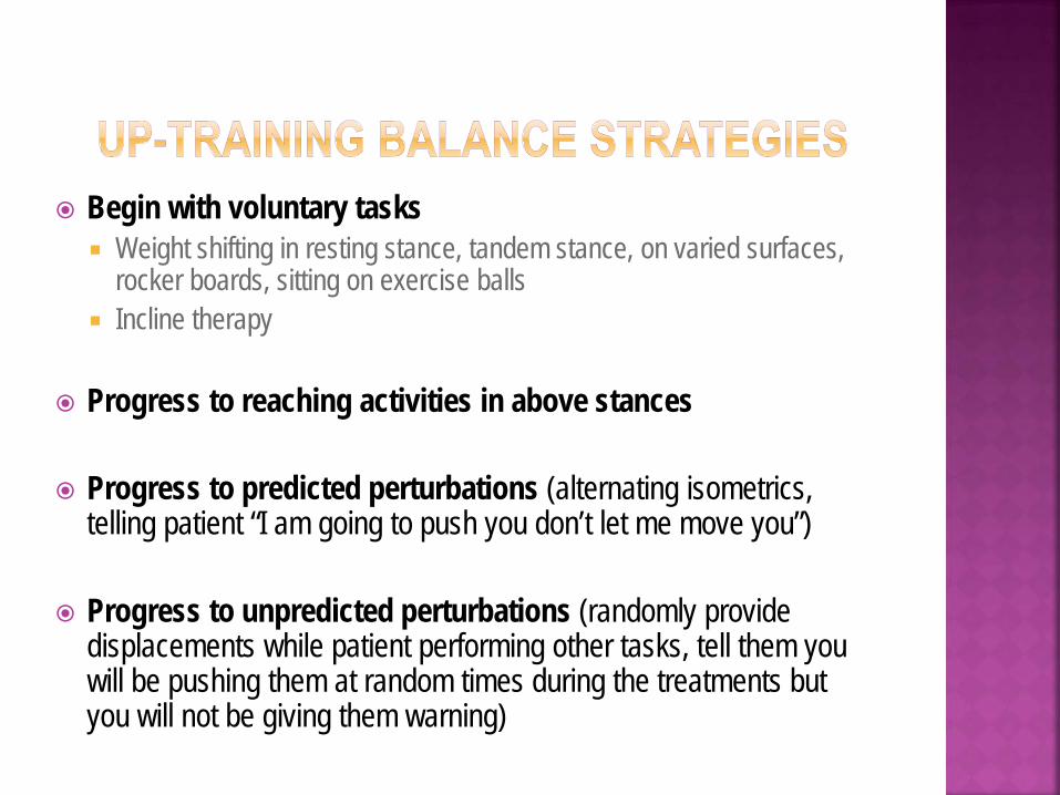
Begin with voluntary tasks Weight shifting in resting stance, tandem stance, on varied surfaces,
rocker boards, sitting on exercise balls Incline therapy
Progress to reaching activities in above stances Progress to predicted perturbations (alternating isometrics,
telling patient “I am going to push you don’t let me move you”) Progress to unpredicted perturbations (randomly provide
displacements while patient performing other tasks, tell them you will be pushing them at random times during the treatments but you will not be giving them warning)


Acute Phase Medications
Chronic Phase Habituation Substitution Adaptation/Gaze stabilization (VOR) Postural control stability (LOS/Strategies) Sensory integration (foam/disc) Substitution for complete loss Gait and ADL activities

• Remove/Alter the preferred balance system(s) and increase use of other systems
• Stimulate the impaired system(s) starting with more simple challenges and working to more difficult challenges
• i.e.: for vestibular stimulation start in supported sitting on a firm surface, move to unsupported sitting, to compliant surface, to standing on firm surface, etc.
• Perform functional activities to improve integration of newly stimulated input, with increasing difficulty levels. ** Gear the functional task around the patient’s goal so it is
important to him/her**
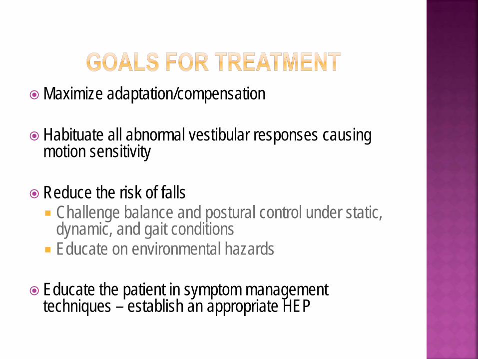
Maximize adaptation/compensation Habituate all abnormal vestibular responses causing
motion sensitivity Reduce the risk of falls Challenge balance and postural control under static,
dynamic, and gait conditions Educate on environmental hazards
Educate the patient in symptom management techniques – establish an appropriate HEP
Evaluate all systems of balance and assess the strengths and weakness of each one individually.
Take into consideration the potential for
improvement of a weak system or the potential for substitution by a stronger system.
Realize that the patient is never performing “to
potential”. The capacity of their balance skill is a direct reflection of the challenge we apply to it.

List tests and findings Vestibular testing
Central Testing Spontaneous Nystagmus: (-) Gaze Evoked Nystagmus: (-) Smooth pursuits: (+) for increased blinking, loss of target, increased tearing, increased
squinting Saccades: (+) for overshooting/undershooting target, increased blinking or squinting Convergence/Divergence: (-) Ocular ROM: comment on if eyes move in coordination, or any abnormalities you see.
Peripheral Testing VOR: (+) for avoidance, loss of target, increased tearing, increased blinking, squinting Head Thrust: (+) to R or L, or may be unable to accurately assess due to avoidance w/
VOR Dix Hallpike: (+) for R or L posterior canalithiasis,
Comment on onset of dizziness, when dizziness resolves, and intensity Roll Test: (+) for R or L canalithiasis
Comment on intensity of dizziness w/ testing on R and L, when resolves
For Gaze Stabilization Pt will demonstrate improved central gaze stabilization as
noted by (-) smooth pursuits testing to allow pt to watch the baseball game on television without dizziness in _____ weeks.
Pt will demonstrate improved gaze stabilization as
indicated achieve a score of ______ on dynamic gait index to allow pt to turn his/her head and maintain a conversation while walking without loss of balance in ______ weeks.

For dynamic balance/optokinetic activities Therapist facilitated use of balance strategies and integration of gaze
stabilization w/ pt performing walking w/ 360 degree turns, performed in 6 steps turning to R, 8 steps turning to L (completing turns in 3 steps is normal), requires CGA and vc’s 90% of the time for safety, and pacing. Demonstrates early stepping strategies 50% of the time to maintain balance and decreased gait speed noted when performing turns.
Therapist facilitated use of vestibular system w/ pt performing head
turns while walking, able to correctly identify targets 50% of the time when looking to the right, and 75% Of the time when looking to the L. Early stepping strategies noted w/ head turns and pt requires CGA to min assist to recover balance w/ slight losses.

Documentation of Settling Exercise Therapist instructed pt in settling exercise to increase
habituation of vestibular system (or recalibration of vestibular system)- pt seated on firm surface, PT providing axial loading through shoulders while pt closes his/her eyes and counts backwards from 100 to 0 by 3’s. Able to complete activity in 3 minutes w/ 15 errors noted in counting. Dizziness at start of exercises graded 9/10, after completion of settling exercise dizziness reported to be 2/10.
Progression shown by decreased time to complete the
exercise, decreased errors, increasing difficulty of activity (instead of counting by 3’s, counting by 7’s, etc)

Be specific with what you see or don’t see that should be happening
Write the note from your perspective, what you did and
why you were there. Not just what the patient performed. Use your imagination, be creative. Have fun with the
treatments!

Christy, Beth SPT and Jeff Walter (oversight). “Vestibular System: Anatomy and Physiology (with Practical Applications).” www.vestibular seminars.com/images/reviewed_vestibularsystem_1_doc.
Hain, Timothy C. MD. “Anatomy and Pathophysiology of the Vestibular System.” www.tcain.com/cv/courses/eng2008/anaphys.pdf
Hedenberg, Ray PT and Kenda Fuller, PT, NCS. (2008) “Stable Mobility, Functional Classification for Differential Diagnosis Related to Disequilibrium, Imbalance and Dizziness.” Innovative Rehabilitation Balance Solutions. pp. 1-81.
Marryott, Kimberly, PT, DHS. (4-5 Jan 2010) “Balance for Life” Amedisys Home Health. Morgan, Barry, PT. (Oct 2015) “Vestibular Rehabilitation Evaluation and Treatment.” North
American Seminars, Inc. (2015) www.healthclick.com Page, Ph. D, OTR/L, CKTP, FAHA. (October 2013 – March 2014) “Stroke Rehabilitation
Specialist Certification. “ Neurorecovery Unlimited. Shepard, Neil T, Phd, CCC-A. “Signs and Symptoms of Central Vestibular Disorders.”
www.asha.org. American Speech-Language-Hearing Association, Feb 2009. Web. 10 September 2017.
Thompson, Timothy MD and Ronald Amedee MD. “Vertigo: A Review of Common Peripheral and Central Vestibular Disorders.” www.ncbi.nlm.nih.gov. The Ochsner Journal, Spring 2009. Web. 10 September 2017.

Email me: [email protected]