joanna sheldon 2006a
TRANSCRIPT
Investigation of monoclonal gammopathies:
what we should know but have probably forgotten!
Dr. Joanna Sheldon Dr. Joanna Sheldon Protein Reference Unit Protein Reference Unit St. George St. George’ ’s Hospital s Hospital
History of the lab 1847 – Bence Jones protein described 1937 – electrophoresis separates plasma protiens 1940 – the term paraprotein introduced by Apitz 1959 the immunoglobulin introduced by Heremans 1965 measurement of immunoglobulin concentration by
radial immuno diffusion described by Mancini 1966 – measurement of proteins by rocket electrophoreiss
described by Laurell 1976 – automated immunonephelometry for protein
measurement introduced
Setting the standards ALL laboratory methods should be: ACCURATE – where possible, be calibrated against an IRP PRECISE
show CVs of <10% realistic (<5% preferable) within batch show CVs of <20% realistic (<10% preferable) between batch
CONSISTENT BETWEEN USERS show CVs of <20% realistic (<10% preferable) in EQA
CLINICALLY SPECIFIC show a low number of false positives
CLINICALLY SENSITIVE show a low number of false NEGATIVES
CLINICALLY SENSITIVE show a low number of false POSITIVES
VALUE FOR MONEY all of the above and cost effective
What is normal? SERUM « polyclonal gamma region on electrophresis « Adult concentrations
IgG 616 g/L, IgA 0.8 – 4.0 g/L, IgM 0.5 – 2.0 g/L « IgG half life ~ 21 days and dependent on concentration « IgA and IgM half life ~ 5 days independent of concentration
URINE «Total protein <0.1 g/L « a trace of albumin should be detectable in every urine « normal urine (adequately concentrated) will also show some other protein e.g. transferrin and some polyclonal free light chains « these free light chains are a normal result of B cell development
Monoclonal proteins development of a monoclonal does not happen
overnight will start as a small band may develop quickly or very slowly may increase in concentration as clone grows may remain at a low and stable concentration may disappear over time may suppress background B cell population the same immunoglobulin concentration may relate to
polyclonal, oligoclonal or monoclonal populations there is NO antibody that is capable of distinguishing
a monoclonal protein from a polyclonal protein
Things to remember monoclonal proteins are not (usually)
normal proteins (in terms of structure) monoclonal proteins do not behave like
polyclonal proteins presence of a monoclonal does not
mean malignancy absence of a monoclonal does not
exclude malignancy
What are the stages?
« Detection « Typing « Quantification « Monitoring
Detection of monoclonal proteins
ALWAYS check serum and urine approx. 20% of myeloma only make BJP BJP is small ~22kDa (but can polymerise) BJP can pass easily through the glomerulus Serum immunoglobulins should always be
done with serum protein electrophoresis International Guidelines for BJP analysis
recommends 2 nd void of the day for detection
Detection of monoclonal proteins
High quality electrophoresis agarose, cellulose acetate or capillary serum (prefereable to plasma) urine – concentrated or sensitive stain (at least a trace of albumin MUST be seen in all urines) monoclonal proteins can appear anywhere
from the alpha1 to the postgamma areas low threshold for immunofixation

IMMUNOFIXATION
use high quality antiserum antitotal (free and bound) light chain antiserum is better than antifree light chain antiserum antilight chain antiserum often shows greater binding to free light chains than to bound light chains
one antiserum will not detect ALL monoclonals immunofixation does increase sensitivity over
electrophoresis (by 1020x) good interpretation increases specificity immunofixation is not quantitative
Typing of monoclonal proteins
Immunofixation is the only reliable way to type monoclonal proteins – it can « confirm the clonality of band detected by electrophoresis
« test for α, γ and µ heavy chains and κ and λ light chains
« test for the δ and ε heavy chains where a serum shows monoclonal light chains without a corresponding α, γ or µ heavy chain
« exclude low concentration monoclonal components even where no band is apparent on electrophoresis but with clinical indications e.g. AL amyloidosis
Typing of monoclonal proteins
Immunofixation is the only reliable way to type monoclonal proteins – it can « exclude the presence of monoclonal IgA or IgM if they are showing raised concentrations without increased staining in the betagamma region of the electrophoresis
« positively identify other proteins that may be mistaken for monoclonal immunoglobulins e.g. fibrinogen, Creactive protein, beta2 microglobulin and complement components
« detect minimal residual disease or complete remission post stem cell transplantation when no monoclonal component is seen on the electrophoretic separation.
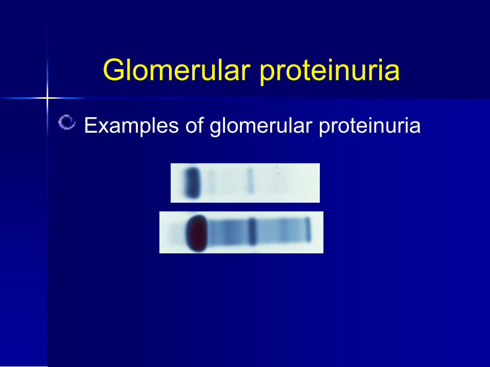
Glomerular proteinuria
Examples of glomerular proteinuria
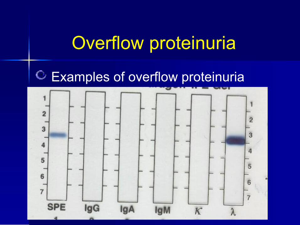
Overflow proteinuria
Examples of overflow proteinuria

Proteinuria
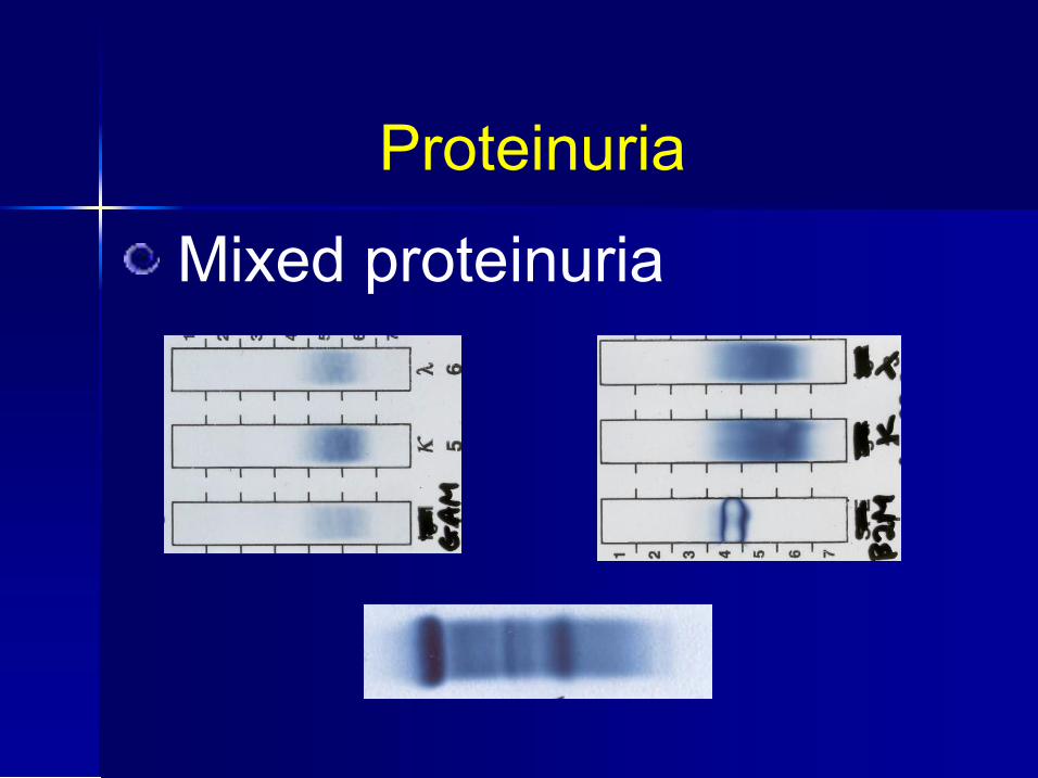
Mixed proteinuria glomerular, tubular and overflow can all occur together patterns hard to classify
Proteinuria
Mixed proteinuria
Light chains polyclonal B cells produce a slight excess of light
chains as part of their normal processes these free light chains arrive at the kidneys and are
filtered by the glomerulus (mwt approx. 25kDa) inflammatory responses can increase the amount of
polyclonal free light chains produced kidneys are important sites of light chain catabolism light chain catabolism (plus dehydration, acidosis etc)
can cause aggregation of excess light chains and tubular damage

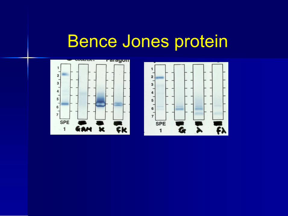
Bence Jones protein MONOCLONAL free light chains first described in 1846! important marker of B cell malignancy rarely seen in benign conditions can form amyloid or myeloma casts kidneys are important sites of light chain catabolism light chain catabolism (plus dehydration, acidosis etc)
can cause aggregation of excess light chains and tubular damage
there is NO antiserum available ANYWHERE that can distinguish monoclonal from polyclonal light chains
Bence Jones protein Free light chains not necessarily BJP BJP is monoclonal free light chains reliable detection of BJP can only be
done by good quality electrophoresis and immunofixation
finding and typing BJP is probably the hardest thing we do in protein labs…..
Bence Jones protein

Don’t forget….. intact monoclonal Ig also appears in the
urine (with or without BJP) will usually have different mobility BJP β2 microglobulin can also be a large
band on urine EP (especially if patient is on alphainterferon) patients with amyloid may have heavy
glomerular or tubular proteinuria and only a small amount of BJP
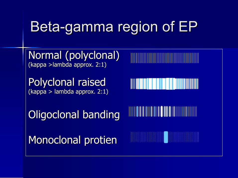
Beta Beta gamma region of EP gamma region of EP
Normal (polyclonal) Normal (polyclonal) (kappa >lambda approx. 2:1) (kappa >lambda approx. 2:1)
Polyclonal raised Polyclonal raised (kappa > lambda approx. 2:1) (kappa > lambda approx. 2:1)
Oligoclonal banding Oligoclonal banding
Monoclonal Monoclonal protien protien
Why? patients with infection and inflammatory conditions
show increased free light chain excretion – not BJP patients with B cell malignancies with BJP can have
glomerular, tubular, overflow or mixed proteinuria elderly patients often have some tubular proteinuria tubular catabolism can make light chains fragments
that aggregate tubular catabolism can make light chains fragments
that aggregate and have similar charge degraded urines show very fuzzy patterns high resolution electrophoresis picks up tiny amounts
of protein
What can we do? use an electrophoretic technique that is
sensitive…to 10mg/L BJP see albumin in every urine confirm with immunofixation increases
sensitivity and specificity don’t be afraid to ask for a fresh sample if the
urine is degraded, smelly or shows an indistinct pattern positive identification important – if there is a
band, what is it (BJP, Hb, β2M, lysozyme etc.)
Quantification – best of a bad job!
electrophoresis, scanning densitometry and total protein NOT ideal
total protein methods are poor EP separation can have a high ‘background’ due to protein fragments
tubular proteins ‘crud’
limitation of urine volume – timed, 24 hour, random
within a patient, urine patterns are surprisingly stable
What is best?
high quality electrophoresis low threshold for fixation skilled interpretation quantification by % BJP and
TP