jobb sttaask aannaallyyssiiss ffoorr arrdmmss ... surveys/vtresults2009.pdf · writing(cv, venous,...
TRANSCRIPT

VT Results for the Web
Page 1 of 13 June 15, 2010
JJoobb TTaasskk AAnnaallyyssiiss ffoorr
AARRDDMMSS VVaassccuullaarr TTeecchhnnoollooggyy
Data Collected: December 27, 2009
Reported: June 15, 2010
AAnnaallyyssiiss SSuummmmaarryy FFoorr:: VVaassccuullaarr TTeecchhnnoollooggyy EExxaamm
Survey Dates 12/11/2009 - 12/27/2009
Invited Respondents 2,000
Surveys with Demographics 523 (completed at least one question)
Completed Surveys 445 (completed all questions)
Response Rate, Completed Surveys 22.2%

VT Results for the Web
Page 2 of 13 June 15, 2010
Demographics
EEdduuccaattiioonnaall LLeevveell
Education
N Percent
Decline to state 0 0%
On-the-job training 63 12%
Certificate program 93 18%
Associates degree 146 28%
Bachelors degree 162 31%
Masters degree 29 6%
MD 26 5%
PhD 4 1%
Total 523 100%
Table 1. Education -- All Survey Respondents
Graphically, the educational level of respondents is represented below.
Figure 1. Educational Level

VT Results for the Web
Page 3 of 13 June 15, 2010
SSoonnooggrraapphheerrss iinn LLaabb
The task analysis included a question regarding the number of sonographers that practice in the
same lab as the respondent.
Sonographers in Lab
N Percent
Decline to State 2 0%
0 - 5 316 60%
6 - 10 117 22%
11 - 15 47 9%
16 - 20 21 4%
20 + 20 4%
Total 523 100%
Table 2. Sonographers in Lab
Below are the results from Table 2.
Figure 2. Sonographers in Lab

VT Results for the Web
Page 4 of 13 June 15, 2010
SSttuuddiieess ppeerr MMoonntthh Below, Table 3. VT Studies per Month shows the number of studies respondents typically conduct per
month in their own practices.
Vascular Studies per Month
N Percent
Decline to State 2 0%
0-50 209 40%
51-100 152 29%
101-150 97 19%
151-200 46 9%
201+ 17 3%
Total 523 100%
Table 3. VT Studies per Month
Figure 3. VT Studies per Month

VT Results for the Web
Page 5 of 13 June 15, 2010
LLaabboorraattoorryy SSttuuddiieess ppeerr MMoonntthh
Table 4, Vascular Technology Exams in Lab/Month, shows the number of VT sonography
exams conducted in the respondent’s laboratory in a typical month
Sonograms in Lab per Month
N Percent
Decline to State 3 1%
0-200 232 44%
201-500 175 33%
501-1,000 74 14%
1,000+ 39 7%
Total 523 100%
Table 4. VT Studies in Respondent’s Laboratory
The results from Table 4 are presented graphically below.
Figure4. VT Exams in Respondent’s Laboratory

VT Results for the Web
Page 6 of 13 June 15, 2010
EExxppeerriieennccee aass aa VVTT SSoonnooggrraapphheerr
The number of years the respondents have spent as a Sonographer are tabulated in Table 5.
Years as a Sonographer.
Years Sonographer
N Percent
Decline to State 3 1%
0 - 5 89 17%
6 - 10 99 19%
11- 15 86 16%
16 - 20 87 17%
20 + 159 30%
Total 523 100%
Table 5. Years as a Sonographer
Figure 5. Years as a Sonographer
VT Results for the Web
Page 7 of 13 June 15, 2010
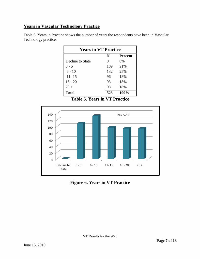
YYeeaarrss iinn VVaassccuullaarr TTeecchhnnoollooggyy PPrraaccttiiccee
Table 6. Years in Practice shows the number of years the respondents have been in Vascular
Technology practice.
Years in VT Practice
N Percent
Decline to State 0 0%
0 - 5 109 21%
6 - 10 132 25%
11- 15 96 18%
16 - 20 93 18%
20 + 93 18%
Total 523 100%
Table 6. Years in VT Practice
Figure 6. Years in VT Practice

VT Results for the Web
Page 8 of 13 June 15, 2010
Survey Topics Listings
Below are the complete topic listings as they appeared in the survey.
ID VT Survey Tasks
1. Anatomy & physiology
1.1. Assess carotid arterial pulse
1.2. Evaluate the effects of limb compression maneuvers on venous flow
1.3. Evaluate the effects of respiration on venous flow
1.4. Assess pressure changes following exercise: lower extremities
1.5. Assess pressure changes following exercise: upper extremities
1.6. Assess segmental pressure gradients: lower extremities
1.7. Assess segmental pressure gradients: upper extremities
1.8. Assess renal arteries
1.9. Assess the hepatic arteries
1.10. Assess the hepatic veins
1.11. Assess the portal venous system
1.12. Evaluate renal veins
1.13. Evaluate the inferior vena cava
1.14. ** Evaluate transcranial vessels
1.15. ** Evaluate transcranial vessels without imaging
1.16. ** Assess the common carotid artery
1.17. ** Assess the external carotid artery
1.18. ** Assess the internal carotid artery
1.19. ** Assess subclavian arteries
1.20. ** Assess vertebral arteries
1.21. ** Evaluate for central venous access: upper extremities
1.22. ** Evaluate for central venous access: lower extremities
1.23. ** Map superficial veins (e.g., cephalic): upper extremities
1.24. ** Map superficial veins (e.g., saphenous): lower extremities
1.25. ** Evaluate the femoral artery for obstruction
1.26. ** Evaluate the common femoral artery for obstruction
1.27. ** Evaluate the common iliac artery for obstruction
1.28. ** Evaluate the deep femoral artery for obstruction
1.29. ** Evaluate the external iliac artery for obstruction
1.30. ** Evaluate the internal iliac artery for obstruction
1.31. ** Evaluate the peroneal artery for obstruction
1.32. ** Evaluate the posterior tibial arteries for obstruction
1.33. ** Evaluate the celiac artery
1.34. ** Evaluate the inferior mesenteric artery
1.35. ** Evaluate the superior mesenteric artery

VT Results for the Web
Page 9 of 13 June 15, 2010
2. Pathology
2.1. Assess arteriovenous malformations
2.2. Assess arteriovenous fistulas
2.3. ** Evaluate the circle of Willis for disease
2.4. Evaluate the carotid arteries following endovascular repair
2.5. Assess dialysis access (fistula or graft): lower extremities
2.6. Assess dialysis access (fistula or graft): upper extremities
2.7. Evaluate post-op bypass grafts: lower extremities
2.8. Evaluate post-op bypass grafts: upper extremities
2.9. Evaluate abdominal aortic aneurysms following endovascular repair
2.10. Evaluate the Aorto-iliac region following endovascular repair (angioplasty and/or stent)
2.11. Evaluate the renal arteries following bypass graft surgery
2.12. Perform follow-up evaluation after interventions for mesenteric artery disease
2.13. Perform follow-up evaluation after interventions for renal artery disease
2.14. Perform follow-up evaluation after portal decompression (TIPS)
2.15. Perform post-renal transplant evaluations
2.16. Evaluate anterior tibial veins to assess for venous obstruction
2.17. Evaluate peroneal veins to assess for venous obstructions
2.18. Evaluate posterior tibial veins to assess for venous obstruction
2.19. Assess response to induced reactive hyperemia: lower extremities
2.20. Assess response to induced reactive hyperemia: upper extremities
2.21. Encountered a peripheral pseudoaneurysm: lower extremities
2.22. Encountered a peripheral pseudoaneurysm: upper extremities
2.23. Evaluated subclavian stenosis and/or occlusion
2.24. Evaluate the femoral artery for obstruction with duplex
2.25. Evaluate the anterior tibial artery for obstruction with duplex
2.26. Evaluate the common femoral artery for obstruction with duplex
2.27. Evaluate the common iliac artery for obstruction with duplex
2.28. Evaluate the deep femoral artery for obstruction with duplex
2.29. Evaluate the external iliac artery for obstruction with duplex
2.30. Evaluate the internal iliac artery for obstruction with duplex
2.31. Evaluate the peroneal artery for obstruction with duplex
2.32. Evaluate the posterior tibial arteries for obstruction with duplex
2.33. Assess for pseudoaneurysms
2.34. Encountered a peripheral aneurysm: lower extremities
2.35. Encountered a peripheral aneurysm: upper extremities
2.36. Encountered arterial dissections
2.37. Encountered steal physiology secondary to dialysis access fistula placement
2.38. Evaluate abdominal aortic aneurysms
2.39. Evaluate the celiac artery
2.40. Evaluate the inferior mesenteric artery

VT Results for the Web
Page 10 of 13 June 15, 2010
2.41. Evaluate the superior mesenteric artery
2.42. Perform food challenge testing for mesenteric ischemia
3. Patient care
3.1. Schedule examinations
3.2. Educate the public and other health care professionals in the application of vascular tests
3.3. Interact with supervising physician as to procedures to be followed for examination
3.4. Use a computer for patient scheduling
3.5. Use a computer for report generation
3.6. Use a computer for storage of demographic data
3.7. **Have contraindications for the performance of exercise testing(CV, Venous, Peripheral arterial, Abdomen)
4. Integration of data
4.1. Use a computer for storage of exam results
4.2. Obtain pertinent clinical history from patient and medical record
4.3. ** Obtain pertinent clinical history from patient and medical record(CV, Venous, Peripheral arterial, Abdomen)
4.4. ** Provide preliminary interpretation to referring physician of test results verbally or in writing(CV, Venous, Peripheral arterial, Abdomen)
4.5. ** Compare results with previous studies(CV, Venous, Peripheral arterial, Abdomen)
4.6. Perform validation studies (e.g., review venograms and/or arteriograms)
4.7. **Report the limitations of the exam(CV, Venous, Peripheral arterial, Abdomen)
4.8. Evaluate and document physical signs of peripheral vascular disease
4.9. Palpate peripheral pulses
4.10. Analyze spectral waveforms: lower extremities
4.11. Analyze spectral waveforms: upper extremities
4.12. Evaluate analog Doppler waveform recordings: lower extremities
4.13. Evaluate analog Doppler waveform recordings: upper extremities
4.14. Provide preliminary interpretation to referring physician of test results verbally or in writing
4.15. Request supervising medical director or designee review studies
5. Protocols
5.1. Evaluate transcranial Doppler spectral waveform analysis with color duplex imaging
5.2. Evaluate transcranial Doppler spectral waveform analysis without imaging
5.3. Perform transcranial Doppler for cardiac septal defects (PFD)
5.4. Assess the common carotid artery with duplex ultrasound
5.5. Assess the external carotid artery with duplex ultrasound
5.6. Assess the internal carotid artery with duplex ultrasound
5.7. Use duplex imaging to assess subclavian arteries
5.8. Use duplex imaging to assess vertebral arteries
5.9. Assess venous valvular competency with patient standing
5.10. Evaluate for central venous access: lower extremities
5.11. Evaluate for central venous access: upper extremities
5.12. Evaluate perforating veins with duplex ultrasound for incompetence
VT Results for the Web
Page 11 of 13 June 15, 2010
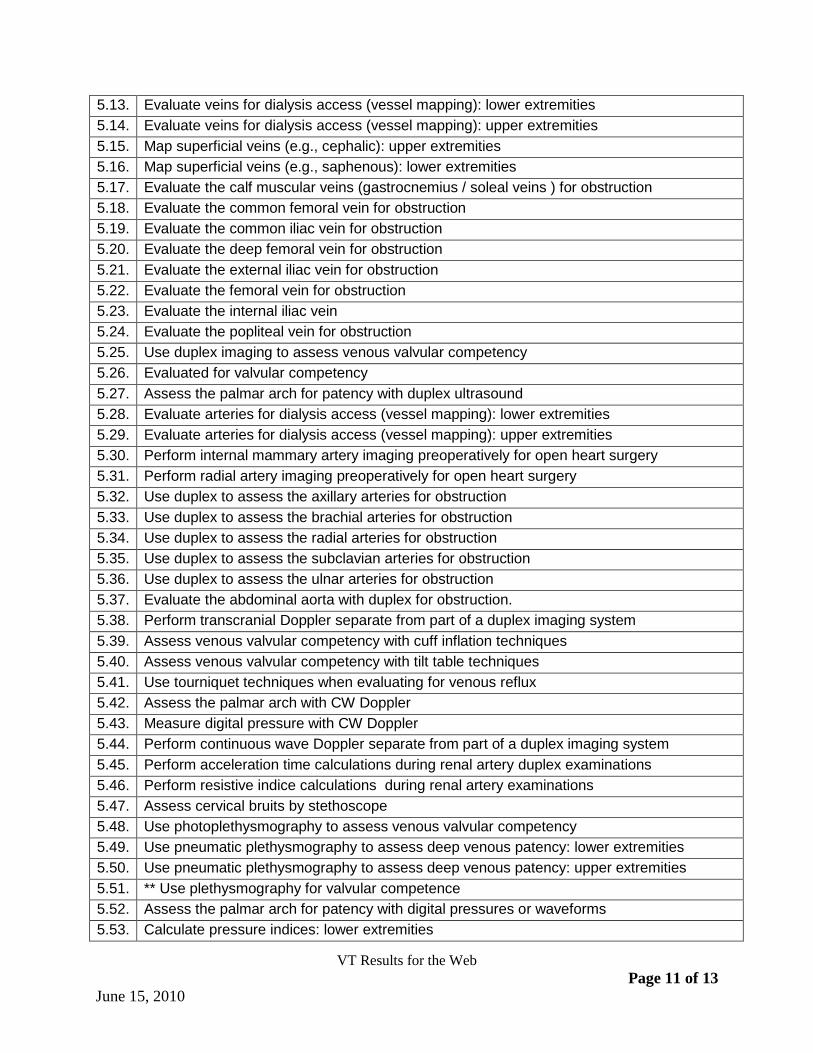
5.13. Evaluate veins for dialysis access (vessel mapping): lower extremities
5.14. Evaluate veins for dialysis access (vessel mapping): upper extremities
5.15. Map superficial veins (e.g., cephalic): upper extremities
5.16. Map superficial veins (e.g., saphenous): lower extremities
5.17. Evaluate the calf muscular veins (gastrocnemius / soleal veins ) for obstruction
5.18. Evaluate the common femoral vein for obstruction
5.19. Evaluate the common iliac vein for obstruction
5.20. Evaluate the deep femoral vein for obstruction
5.21. Evaluate the external iliac vein for obstruction
5.22. Evaluate the femoral vein for obstruction
5.23. Evaluate the internal iliac vein
5.24. Evaluate the popliteal vein for obstruction
5.25. Use duplex imaging to assess venous valvular competency
5.26. Evaluated for valvular competency
5.27. Assess the palmar arch for patency with duplex ultrasound
5.28. Evaluate arteries for dialysis access (vessel mapping): lower extremities
5.29. Evaluate arteries for dialysis access (vessel mapping): upper extremities
5.30. Perform internal mammary artery imaging preoperatively for open heart surgery
5.31. Perform radial artery imaging preoperatively for open heart surgery
5.32. Use duplex to assess the axillary arteries for obstruction
5.33. Use duplex to assess the brachial arteries for obstruction
5.34. Use duplex to assess the radial arteries for obstruction
5.35. Use duplex to assess the subclavian arteries for obstruction
5.36. Use duplex to assess the ulnar arteries for obstruction
5.37. Evaluate the abdominal aorta with duplex for obstruction.
5.38. Perform transcranial Doppler separate from part of a duplex imaging system
5.39. Assess venous valvular competency with cuff inflation techniques
5.40. Assess venous valvular competency with tilt table techniques
5.41. Use tourniquet techniques when evaluating for venous reflux
5.42. Assess the palmar arch with CW Doppler
5.43. Measure digital pressure with CW Doppler
5.44. Perform continuous wave Doppler separate from part of a duplex imaging system
5.45. Perform acceleration time calculations during renal artery duplex examinations
5.46. Perform resistive indice calculations during renal artery examinations
5.47. Assess cervical bruits by stethoscope
5.48. Use photoplethysmography to assess venous valvular competency
5.49. Use pneumatic plethysmography to assess deep venous patency: lower extremities
5.50. Use pneumatic plethysmography to assess deep venous patency: upper extremities
5.51. ** Use plethysmography for valvular competence
5.52. Assess the palmar arch for patency with digital pressures or waveforms
5.53. Calculate pressure indices: lower extremities

VT Results for the Web
Page 12 of 13 June 15, 2010
5.54. Calculate pressure indices: upper extremities
5.55. Determine systolic pressure at rest: lower extremities
5.56. Determine systolic pressure at rest: upper extremities
5.57. Evaluate for cold sensitivity
5.58. Perform air plethysmography (APG)(volume pulse recording)
5.59. Perform digital photoplethysmographic pressures : lower extremities
5.60. Perform digital photoplethysmographic pressures: upper extremities
5.61. Perform digital photoplethysmographic waveforms : lower extremities
5.62. Perform digital photoplethysmographic waveforms: upper extremities
5.63. Perform ice immersion testing for cold sensitivity
5.64. Perform TCPO2 evaluations
5.65. Perform volume pulse recording: lower extremities
5.66. Perform volume pulse recording: upper extremities
5.67. Record bilateral brachial pressures
5.68. Use photoplethysmography (digit waveforms) to assess cold sensitivity
5.69. Use photoplethysmography (on digits for pressure determination) to assess cold sensitivity
5.70. Use PPG for digital pressures
5.71. Use PPG for digital waveform analysis
6. Physics & instrumentation
6.1. Use power Doppler
6.2. Record images using a video paper printer
6.3. Record images using a video taping system
6.4. ** Recognize the presence of imaging artifacts
6.5. Record images using digital storage
6.6. Use a linear array transducer
6.7. Use a phased array transducer
6.8. Use an annular array transducer
6.9. Instruct physicians in the use of vascular tests
6.10. Instruct technologists in the use of vascular tests
6.11. Perform quality assurance checks on equipment
6.12. Compute statistics on lab data to document accuracy of testing
7. Treatment
7.1. ** Evaluate vessels after open repair or bypass survey
7.2. Evaluate the femoral-popliteal arteries following endovascular repair
7.3. Evaluate the tibial arteries following endovascular repair
7.4. Provide intraoperative duplex assessment during carotid endarterectomy
7.5. Provide intraoperative monitoring via transcranial Doppler
7.6. Provide intraoperative duplex assessment during venous ablation procedures
7.7. Provide intraoperative duplex assessment during percutaneous angioplasty
7.8. Provide intraoperative monitoring during lower extremity bypasses
7.9. Provide intraoperative monitoring during abdominal splanchnic revascularization

VT Results for the Web
Page 13 of 13 June 15, 2010
7.10. Provide intraoperative monitoring via IVUS
7.11. Assist in ultrasound guided pseudoaneurysm thrombin treatment
7.12. Perform pseudoaneurysm compression: lower extremities
7.13. Perform pseudoaneurysm compression: upper extremities
8. Other
8.1. Use laser Doppler
8.2. Use ultrasound contrast
8.3. Evaluate vessel injury following trauma
8.4. Assess for temporal arteritis with duplex ultrasound
8.5. Assess the tibial arteries prior to reconstructive surgery
8.6. Measure brachial artery reactivity (BART)
8.7. Measure intimal medial thickness (IMT)
8.8. Encountered popliteal artery cysts
8.9. Encountered renal cysts
8.10. Evaluated thoracic outlet syndrome
**These tasks were added after the survey