journal of pharmaceutical and biomedical analysis - core · journal of pharmaceutical and...
TRANSCRIPT

Apa
PS
a
ARRAA
KEGAAD
1
psth
cme
FC
h04
Journal of Pharmaceutical and Biomedical Analysis 129 (2016) 542–550
Contents lists available at ScienceDirect
Journal of Pharmaceutical and Biomedical Analysis
j o ur nal ho me page: www.elsev ier .com/ lo cate / jpba
nalytical comparison of a US generic enoxaparin with the originatorroduct: The focus on comparative assessment ofntithrombin-binding components
ierre A.J. Mourier, Fréderic Herman, Philippe Sizun, Christian Viskov ∗
anofi R&D, Centre de Recherche Vitry-sur-Seine, 13, Quai Jules Guesde, 94403 Vitry-sur Seine, France
r t i c l e i n f o
rticle history:eceived 2 May 2016eceived in revised form 18 July 2016ccepted 19 July 2016vailable online 28 July 2016
eywords:noxaparin sodiumenericnalytical comparabilityntithrombin affine oligosaccharidesisaccharide quantification
a b s t r a c t
Enoxaparin sodium, a low-molecular-weight heparin (LMWH) prepared from porcine intestinal heparin,is widely used for the prevention and treatment of venous thromboembolism. The antithrombotic activityof heparin is mediated mainly through its activation of antithrombin (AT) and subsequent inhibition ofcoagulation factors. Heparin is a complex heteropolymer and the sulfation pattern of its alternating uronicacid and glucosamine sugar units is a major factor influencing its biological activity. The manufacturingprocess itself is associated with the introduction of exogenous microheterogeneities that may furtheraffect its biological efficacy. This is important since enoxaparin is prepared by depolymerizing the heparinwith the aim of optimizing its biological activity and safety. Changes during its manufacture could thusaffect its biological activity and safety. The current study was performed to assess potential differencesbetween the originator enoxaparin and a new generic enoxaparin commercialized by Teva. Heparinasedigestion, AT affinity chromatography, gel permeation chromatography, anion exchange chromatogra-phy, and nuclear magnetic resonance methodologies were used. The results indicated differences inoligosaccharides related to the cleavage selectivity around the heparin AT-binding sequences of the TevaEnoxaparin Sodium Injection, USP and the originator Sanofi enoxaparin. These differences influence the
strength of the AT-binding affinity of the individual oligosaccharides, their ability to activate AT and,therefore, the inhibitory potency on the proteases of the coagulation cascade. This study, together withother published analytical reports, describes specific compositional differences between generics andoriginator LWMHs. However, it is yet to be established whether such variations might have any clinicalrelevance.© 2016 The Author(s). Published by Elsevier B.V. This is an open access article under the CCBY-NC-ND license (http://creativecommons.org/licenses/by-nc-nd/4.0/).
. Introduction
Enoxaparin sodium, a low-molecular-weight heparin (LWMH)repared from United States Pharmacopeia (USP)-grade heparin
odium, is generally used as an anticoagulant for the preven-ion and treatment of venous thromboembolism. Heparin is aighly sulfated linear heteropolymer composed of alternating unitsAbbreviations: AT, antithrombin; BSA, bovine serum albumin; CTA,etyltrimethylammonium; Gal, galactose; GlcA, glucuronic acid; LMWH, low-olecular-weight heparin; NMR, nuclear magnetic resonance; SAX, strong anion
xchange; USP, United States Pharmacopeia.∗ Corresponding author.
E-mail addresses: [email protected] (P.A.J. Mourier),[email protected] (F. Herman), [email protected] (P. Sizun),[email protected] (C. Viskov).
ttp://dx.doi.org/10.1016/j.jpba.2016.07.033731-7085/© 2016 The Author(s). Published by Elsevier B.V. This is an open access articl.0/).
of uronic acids (�-l-iduronic acid or �-d-glucuronic acid) andD-glucosamines. The latter sugar residues may be N-sulfated, N-acetylated, or O-sulfated at position 6, and less frequently, atposition 3. The uronic-acid residues can be O-sulfated at posi-tion 2, although this is far more unusual for glucuronic acidthan for iduronic acid. The sulfation pattern responsible for thesemicroheterogeneities in heparin has an impact on its pleiotropiceffects and interaction with hundreds of proteins and enzymes[1].
The antithrombotic activity of heparin is mediated mainlythrough the activation of antithrombin (AT) [2] and the forma-tion of ternary complexes with coagulation factors, principallyfactors Xa and IIa [3]. Rare 3-O sulfation moieties present on the
d-glucosamine residues are responsible for the specific binding ofheparin to AT [4,5]. The endogenous structure of heparin can, how-ever, be partially denatured by chemical side reactions that maye under the CC BY-NC-ND license (http://creativecommons.org/licenses/by-nc-nd/

P.A.J. Mourier et al. / Journal of Pharmaceutical an
Nomenclature
U Uronic acidIdoA l-Iduronic acid: �-l–Idopyranosyluronic acid�U 4,5-Unsaturated uronic acid, e.g. �GlcA: 4-deoxy-
�-l-threo-hex-enepyranosyluronic acid�U2S Trisulfated disaccharide2S 2-O-Sulfate3S 3-O-Sulfate6S 6-O-Sulfate
Structural Symbols�IVa �U-GlcNAc�IVs �U-GlcNS�IIa �U-GlcNAc,6S�IIIa �U 2S-GlcNAc�IIs �U-GlcNS,6S�IIIs �U 2S-GlcNS�Ia �U 2S-GlcNAc,6S�Is �U 2S-GlcNS,6S�IVsgal �GalA-GlcNS�IIsgal �GalA-GlcNS,6S
The iduronic (id) or glucuronic (glu) structure of uronic acids isindicated for oligosaccharides. Underlined disaccharides have a3-O-sulfated glucosamine�IIs �U-GlcNS,3S,6S�Is �U2S-GlcNS,3S,6SIIsglu GlcA-GlcNS,3S,6S�IIa-IIsglu �U-GlcNAc,6S- glcA-GlcNS,3S,6S�U 2S Percentage of oligosaccharides starting with 2-O
sulfated uronic acid�U Percentage of oligosaccharides starting with uronic
acid without 2-O sulfationNHAc 1 Acetyl group at the non-reducing end, on the first
disaccharide building blockNHAc cent Acetyl group inside the oligosaccharide chainNAc nbr/chain Number of acetyl groups per oligosaccharide
osifi
tlemntditoatt[
dopn
chain
ccur during the manufacturing process. Indeed, several chemicalteps required during the preparation of pure USP-grade heparinntroduce new, exogenous microheterogeneities that can act as angerprint for the origin of the heparin [6].
Enoxaparin is prepared by the chemical depolymerization ofhe heparin heteropolymer with the aim of optimizing its bio-ogical activity and improving its efficacy and safety profile. Thenoxaparin manufacturing process described in our previous workodified the endogenous backbone through the introduction of
ew moieties, such as 1,6 anhydro bicyclic rings [7,8]. Briefly,he polysaccharide was esterified using benzyl ester and thenepolymerized by sodium hydroxide under appropriate operat-
ng conditions. The overall reaction conditions and the nature ofhe depolymerizing base determine the structure and amountsf the specific sequences generated, as well as the biologicalctivity profile of the end product. Significant changes such ashe choice of the base, e.g. a phosphazene base, were showno change the pharmacologic activity of the resulting product9].
The first two US-approved [10] generic enoxaparins pro-
uced by Sandoz and Amphastar were compared with the Sanofiriginator enoxaparin in 2015. Two studies, the first based on high-erformance liquid chromatography (HPLC) [11] and the second onuclear magnetic resonance (NMR) [12], indicated that the genericd Biomedical Analysis 129 (2016) 542–550 543
enoxaparins showed specific differences when compared to theoriginator. The use of compositional building block analysis [6,13]was sufficient to distinguish generic enoxaparins from the origina-tor.
To study the Enoxaparin Sodium injection, USP by Amphastarand Sandoz, methodologies and analytical procedures such asAT affinity chromatography, gel permeation chromatography(GPC), and cetyltrimethylammonium–strong anion exchange (CTA-SAX) chromatography were used [11]. The structural differencesbetween the generic and originator compounds were more pro-nounced than would have been expected in any batch-to-batchvariations likely to occur during manufacture of biological prod-ucts. These differences were statistically significant and permittedto distinguish between the generic enoxaparins produced by thetwo manufacturers.
Other methods of analysis, the majority based on a single chro-matographic separation followed by mass spectroscopy analysis[14–18], have been proposed as suitable for the characterization ofLMWHs, however, these methods may not be sufficient since theydo not include any AT affinity chromatography step, which sup-plements the process with a key additional orthogonal resolvingpower.
In February 2015, an Enoxaparin Sodium Injection, USP Tevawas launched in the US market. The current study compares thestructure of the Teva generic enoxaparin with the originator Sanoficompound using identical methodologies as those used previ-ously to study the Amphastar and Sandoz generic enoxaparins[11].
Our study also includes data on semuloparin. Semuloparin is anexperimental ultra-LMWH. Its development was stopped in 2012.The inclusion of the semuloparin data is not intended as a compar-ison with the enoxaparin compounds. Instead semuloparin is usedas an analytical reference, as extensive structural studies conductedwith semuloparin identified many AT binding oligosaccharides.Therefore, semuloparin data are intended as a HPLC reference foridentification of oligosaccharides and a NMR reference for detectionof important characteristic signals.
2. Materials and methods
2.1. Materials
Two samples of the Enoxaparin Sodium Injection, USP Tevamanufactured by Italfarmaco, AB 14023, expiry date 12–2017 (T1)and AB 15027, expiry date 02-2018 (T2), were purchased in the USAas prefilled syringes. A batch of Sanofi enoxaparin, EEA 1412E (S1),in powder form was used as the reference enoxaparin for this com-parability exercise (S1 was one of the batches used in the previousAmphastar and Sandoz study [11]). Semuloparin was also obtainedfrom Sanofi (Vitry-sur-Seine, France). Heparinase I (EC 4.2.7), hep-arinase II (no EC number), and heparinase III (EC 4.2.2.8) fromFlavobacterium heparinum were obtained from Grampian Enzymes(Orkney, UK). All other reagents and chemicals were of the high-est quality available. Water was purified using a Millipore Milli-Qpurification system (Darmstadt, Germany).
2.2. Exhaustive depolymerization using the heparinase mixture(disaccharide building block analysis)
Enoxaparin samples were digested in the heparinase mixtureas described previously [6,11]. Briefly, enoxaparin samples (20 �l
of a 20 mg/ml solution in water) were digested at room temper-ature for 48 h in a total volume of 170 �l containing 20 �l of amixture of heparinase I, II, and III. Each heparinase was at 0.5IU/ml in a potassium phosphate buffer (pH 7.0; 10 mM KH2PO4 and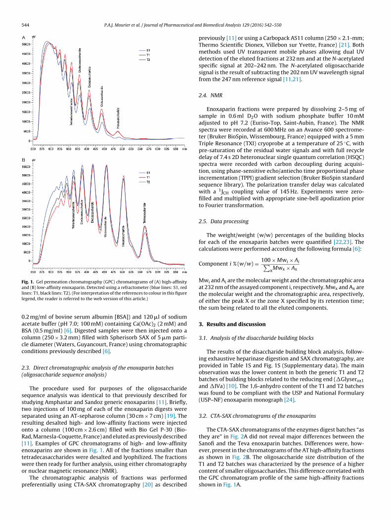
544 P.A.J. Mourier et al. / Journal of Pharmaceutical an
Fig. 1. Gel permeation chromatography (GPC) chromatograms of (A) high-affinityand (B) low-affinity enoxaparin. Detected using a refractometer (blue lines: S1, redll
0aBccc
2(
sstsroR[etwo
p
ines: T1, black lines: T2). (For interpretation of the references to colour in this figureegend, the reader is referred to the web version of this article.)
.2 mg/ml of bovine serum albumin [BSA]) and 120 �l of sodiumcetate buffer (pH 7.0; 100 mM) containing Ca(OAc)2 (2 mM) andSA (0.5 mg/ml) [6]. Digested samples were then injected onto aolumn (250 × 3.2 mm) filled with Spherisorb SAX of 5 �m parti-le diameter (Waters, Guyancourt, France) using chromatographiconditions previously described [6].
.3. Direct chromatographic analysis of the enoxaparin batchesoligosaccharide sequence analysis)
The procedure used for purposes of the oligosaccharideequence analysis was identical to that previously described fortudying Amphastar and Sandoz generic enoxaparins [11]. Briefly,wo injections of 100 mg of each of the enoxaparin digests wereeparated using an AT-sepharose column (30 cm × 7 cm) [19]. Theesulting desalted high- and low-affinity fractions were injectednto a column (100 cm × 2.6 cm) filled with Bio Gel P-30 (Bio-ad, Marnesla-Coquette, France) and eluted as previously described11]. Examples of GPC chromatograms of high- and low-affinitynoxaparins are shown in Fig. 1. All of the fractions smaller thanetradecasaccharides were desalted and lyophilized. The fractions
ere then ready for further analysis, using either chromatographyr nuclear magnetic resonance (NMR).The chromatographic analysis of fractions was performed
referentially using CTA-SAX chromatography [20] as described
d Biomedical Analysis 129 (2016) 542–550
previously [11] or using a Carbopack AS11 column (250 × 2.1-mm;Thermo Scientific Dionex, Villebon sur Yvette, France) [21]. Bothmethods used UV transparent mobile phases allowing dual UVdetection of the eluted fractions at 232 nm and at the N-acetylatedspecific signal at 202–242 nm. The N-acetylated oligosaccharidesignal is the result of subtracting the 202 nm UV wavelength signalfrom the 247 nm reference signal [11,21].
2.4. NMR
Enoxaparin fractions were prepared by dissolving 2–5 mg ofsample in 0.6 ml D2O with sodium phosphate buffer 10 mMadjusted to pH 7.2 (Euriso-Top, Saint-Aubin, France). The NMRspectra were recorded at 600 MHz on an Avance 600 spectrome-ter (Bruker BioSpin, Wissembourg, France) equipped with a 5 mmTriple Resonance (TXI) cryoprobe at a temperature of 25 ◦C, withpre-saturation of the residual water signals and with full recycledelay of 7.4 s 2D heteronuclear single quantum correlation (HSQC)spectra were recorded with carbon decoupling during acquisi-tion, using phase-sensitive echo/antiecho time proportional phaseincrementation (TPPI) gradient selection (Bruker BioSpin standardsequence library). The polarization transfer delay was calculatedwith a 1JCH coupling value of 145 Hz. Experiments were zero-filled and multiplied with appropriate sine-bell apodization priorto Fourier transformation.
2.5. Data processing
The weight/weight (w/w) percentages of the building blocksfor each of the enoxaparin batches were quantified [22,23]. Thecalculations were performed according the following formula [6]:
Component i % (w/w) = 100 × Mwi × Ai∑xMwx × Ax
Mwi and Ai are the molecular weight and the chromatographic areaat 232 nm of the assayed component i, respectively. Mwx and Ax arethe molecular weight and the chromatographic area, respectively,of either the peak X or the zone X specified by its retention time;the sum being related to all the eluted components.
3. Results and discussion
3.1. Analysis of the disaccharide building blocks
The results of the disaccharide building block analysis, follow-ing exhaustive heparinase digestion and SAX chromatography, areprovided in Table 1S and Fig. 1S (Supplementary data). The mainobservation was the lower content in both the generic T1 and T2batches of building blocks related to the reducing end (�Glyserox1and �IVa) [10]. The 1,6-anhydro content of the T1 and T2 batcheswas found to be compliant with the USP and National Formulary(USP–NF) enoxaparin monograph [24].
3.2. CTA-SAX chromatograms of the enoxaparins
The CTA-SAX chromatograms of the enzymes digest batches “asthey are” in Fig. 2A did not reveal major differences between theSanofi and the Teva enoxaparin batches. Differences were, how-ever, present in the chromatograms of the AT high-affinity fractionsas shown in Fig. 2B. The oligosaccharide size distribution of the
T1 and T2 batches was characterized by the presence of a highercontent of smaller oligosaccharides. This difference correlated withthe GPC chromatogram profile of the same high-affinity fractionsshown in Fig. 1A.
P.A.J. Mourier et al. / Journal of Pharmaceutical and Biomedical Analysis 129 (2016) 542–550 545
Fig. 2. Cetyltrimethylammonium–strong anion exchange (CTA-SAX) chro-matograms of the originator Sanofi enoxaparin S1 and the generic Teva T1 and T2enoxaparin batches (A) “as they are” and (B) their high affinity fractions (blacklc
fhuaac
htpeac
Fig. 3. Cetyltrimethylammonium–strong anion exchange (CTA-SAX) chro-matograms of the originator Sanofi enoxaparin S1 and the generic Teva T1and T2 enoxaparin batches (black line: UV 232 nm, red line: UV 202–242 nm).(A) High-affinity octasaccharides and (B) High-affinity decasaccharides o1:�Is-IIaid-IIsglu-IIIsid; o2: �IIa-IIsglu-Isid-Isid
1,6anhydro; o3: �IIa-IIsglu-Isid-Isid;
o4: �IIa-IIsglu-Isid-Isidepi; o5: �Is-IIaid-IIsglu-Isid
1,6anhydro; o6: �Is- IIaid-
IIsglu-Isid1,6anhydro−epi; o7: �Is-IIaid-IIsglu-Isid. d1: �IIa-IVsglu-Isid-Isid-Isid; d2:
�IIa-IIsglu-Isid-Isid-IIsglu; d3: �IIa-IIsglu-Isid-Isid-Isid; d4: �Is-IIaid-IIsglu-Isid-Isid;
d5: �Is-IIaid-IIsglu-Isid-Isid1,6anhydro; d6: �Is-Isid-IIaid-IIsglu-Isid. (For interpretation
of the references to colour in this figure legend, the reader is referred to the webversion of this article.)
ine: UV 232 nm, red line: UV 202–242 nm). (For interpretation of the references toolour in this figure legend, the reader is referred to the web version of this article.)
The next step was fractionation of the whole AT high-affinityraction on the basis of oligosaccharide size. Each size-definedigh-affinity fraction obtained after GPC purification was comparedsing CTA-SAX. Differences were already detected in the high-ffinity hexasaccharides (Supplementary data S.2.2.) and furthernalysis of the higher fraction sizes provided additional informationoncerning the nature of the discrepancies.
The CTA-SAX chromatograms of the main constituents of theigh-affinity octasaccharides and decasaccharides present in thehree enoxaparin batches are shown in Figs. 3A and B. Theresence of supplementary non-endogenous derivatives in the
noxaparins, such as 1,6-anhydro derivatives, mannose epimers,nd odd oligosaccharides, increased the complexity of the decasac-haride mixture and were not fully resolved by any method.Following the structural identification of the main high affinityoctasaccharides in Fig. 3A, two groups can be distinguished. TheAT high-affinity pentasaccharide was on the reducing side of thechain in o1, o5, o6, and o7 in the first group but on the non-reducingside in o2, o3, and o4 in the second group. The first group is themajor one present in enoxaparin as a result of the selectivity of thedepolymerization conditions. This observation can be summarized
as shown below: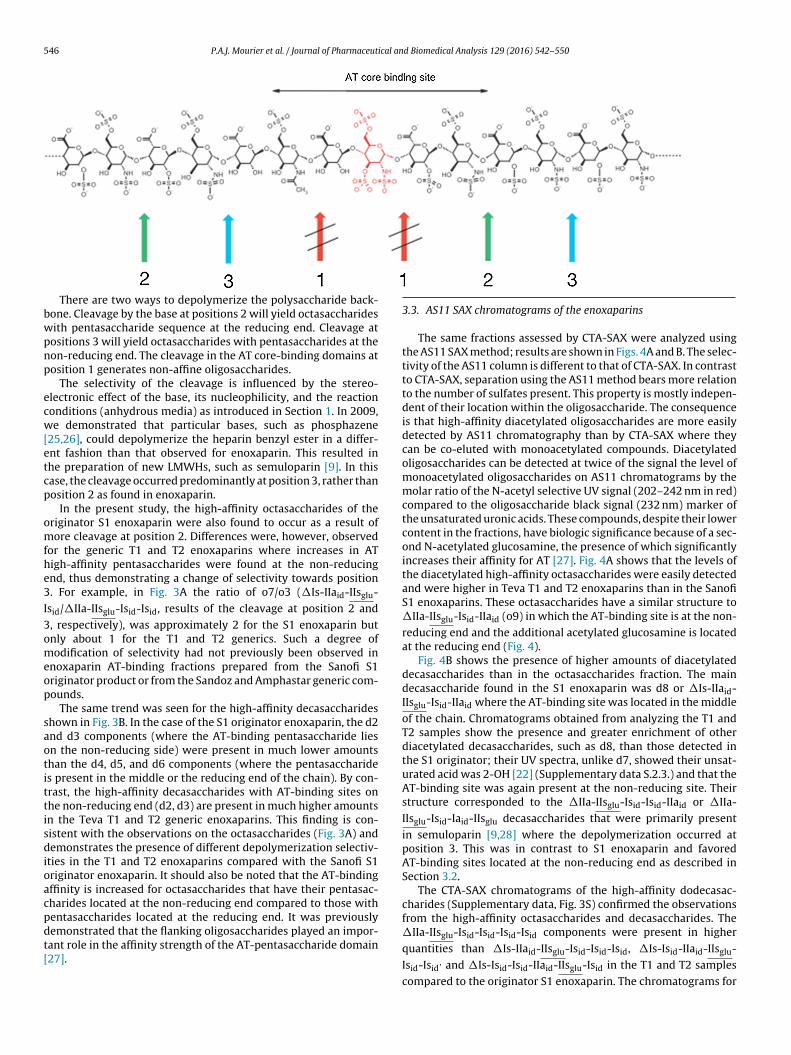
5 ical and Biomedical Analysis 129 (2016) 542–550
bwpnp
ecw[etcp
omfhe3
I3omeop
saotittisdioacpdt[
46 P.A.J. Mourier et al. / Journal of Pharmaceut
There are two ways to depolymerize the polysaccharide back-one. Cleavage by the base at positions 2 will yield octasaccharidesith pentasaccharide sequence at the reducing end. Cleavage atositions 3 will yield octasaccharides with pentasaccharides at theon-reducing end. The cleavage in the AT core-binding domains atosition 1 generates non-affine oligosaccharides.
The selectivity of the cleavage is influenced by the stereo-lectronic effect of the base, its nucleophilicity, and the reactiononditions (anhydrous media) as introduced in Section 1. In 2009,e demonstrated that particular bases, such as phosphazene
25,26], could depolymerize the heparin benzyl ester in a differ-nt fashion than that observed for enoxaparin. This resulted inhe preparation of new LMWHs, such as semuloparin [9]. In thisase, the cleavage occurred predominantly at position 3, rather thanosition 2 as found in enoxaparin.
In the present study, the high-affinity octasaccharides of theriginator S1 enoxaparin were also found to occur as a result ofore cleavage at position 2. Differences were, however, observed
or the generic T1 and T2 enoxaparins where increases in ATigh-affinity pentasaccharides were found at the non-reducingnd, thus demonstrating a change of selectivity towards position. For example, in Fig. 3A the ratio of o7/o3 (�Is-IIaid-IIsglu-
sid/�IIa-IIsglu-Isid-Isid, results of the cleavage at position 2 and, respectively), was approximately 2 for the S1 enoxaparin butnly about 1 for the T1 and T2 generics. Such a degree ofodification of selectivity had not previously been observed in
noxaparin AT-binding fractions prepared from the Sanofi S1riginator product or from the Sandoz and Amphastar generic com-ounds.
The same trend was seen for the high-affinity decasaccharideshown in Fig. 3B. In the case of the S1 originator enoxaparin, the d2nd d3 components (where the AT-binding pentasaccharide liesn the non-reducing side) were present in much lower amountshan the d4, d5, and d6 components (where the pentasaccharides present in the middle or the reducing end of the chain). By con-rast, the high-affinity decasaccharides with AT-binding sites onhe non-reducing end (d2, d3) are present in much higher amountsn the Teva T1 and T2 generic enoxaparins. This finding is con-istent with the observations on the octasaccharides (Fig. 3A) andemonstrates the presence of different depolymerization selectiv-
ties in the T1 and T2 enoxaparins compared with the Sanofi S1riginator enoxaparin. It should also be noted that the AT-bindingffinity is increased for octasaccharides that have their pentasac-harides located at the non-reducing end compared to those withentasaccharides located at the reducing end. It was previously
emonstrated that the flanking oligosaccharides played an impor-ant role in the affinity strength of the AT-pentasaccharide domain27].3.3. AS11 SAX chromatograms of the enoxaparins
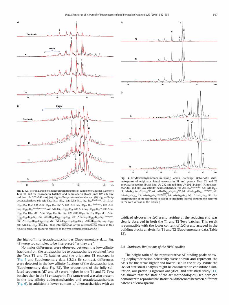
The same fractions assessed by CTA-SAX were analyzed usingthe AS11 SAX method; results are shown in Figs. 4A and B. The selec-tivity of the AS11 column is different to that of CTA-SAX. In contrastto CTA-SAX, separation using the AS11 method bears more relationto the number of sulfates present. This property is mostly indepen-dent of their location within the oligosaccharide. The consequenceis that high-affinity diacetylated oligosaccharides are more easilydetected by AS11 chromatography than by CTA-SAX where theycan be co-eluted with monoacetylated compounds. Diacetylatedoligosaccharides can be detected at twice of the signal the level ofmonoacetylated oligosaccharides on AS11 chromatograms by themolar ratio of the N-acetyl selective UV signal (202–242 nm in red)compared to the oligosaccharide black signal (232 nm) marker ofthe unsaturated uronic acids. These compounds, despite their lowercontent in the fractions, have biologic significance because of a sec-ond N-acetylated glucosamine, the presence of which significantlyincreases their affinity for AT [27]. Fig. 4A shows that the levels ofthe diacetylated high-affinity octasaccharides were easily detectedand were higher in Teva T1 and T2 enoxaparins than in the SanofiS1 enoxaparins. These octasaccharides have a similar structure to�IIa-IIsglu-Isid-IIaid (o9) in which the AT-binding site is at the non-reducing end and the additional acetylated glucosamine is locatedat the reducing end (Fig. 4).
Fig. 4B shows the presence of higher amounts of diacetylateddecasaccharides than in the octasaccharides fraction. The maindecasaccharide found in the S1 enoxaparin was d8 or �Is-IIaid-IIsglu-Isid-IIaid where the AT-binding site was located in the middleof the chain. Chromatograms obtained from analyzing the T1 andT2 samples show the presence and greater enrichment of otherdiacetylated decasaccharides, such as d8, than those detected inthe S1 originator; their UV spectra, unlike d7, showed their unsat-urated acid was 2-OH [22] (Supplementary data S.2.3.) and that theAT-binding site was again present at the non-reducing site. Theirstructure corresponded to the �IIa-IIsglu-Isid-Isid-IIaid or �IIa-IIsglu-Isid-Iaid-IIsglu decasaccharides that were primarily presentin semuloparin [9,28] where the depolymerization occurred atposition 3. This was in contrast to S1 enoxaparin and favoredAT-binding sites located at the non-reducing end as described inSection 3.2.
The CTA-SAX chromatograms of the high-affinity dodecasac-charides (Supplementary data, Fig. 3S) confirmed the observationsfrom the high-affinity octasaccharides and decasaccharides. The
�IIa-IIsglu-Isid-Isid-Isid-Isid components were present in higherquantities than �Is-IIaid-IIsglu-Isid-Isid-Isid, �Is-Isid-IIaid-IIsglu-
Isid-Isid, and �Is-Isid-Isid-IIaid-IIsglu-Isid in the T1 and T2 samples
compared to the originator S1 enoxaparin. The chromatograms for
P.A.J. Mourier et al. / Journal of Pharmaceutical and Biomedical Analysis 129 (2016) 542–550 547
Fig. 4. AS11 strong anion exchange chromatograms of Sanofi enoxaparin S1, genericTeva T1 and T2 enoxaparin batches and semuloparin (black line: UV 232 nm;red line: UV 202–242 nm). (A) High-affinity octasaccharides and (B) High-affinitydecasaccharides. o1: �Is-IIaid-IIsglu-IIIsid, o2: �IIa-IIsglu-Isid-Isid
1,6anhydro, o3: �IIa-
IIsglu-Isid-Isid; o4: �IIa-IIsglu-Isid-Isidepi, o5: �Is-IIaid-IIsglu-Isid
1,6anhydro, o6: �Is-
IIaid-IIsglu-Isid1,6anhydro−epi, o7: �Is-IIaid-IIsglu-Isid, o8: �Is-IIaid-IIsglu-Isid
ep, o9: �IIa-
IIsglu-Isid-IIaid, d1: �IIa-IVsglu-Isid-Isid-Isid, d2: �IIa-IIsglu-Isid-Isid-IIsglu, d3: �IIa-
IIsglu-Isid-Isid-Isid, d4: �Is-IIaid-IIsglu-Isid-Isid, d5: �Is-IIaid-IIsglu-Isid-Isid1,6anhydro,
d6: �Is-Isid-IIaid-IIsglu-Isid, d7: �IIa-IIsglu-Isid-Isid-IIaid + �IIa-IIsglu-Isid-Iaid-IIsglu,
d
fi
t4
ft(w(fbi(
Fig. 5. Cetyltrimethylammonium–strong anion exchange (CTA-SAX) chro-matograms of originator Sanofi enoxaparin S1 and generic Teva T1 and T2enoxaparin batches (black line: UV 232 nm, red line: UV 202–242 nm) (A) tetrasac-charides and (B) low-affinity hexasaccharides. t1: �Is-Isid
1,6anhydro, t2: �Is-Isglu,t3: �Is-Isid, t4: �Is-Isid
epi, o4: �IIa-IIsglu-Isid-Isid
epi, h1: �Is-Isid-IIsglu1,6anhydro, h2:
�Is-Isid-IIsglu, h3: �Is-Isid-Isid1,6anhydro, h4: �Is-Isid-Isid, h5: �Is-Isid-Isid
epi. (Forinterpretation of the references to colour in this figure legend, the reader is referred
8: �Is-IIaid-IIsglu-Isid-IIaid. (For interpretation of the references to colour in this
gure legend, the reader is referred to the web version of this article.)
he high-affinity tetradecasaccharides (Supplementary data, Fig.S) were too complex to be interpreted “as they are”.
No major differences were observed between the low-affinityractions from the tetrasaccharide to octasaccharide obtained fromhe Teva T1 and T2 batches and the originator S1 enoxaparinFig. 5 and Supplementary data S.2.2.). By contrast, differencesere detected in the low-affinity fractions of the decasaccharides
Supplementary data Fig. 7S). The proportions of the fully sul-
ated sequences (d7 and d8) were higher in the T1 and T2 Tevaatches than in the S1 enoxaparin. The same trend was also presentn the low-affinity dodecasaccharides and tetradecasaccharidesFig. 6). In addition, a lower content of oligosaccharides with an
to the web version of this article.)
oxidized glycoserine �Glyserox residue at the reducing end wasclearly observed in both the T1 and T2 Teva batches. This resultis compatible with the lower content of �Glyserox assayed in thebuilding blocks analysis for T1 and T2 (Supplementary data, Table1S).
3.4. Statistical limitations of the HPLC studies
The height ratio of the representative AT binding peaks show-ing deploymerization selectivity were shown and represent thebasis for the terms higher and lower used in the study. While thelack of statistical analysis might be considered to constitute a lim-itation, our previous rigorous analytical and statistical study [11]has shown that the state of the art methodologies used here can
demonstrate reproducible statistical differences between differentbatches of enoxaparins.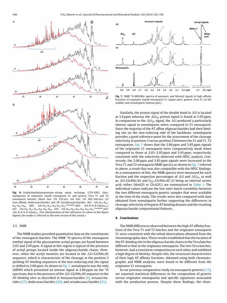
548 P.A.J. Mourier et al. / Journal of Pharmaceutical and Biomedical Analysis 129 (2016) 542–550
Fig. 6. Cetyltrimethylammonium–strong anion exchange (CTA-SAX) chro-matograms of originator Sanofi enoxaparin S1 and generic Teva T1 and T2enoxaparin batches (black line: UV 232 nm; red line: UV 202–242 nm) (A)Low-affinity dodecasaccharides and (B) tetradecasaccharides. dd1: �Is-Isid-Isid-Isid-Isid-Isid, dd2: �Is-Isid-Isid-Isid-Isid-Isid
1,6anhydro,dd3: �Is-X-X-X-Glyserox),td1: �Is-Is -Is -Is -Is -Is -Is , td2: �Is-Is -Is -Is -Is -Is -Is 1,6anhydro,td3:�l
3
om2oesyiLsAr
1
id id id id id id id id id id id id
Is-X-X-X-X-Glyserox. (For interpretation of the references to colour in this figureegend, the reader is referred to the web version of this article.)
.5. NMR
The NMR studies provided quantitative data on the constituentsf the enoxaparin batches. The NMR 1H spectra of the enoxaparinethyl signal of the glucosamine acetyl groups are found between
.03 and 2.05 ppm. A signal in this region is typical of the presencef acetyl groups located inside the oligosaccharide chains. How-ver, when the acetyl moieties are located in the �U-GlcNAc,6Sequence, which is characteristic of the cleavage at the position 3ielding AT-binding sequences at the non-reducing end, the signals shifted to 2.06 ppm. As shown in Fig. 7, semuloparin was the only
1
MWH which presented an intense signal at 2.06 ppm on the Hpectrum, due to the presence of the �U-GlcNAc,6S sequence in theT-binding sites as described in hexasaccharides [9], octasaccha-ides [27], dodecasaccharides [29], and octadecasaccharides [21].Fig. 7. NMR H 600 MHz spectra of anomeric and NAcetyl signals in high affinityfractions of originator Sanofi enoxaparin S1 (upper part), generic Teva T1 (at themiddle) and semuloparin (bottom part).
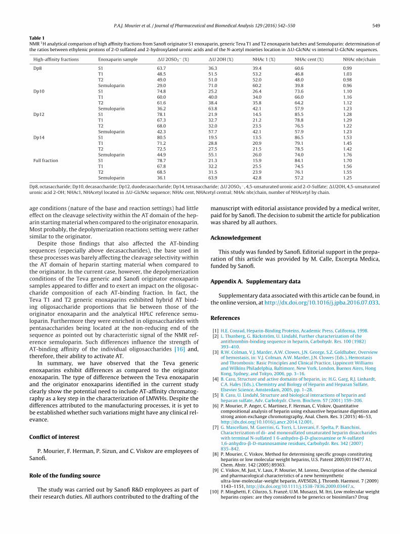
Similarly, the proton signal of the double bond in �U is locatedat 5.8 ppm whereas the �U2S proton signal is found at 5.95 ppm.In comparison to the �U2S signal, the �U produced a particularlyintense signal in semuloparin when compared to S1 enoxaparin.Since the majority of the AT affine oligosaccharides had their bind-ing site on the non-reducing side of the backbone, semuloparinprovides a good reference point for the assessment of the cleavageselectivity at position 3 versus position 2 between the S1 and T1, T2enoxaparins. Fig. 7 shows that the 2.06 ppm and 5.85 ppm signalsof the originator S1 enoxaparin were comparatively weak whencompared to those at 2.03–2.05 ppm and 5.95 ppm, respectively,consistent with the selectivity observed with HPLC analysis. Con-versely, the 2.06 ppm and 5.85 ppm signals were increased in theTeva T1 and T2 enoxaparin NMR spectra as shown in Fig. 7 referredto above, a result that was also compatible with the HPLC findings.As a consequence of this, the NMR spectra were measured for eachfraction and the respective percentages of �U and �U2S, as wellas �U-GlcNAc,6S and U2S-GlcNAc,6S (U being an internal uronicacid, either (IdoA2S or GlcA2S)) are summarized in Table 1. Theindividual values indicate the low inter-batch variability betweenthe two different enoxaparin generic samples that were availableat the time of the study. The results were also compared with dataobtained from semuloparin further supporting the differences incleavage selectivity of heparin AT binding domain and the resultingoligosaccharide compositional features.
4. Conclusions
The NMR differences observed between the high AT affinity frac-tions of the Teva T1 and T2 batches and the originator enoxaparinS1 were consistent with the initial observations obtained from thechromatographic data. These results established that the location ofthe AT-binding site in the oligosaccharide chains in the Teva batchesdiffered to that in the originator enoxaparin. The two Teva batches,however, had a consistent composition to each other and exhibiteda high degree of identity. Despite this, the structural characteristicsof their high AT affinity fractions, obtained using both chromato-graphic and NMR analyses, were found to be different from theoriginator S1 enoxaparin.
In our previous comparative study on enoxaparin generics [11],we reported statistical differences in the composition of genericversus originator enoxaparins and specific signatures associatedwith the production process. Despite these findings, the cleav-
P.A.J. Mourier et al. / Journal of Pharmaceutical and Biomedical Analysis 129 (2016) 542–550 549
Table 1NMR 1H analytical comparison of high affinity fractions from Sanofi originator S1 enoxaparin, generic Teva T1 and T2 enoxaparin batches and Semuloparin: determination ofthe ratios between ethylenic protons of 2-O sulfated and 2-hydroxylated uronic acids and of the N-acetyl moieties location in �U-GlcNAc vs internal U-GlcNAc sequences.
High-affinity fractions Enoxaparin sample �U 2OSO3− (%) �U 2OH (%) NHAc 1 (%) NHAc cent (%) NHAc nbr/chain
Dp8 S1 63.7 36.3 39.4 60.6 0.99T1 48.5 51.5 53.2 46.8 1.03T2 49.0 51.0 52.0 48.0 0.98Semuloparin 29.0 71.0 60.2 39.8 0.96
Dp10 S1 74.8 25.2 26.4 73.6 1.10T1 60.0 40.0 34.0 66.0 1.16T2 61.6 38.4 35.8 64.2 1.12Semuloparin 36.2 63.8 42.1 57.9 1.23
Dp12 S1 78.1 21.9 14.5 85.5 1.28T1 67.3 32.7 21.2 78.8 1.29T2 68.0 32.0 23.5 76.5 1.22Semuloparin 42.3 57.7 42.1 57.9 1.23
Dp14 S1 80.5 19.5 13.5 86.5 1.53T1 71.2 28.8 20.9 79.1 1.45T2 72.5 27.5 21.5 78.5 1.42Semuloparin 44.9 55.1 26.0 74.0 1.76
Full fraction S1 78.7 21.3 15.9 84.1 1.70T1 67.8 32.2 25.5 74.5 1.56T2 68.5 31.5 23.9 76.1 1.55Semuloparin 36.1 63.9 42.8 57.2 1.25
D ccharu HAcet
aeaMs
stttcscTiolpseAt
eeacrdbe
C
S
R
t
p8, octasaccharide; Dp10, decasaccharide; Dp12, duodecasaccharide; Dp14, tetrasaronic acid 2-OH; NHAc1, NHAcetyl located in �U-GlcNAc sequence; NHAc cent, N
ge conditions (nature of the base and reaction settings) had littleffect on the cleavage selectivity within the AT domain of the hep-rin starting material when compared to the originator enoxaparin.ost probably, the depolymerization reactions setting were rather
imilar to the originator.Despite those findings that also affected the AT-binding
equences (especially above decasaccharides), the base used inhese processes was barely affecting the cleavage selectivity withinhe AT domain of heparin starting material when compared tohe originator. In the current case, however, the depolymerizationonditions of the Teva generic and Sanofi originator enoxaparinamples appeared to differ and to exert an impact on the oligosac-haride composition of each AT-binding fraction. In fact, theeva T1 and T2 generic enoxaparins exhibited hybrid AT bind-ng oligosaccharide proportions that lie between those of theriginator enoxaparin and the analytical HPLC reference semu-oparin. Furthermore they were enriched in oligosaccharides withentasaccharides being located at the non-reducing end of theequence as pointed out by characteristic signal of the NMR ref-rence semuloparin. Such differences influence the strength ofT-binding affinity of the individual oligosaccharides [16] and,
herefore, their ability to activate AT.In summary, we have observed that the Teva generic
noxaparins exhibit differences as compared to the originatornoxaparin. The type of difference between the Teva enoxaparinnd the originator enoxaparins identified in the current studylearly show the potential need to include AT-affinity chromatog-aphy as a key step in the characterization of LMWHs. Despite theifferences attributed to the manufacturing processes, it is yet toe established whether such variations might have any clinical rel-vance.
onflict of interest
P. Mourier, F. Herman, P. Sizun, and C. Viskov are employees ofanofi.
ole of the funding source
The study was carried out by Sanofi R&D employees as part ofheir research duties. All authors contributed to the drafting of the
[
ide; �U 2OSO3− , 4,5-unsaturated uronic acid 2-O-Sulfate; �U2OH, 4,5-unsaturated
yl central; NHAc nbr/chain, number of NHAcetyl by chain.
manuscript with editorial assistance provided by a medical writer,paid for by Sanofi. The decision to submit the article for publicationwas shared by all authors.
Acknowledgement
This study was funded by Sanofi. Editorial support in the prepa-ration of this article was provided by M. Calle, Excerpta Medica,funded by Sanofi.
Appendix A. Supplementary data
Supplementary data associated with this article can be found, inthe online version, at http://dx.doi.org/10.1016/j.jpba.2016.07.033.
References
[1] H.E. Conrad, Heparin-Binding Proteins, Academic Press, California, 1998.[2] L. Thunberg, G. Bäckström, U. Lindahl, Further characterization of the
antithrombin-binding sequence in heparin, Carbohydr. Res. 100 (1982)393–410.
[3] R.W. Colman, V.J. Marder, A.W. Clowes, J.N. George, S.Z. Goldhaber, Overviewof hemostasis, in: V.J. Colman, A.W. Marder, J.N. Clowes (Eds.), Hemostasisand Thrombosis: Basic Principles and Clinical Practice, Lippincott Williamsand Wilkins Philadelphia, Baltimore, New York, London, Buenos Aires, HongKong, Sydney, and Tokyo, 2006, pp. 3–16.
[4] B. Casu, Structure and active domains of heparin, in: H.G. Garg, R.J. Linhardt,C.A. Hales (Eds.), Chemistry and Biology of Heparin and Heparan Sulfate,Elsevier Science, Amsterdam, 2005, pp. 1–28.
[5] B. Casu, U. Lindahl, Structure and biological interactions of heparin andheparan sulfate, Adv. Carbohydr. Chem. Biochem. 57 (2001) 159–206.
[6] P. Mourier, P. Anger, C. Martinez, F. Herman, C. Viskov, Quantitativecompositional analysis of heparin using exhaustive heparinase digestion andstrong anion exchange chromotography, Anal. Chem. Res. 3 (2015) 46–53,http://dx.doi.org/10.1016/j.ancr.2014.12.001.
[7] G. Mascellani, M. Guerrini, G. Torri, L. Liverani, F. Spelta, P. Bianchini,Characterization of di- and monosulfated unsaturated heparin disaccharideswith terminal N-sulfated 1 6-anhydro-�-D-glucosamine or N-sulfated1,6-anhydro-�-D-mannosamine residues, Carbohydr. Res. 342 (2007)835–842.
[8] P. Mourier, C. Viskov, Method for determining specific groups constitutingheparins or low molecular weight heparins, U.S. Patent 2005/0119477 A1,Chem. Abstr. 142 (2005) 89363.
[9] C. Viskov, M. Just, V. Laux, P. Mourier, M. Lorenz, Description of the chemical
and pharmacological characteristics of a new hemisyntheticultra-low-molecular-weight heparin, AVE5026, J. Thromb. Haemost. 7 (2009)1143–1151, http://dx.doi.org/10.1111/j.1538-7836.2009.03447.x.10] P. Minghetti, F. Cilurzo, S. Franzé, U.M. Musazzi, M. Itri, Low molecular weightheparins copies: are they considered to be generics or biosimilars? Drug

5 ical an
[
[
[
[
[
[
[
[
[
[
[
[
[
[
[
[
[
[
50 P.A.J. Mourier et al. / Journal of Pharmaceut
Discov. Today 18 (2013) 305–311, http://dx.doi.org/10.1016/j.drudis.2012.11.002.
11] P.A. Mourier, C. Agut, H. Souaifi-Amara, F. Herman, C. Viskov, Analytical andstatistical comparability of generic enoxaparins from the US market with theoriginator product, J. Pharm. Biomed. Anal. 115 (2015) 431–442, http://dx.doi.org/10.1016/j.jpba.2015.07.038.
12] M. Guerrini, T.R. Rudd, L. Mauri, E. Macchi, J. Fareed, E.A. Yates, A. Naggi, G.Torri, Differentiation of generic enoxaparins marketed in the United States byemploying NMR and multivariate analysis, Anal. Chem. 87 (2015) 8275–8283,http://dx.doi.org/10.1021/acs.analchem.5b01366.
13] Z. Wang, D. Li, X. Sun, X. Bai, L. Jin, L. Chi, Liquid chromatography-diode arraydetection-mass spectrometry for compositional analysis of low molecularweight heparins, Anal. Biochem. 451 (2014) 35–41, http://dx.doi.org/10.1016/j.ab.2014.02.005.
14] Q. Zhang, X. Chen, Z. Zhu, X. Zhan, Y. Wu, L. Song, J. Kang, Structural analysis oflow molecular weight heparin by ultraperformance size exclusionchromatography/time of flight mass spectrometry and capillary zoneelectrophoresis, Anal. Chem. 85 (2013) 1819–1827, http://dx.doi.org/10.1021/ac303185w.
15] L. Li, F. Zhang, J. Zaia, R.J. Linhardt, Top-down approach for the directcharacterization of low molecular weight heparins using LC-FT–MS, Anal.Chem. 84 (2012) 8822–8829, http://dx.doi.org/10.1021/ac302232c.
16] G. Li, J. Steppich, Z. Wang, Y. Sun, C. Xue, R.J. Linhardt, L. Li, Bottom-up lowmolecular weight heparin analysis using liquid chromatography–fouriertransform mass spectrometry for extensive characterization, Anal. Chem. 86(2014) 6626–6632, http://dx.doi.org/10.1021/ac501301v.
17] S.N. Oliveira, G.R. Santos, B.F. Glauser, N.V. Capillé, I.N. Queiroz, M.S. Pereira,V.H. Pomin, P.A. Mourão, Structural and functional analyses of biosimilarenoxaparins available in Brazil, Thromb. Haemost. 113 (2015) 53–65, http://dx.doi.org/10.1160/TH14-05-0411.
18] X. Xu, D. Li, L. Chi, X. Du, X. Bai, L. Chi, Fragment profiling of low molecularweight heparins using reversed phase ion pair liquidchromatography–electrospray mass spectrometry, Carbohydr. Res. 407
(2015) 26–33, http://dx.doi.org/10.1016/j.carres.2015.01.016.19] M. Höök, I. Björk, J. Hopwood, U. Lindahl, Anticoagulant activity of heparin:separation of high-activity and low-activity heparin species by affinitychromatography on immobilized antithrombin, FEBS Lett. 66 (1976) 90–93,http://dx.doi.org/10.1016/0014-5793(76)80592-3.
[
d Biomedical Analysis 129 (2016) 542–550
20] P.A. Mourier, C. Viskov, Chromatographic analysis and sequencing approachof heparin oligosaccharides using cetyltrimethylammonium dynamicallycoated stationary phases, Anal. Biochem. 332 (2004) 299–313.
21] P.A. Mourier, O.Y. Guichard, F. Herman, C. Viskov, Isolation of a pureoctadecasaccharide with antithrombin activity from anultra-low-molecular-weight heparin, Anal. Biochem. 453 (2014) 7–15, http://dx.doi.org/10.1016/j.ab.2014.02.013.
22] A. Pervin, C. Gallo, K.A. Jandik, X.J. Han, R.J. Linhardt, Preparation andstructural characterization of large heparin-derived oligosaccharides,Glycobiology 5 (1995) 83–95.
23] K.G. Rice, R.J. Linhardt, Study of structurally defined oligosaccharidesubstrates of heparin and heparin monosulfate lyases, Carbohydr. Res. 190(1989) 219–233.
24] U.S. Pharmacopeia–National Formulary (USP–NF), Test for1,6-anhydroderivative for enoxaparin sodium, U.S. PharmacopeialConvention, 2009. http://www.usp.org/sites/default/files/usp pdf/EN/USPNF/genChapter207.pdf (accessed 20.03.16).
25] R. Schwesinger, J. Willaredt, H. Schlemper, M. Keller, D. Schmitt, H. Fritz,Novel, very strong, uncharged auxiliary bases; design and synthesis ofmonomeric and polymer-bound Triaminoiminophosphorane bases of broadlyvaried steric demand, Chem. Ber. 127 (1994) 2435–2454, http://dx.doi.org/10.1002/cber.19941271215.
26] R. Schwesinger, H. Schlemper, C. Hasenfratz, Extremely strong, unchargedauxiliary bases; monomeric and polymer-supported polyaminophosphazenes(P2–P5), Liebigs Ann. (1996) 1055–1081, http://dx.doi.org/10.1002/jlac.199619960705.
27] M. Guerrini, S. Guglieri, B. Casu, G. Torri, P. Mourier, C. Boudier, C. Viskov,Antithrombin-binding octasaccharides and role of extensions of the activepentasaccharide sequence in the specificity and strength of interaction.Evidence for very high affinity induced by an unusual glucuronic acid residue,J. Biol. Chem. 283 (2008) 26662–26675, http://dx.doi.org/10.1074/jbc.M801102200.
28] V. Biberovic, L. Grondard, P. Mourier, C. Viskov, Mixture of sulfatedoligosaccharides, U.S. Patent (2011) 8003623 B2.
29] C. Viskov, S. Elli, E. Urso, D. Gaudesi, P. Mourier, F. Herman, C. Boudier, B. Casu,G. Torri, M. Guerrini, Heparin dodecasaccharide containing twoantithrombin-binding pentasaccharides: structural features and biologicalproperties, J. Biol. Chem. 288 (2013) 25895–25907, http://dx.doi.org/10.1074/jbc.M113.485268.