jshs research paper
TRANSCRIPT

1
HIV-1 Nef Gene’s Effect on the Production of Beta Amyloid
Parth Patel
Life Sciences
Woodstock High School
2010 Towne Lake Hills South Dr.
Woodstock, GA 30189

2
Synopsis
HIV-1 Nef Gene’s Effect on the Production of Amyloid
HIV/AIDS is a pandemic that kills millions of people. HIV-1 may induce Alzheimer-like
dementia. Alzheimer’s destroys the lives of millions people through the slow decay of brain cells
causing memory loss. In Alzheimer’s the protein beta amyloid is responsible for producing
plaques, which lead to inflammation and cell death. Studies have shown beta amyloid peptides
secreted from cells in association with exosomes. Exosomal proteins were also found to
accumulate in plaques, suggesting a role for exosomes in the pathogenesis of Alzheimer’s. HIV
Nef protein has shown to be secreted in exosomes and enhances the production of exosomes. The
hypothesis is “If beta amyloid is expressed in association with Nef exosomes, and I transfect
NS20Y cells with the nef gene, collect that supernatant, and place it on un-transfected NS20Y
cells, then the production of beta amyloid by the treated cells should be enhanced”. Nef-GFP was
transfected into NS20Y cells to produce Nef exosomes. Medium, containing exosomes, was
collected and placed onto untreated NS20Y cells. ELISA and Western blot measured beta
amyloid production from treated cells. Results show that Nef exosomes (applied exogenously)
enhance the secretion of beta amyloid peptides.
3
Table of Contents
Title Page Page 1
Synopsis Page 2
Table of Contents Page 3
Introduction Page 4
Purpose, Procedure, and Materials Page 6
Results Page 8
Conclusion Page 9
Literature Cited Page 12
Appendices Page 13
Acknowledgments Page 17
4
Introduction
HIV/AIDS infection is a worldwide epidemic and according to According to UNAIDS
there were 1.7 million AIDS deaths in 2011, which is actually down from 2.3 million in 2005.
This shows that treatments for HIV/AIDS are improving and more lives are being saved.
However, one complication of HIV-1 infection known as HIV Associated Dementia (HAD),
which is a form of dementia similar to that found in Alzheimer’s patients, has been increasing in
incidence even with modern drug treatments according to an HIV associated neurological
incidence study.
The interesting thing about HAD which was sought to exploit through the research was
its similarities to Alzheimer’s disease. Alzheimer’s is another worldwide epidemic that is
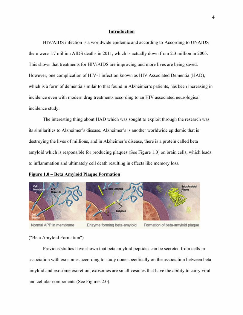
destroying the lives of millions, and in Alzheimer’s disease, there is a protein called beta
amyloid which is responsible for producing plaques (See Figure 1.0) on brain cells, which leads
to inflammation and ultimately cell death resulting in effects like memory loss.
Figure 1.0 – Beta Amyloid Plaque Formation
("Beta Amyloid Formation")
Previous studies have shown that beta amyloid peptides can be secreted from cells in
association with exosomes according to study done specifically on the association between beta
amyloid and exosome excretion; exosomes are small vesicles that have the ability to carry viral
and cellular components (See Figures 2.0).

5
Figure 2.0 – Exosome Basics
(Théry)
Subsequently, exosomal proteins were also found to accumulate in the plaques of
Alzheimer’s patient brains, which could be suggesting a role for exosomes in the pathogenesis of
Alzheimer’s disease. Interestingly, HIV Nef, a product of the nef gene (one of six accessory
genes), has shown to be secreted in exosomes and enhances the production of exosomes, so if
more exosomes are associated with the enhanced produciton of beta amyloid, then in turn the nef
gene could be possibly enhancing the secretion of beta amyloid.
6
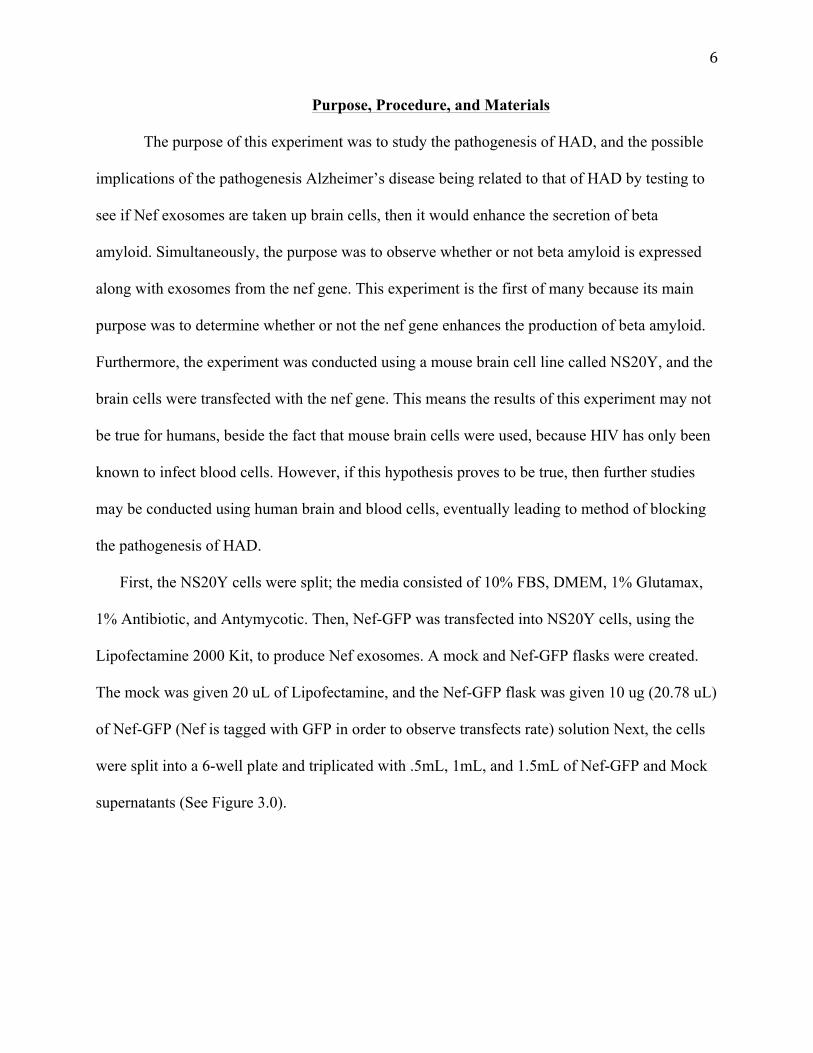
Purpose, Procedure, and Materials
The purpose of this experiment was to study the pathogenesis of HAD, and the possible
implications of the pathogenesis Alzheimer’s disease being related to that of HAD by testing to
see if Nef exosomes are taken up brain cells, then it would enhance the secretion of beta
amyloid. Simultaneously, the purpose was to observe whether or not beta amyloid is expressed
along with exosomes from the nef gene. This experiment is the first of many because its main
purpose was to determine whether or not the nef gene enhances the production of beta amyloid.
Furthermore, the experiment was conducted using a mouse brain cell line called NS20Y, and the
brain cells were transfected with the nef gene. This means the results of this experiment may not
be true for humans, beside the fact that mouse brain cells were used, because HIV has only been
known to infect blood cells. However, if this hypothesis proves to be true, then further studies
may be conducted using human brain and blood cells, eventually leading to method of blocking
the pathogenesis of HAD.
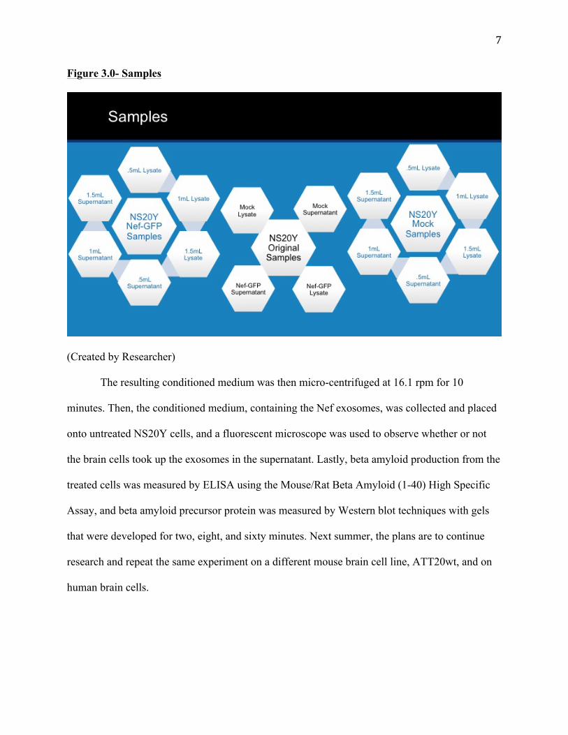
First, the NS20Y cells were split; the media consisted of 10% FBS, DMEM, 1% Glutamax,
1% Antibiotic, and Antymycotic. Then, Nef-GFP was transfected into NS20Y cells, using the
Lipofectamine 2000 Kit, to produce Nef exosomes. A mock and Nef-GFP flasks were created.
The mock was given 20 uL of Lipofectamine, and the Nef-GFP flask was given 10 ug (20.78 uL)
of Nef-GFP (Nef is tagged with GFP in order to observe transfects rate) solution Next, the cells
were split into a 6-well plate and triplicated with .5mL, 1mL, and 1.5mL of Nef-GFP and Mock
supernatants (See Figure 3.0).
7
Figure 3.0- Samples
(Created by Researcher)
The resulting conditioned medium was then micro-centrifuged at 16.1 rpm for 10
minutes. Then, the conditioned medium, containing the Nef exosomes, was collected and placed
onto untreated NS20Y cells, and a fluorescent microscope was used to observe whether or not
the brain cells took up the exosomes in the supernatant. Lastly, beta amyloid production from the
treated cells was measured by ELISA using the Mouse/Rat Beta Amyloid (1-40) High Specific
Assay, and beta amyloid precursor protein was measured by Western blot techniques with gels
that were developed for two, eight, and sixty minutes. Next summer, the plans are to continue
research and repeat the same experiment on a different mouse brain cell line, ATT20wt, and on
human brain cells.
8
Results
The .5mL NS20Y control (mock) treated cells showed no green fluorescent glow, but the
.5mL Nef-GFP treated cells did express a green fluorescent glow as a result of the nef gene being
tagged with GFP, which indicated that an uptake of vesicles (exosomes) occurred. Additionally,
the 1mL NS20Y control treated cells showed no green fluorescent glow while the 1ml Nef-GFP
treated cells showed a greater uptake of exosomes occurred. However, the 1.5mL NS20Y Nef-
GFP treated cells showed a decrease in the uptake of exosomes while the NS20Y control treated
cells showed no green fluorescent glow, which could be suggesting that there is a limit to how
many exosomes can be taken up.
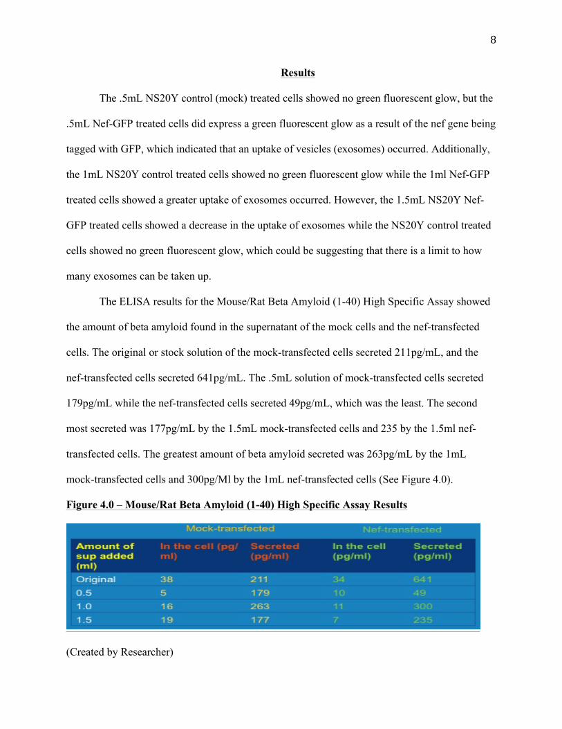
The ELISA results for the Mouse/Rat Beta Amyloid (1-40) High Specific Assay showed
the amount of beta amyloid found in the supernatant of the mock cells and the nef-transfected
cells. The original or stock solution of the mock-transfected cells secreted 211pg/mL, and the
nef-transfected cells secreted 641pg/mL. The .5mL solution of mock-transfected cells secreted
179pg/mL while the nef-transfected cells secreted 49pg/mL, which was the least. The second
most secreted was 177pg/mL by the 1.5mL mock-transfected cells and 235 by the 1.5ml nef-
transfected cells. The greatest amount of beta amyloid secreted was 263pg/mL by the 1mL
mock-transfected cells and 300pg/Ml by the 1mL nef-transfected cells (See Figure 4.0).
Figure 4.0 – Mouse/Rat Beta Amyloid (1-40) High Specific Assay Results
(Created by Researcher)

9
The sixty minute developed western blot showed the clearest results and was used to
observe the amount of beta amyloid precursor protein found within the lysate mock and nef-
transfected cells. The western blot results showed that the 1mL nef-transfect cells had the
greatest trace or amount of beta amyloid precursor protein; however, other specific results are
not presentable because the data is too difficult to examine (See Figure 5.0).
Figure 5.0 – Western Blot Results
(Created by Researcher)
Conclusion
The experiment has supported the hypothesis that the uptake of Nef exosomes by brain
cells would enhance the secretion of beta amyloid. Nef-GFP vesicles produced from transfected
NS20Y cells appear to be able to change the amount of beta amyloid secreted when applied to
un-transfected cells, and therefore, it can be concluded that Nef may play a role in HIV
Associated Dementia by modulating the secretion of beta amyloid. However, in the ELISA and
western blot results there may have been a mix up with the .5mL supernatant solution for the

10
mock-transfacted and nef-transfected cells. However, this does not the affect the conclusion that
the uptake of exosomes by brain cells enhances the production of beta amyloid. As a result,
further conclusions cannot be drawn until more trials are conducted and experimentation is
conducted in consideration of the human body’s immune system response.
In the continuation of this project, there are various paths that may be taken. This
summer, the plan is to conduct this same experiment on human brain cells and then with human
blood cells because HIV cannot directly infect brain cells because of the blood barrier, but it is
possible that HIV hijacks a pathway to the brain and sends exosomes to infect the brain cells.
This seems logical and plausible because the vesicles would be able to bypass the brain’s blood
barrier. As a result, the Nef exosomes would be mimicking how HIV attacks T-cells (See Figure
6.0).
Figure 6.0 – HIV T-cell Infection
(Created by Dr. Andrea Raymond)

11
However, it is extremely difficult to obtain live human brain cells, but once this obstacle
is overcome, blocking the Nef enhancement of beta amyloid production could represent a new
treatment for AIDS dementia.

12
Literature Cited
United Nations. UNAIDS. 2012 UNAIDS Report on the Global AIDS Epidemic. N.p.: Joint United Nations Programme on HIV/AIDS (UNAIDS), 2012. Print. Sacktor, N. HIV-associated neurologic disease incidence changes: Multicenter AIDS Cohort Study, 1990–1998. Comp. R. H. Lyles et al. N.p.: n.p., 2001. Print. Planck, Max. Alzheimer's disease beta-amyloid peptides are released in association with exosomes. Comp. L. Rajendran et al. N.p.: n.p., 2006. Print. "Beta Amyloid Formation." Elements4Health. Elements4Health, 2014. Web. 17 June 2013. <http://www.elements4health.com/images/stories/ amyloid-plaque-formation.jpg>. Théry, Clotilde. "Exosome Basics." TheScientist. The Scientist, 1 July 2011. Web. 12 June 2013. <http://www.the-scientist.com/?articles.view/ articleNo/30791/title/Exosome-Basics/>.
13
Appendix
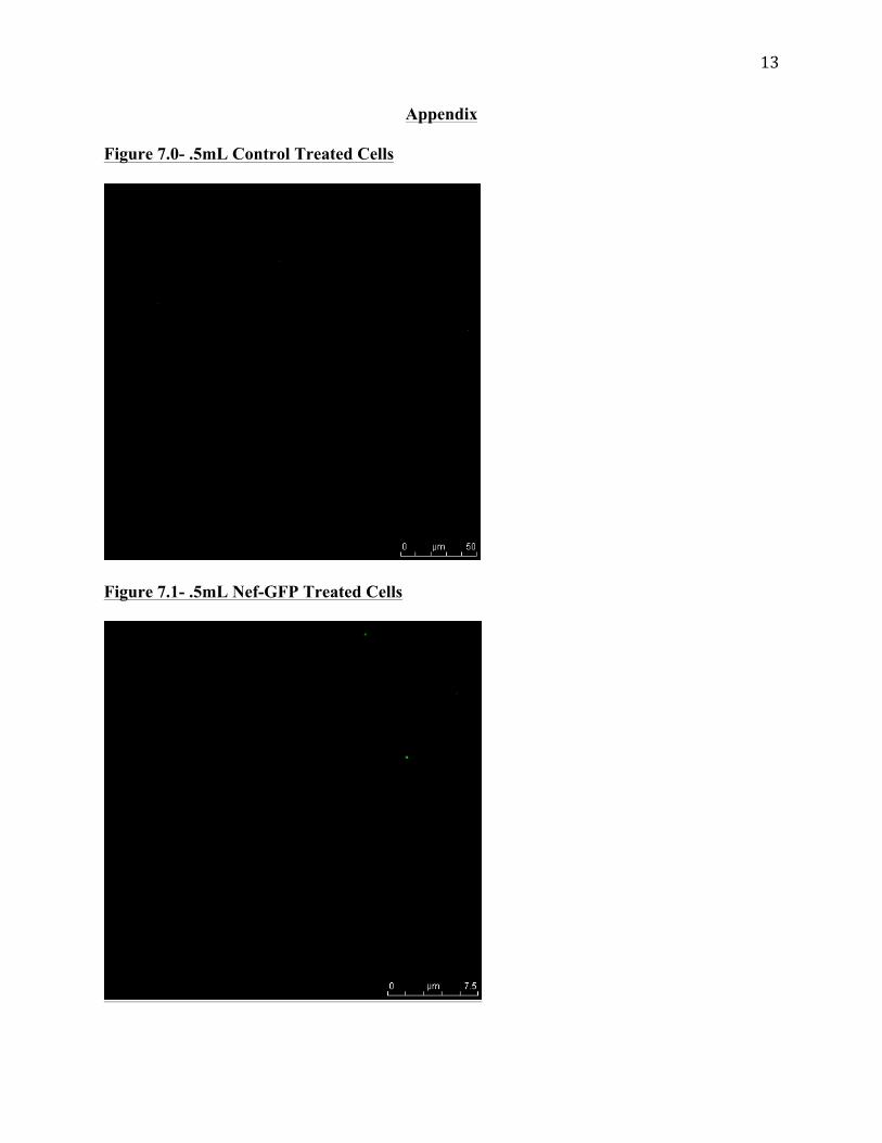
Figure 7.0- .5mL Control Treated Cells
Figure 7.1- .5mL Nef-GFP Treated Cells

14
Figure 8.0 – 1mL Control Treated Cells
Figure 8.1 – 1mL Nef-GFP Treated Cells

15
Figure 9.0 – 1.5mL Control Treated Cells
Figure 9.1 – 1.5mL Nef-GFP Treated Cells

16
Figures 7.1, 8.1, and 9.1 are not the pictures of the NS20Y cells from this experiment
because the pictures from this experiment were lost. However, these pictures are accurate
measurements in the ratio of the amount NS20Y cells that took up the exosomes and displayed
green fluorescent glow.

17
Acknowledgments
This experiment and research was conducted at Morehouse School of Medicine in
Biosafety Hazard Level 2 laboratory for six weeks throughout June and July of 2013.
Dr. Michael D. Powell as the primary investigator expertly taught the biological part of
the experiment and presented various types of literature consisting of background information
regarding the research. This tremendously helped in grasping the concepts firmly and quickly.
Dr. Powell also provided assistance in formulating the hypothesis and drawing conclusions,
making sure none of the conclusions were invalid or extrapolation. Dr. Powell was a great
mentor, teacher, and friend throughout the duration of the research. He is also responsible for
retrieving financial sponsorship and providing the brain cells, Lipofectamine Kit, media, and
Mouse/Rat Beta Amyloid (1-40) High Specific Assay Kit.
Mrs. Mahfuz Khan, the research assistant, assisted in the experimental design,
demonstrated how special equipment, such as the NanoDrop and fluorescent microscope, were to
be used, and taught various lab techniques that were vital to gathering the most accurate data.
She also demonstrated how to conduct ELISA and western blot tests.
Lastly, the Morehouse School of Medicine was generous enough to allow access to its
equipment and providing this research experience through the Vivien Thomas Summer Research
Program.