kabat rehabilitation for facial nerve paralysis ...during the kabat rehabilitation session, perform...
TRANSCRIPT

International Journal of Academic Scientific ResearchISSN: 2272-6446 Volume 6, Issue 1 (February - March 2018), PP 38-46www.ijasrjournal.org
www.ijasrjournal.org 38 | Page
KABAT REHABILITATION FOR FACIAL NERVEPARALYSIS: PERSPECTIVE ON NEUROKINETIC
RECOVERY AND REVIEW OF CLINICALEVALUATION TOOLS
Andrea Giacalone 1*, Teresa Sciarrillo1,Gennaro Rocco2, Enzo Ruberti3
1Department of Medicine, Faculty of Physiotherapy, Catholic University “Our Lady of GoodCounsel”, Albania2Department of Medicine, President of Physiotherapy Degree Course, Catholic University “Our Ladyof Good Counsel”, Albania3PhD s. “ Sapienza” University, Italy
Corresponding author*: Dr. Andrea Giacalone. Address: Catholic University “Our lady of GoodCounsel” Rr. DritanHoxha, Tirane, Albania. Email: [email protected]
Abstract: Background: Facial nerve paralysis (cranial nerve VII) is one of the most commonneurological disorders affecting the cranial nerves. Symptoms of this disorder are not life threatening butcan cause worry and discomfort. Peripheral paralysis of the facial nerve is a disabling pathology; itaffects the functional, psychological, social and occupational aspects of life. The Kabatre habilitationmethod for facial paralysis can evoke or restore the neuromuscular circuit through stimulation of theproprioceptors, restoring the normal functionality of nerve endings in the muscles. Objectives: This studyintends to demonstrate how patients can benefit, both aesthetically and functionally, from proper clinicalevaluation followed by a rehabilitation program based on the Kabat method. Methods :The study isqualitative and has an experimental and multicenter design. Data was collected through a clinical recordand using a specific questionnaire for this type of pathology for monitoring the satisfaction andimprovements of the patient; Functionality evaluation of the facial nerve was based on the SFG Sand H-B scales. Results :The study has shown that patients, who were treated with steroid-antiviral therapy andPNF rehabilitation method, recovered motor abilities of the paralyzed hemi facial. In addition, patientsimproved thermal, pain and position sensitivity. Conclusions: It is possible to conclude that a correctand timely diagnosis is very important, as well as using valid evaluation and data collection tools,properly managing the improvement through standardized clinical evaluation scales and a propermedical and physiotherapy treatment plan. Limitations: The limits encountered during thestudy were: limited number of samples; the short period of observation, not enough to include severeprognosis and full improvement of expressive gestures.
Keywords: facial nerve, facial palsy, neuromuscular re-education, proprioceptive facilitation,rehabilitation.
Abbreviations: PNF= Proprioceptive Neuromuscular Facilitation; CN = cranial nerve; H-B = HouseBrackmann; SFGS = Sunnybrook Facial Grading System
INTRODUCTION
With 23-35 out of 100.000 cases, the peripheral paralysis of the seventh CN is the mostfrequent paralysis of the peripheral nervous system.[1]
Occurring in response to various etiologies (congenital, iatrogenic, tumor, traumatic,infectious, or idiopathic), this highly debilitating [2] pathology can affect the seventh CN at any level
International Journal of Academic Scientific ResearchISSN: 2272-6446 Volume 6, Issue 1 (February - March 2018), PP 38-46
www.ijasrjournal.org 39 | Page
along its course, starting from the nucleus up to the neuromuscular junction. The motor ability of theface is altered and can no longer be controlled by the patient; the appearance of the face changes, bothat rest and, most of all, during movement.
In several cases, there is a midline deviation on the face caused by the prevailing muscularityof the healthy side, also at rest.[3]
Damage is mainly caused by some motor fibers of the facial nerve, which lose their ability tofunction and for this reason, physiotherapy for facial paralysis intends to reinforce the undamagedneuromuscular part.
Facial nerve paralysis is a dysfunctional problem affecting, and in this case altering a delicatepart of the body that provides personality and identity to the subject.[4]The patients require specialconsideration because their communication is affected in social circumstances;[5] as speech,mastication and the ability to express feelings and emotions depend on facial muscle movement.
Either way, functional, psychological, social and occupational aspects of life are seriouslyaffected. [6,7]
In recent years, clinical research on peripheral facial paralysis has gained larger interest;knowledge is expanding in regards to the implementation of manual therapy,[8,9]exercises,acupuncture,[10]laser[11] and electrical stimulation.[12]
This study intends to evaluate the effects of the Kabat method as a form of rehabilitation forrestoring the seventh cranial nerve,[13]associated with corticosteroid and antiviral pharmacologicaltreatment, and the effectiveness of clinical evaluation tools for improving data collection andmonitoring both progress made and results obtained after the treatment. The study was conducted in thecity of Tirana, in Albania, a State in which rehabilitation started in the 90's and is still growing. Only afew specialists who studied abroad recognize and use physiotherapeutic methods within a rehabilitationfacility. Rehabilitators, particularly facing cases of facial paralysis, do not have a standard fororganizing and collecting data from the patient, which can be easily transmitted and help monitor theprogress made and the results obtained following the treatment.
MATERIALS AND METHODS
2.1 SAMPLEThe study is qualitative and has an experimental and multicenter design.
After specialized medical examination, patients affected by peripheral facial paralysis fromvarying etiology were treated with corticosteroid and antiviral medicines and followed a physiotherapyprogram based on the PNF method.
Prior evaluation, each subject was informed about the study and had to complete aquestionnaire and provide signed permission for allowing processing of sensitive data that were to beobtained from each patient's sample.
The study was carried out with the approval of the Governing Council of QSTU "NeneTereza" hospital in Tirana, during a period extending between March 2015 and November 2016.
Ten patients (5 males and 5 females) affected by peripheral facial paralysis werechosenaccording to the following criteria:

International Journal of Academic Scientific ResearchISSN: 2272-6446 Volume 6, Issue 1 (February - March 2018), PP 38-46
www.ijasrjournal.org 40 | Page
Patients of both genders; Specialized diagnosis of the peripheral facial paralysis; Corticosteroid and antiviral treatment; Patients undergoing physiotherapeutic treatment according to Proprioceptive Neuromuscular
Facilitation Techniques.
Criteria excluded from the study were: Patients diagnosed with central facial paralysis; Patients with cognitive deficits; Presence of severe pathologies involving the central/peripheral nervous system; Patients presenting active infection or inflammation; Presence of important complex clinical situations or comorbidities such as serious psychiatric
disorders, Ictus. Patients undergoing different medical or physiotherapy treatment from those proposed in the
study. The average age was 36. Etiologies included 40% idiopathic paralysis, 20% acute otitis media,
20% of infection caused by Herpes Simplex, 10%ofinfection caused by Herpes Zoster, 10%caused by neuronal tumor.
2.2 TOOLSData was collected from all patients, which participated in the study, using the following tools:
Clinical rehabilitation folder.
Ad hoc questionnaire filled by the patient.
Functional evaluation of the seventh CN, both on first observation and at full recovery of thefunctional framework, nevertheless, for a maximum period of six months.The grading system includes the following scales: House-Brackmann (HB)[14]and the
Sunnybrook Facial Grading System.[15]
This grading system could measure facial dysfunctions in a subtler way compared to the H-Band was particularly sensitive evaluating the improvements following facial rehabilitation.
- Physiotherapeutic Techniques: Patients, who are followed by the Physiotherapistduring the Kabat Rehabilitation session, perform specific movements with predefined patterns. Thesespiral and diagonal movements involve the muscles, which are brought to train in global patterns.Precisely, during rehabilitation of the facial nerve, 3 fulcrums are found, whose muscles can bestimulated:
- Upper fulcrum: includes the frontalis, corrugator and orbicularis muscles of the eye;
- Intermediate fulcrum: Includes the common elevator muscle of the upper lip andwing of nose, the dilator naris and the mirtiforme
- Lower fulcrum: includes the zygomaticus major, the zygomaticus minor, the risorius,the orbicularis, the triangular of the lower lip, buccinator, chin muscle and square muscle of the chin.
As with all cranial nerves, some specific techniques can also be applied on the facial nerve toenhance responses and improve recovery; the most important are:
The rhythmic start: shows and guides the subject into the movements and the scheme;
The inversion of the agonist: utilizes sequential concentric and eccentric contractions;
The inversion of the antagonist: reinforces responses through the induction phenomenon;
Repeated contractions: calls for a response with repeated stretching that briefly enables movement;
Isometric contractions: for stabilizing and reinforcing the response.
International Journal of Academic Scientific ResearchISSN: 2272-6446 Volume 6, Issue 1 (February - March 2018), PP 38-46
www.ijasrjournal.org 41 | Page
Therefore, by applying pressure on the face, in combination with traction movements, bothsomatic and proprioceptive sensitivity is stimulated; this, coupled with the patient's willingness to try tomaintain and\or obtain contraction of the muscles, stimulates central and peripheral nervous structureswith the result of reactivating the damaged muscle areas.
It includes the use of five of the seven basic techniques: scheme, manual contact, resistance,verbal command, stretching.
RESULTS
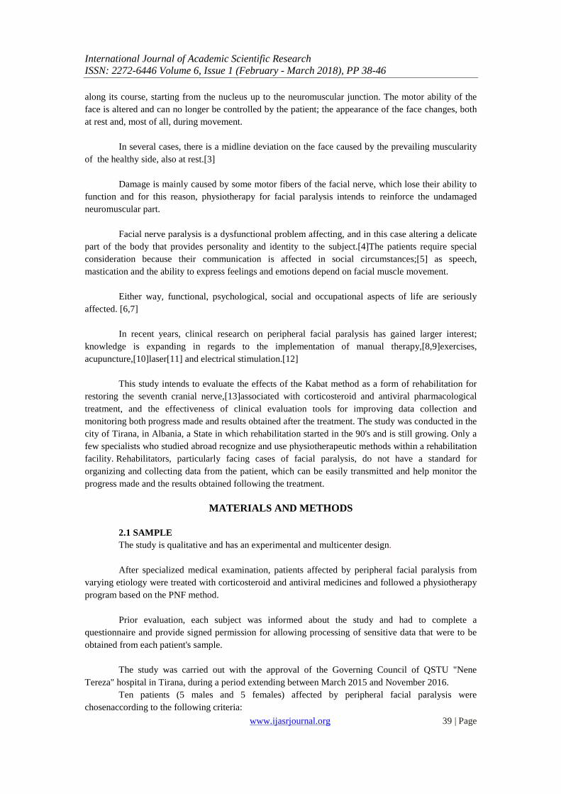
The collected data using the adopted evaluation tools show, through Table 1,that 40% (4 pcs)of10 patients (5 males and 5 females) between 10 and 67 years old (mean age of 36), showed lesion tothe left CN VII and 60% (6 pcs) to the right, no patient showed bilateral facial nerve lesion.
Patients were observed for an average of 6 months for significant changes of the facialfunction, especially in younger adults.
Table (1): Changes of the facial function after 6 monthPATIENTS AGE GENDER LESION H-B SFGS
BEFORE AFTER BEFORE AFTER1 10 F DX V I 4 962 11 F DX V I 4 96
3 15 M DX III I 74 100
4 22 M DX IV I 20 1005 28 F SX IV II 20 826 45 F SX IV II 8 787 51 M DX IV II 8 848 55 F SX V II 4 829 60 M SX IV II 2 7610 67 M SX V III 4 66
MEAN 36,4 / / / / 14,8 86MODE / / / IV II / /
MEDIAN / / / IV II / /DS / / / 0,674949 0,674949 21,81131 11,50845
According to the House Brackmann scale, during the initial state, 10% (1 pcs) showed gradeIII, 50% (5 pcs) grade IV and 40% (4 pcs) grade V with MODE of grade IV and MEDIAN of grade IV.
After treatment, patients improved with 40% grade I, 50% grade II and 10% grade III; MODEand MEDIAN grades decreased to II.
Similarly, for the Sunnybrook Facial Grading System, patients before rehabilitation showed aglobal score of the facial function, which is obtained by the difference between the voluntarymovement score, the symmetry at rest and the synkinesis of 14.8. Following rehabilitation, higher scoreis obtained from improvement of the three subscales composing the scale, resulting in an average scoreof 86.
International Journal of Academic Scientific ResearchISSN: 2272-6446 Volume 6, Issue 1 (February - March 2018), PP 38-46
www.ijasrjournal.org 42 | Page
Out of 10 patients, only one manifested synkinesis at the beginning of treatment with a totalscore of 6/15 and a final score of 2/15.
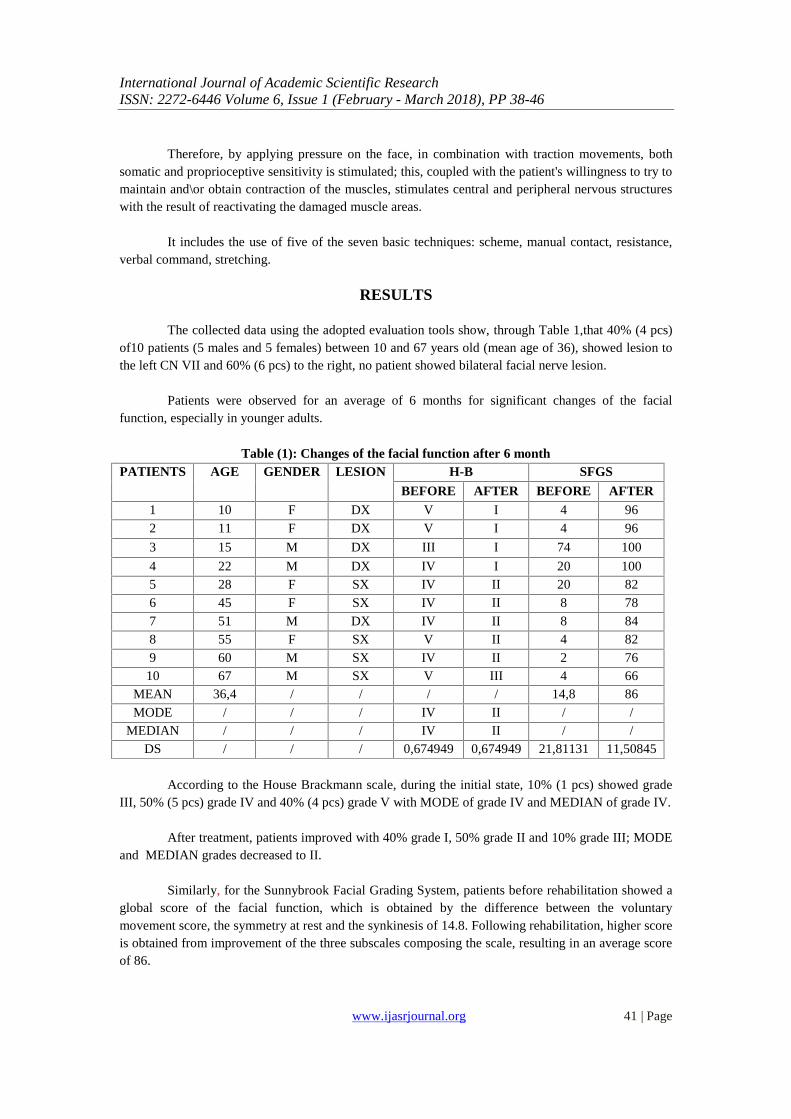
Data collected from each patient through the questionnaire, before and after the treatment, hasproven to be useful. According to the patient, this tool revealed an improvement of theirpsychophysical conditions.
Health has improved from MEDIOCRE (60%) and POOR (40%) to GOOD (90%) (Chart 1).Only one patient remains in the MEDIOCRE state and none are EXCELLENT, as there are somebreaches that can be filled by extending treatment beyond the 6 months period of the study.
Chart.1: Health status before and after treatment
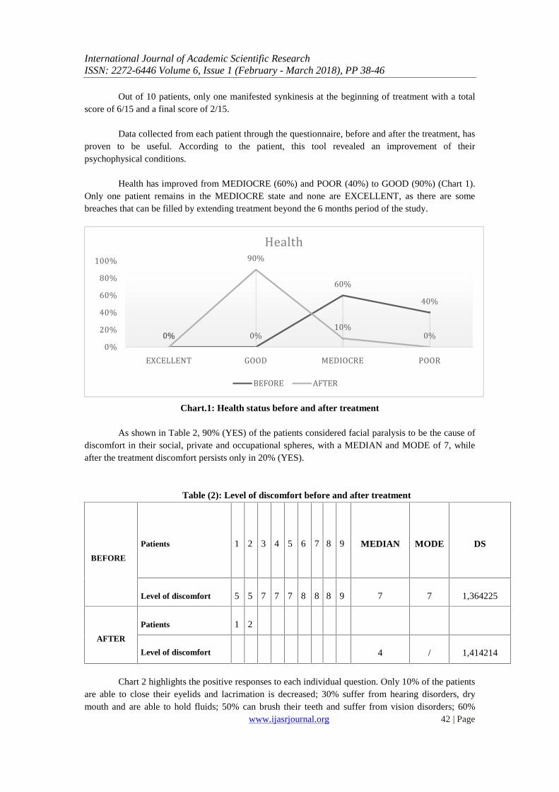
As shown in Table 2, 90% (YES) of the patients considered facial paralysis to be the cause ofdiscomfort in their social, private and occupational spheres, with a MEDIAN and MODE of 7, whileafter the treatment discomfort persists only in 20% (YES).
Table (2): Level of discomfort before and after treatment
BEFORE
Patients 1 2 3 4 5 6 7 8 9 MEDIAN MODE DS
Level of discomfort 5 5 7 7 7 8 8 8 9 7 7 1,364225
AFTER
Patients 1 2
Level of discomfort 5 3 4 / 1,414214
Chart 2 highlights the positive responses to each individual question. Only 10% of the patientsare able to close their eyelids and lacrimation is decreased; 30% suffer from hearing disorders, drymouth and are able to hold fluids; 50% can brush their teeth and suffer from vision disorders; 60%
0% 0%60% 40%
0%
90%
10% 0%0%20%40%60%80%100%
EXCELLENT GOOD MEDIOCRE POOR
Health
BEFORE AFTER
International Journal of Academic Scientific ResearchISSN: 2272-6446 Volume 6, Issue 1 (February - March 2018), PP 38-46
www.ijasrjournal.org 43 | Page
suffer from hyper lacrimation and salivation disorders; 80% suffer from gustatory sensitivity disordersand paraesthesia; 90% suffer from hypo/anesthesia of the skin and facial pain.
Chart.2: Deficit before treatment
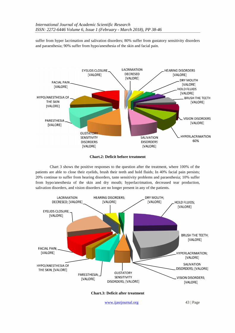
Chart 3 shows the positive responses to the question after the treatment, where 100% of thepatients are able to close their eyelids, brush their teeth and hold fluids; In 40% facial pain persists;20% continue to suffer from hearing disorders, taste sensitivity problems and paraesthesia; 10% sufferfrom hypo/anesthesia of the skin and dry mouth; hyperlacrimation, decreased tear production,salivation disorders, and vision disorders are no longer present in any of the patients.
Chart.3: Deficit after treatment
International Journal of Academic Scientific ResearchISSN: 2272-6446 Volume 6, Issue 1 (February - March 2018), PP 38-46
www.ijasrjournal.org 43 | Page
suffer from hyper lacrimation and salivation disorders; 80% suffer from gustatory sensitivity disordersand paraesthesia; 90% suffer from hypo/anesthesia of the skin and facial pain.
Chart.2: Deficit before treatment
Chart 3 shows the positive responses to the question after the treatment, where 100% of thepatients are able to close their eyelids, brush their teeth and hold fluids; In 40% facial pain persists;20% continue to suffer from hearing disorders, taste sensitivity problems and paraesthesia; 10% sufferfrom hypo/anesthesia of the skin and dry mouth; hyperlacrimation, decreased tear production,salivation disorders, and vision disorders are no longer present in any of the patients.
Chart.3: Deficit after treatment

International Journal of Academic Scientific ResearchISSN: 2272-6446 Volume 6, Issue 1 (February - March 2018), PP 38-46
www.ijasrjournal.org 44 | Page
No changes were reported by any of the patients following the pathological event of facialparalysis, while 40% (4 pts) reported suffering concomitant pathologies, which include 20% diabetes,10% hypertension, 10% thyroid pathologies.
DISCUSSION
The ten patients treated with steroid-antiviral therapy and the PNF rehabilitation therapymethod recovered functional motor abilities of the paralyzed hemi facial muscles. Eyelid closureimproved during the first session, so it was possible to remove the eye bandage, and there was a generalimprovement of the buccinator and orbicularis muscles, allowing the lips to close tight. This evolutionwas so successful that it would be possible to exclude the usage of chewy sticks from the treatment,which would help stimulate the muscles during mastication.
During the following sessions, patients gradually recovered most functionality of all facialmuscles: the corrugator muscle, with the re-appearance of forehead wrinkles, and the orbicularis andpalpebral muscles with the re-appearance of eye blinking.
Tonicity recovery of the zygomatizyc muscle and functionality of the common elevatormuscle of the nose improved to the extent of favoring the re-appearance of nose-palpebral reflexes.
Also, lower compartment muscles recovered well: patients are able to blow up their cheeks,so, in general, recovery of the face to asymmetrical condition is likely expectable.
Patients also show positive improvement of positional, thermal and pain sensitivity, and noincidence of synkinesis.[16,17]
CONCLUSIONS
Treatment of facial nerve paralysis can be a difficult process that requires persistence. [18]
In this study, all patients recovered a positive static dynamic symmetry, despite the severity,various etiologies and different levels of evaluation. It is possible to conclude that a correct and timelydiagnosis is very important, as well as using valid evaluation and data collection tools, properlymanaging the improvement through standardized clinical evaluation scales and a proper medical andphysiotherapy treatment plan.
Rehabilitation combined with medical treatment will help achieve better results while alsoreducing the time for recovery. To promote functional recovery[19] it must be adapted to therequirements of the patient, while considering the medical diagnosis and proper initial evaluation anddefining individual goals.
The Proprioceptive Neuromuscular Facilitation method [20] is among the rehabilitativemethods that offer treatment plans for patients that suffer from facial paralysis. It has been proposedand used in this study and demonstrates the studies published in literature, which support and considerthis rehabilitation protocol as an effective and useful tool for healing facial nerve disabilities. This typeof physical rehabilitation is considered as an integral part of the medical treatment to achieve a betterand more rapid recovery of patients that suffer from facial paralysis, especially in severecases,[21]preventing the development of other complications related to this disease, such as synkinesisand hemispasm. Additionally, when applied at an early stage, recovery proves to be faster and betterthan for non-rehabilitated patients.[22]

International Journal of Academic Scientific ResearchISSN: 2272-6446 Volume 6, Issue 1 (February - March 2018), PP 38-46
www.ijasrjournal.org 45 | Page
The decision to propose and carry out a study in Albania came from the desire to contribute tothe development of this country, which, as a EU membership candidate, is a country that is currentlyapplying reforms in all sectors and a place where innovation and improvement of the quality of healthcare interventions and services is one of the priorities of the Ministry of Health. Therefore, health andrehabilitation services must be provided with standard tools, certified tools and according to Europeanguidelines.
In regard to rehabilitation services that concern peripheral facial nerve paralysis, there is a lackof standard tools form easuring a dysfunction. Furthermore, patients can benefit from the effects of theKabat (PNF) therapy method if the therapist is well-trained and able to establish a physiotherapist-patient relationship with perseverance and motivation. Therefore, the presence of a qualifiedrehabilitation professional is crucial.
LIMITS
The limit of the study is related to the limited number of samples, as only 10 patients werechosen as they fulfilled the inclusion criteria and agreed to participate in the study. Also, since therewere patients with worse prognosis, the predetermined 6 months period of observation would be tooshort for following the total improvement on the mimicry of expressions, requiring an extended amountof time for the treatment.
REFERENCES
[1] Pereira, L. M., Obara, K., Dias, J. M., Menacho, M. O., Lavado, E. L., & Cardoso, J. R. (2011). Facial exercisetherapy for facial palsy: systematic review and meta-analysis. Clinical rehabilitation, 25(7), 649-658.
[2] De Swart BJ, Verheij JC, BeurskensCH.Problems with eating and drinking in patients with unilateral peripheralfacialparalysis. Dysphagia. 2003;18:267–273.
[3] Mattox DE. Clinical disorders of the facial nerve. In: Flint PW, Haughey BH, Lund VJ, Niparko JK, Richardson MA,Robbins KT, et al., editors. Cummings Otolaryngology: Head & Neck Surgery. 6th ed. Philadelphia, PA: ElsevierScience Health Science; 2014. pp. 2617–2628.
[4] Coulson SE, O’Dwyer NJ, Adams RD, Croxson GR. Expression of emotion and quality of life after facial nerveparalysis. OtolNeurotol. 2004;25:1014–1019.
[5] Ekman P. Psychosocial aspects of facial paralysis. In: May M, ed. The Facial Nerve. New York, NY: ThiemeInc;1986:781– 787.
[6] Coulson SE, O’Dwyer NJ, Adams RD, Croxson GR. Expression of emotion and quality of life after facial nerveparalysis. OtolNeurotol. 2004;25:1014 –1019.
[7] Walker D, Hallam M, Ni Mhurchadha S, et al. Our experience: the psychosocial impact of facial palsy. ClinOtolaryngol.2012;37:474–477.
[8] [8] Flores PF, Medina RZ, Haro LG. Idiopathic peripheral facial paralysis treatment: physic therapy versusprednisone [Tratamiento de la parálisis facial periféricaidiopática: terapiafísica versus prednisona]. Revistamédicadel InstitutoMexicano del Seguro Social 1998;36:217-21.
[9] Manikandan N. Effect of facial neuromuscular re-education on facial symmetry in patients with Bell’s palsy: arandomized controlled trial. Clin Rehabilitation 2007;21:338-43.
[10] Wang XH, Zhang LM, Han M, et al. Treatment of Bell’s palsy with combination of traditional Chinese medicine andwestern medicine. Hua Xi Kou Qiang Yi XueZaZhi 2004;22:211-13.
[11] Alayat MS, Elsodany AM, El Fiky AA. Efficacy of high and low level laser therapy in the treatment of Bell’s palsy: arandomized double blind placebo-controlled trial. Lasers Med Sci 2014;29:335-42.
[12] Alakram P, Puckree T. Effects of electrical stimulation on House-Brackmann scores in early Bell’s palsy.Physiotherapy Theory Pract 2010;26:160-6.
[13] Kabat H, Knott M. Proprioceptive facilitation therapy for paralysis.Physiotherapy1954;40:171-6.[14] House JW, Brackmann DE. Facial nerve grading system. Otolaryngol Head Neck Surg 1985;93:146–147.[15] Ross BG, Fradet G, Nedzelski JM. Development of a sensitive clinical facial grading system. Otolaryngol Head Neck
Surg 1996;114:380–386.[16] ÇELIK, M., FORTA, H., & VURAL, Ç. (2000). The development of synkinesis after facial nerve paralysis. European
neurology, 43(3), 147-151.[17] ISHII, L., GODOY, A., ENCARNACION, C. O., BYRNE, P. J., BOAHENE, K., & ISHII, M. (2012). Not just
another face in the crowd: society's perceptions of facial paralysis. The Laryngoscope, 122(3), 533-538.[18] SARDARU, D., & PENDEFUNDA, L. (2012). Neuro-proprioceptive facilitation in the re-education of functional
problems in facial paralysis. A practical approach. Revista medico-chirurgicala a Societatii de Medici si Naturalistidin Iasi, 117(1), 101-106.

International Journal of Academic Scientific ResearchISSN: 2272-6446 Volume 6, Issue 1 (February - March 2018), PP 38-46
www.ijasrjournal.org 46 | Page
[19] VANSWEARINGEN, J. M., COHN, J. F., TURNBULL, J., MRZAI, T., & JOHNSON, P. (1998). Psychologicaldistress linking impairment with disability in facial neuromotor disorders. Otolaryngology--head and necksurgery, 118(6), 790-796.
[20] MONARI, G. (2004). FNP: facilitazioni neurocinetiche progressive: elaborazione del concetto Kabat. Edi-ermes.[21] MONINI, S., IACOLUCCI, C. M., DI TRAGLIA, M., LAZZARINO, A. I., & BARBARA, M. (2016). Role of Kabat
rehabilitation in facial nerve palsy: a randomised study on severe cases of Bell's palsy. Acta OtorhinolaryngologicaItalica, 36(4), 282
[22] BARBARA, M., ANTONINI, G., VESTRI, A., VOLPINI, L., & MONINI, S. (2010). Role of Kabat physicalrehabilitation in Bell's palsy: a randomized trial. Actaoto-laryngologica, 130(1), 167-172.