kansas state university college of veterinary …...institution name: kansas state university...
TRANSCRIPT
ACVR Residency Training Program Application Form:
Institution Name: Kansas State University College of Veterinary Medicine
This document is to act as a guide for institutions desiring ACVR accreditation of their residency
training program. It should be used in concert with the requirements set out in the ACVR
Essentials of Residency Training document and it follows the headings of that document. It is
intended to streamline the application process and help define what information the RSEC needs
to evaluate the program. All terms used in this application have same definitions as defined in
the Essentials.
II. Objectives:
Succinctly state the objectives of the training program.
1. Advanced training in diagnostic imaging
a. Develop skills in diagnostic imaging to include: diagnostic and special
radiographic procedures; ultrasound; CT; MRI; nuclear imaging.
2. To provide instruction in the basic principles of radiobiology, radiation physics,
radiation protection, radiation dosimetry, radiation safety, radiological anatomy
and physiology, and pathologic physiology.
3. Training in clinical investigation
4. Training in scientific writing and literature evaluation
5. Training in didactic teaching and scientific presentations
6. Preparation for certification examination by the American College of Veterinary
Radiology
7. Develop skills for communication with clients and referring veterinarians
III. Training period:
What is the total length of the training program in months? 36
If this is a 4 year program, during what year will the resident be eligible to take the
ACVR Preliminary Exam? If the resident is not eligible to take the exam during the
beginning of the 3rd year (September), please state the reason.
What is the total duration of supervised clinical training in the program? 30 months
What are the responsibilities of the resident in the remaining non-clinical portion of the
program?
1)Vacation,
2) Planning, completion and presentation of a research project.
3) Preparation for boards
4) Externships (ie. Cardiology, exotics)

This is determined on an individual basis. Past has included opportunities for
equine or seeing more MRI (neuro cases).
IV. Direction and Supervision:
Program Director:
Who is the Director of Residency training? David S. Biller, DVM, DACVR
What percentage of this individual’s time is committed to clinical service and teaching of
residents? 75%
From May 2017 there will be a European Boarded Radiologist, A European board
qualified radiologist (taking orals in February 2018. Presently interviewing for a 4th
radiologist.
Faculty:
Please list the faculty member of the program accepting PRIMARY responsibility for
training in each of the following core areas:
Roentgen diagnosis:
Faculty: David Biller
Percentage clinical service: 75%
Diagnostic ultrasound:
Faculty: David Biller
Percentage clinical service: 75%
Computed Tomography
Faculty: David Biller
Percentage clinical service: 70%
Magnetic Resonance Imaging:
Faculty: Nicky Cassel
Percentage clinical service: 70%
Nuclear Medicine:
Faculty: Nicky Cassel
Percentage clinical service: 70%
List the names and percentage clinical commitment of additional imaging faculty in the
program, and their area(s) of instructional responsibility. For each imaging faculty in the
program please provide a one page CV documenting their expertise in the area(s) of
assigned responsibility.
Dr Carlo Anselmi will be joining us May 2017. Board eligible and taking boards in
2/2018. He will be a clinical track faculty with 75A% clinical duties.
For each of the specialty colleges listed below please list at least two Diplomates of these
colleges who can be expected to regularly interact with radiology residents:

ACVIM
Ken Harkin
Tom Schermerhorn
ACVS
Jim Roush
Walter Renberg
ACVP
Kelly Almes
Brad Njaa
V. Affiliation agreement:
If all of the training will not be accomplished on-site, please attach a copy of the
affiliations agreement(s). Include the scope of the training and amount of time the
resident will be away from the home institution.
All training will be accomplished on site. Have previously had residents visit,
Mississippi State, The Ohio State and Washington State.
VI. Facilities:
Briefly describe how the program meets the facility requirements.
The radiology facilities include a main reading area (2 diagnostic work stations) and
an LCD projector utilized for rounds, another reading area with 4 triple view boxes
and a diagnostic work station., and a separate reading area used almost exclusively
for ultrasound interpretation. There is 1 small animal diagnostic radiology room
with Agfa DR. There is a shared large and small animal diagnostic room with Agfa
DR and Eklin DR portable xray unit and overhead X-ray tube. There is a small
animal diagnostic room with a digital fluoroscopy unit that can also use the AGFA
DR.
We have a second equine diagnostic imaging area in a separate building (Equine
performance testing center) with an Eklin DR unit.
We have one room used for nuclear medicine. We have a Siemens gamma camera
with NucLear Mac computer.
We have one CT room with a GE 16 slice machine. We have one MR room with a
Hitachi MRP-7000 (0.3T Permanent magnet). We are in the process of building a
second MRI space for a 3T Toshiba unit. Once the new MRI is in and running we
intend to get rid of the 0.3 T magnet and make that room ultrasound with 2 units
(second unit will be determined later). We have one ultrasound room with a Toshiba
Aplio 500.
We have a AGFA PAC unit that connects to ultrasound, nuclear medicine, CT, MR,
and all DR units.
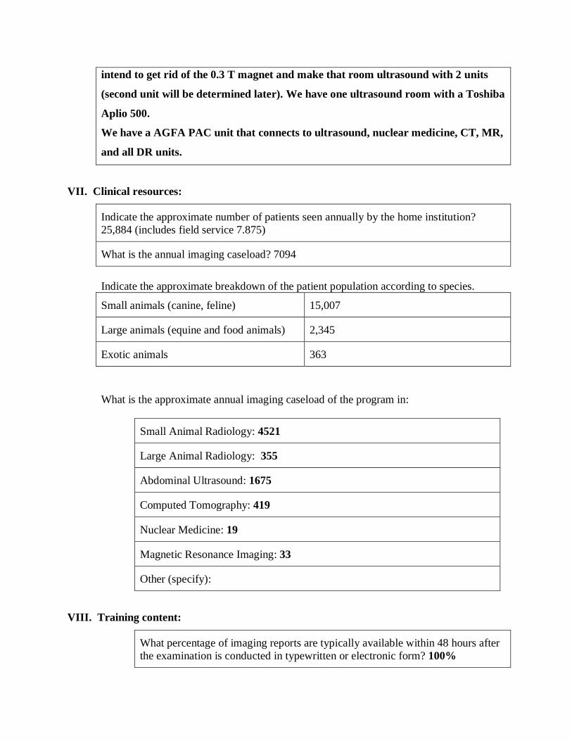
VII. Clinical resources:
Indicate the approximate number of patients seen annually by the home institution?
25,884 (includes field service 7.875)
What is the annual imaging caseload? 7094
Indicate the approximate breakdown of the patient population according to species.
Small animals (canine, feline) 15,007
Large animals (equine and food animals) 2,345
Exotic animals 363
What is the approximate annual imaging caseload of the program in:
Small Animal Radiology: 4521
Large Animal Radiology: 355
Abdominal Ultrasound: 1675
Computed Tomography: 419
Nuclear Medicine: 19
Magnetic Resonance Imaging: 33
Other (specify):
VIII. Training content:
What percentage of imaging reports are typically available within 48 hours after
the examination is conducted in typewritten or electronic form? 100%

If your answer is less than 75% please explain how reports are generated and
how long it takes for the report to be available for review in typewritten form.
Of the preliminary reports generated from the imaging caseload what percentage
are initially produced by the resident? Depends on what year the residents are.
During first year 10%, second year >50%, third year while on duty >90%
What percentage of resident reports are reviewed by the imaging faculty prior to
finalization of the report? 100%
When preliminary resident reports are reviewed and edited by the imaging
faculty responsible for training, what percentage of the time are two or more
faculty present? 0%. This will change to 75% with the addition of the 2 new
radiologists.
Please complete the table below
Approximate number of
cases in the 30 months
clinical experience
Small Animal Radiology: 4200
Large Animal Radiology: 700
Abdominal Ultrasound: 1300
Computed Tomography: 350
Nuclear Medicine: 45
Magnetic Resonance Imaging: 40
Elective (any of above)
Required elective (specify):
Total 6,635
Please indicate the course number and unit assignment residents are required to take to
meet the educational objectives for formal instruction as outlined in the Essentials in the
following:
Topic Course number Units
Radiobiology:
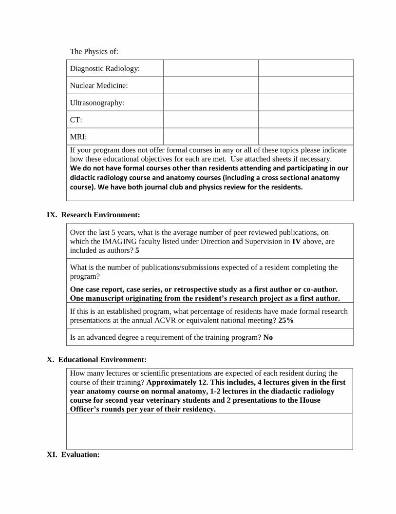
The Physics of:
Diagnostic Radiology:
Nuclear Medicine:
Ultrasonography:
CT:
MRI:
If your program does not offer formal courses in any or all of these topics please indicate
how these educational objectives for each are met. Use attached sheets if necessary.
We do not have formal courses other than residents attending and participating in our didactic radiology course and anatomy courses (including a cross sectional anatomy course). We have both journal club and physics review for the residents.
IX. Research Environment:
Over the last 5 years, what is the average number of peer reviewed publications, on
which the IMAGING faculty listed under Direction and Supervision in IV above, are
included as authors? 5
What is the number of publications/submissions expected of a resident completing the
program?
One case report, case series, or retrospective study as a first author or co-author.
One manuscript originating from the resident’s research project as a first author.
If this is an established program, what percentage of residents have made formal research
presentations at the annual ACVR or equivalent national meeting? 25%
Is an advanced degree a requirement of the training program? No
X. Educational Environment:
How many lectures or scientific presentations are expected of each resident during the
course of their training? Approximately 12. This includes, 4 lectures given in the first
year anatomy course on normal anatomy, 1-2 lectures in the diadactic radiology
course for second year veterinary students and 2 presentations to the House
Officer’s rounds per year of their residency.
XI. Evaluation:
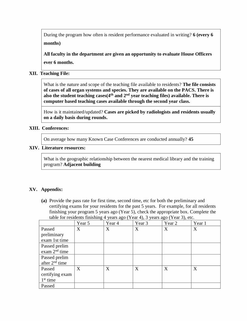
During the program how often is resident performance evaluated in writing? 6 (every 6
months)
All faculty in the department are given an opportunity to evaluate House Officers
ever 6 months.
XII. Teaching File:
What is the nature and scope of the teaching file available to residents? The file consists
of cases of all organ systems and species. They are available on the PACS. There is
also the student teaching cases(4th and 2nd year teaching files) available. There is
computer based teaching cases available through the second year class.
How is it maintained/updated? Cases are picked by radiologists and residents usually
on a daily basis during rounds.
XIII. Conferences:
On average how many Known Case Conferences are conducted annually? 45
XIV. Literature resources:
What is the geographic relationship between the nearest medical library and the training
program? Adjacent building
XV. Appendix:
(a) Provide the pass rate for first time, second time, etc for both the preliminary and
certifying exams for your residents for the past 5 years. For example, for all residents
finishing your program 5 years ago (Year 5), check the appropriate box. Complete the
table for residents finishing 4 years ago (Year 4), 3 years ago (Year 3), etc.
Year 5 Year 4 Year 3 Year 2 Year 1
Passed
preliminary
exam 1st time
X X X X X
Passed prelim
exam 2nd time
Passed prelim
after 2nd time
Passed
certifying exam
1st time
X X X X X
Passed
certifying exam
2nd time
Passed
certifying exam
after 2nd time
Unsuccessful in
all attempts
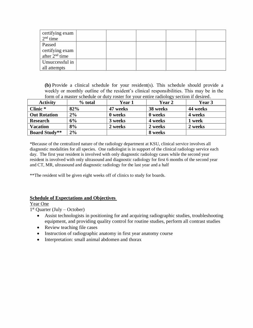
(b) Provide a clinical schedule for your resident(s). This schedule should provide a
weekly or monthly outline of the resident’s clinical responsibilities. This may be in the
form of a master schedule or duty roster for your entire radiology section if desired.
Activity % total Year 1 Year 2 Year 3
Clinic * 82% 47 weeks 38 weeks 44 weeks
Out Rotation 2% 0 weeks 0 weeks 4 weeks
Research 6% 3 weeks 4 weeks 1 week
Vacation 8% 2 weeks 2 weeks 2 weeks
Board Study** 2% 8 weeks
*Because of the centralized nature of the radiology department at KSU, clinical service involves all
diagnostic modalities for all species. One radiologist is in support of the clinical radiology service each
day. The first year resident is involved with only diagnostic radiology cases while the second year
resident is involved with only ultrasound and diagnostic radiology for first 6 months of the second year
and CT, MR, ultrasound and diagnostic radiology for the last year and a half
**The resident will be given eight weeks off of clinics to study for boards.
Schedule of Expectations and Objectives
Year One
1st Quarter (July – October)
• Assist technologists in positioning for and acquiring radiographic studies, troubleshooting
equipment, and providing quality control for routine studies, perform all contrast studies
• Review teaching file cases
• Instruction of radiographic anatomy in first year anatomy course
• Interpretation: small animal abdomen and thorax

2nd quarter (October – December)
• Quality control for radiographic studies, contrast studies
• Come in for emergency with Faculty
3rd quarter (Jan – March)
• Quality control of radiographic studies, contrast studies
• Interpretation – take on more cases
• Attend second year radiology lectures and assist in lab
• Create a plan for research project and submit grant proposals
• Attend course on Cross sectional anatomy
• Attending ultrasound cases, initially to observe and then to perform partial studies under
guidance
4th quarter (April – June)
• Continue 3rd quarter clinical duties
• Spend more time doing ultrasound studies under direct supervision
Year Two
1st & 2nd Quarters (July – December)
• Begin CT, MR, and NM acquisition with technologists, and interpretation
• Continue with past clinical duties as scheduled.
• Progress on research project
3rd – 4th Quarters (Jan – June)
• Continue clinical duties as scheduled
• Complete research project
Year Three
1st Quarter (July – Sept)
• Continue clinical duties as scheduled
• Get July and August off to study for written boards
2nd – 4th Quarters (October – June)
• Continue clinical duties as scheduled
• Submit research abstracts & prepare research manuscript
• Continue radiographic, US, and CT/MR interpretations
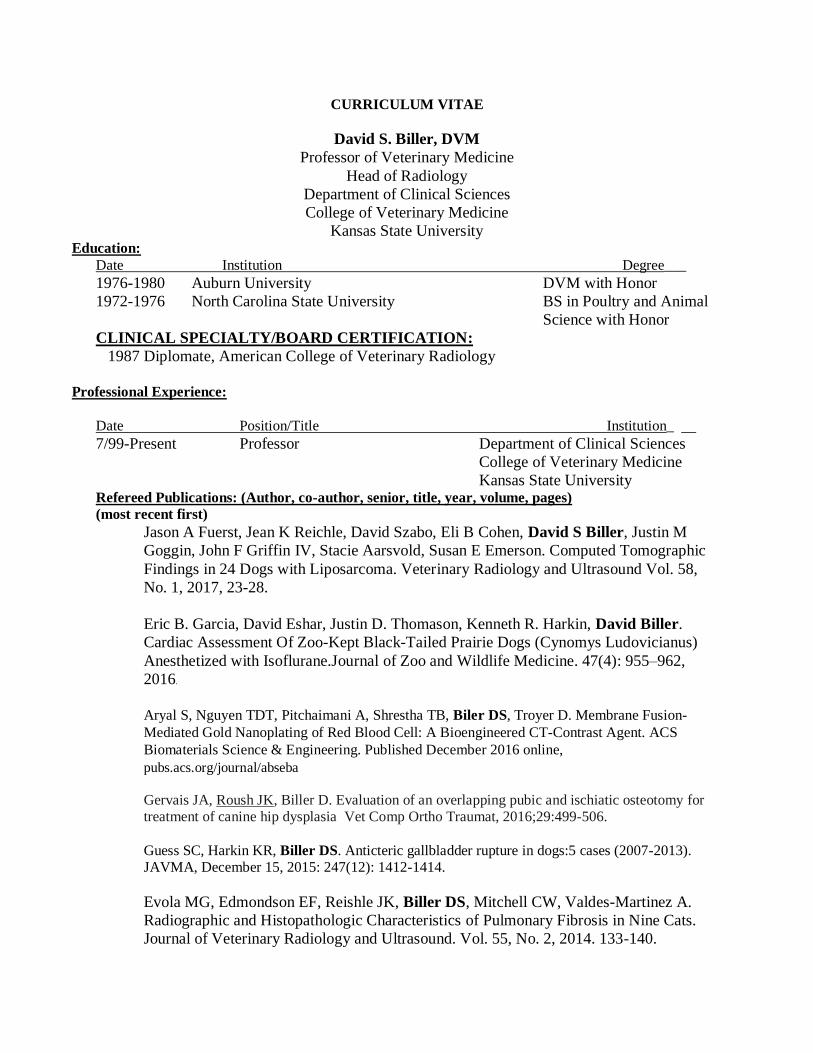
CURRICULUM VITAE
David S. Biller, DVM
Professor of Veterinary Medicine
Head of Radiology
Department of Clinical Sciences
College of Veterinary Medicine
Kansas State University Education:
Date Institution Degree___
1976-1980 Auburn University DVM with Honor
1972-1976 North Carolina State University BS in Poultry and Animal
Science with Honor
CLINICAL SPECIALTY/BOARD CERTIFICATION:
1987 Diplomate, American College of Veterinary Radiology
Professional Experience:
Date Position/Title Institution_
7/99-Present Professor Department of Clinical Sciences
College of Veterinary Medicine
Kansas State University Refereed Publications: (Author, co-author, senior, title, year, volume, pages)
(most recent first)
Jason A Fuerst, Jean K Reichle, David Szabo, Eli B Cohen, David S Biller, Justin M
Goggin, John F Griffin IV, Stacie Aarsvold, Susan E Emerson. Computed Tomographic
Findings in 24 Dogs with Liposarcoma. Veterinary Radiology and Ultrasound Vol. 58,
No. 1, 2017, 23-28.
Eric B. Garcia, David Eshar, Justin D. Thomason, Kenneth R. Harkin, David Biller.
Cardiac Assessment Of Zoo-Kept Black-Tailed Prairie Dogs (Cynomys Ludovicianus)
Anesthetized with Isoflurane.Journal of Zoo and Wildlife Medicine. 47(4): 955–962,
2016.
Aryal S, Nguyen TDT, Pitchaimani A, Shrestha TB, Biler DS, Troyer D. Membrane Fusion-
Mediated Gold Nanoplating of Red Blood Cell: A Bioengineered CT-Contrast Agent. ACS
Biomaterials Science & Engineering. Published December 2016 online,
pubs.acs.org/journal/abseba
Gervais JA, Roush JK, Biller D. Evaluation of an overlapping pubic and ischiatic osteotomy for
treatment of canine hip dysplasia Vet Comp Ortho Traumat, 2016;29:499-506.
Guess SC, Harkin KR, Biller DS. Anticteric gallbladder rupture in dogs:5 cases (2007-2013).
JAVMA, December 15, 2015: 247(12): 1412-1414.
Evola MG, Edmondson EF, Reishle JK, Biller DS, Mitchell CW, Valdes-Martinez A.
Radiographic and Histopathologic Characteristics of Pulmonary Fibrosis in Nine Cats.
Journal of Veterinary Radiology and Ultrasound. Vol. 55, No. 2, 2014. 133-140.
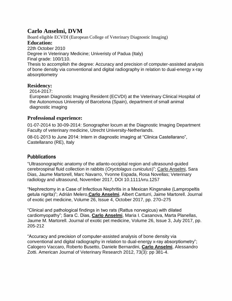
Carlo Anselmi, DVM Board eligible ECVDI (European College of Veterinary Diagnostic Imaging)
Education: 22th October 2010 Degree in Veterinary Medicine; Univeristy of Padua (Italy) Final grade: 100/110. Thesis to accomplish the degree: Accuracy and precision of computer-assisted analysis of bone density via conventional and digital radiography in relation to dual-energy x-ray absorptiometry
Residency: 2014-2017: European Diagnostic Imaging Resident (ECVDI) at the Veterinary Clinical Hospital of the Autonomous University of Barcelona (Spain), department of small animal diagnostic imaging
Professional experience:
01-07-2014 to 30-09-2014: Sonographer locum at the Diagnostic Imaging Department Faculty of veterinary medicine, Utrecht University-Netherlands.
08-01-2013 to June 2014: Intern in diagnostic imaging at “Clinica Castellarano”, Castellarano (RE), Italy
Pubblications
“Ultrasonographic anatomy of the atlanto-occipital region and ultrasound-guided cerebrospinal fluid collection in rabbits (Oryctolagus cuniculus)”; Carlo Anselmi, Sara Dias, Jaume Martorell, Marc Navarro, Yvonne Espada, Rosa Novellas; Veterinary radiology and ultrasound, November 2017, DOI 10.1111/vru.1257
“Nephrectomy in a Case of Infectious Nephritis in a Mexican Kingsnake (Lampropeltis getula nigrita)”; Adrián Melero,Carlo Anselmi, Albert Canturri, Jaime Martorell. Journal of exotic pet medicine, Volume 26, Issue 4, October 2017, pp. 270–275
“Clinical and pathological findings in two rats (Rattus norvegicus) with dilated cardiomyopathy”; Sara C. Dias, Carlo Anselmi, Maria I. Casanova, Marta Planellas, Jaume M. Martorell. Journal of exotic pet medicine, Volume 26, Issue 3, July 2017, pp. 205-212
“Accuracy and precision of computer-assisted analysis of bone density via conventional and digital radiography in relation to dual-energy x-ray absorptiometry”; Calogero Vaccaro, Roberto Busetto, Daniele Bernardini, Carlo Anselmi, Alessandro Zotti. American Journal of Veterinary Research 2012, 73(3): pp 381-4.
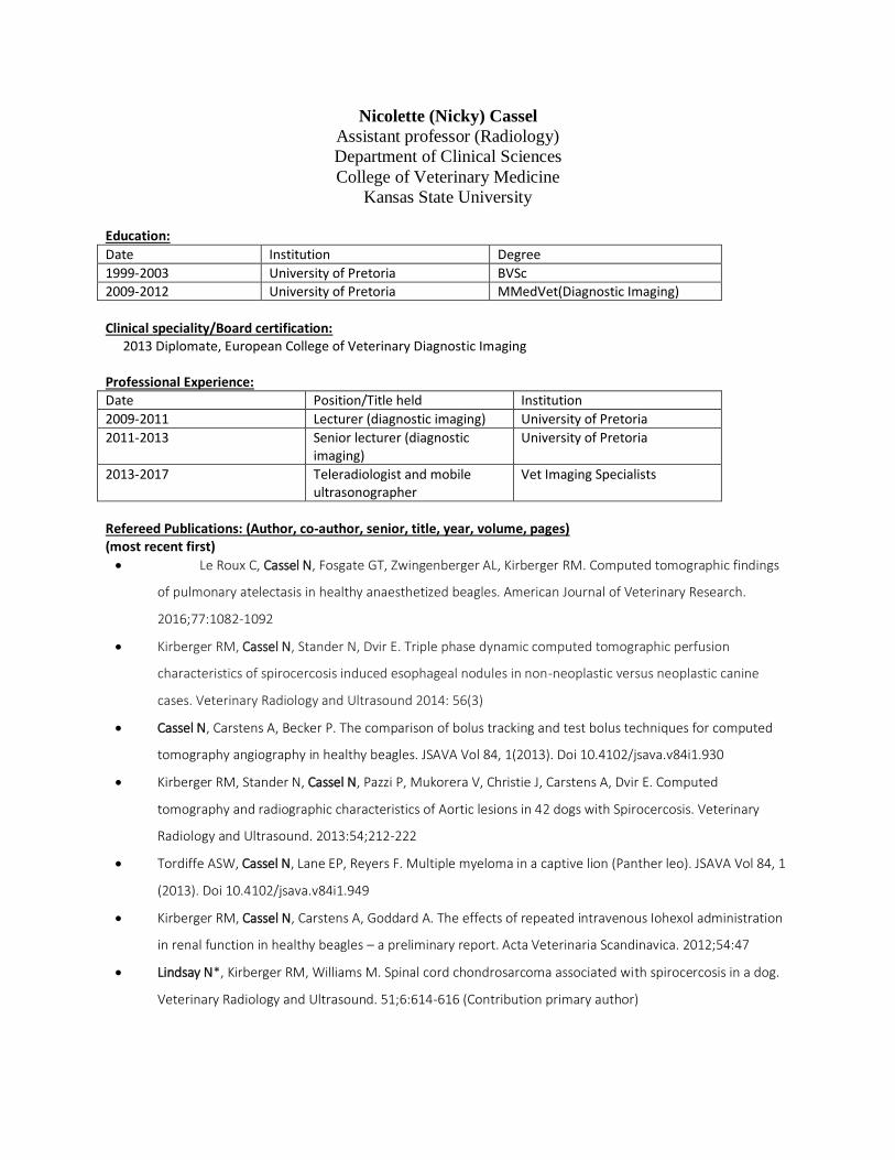
Nicolette (Nicky) Cassel
Assistant professor (Radiology)
Department of Clinical Sciences
College of Veterinary Medicine
Kansas State University
Education:
Date Institution Degree
1999-2003 University of Pretoria BVSc
2009-2012 University of Pretoria MMedVet(Diagnostic Imaging)
Clinical speciality/Board certification:
2013 Diplomate, European College of Veterinary Diagnostic Imaging
Professional Experience: Date Position/Title held Institution
2009-2011 Lecturer (diagnostic imaging) University of Pretoria
2011-2013 Senior lecturer (diagnostic imaging)
University of Pretoria
2013-2017 Teleradiologist and mobile ultrasonographer
Vet Imaging Specialists
Refereed Publications: (Author, co-author, senior, title, year, volume, pages) (most recent first)
• Le Roux C, Cassel N, Fosgate GT, Zwingenberger AL, Kirberger RM. Computed tomographic findings
of pulmonary atelectasis in healthy anaesthetized beagles. American Journal of Veterinary Research.
2016;77:1082-1092
• Kirberger RM, Cassel N, Stander N, Dvir E. Triple phase dynamic computed tomographic perfusion
characteristics of spirocercosis induced esophageal nodules in non-neoplastic versus neoplastic canine
cases. Veterinary Radiology and Ultrasound 2014: 56(3)
• Cassel N, Carstens A, Becker P. The comparison of bolus tracking and test bolus techniques for computed
tomography angiography in healthy beagles. JSAVA Vol 84, 1(2013). Doi 10.4102/jsava.v84i1.930
• Kirberger RM, Stander N, Cassel N, Pazzi P, Mukorera V, Christie J, Carstens A, Dvir E. Computed
tomography and radiographic characteristics of Aortic lesions in 42 dogs with Spirocercosis. Veterinary
Radiology and Ultrasound. 2013:54;212-222
• Tordiffe ASW, Cassel N, Lane EP, Reyers F. Multiple myeloma in a captive lion (Panther leo). JSAVA Vol 84, 1
(2013). Doi 10.4102/jsava.v84i1.949
• Kirberger RM, Cassel N, Carstens A, Goddard A. The effects of repeated intravenous Iohexol administration
in renal function in healthy beagles – a preliminary report. Acta Veterinaria Scandinavica. 2012;54:47
• Lindsay N*, Kirberger RM, Williams M. Spinal cord chondrosarcoma associated with spirocercosis in a dog.
Veterinary Radiology and Ultrasound. 51;6:614-616 (Contribution primary author)