kidney disease
TRANSCRIPT

Multicystic Dysplastic Kidney Disease
Presenter:
Ifeanyi N. Malu, BSc., MA., RDMS (expected)
Instructor:
Mrs. Shelia Chong,MBA,RDMS
May 2015

Archiving Images and HIPPA
• Privacy and confidentiality• Images sent to your doctor via the Picture
Archiving and communication Systems(PACS)
-Method of storing sonographic images in digital format to ensure complete privacy. Once stored, only your doctor will access your medical information.
Your doctor will review your images and contact you if there's any need.
-Craig, M(2006)
Format by Sheila Chong ©
New or Difficult Terms
• Kidney dysplasia
• Potter type 2
-http://www.fetalsono.com/teachfiles/mcdk.lass
--http://library.med.utah.edu/WebPath/TUTORIAL/RENCYST/RENCYST.html
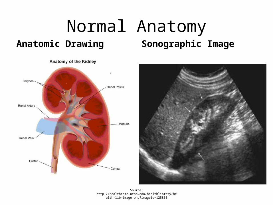
Normal AnatomyAnatomic Drawing Sonographic Image
Source: http://healthcare.utah.edu/healthlibrary/health-lib-image.php?imageid=125836

Fetal Kidney
May 2015
• The baby's kidneys start to produce urine beginning between the 11th and 12th week.
• The kidneys are visible at 15th – 17th weeks
-Chong, S. (2015)-Hagen-Ansert, S.L.(2012).
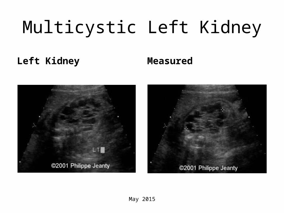
Multicystic Left Kidney
Left Kidney Measured
May 2015
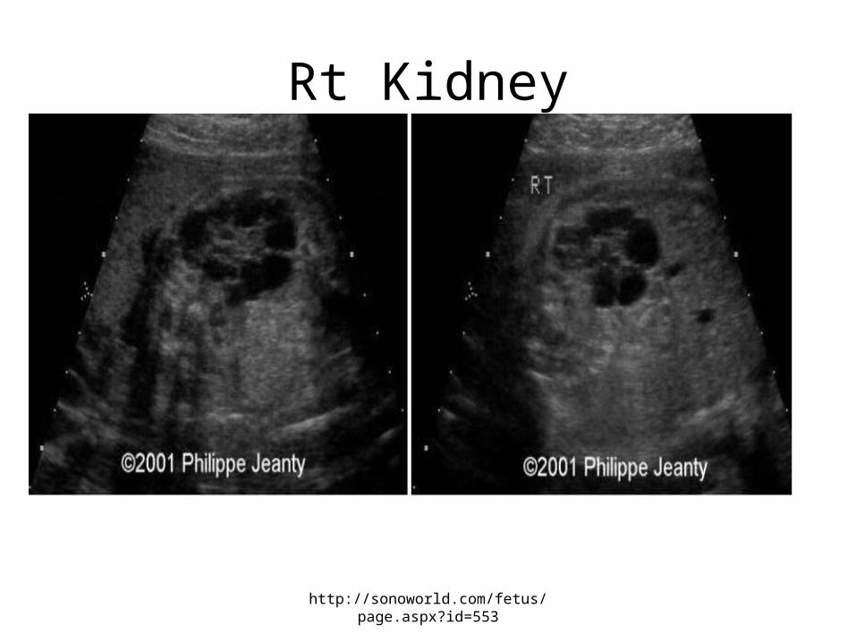
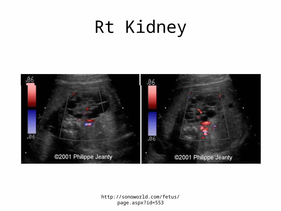
Rt Kidney
http://sonoworld.com/fetus/page.aspx?id=553
Rt Kidney
http://sonoworld.com/fetus/page.aspx?id=553

Multicystic dysplastic kidney (MCDK)
• Multicystic dysplastic kidney (MCDK) is a congenital maldevelopment in which the renal cortex is replaced by numerous cysts of multiple sizes
-Medscape,2015. • Enlarged, echogenic kidneys
with multiple cysts, unilateral or bilateral, probably due to an early obstructive defect.
-Sleurs & Valero, 2001
• Anatomic Drawing
May 2015
May 2015
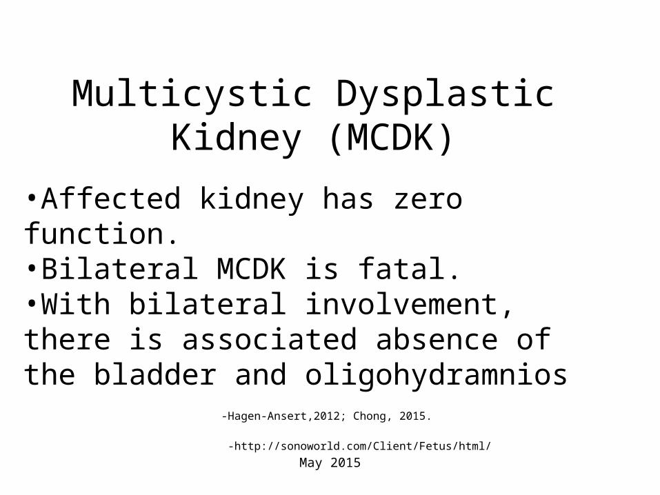
Multicystic Dysplastic Kidney (MCDK)
•Affected kidney has zero function. •Bilateral MCDK is fatal.•With bilateral involvement, there is associated absence of the bladder and oligohydramnios -Hagen-Ansert,2012; Chong, 2015.
-http://sonoworld.com/Client/Fetus/html/
May 2015
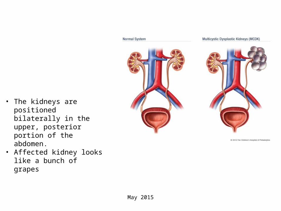
• The kidneys are positioned bilaterally in the upper, posterior portion of the abdomen.
• Affected kidney looks like a bunch of grapes

-Fetal Multicystic Dysplastic Kidney. Available at https://www.luriechildrens.org/en-us/care-
services/conditions-treatments/fetal-multicystic-dysplastic-kidney/Pages/index.aspx
• Most common in Caucasians• Most common renal cystic in
childhood• Occurs at the same rate in both
females and males
Epidemiology and Demographics

Epidemiology and Demographics
• In the United States, the incidence of MCDK is estimated to be 1 in every 2400 live births
• Occurs at the same rate in both females and males
– Luriechildren.org(n.d).
Source: http://www.childrenshospital.org/conditions-and-treatments/conditions/multicystic-dysplastic-kidney
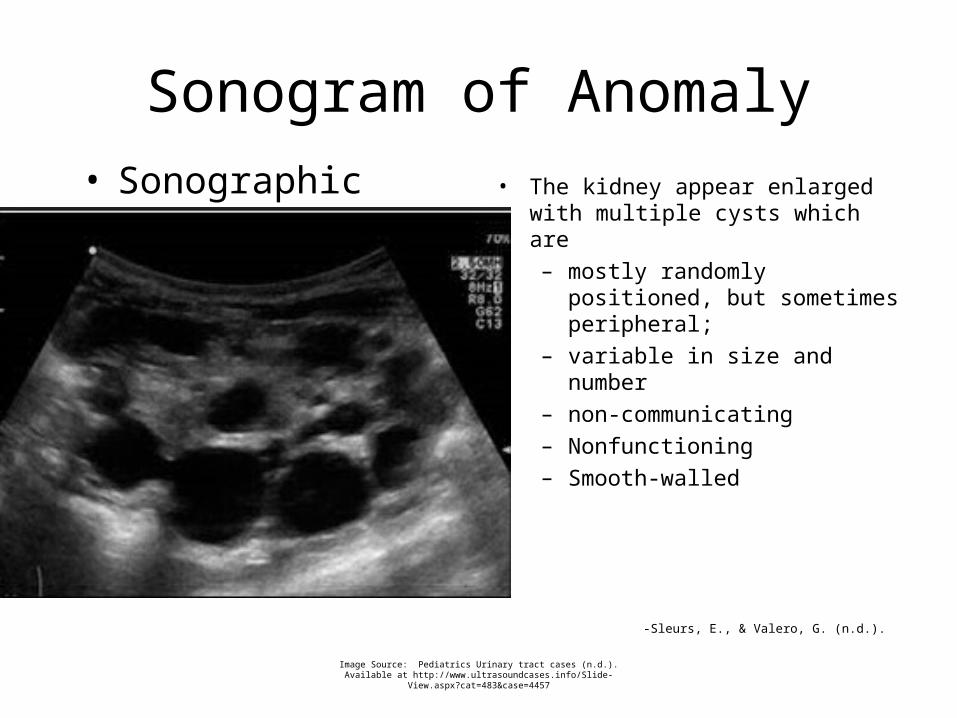
Sonogram of Anomaly• Sonographic Image • The kidney appear enlarged with
multiple cysts which are
– mostly randomly positioned, but sometimes peripheral;
– variable in size and number
– non-communicating
– Nonfunctioning
– Smooth-walled
-Sleurs, E., & Valero, G. (n.d.).
Image Source: Pediatrics Urinary tract cases (n.d.). Available at http://www.ultrasoundcases.info/Slide-
View.aspx?cat=483&case=4457
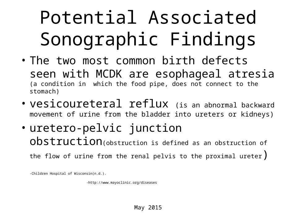
Potential Associated Sonographic Findings
• The two most common birth defects seen with MCDK are esophageal atresia (a condition in which the food pipe, does not connect to the stomach)
• vesicoureteral reflux (is an abnormal backward movement of urine from the bladder into ureters or kidneys)
• uretero-pelvic junction obstruction(obstruction is defined as an obstruction of the flow of urine from the renal pelvis to the
proximal ureter)
-Children Hospital of Wisconsin(n.d.).
-http://www.mayoclinic.org/diseases
May 2015

Etiology
• Unknown
• Some studies suggest certain viral infections and some drugs might also play a role if exposure occurs at a critical stage of development.
• There are rare cases when multicystic dysplastic kidney runs in families because of a genetic trait.
-nationwidechildrens.org, (n.d.).
May 2015

Pathophysiology
• The pathogenesis is unknown but a hypothesis is that an early obstructive defect of the developing kidney causes the disorder
• Exposure to viral infections in utero has been associated with multicystic dysplastic kidney
• Teratogens may also play a role in abnormal renal development
Swiatecka-Urba, A., & Langman, C. (2013)
Sleur & Valero, (2001).
May 2015

Signs & Symptoms
• Severe deformities or polysystemic malformation syndromes
• In bilateral cases, the newborn has oligohydramnios
• Baby born with unilateral multicystic dysplastic kidney disease and a normal, working kidney on the other side should have very little effect
• Rare problem is hypertension -Kishikawa, T; Toda, T et al. ,1981,Wikipedia,2015
May 2015
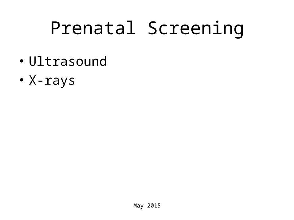
Prenatal Screening
• Ultrasound
• X-rays
May 2015

Prenatal Diagnosis
• A fetal MCDK is generally diagnosed by ultrasound (sonogram) examination before birth
• The condition is usually found during a prenatal ultrasound or when a doctor discovers an abdominal mass during a routine physical examination.
• Sometimes a nuclear scan is required to differentiate a multicystic dysplastic kidney from a blocked kidney
nationwidechildrens.org/multicystic-dysplastic-kidney(n.d.).
Format by Sheila Chong ©

Prenatal Treatment
• The multicystic dysplastic kidney requires no specific treatment. Over time, the abnormal kidney regresses and just goes away
luriechildrens.org/en-us(n.d.).
May 2015

Postnatal Treatment
• After the baby is born, the progress of the MCDK is tracked through a series of ultrasound examinations every six months to a year. It is monitored to make sure that it does not grow or develop a tumor.
• Voiding cystourethrogram: special X-ray that watches the kidney as it makes urine and watches the filling and emptying of the bladder
-www.luriechildrens.org
May 2015

Complications
• Hypertension, hematuria, infection, flank pain
• MCDK should not be confused with polycystic kidney disease (PCKD) or other renal cystic diseases
-nationwidechildrens.org(n.d.)
May 2015

Monitoring
• The condition is being monitored by a national registry (i.e.,National Multicystic Kidney Registry)
• By age 5, the kidney may no longer be visible in x-ray or ultrasound examinations
• Surgical removal is an option.
May 2015

Sonographic monitoring
• Ultrasound is an excellent diagnostic test with a “high degree of confidence.”
-http://emedicine.medscape.com/article/411365-overview#a22
May 2015

Lab monitoring
Alpha fetoprotein assay
•Can detect fetal defects
•Monitor fetal distress
•Monitor fetal abnormalities
•Usually performed at 16 weeks
-Craig, M (2006)
May 2015

CASE PRESENTATION
Multicystic Dysplastic Kidney
May 2015
Case information
May 2015
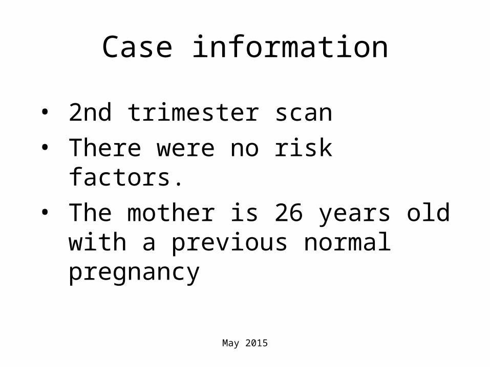
• 2nd trimester scan
• There were no risk factors.
• The mother is 26 years old with a previous normal pregnancy
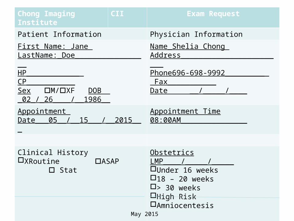
Chong Imaging Institute CII Exam Request
Patient Information Physician Information
First Name: Jane LastName:_Doe_________________HP_____________ CP___________Sex M/XF DOB _02_/_26____/__1986__
Name_Shelia Chong Address________________________Phone696-698-9992__________ Fax___________Date __/_____/____
Appointment Date___05__/__15___/__2015___
Appointment Time 08:00AM_______________
Clinical HistoryXRoutine ASAP Stat
Obstetrics LMP____/_____/_____Under 16 weeks18 – 20 weeks > 30 weeksHigh RiskAmniocentesis
Doctor’s Signature Copy To
May 2015
Room Set-Up
• Ultrasound machine
• Curved linear transducer
• Examination table(powered adjustable)
• Acoustic gel(pre-warmed)
• Latex gloves
• Towel
• pillow
May 2015

Prevent nosocomial infections
• Hand washing
• Wear gloves
• Gown( lab coat,uniform,scrups)
May 2015

Wash your hands, Apply Gloves
-IMAGES SOURCE: MooreMedical(n.d.).

Assisting patients with special needs
• Adopting friendly and nonjudgmental approach
• Be sensitive about privacy concerns
• Assure confidentiality
• Respect ethnic and culture
-Craig,M. (2006).

Patient Rights
• Health Insurance Portability and Accountability Act (HIPPA)
• Right to refuse procedure
• Have informed consent
-Craig, M(2006).
May 2015

Get the Patient
Hi, my name is Ifeanyi Malu. I am the sonographer for your exam. I will perform your second trimester ultrasound exam. The exam will take about 30 minutes. You will be provided with appropriate dress for the exam. The exam will involve exposing your lower abdomen by raising your dress. Liquid gel will be applied on your body; if you feel uncomfortable, please let me know.
May 2015

Ensure you have the right Patient • Confirmation of name and date of birth
• Check the wristband to reconfirm identity
• Check clinical information
May 2015

Sonographer’s Interview
1 Q What is your name? What is your date of birth?
A Jane Doe
2 Q What is your address?
A 711 Steward Ave, Garden City NY
3 Q What are you here for?
A Second trimester sonogram
4 Q What is the date of your last normal menstrual period?
A: 02/15/2015
5 Q: Do you have regular or irregular period?
A: RegularMay 2015
Interview
• 6Q:Is there any complication with previous pregnancy?
• A: No
• 7Q:Is there previous surgery?
• A:No
• 8Q: Are you taking any medication?
• A:No
• 9Q:Do you smoke?
• A: NoMay 2015

Explain Dress for exam
• This exam will take about 30 minutes to complete. It involves exposing your abdomen. I will apply gel on your body. You will change into an exam gown. For your privacy, there’s a room to change your clothes. Ultrasound is highly safe and will not harm you or your fetus. Let me know if you need additional information and privacy.
May 2015

MCKD Video
Source: YouTube Video Available at https://www.youtube.com/watch?v=0IbITe_rULU&list=PLY73TA67Kf_kL4UWRN6DAAXwpb90dJAI7&i
ndex=2
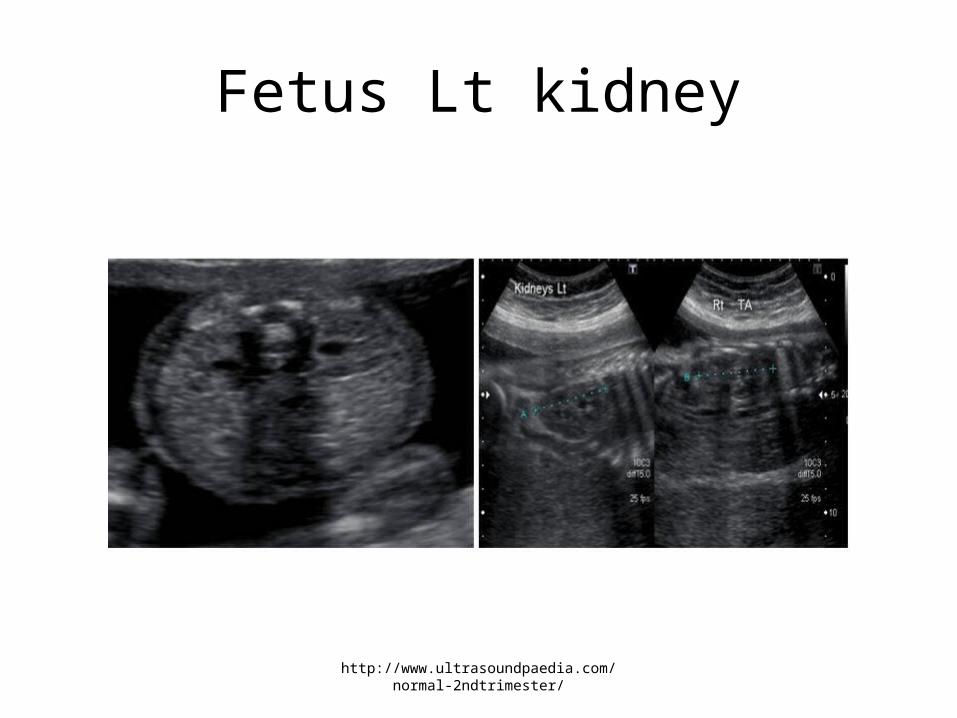
Fetus Lt kidney
http://www.ultrasoundpaedia.com/normal-2ndtrimester/
Image(s)
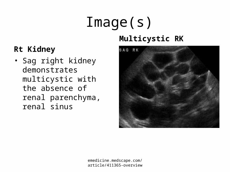
Rt Kidney
• Sag right kidney demonstrates multicystic with the absence of renal parenchyma, renal sinus
Multicystic RK
emedicine.medscape.com/article/411365-overview
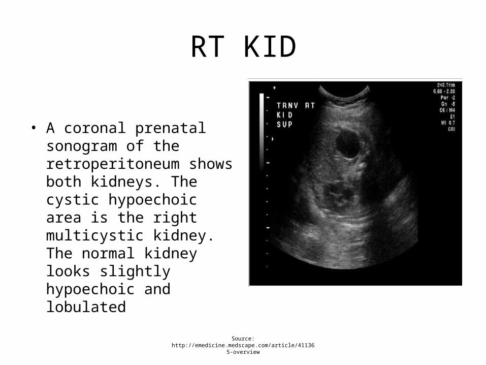
RT KID
• A coronal prenatal sonogram of the retroperitoneum shows both kidneys. The cystic hypoechoic area is the right multicystic kidney. The normal kidney looks slightly hypoechoic and lobulated
Source: http://emedicine.medscape.com/article/411365-overview

Sag Rt Kidney
• A prenatal sonogram shows the right multicystic dysplastic kidney in a longitudinal view. The spine underlies the affected kidney.
Sag RK
Source: http://emedicine.medscape.com/article/411365-
overview
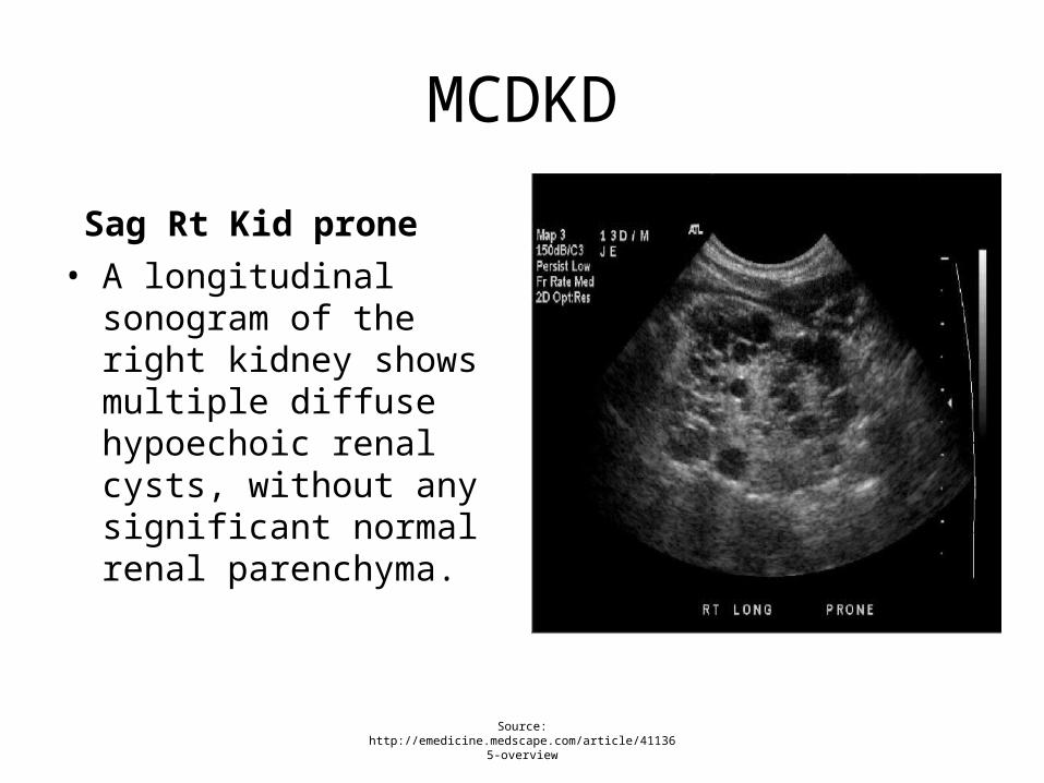
MCDKD
Sag Rt Kid prone
• A longitudinal sonogram of the right kidney shows multiple diffuse hypoechoic renal cysts, without any significant normal renal parenchyma.
Source: http://emedicine.medscape.com/article/411365-overview
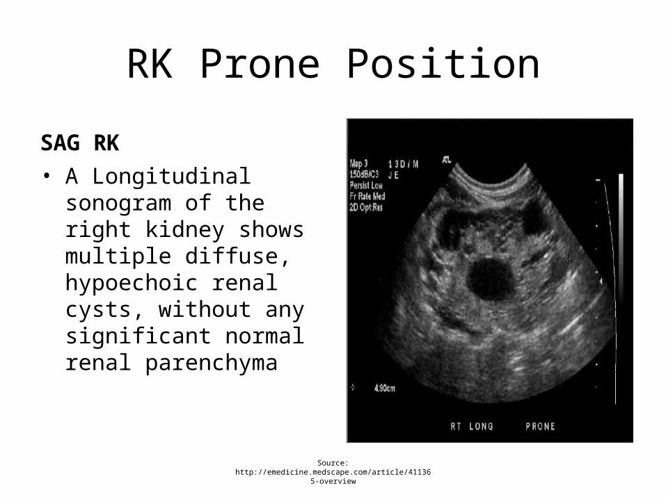
RK Prone Position
SAG RK
• A Longitudinal sonogram of the right kidney shows multiple diffuse, hypoechoic renal cysts, without any significant normal renal parenchyma
Source: http://emedicine.medscape.com/article/411365-overview

• A nongenetic defect due to malformation of the kidney which appears as a bunch of grapes with multiple renal cysts but lacking the normal renal bean shape, and the collection drainage system

Wash your hands, Apply Gloves Begin the Exam
IMAGES SOURCE: MooreMedical(n.d.).

OB Exam Basic Protocol Images with sonographic images & measurements
• Head, face, and neck:
• Lateral cerebral ventricles;
• Choroid plexus;
• Midline falx;
• Cavum septi pellucidi;
• Cerebellum;
• Cistern magna; and
• Upper lip.AIUM(2013). Obstetric Ultrasound Examinations. Available at http://www.aium.org/resources/guidelines/obstetric.pdf

OB Exam Basic Protocol
• Chest:
• Heart:
• Four-chamber view;
• Left ventricular outflow tract; and
• Right ventricular outflow tract.
AIUM(2013). Obstetric Ultrasound Examinations. Available at http://www.aium.org/resources/guidelines/obstetric.pdf
May 2015
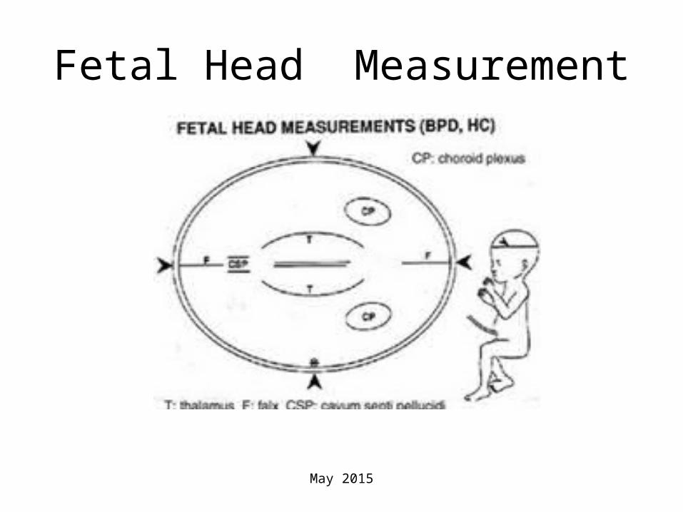
Fetal Head Measurement
May 2015

OB Exam Basic Protocol
• Abdomen:
• Stomach (presence, size, and situs);
• Kidneys;
• Urinary bladder;
• Umbilical cord insertion site into the fetal abdomen; and
• Umbilical cord vessel number.AIUM(2013). Obstetric Ultrasound
Examinations. Available at http://www.aium.org/resources/gui
delines/obstetric.pdf
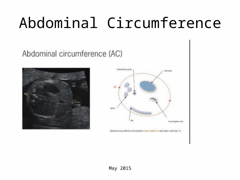
Abdominal Circumference
May 2015
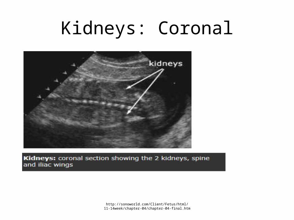
Kidneys: Coronal
http://sonoworld.com/Client/Fetus/html/11-14week/chapter-04/chapter-04-final.htm

OB Exam Basic Protocol
• Spine:
• Cervical, thoracic, lumbar, and sacral spine.
• Extremities:
• Legs and arms.
AIUM(2013). Obstetric Ultrasound Examinations. Available at
http://www.aium.org/resources/guidelines/obstetric.pdf
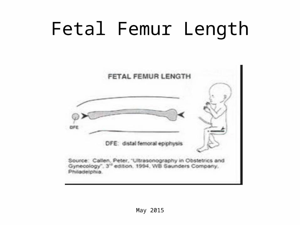
Fetal Femur Length
May 2015

Pathology Sonographic Image(s)with Sonographer Report of this (these) Image(s)
ImageSonographers Report as done at your site
• The left kidney looks enlarged with multiple randomly positioned non-communicating cysts of various sizes.
-www.med-ed.virginia.edu

Case presentation:Fetal Bladder
Fetal BladderSonographic image of fetal bladder
Shepherd, W. (n.d.). Multicystic Dysplastic Kidney . Reterived from http://sonoworld.com/CaseDetails/multicystic_Dysplastic_Kidney.as
px?ModuleCategoryId=1478
• Fetal Bladder
• The Bladder filled and emptied during the 30 minute exam
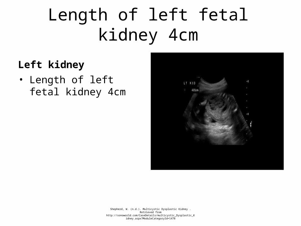
Length of left fetal kidney 4cm
Left kidney
Shepherd, W. (n.d.). Multicystic Dysplastic Kidney . Retrieved from http://sonoworld.com/CaseDetails/multicystic_Dysplastic_Kidney.as
px?ModuleCategoryId=1478
• Length of left fetal kidney 4cm

Right kidney
Right kidney measures 2.76cmSonographic image of fetal right kidney
Shepherd, W. (n.d.). Multicystic Dysplastic Kidney . Reterived from http://sonoworld.com/CaseDetails/multicystic_Dysplastic_Kidney.as
px?ModuleCategoryId=1478
• Length of fetal right kidney 2.76cm
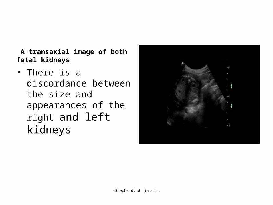
A transaxial image of both fetal kidneys
-Shepherd, W. (n.d.).
• There is a discordance between the size and appearances of the right and left kidneys

Enlargement of the transaxial view of the kidneys.
-Shepherd, W. (n.d.).
• There are many cysts within the left kidney. There is mild dilation of the right renal pelvis. The extent of this dilation varied between 2-5mm during the scan

Preliminary Completion of the Exam
• Wash hands after the exam
• I will show the images to the Radiologist who will provides a report to your Obstetrician. The Radiologist might also carry out further scanning . A full report of your examination will be sent to your doctor. If you need further information about your exams today, please contact your doctor.
May 2015

Other Useful Imaging Test
• X-rays
-Kidney, ureter, and bladder (KUB) images of an infant with a right multicystic dysplastic kidney demonstrate displacement of bowel loops away from the right abdomen
Source: http://emedicine.medscape.com/article/411365-overview

Review Images with the Interpreting Physician
• The enlarged left kidney could contain a mass, be obstructed, or be a multicystic dysplastic kidney. The normal renal cortex is progressively replaced by many cysts of different sizes. Multicyctic Dysplastic Kidney Disease is diagnosed
Format by Sheila Chong ©

Differential considerations
• Hydronephrosis (condition that typically occurs when one kidney becomes swollen due to the failure of normal drainage of urine from the kidney to the bladder)
http://www.healthline.com/health/unilateral-hydronephrosis#Overview1
-Hagen-Ansert, S.L. (2012).
Format by Sheila Chong ©

Release the Patient
Q: The exam is now completed. I will not provide the results or diagnostic information to you. Only your doctor can provide diagnostic information.
I will show the images to the Radiologist who will provides a report to your Obstetrician . A full report of your examination will be sent to your doctor. Your doctor may contact you if need be.
Q: Do you have any questions?
A: NO
Q: You may proceed to the reception area.May 2015

Consent Form
Patient Name: Patient Name:
I approve Dr. I approve Dr. ChongChong to perform an to perform an Ultrasound exams Ultrasound exams second trimestersecond trimester
I understand the general risks for I understand the general risks for SonogramSonogram are: are:•NoneNone
May 2015
Consent Form
I understand the benefits of I understand the benefits of second trisecond tri are: are:•To detect birth defect (multicystic kidney in your baby)To detect birth defect (multicystic kidney in your baby)•Multicystic dysplasia of the kidney is the most common cause of an abdominal mass in a newborn
SignaturesSignatures
Patient _____________________ Date ____/____/____Patient _____________________ Date ____/____/____
Doctor _____________________ Date ____/____/____Doctor _____________________ Date ____/____/____
Witness _____________________ Date ____/____/____Witness _____________________ Date ____/____/____
May 2015
Explanation of Time-Out Procedure
• Ensure that the correct patient is present
• Correct examination is being performed
• Clinical history corresponds to the requested examination
• Correct side/site is being examined
• All patient identification documentation is completed.
May 2015
References
•AIUM.(2013). Obstetric Ultrasound Examinations. Available at ahttp://www.aium.org/resources/guidelines/obstetric.pdf
•Aslam, M., Watson, A.R on behalf of the Trent & Anglia MCDK Study Group.(2006). Unilateral multicystic dysplastic kidney (MCDK): long-term outcomes. Arch Dis Child 2006;91: 820-823
•Children Hospital of Wisconsin(n.d.). Multicystic dysplastic kidney. Available at http://www.chw.org/medical-care/fetal-concerns-center/conditions/infant-complications/multicystic-dysplastic-kidney/
•Chong,S. (March, 2015) Lecture Note. Sanford-Brown Institute, Garden City NY
•Craig, M. (2006). Essentials of Sonography and Patient Care. Second Edition. Saunders, St. Louis, MO.
•eMedicine(n.d.). Multicystic Dysplastic Kidney Imaging .Retrieved electronically from http://emedicine.medscape.com/article/411365-overview
May 2015

References
• Hagen-Ansert, S.L. (2012). Textbook of Diagnostic Sonography, Seventh Edition, Elsevier Mosby St, Louis, MO.
• Kiyak, A., Yilma,z A., Turha,n P., Sander, S., Aydin, G.,& Aydogan, G. (1997). Unilateral multicystic dysplastic kidney: single-center experience. Pediatr Nephrol. Jan 2009;24(1):99-104
• Shepherd, W.(n.d.). Multicystic Dysplastic Kidney. Retrieved from http://sonoworld.com/CaseDetails/multicystic_Dysplastic_Kidney.aspx?ModuleCategoryId=1478
• Singh, J.K., Kanojia, R.P.&, Narasimhan KL.(2014). Multicystic dysplastic kidney in children--a need for conservative and long term approach. Indian J Pediatric. Aug 2009;76(8):809-12
• Sleurs, E., & Valero, G. (2001). Multicystic Dysplastic Kidney Disease. Available at http://sonoworld.com/fetus/page.aspx?id=553
• Sukthankar, S.,& Watson ,A.R.(2000), Unilateral multicystic dysplastic kidney disease: defining the natural history. Acta Paediatrica 2000;89:811-813.
• .
May 2015
References
• Swiatecka-Urba, A., & Langman, C. (2013). Multicystic Renal Dysplasia: Pathophysiology. Retrieved from http://emedicine.medscape.com/article/982560-overview#a0104
• Moore Medical(n.d.). Electronically retrieved from https://www.mooremedical.com/index.cfm?/Flexam -Sterile-Powder-Free-�Nitrile-Exam-Gloves/&PG=CTL&CS=HOM&FN=ProductDetail&PID=18014&spx=1
• Miller-Keane Encyclopedia & Dictionary• US Department of Health and Human Services. Available at
http://www.niddk.nih.gov/health-information/health-topics/kidney-disease/kidney-dysplasia/Pages/facts.aspx
May 2015

Thank You
• Q/A
May 2015