kidney sturcture
TRANSCRIPT

Kidneys structure

Kidneys : are reddish organs shaped like kidney beans, they are located above the waist between the peritoneum and the posterior wall of the abdomen.-since their position is behind the peritoneum of the abdominal cavity they are said retroperitoneal.
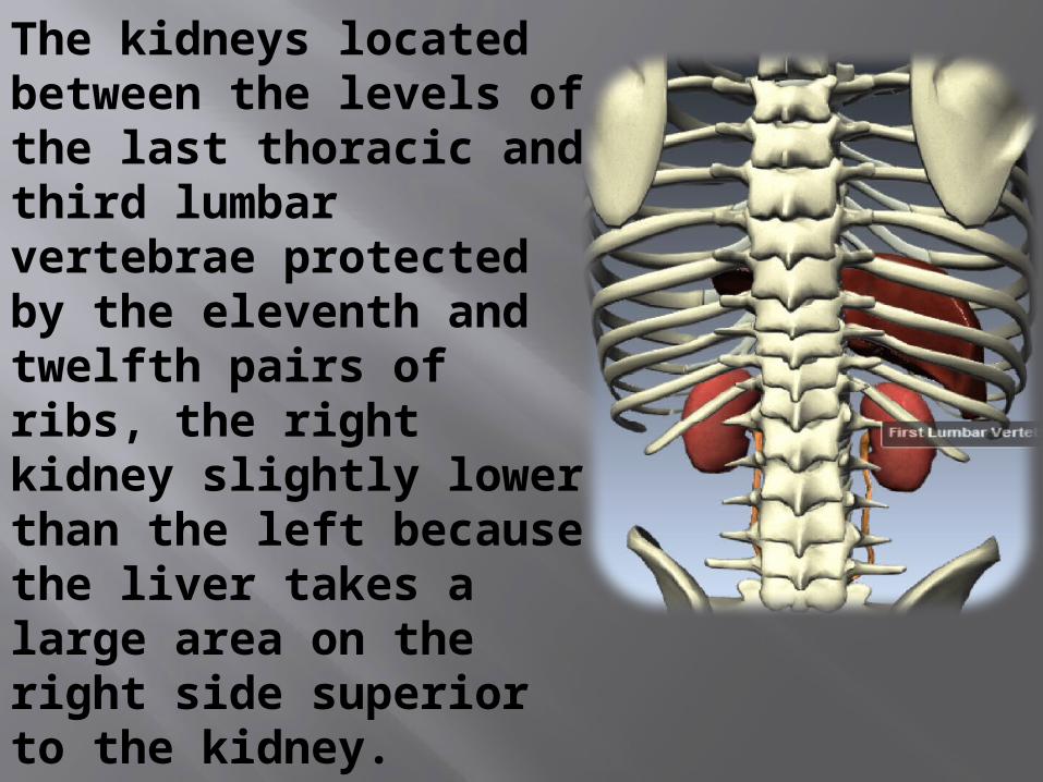
The kidneys located between the levels of the last thoracic and third lumbar vertebrae protected by the eleventh and twelfth pairs of ribs, the right kidney slightly lower than the left because the liver takes a large area on the right side superior to the kidney.

An average adult kidney is 10-12 cm long, 5-7.5 cm wide and 2.5 thick.It’s concave medial border faces the vertebral column.

There are three layers of tissue surround each kidney :
1- renal capsule.2- adipose capsule (perirenal fat).3- renal fascia .

1-renal capsule: is the deeper layer and it’s smooth, transparent, fibrous membrane that is continuous with the coat of the ureter . It serves as a barrier against trauma and helps to maintain the shape of the kidney .

2- adipose capsule: is a mass of fatty tissue surrounding the renal capsule. It also protects the kidney from trauma and holds it firmly in place within the abdominal cavity.

3- renal fascia: is the superficial layer, and it’s a thin layer of dense, irregular connective tissue that anchors the kidney to surrounding structure and to the abdominal wall . On the anterior surface of the kidneys, the renal fascia is deep to the peritoneum.

Adrenal glands : are a type of endocrine gland that are triangle-shaped and located on top of the kidneys. The outer part of the adrenal glands is known as the
cortex and releases hormones
including testosterone and cortical. The inner area of
the adrenal glands is known as the medulla and
produces the hormones norepinephrine and
epinephrine. When the adrenal glands produce too much or too
little of a hormone, illness can result.

Renal hilus: is located near the center of the concave border and it’s vertical fissure so through which the ureter leaves the kidney, blood and lymphatic vessels and nerves also enter and exit the kidney through the renal hilus. Also the renal hilus is the entrance to a cavity within the kidney called (renal sinus).


The renal cortex: is part of the kidneys it’s reddish area containing mostly nephrons and blood vessels. Its function is to filter the blood and remove waste products inside the body.

And a deep reddish-brown region called Renal medulla within the medulla 8-18 cone-shaped structures termed renal pyramids. The base of each pyramid faces the renal cortex and it’s apex called renal papilla (points toward the center of the kidney .

The renal cortex is the smooth-textured area extending from the renal capsule to the base of the renal pyramid and into the spaces between them.Those portions of the renal cortex that extend between the renal pyramids are called the renal columns.

The renal cortex and renal pyramids constitute the parenchyma of the kidney, within the parenchyma are about 1 million microscopic structure called (nephrons). Which are the functional units of the kidney. The number of nephron is constant from birth ; increase in kidney size is due solely the growth of individual nephrons. If nephrons are become diseased the new ones can’t form .The two kidneys are filter blood at 80% in normal condition.


Urine formed by the nephrons ultimately drains into large ducts called papillary ducts.they lead to cuplike structure called minor and major calyces.Each kidney has 8-18 minor calyces and 2-3 major calyces.

A minor calyx receives urine from the papillary ducts of one renal pyramid and delivers urine to a major calyx.From the major calyces, the urine drains into a large cavity called the renal pelvis and then out through the ureter to the urinary bladder.

A nephron consists of two portions ; renal corpuscle where the plasma is filtered and a renal tubule into which the filtered fluid passes. Nephrons perform three basic functions:1-glomerular filtration .2-tubular secretion. 3-tubular reabsorption.

1-glomerular filtration: substance in the blood that are small enough pass across the wall of the glomerular capillaries into the renal tubule.2-renal tubule: the fluid moves along it, many materials are returned to the blood.

renal corpuscle:filtration unit of vertebrate nephrons, functional units of the kidney. It consists of a knot of capillaries (glomerulus) surrounded by a double-walled capsule (Bowman’s capsule) that opens into a tubule.

Blood pressure forces plasma minus its macromolecules (e.g., proteins) from the glomerular capillaries into the Bowman’s capsule. This filtrate, called capsular urine, then passes into the tubule for further processing.

Renal Artery: This is the artery responsible for bringing blood to the kidney from the left side of the heart. 200 quarts of blood passes through the kidneys each day, coming through the renal arteries. That means that this blood contains glucose and oxygen. The incoming artery going in to each kidney divides into four or five branches, and then form arterioles. the abdominal aorta which branches off into the renal arteries in the right and left kidney.


Renal Vein: In this vein the remainder, filtered blood is returned to the right side of the heart after all of the urea and impurities have been removed. the inferior Vena Cava. It then branches off into the renal veins to the left and right kidney.

THANK YOU FOR ATTENTION
BY : MUSTAFA KHALIL IBRAHIMTSMU