kidneys metabolize vita d into calcitrol to synthesize...
TRANSCRIPT

4. VITA D- absorbs CALCIUM for healthy bones
● The skin soaks in vita D from sun exposure● 15-20 min or 30 min for darker complexions
to synthesize vita D● Kidneys metabolize vita D into CALCITROL

Difference between Ureter vs Urethra- Miss Thoni,p3
Aretha (Urethra) Franklin
Performed AT LAST, 1960
Originally performed by Etta James

Anatomy of Urinary System- pg. 961, fig. 25.1
Urethra- ending sounds like throw, what pitchers do.
First period: Mr. Ruibal
Mr. Fletcher- class of 2017

POSITION OF THE KIDNEYS➔ Superior Lumbar Position
T12 - L3 ➔ Protected by the lower
rib cage➔ RIGHT kidney is slightly
lower than the left➔ Adrenal Glands sit on top
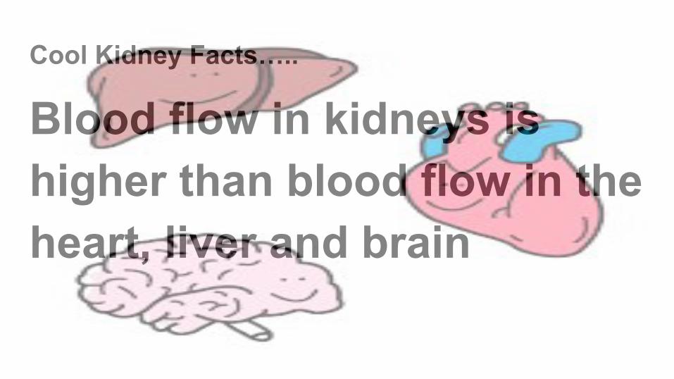
Cool Kidney Facts…..
Blood flow in kidneys is higher than blood flow in the heart, liver and brain
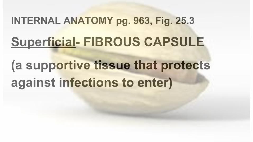
INTERNAL ANATOMY pg. 963, Fig. 25.3
Superficial- FIBROUS CAPSULE
(a supportive tissue that protects against infections to enter)

Regional ANATOMY pg. 964, Fig. 25.4
Renal Cortex: perimeter of the cortex that includes a passageway for blood vessels

Regional ANATOMY pg. 963, Fig. 25.3
Renal Medulla/Pyramids:centrally located triangular structures surrounded by major blood vessels; apex is inward

Regional ANATOMY pg. 963, Fig. 25.3
Renal Pelvis: canals made up of major and minor calyces (singular= calyx) that lead out to the ureter

Blood Vessels by region of the Kidney, pg. 964, fig. 25.4
Cortical Radiate vessels are in the renal cortex & are perpendicular to the Arcuate vessels which run parallel to the renal pyramids

Cool Kidney Fact:
We lose about 2 quarts of water through urine, reserving 198 quarts every 24 hours

RENAL TISSUE- pg.965, fig.25.5
● NEPHRON: subunit that make up the kidney
● > million nephrons per kidney● 2 types of nephrons:
○ Cortical○ Juxtamedullary

2 major parts of a Nephron- pg. 965, fig 25.5
Renal Corpuscle: made up of the Glomerulus and Bowman’s capsule extending to 3 parts of the tubule:
1. PCT: proximal convoluted tubule2. Loop of Henle3. DCT: distal convoluted tubule

Cortical vs. Juxtamedullary NephronsNephron Region Fact Length of loop
of Henle
Cortical Cortex makes up 85% of kidney
Short
Juxtamedullary Medulla Produces concentrated urine Long

URETERS, pg. 985
● Connecting kidney to bladder● Muscular tubes use peristalsis ● Bladder pressure prevents backflow

URINARY BLADDER, pg. 986
● Smooth collapsible muscular sac● MALES: prostate is inferior to bladder● FEMALES: bladder is anterior to
vagina/uterus● When empty, it converts to a pyramidal
shape

URETHRA, pg. 987
● Thin walled muscular tube extends from bladder to exit the body
● Internal urethra sphincter is voluntarily controlled
● Females: 3-4 cm (1.5 in)● Males: 20 cm (8 in)- excrete semen & urine

Cool Kidney Fact:
children born with one kidney grows to the equivalent of two

Renal Blood Flow 1.Renal artery → 2. Interlobar arteries → 3. Arcuate arteries →
4. Interlobular arteries → 5. Afferent arterioles →6. Glomerular
capillaries → 7. Efferent arterioles → 8. Peritubular
capillaries/vasa recta → 9. Interlobular veins → 10. Arcuate veins
→11. Interlobar veins → 12. Renal vein

URINE FORMATION, pg. 969-978
Three processes:
1. Glomerular Filtration 2. Tubular Reabsorption3. Tubular Secretion

Diuretics- enhance urine output
1. Alcohol- a sedative that inhibits ADH2. Caffeine and 3. certain meds-inhibit Na+
reabsorption

Urine- Color, Odor, and pH
Color: urochrome is urine’s pigment made up of destroyed hemoglobin. Concerns:
Pink- blood
Brown- bile
Cloudy- infection

Urine- color, ODOR and pH, pg. 985
*Ammonia odor comes from bacteria metabolizes urea (urea comes from converting nitrogen)
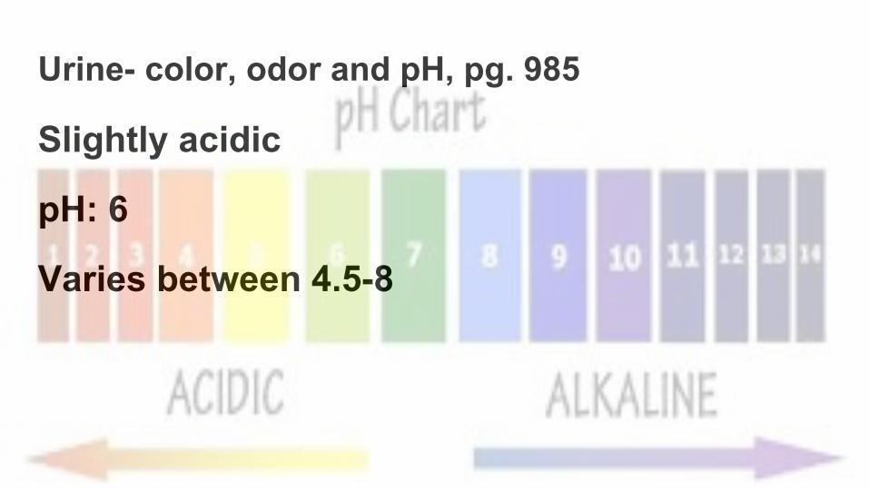
Urine- color, odor and pH, pg. 985
Slightly acidic
pH: 6
Varies between 4.5-8

Medical Term Causes
Pg. 962 Hydronephrosis Water in kidneys that leads to necrosis
Pg. 986 Renal Caliculi Kidney stones are formed from crystallized calcium, magnesium, uric acid salts. Large stones can block urine drainageFrequent uti’s, urine retention, high blood levels of calcium
Pg. 988 Incontinence Unable to retain urine from:1. Emotional problems 3. laugh/cough2. Pressure during pregnancy 4. Overfilled bladder

420 KIDNEY STONES from 1 Patient in CHINA

Hemodialysis
Can be done at home or in the hospital
Takes 3-5 hours at a time.
Must be done 3x a week
People can be on dialysis for 20-30 years

Hemodialysis needs an AV fistula
Vascular access
Surgically implanted of a U-shaped tube that attaches an artery to a vein
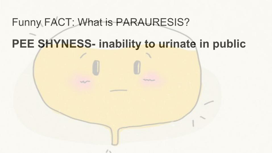
Funny FACT: What is PARAURESIS?
PEE SHYNESS- inability to urinate in public