king s research portal · †department of biotechnology and food sciences, ... §department of...
TRANSCRIPT

King’s Research Portal
DOI:10.1021/jacs.6b09012
Document VersionPublisher's PDF, also known as Version of record
Link to publication record in King's Research Portal
Citation for published version (APA):Nagy, G. N., Suardiaz, R., Lopata, A., Ozohanics, O., Vékey, K., Brooks, B. R., ... Rosta, E. (2016). StructuralCharacterization of Arginine Fingers: Identification of an Arginine Finger for the Pyrophosphatase dUTPases.Journal of the American Chemical Society, 138(45), 15035-15045. 10.1021/jacs.6b09012
Citing this paperPlease note that where the full-text provided on King's Research Portal is the Author Accepted Manuscript or Post-Print version this maydiffer from the final Published version. If citing, it is advised that you check and use the publisher's definitive version for pagination,volume/issue, and date of publication details. And where the final published version is provided on the Research Portal, if citing you areagain advised to check the publisher's website for any subsequent corrections.
General rightsCopyright and moral rights for the publications made accessible in the Research Portal are retained by the authors and/or other copyrightowners and it is a condition of accessing publications that users recognize and abide by the legal requirements associated with these rights.
•Users may download and print one copy of any publication from the Research Portal for the purpose of private study or research.•You may not further distribute the material or use it for any profit-making activity or commercial gain•You may freely distribute the URL identifying the publication in the Research Portal
Take down policyIf you believe that this document breaches copyright please contact [email protected] providing details, and we will remove access tothe work immediately and investigate your claim.
Download date: 18. Feb. 2017
Structural Characterization of Arginine Fingers: Identification of anArginine Finger for the Pyrophosphatase dUTPasesGergely N. Nagy,*,†,‡ Reynier Suardíaz,§ Anna Lopata,‡,∇ Oliver Ozohanics,∥ Karoly Vekey,⊥,○
Bernard R. Brooks,# Ibolya Leveles,†,‡ Judit Toth,‡ Beata G. Vertessy,*,†,‡ and Edina Rosta*,§
†Department of Biotechnology and Food Sciences, Budapest University of Technology and Economics, Budapest 1111, Hungary‡Institute of Enzymology, Research Centre for Natural Sciences, Hungarian Academy of Sciences, Budapest 1117, Hungary§Department of Chemistry, King’s College London, London SE1 1DB, United Kingdom∥MS Proteomics Research Group, Institute of Organic Chemistry, Research Centre for Natural Sciences, Hungarian Academy ofSciences, Budapest 1117, Hungary⊥Core Technologies Centre, Research Centre for Natural Sciences, Hungarian Academy of Sciences, Budapest 1117, Hungary#Laboratory of Computational Biology, National Heart, Lung and Blood Institute, National Institutes of Health, Rockville, Maryland10892-9314, United States
*S Supporting Information
ABSTRACT: Arginine finger is a highly conserved and essentialresidue in many GTPase and AAA+ ATPase enzymes thatcompletes the active site from a distinct protomer, forming contactswith the γ-phosphate of the nucleotide. To date, no pyrophospha-tase has been identified that employs an arginine finger fulfilling allof the above properties; all essential arginine fingers are used tocatalyze the cleavage of the γ-phosphate. Here, we identify andunveil the role of a conserved arginine residue in trimeric dUTPasesthat meets all the criteria established for arginine fingers. We foundthat the conserved arginine adjacent to the P-loop-like motif enablesstructural organization of the active site for efficient catalysis via itsnucleotide coordination, while its direct electrostatic role intransition state stabilization is secondary. An exhaustive structure-based comparison of analogous, conserved arginines fromnucleotide hydrolases and transferases revealed a consensus amino acid location and orientation for contacting the γ-phosphateof the substrate nucleotide. Despite the structurally equivalent position, functional differences between arginine fingers ofdUTPases and NTPases are explained on the basis of the unique chemistry performed by the pyrophosphatase dUTPases.
■ INTRODUCTIONProtein−protein interactions play crucial roles in ensuringoptimal catalytic activity. Notably, interprotomer interactionsoften modulate the nucleotide triphosphate (NTP) hydrolysisand transfer reaction rates in NTP processing enzymes, e.g.,AAA+ enzymes1 (ATPases associated with various cellularactivities), GTPases,2 polymerases,3 kinases,4,5 and dUTPases6
(DUT) assembled as homo- or heteropolymers. In many ofthese enzymes, an essential conserved residue, the arginine(Arg) finger, has been identified as a key entity in catalytic rateacceleration. The Arg finger constitutes a catalytically activecomplex with a neighboring enzyme monomer throughinteractions with the γ-phosphate of the nucleotide substrate.7
The Arg finger can originate either from a neighboring enzymeprotomer (e.g., AAA+ enzymes,8 large GTP binding proteins9)or from an effector protein (e.g., small GTPases7). This residuewas proposed to possess a catalytic role via nucleotidepolarization and/or transition state (TS) stabilization inGTPases,7,10 ATPase motors,11,12 and helicases,13 and in signaltransduction ATPases.14 Experimental data established that
even the conservative replacement of the Arg finger7,15
diminished the catalytic rates while maintaining local structuralintegrity including P-loop coordination.All proteins with catalytically essential Arg fingers reported in
the literature to date catalyze the hydrolysis at the γ-phosphategroup of the NTP. However, highly conserved arginines alsoassist the mechanism of NTP pyrophosphatases or transferases.Among them, dUTPases catalyze the pyrophosphorolysis ofdUTP, a noncanonical DNA nucleotide.16 With this catalyticactivity, dUTPases participate in preventive DNA repair andwere recently shown to also exhibit regulatory functions relatedto, e.g., viral gene expression.17,18 Trimeric dUTPases hostthree active sites formed with the essential contribution of eachprotomer19 (Figure 1a). The nucleotide interactions at eachactive site are provided by five conserved motifs. TheC‑terminal arm of trimeric dUTPases encompasses thedynamic fifth conserved motif,20 which contains a conserved
Received: September 5, 2016Published: October 14, 2016
Article
pubs.acs.org/JACS
© 2016 American Chemical Society 15035 DOI: 10.1021/jacs.6b09012J. Am. Chem. Soc. 2016, 138, 15035−15045
This is an open access article published under a Creative Commons Attribution (CC-BY)License, which permits unrestricted use, distribution and reproduction in any medium,provided the author and source are cited.

Arg directly preceding a glycine-rich P-loop-like motif (Figure1). This Arg residue forms multiple hydrogen-bonding contactswith the γ‑phosphate of the substrate and contributes to thefunctionally critical dUTP/dUDP substrate discrimination ofdUTPases.20,21 Despite the wealth of biochemical, structural,and computational studies concerning dUTPases,20,22−27
structural and computational evidence assessing the role ofthis conserved Arg was missing.Here, we determined crystallographic structures of Myco-
bacterium tuberculosis dUTPase (mtDUT), to establish the roleof R140 as an arginine finger, performing kinetic and ligandbinding measurements as well as molecular dynamics (MD)and QM/MM calculations. Our results together reveal theessential role of R140 in catalysis by primarily promoting activesite organization. We further provide an exhaustive structure-based comparison of analogous conserved arginine residuespresent in a large number of nucleotide hydrolase andtransferase enzyme classes, identifying a conserved spatialposition for these positively charged Arg residues in one metal-ion catalytic28 nucleotide hydrolase and transferase enzymes.
■ METHODSChemicals. DpnI restriction enzyme as well as Phusion polymerase
were obtained from New England Biolabs (Ipswich, MA). IPTG wasobtained from Fisher Scientific GmbH, Germany. Ni-NTA was fromQiagen. Materials for electrophoresis were from Bio-Rad. Phenol redwas obtained from Merck (Kenilworth, NJ); dUTP, α,β-imido dUTP(dUPNPP), and dUDP were from Jena Biosciences. All otherchemicals were of analytical grade and were purchased from Sigma-Aldrich (St. Louis, MO).Mutagenesis and Protein Expression. Site-directed mutagenesis
was performed by the Quikchange method (Agilent) and was verifiedby sequencing (MWG Eurofins GmbH, Martinsried, Germany). Weinterrogated the effect of a lysine surrogate to the R140 residue by
performing the arginine/lysine exchange in mtDUTH145W constructwhich displays similar kinetic parameters to the wild type mtDUT.29
The following primers were used to create mtDUTR140K,H145W from them t D U T H 1 4 5 W c o n s t r u c t : 5 ′− 3 ′ GGCC T CG A C -A TCCAAGGGCGACGGTGGCTGGGG a n d 3 ′− 5 ′CCCCAGCCACCGTCGCCCTTGGATGTCGAGGCC, respec-tively. For mtDUTG143STOP, the primers 5′−3′ CCCGCGGCGACT-GAGGCCACGGTTCCTC and 3 ′−5 ′ GAGGAACCG-TGGCCTCAGTCGCCGCGGG enabled the introduction of a stopcodon in place of G143 residue of mtDUTH145W, thus disrupting thefirst β-turn of the P-loop-like motif. Proteins were expressed andpurified as described previously.29 Briefly, recombinant dUTPase withan N-terminal 6xHis-tag was expressed in E. coli strain BL21(DE3)-pLysS. The final supernatant after cell extraction was loaded onto a Ni-NTA column and purified according to the Novagen protocol.Proteins were dialyzed against a buffer containing 20 mM HEPES/NaOH pH 7.5, 100 mM NaCl, 2 mM MgCl2, and 1 mM DTT. Proteinconcentration was measured by UV absorbance using extinctioncoefficients ε280 = 8480 M−1 cm−1 for mtDUTR140K,H145W and ε280 =2980 M−1 cm−1 for mtDUTG143STOP.
ESI-MS. In the mass spectrometric study of protein complexes, acommercial Waters QTOF Premier instrument (Waters, Milford, MA)equipped with an electrospray ionization source (Waters, Milford,MA) was used in the positive ion mode. Mass spectra were obtainedunder native conditions; the ions were generated from aqueous 10mM NH4HCO3 pH 7.5 buffer solution containing M. tuberculosisdUTPase protein constructs at 0.4 μM monomer concentration. Theseconditions allowed transfer of the native protein complex present inthe solution into the gas phase. The capillary voltage was 3600 V; thesampling cone voltage was 125 V, and the temperature of the sourcewas kept at 80 °C. Collision cell pressure was 3.38 × 10⁻³ mbar, andion guide gas flow was 35.00 mL/min. Mass spectra were recordedusing the software MassLynx 4.1 (Waters, Milford, MA) in the massrange 1400−5000 m/z.
Enzyme Activity Assay. dUTPase activity was followed by acontinuous spectrophotometric phenol red assay as described in ref 30.
Figure 1. Structural organization and catalytic mechanism of trimeric dUTPases. (a) Schematic representation of the trimer assembly and active siteorganization. Conserved arginine residues (R64 and R140) that coordinate the nucleotide triphosphate together with the catalytic Mg2+ ions andcoordinating water are highlighted (licorice and spheres). (b) Reaction scheme for the rate limiting nucleotide hydrolysis reaction step. Nucleophilicattack on the Pα atom is supported by the conserved proton-abstracting residue D83. (c) Sequence conservation of motifs 1−5 in all-β folded(trimeric or monomeric) dUTPases. The conserved arginines of 2nd and 5th motifs are highlighted with green and red asterisks, respectively. ThemtDUT sequence is also shown for comparison.
Journal of the American Chemical Society Article
DOI: 10.1021/jacs.6b09012J. Am. Chem. Soc. 2016, 138, 15035−15045
15036

Briefly, the assay that measures proton release upon dUTP hydrolysiswas performed in 10 mm path length cuvettes thermostated to 20 °Cin a Specord 200 spectrophotometer with a reaction volume of 1000μL. Due to low enzyme activity of the investigated enzyme pointmutants, mtDUTR140K,H145W and mtDUTG143STOP enzyme concen-trations were set to 1 and 5 μM, respectively, in 1 mM HEPES pH 7.5buffer containing 100 mM KCl, 40 μM phenol red, and 5 mM MgCl2.The reaction was started by a final quick addition of 5−80 μM dUTPto the reaction mixture; then, absorbance decrease was recorded for1−5 min. The measured absorbance decline was corrected for thechange of the absorbance for the same time interval in absence ofdUTP substrate. The average of three measurements was calculated ateach data point. Kinetic data were fitted with Michaelis−Mentenequation.Circular Dichroism Intensity Titrations. Near-UV CD spectra
were recorded at 20 °C on a JASCO 720 spectropolarimeter using a 10mm path length quartz cuvette. Protein (50 μM) was titrated withstepwise addition of α,β-imido-dUTP (dUPNPP) in a buffercontaining 10 mM potassium phosphate pH 7.5 and 1 mM MgCl2,as described in ref 20. A spectrum in the range λ = 250−290 nm wasrecorded at each nucleotide concentration. Differential curves wereobtained by subtracting the CD spectra of nucleotide−buffer titrationsfrom that of the corresponding nucleotide−protein titration point.Differential ellipticity at λmax = 269 nm was plotted against thenucleotide concentration by using Origin software (OriginLab Corp.,Northampton, MA). Dissociation constants were determined by fittingthe data with quadratic eq 1:
= ++ + − + + −
y sA c x K c x K cx
c[( ) ( ) 4 ]
2
2
(1)
Here s = y at x = 0; A is the amplitude of the circular dichroismintensity change. c is the concentration of the protein, and K is thedissociation constant.Crystallization. mtDUTR140K,H145W and mtDUTG143STOP proteins
were crystallized using the hanging drop method and applying thesame crystallization condition that was used for the wild typeenzyme.29 Briefly, 4.5 mg/mL dUTPase together with 6.7 mMdUPNPP, 20 mM MgCl2, and 0.1% sodium azide were mixed with thereservoir solution containing 0.1 M TRIS/HCl pH 7.5, 1.5 Mammonium sulfate, and 12.5% glycerol in a 1 μL:1 μL ratio.Data Collection and Structure Determination. Complete
crystallographic data sets of the acquired crystals were recorded at aSupernova sealed tube home source diffractometer (Agilent) formtDUTR140K,H145W, as well as at the ID23-2 beamline of ESRF(Grenoble), for mtDUTG143STOP. Data were processed using XDS andXScale31 programs. An initial electron density map was obtained forboth mutant enzyme:dUPNPP:Mg2+ complexes by Fourier synthesiscalculating with the phases of the mtDUTH145W structure (PDB:3HZA) as the model. Model building and refinement was carried outusing the Coot32 program and REFMAC33 from the CCP4 suite.34 Tomodel anisotropic displacements of the protein complexes, TLSparametrization was implemented within the program REFMAC.Later, refinement of high resolution mtDUTG143STOP structure waspursued using anisotropic B factors. At the final cycles of thisrefinement, B factor restraints were reduced to 0.7. Final modelscontain the bound substrate analogue dUPNPP:Mg2+ complex. Forthe sake of clarity, residues are numbered according to thephysiological M. tuberculosis dUTPase sequence, disregarding therecombinant tag in the atomic coordinate files as well as figures and
throughout the text. Coordinates and structure factor data ofmtDUTR140K,H145W and mtDUTG143STOP were deposited to the ProteinData Bank with accession codes 5EDD and 5ECT, respectively (cf.Table 2).
Molecular Dynamics Simulations. The coordinates of the wildtype (WT) mycobacterial dUTPase enzyme in complex with theisosteric substrate analogue, α,β-imido-dUTP, and Mg2+ were takenfrom the PDB 2PY4 structure of 1.49 Å resolution. To convert thecrystallized substrate analogue to dUTP, the imido NH moiety wasreplaced by O. The R140K and G143STOP mutants were generatedusing the Mutagenesis utility of Pymol35 starting from the wild type2PY4 structure. The simulations were set-up using standard simulationprotocols with CHARMM-GUI.36,37 Hydrogen coordinates weregenerated using standard protonation states for all ionizable residuesusing CHARMM.38 The system was solvated with a pre-equilibratedTIP3P cubic water box of 80 Å3. Water molecules were randomlyreplaced by ions to ensure the neutralization of the system, and anadditional KCl salt concentration corresponding to 0.15 M. We runproduction trajectories of 300 ns after 10 ns of equilibration usingNAMD.39 Temperature and pressure were held constant at 300 K and1 atm, respectively. We used the CHARMM3640 force field withperiodic boundary conditions and the particle mesh Ewald method41
for the long-range electrostatics in combination with a 12 Å cutoff forthe evaluation of the nonbonded interactions, and the same protocolwas followed for the corresponding MD simulations.
QM/MM Calculations. Geometries of the reactant and transitionstates of the phosphate cleavage reaction in WT dUTPase were takenfrom our previous work.24 The system was trimmed to a sphere of 21Å radius centered at the Pα atom of the dUTP. Atoms further than 15Å from the Pα atom of dUTP were kept frozen during all QM/MMcalculations. No cutoffs were introduced for the nonbonding MM andQM/MM interactions. We have used Q-Chem v4.142,43 andGaussian09 D0144 program packages to perform the QM calculationsusing the B3LYP functional in combination with 6-31+G(d,p) basissets. The quantum mechanical system was coupled with theCHARMM program38 for the MM region. A full electrostaticembedding scheme45 has been adopted in all the calculations, andhydrogen link atoms have been used to treat the QM/MM boundaries.The energy minimized reaction pathway was obtained using a QMregion consisting of the dUTP molecule; the backbone of T81 andI82; the side chains of D83, S65, H145, S147, and S148; the amidegroup of Q113; the guanidine group of R140; the Mg2+ ion with its fullcoordination shell; and nearby crystallographic waters to account for132 atoms. We investigated the reaction barrier using different QM/MM model systems. To separate the geometrical factors from theelectrostatic and charge transfer interactions, we used the fullygeometry optimized WT reaction profile,24 and recalculated the barrierheight using different models for the R64 and R140 side chains. Weincluded them either (i) in the QM region, corresponding to theminimized profile, accounting fully for charge transfer and electrostaticeffects; (ii) in the MM region, allowing only electrostatic effects; or(iii) with its atomic charges set to zero (neutralized) during theevaluations of the QM/MM wave function and energies, onlypreserving its geometrical effects.
■ RESULTS
Integrity of Both R140 and the P-Loop-like Element IsEssential for Catalytic Efficiency. We engineered twomutant proteins (mtDUTR140K,H145W and mtDUTG143STOP) to
Table 1. Kinetic and Ligand Binding Parameters of the dUTPase Variants
enzyme mtDUTWT mtDUTH145Wa mtDUTT138STOPa mtDUTR140K,H145W mtDUTG143STOP
kcat (s−1) 3.0 ± 0.3 0.7 ± 0.04 0.0035 ± 0.0001 0.0045 ± 0.0003 0.00089 ± 0.00004
KM (μM) 1.4 ± 0.4 1.3 ± 0.4 6.7 ± 0.4 1.1 ± 0.3 8.2 ± 2.5b
KddUPNPPb (μM) n.d. 0.9 ± 0.5a 3.9 ± 1.3 5.7 ± 1.5 5.2 ± 1.0
aFrom ref 24. bKd measurements were performed by near-UV CD titration, as described in the Methods section; for titration curves, see also FigureS2.
Journal of the American Chemical Society Article
DOI: 10.1021/jacs.6b09012J. Am. Chem. Soc. 2016, 138, 15035−15045
15037

elucidate the role of key constituents of the C-terminal armincluding the R140 in substrate coordination and catalysis.Notably, the conserved arginine is replaced by a lysine in the
first construct, whereas the C-terminal truncation of the secondconstruct at the 143th residue position (mtDUTG143STOP)disrupts only the P-loop-like element, while the R140 is
Table 2. Crystallographic Data Collection and Refinement Statistics
mtDUTR140K mtDUTG143STOP
PDB 5EDD 5ECTData Collection
space group P63 P63cell dimensions a, c (Å) 55.05, 83.83 54.78, 84.08resolution (Å) 41.9−1.97 (2.02−1.97)a 26.0−1.30 (1.33−1.30)a
wavelength (Å) 1.5406 0.8729Rmerge 0.130 (0.497) 0.028 (0.561)⟨I/σ(I)⟩ 6.1 (1.6) 21.23 (1.95)completeness (%) 99.6 (99.6) 99.1 (99.3)redundancy 3.5 (2.5) 3.3 (3.3)
Refinement Statisticsresolution (Å) 19.19−1.97no. reflns 35829 34820Rwork/Rfree 0.160/0.211 0.123/0.157no. atomsprotein 1026 1070ligand/ion 37 39water 159 127B factorsprotein 21.0 22.3ligand/ion 20.2 19.1water 32.0 31.6rms deviationsbond length (Å) 0.019 0.019bond angle (deg) 1.95 1.76
aValues in parentheses are for the highest resolution shell.
Figure 2. Flexibility and disorder of mtDUT and its mutants affecting the Arg finger and the P-loop-like elements from crystallographic structures(top) and MD simulations (bottom). Cartoon representation is shown of the (a) wild type mtDUT (PDB: 2PY4), (b) mtDUTR140K,H145W, and (c)mtDUTG143STOP structures. The local thermal motions defined by B factors in crystal structures (upper row, a−c) are compared with the observedflexibility in MD simulations (lower row, d−f). The superposition of MD simulation snapshots collected at uniform intervals is shown. Thedisordered C-terminal region and the loop S18−D24 are highlighted; the dUTP and the R/K140 are shown as licorice. Color code: blue, more rigid;red, more flexible. Only one dUTPase protomer is highlighted with the other two protomers shown as transparent.
Journal of the American Chemical Society Article
DOI: 10.1021/jacs.6b09012J. Am. Chem. Soc. 2016, 138, 15035−15045
15038
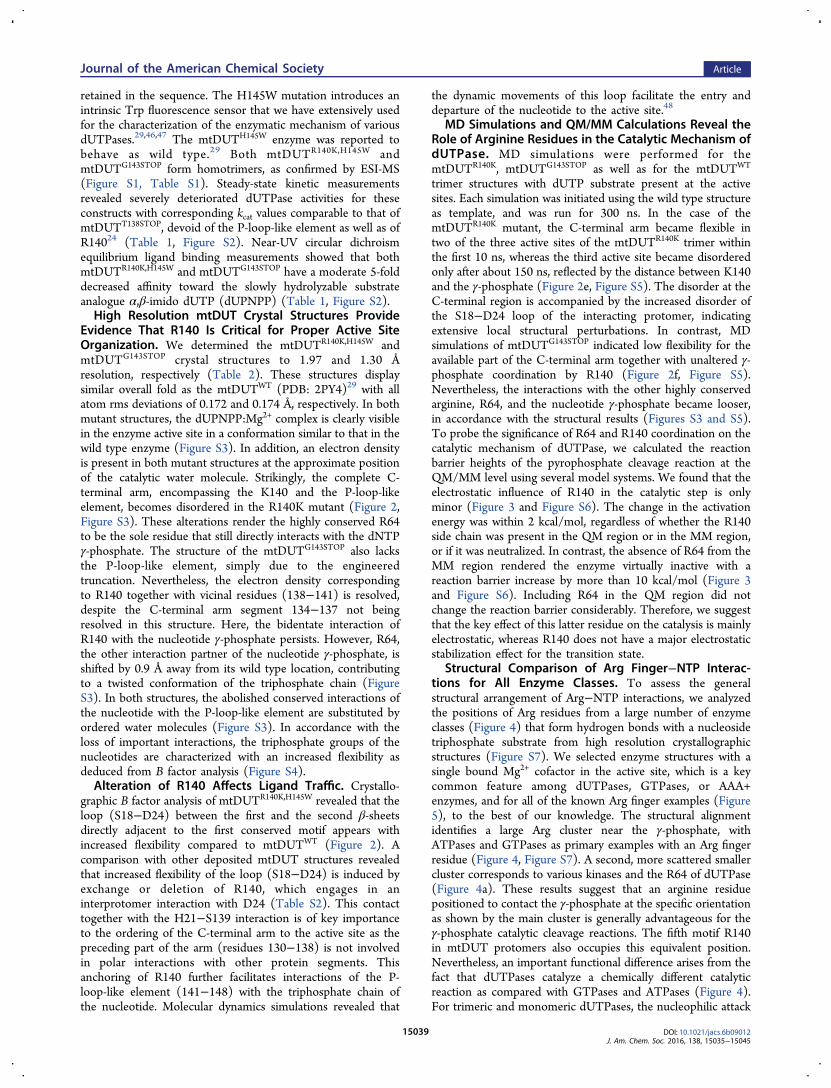
retained in the sequence. The H145W mutation introduces anintrinsic Trp fluorescence sensor that we have extensively usedfor the characterization of the enzymatic mechanism of variousdUTPases.29,46,47 The mtDUTH145W enzyme was reported tobehave as wild type.29 Both mtDUTR140K,H145W andmtDUTG143STOP form homotrimers, as confirmed by ESI-MS(Figure S1, Table S1). Steady-state kinetic measurementsrevealed severely deteriorated dUTPase activities for theseconstructs with corresponding kcat values comparable to that ofmtDUTT138STOP, devoid of the P-loop-like element as well as ofR14024 (Table 1, Figure S2). Near-UV circular dichroismequilibrium ligand binding measurements showed that bothmtDUTR140K,H145W and mtDUTG143STOP have a moderate 5-folddecreased affinity toward the slowly hydrolyzable substrateanalogue α,β-imido dUTP (dUPNPP) (Table 1, Figure S2).High Resolution mtDUT Crystal Structures Provide
Evidence That R140 Is Critical for Proper Active SiteOrganization. We determined the mtDUTR140K,H145W andmtDUTG143STOP crystal structures to 1.97 and 1.30 Åresolution, respectively (Table 2). These structures displaysimilar overall fold as the mtDUTWT (PDB: 2PY4)29 with allatom rms deviations of 0.172 and 0.174 Å, respectively. In bothmutant structures, the dUPNPP:Mg2+ complex is clearly visiblein the enzyme active site in a conformation similar to that in thewild type enzyme (Figure S3). In addition, an electron densityis present in both mutant structures at the approximate positionof the catalytic water molecule. Strikingly, the complete C-terminal arm, encompassing the K140 and the P-loop-likeelement, becomes disordered in the R140K mutant (Figure 2,Figure S3). These alterations render the highly conserved R64to be the sole residue that still directly interacts with the dNTPγ-phosphate. The structure of the mtDUTG143STOP also lacksthe P-loop-like element, simply due to the engineeredtruncation. Nevertheless, the electron density correspondingto R140 together with vicinal residues (138−141) is resolved,despite the C-terminal arm segment 134−137 not beingresolved in this structure. Here, the bidentate interaction ofR140 with the nucleotide γ-phosphate persists. However, R64,the other interaction partner of the nucleotide γ-phosphate, isshifted by 0.9 Å away from its wild type location, contributingto a twisted conformation of the triphosphate chain (FigureS3). In both structures, the abolished conserved interactions ofthe nucleotide with the P-loop-like element are substituted byordered water molecules (Figure S3). In accordance with theloss of important interactions, the triphosphate groups of thenucleotides are characterized with an increased flexibility asdeduced from B factor analysis (Figure S4).Alteration of R140 Affects Ligand Traffic. Crystallo-
graphic B factor analysis of mtDUTR140K,H145W revealed that theloop (S18−D24) between the first and the second β-sheetsdirectly adjacent to the first conserved motif appears withincreased flexibility compared to mtDUTWT (Figure 2). Acomparison with other deposited mtDUT structures revealedthat increased flexibility of the loop (S18−D24) is induced byexchange or deletion of R140, which engages in aninterprotomer interaction with D24 (Table S2). This contacttogether with the H21−S139 interaction is of key importanceto the ordering of the C-terminal arm to the active site as thepreceding part of the arm (residues 130−138) is not involvedin polar interactions with other protein segments. Thisanchoring of R140 further facilitates interactions of the P-loop-like element (141−148) with the triphosphate chain ofthe nucleotide. Molecular dynamics simulations revealed that
the dynamic movements of this loop facilitate the entry anddeparture of the nucleotide to the active site.48
MD Simulations and QM/MM Calculations Reveal theRole of Arginine Residues in the Catalytic Mechanism ofdUTPase. MD simulations were performed for themtDUTR140K, mtDUTG143STOP as well as for the mtDUTWT
trimer structures with dUTP substrate present at the activesites. Each simulation was initiated using the wild type structureas template, and was run for 300 ns. In the case of themtDUTR140K mutant, the C-terminal arm became flexible intwo of the three active sites of the mtDUTR140K trimer withinthe first 10 ns, whereas the third active site became disorderedonly after about 150 ns, reflected by the distance between K140and the γ-phosphate (Figure 2e, Figure S5). The disorder at theC-terminal region is accompanied by the increased disorder ofthe S18−D24 loop of the interacting protomer, indicatingextensive local structural perturbations. In contrast, MDsimulations of mtDUTG143STOP indicated low flexibility for theavailable part of the C-terminal arm together with unaltered γ-phosphate coordination by R140 (Figure 2f, Figure S5).Nevertheless, the interactions with the other highly conservedarginine, R64, and the nucleotide γ-phosphate became looser,in accordance with the structural results (Figures S3 and S5).To probe the significance of R64 and R140 coordination on thecatalytic mechanism of dUTPase, we calculated the reactionbarrier heights of the pyrophosphate cleavage reaction at theQM/MM level using several model systems. We found that theelectrostatic influence of R140 in the catalytic step is onlyminor (Figure 3 and Figure S6). The change in the activationenergy was within 2 kcal/mol, regardless of whether the R140side chain was present in the QM region or in the MM region,or if it was neutralized. In contrast, the absence of R64 from theMM region rendered the enzyme virtually inactive with areaction barrier increase by more than 10 kcal/mol (Figure 3and Figure S6). Including R64 in the QM region did notchange the reaction barrier considerably. Therefore, we suggestthat the key effect of this latter residue on the catalysis is mainlyelectrostatic, whereas R140 does not have a major electrostaticstabilization effect for the transition state.
Structural Comparison of Arg Finger−NTP Interac-tions for All Enzyme Classes. To assess the generalstructural arrangement of Arg−NTP interactions, we analyzedthe positions of Arg residues from a large number of enzymeclasses (Figure 4) that form hydrogen bonds with a nucleosidetriphosphate substrate from high resolution crystallographicstructures (Figure S7). We selected enzyme structures with asingle bound Mg2+ cofactor in the active site, which is a keycommon feature among dUTPases, GTPases, or AAA+enzymes, and for all of the known Arg finger examples (Figure5), to the best of our knowledge. The structural alignmentidentifies a large Arg cluster near the γ-phosphate, withATPases and GTPases as primary examples with an Arg fingerresidue (Figure 4, Figure S7). A second, more scattered smallercluster corresponds to various kinases and the R64 of dUTPase(Figure 4a). These results suggest that an arginine residuepositioned to contact the γ-phosphate at the specific orientationas shown by the main cluster is generally advantageous for theγ-phosphate catalytic cleavage reactions. The fifth motif R140in mtDUT protomers also occupies this equivalent position.Nevertheless, an important functional difference arises from thefact that dUTPases catalyze a chemically different catalyticreaction as compared with GTPases and ATPases (Figure 4).For trimeric and monomeric dUTPases, the nucleophilic attack
Journal of the American Chemical Society Article
DOI: 10.1021/jacs.6b09012J. Am. Chem. Soc. 2016, 138, 15035−15045
15039

occurs at the α-phosphate; therefore, their cognate Argcorresponding to R140 is not involved in the direct electronicstabilization of the transition state. In contrast, the γ-phosphateserves as the reaction center upon the nucleophilic attack inGTPases and ATPases, in which the Arg fingers can indeedstabilize the transition state.
■ DISCUSSIONFunctional Roles of Conserved Arginines at the Active
Site of dUTPases. Three conserved arginine residues arepresent in the active site of dUTPases: R64 (second motif),R110 (fourth motif), and R140 (fifth motif). Both R64 andR140 directly contact the triphosphate chain (Figure 1c).Arg140 coordinates the nucleotide γ-phosphate, analogously tothe Arg fingers in AAA+ enzymes. The fourth motif R110 doesnot directly coordinate the substrate nucleotide, and thecorresponding R111A mutant of Saccharomyces cerevisiae
dUTPase showed only 30−50% decrease in activity.27 R64contacts both the β- and γ-phosphates (Figure 4b), and at thesame time completes the active site at the interface betweentwo protomers. R64 is part of a different protomer than the onecoordinating the nucleoside group. In contrast to R140, ourQM/MM results predicted that R64 electrostatically stabilizesthe TS of the pyrophosphorolysis reaction. This is also inaccordance with a previous mutational study in Saccharomycescerevisiae that reported very low activity for the correspondingAla mutant R68A.27 Furthermore, a positively charged residue(R/K) occupies this position almost invariably, and the highlyconserved R64 is substituted by Lys in only about 3% of fullyfunctional trimeric dUTPases (e.g., ref 22) (Figure 1c andTable S2).R140 is a highly conserved residue among trimeric and
monomeric dUTPases (Figure 1b). Its naturally occurringhomologues are very rare (less than 0.3%, see Table S3), anddo not favor the conservative Lys exchange. This is in goodagreement with our results that conservative R140K mutationrenders the enzyme inactive. Accordingly, Arg/Lys, Arg/Alapoint mutants of dUTPases were also seriously compromised intheir catalytic efficiency on the basis of prior studies includingherpes viral, lentiviral, retroviral, yeast, and human sour-ces.20,23,25−27
R140 displays an exceptional combination of sequential andsteric positioning in dUTPases that enables its major effects onthe P-loop-like motif coordination. R140 directly precedes theP-loop motif, atypical for NTPases with the conventionalGxxxxGK(T/S) P-loop or Walker A type sequence motif.50
Our results demonstrate that deletion of the P-loop-likeelement renders the M. tuberculosis dUTPase inactive. On thebasis of our combined in vitro, computational, and structuraldata of mtDUTR140K,H145W and mtDUTG143STOP, we proposedifferent mechanisms for the activity loss of the two mutantenzyme constructs. In the presence of Arg140 and in theabsence of the P-loop-like motif, the conserved Arg residueprovides similar contacts to the nucleotide as in the wild typeenzyme. Therefore, low enzymatic activity shows that R140 initself cannot provide sufficient catalytic rate enhancement, andthe loss of catalytically competent P-loop coordination is likelythe main reason for the lack of activity. Accordingly, severalearlier studies demonstrated that mutant constructs devoid ofthe P-loop region23,51 or those lacking the complete C-terminalarm including the conserved Arg20,22−24 are nearly inactive.In contrast, when R140 is exchanged to a Lys, the
deteriorated enzyme activity together with structure-basedand MD observations reflects that Arg/Lys substitution is notfunctional at this position. Our results indicate that R140residue is therefore an essential factor for the structural integrityof the active site to enable efficient catalysis: (i) via its directnucleotide coordination or (ii) via interprotomer contacts.These interactions enable optimal positioning of hallmarkresidues from the P-loop-like element,20,24,52 including S147and S148 that have a decisive role in ensuring catalyticefficiency. This is also in agreement with QM/MM calculationsdemonstrating that the absence of the complete C-terminalarm24 but not of R140 in itself corresponds to a marked largerincrease of the transition state energy barrier, suggesting apotent yet indirect role of R140 in catalytic rate enhancement.It is worth noting that in our QM/MM calculations we used theoptimized geometries along the profile obtained for the wildtype reaction, and only single point calculations wereperformed for our model systems to distinguish between the
Figure 3. Role of arginine residues in mtDUT catalysis as assessed byQM/MM calculations. (a) Transition state configuration of the M.tuberculosis dUTPase active site. Residues always included in the QMregion (licorice, sphere for the Mg2+) and the R64 and R140 moieties(transparent thin sticks) are shown. The reaction coordinate wasdefined as a linear combination of distances involving the breaking andforming of bonds (yellow dashed lines): r(O−Pα) − r(Ow−Pα) +0.5r(Ow−Hw) − 0.5r(Hw−OCO). (b) QM/MM energy barriers forthe catalytic step when the side chains of R64 and/or R140 are (i)included in the QM region (QM), (ii) included in the MM region(MM), or (iii) neutralized in the MM region (MM0) during QM/MMcalculations.
Journal of the American Chemical Society Article
DOI: 10.1021/jacs.6b09012J. Am. Chem. Soc. 2016, 138, 15035−15045
15040

electrostatic and structural contributions to catalysis. Moredetailed analysis could also be carried out to evaluate theelectrostatic preorganization toward the transition state, asdone, e.g., by Florian and Warshel.53 To compare with ourbiochemical data, calculations of accurate free energy barriersare needed, taking into account geometrical relaxation thatrequires more extensive free energy calculations.Identification of an Arg Finger in dUTPases. On the
basis of the following properties, we propose that R140 fulfillsthe requirements of being an Arg finger: (i) It is a highlyconserved residue. (ii) Its mutation (even conservative)seriously compromises the catalytic activity. (iii) It emergesfrom a separate protein subunit that forms the substrate bindingsite and establishes interactions with the γ-phosphate of thesubstrate NTP. The conserved arginine residue of dUTPasesthus displays functional and structural similarities to the Argfinger motifs of AAA+ enzymes, GTPase activating proteins
(GAPs) for small G proteins, and other nucleoside triphosphatehydrolyzing enzymes that form interactions with the γ-phosphate of the substrate. For a more in-depth structuralanalysis, we provided a thorough structure-based comparison ofarginines and, within, Arg fingers employed by NTP hydrolaseenzymes (Figure 4). Importantly, while the P-loop-like elementwas identified in other pyrophosphatase enzymes as well,54−56
they do not employ an Arg finger. Notably, conserved Argresidues are also present in two metal-ion catalyst enzymes57
(e.g., dimeric dUTPases, polymerases); however, these residuesoriginated from the same protein subunit as the protomerforming the active site. Therefore, these are not considered Argfingers. Moreover, the positioning of the arginines in theseenzymes does not seem to follow an obvious cluster around thetriphosphate chain (Figure S8).Interestingly, R64 also fulfils the requirements for an arginine
finger, thus presenting dUTPases with two arginine fingers.
Figure 4. Arg fingers from various NTP hydrolase/transferase enzymes. (a) Arg residues occupy clusters in specific positions around the γ-phosphatein NTP cleaving enzymes. The nucleoside group (gray) is shown for dUTPase. The triphosphate group and its coordinating Arg residues are shownas sticks (blue for the same protomer as the one that coordinates the substrate, green or pink for additional different protomers). Mg2+ ions areshown as green spheres. (b) Analogous positions of the Arg fingers around the nucleotide for dUTPase (PDB: 2PY4,29 note the presence of α,β-imido dUTP substrate analogue instead of dUTP) and for the MoeB protein (transparent, 1JWA49). (c, d) Analogous positions of the Arg fingersfrom a GTPase (PDB: 2QTV, c) and an ATPase (PDB: 2JIZ, d). Note that the nucleophilic attack (red arrows) of the respective nucleophile watermolecule (Wcat) takes place on different phosphate moieties for the pyrophosphatase dUTPases and MoeB vs ATPases and GTPases.
Journal of the American Chemical Society Article
DOI: 10.1021/jacs.6b09012J. Am. Chem. Soc. 2016, 138, 15035−15045
15041

Dual essential arginine fingers were reported for severalGTPase and AAA+ enzymes, including Myo9b-RhoGAP,58
ClpB/Hsp104,59 ClpA, ClpC, p97/VCP/Cdc48,60 and NSF.61
In these enzymes the available structural data suggests that thesecond arginine contacts the nucleotide sugar group, not thetriphosphate chain. Our structural analysis over many NTPhydrolysis enzymes has identified that there is a well-definedstructural region occupied by arginines (Figure 4a) around theγ-phosphate. This supports a consensus position of argininefingers (either intrinsic10 or “classical” from a distinct subunit)corresponding to R140, which is located at the same regionwith respect to the phosphate chain and the magnesium ion ina large number of nucleotide triphosphate converting enzymes.Besides dUTPases, we identified only one other example
among NTP pyrophosphatase or pyrophosphorylase enzymesthat employs an Arg finger (Figure 5). This enzyme familyperforms various essential signaling and regulatory functions,and belongs to the eukaryotic ubiquitin activating (E1)enzymes62 or to the analogous prokaryotic protein families,e.g., MoeB/MoaD49 and Thif/ThiS63 playing roles in thebiosynthesis of molybdopterin, and thiamine, respectively.These Arg fingers, however, are not required for catalyticactivity with the respective Ala mutations representing an activeenzyme.49 Note also that the currently available crystallographicstructures do not have a Mg2+ ion cofactor bound, which mayaffect the positioning of the Arg residues (in particular for Thif,see Figure S7).While the E1-like pyrophosphatase enzymes were reported
to possess an Arg finger, they do not strictly fulfill all therequired criteria, as the R/K or even R/A mutations do notaffect the catalytic activity49 (cf., criteria for Arg fingers above).This fact implies that E1-like pyrophosphatases in addition todUTPases do not use their structurally analogous Arg fingersfor the electrostatic stabilization of the TS. This contrasts thefunctional role of Arg fingers from ATPases and GTPaseswhere these catalytically essential residues directly stabilize theTS. Nevertheless, as the R140 Arg finger structurally positionsthe P-loop-like motif of dUTPases, its presence is critical forensuring optimal catalytic activity.Evolutionary Development of dUTPases: Recruiting
the Arg Finger and P-Loop-like Motif, and Emergence ofNovel Functions. The β-fold dUTPase superfamily includes
dCTP deaminases (DCD), bifunctional dCTP deaminase/dUTPases (DCD-DUT), and dUTPases. These enzymes sharesimilar overall fold and subunit assembly, yet the active site ofDCD is assembled from only two and not from threesubunits.64 Among these enzymes, only monofunctionaldUTPases employ the P-loop-like motif, even though DCD-DUT is also acting as a pyrophosphatase. It has been proposedthat the Last Universal Common Ancestor (LUCA) ofdUTPases could have possessed bifunctional DCD/DUTactivity.65 The monofunctional DUT enzymes were evolvedlater from this ancient promiscuous enzyme, according to thecommon proposed model of enzyme catalysis evolution.66 AnArg or Lys, not coupled to a P-loop-like motif, is present insome DCD-DUTs at a sequential position corresponding to thefifth motif Arg (R140) of trimeric DUTs.67 On the basis of thecurrently available structural information, however, theseresidues do not coordinate the cognate dCTP or dUTPnucleotides (e.g., PDB 2HXD). Instead, another conserved Lyscoordinates the γ-phosphate of the nucleotide in DCD andDCD-DUT enzymes.20 On the basis of these observations, wesuggest that the acquisition of the P-loop-like motif might haveenabled the antecedent arginine to adopt the function of an Argfinger. The novel Arg finger function together with the P-loop-like fifth motif probably contributed to their increased substratespecificity and dUTPase catalytic rate acceleration.20
Intriguingly, it has recently been revealed that some trimericdUTPases possess additional functionality beyond their role inthe nucleotide metabolism.17,23,47 Moonlighting activity gen-erally associated with viral transfection and expressionregulation was reported for many viral dUTPases.18,68−70 Ithas been previously suggested that several viruses acquired theirdut gene from their host organism via horizontal genetransfer.71 Although the underlying molecular mechanism isnot yet fully deciphered, the acquired fifth motif together withother elements was suggested to play a role in the observedmoonlighting function,72 and within, the involvement of theArg finger may also be envisaged. Nevertheless, in some casesthe novel immunoregulatory function of viral dUTPases waspresented in the absence of cognate dUTPase activity73 and ofthe P-loop-like motif.74,75
Figure 5. Biological role and biochemical function of enzymes employing arginine fingers. The diverse physiological functions fulfilled by ATPases,GTPases, and PPi hydrolysis or transfer enzymes are displayed together with enzyme representatives (for additional structural information, seeFigure S7).
Journal of the American Chemical Society Article
DOI: 10.1021/jacs.6b09012J. Am. Chem. Soc. 2016, 138, 15035−15045
15042

■ CONCLUSION
We showed that the Arg from the fifth motif (R140) fulfills thecriteria used to identify Arg fingers, therefore associating thisconserved arginine of dUTPases with Arg fingers for the firsttime. We demonstrated that this highly conserved residue isrequired for full catalytic activity, promoting catalysis whilebeing located on distinct subunit and exerting its effects oninterprotomer interactions around the substrate. Functionaldifferences between Arg fingers of dUTPases and of other Argfinger-employing P-loop NTPases may be explained on thebasis of the different role of the cognate Arg finger in thecatalytic mechanism. Notably, currently known enzymes withArg fingers catalyze the cleavage between the β−γ phosphategroups of the respective nucleotides, with the single exceptionof E1-like enzymes, for which, however, the conserved Argfingers are not essential for catalysis. As the cleavage takes placebetween the α−β phosphates for pyrophosphatases, R140 beingfurther from the cleavage site has a more pronounced structuralrole, whereas R64, another conserved Arg residue from adifferent subunit, can be directly involved in TS stabilization.Together, these findings support our hypothesis that the role ofthe Arg finger in dUTPases is to correctly position the P-loopresidues.The distinct structural composition of dUTPases renders
them highly analogous to NTPases, and to GTPases inparticular, albeit with a unique chemistry. It remains to beexplored how these structural features are related to therecently identified moonlighting function that dUTPaseenzymes play in viral signaling and how their evolutionarydevelopment took place.
■ ASSOCIATED CONTENT
*S Supporting InformationThe Supporting Information is available free of charge on theACS Publications website at DOI: 10.1021/jacs.6b09012.
Additional figures, including ESI-MS spectra, steady-statekinetic measurements and ligand binding isotherms,active site structural observations, QM/MM energyprofiles, structural comparison, and position of conservedArg residues in enzymes with two metal-ion catalyticmechanism; additional tables, including molecularmasses, crystallographic B factor analysis, and conserva-tion of active site Arg positions in dUTPases (PDF)
■ AUTHOR INFORMATION
Corresponding Authors*[email protected]*[email protected]*[email protected]
Present Addresses∇Laboratory of Molecular Physiology, National Heart, Lungand Blood Institute, National Institutes of Health, Bethesda,MD 20892, United States, and Astbury Centre for StructuralMolecular Biology, Faculty of Biological Sciences, University ofLeeds, Leeds LS2 9JT, United Kingdom.○Szent Istvan University, Ybl Miklos Faculty of Architectureand Civil Engineering, Budapest 1442, Hungary.
NotesThe authors declare no competing financial interest.
■ ACKNOWLEDGMENTS
E.R. gratefully acknowledges Dr. Kei-ichi Okazaki for helpfuldiscussions, and P.G. Jambrina for help with structuralalignments. We acknowledge the European SynchrotronRadiation Facility for provision of synchrotron radiationfacilities, and we would like to thank Alexander Popov forassistance in using beamline ID23-2. We gratefully thankVeronika Harmat for home source and synchrothron datacollection, respectively. We acknowledge computer time onARCHER granted via the UK High-End Computing Con-sortium for Biomolecular Simulation, HECBioSim (http://www.hecbiosim.ac.uk), supported by EPSRC (Grant EP/L000253/1) and the computational resources of the NIHbiowulf cluster. R.S. acknowledges the EC for a Marie Curiefellowship (Project 622711). E.R. acknowledges the BB/N007700/1 BBSRC grant. This work was supported by theNational Research, Development and Innovation OfficeHungarian Scientific Research Fund [OTKA K115993,K119493, K109486], the MedinProt program of the HungarianAcademy of Sciences, the International Centre for GeneticEngineering and Biotechnology [ICGEB CRP/HUN14-01],and the European Commission FP7 Biostruct-X project[Contract 283570]. J.T. is supported by the Bolyai JanosResearch Scholarship of the Hungarian Academy of Sciences.
■ REFERENCES(1) Hanson, P. I.; Whiteheart, S. W. Nat. Rev. Mol. Cell Biol. 2005, 6(7), 519.(2) Wittinghofer, A.; Vetter, I. R. Annu. Rev. Biochem. 2011, 80, 943.(3) Glover, B. P.; McHenry, C. S. Cell 2001, 105 (7), 925.(4) Jambrina, P. G.; Bohuszewicz, O.; Buchete, N.-V.; Kolch, W.;Rosta, E. Biochem. Soc. Trans. 2014, 42 (4), 784.(5) Jambrina, P. G.; Rauch, N.; Pilkington, R.; Rybakova, K.; Nguyen,L. K.; Kholodenko, B. N.; Buchete, N.-V.; Kolch, W.; Rosta, E. Angew.Chem., Int. Ed. 2016, 55 (3), 983.(6) Szabo, J. E.; Takacs, E.; Merenyi, G.; Vertessy, B. G.; Toth, J. Sci.Rep. 2016, 6, 24219.(7) Ahmadian, M. R.; Stege, P.; Scheffzek, K.; Wittinghofer, A. Nat.Struct. Biol. 1997, 4, 686−689.(8) Hanson, P. I.; Whiteheart, S. W. Nat. Rev. Mol. Cell Biol. 2005, 6(7), 519.(9) Gasper, R.; Meyer, S.; Gotthardt, K.; Sirajuddin, M.;Wittinghofer, A. Nat. Rev. Mol. Cell Biol. 2009, 10 (6), 423.(10) Schroter, G.; Mann, D.; Kotting, C.; Gerwert, K. J. Biol. Chem.2015, 290 (28), 17085.(11) Kagawa, R.; Montgomery, M. G.; Braig, K.; Leslie, A. G. W.;Walker, J. E. EMBO J. 2004, 23, 2734.(12) Komoriya, Y.; Ariga, T.; Iino, R.; Imamura, H.; Okuno, D.; Noji,H. J. Biol. Chem. 2012, 287, 15134.(13) Crampton, D. J.; Guo, S.; Johnson, D. E.; Richardson, C. C.Proc. Natl. Acad. Sci. U. S. A. 2004, 101, 4373.(14) Danot, O.; Marquenet, E.; Vidal-Ingigliardi, D.; Richet, E.Structure 2009, 17 (2), 172.(15) Yukawa, A.; Iino, R.; Watanabe, R.; Hayashi, S.; Noji, H.Biochemistry 2015, 54 (2), 472.(16) Vertessy, B. G.; Toth, J. Acc. Chem. Res. 2009, 42, 97.(17) Tormo-Mas, M. A.; Mir, I.; Shrestha, A.; Tallent, S. M.;Campoy, S.; Lasa, I.; Barbe, J.; Novick, R. P.; Christie, G. E.; Penades,J. R. Nature 2010, 465 (7299), 779.(18) Szabo, J. E.; Nemeth, V.; Papp-Kadar, V.; Nyíri, K.; Leveles, I.;Bendes, A. A.; Zagyva, I.; Rona, G.; Palinkas, H. L.; Besztercei, B.;Ozohanics, O.; Vekey, K.; Liliom, K.; Toth, J.; Vertessy, B. G. NucleicAcids Res. 2014, 42 (19), 11912.(19) Mol, C. D.; Harris, J. M.; McIntosh, E. M.; Tainer, J. A. Structure1996, 4 (9), 1077.
Journal of the American Chemical Society Article
DOI: 10.1021/jacs.6b09012J. Am. Chem. Soc. 2016, 138, 15035−15045
15043

(20) Pecsi, I.; Szabo, J. E.; Adams, S. D.; Simon, I.; Sellers, J. R.;Vertessy, B. G.; Toth, J. Proc. Natl. Acad. Sci. U. S. A. 2011, 108 (35),14437.(21) Vertessy, B. G.; Larsson, G.; Persson, T.; Bergman, A. C.;Persson, R.; Nyman, P. O. FEBS Lett. 1998, 421, 83.(22) Nord, J.; Kiefer, M.; Adolph, H. W.; Zeppezauer, M. M.;Nyman, P. O. FEBS Lett. 2000, 472 (2−3), 312.(23) Nemeth-Pongracz, V.; Barabas, O.; Fuxreiter, M.; Simon, I.;Pichova, I.; Rumlova, M.; Zabranska, H.; Svergun, D.; Petoukhov, M.;Harmat, V.; Klement, E.; Hunyadi-Gulyas, E.; Medzihradszky, K. F.;Konya, E.; Vertessy, B. G. Nucleic Acids Res. 2007, 35 (2), 495.(24) Lopata, A.; Jambrina, P. G.; Sharma, P. K.; Brooks, B. R.; Toth,J.; Vertessy, B. G.; Rosta, E. ACS Catal. 2015, 5 (6), 3225.(25) Freeman, L.; Buisson, M.; Tarbouriech, N.; Van der Heyden, A.;Labbe, P.; Burmeister, W. P. J. Biol. Chem. 2009, 284 (37), 25280.(26) Shao, H.; Robek, M. D.; Threadgill, D. S.; Mankowski, L. S.;Cameron, C. E.; Fuller, F. J.; Payne, S. L. Biochim. Biophys. Acta,Protein Struct. Mol. Enzymol. 1997, 1339 (2), 181.(27) Tchigvintsev, A.; Singer, A. U.; Flick, R.; Petit, P.; Brown, G.;Evdokimova, E.; Savchenko, A.; Yakunin, A. F. Biochem. J. 2011, 437(2), 243.(28) Yang, W. Nat. Struct. Mol. Biol. 2008, 15 (11), 1228.(29) Varga, B.; Barabas, O.; Takacs, E.; Nagy, N.; Nagy, P.; Vertessy,B. G. Biochem. Biophys. Res. Commun. 2008, 373 (1), 8.(30) Vertessy, B. G.; Persson, R.; Rosengren, A. M.; Zeppezauer, M.;Nyman, P. O. Biochem. Biophys. Res. Commun. 1996, 219 (2), 294.(31) Kabsch, W. Acta Crystallogr., Sect. D: Biol. Crystallogr. 2010, 66(2), 125.(32) Emsley, P.; Lohkamp, B.; Scott, W. G.; Cowtan, K. ActaCrystallogr., Sect. D: Biol. Crystallogr. 2010, 66 (4), 486.(33) Murshudov, G. N.; Skubak, P.; Lebedev, A. A.; Pannu, N. S.;Steiner, R. A.; Nicholls, R. A.; Winn, M. D.; Long, F.; Vagin, A. A. ActaCrystallogr., Sect. D: Biol. Crystallogr. 2011, 67 (4), 355.(34) Winn, M. D.; Ballard, C. C.; Cowtan, K. D.; Dodson, E. J.;Emsley, P.; Evans, P. R.; Keegan, R. M.; Krissinel, E. B.; Leslie, A. G.W.; McCoy, A.; McNicholas, S. J.; Murshudov, G. N.; Pannu, N. S.;Potterton, E. A.; Powell, H. R.; Read, R. J.; Vagin, A.; Wilson, K. S.Acta Crystallogr., Sect. D: Biol. Crystallogr. 2011, 67 (4), 235.(35) The PyMOL Molecular Graphics System, Version 1.8; Schrodinger,LLC.(36) Jo, S.; Kim, T.; Iyer, V. G.; Im, W. J. Comput. Chem. 2008, 29(11), 1859.(37) Lee, J.; Cheng, X.; Swails, J. M.; Yeom, M. S.; Eastman, P. K.;Lemkul, J. A.; Wei, S.; Buckner, J.; Jeong, J. C.; Qi, Y.; Jo, S.; Pande, V.S.; Case, D. A.; Brooks, C. L.; MacKerell, A. D.; Klauda, J. B.; Im, W. J.Chem. Theory Comput. 2016, 12 (1), 405.(38) Brooks, B. R.; Brooks, C. L.; Mackerell, A. D.; Nilsson, L.;Petrella, R. J.; Roux, B.; Won, Y.; Archontis, G.; Bartels, C.; Boresch,S.; Caflisch, A.; Caves, L.; Cui, Q.; Dinner, A. R.; Feig, M.; Fischer, S.;Gao, J.; Hodoscek, M.; Im, W.; Kuczera, K.; Lazaridis, T.; Ma, J.;Ovchinnikov, V.; Paci, E.; Pastor, R. W.; Post, C. B.; Pu, J. Z.; Schaefer,M.; Tidor, B.; Venable, R. M.; Woodcock, H. L.; Wu, X.; Yang, W.;York, D. M.; Karplus, M. J. Comput. Chem. 2009, 30 (10), 1545.(39) Phillips, J. C.; Braun, R.; Wang, W.; Gumbart, J.; Tajkhorshid,E.; Villa, E.; Chipot, C.; Skeel, R. D.; Kale, L.; Schulten, K. J. Comput.Chem. 2005, 26 (16), 1781−1802.(40) Huang, J.; MacKerell, A. D. J. Comput. Chem. 2013, 34 (25),2135.(41) Darden, T.; York, D.; Pedersen, L. J. Chem. Phys. 1993, 98 (12),10089.(42) Shao, Y.; Molnar, L. F.; Jung, Y.; Kussmann, J.; Ochsenfeld, C.;Brown, S. T.; Gilbert, A. T. B.; Slipchenko, L. V.; Levchenko, S. V.;O’Neill, D. P.; DiStasio, R. A., Jr; Lochan, R. C.; Wang, T.; Beran, G. J.O.; Besley, N. A.; Herbert, J. M.; Lin, C. Y.; Van Voorhis, T.; Chien, S.H.; Sodt, A.; Steele, R. P.; Rassolov, V. A.; Maslen, P. E.; Korambath,P. P.; Adamson, R. D.; Austin, B.; Baker, J.; Byrd, E. F. C.; Dachsel, H.;Doerksen, R. J.; Dreuw, A.; Dunietz, B. D.; Dutoi, A. D.; Furlani, T. R.;Gwaltney, S. R.; Heyden, A.; Hirata, S.; Hsu, C.-P.; Kedziora, G.;Khalliulin, R. Z.; Klunzinger, P.; Lee, A. M.; Lee, M. S.; Liang, W.;
Lotan, I.; Nair, N.; Peters, B.; Proynov, E. I.; Pieniazek, P. A.; Rhee, Y.M.; Ritchie, J.; Rosta, E.; Sherrill, C. D.; Simmonett, A. C.; Subotnik, J.E.; Woodcock, H. L.; Zhang, W.; Bell, A. T.; Chakraborty, A. K.;Chipman, D. M.; Keil, F. J.; Warshel, A.; Hehre, W. J.; Schaefer, H. F.,III; Kong, J.; Krylov, A. I.; Gill, P. M. W.; Head-Gordon, M. Phys.Chem. Chem. Phys. 2006, 8 (27), 3172.(43) Shao, Y.; Gan, Z.; Epifanovsky, E.; Gilbert, A. T. B.; Wormit, M.;Kussmann, J.; Lange, A. W.; Behn, A.; Deng, J.; Feng, X.; Ghosh, D.;Goldey, M.; Horn, P. R.; Jacobson, L. D.; Kaliman, I.; Khaliullin, R. Z.;Kus, T.; Landau, A.; Liu, J.; Proynov, E. I.; Rhee, Y. M.; Richard, R. M.;Rohrdanz, M. A.; Steele, R. P.; Sundstrom, E. J.; Woodcock, H. L.;Zimmerman, P. M.; Zuev, D.; Albrecht, B.; Alguire, E.; Austin, B.;Beran, G. J. O.; Bernard, Y. A.; Berquist, E.; Brandhorst, K.; Bravaya,K. B.; Brown, S. T.; Casanova, D.; Chang, C.-M.; Chen, Y.; Chien, S.H.; Closser, K. D.; Crittenden, D. L.; Diedenhofen, M.; DiStasio, R. A.;Do, H.; Dutoi, A. D.; Edgar, R. G.; Fatehi, S.; Fusti-Molnar, L.;Ghysels, A.; Golubeva-Zadorozhnaya, A.; Gomes, J.; Hanson-Heine,M. W. D.; Harbach, P. H. P.; Hauser, A. W.; Hohenstein, E. G.;Holden, Z. C.; Jagau, T.-C.; Ji, H.; Kaduk, B.; Khistyaev, K.; Kim, J.;Kim, J.; King, R. A.; Klunzinger, P.; Kosenkov, D.; Kowalczyk, T.;Krauter, C. M.; Lao, K. U.; Laurent, A. D.; Lawler, K. V.; Levchenko, S.V.; Lin, C. Y.; Liu, F.; Livshits, E.; Lochan, R. C.; Luenser, A.;Manohar, P.; Manzer, S. F.; Mao, S.-P.; Mardirossian, N.; Marenich, A.V.; Maurer, S. A.; Mayhall, N. J.; Neuscamman, E.; Oana, C. M.;Olivares-Amaya, R.; O’Neill, D. P.; Parkhill, J. A.; Perrine, T. M.;Peverati, R.; Prociuk, A.; Rehn, D. R.; Rosta, E.; Russ, N. J.; Sharada, S.M.; Sharma, S.; Small, D. W.; Sodt, A.; Stein, T.; Stuck, D.; Su, Y.-C.;Thom, A. J. W.; Tsuchimochi, T.; Vanovschi, V.; Vogt, L.; Vydrov, O.;Wang, T.; Watson, M. A.; Wenzel, J.; White, A.; Williams, C. F.; Yang,J.; Yeganeh, S.; Yost, S. R.; You, Z.-Q.; Zhang, I. Y.; Zhang, X.; Zhao,Y.; Brooks, B. R.; Chan, G. K. L.; Chipman, D. M.; Cramer, C. J.;Goddard, W. A.; Gordon, M. S.; Hehre, W. J.; Klamt, A.; Schaefer, H.F.; Schmidt, M. W.; Sherrill, C. D.; Truhlar, D. G.; Warshel, A.; Xu, X.;Aspuru-Guzik, A.; Baer, R.; Bell, A. T.; Besley, N. A.; Chai, J.-D.;Dreuw, A.; Dunietz, B. D.; Furlani, T. R.; Gwaltney, S. R.; Hsu, C.-P.;Jung, Y.; Kong, J.; Lambrecht, D. S.; Liang, W.; Ochsenfeld, C.;Rassolov, V. A.; Slipchenko, L. V.; Subotnik, J. E.; Van Voorhis, T.;Herbert, J. M.; Krylov, A. I.; Gill, P. M. W.; Head-Gordon, M. Mol.Phys. 2015, 113 (2), 184.(44) Frisch, M. J.; Trucks, G. W.; Schlegel, H. B.; Scuseria, G. E.;Robb, M. A.; Cheeseman, J. R.; Scalmani, G.; Barone, V.; Mennucci,B.; Petersson, G. A.; Nakatsuji, H.; Caricato, M.; Li, X.; Hratchian, H.P.; Izmaylov, A. F.; Bloino, J.; Zheng, G.; Sonnenberg, J. L.; Hada, M.;Ehara, M.; Toyota, K.; Fukuda, R.; Hasegawa, J.; Ishida, M.; Nakajima,T.; Honda, Y.; Kitao, O.; Nakai, H.; Vreven, T.; Montgomery, J. A., Jr.;Peralta, J. E.; Ogliaro, F.; Bearpark, M.; Heyd, J. J.; Brothers, E.; Kudin,K. N.; Staroverov, V. N.; Kobayashi, R.; Normand, J.; Raghavachari, K.;Rendell, A.; Burant, J. C.; Iyengar, S. S.; Tomasi, J.; Cossi, M.; Rega,N.; Millam, J. M.; Klene, M.; Knox, J. E.; Cross, J. B.; Bakken, V.;Adamo, C.; Jaramillo, J.; Gomperts, R.; Stratmann, R. E.; Yazyev, O.;Austin, A. J.; Cammi, R.; Pomelli, C.; Ochterski, J. W.; Martin, R. L.;Morokuma, K.; Zakrzewski, V. G.; Voth, G. A.; Salvador, P.;Dannenberg, J. J.; Dapprich, S.; Daniels, A. D.; Farkas, O.;Foresman, J. B.; Ortiz, J. V.; Cioslowski, J.; Fox, D. J. Gaussian 09,Revision E.01; Gaussian, Inc.: Wallingford, CT, 2009.(45) Woodcock, H. L.; Hodoscek, M.; Gilbert, A. T. B.; Gill, P. M.W.; Schaefer, H. F.; Brooks, B. R. J. Comput. Chem. 2007, 28 (9), 1485.(46) Takacs, E.; Nagy, G.; Leveles, I.; Harmat, V.; Lopata, A.; Toth,J.; Vertessy, B. G. FEBS Lett. 2010, 584 (14), 3047.(47) Pecsi, I.; Hirmondo, R.; Brown, A. C.; Lopata, A.; Parish, T.;Vertessy, B. G.; Toth, J. PLoS One 2012, 7 (5), e37461.(48) Lopata, A.; Leveles, I.; Bendes, A. A.; Viskolcz, B.; Vertessy, B.G.; Jojart, B.; Toth, J. J. Biol. Chem. Under revision.(49) Lake, M. W.; Wuebbens, M. M.; Rajagopalan, K. V.; Schindelin,H. Nature 2001, 414 (6861), 325.(50) Walker, J. E.; Saraste, M.; Runswick, M. J.; Gay, N. J. EMBO J.1982, 1, 945.(51) Vertessy, B. G. Proteins: Struct., Funct., Genet. 1997, 28 (4), 568.
Journal of the American Chemical Society Article
DOI: 10.1021/jacs.6b09012J. Am. Chem. Soc. 2016, 138, 15035−15045
15044

(52) Pecsi, I.; Leveles, I.; Harmat, V.; Vertessy, B. G.; Toth, J. NucleicAcids Res. 2010, 38 (20), 7179.(53) Florian, J.; Warshel, A.; Goodman, M. F. J. Phys. Chem. B 2002,106 (22), 5754.(54) Bork, P.; Koonin, E. V. Proteins: Struct., Funct., Genet. 1994, 20,347.(55) Law, A.; Boulanger, M. J. J. Biol. Chem. 2011, 286, 15577.(56) Tesmer, J. J.; Klem, T. J.; Deras, M. L.; Davisson, V. J.; Smith, J.L. Nat. Struct. Biol. 1996, 3, 74.(57) Yang, W.; Lee, J. Y.; Nowotny, M. Mol. Cell 2006, 22 (1), 5.(58) Yi, F.; Kong, R.; Ren, J.; Zhu, L.; Lou, J.; Wu, J. Y.; Feng, W. J.Mol. Biol. 2016, 428 (15), 3043.(59) Zeymer, C.; Fischer, S.; Reinstein, J. J. Biol. Chem. 2014, 289(47), 32965.(60) Wang, Q.; Song, C.; Irizarry, L.; Dai, R.; Zhang, X.; Li, C.-C. H.J. Biol. Chem. 2005, 280 (49), 40515.(61) Zhao, C.; Matveeva, E. A.; Ren, Q.; Whiteheart, S. W. J. Biol.Chem. 2010, 285 (1), 761.(62) Schulman, B. A.; Harper, J. W. Nat. Rev. Mol. Cell Biol. 2009, 10(5), 319.(63) Fujishiro, T.; Kahnt, J.; Ermler, U.; Shima, S. Nat. Commun.2015, 6, 6895.(64) Johansson, E.; Fanø, M.; Bynck, J. H.; Neuhard, J.; Larsen, S.;Sigurskjold, B. W.; Christensen, U.; Willemoes, M. J. Biol. Chem. 2005,280 (4), 3051.(65) Iyer, L. M.; Aravind, L. Proteins: Struct., Funct., Genet. 2004, 55(4), 977.(66) Khersonsky, O.; Roodveldt, C.; Tawfik, D. S. Curr. Opin. Chem.Biol. 2006, 10 (5), 498.(67) Helt, S. S.; Thymark, M.; Harris, P.; Aagaard, C.; Dietrich, J.;Larsen, S.; Willemoes, M. J. Mol. Biol. 2008, 376 (2), 554.(68) Weiss, R. S.; Lee, S. S.; Prasad, B. V.; Javier, R. T. J. Virol. 1997,71 (3), 1857.(69) Davison, A. J.; Stow, N. D. J. Virol. 2005, 79 (20), 12880.(70) Tormo-Mas, M. A.; Donderis, J.; García-Caballer, M.; Alt, A.;Mir-Sanchis, I.; Marina, A.; Penades, J. R. Mol. Cell 2013, 49 (5), 947.(71) McClure, M. A. Curr. Protein Pept. Sci. 2001, 2 (4), 313.(72) Maiques, E.; Quiles-Puchalt, N.; Donderis, J.; Ciges-Tomas, J.R.; Alite, C.; Bowring, J. Z.; Humphrey, S.; Penades, J. R.; Marina, A.Nucleic Acids Res. 2016, 44 (11), 5457.(73) Madrid, A. S.; Ganem, D. J. Virol. 2012, 86 (16), 8693.(74) Voronin, N.; Herzig, E.; Hizi, A. Retrovirology 2014, 11 (1), 60.(75) Hizi, A.; Herzig, E. Retrovirology 2015, 12 (1), 70.
Journal of the American Chemical Society Article
DOI: 10.1021/jacs.6b09012J. Am. Chem. Soc. 2016, 138, 15035−15045
15045