kultur & kongresshaus aarautranslate this pagepdf...2018-03-12- zusammenfassung, résumé und...
TRANSCRIPT
März | 2018 ISSN 1660-3656
Epileptologie
35. Jahrgang
Schweizerische Epilepsie-LigaLigue Suisse contre l’Epilepsie Lega Svizzera contro l’EpilessiaSwiss League Against Epilepsy
GENETIK
Conséquences pratiques du diagnostic génétique des épilepsies mendéliennes
Genetics in Epilepsy “plus”: Focus on the Role of CGH Array
Seizures and Epilepsies due to Channelopathies and Neurotransmitter Receptor Dysfunction: A Parallel Between Genetic and Immune Aspects
Genetic Testing for Epilepsy Surgery
Somatic Mosaicism in Epilepsy with Focal Cortical Dysplasia
35. Jahrgang März | 2018
Epileptologie
Gemeinsame Jahrestagung der SGKN und der Epilepsie-Liga
Mittwoch | WednesdayDonnerstag | Thursday
30. und 31. Mai 2018Kultur & Kongresshaus Aarau
Informationen & Registration | www.sgkn-congress.ch
shut
ters
tock
.com
/ And
rii V
odol
azhs
kyi

März | 2018 ISSN 1660-3656Epileptologie | 35. Jahrgang
Epilepsie-LigaSeefeldstrasse 84CH-8008 Zürich
Redaktionskommission
Martinus Hauf | Tschugg Dörthe Heinemann | BernGünter Krämer | Zürich (Vorsitz)Oliver Maier | St. GallenJan Novy | LausanneFabienne Picard | GenèveStephan Rüegg | BaselMatthias Schmutz | ZürichSerge Vulliémoz | GenèveFrédéric Zubler | Bern
Beirat
Pamela Agazzi | LuganoAlexandre Datta | BaselThomas Grunwald | ZürichChristian W. Hess | BernAnna Marie Hew-Winzeler | ZürichGünter Krämer | ZürichTheodor Landis | GenèveNoëlle Mercier | LavignyKlaus Meyer | TschuggAndrea O. Rossetti | Lausanne Stephan Rüegg | BaselKaspar Schindler | BernMarkus Schmutz | BaselMargitta Seeck | Genève Urs Sennhauser | HettlingenFranco Vassella | BremgartenElmar Zwahlen | Tschugg
Inhalt
Editorial 1 - 3
Conséquences pratiques du diagnostic génétique des épilepsies mendéliennes Gaëtan Lesca et Christian M. Korff 4 - 9
Genetics in Epilepsy “plus”: Focus on the Role of CGH Array Sarah E. Bürki 10 - 14
Seizures and Epilepsies due to Channelopathies and Neurotransmitter Receptor Dysfunction: A Parallel Between Genetic and Immune Aspects Christian M. Korff and Fabienne Picard 15 - 20
Genetic Testing for Epilepsy SurgeryBobby P.C. Koeleman, Maurits W.C.B. Sandersand Kees P.J. Braun 21 - 28
Somatic Mosaicism in Epilepsy with Focal Cortical Dysplasia Sara Baldassari and Stéphanie Baulac 29 - 36
Epilepsie-Liga-Mitteilungen 37 - 41 Kongresskalender 42 - 44
Schweizerische Epilepsie-LigaLigue Suisse contre l’Epilepsie Lega Svizzera contro l’EpilessiaSwiss League Against Epilepsy

- Zusammenfassung, Résumé und englischer Ab-stract (mit Titel der Arbeit): Ohne Literaturzitate und Akronyme sowie unübliche Abkürzungen ( je maximal 250 Wörter).
- Text: Dabei bei Originalarbeiten Gliederung in Ein-leitung, Methode (inkl. Untersuchungsmaterial, Pa-tienten, Versuchstiere etc., ggf. auch Angabe über Einwilligung bzw. Einhaltung der Deklaration von Helsinki inkl. Votum einer Ethikkommission), Ergeb-nisse und Diskussion. Abkürzungen sind bei ihrem ersten Erscheinen im Text voll auszuschreiben.
- Literaturverzeichnis: Am Ende der Arbeit werden die Literaturstellen in der im Text zitierten Reihen-folge aufgeführt und nach untenstehendem Muster zitiert. Persönliche Mitteilungen, unveröffentlichte Befunde oder zur Publikation eingereichte Manu-skripte werden nicht aufgenommen, sondern ent-sprechend im Text vermerkt. Zitierungen „im Druck“ bzw. „in press“ beziehen sich nur auf von einer Zeit-schrift bereits angenommene Arbeiten (mit Angabe von Zeitschrift und – soweit bekannt – Band und Erscheinungsjahr. Das Zitieren von Arbeiten als „in Vorbereitung“ oder „in preparation“ ist nicht zuläs-sig. Kongressmitteilungen können nur als zitierbare Abstracts oder Beitrag in Proceedings-Bänden be-rücksichtigt werden.
- Tabellen: Jede Tabelle steht auf einer neuen Seite und hat eine kurze erklärende Überschrift. Abkür-zungen oder Zeichen sind in einer Fussnote zu erklä-ren.
- Abbildungslegenden: Die Legende für jede Abbil-dung steht auf einer neuen Seite; alle Abkürzungen oder Zeichen sind darin zu erklären.
- Abbildungen: Strichzeichnungen, schattierte Zeich-nungen oder Fotografien (SW oder Farbe).
- Zitierweise: Zeitschriftenartikel: Daoud AS, Bati-eha A, Abu-Ekteish F et al. Iron status: a possible risk factor for the first febrile seizure. Epilepsia 2002; 43: 740-743 (bei bis zu vier Autoren werden alle genannt; Abkürzungen der Zeitschriften nach der „List of Journals indexed in Index Medicus“); Bücher: Shorvon S. Status Epilepticus. Its Clinical Features and Treatment in Children and Adults. Cambridge: Cambridge University Press, 1994; Buchkapitel: Holthausen H, Tuxhorn I, Pieper T et al. Hemispherectomy in the treatment of neuronal migrational disorders. In: Kotagal P, Lüders HO (eds): The Epilepsies. Etiologies and Prevention. San Diego, London, Boston et al.: Academic Press, 1999: 93-102
Was ist an die Redaktion einzureichen?
Alle Manuskripte sind inklusive Abbildungen und Tabellen in dreifacher Ausführung einzureichen. Bevor-zugt wird eine elektronische Manuskripteinreichung per e-mail (Textverarbeitung: MS Word).
Richtlinien für die Autoren
Allgemeines
Epileptologie veröffentlicht sowohl angeforderte als auch unaufgefordert eingereichte Manuskripte über al-le Themen der Epileptologie. Es werden in der Regel nur bislang unveröffentlichte Arbeiten angenommen. Die Manuskripte oder wesentliche Teile daraus dürfen auch nicht gleichzeitig anderen Zeitschriften angeboten wer-den oder anderweitig bereits zur Publikation angenom-men worden sein. Alle Manuskripte werden zweifach begutachtet. Von den Beiträgen werden keine Sonder-drucke erstellt, sie werden jedoch als pdf-Datei zusätz-lich auf der Liga-Homepage (www.epi.ch) veröffentlicht und können von dort heruntergeladen werden.
Redaktionsanschrift
Unaufgefordert eingereichte Manuskripte (inkl. Briefe an die Herausgeber) sind zu richten an: Frau M. Becker, Redaktion Epileptologie, Schwei-zerische Liga gegen Epilepsie, Seefeldstr. 84, 8008 Zürich. Tel. 043 477 01 39, Fax 043 488 67 78, e-mail: [email protected].
Hinweise zur Manuskripterstellung
Manuskripte werden nur akzeptiert, wenn sie den folgenden Kriterien entsprechen. Nicht entsprechend abgefasste Manuskripte werden vor der Begutachtung zurückgesandt.1. Sprache: Neben deutsch auch englisch und franzö-
sisch möglich.2. Schreibweise (deutsch): Als Schreibweise gilt die
deutsche Form mit „z“ und „k“ (also z. B. Karzinom), lateinische Fachtermini behalten aber ihre Schreib-weise (also z. B. Arteria carotis).
3. Form: Der gesamte Text, einschliesslich Literaturver-zeichnis, Tabellen und Abbildungslegenden, ist fol-gendermassen zu formatieren:
- DIN-A4-Papier, einseitig (1 1/2- oder 2-zeilig mit max. 30 Zeilen je Seite).
- Literaturverweise werden gemäss der Reihenfolge, in der sie im Text vorkommen, arabisch nummeriert; im Text erscheinen die Verweiszahlen in eckigen Klammern.
- Tabellen und Abbildungen haben eine jeweils fort-laufende arabische Nummerierung.
4. Reihenfolge: 1. Titelblatt (ggf. inkl. Danksagung, För-derung durch Hilfe anderer oder Drittmittelfinanzie-rung), 2. Zusammenfassung in Deutsch, Résumé in Französisch und Summary in Englisch sowie je drei bis fünf Schlüsselwörter, 3. Text, 4. Literatur, 5. Ta-bellen, 6. Abbildungslegenden und 7. Abbildungen:
- Das Titelblatt enthält den vollen Titel der Arbeit (deutsch und englisch), Namen und Titel der Auto-ren, die Kliniken bzw. Institutionen, an denen alle Autoren arbeiten, sowie die vollständige Adresse des federführenden Autors mit Telefon- und Fax-nummer sowie e-mail.

1Epileptologie 2018; 35
Génétique
Chers Collègues,
Des avancées majeures se sont poursuivies ces der-nières années dans le domaine de la génétique des épilep-sies, avec de plus en plus de conséquences thérapeutiques et une implication dans la compréhension de certains mécanismes de genèse de l’épilepsie. Depuis le dernier numéro sur le même thème (Epileptologie 2015), nous sommes en effet confrontés à une explosion de nouveaux gènes identifiés, liée à une amélioration technique des méthodes d’analyse, un accès facilité et meilleur marché au séquençage d’exome. Une avancée dans la compréhen-sion de la physiopathologie des dysplasies corticales fo-cales, grandes pourvoyeuses d’épilepsies focales pharma-corésistantes est également constatée. Un numéro faisant part de ces nouveautés nous paraissait indispensable à ce stade. Nous avons souhaité mettre l’accent sur certains as-pects potentiellement utiles dans la pratique quotidienne des cliniciens, en particulier sur le plan thérapeutique.
Le but de ce numéro est donc d’apporter un nouvel éclairage sur les découvertes récentes et les progrès en gé-nétique des épilepsies effectués depuis 2015, en insistant sur certaines implications pratiques.
Gaëtan Lesca, du service de Génétique des Hospices Civils de Lyon (Bron, France), en association avec Christian Korff, de l’Unité de Neuropédiatrie des HUG (Genève) pro-pose une mise à jour des gènes impliqués dans des formes mendéliennes d’épilepsie et décrit les conséquences thé-rapeutiques de l’identification de certaines mutations.
Sarah Bürki, du département de neuropédiatrie de l’Inselspital de Bern (Suisse) nous décrit le rôle de l’analyse CGH array dans l’épilepsie “plus”, c’est-à-dire une épilep-sie associée à au moins un des troubles neurologiques ou psychiatriques suivants : retard du développement, déficit intellectuel, autisme ou anomalies congénitales multiples.
Adoptant la possibilité désormais offerte aux auteurs de ce Journal de publier des textes au format moins clas-sique que les traditionnels articles de revue, nous sous-signés proposons le résumé d’un article que nous avons publié avec Agustina Lascano en 2016 portant sur le paral-lèle entre les étiologies génétiques et auto-immunes de certaines épilepsies liées à des canalopathies et des dys-fonctions d’autres récepteurs de neurotransmetteurs. Ce résumé inclut une mise à jour de l’article.
Bobby Koeleman et son équipe du département de neuropédiatrie du « Brain Center Rudolf Magnus » de la Faculté de médecine d’Utrecht (Pays-Bas) nous apporte une revue inédite de l’implication des découvertes géné-tiques chez des patients souffrant d’épilepsie dans le cadre de la chirurgie de l’épilepsie.
Enfin, Sara Baldassari et Stéphanie Baulac de l’Institut du Cerveau et de la Moelle épinière de Paris proposent une revue sur le sujet des mutations somatiques identifiées dans les épilepsies avec dysplasies corticales focales, en complément aux mutations germinales identifiées depuis 2013 dans les gènes de la voie GATOR1.
Nous remercions chaleureusement les auteurs de ce numéro, pour leur implication et la qualité de leurs ar-ticles, et vous souhaitons une excellente lecture !
Fabienne Picard
Christian M. Korff
PD Dr Fabienne Picard, PD Dr Christian M. Korff

2 Epileptologie 2018; 35
Verehrte Kolleginnen und Kollegen
In den letzten Jahren gab es in der Epilepsiegenetik grosse Fortschritte zu verzeichnen. Diese zeitigen immer mehr Auswirkungen in der Therapie und wirken sich auch im Verständnis bestimmter Entstehungsprozesse aus. Seit der letzten Ausgabe zu diesem Thema (Epileptologie 2015) erleben wir in der Tat einen explosionsartigen Anstieg neu identifizierter Gene, im Verbund mit einer verbesserten Analysetechnik und eines leichteren und kostengünsti-geren Zugangs zur Exom-Sequenzierung. Fortschritte gab es auch im Verständnis der Physiopathologie fokaler kor-tikaler Dysplasien, die als signifikante Ursache für fokale pharmakoresistente Epilepsien stehen. Angesichts dieser Entwicklungen erschien es uns daher angebracht, in die-ser Ausgabe darüber zu berichten. Dabei wollten wir un-ser Hauptaugenmerk auf bestimmte Aspekte richten, die – insbesondere in der Therapie – vor allem im praktischen Klinikalltag potenziell relevant sind.
Es ist unser Bestreben, jüngste Entdeckungen und Fort-schritte in der Epilepsiegenetik seit 2015 intensiver zu be-leuchten und vor allem vor dem Hintergrund praktischer Implikationen darzustellen.
Gaëtan Lesca von der Humangenetik der Hospices Ci-vils de Lyon (Bron, Frankreich) befasst sich zusammen mit Christian Korff von der Neuropädiatrie der HUG (Genf) mit einer Aktualisierung der in monogenetischen Epilepsien implizierten Gene und beschreibt die therapeutischen Fol-gen, die sich durch die Identifizierung bestimmter Mutati-onen ergeben.
Sarah Bürki von der Neuropädiatrie des Inselspitals in Bern beschreibt uns die Rolle der Array-CGH-Untersu-chung bei GEFA+, d. h. bei einer generalisierten Epilepsie mit Fieberanfällen, bei der zumindest eine der nachfol-genden neurologischen oder psychiatrischen Störungen auftritt: Entwicklungsstörung, intellektuelle Retardierung, Autismus oder multiple kongenitale Anomalien.
Genetik
PD Dr. Fabienne Picard, PD Dr. Christian M. Korff
Künftig können die Autoren dieser Fachzeitschrift Texte nicht nur als klassische Fachartikel veröffentlichen, sondern auch in weniger traditionellen Formaten. Vor die-sem Hintergrund bieten die Unterzeichneten die Zusam-menfassung eines 2016 zusammen mit Agustina Lascano publizierten Artikels zu den Parallelen zwischen den ge-netischen und autoimmunen Ätiologien bestimmter Epi-lepsien im Verbund mit Kanalopathien und Dysfunktionen anderer Neurotransmitter-Rezeptoren. Das Resumé ent-hält eine aktualisierte Version des Artikels.
Bobby Koeleman und sein Team von der Neuropädiat-rie des «Brain Center Rudolf Magnus» der medizinischen Fakultät der Universität Utrecht (Niederlande) bieten uns eine bislang unveröffentlichte Darstellung der Implikati-onen, die sich aus genetischen Entdeckungen bei Epilepti-kern im Rahmen der Epilepsiechirurgie ergeben.
Und last but not least geben uns Sara Baldassari und Stéphanie Baulac vom Institut du Cerveau et de la Moelle épinière in Paris einen Überblick über die somatischen Mutationen in Epilepsien mit fokalen kortikalen Dyspla-sien, ergänzend zu den seit 2013 in den Genen des Signal-wegs GATOR1 identifizierten Keimbahnmutationen.
Wir danken allen Autoren dieser Ausgabe herzlich für ihr Engagement und die Qualität ihrer Texte und wün-schen Ihnen eine angenehme Lektüre!
Fabienne Picard
Christian M. Korff

Genetics
Dear Colleagues,
Major advances have continued to be made in re-cent years in the area of the genetics of epilepsy with an increasing number of treatment-related conse-quences and an implication for how we understand certain mechanisms of epileptogenesis. As a matter of fact, since the last issue on the same topic (Epileptolo-gie 2015), we have been confronted with an explosion of newly identified genes, associated with a technical improvement in analysis methods, easier and cheaper access to exome sequencing. We have also confirmed an advance in our understanding of the physiopathol-ogy of focal cortical dysplasia, a significant cause of drug-resistant focal epilepsy. An issue announcing these new discoveries seemed essential to us at this stage. We wanted to highlight certain aspects, which could potentially be useful in daily clinical practice, par-ticularly in terms of treatment.
Therefore, the aim of this issue is to shed more light on recent discoveries and progress made since 2015 in the genetics of epilepsy, underlining certain practical implications.
Gaëtan Lesca, from the Genetics Department of the Hospices Civils de Lyon (Bron, France), in associa-tion with Christian Korff, from the Paediatric Neurology Unit of Geneva University Hospitals, proposes updating the genes involved in Mendelian forms of epilepsy and described the treatment-related consequences of iden-tifying certain mutations.
Sarah Bürki, from the Paediatric Neurology Depart-ment of the Inselspital in Bern (Switzerland) describes for us the role of array CGH analysis in “plus” epilepsy, i.e. a form of epilepsy associated with at least one of the following neurological or psychiatric disorders: de-velopmental delay, intelligence deficit, autism or multi-ple congenital anomalies.
Making it possible from now on for authors of this Journal to publish texts in a less conventional format than traditional review articles, we, the undersigned, submit the extract of an article we published with Agustina Lascano in 2016 concerning the parallel be-tween the genetic and autoimmune aetiologies of cer-tain forms of epilepsy associated with channelopathies and dysfunctions of other neurotransmitter receptors. This extract includes an update to the article.
Bobby Koeleman and his team at the Paediatric Neurology Department of the “Rudolf Magnus Brain Center” at the Medical Faculty of the University of Utrecht (Netherlands) contribute a new review of the implication of genetic discoveries in epilepsy sufferers as part of epilepsy-related surgery.
Finally, Sara Baldassari and Stéphanie Baulac from the Brain & Spine Institute in Paris, submit a review on the subject of somatic mutations identified in forms of epilepsy with focal cortical dysplasia, in addition to germline mutations identified since 2013 in the genes of the GATOR1 pathway.
Thank you very much to the authors of this issue for their involvement and the quality of their articles. We hope you enjoy reading the issue!
Fabienne Picard
Christian M. Korff
PD Dr. Fabienne Picard, PD Dr. Christian M. Korff
3Epileptologie 2018; 35

4 Epileptologie 2018; 35 Conséquences pratiques du diagnostic génétique... | G. Lesca, C. M. Korff
Résumé
L’évolution rapide des technologies de séquençage a permis l’identification de nombreux gènes respon-sables de formes mendéliennes d’épilepsie et de confir-mer la très grande hétérogénéité génétique de certains syndromes électro-cliniques, particulièrement marquée dans le cas des encéphalopathies épileptiques. Ces nouvelles données ont permis de faire évoluer la clas-sification internationale des épilepsies et sont mainte-nant transférées dans la pratique clinique quotidienne, permettant d’analyser de nombreux gènes de façon simultanée et de porter un diagnostic étiologique chez un nombre croissant de patient. L’identification de la mutation causale d’une forme mendélienne d’épilepsie représente une étape indispensable pour prodiguer un conseil génétique fiable et peut avoir des conséquences au niveau thérapeutique. Le regroupement de patients porteurs d’une forme donnée d’épilepsie mendélienne rare autours d’équipes médico-scientifiques pratiquant une recherche de pointe est une condition indispen-sable au développement d’une médecine de précision, permettant de cibler le mécanisme pathologique au niveau moléculaire.
Epileptologie 2018; 35: 4 – 9
Mots clés : Génétique, épilepsie, séquençage à haut dé-bit, conseil génétique, médecine personnalisée
Gaëtan Lesca 1,2,3,4 et Christian M. Korff 4,5
1 Service de Génétique, Hospices Civils de Lyon, Bron, France2 INSERM U1028, CNRS UMR5292, Centre de Recherche en Neurosciences de Lyon, Bron, France3 Université Claude Bernard Lyon 1, Université de Lyon, Lyon, France4 Groupe franco-Romand génétique et épilepsies5 Unité de Neuropédiatrie, Service des Spécialités Pédiatriques, Département de l’Enfant et de l’Adolescent, Hôpitaux Universitaires, Genève
Practical consequences of the genetic diagnosis of Mendelian epilepsies
The rapid evolution of sequencing technologies has made it possible to identify numerous genes responsi-ble for Mendelian forms of epilepsy and to confirm the high level of genetic heterogeneity of many electro-clinical syndromes, which is particularly marked in the case of epileptic encephalopathies. This new genetic data influenced the evolution of the international clas-sification of the epilepsies and is now transferred to daily clinical practice, allowing the simultaneous analy-sis of high numbers of genes, and achieving an etiologi-cal diagnosis in a growing number of patients. Identi-fying the causal mutation of a Mendelian form of epi-lepsy is a mandatory step to provide accurate genetic counseling and may have therapeutic consequences. Gathering patients with a given form of rare Mende-lian epilepsy and medical-scientific teams conducting cutting-edge research is a prerequisite for the develop-ment of precision medicine, which aims at targeting the pathological mechanism at the molecular level.
Key words: Genetics, epilepsy, high-throughput sequen-cing, genetic counseling, personalized medicine
Praktische Konsequenzen der genetischen Diagnose Mendelscher Epilepsien
Die rasante Entwicklung der Sequenzierungstech-nologien hat die Identifizierung zahlreicher für men-delsche Epilepsieformen verantwortlicher Gene und die Bestätigung der enormen genetischen Heterogenität bestimmter elektroklinischer Syndrome, die bei epilep-tischen Enzephalopathien besonders ausgeprägt ist, ermöglicht. Diese neuen Daten führten zur Weiterent-
Conséquences pratiques du diagnostic génétique des épilepsies mendéliennes
5Epileptologie 2018; 35Conséquences pratiques du diagnostic génétique... | G. Lesca, C. M. Korff
Conséquences pratiques du diagnostic génétique des épilepsies mendéliennes wicklung der internationalen Klassifikation der Epilep-sien und finden nun Eingang in die tägliche klinische Praxis, wo sie bei einer zunehmenden Zahl von Patient-en die simultane Multigenanalyse und eine ätiologis-che Diagnosestellung gestatten. Die Feststellung der ursächlichen Mutation einer mendelschen Epilepsie ist ein unverzichtbarer Schritt im Rahmen einer fundierten genetischen Beratung und kann einen Einfluss auf das therapeutische Vorgehen haben. Patienten mit bestim-mten seltenen mendelschen Epilepsien mit Teams aus der medizinisch-wissenschaftlichen Spitzenforschung zusammenzubringen, ist eine unabdingbare Voraus-setzung für die Entwicklung einer Präzisionsmedizin zur Aufklärung des pathologischen Mechanismus auf Molekülebene.
Schlüsselwörter: Genetik, Epilepsie, Hochdurchsatz- Sequenzierung, genetische Beratung, personalisierte Medizin
Introduction
L’évolution des technologies d’analyse génétique a été abordée dans un précédent article publié dans ce même journal [1]. Le séquençage à haut débit, qui a d’abord été un outil puissant pour l’identification des gènes impliqués dans les maladies humaines, et qui a été ensuite transféré au diagnostic, permet d’étudier des panels de plusieurs dizaines ou centaines de gènes et même l’exome, c’est-à-dire l’ensemble des régions co-dantes des gènes humains. Dans cet article, nous allons aborder les conséquences pratiques de la confirmation moléculaire d’une forme mendélienne d’épilepsie.
Evolution récente des connaissances des bases génétiques des épilepsies
Les premiers gènes impliqués dans les épilepsies mendéliennes ont été identifiés grâce aux études de liaison génétiques, qui étaient basées sur des grandes familles dont les membres présentaient des syn-dromes électro-cliniques bien caractérisés. Le premier gène d’épilepsie monogénique à avoir été identifié est CHRNA4, qui code pour une sous-unité du récepteur muscarinique à l’acétylcholine, dont certains variants causent une forme d’épilepsie frontale nocturne auto-somique dominante (« autosomal dominant nocturnal frontal lobe epilepsy », récemment rebaptisée « sleep-related hypermotor epilepsy ») [2]. D’autres gènes ont été identifiés de cette manière, la plupart codant pour des canaux ioniques, donnant naissance au concept de canalopathie. Cependant, c’est l’évolution considérable des technologies, à la fin des années 2000, qui a permis une accélération exponentielle des découvertes, en per-mettant l’identification de formes d’épilepsies causées par des mutations de novo, comme c’est fréquemment
le cas des encéphalopathies épileptiques, et de facili-ter l’étude des formes autosomiques récessives, même à partir d’un nombre limité de familles. On connait à l’heure actuelle plus d’une centaine de gènes dont les mutations peuvent être tenues pour responsables de différentes formes d’épilepsies mendéliennes (Tableau 1). L’hétérogénéité est considérable en ce qui concerne les encéphalopathies épileptiques et en particulier celles qui débutent au cours des premiers jours ou de la première année de vie. Ainsi, alors que les bases génétiques du syndrome de West demeuraient encore obscures il y a quelques années seulement, en dehors des causes classiques comme la trisomie 21 ou la sclérose tubéreuse de Bourneville, on a maintenant identifié plus d’une cinquantaine de gènes dont les mutations peuvent causer ce syndrome, d’autres étant encore régulièrement découverts.
Sur le plan des mécanismes physiopathologiques, ces nombreuses découvertes génétiques ont permis de confirmer le concept de canalopathie, puisqu’environ un tiers des gènes responsables d’épilepsies mendé-liennes codent pour des canaux ioniques. Ces travaux ont également montré la très grande diversité des mécanismes impliqués, puisque d’autres gènes codent pour des protéines synaptiques, des récepteurs, des transporteurs, des facteurs de transcription, des pro-téines impliquées dans le remodelage de la chroma-tine, dans le métabolisme de nombreuses molécules et neurotransmetteurs, etc..
Initialement utilisées en recherche, la cytogéné-tique moléculaire et le séquençage à haut débit ont maintenant remplacé, dans la plupart des indications, les outils traditionnels comme le séquençage par la mé-thode de Sanger ou le caryotype. Le séquençage à haut débit permet d’étudier de nombreux gènes de façon si-multanée, souvent pour le même coût que l’étude d’un ou de quelques gènes par la méthode de Sanger.
Conséquences sur la classification des épilepsies
L’évolution très rapide de la connaissance des bases génétiques des épilepsies a eu des conséquences ma-jeures sur la classification proposée par la ligue inter-nationale contre l’épilepsie (ILAE, International League Against Epilepsy). Depuis 1989, celle-ci distinguait trois grandes catégories étiologiques, caractérisées à l’aide des données électro-cliniques et de l’imagerie cérébrale : idiopathique, cryptogénique et symptomatique. Il a été proposé en 2010 de remplacer les termes d’idiopathique par génétique et de cryptogénique par structural/mé-tabolique. Ces modifications introduisaient toutefois plusieurs niveaux de confusion. Le premier concernait la notion même de facteur génétique. En effet, le plus souvent, les épilepsies idiopathiques ne suivent pas un mode d’hérédité mendélien mais plutôt multifactoriel, dans lequel les facteurs génétiques exercent chacun un effet modéré et interagissent entre eux et avec des

6 Epileptologie 2018; 35 Conséquences pratiques du diagnostic génétique... | G. Lesca, C. Korff
Tableau 1: Liste (non exhaustive) des gènes impliqués dans des formes mendéliennes d’épilepsie. Les gènes figurés en gras sont ceux qui sont les plus fréquemment impliqués.

7Epileptologie 2018; 35
facteurs environnementaux. Ces facteurs de prédispo-sition génétique sont encore mal compris et exercent de toute façon un effet modeste, contrairement aux mutations responsables des épilepsies mendéliennes. Le deuxième niveau de confusion était lié à la mise sur le même plan des facteurs étiologiques et pronos-tiques, les épilepsies idiopathiques étant considérés comme ayant globalement un pronostic moins sévère. Or, l’utilisation du terme « génétique » avec cette signi-fication pronostique a créé une ambiguïté puisque de nombreuses formes d’encéphalopathies épileptiques développementales ou d’épilepsies associées à des troubles cognitifs sévères sont précisément causées par des mutations génétiques. La version récemment révi-sée de cette classification distingue différents niveaux de description : sémiologique, syndromique, radiolo-gique et étiologique, plaçant ainsi plus clairement les données génétiques dans la catégorie étiologique [3]. Cette évolution montre aussi que désormais, les tests génétiques sont entrés dans la démarche diagnostique quotidienne des épilepsies, comme l’avaient fait aupa-ravant l’EEG puis l’imagerie cérébrale, en apportant un nouveau niveau de description.
Conséquences du diagnostic génétique pour le patient et sa famille
L’établissement d’un diagnostic de certitude
Outre la contribution du test génétique à la carac-térisation étiologique de certains syndromes épilep-tiques, l’identification de la cause moléculaire d’une épilepsie mendélienne est une étape importante pour les patients et leurs familles. Il peut être la conclusion d’une véritable odyssée diagnostique en permettant de mettre fin aux investigations répétées, coûteuses et parfois invasives, comme les ponctions lombaires ou les biopsies, et parfois en évitant une intervention chirur-gicale dont l’efficacité pourrait être limitée.
Des informations à visée pronostique
Un diagnostic établi précocement peut aussi avoir un intérêt en termes pronostiques. Dans le cas des épi-lepsies débutant dans les premiers jours ou semaines de vie, la mise en évidence d’une mutation du gène PRRT2 ou d’une mutation du gène KCNQ2 conduisant à une perte de fonction, seront plutôt en faveur d’un pronostic favorable. Les données pronostiques ont en-core un impact limité parce que trop peu de variants identifiés chez les patients ont été suivis d’études fonc-tionnelles. Ce type d’études, réalisé sur des modèles cellulaires ou animaux ne peut être effectué que dans le cadre de collaborations internationales permettant de regrouper les données des patients ayant bénéficié
d’un diagnostic génétique autour d’équipes pratiquant une recherche de haut niveau.
Le conseil génétique
L’une des conséquences pratiques du diagnostic moléculaire est la possibilité d’apporter un conseil gé-nétique fiable. A un syndrome électrochimique donné peuvent correspondre plusieurs modes de transmis-sion. Par exemple, le syndrome de West peut être causé par des mutations du gène AP3B2, de transmission au-tosomique récessive, du gène ARX, situé sur le chromo-some X ou par une mutation de novo du gène STXBP1. Evidemment, les conséquences en termes de risque de récurrence familiale ne sont pas du tout les mêmes. Les options pour les couples à risque de récurrence d’une épilepsie monogénique ou pour les apparentés poten-tiellement conducteurs, telles que le diagnostic pré-natal ou préimplantatoire, ne sont possibles que si la mutation pathogène a été identifiée chez le cas index. La mise en évidence d’une mutation de novo chez un patient limite le risque de transmission dans la famille. Toutefois, le risque pour un futur enfant du couple parental (ou de chacun des parents s’ils sont séparés) ne peut pas être considéré comme nul. Cela est lié à l’impossibilité d’exclure une mosaïque germinale, qui correspond au fait que seule une petite proportion de cellules sont porteuses de la mutation, celles-ci étant parfois limitées aux gonades. L’existence de mosaïques parentales a pu être démontrée chez environ 9% des parents d’enfant atteint du syndrome de Dravet, par exemple, avec un taux de mosaïcisme variant entre 4 et 85% des cellules [4]. Dans ces situations, un diagnostic anténatal est également possible. L’estimation précise du risque de récurrence permet, pour les couples qui le souhaitent, d’éviter la naissance d’un autre enfant por-teur d’une affection neurologique associée à un handi-cap sévère.
Conséquences thérapeutiques
Adaptations thérapeutiques
La confirmation diagnostique peut, dans certains cas, avoir un impact thérapeutique immédiat. C’est le cas du déficit en transporteur du glucose (Glut-1), lié à des mutations du gène SLC2A1, qui peut être à l’origine de syndromes épileptiques et neurologiques de sévérité variable, incluant le syndrome de De Vivo « classique » et les absences myocloniques pharma-corésistantes mais répondant de façon spectaculaire au régime cétogène, qui permet d’apporter des corps cétoniques comme source alternative d’énergie pour les neurones. C’est également le cas des déficits du mé-tabolisme de la vitamine B6, liés à des mutations des
Conséquences pratiques du diagnostic génétique... | G. Lesca, C. M. Korff
8 Epileptologie 2018; 35
gènes ALDH7A1 ou PNPO, qui causent des épilepsies à début néonatal, mal contrôlées par les anti-épilep-tiques mais répondant à la pyridoxine ou au pyridoxal phosphate, selon le gène impliqué. Cette substitution, lorsqu’elle est mise en place précocement, permet de contrôler l’épilepsie et de limiter le risque de séquelles neurologiques, en particulier cognitives [5, 6]. Dans d’autres cas, même en l’absence de traitement spéci-fique, la confirmation du diagnostic étiologique permet d’adapter le traitement antiépileptique, en évitant cer-tains médicaments, comme par exemple les bloqueurs de canaux sodiques chez des patients avec syndrome de Dravet lié à une mutation du gène SCN1A, alors que d’autres molécules sont au contraire bénéfiques comme le Topiramate ou le Stiripentol [7]. Les bloqueurs des canaux sodiques sont particulièrement efficaces chez les patients porteurs de certaines mutations faux-sens du gène SCN8A causant un gain de fonction, c’est-à-dire une hyperactivité du canal, souvent à l’origine d’encé-phalopathies avec épilepsie pharmacorésistante. Enfin, une étude collaborative internationale a permis de dis-tinguer plusieurs catégories de mutations de SCN2A, en fonction de leur effet fonctionnel et de la présen-tation électro-clinique des patients [8]. Les bloqueurs de canaux sodiques sont, dans ce cas également, plus efficaces lorsque la mutation cause un gain de fonction.
Vers une médecine personnalisée
Développement de traitements ciblés
L’enjeu ultime de l’identification des gènes res-ponsables d’épilepsies mendéliennes est bien sûr le développement de traitements qui, contrairement à la majorité des traitements antiépileptiques disponibles à l’heure actuelle, permettrait de cibler de manière spé-cifique les mécanismes physiopathologiques liés aux mutations d’un gène donné, voire même d’une muta-tion donnée, exerçant un effet fonctionnel particulier. Quelques traitements ont déjà été proposés sur la base des mécanismes physiopathologiques. Le cas le plus notable est celui de la sclérose tubéreuse de Bourne-ville, liée à des mutations des gènes TCS1 et TSC2, qui conduit à un défaut de répression de la voie mTORC1 qui régule, au niveau cérébral, la neurogénèse, la mor-phologie axono-dendritique, ainsi que le fonctionne-ment et la plasticité synaptique. La Rapamycine, un inhibiteur de la voie mTORC1 a montré son efficacité dans le traitement de l’épilepsie réfractaire chez les pa-tients atteints de sclérose tubéreuse de Bourneville [9]. Or, il a été récemment montré que les mutations des gènes DEPDC5, NPRL2 et NPRL3, qui codent pour des protéines du complexe GATOR1, qui inhibe également la voie mTORC1, étaient une cause fréquente d’épilep-sies familiales focales, parfois associées à des dyspla-sies cérébrales [10]. Chez ces patients dont l’épilepsie
est fréquemment pharmacorésistante, les inhibiteurs de la voie mTORC1 représentent également une option prometteuse. Les mutations du gène KCNT1, qui code pour un canal potassique activé par le calcium, sont une autre cause fréquente d’épilepsies focales fami-liales [11]. Des mutations de ce gène, provoquant un gain de fonction, sont également retrouvées dans la moitié des épilepsies avec crises focales migrantes du nourrisson, ainsi que dans d’autres formes d’encéphalo-pathies avec épilepsie à début précoce, pharmacorésis-tantes et de pronostic sévère [12]. Des études réalisées in vitro et sur des modèles animaux ont montré un effet inhibiteur de la quinidine sur ces canaux mutés, mais les premiers essais chez l’homme, réalisés sur un nombre limité de patients, n’a pour l’instant pas mon-tré d’efficacité notable [13].
La rétigabine, qui favorise l’ouverture des ca-naux potassiques KV7, a été proposée pour traiter les patients porteurs de mutations faux-sens du gène KCNQ2 qui causent une encéphalopathie épileptique à début néonatal. L’utilisation de ce traitement a cepen-dant dû être interrompue du fait d’effets secondaires, comme une coloration bleue des muqueuses et des doigts [14]. D’autres exemples illustrant bien le fait que l’identification des mutations causales d’une épilepsie mendélienne et les mécanismes physiopathologiques qu’elles engendrent peut permettre de concevoir des thérapeutiques ciblées, comme par exemple certaines mutations faux-sens des gènes GRIN2A ou GRIN2D, causant un gain de fonction du récepteur NMDA, pour lesquels un traitement par la mémantine, un inhibiteur de ce récepteur, a été prescrit chez quelques enfants [15, 16]. Ces approches thérapeutiques ciblées n’en sont encore qu’à leurs prémices. Elles seront facilitées par le regroupement de patients présentant des muta-tions d’un gène donné avec des effets semblables. On peut penser que ces traitements auront plus de chance d’être efficaces s’ils sont prescrits plus précocement, ce qui implique la réalisation d’un diagnostic moléculaire rapide.
La pharmacogénétique
Au cours des prochaines années, les données de sé-quençage à haut débit apporteront probablement éga-lement des données pharmacogénétiques concernant le métabolisme et les effets secondaires potentiels des traitements [17]. L’exemple classique est celui de l’hé-patotoxicité liée au Valproate chez les patients porteurs de mutations du gène POLG [18]. Dans ce cas, les effets secondaires du médicament sont liés à la cause de la maladie. La plupart des antiépileptiques ont des effets secondaires qui sont probablement, en partie, liés à des facteurs génétiques, impliquant en particulier des gènes de protéines impliquées dans le métabolisme de ces molécules [19]. L’exemple le mieux étudié est celui de certains allèles des gènes CYPC9 et CYPC19 du sys-
8 Epileptologie 2018; 35 Conséquences pratiques du diagnostic génétique... | G. Lesca, C. M. Korff
9Epileptologie 2018; 35Conséquences pratiques du diagnostic génétique... | G. Lesca, C. M. Korff
tème des cytochromes P450 qui influencent le métabo-lisme des anti-épileptiques, pouvant favoriser une neu-rotoxicité ou des réactions cutanées sévères. Un autre exemple classique est celui de l’allèle HLA-B*15-02, particulièrement associé au risque de syndrome de Stevens-Johnson induit par la Carbamazépine chez les patients Chinois ou du Sud-Est asiatique [20]. La re-cherche dans ce domaine n’en est qu’à ses débuts mais représente un champ d’application prometteur.
Conclusion
L’évolution rapide des technologies de séquençage du génome humain a permis l’identification de nom-breux gènes responsables de formes mendéliennes d’épilepsie. Ces technologies sont maintenant transfé-rées dans la pratique clinique, permettant d’analyser de nombreux gènes de façon simultanée et de porter un diagnostic étiologique chez un nombre croissant de patient. Une telle confirmation représente une étape indispensable pour le conseil génétique et peut avoir des conséquences importantes dans le domaine théra-peutique.
Références
1. Ranza E, Makrythanasis P, Antonarakis SE. Genome analysis for genetic
diagnosis of epilepsies and its challenges in clinical practice. Epileptolo-
gie 2015; 32: 118-124
2. Steinlein OK, Mulley JC, Propping P et al. A missense mutation in the neu-
ronal nicotinic acetylcholine receptor alpha 4 subunit is associated with
autosomal dominant nocturnal frontal lobe epilepsy. Nat Genet 1995;
11: 201-203
3. Scheffer IE, Berkovic S, Capovilla G et al. ILAE classification of the epilep-
sies: Position paper of the ILAE Commission for Classification and Termi-
nology. Epilepsia 2017; 58: 512-521
4. Depienne C, Trouillard O, Gourfinkel-An I et al. Mechanisms for variable
expressivity of inherited SCN1A mutations causing Dravet syndrome. J
Med Genet 2010; 47: 404-410
5. Plecko B, Paul K, Mills P et al. Pyridoxine responsiveness in novel muta-
tions of the PNPO gene. Neurology 2014; 82: 1425-1433
6. Oliveira R, Pereira C, Rodrigues F et al. Pyridoxine-dependent epilepsy
due to antiquitin deficiency: achieving a favourable outcome. Epileptic
Disord 2013; 15: 400-406
7. McTague A, Howell KB, Cross JH et al. The genetic landscape of the epi-
leptic encephalopathies of infancy and childhood. Lancet Neurol 2016;
15: 304-316
8. Wolff M, Johannesen KM, Hedrich UB et al. Genetic and phenotypic he-
terogeneity suggest therapeutic implications in SCN2A-related disorders.
Brain 2017; 140: 1316-1336
9. Krueger DA, Wilfong AA, Holland-Bouley K et al. Everolimus treatment of
refractory epilepsy in tuberous sclerosis complex. Ann Neurol 2013; 74:
679-687
10. Baulac S. mTOR signaling pathway genes in focal epilepsies. Prog Brain
Res 2016; 226: 61-79
11. Heron SE, Smith KR, Bahlo M et al. Missense mutations in the sodium-
gated potassium channel gene KCNT1 cause severe autosomal dominant
nocturnal frontal lobe epilepsy. Nat Genet 2012; 44: 1188-1190
12. Barcia G, Fleming MR, Deligniere A et al. De novo gain-of-function
KCNT1 channel mutations cause malignant migrating partial seizures of
infancy. Nat Genet 2012; 44: 1255-1259
13. Milligan CJ, Li M, Gazina EV et al. KCNT1 gain of function in 2 epilepsy
phenotypes is reversed by quinidine. Ann Neurol 2014; 75: 581-590
14. Millichap JJ, Park KL, Tsuchida T et al. KCNQ2 encephalopathy: Features,
mutational hot spots, and ezogabine treatment of 11 patients. Neurol
Genet 2016; 2: e96
15. Pierson TM, Yuan H, Marsh ED et al. GRIN2A mutation and early-onset
epileptic encephalopathy: personalized therapy with memantine. Ann
Clin Transl Neurol 2014; 1: 190-198
16. Li D, Yuan H, Ortiz-Gonzalez XR et al. GRIN2D recurrent de novo domi-
nant mutation causes a severe epileptic encephalopathy treatable with
NMDA receptor channel blockers. Am J Hum Genet 2016; 99: 802-816
17. Symonds JD, Zuberi SM, Johnson MR. Advances in epilepsy gene discovery
and implications for epilepsy diagnosis and treatment. Curr Opin Neurol
2017; 30: 193-199
18. Stewart JD, Horvath R, Baruffini E et al. Polymerase – gene POLG deter-
mines the risk of sodium valproate-induced liver toxicity. Hepatology
2010; 52: 1791-1796
19. EpiPM Consortium. A roadmap for precision medicine in the epilepsies.
Lancet Neurol 2015; 14: 1219-1228
20. Balestrini S, Sisodiya SM. Pharmacogenomics in epilepsy. Neurosci Lett
2017; S0304-3940: 30024-1. Epub ahead of print
Adresse de correspondance :Dr. Gaëtan LescaService de génétiqueGroupement Hospitalier EstHospices Civils de Lyon59 Boulevard Pinel F 69677 BRON Tél. 0033 4 27 85 55 [email protected]
10 Epileptologie 2018; 35 Genetics in Epilepsy “plus”: Focus on the Role of CGH Array | Sarah E. Bürki
Genetics in Epilepsy “plus”: Focus on the Role of CGH Array
Summary
Up to 30 - 40% of children with epilepsy are refrac-tory to treatment, and despite numerous invasive and costly investigations, approximately 30% of cases will not identify the cause. Array comparative genomic hy-bridization (CGH array) as a diagnostic tool in molecular genetics has facilitated recognition of microdeletions and microduplications as risk factors for both general-ized and focal epilepsies. There is evidence that many microdeletions/duplications or so called copy number variants (CNVs) predispose to a range of epilepsy plus at least one other neurological/psychiatric disorder in-cluding developmental delay, neuropsychiatric intellec-tual disability, autism or multiple congenital anomalies (epilepsy “plus”). Studies suggest a diagnostic yield of 15 - 20% of CGH array for this type of condition. Fur-thermore, CGH array can lead to the discovery of candi-date epilepsy or other disease associated genes, provid-ing insight into new treatment and control of seizures.
The identification of pathogenic CNVs implicated in epilepsy can have a significant impact on early diag-nosis and successful treatment of childhood epilepsy. Proper management of seizures can prevent negative effects on a child’s brain development, offer the possi-bility of genetic counselling for families and avoid cost-ly testing for other rare forms of seizure disorders.
Epileptologie 2018; 35: 10 – 14
Key words: Array Comparative Genomic Hybridization (CGH array), Copy Number Variant (CNV), childhood epilepsy, Intellectual Disability (ID), Autism Spectrum Disease (ASD)
Sarah E. BürkiDepartment of Neuropediatrics, Development and Rehabilitation, University Children´s Hospital, Inselspital, Berne
Genetische Abklärung bei Epilepsie “plus” mit Fo-kus auf die Rolle von CGH Array-Analysen
Mehr als ein Drittel der Kinder mit Epilepsie leiden unter einer therapierefraktären Form, und bei vielen dieser Patienten findet man trotz unzähligen invasi-ven und teuren Zusatzuntersuchungen keine Ursache. Die Entdeckung der diagnostischen Methode der soge-nannten CGH array (Comparative Genomic Hybridiza- tion)-Analyse hat es möglich gemacht, dass geneti-sche Veränderungen in Form von Mikrodeletionen und Mikroduplikationen als Ursache beziehungsweise Ri-sikofaktoren sowohl für generalisierte als auch für fokale Epilepsien gefunden wurden. Es gibt Hinweise darauf, dass viele dieser sogenannten CNVs (Copy Num-ber Variants) zu einem breiten Spektrum von Epilepsie plus mindestens einer anderen neurologischen oder psychiatrischen Erkrankung wie generelle Entwick-lungsverzögerung, geistige Behinderung, Autismus- Spektrum-Störung oder multiple kongenitale Anomali-en führen (Epilepsie “plus”). Studien lassen den Schluss zu, dass die diagnostische Ausbeute einer CGH array bei dieser Art von Symptomkomplex 15 - 20% betra-gen kann. Forschungsmässig konnte mehrfach gezeigt werden, dass mittels CGH array-Analysen neue Epilep-siekandidaten oder andere krankheitsassoziierte Gene entdeckt wurden.
Die Ermittlung einer potenziell pathogenen CNV kann grossen Einfluss haben auf eine frühe Diagnose und bestenfalls entsprechend erfolgreiche Behandlung der Epilepsie, was sich wiederum positiv auf die kind-liche Hirnentwicklung auswirkt. Ausserdem kann den Familien eine spezifische genetische Beratung angebo-ten werden, und andere Zusatzuntersuchungen kön-nen vermieden werden.
Schlüsselwörter: Array-CGH (Comparative Genomic Hybridization), CNV (Copy Number Variant), kindli-che Epilepsie, geistige Behinderung, Autismus-Spekt-rum-Störung

11Epileptologie 2018; 35Genetics in Epilepsy “plus”: Focus on the Role of CGH Array | Sarah E. Bürki
Genetics in Epilepsy “plus”: Focus on the Role of CGH Array Tests génétiques en cas d‘épilepsie “plus”: rôle de la CGH-Array
Jusqu’à 30 - 40% des enfants avec épilepsie sont réfractaires au traitement. Dans environ 30% des cas la cause reste inconnue, malgré de nombreux tests inva-sifs et coûteux. L’arrivée de la CGH array (Comparative Genomic Hybridization) a permis d’identifier des modi-fications génétiques telles que microdélétions et micro-duplications, concernant aussi bien les épilepsies géné-ralisées que focales. Il semblerait que de nombreuses microdélétions ou duplications (aussi appelées CNVs, Copy Number Variants) soient un facteur de prédispo-sition pour toute une série d’épilepsies accompagnées d’au moins un autre symptôme neurologique ou psy-chiatrique, tel que retard du développement, retard in-tellectuel, autisme, ou autre anomalie congénitale (épi-lepsie « plus »). Des études suggèrent que la CGH array peut contribuer au diagnostic dans jusqu’à 15% des cas. De plus la CGH array peut conduire à la découverte de nouveaux syndromes ou gènes associés à une maladie, ce qui augmente notre connaissance du traitement et du contrôle des crises.
L’identification de CNVs pathogènes impliquées dans l’épilepsie peut avoir un impact important sur le diagnostic précoce et le traitement de l’épilepsie chez l’enfant. Une thérapie adéquate des crises peut empê-cher les effets néfastes sur le développement du cer-veau de l’enfant, offre des possibilités de conseil pour des familles, et évite des examens coûteux pour d’autre maladies rares.
Mots clés: CGH array (Comparative Genomic Hybridiza-tion), CNVs (Copy Number Variants), épilepsie chez l’en-fant, retard intellectuel, autisme
Introduction
About one percent of children are affected by epi-lepsy and in most cases onset occurs in infancy or early childhood. Unfortunately, 30 - 40% of children with epilepsy will be refractory to treatment, and despite numerous invasive and costly investigations, 30% of cases will not identify the cause [1]. Prolonged uncon-trolled seizures especially in the developing immature brain can impair the functional development, resulting in disorders such as developmental delay/intellectual disability, psychiatric illnesses (epilepsy “plus”). How-ever, it is likely that the underlying genetic cause also explains the developmental encephalopathy, as many of the associated epilepsies are suspected to be caused or influenced by genetic factors [2].
Clinical cytogenetic testing, in the past, relied on g-banded karyotyping. This technique only detects ab-normalities in about 3% of patients with unexplained global developmental delay/intellectual disability (GDD/ID), autism spectrum disorder (ASD), or multiple
congenital anomalies (MCA), whereas with using CGH array (array comparative genomic hybridization), a mo-lecular cytogenetic method for analysing copy number variations (CNVs), the diagnostic yield rises to 15 - 20% [3]. Prospective and observational studies of patients with genetic generalized, idiopathic focal epilepsies, or epileptic encephalopathies have provided similar diag-nostic yields of CGH array [4]. However, a case-control study has shown that CNVs were more common in individuals with a combination of intellectual disabil-ity (ID) and genetic generalized epilepsy (GGE) than in those with either phenotype alone [5]. These findings reflect data that have been published very recently, where Borlot et al. found that in a large cohort of adults with childhood-onset epilepsy and intellectual disabil-ity pathogenic and/or likely pathogenic copy number variations in 16.1% of the 143 probands investigated using CGH array. In this cross-sectional study eight non-recurrent rare CNVs that overlapped one or more genes associated with intellectual disability, autism, and/or epilepsy were identified [6]. In addition, the method of CGH array has a significant impact on epilepsy research and could lead to the discovery of candidate epilepsy genes.
To illustrate the complexity and broad variability of genotype-phenotype correlations this brief overview will focus on recurrent and rare microdeletion and du-plication syndromes and their association with epilepsy “plus”. The genetic variability of epilepsy “plus” asso-ciated with CNVs e.g. involving known epilepsy genes will be addressed by providing recent examples from the literature.
Methodical aspects of CGH array
CGH array, also referred to as the “molecular karyo-type”, has replaced the routine karyotype, because the resolution of CGH array is 100 - 1000 times greater than that of routine karyotyping, meaning that CGH array can detect CNVs as small as ~1 kb. CNVs could contain zero up to several genes and be both, normal genetic variants and or pathogenic mutations. The technique of CGH array is based on competitive hybridi-zation of reference and patient DNA to an immobilized target sequence on a glass platform or other flat sur-face [7]. Copy number variants (CNVs) are deletions and duplications ranging from 1 kilobase (kb) to an entire chromosome. They are an important source of nor-mal genomic variation, but some act as risk factors or causes of disease. The interpretation of results may be challenging for different reasons, first of all one has to consider technical aspects of the analysis such as the applied platform, accuracy of reference databases, fil-ter settings determining threshold values (e.g. 200 kb) for deletions and 400 kb for duplications and number of probes (e.g. 50 markers). In terms of potential patho-genicity of a deletion or duplication many factors have
12 Epileptologie 2018; 35 Genetics in Epilepsy “plus”: Focus on the Role of CGH Array | Sarah E. Bürki
to be considered: location, size, contented gene(s), pa-rental inheritance, presence or absence in control stud-ies and of course any of the individual’s phenotypes are all very relevant. Furthermore, one has to consider an extensive phenotypic heterogeneity as well as incom-plete penetrance, variable expressivity and suscepti-bility loci of CNVs [8]. In general, de novo CNVs, larger CNVs (> 500 kb) and deletions more often than duplica-tions have a higher likelihood to be possibly pathogen-ic. CNVs are usually categorized as either pathogenic, benign or variants of unknown significance (VUS).
CNVs predisposing to epilepsy in individuals in microdeletion syndromes associated with epilep-sy
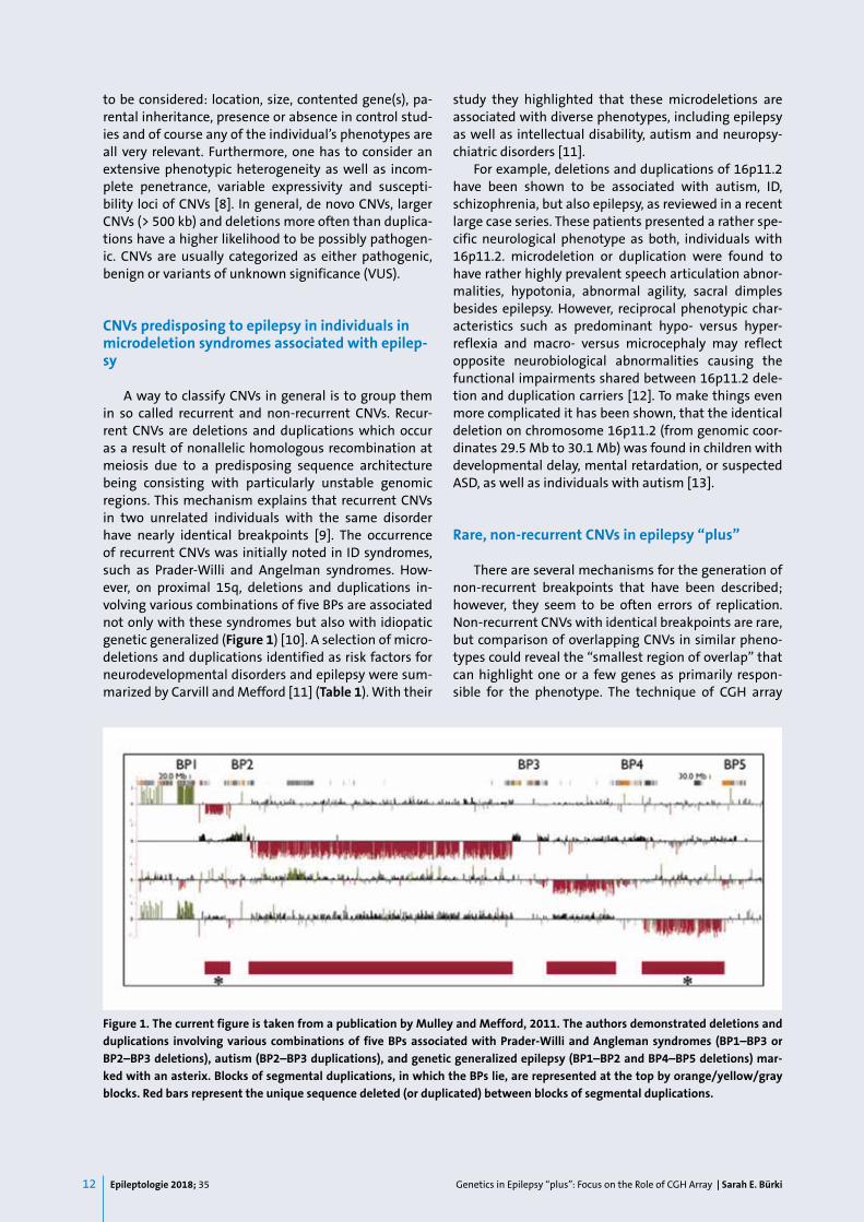
A way to classify CNVs in general is to group them in so called recurrent and non-recurrent CNVs. Recur-rent CNVs are deletions and duplications which occur as a result of nonallelic homologous recombination at meiosis due to a predisposing sequence architecture being consisting with particularly unstable genomic regions. This mechanism explains that recurrent CNVs in two unrelated individuals with the same disorder have nearly identical breakpoints [9]. The occurrence of recurrent CNVs was initially noted in ID syndromes, such as Prader-Willi and Angelman syndromes. How-ever, on proximal 15q, deletions and duplications in-volving various combinations of five BPs are associated not only with these syndromes but also with idiopatic genetic generalized (Figure 1) [10]. A selection of micro-deletions and duplications identified as risk factors for neurodevelopmental disorders and epilepsy were sum-marized by Carvill and Mefford [11] (Table 1). With their
study they highlighted that these microdeletions are associated with diverse phenotypes, including epilepsy as well as intellectual disability, autism and neuropsy-chiatric disorders [11].
For example, deletions and duplications of 16p11.2 have been shown to be associated with autism, ID, schizophrenia, but also epilepsy, as reviewed in a recent large case series. These patients presented a rather spe-cific neurological phenotype as both, individuals with 16p11.2. microdeletion or duplication were found to have rather highly prevalent speech articulation abnor-malities, hypotonia, abnormal agility, sacral dimples besides epilepsy. However, reciprocal phenotypic char-acteristics such as predominant hypo- versus hyper-reflexia and macro- versus microcephaly may reflect opposite neurobiological abnormalities causing the functional impairments shared between 16p11.2 dele-tion and duplication carriers [12]. To make things even more complicated it has been shown, that the identical deletion on chromosome 16p11.2 (from genomic coor-dinates 29.5 Mb to 30.1 Mb) was found in children with developmental delay, mental retardation, or suspected ASD, as well as individuals with autism [13].
Rare, non-recurrent CNVs in epilepsy “plus”
There are several mechanisms for the generation of non-recurrent breakpoints that have been described; however, they seem to be often errors of replication. Non-recurrent CNVs with identical breakpoints are rare, but comparison of overlapping CNVs in similar pheno-types could reveal the ‘‘smallest region of overlap’’ that can highlight one or a few genes as primarily respon-sible for the phenotype. The technique of CGH array
Figure 1. The current figure is taken from a publication by Mulley and Mefford, 2011. The authors demonstrated deletions and duplications involving various combinations of five BPs associated with Prader-Willi and Angleman syndromes (BP1–BP3 or BP2–BP3 deletions), autism (BP2–BP3 duplications), and genetic generalized epilepsy (BP1–BP2 and BP4–BP5 deletions) mar-ked with an asterix. Blocks of segmental duplications, in which the BPs lie, are represented at the top by orange/yellow/gray blocks. Red bars represent the unique sequence deleted (or duplicated) between blocks of segmental duplications.

13Epileptologie 2018; 35Genetics in Epilepsy “plus”: Focus on the Role of CGH Array | Sarah E. Bürki
led to the detection of rare CNVs in 8.8 % (7/80) of the adults and children with ID or developmental delay, and childhood-onset epilepsy. The CNVs involved known mi-crodeletion syndromes (16p11.2, 16p13.11 and 2q13) but also rare CNV encompassing known disease genes, such as SCN1A in four individuals. Such a finding is rel-evant, as deletions disrupting SCN1A might be associ-ated with single gene abnormalities [14]. In the pres-ence of SCN1A abnormalities certain anti-seizure medi-cations should be avoided because they make seizures worse, and other medications or diet are more likely to be associated with improved seizure control.
As mentioned before, the importance of CGH ar-ray has been further increased through its role in the identification of disease-causing genes. The identifica-tion of regions of overlapping CNVs between patients with similar neurodevelopmental phenotypes and epilepsy, for example deletions of CDKL5 in girls with severe epilepsy and a Rett syndrome-like phenotype, might be a starting point toward identifying additional epilepsy genes [15]. The former only candidate epilep-sy gene CHD2 was identified by being the only shared gene within several reported overlapping CNVs of the chromosome 15q26.1 region associated with complex
Table 1: Selected microdeletion and -duplication syndromes associated with phenotypic heterogeneity
Table adapted from Carvill and Mefford, Curr Opin Gen Dev 2013 [11]
Genomic location Coordinates (hg19) for critical region (Mb)
Associated phenotypes, with focus on ID: Intellectual disability, EPI: epilepsy,
ASD: Autism spectrum disorder, and MCA (multiple congenital anomalies
1q21.1 Chr1: 146.5–147.5 ID, EPI, MCA
3q29 Chr3: 195.8–197.4 ID, EPI
10q22q23 Chr10: 81.5–89.0 ID
15q11.2 Chr15: 22.8–23.1 ID, EPI, ASD
15q13.3 Chr15: 31.3–32.5 ID, EPI, ASD
15q24 Chr15: 74.4–75.5 ID, ASD
16p11.2 Chr16: 28.8–30.2 ID, ASD, specifically speech disorder
16p12.2 Chr16: 21.9–22.5 ID, EPI
16p13.11 Chr16: 15.0–16.3 ID, ASD, EPI
17q12 Chr17: 34.8–36.3 ID, ASD, EPI
17q21.3 Chr17: 43.7–44.3 ID
22q11.2 distal Chr22: 21.8–23.7 ID, MCA
22q11.2 distal Chr22: 21.8–23.7 ID, MCA
14 Epileptologie 2018; 35 Genetics in Epilepsy “plus”: Focus on the Role of CGH Array | Sarah E. Bürki
phenotypes including not only developmental delay but epilepsy with photosensitivity [16]. Another similar example is that of a de novo microdeletion at 9q33.3-q34.11 encompassing STXBP1 in a girl with mental re-tardation and Early Infantile Epileptic Encephalopathy (EIEE), characterized by tonic seizures, seizure intrac-tability and characteristic suppression-burst pattern on EEG. After mutation analysis of the candidate gene STXBP1 four other unrelated EIEE patients revealed het-erozygous missense mutations in the same gene [17].
Conclusion
CGH array is a method of genetic testing that has been shown to have a significant impact on epilepsy re-search and diagnosis, being that CNVs are an important genetic cause of epilepsy in children. Pathogenic CNVs are most likely to be found in cases where epilepsy is associated with developmental delay, intellectual disa-bility or autism spectrum disorder, especially in the set-ting of dysmorphic features. CGH array testing to evalu-ate for potential pathogenic CNVs should be performed in patients with epilepsy “plus” (epilepsy combined with intellectual disability, malformations, dysmorphic stigmata, ASD or other features). However, evaluating the implications of a potentially pathogenic CNV for an individual patient can be challenging. To interpret the findings of a particular CNV and its possible degree of pathogenicity and the reproductive risk for the family the clinician should seek the assistance of a geneticist. Once a possibly disease causing or contributing CNV has been found the advantages to the patient and clini-cian include ending the often long diagnostic „odyssey“, providing families with prognostic information and possible features of the syndrome that may be relevant for clinical management and in an increasing number of cases providing families access to syndrome-specific research or patient organizations.
References
1. Sillanpaa M, Schmidt D. Natural history of treated childhood-onset epi-
lepsy: prospective, long-term population-based study. Brain 2006; 129:
617-624
2. Thomas RH, Berkovic SF. The hidden genetics of epilepsy – a clinically
important new paradigm. Nat Rev Neurol 2014; 10: 283-292
3. Miller DT, Adam MP, Aradhya S et al. Consensus statement: chromosomal
microarray is a first-tier clinical diagnostic test for individuals with deve-
lopmental disabilities or congenital anomalies. Am J Hum Genet 2010;
86: 749-764
4. Olson, H, Shen Y, Avallone J et al. Copy number variation plays an impor-
tant role in clinical epilepsy. Ann Neurol 2014; 75: 943-958
5. Mullen SA, Carvill GL, Bellows S et al. Copy number variants are frequent
in genetic generalized epilepsy with intellectual disability. Neurology
2013; 81: 1507-1514
6. Borlot F, Regan BM, Basset AS et al. Prevalence of pathogenic copy
number variation in adults with pediatric-onset epilepsy and intellectual
disability. JAMA Neurol 2017; 74: 1301-1311
7. Zhang ZF, Ruivenkamp C, Staaf J et al. Detection of submicroscopic con-
stitutional chromosome aberrations in clinical diagnostics: a validation
of the practical performance of different array platforms. Eur J Hum Ge-
net 2008; 16: 786-792
8. Nowakowska B. Clinical interpretation of copy number variants in the
human genome. J Appl Genetics 2017; 58: 449-457
9. Mefford HC. CNVs in Epilepsy. Curr Genet Med Rep 2014; 2: 162-167
10. Mulley JC, Mefford HC. Epilepsy and the new cytogenetics. Epilepsia
2011; 52: 423-432
11. Carvill GL, Mefford HC. Microdeletion syndromes. Curr Opin Gen Dev
2013, 23: 232-239
12. Steinmann KJ, Spence SJ, Ramocki B et al. 16p11.2 deletion and duplica-
tion: Characterizing neurological phenotypes in a large clinically ascer-
tained cohort. Am J Med Genet A 2016; 170: 2943-2955
13. Weiss LA, Shen Y, Korn JM. Association between microdeletion and micro-
duplication at 16p11.2 and autism. N Engl J Med 2008; 358: 667-675
14. Fry AE, Rees E, Thompson R et al. Pathogenic CVN and SCN1A mutations
in patients with ID and childhood onset epilepsy. BMC Med Genet 2016;
17: 34
15. Erez A, Patel AJ, Wang X et al. Alu-specific microhomology-mediated de-
letions in CDKL5 in females with early-onset seizure disorder. Neuroge-
netics 2009; 10: 363-369
16. Galizia EC, Myers CT, Leu C et al. CHD2 variants are a risk factor for pho-
tosensitivity in epilepsy. Brain 2015; 138: 1198-1207
17. Saitsu H, Kato M, Mizuguchi T et al. De novo mutations in the gene enco-
ding STXBP1 (MUNC18-1) cause early infantile epileptic encephalopathy.
Nat Genet 2008; 40:782-788
Address for correspondence:Dr. Sarah E. BürkiDepartment of Neuropediatrics,Development and RehabilitationUniversity Chidren’s HospitalInselspitalCH 3010 BernTel. 0041 31 632 94 [email protected]

15Epileptologie 2018; 35Seizures and Epilepsies due to Channelopathies and... | C. M. Korff, F. Picard
Epileptologie 2018; 35: 15 – 16
Key words: Ion channels, neurotransmitters, immunity, genetics Introduction
The study of genetic and immune aspects in epi-lepsy has been increasing exponentially over the past decade. Both components have helped to better under-stand the pathophysiological mechanisms at the basis of seizure genesis. Hundreds of mutations in genes coding for ion channels, neurotransmitter receptors or synaptic proteins have been reported as causing vari-able types of epilepsies. During the same period, auto-antibodies targeting the very same molecules have been discovered and recognized as causing acute en-cephalitides with seizures. These parallel mechanisms at the basis of multiple situations in which seizures can be observed have been the object of an article we pub-lished in a recent issue of Molecular Syndromology [1]. We here summarize and update the illustrative exam-ples described in this paper in order to draw attention of the readers on this fascinating topic.
NMDA-receptor, NR1 subunit
The NMDA receptor is an ion channel permeable to sodium, potassium, and calcium. It is found at excita-tory synapses throughout the brain, and is composed of four subunits. Two of them are of the NR1 glycine-bind-ing subtype and are ubiquitous. The two other subunits are glutamate-binding and of variable subtypes (NR2A, NR2B, NR2C, NR2D, NR3A or NR3B). The gene that en-codes NR1 is GRIN1 (OMIM : 138249); it is located at 9q34.3. GRIN1 mutations that generate protein loss-of-function have been recently related to an encepha-lopathy characterized by profound and early-onset de-velopmental delay, seizures of variable types (including infantile spasms, tonic and atonic seizures, hypermotor seizures, focal dyscognitive seizures, febrile seizures,
Christian M. Korff1 and Fabienne Picard2
1 Pediatric Neurology Unit, Child and Adolescent Department, University Hospitals, Geneva2 Epileptology Unit, Clinical Neurosciences Department, University Hospitals, Geneva
generalized seizures, and status epilepticus), abnormal movements, autistic features and sleep difficulties [2].
This phenotype is very similar to that reported earli-er in association with auto-antibodies directed against the same subunit. After an initial report published in 2007, 100 patients aged 5 to 76 years were reported as having a homogeneous association of symptoms re-lated to the presence of circulating anti-NMDA receptor antibodies [3]. Symptoms included in the majority of patients acute- or subacute-onset psychiatric troubles, seizures, abnormal movements and dysautonomic fea-tures. Severe sleep disorders have also been reported later. Immunohistochemical analyses from all patients’ sera and cerebrospinal fluid samples showed that NR1 was the precise target against which these antibodies reacted. Up to now, the pathophysiology of these anti-bodies is explained by internalization and reduction of the number of synaptic NMDA receptors. The prognosis of this encephalitis is considered as rather favorable, as most patients will respond to promptly initiated immu-nomodulatory or immunosuppressive therapies. A cer-tain number of women present an underlying ovarian tumor at the basis of the auto-immune reaction, whose removal usually allows complete recovery.
LGI
Leucin-rich glioma-inactivated 1 (LGI1) is a synaptic protein dimer that binds to the presynaptic and post-synaptic metalloproteinases ADAM22 and 23. This complex regulates the function of AMPA glutamater-gic receptors and voltage-gated potassium channels [4]. LGI1 (OMIM : 604619), also called epitempin, is the gene that encodes LGI1. It is located at 10q23.33.
LGI1 mutations have been associated with autoso-mal dominant partial epilepsy with auditory features (ADPEAF) (or autosomal dominant lateral temporal lobe epilepsy (ADLTLE)). This epilepsy is characterized by variable ictal auditory symptoms, which include unformed simple sounds, including humming, buzz-ing, or ringing, distortions, such as volume changes,
Seizures and Epilepsies due to Channelopathies and Neurotransmitter Receptor Dysfunction: A Parallel Between Genetic and Immune Aspects

16 Epileptologie 2018; 35 Seizures and Epilepsies due to Channelopathies and... | C. M. Korff, F. Picard
or complex sounds, such as specific songs or voices, or a receptive aphasia [5]. Convulsions may follow these initial features. The course of the epilepsy is usually fa-vorable with excellent response to antiepileptic drugs. Sporadic cases have been described, but inherited mu-tations represent the vast majority of all those report-ed. All known mutations seem to induce a loss of func-tion of LGI1, be it by loss of expression, loss of synaptic secretion, or loss of interaction with its main receptor, ADAM22.
Auto-antibodies against LGI1 have been reported in 2010 as being responsible for a form of limbic en-cephalitis previously attributed to potassium channels [6]. This encephalitis is characterized by acute onset of seizures (often, but not exclusively, facio-brachial and dystonic), cognitive decline involving memory and be-havior, and sleep disturbance. Abnormal MRI signals are often noted in the mesiotemporal regions, and hy-ponatremia is frequently diagnosed.
In presence of these antibodies, the interaction be-tween LGI1 and ADAM22/ADAM23 is inhibited (i.e., the ligand-receptor interaction is blocked). It is thought that consequently, the reduced AMPA receptor function on inhibitory interneurons could cause disinhibition of excitatory neurons, at the basis of seizures and cogni-tive symptoms observed in these patients. A loss of maturation of synapses through dysfunctional NMDA receptors and AMPA receptors is also suspected [7]. The course of the disease is usually favorable with a rapid response to immunomodulatory or immunosuppres-sive therapies. Cognitive sequelae may persist.
Conclusion
Genetic or dysimmune causes are at the basis of certain epilepsies involving ion channels, neurotrans-mitters, or their receptors. For unknown reasons, the clinical presentation associated with certain mutations or circulating auto-antibodies targeting their product may either be similar, or very different. Thus, the basic mechanisms at the basis of such diseases remain to be fully understood.
Acknowledgements
This summary of the original article by Lascano AM, Korff CM, Picard F. Seizures and epilepsies due to channelopathies and neurotransmitter recep-tor dysfunction: A parallel between genetic and im-mune aspects. Mol Syndromol 2016; 7: 197-209 (DOI:10.1159/000447707), is published with kind per-mission granted by S. Karger AG, Basel, publisher of Molecular Syndromology.
References
1. Lascano AM, Korff CM, Picard F. Seizures and epilepsies due to channelo-
pathies and neurotransmitter receptor dysfunction: A parallel between
genetic and immune aspects. Mol Syndromol 2016; 7: 197-209
2. Lemke JR, Geides K, Helbig KL et al. Delineating the GRIN1 phenotypic
spectrum: A distinct genetic NMDA receptor encephalopathy. Neurology
2016; 86: 2171-2178
3. Dalmau J, Gleichman AJ, Hughes EG et al. Anti-NMDA-receptor ence-
phalitis: case series and analysis of the effects of antibodies. Lancet Neu-
rol 2008; 7: 1091-1098
4. van Sonderen A, Petit-Pedrol M. Dalmau J, Tilulaer MJ. The value of LGI1,
Caspr2 and voltage-gated potassium channel antibodies in encephalitis.
Nat Rev Neurol 2017; 13: 290-301
5. Ottman R. Autosomal Dominant Partial Epilepsy with Auditory Features,
in GeneReviews® [Internet]. Seattle: University of Washington, 2015
6. Lai M, Huijbers MG, Lancater E et al. Investigation of LGI1 as the anti-
gen in limbic encephalitis previously attributed to potassium channels: a
case series. Lancet Neurol 2010; 9: 776-785
7. Fukata Y, Yokoi N, Miyazaki Y, Fukata M. The LGI1-ADAM22 protein com-
plex in synaptic transmission and synaptic disorders. Neurosci Res 2017;
116: 39-45
Address for correspondence: PD Dr Christian M. KorffPediatric Neurology UnitChildren’s Hospital6 Rue Willy-DonzéCH 1211 Geneva 14 Tel. 0041 22 372 45 72Fax 0041 22 372 54 89 [email protected]

17Epileptologie 2018; 35Seizures and Epilepsies due to Channelopathies and... | C. M. Korff, F. Picard
Epileptologie 2018; 35: 17 – 18
Schlüsselwörter: Ionenkanäle, Neurotransmitter, Immu-nität, Genetik
Einleitung
Das Forschungsaufkommen zu genetischen und im-munologischen Aspekten der Epilepsie ist innerhalb der letzten zehn Jahre exponentiell gestiegen. Beide Kom-ponenten haben zu einem besseren Verständnis der pathophysiologischen Mechanismen der Anfallsgenese beigetragen. Für Hunderte von Mutationen in Genen, die für Ionenkanäle, Neurotransmitter-Rezeptoren oder synaptische Proteine kodieren, wurde eine ursächliche Beteiligung an unterschiedlichen Epilepsieformen be-schrieben. Im selben Zeitraum wurden Autoantikörper gegen ebendiese Moleküle entdeckt und als Ursache für akute Enzephalitiden mit Anfällen identifiziert. Die-se parallelen ursächlichen Mechanismen der vielfälti-gen Anfallssituationen waren Gegenstand eines Arti-kels, den wir in einer der letzten Ausgaben von Mole-cular Syndromology veröffentlicht haben [1]. Im Vorlie-genden werden die zur Veranschaulichung dienenden Beispiele aus diesem Fachartikel zusammengefasst und aktualisiert, um dem Leser diese faszinierende Thema-tik nahezubringen.
NMDA-Rezeptor, NR1-Untereinheit
Der NMDA-Rezeptor ist ein für Natrium, Kalium und Kalzium durchlässiger Ionenkanal. Er kommt an erre-genden Synapsen im gesamten Gehirn vor und besteht aus vier Untereinheiten. Zwei dieser Untereinheiten ge-hören zum Glycin-bindenden NR1-Subtyp und sind ubi-quitär. Zwei weitere Untereinheiten binden Glutamat und gehören unterschiedlichen Subtypen an (NR2A, NR2B, NR2C, NR2D, NR3A oder NR3B). Für NR1 kodiert das auf 9q34.3 lokalisierte Gen GRIN1 (OMIM: 138249). GRIN1-Mutationen, die mit einem Protein-Funkti-onsverlust einhergehen, wurden in jüngerer Zeit mit einer Enzephalopathie in Zusammenhang gebracht,
Christian M. Korff1 and Fabienne Picard2
1 Pediatric Neurology Unit, Child and Adolescent Department University Hospitals, Geneva2 Epileptology Unit, Clinical Neurosciences Department, University Hospitals, Geneva
die durch eine tiefgreifende und früh einsetzende Ent-wicklungsverzögerung, Anfälle unterschiedlicher Art (zum Beispiel infantile Spamen, tonische und atonische Anfälle, hypermotorische Anfälle, fokale dyskog-nitive Anfälle, febrile Anfälle, generalisierte Anfälle und Status epilepticus), abnorme Bewegungen, autistische Merkmale und Schlafprobleme gekennzeichnet ist [2].
Dieser Phänotyp weist grosse Ähnlichkeit auf mit dem Phänotyp, der bereits im Zusammenhang mit ge-gen dieselbe Untereinheit gerichteten Autoantikörpern beschrieben wurde. Nach einem ersten, 2007 veröffent-lichten Bericht wiesen 100 Patienten im Alter zwischen 5 und 76 Jahren einen einheitlichen Symptomenkom-plex auf, der durch zirkulierende Antikörper gegen den NMDA-Rezeptor bedingt war [3]. Zu den Symptomen gehörten bei der Mehrzahl dieser Patienten akute oder subakute psychiatrische Störungen, Anfälle, abnorme Bewegungen und Merkmale einer dysautonomen Stö-rung. Später wurde ausserdem über schwere Schlafstö-rungen berichtet. Immunhistochemische Serum- und Liquoranalysen aller Patienten zeigten, dass sich diese Antikörper gezielt gegen NR1 richteten. Bislang erklärt man sich die Pathophysiologie dieser Antikörper durch die Internalisierung und zahlenmässige Verminderung der synaptischen NMDA-Rezeptoren. Die Prognose gilt bei dieser Enzephalitis als relativ günstig, da die meis-ten Patienten auf zeitnah eingeleitete immunmodu-latorische oder immunsuppressive Therapien anspre-chen. Bei manchen Frauen bildet ein zugrundeliegen-des Ovarialkarzinom den Ausgangspunkt der Autoim-munreaktion; nach dessen Entfernung ist in der Regel eine vollständige Genesung möglich.
LGI
Das Leucin-rich glioma-inactivated 1 (LGI1) ist ein dimeres synaptisches Protein, das an die prä- und post-synaptischen Metalloproteasen ADAM22 und 23 bindet. Dieser Komplex reguliert die Funktion von glutamater-gen AMPA-Rezeptoren und spannungsabhängigen Ka-liumkanälen [4]. Für LGI1 kodiert das Gen LGI1 (OMIM: 604619), das auch als Epitempin bezeichnet wird. Gen-ort ist 10q23.33.
Seizures and Epilepsies due to Channelopathies and Neurotransmitter Receptor Dysfunction: A Parallel between Genetic and Immune Aspects

18 Epileptologie 2018; 35 Seizures and Epilepsies due to Channelopathies and... | C. M. Korff, F. Picard
LGI1-Mutationen sind mit autosomal-dominanter partieller Epilepsie mit auditorischen Auren (ADPE-AF, auch autosomal-dominante laterale Temporallap-pen-Epilepsie (ADLTLE) assoziiert. Diese Epilepsie ist gekennzeichnet durch unterschiedliche iktale audito-rische Symptome, darunter einfache ungeformte Ge-räusche (z. B. Summen, Brummen oder Klingeln), Ver-zerrungen (z. B. Lautstärkenänderungen), komplexe Geräusche (z. B. bestimmte Lieder oder Stimmen) oder rezeptive Aphasie [5]. An diese anfänglichen Störun-gen können sich Konvulsionen schliessen. Die Epilepsie zeigt in der Regel einen günstigen Verlauf und spricht hervorragend auf Antiepileptika an. Zwar wurden spo-radische Fälle beschrieben, doch beruhen die weitaus meisten berichteten Fälle auf vererbten Mutationen. Alle bekannten Mutationen scheinen einen Funktions-verlust von LGI1 zu induzieren – sei es durch einen Ex-pressionsausfall, einen Verlust der synaptischen Sekre-tion oder den Interaktionsverlust mit seinem wichtigs-ten Rezeptor, ADAM22.
Autoantikörper gegen LGI1 sind laut einem Bericht aus dem Jahr 2010 verantwortlich für eine Form der limbischen Enzephalitis, die bis dahin auf Kaliumkanä-le zurückgeführt worden war [6]. Diese Enzephalitis ist charakterisiert durch das akute Einsetzen von Anfällen (häufig, aber nicht ausschliesslich faziobrachial und dystonisch), eine Verschlechterung der gedächtnis- und verhaltensbezogenen kognitiven Funktionen sowie Schlafstörungen. Oft werden abnorme MRT-Signale in den mesiotemporalen Regionen beobachtet, und häu-fig wird eine Hyponatriämie diagnostiziert.
In Gegenwart dieser Antikörper ist die Wechselwir-kung zwischen LGI1 und ADAM22/ADAM23 gehemmt (d. h., die Ligand-Rezeptor-Interaktion ist blockiert). Man geht davon aus, dass die infolgedessen verminder-te Funktion der AMPA-Rezeptoren auf inhibitorischen Interneuronen eine Disinhibition exzitatorischer Neu-ronen hervorruft, die wiederum für die bei diesen Pa-tienten beobachteten Anfälle und kognitiven Ausfälle verantwortlich ist. Eine Beeinträchtigung der Synapsen-reifung durch dysfunktionelle NMDA- und AMPA-Re-zeptoren wird ebenfalls vermutet [7]. Die Erkrankung zeigt in der Regel einen günstigen Verlauf und spricht rasch auf immunmodulatorische oder immunsuppres-sive Therapien an. Die kognitiven Folgeerscheinungen können fortbestehen.
Schlussfolgerung
Bestimmte Epilepsien mit Beteiligung von Ionen-kanälen, Neurotransmittern oder deren Rezeptoren haben genetische oder immunologische Ursachen. Aus unbekannten Gründen kann das jeweilige klinische Bild, das mit bestimmten Mutationen oder zirkulieren-den Autoantikörpern gegen deren Produkte einhergeht, entweder einheitlich oder sehr unterschiedlich sein. Zur vollständigen Aufklärung der Mechanismen, die derar-
tigen Erkrankungen zugrunde liegen, sind daher weite-re Untersuchungen erforderlich.
Danksagungen
Die Veröffentlichung dieser Zusammenfassung der Originalarbeit von Lascano AM, Korff CM, Picard F, «Seizures and epilepsies due to channelopathies and neurotransmitter receptor dysfunction: A parallel bet-ween genetic and immune aspects.» Mol Syndromol 2016; 7: 197-209 (DOI:10.1159/000447707), erfolgt mit freundlicher Genehmigung der S. Karger AG, Basel, der Herausgeberin von Molecular Syndromology.
References
1. Lascano AM, Korff CM, Picard F. Seizures and epilepsies due to channelo-
pathies and neurotransmitter receptor dysfunction: A parallel between
genetic and immune aspects. Mol Syndromol 2016; 7: 197-209
2. Lemke JR, Geides K, Helbig KL et al. Delineating the GRIN1 phenotypic
spectrum: A distinct genetic NMDA receptor encephalopathy. Neurology
2016; 86: 2171-2178
3. Dalmau J, Gleichman AJ, Hughes EG et al. Anti-NMDA-receptor ence-
phalitis: case series and analysis of the effects of antibodies. Lancet Neu-
rol 2008; 7: 1091-1098
4. van Sonderen A, Petit-Pedrol M. Dalmau J, Tilulaer MJ. The value of LGI1,
Caspr2 and voltage-gated potassium channel antibodies in encephalitis.
Nat Rev Neurol 2017; 13: 290-301
5. Ottman R. Autosomal Dominant Partial Epilepsy with Auditory Features,
in GeneReviews® [Internet]. Seattle: University of Washington, 2015
6. Lai M, Huijbers MG, Lancater E et al. Investigation of LGI1 as the anti-
gen in limbic encephalitis previously attributed to potassium channels: a
case series. Lancet Neurol 2010; 9: 776-785
7. Fukata Y, Yokoi N, Miyazaki Y, Fukata M. The LGI1-ADAM22 protein com-
plex in synaptic transmission and synaptic disorders. Neurosci Res 2017;
116: 39-45
Address for correspondence: PD Dr Christian M. KorffPediatric Neurology UnitChildren’s Hospital6 Rue Willy-DonzéCH 1211 Geneva 14 Tel. 0041 22 372 45 72Fax 0041 22 372 54 89 [email protected]

19Epileptologie 2018; 35Seizures and Epilepsies due to Channelopathies and... | C. M. Korff, F. Picard
Christian M. Korff1 and Fabienne Picard2
1 Pediatric Neurology Unit, Child and Adolescent Department, University Hospitals, Geneva2 Epileptology Unit, Clinical Neurosciences Department, University Hospitals, Geneva
Seizures and Epilepsies due to Channelopathies and Neurotransmitter Receptor Dysfunction: A Parallel between Genetic and Immune Aspects
Epileptologie 2018; 35: 19 – 20
Mots clés : Canaux ioniques, neurotransmitter, immuni-té, génétique
Introduction
L’étude des aspects génétiques et immunologiques de l’épilepsie a connu un développement exponentiel ces dix dernières années. Ces deux composantes ont permis de mieux comprendre les mécanismes physio-pathologiques à l’origine des crises. Des centaines de mutations observées dans des gènes codant des ca-naux ioniques, des récepteurs de neurotransmetteurs ou des protéines synaptiques ont été recensées pour leur rôle dans des formes variables d’épilepsie. Durant la même période, des auto-anticorps dirigés contre ces mêmes molécules ont été découverts et identifiés comme la cause d’encéphalites aiguës accompagnées de crises. Ces mécanismes parallèles à l’origine de di-verses situations de crises ont fait l’objet d’un article que nous avons publié dans un récent numéro de Mole-cular Syndromology [1]. Nous résumons et actualisons ici les exemples illustratifs décrits dans cet article afin d’attirer l’attention du lecteur sur cette thématique fas-cinante.
Récepteur NMDA, sous-unité NR1
Le récepteur NMDA est un canal ionique perméable au sodium, au potassium et au calcium. Localisé sur les synapses excitatrices dans tout le cerveau, il est com-posé de quatre sous-unités. Deux d’entre elles font partie du sous-type NR1 liant la glycine et sont obliga-toires. Les deux autres sous-unités lient le glutamate et sont de sous-types variables (NR2A, NR2B, NR2C, NR2D, NR3A ou NR3B). Le gène qui code NR1 est GRIN1 (OMIM : 138249) localisé sur le chromosome 9q34.3. Les mutations du gène GRIN1 qui génèrent une perte de fonction des protéines ont été récemment mises en lien avec une encéphalopathie caractérisée par un retard de développement profond et précoce, des crises
de différents types (notamment des spasmes infantiles, des crises toniques et atoniques, des crises hypermo-trices, des crises dyscognitives focales, des convulsions fébriles, des crises généralisées et un état de mal épi-leptique), des mouvements anormaux, des caractéris-tiques autistiques et des troubles du sommeil [2].
Ce phénotype est très similaire à celui déjà rapporté en lien avec des auto-anticorps dirigés contre la même sous-unité. Après un premier rapport publié en 2007, les cas de 100 patients âgés de 5 à 76 ans ont été rap-portés comme présentant un ensemble homogène de symptômes lié à la présence d’anticorps circulants diri-gés contre le récepteur NMDA [3]. Chez la majorité des patients, les symptômes incluaient des troubles psy-chiatriques d’apparition aiguë ou subaiguë, des crises, des mouvements anormaux et des caractéristiques dysautonomiques. Des troubles sévères du sommeil ont également été rapportés ultérieurement. Les analyses immunohistochimiques des échantillons de liquide céphalorachidien et de sérum de tous les patients ont montré que ces anticorps étaient précisément dirigés contre le gène NR1. Jusqu’à présent, la physiopatholo-gie de ces anticorps s’explique par l’internalisation et la réduction du nombre de récepteurs NMDA synaptiques. Le pronostic de cette encéphalite est considéré comme plutôt favorable, car la plupart des patients répondent aux thérapies immunomodulatrices ou immunosup-pressives initiées rapidement. Un certain nombre de femmes présentent une tumeur ovarienne sous-ja-cente à l’origine de la réaction auto-immune. L’ablation de cette tumeur permet généralement une guérison complète.
LGI
LGI1 (Leucin-rich glioma-inactivated 1) est une pro-téine synaptique dimère qui se lie aux métalloprotéi-nases présynaptiques et postsynaptiques ADAM22 et 23. Ce complexe régule la fonction des récepteurs glutama-tergiques AMPA et des canaux potassiques voltage-dé-pendants [4]. LGI1 (OMIM : 604619), également appelé épitempine, est le gène qui code LGI1. Il est localisé sur le chromosome 10q23.33.

20 Epileptologie 2018; 35 Seizures and Epilepsies due to Channelopathies and... | C. M. Korff, F. Picard
Les mutations de LGI1 sont associées à une épilep-sie partielle autosomique dominante avec des signes auditifs (ADPEAF) (ou épilepsie du lobe temporal latéral autosomique dominante (ADLTLE)). Cette épilepsie est caractérisée par des symptômes auditifs ictaux variables, parmi lesquels des sons simples informes (notamment des fredonnements, bourdonnements, tintements), des distorsions (telles que des changements de volume), des sons complexes (tels que des chansons ou voix spéci-fiques) ou une aphasie réceptive [5]. Ces caractéristiques initiales peuvent être suivies de convulsions. L’évolution de cette épilepsie est généralement favorable, avec une excellente réponse aux médicaments antiépileptiques. Si des cas sporadiques ont été décrits, la très grande majori-té des cas rapportés repose néanmoins sur des mutations héréditaires. Toutes les mutations connues semblent in-duire une perte de fonction de LGI1, que ce soit par perte d’expression, perte de sécrétion synaptique ou perte d’in-teraction avec son récepteur principal, ADAM22.
Selon un rapport de 2010, des auto-anticorps dirigés contre LGI1 sont responsables d’une forme d’encéphalite limbique jusque-là attribuée aux canaux potassiques. [6]. Cette encéphalite se caractérise par une apparition aiguë des crises (souvent, mais pas exclusivement, facio-brachiales et dystoniques), un déclin cognitif touchant la mémoire et le comportement, et des troubles du som-meil. Des signaux anormaux sont souvent observés à l’IRM dans les régions mésiotemporales et une hypona-trémie est fréquemment diagnostiquée.
En présence de ces anticorps, l’interaction entre LGI1 et ADAM22/ADAM23 est inhibée (l’interaction ligand/récepteur est bloquée). On suppose qu’en conséquence, la réduction de la fonction des récepteurs AMPA sur les interneurones inhibiteurs provoque une désinhibition de neurones excitateurs à son tour responsable des crises et des symptômes cognitifs observés chez ces patients. On suspecte également une perte de maturation des synapses due à un dysfonctionnement des récepteurs NMDA et AMPA [7]. Cette maladie connaît généralement une évolution favorable avec une réponse rapide aux thé-rapies immunomodulatrices ou immunosuppressives. Des séquelles cognitives peuvent subsister.
Conclusion
Certaines formes d’épilepsie impliquant des canaux ioniques, des neurotransmetteurs ou leurs récepteurs ont des causes génétiques ou immunologiques. Pour des raisons inconnues, la présentation clinique associée à certaines mutations ou certains auto-anticorps circu-lants ciblant leurs produits peut être soit similaire, soit très différente. Par conséquent, les mécanismes fonda-mentaux à la base de ces maladies sont encore entière-ment à élucider.
Remerciements
Ce résumé de l’article original de Lascano AM, Korff CM, Picard F, « Seizures and epilepsies due to channelo-pathies and neurotransmitter receptor dysfunction: A parallel between genetic and immune aspects. » Mol Syn-dromol 2016; 7: 197-209 (DOI:10.1159/000447707), est publié avec l’aimable autorisation de S. Karger AG, Bâle, éditeur de Molecular Syndromology.
References
1. Lascano AM, Korff CM, Picard F. Seizures and epilepsies due to channelo-
pathies and neurotransmitter receptor dysfunction: A parallel between
genetic and immune aspects. Mol Syndromol 2016; 7: 197-209
2. Lemke JR, Geides K, Helbig KL et al. Delineating the GRIN1 phenotypic
spectrum: A distinct genetic NMDA receptor encephalopathy. Neurology
2016; 86: 2171-2178
3. Dalmau J, Gleichman AJ, Hughes EG et al. Anti-NMDA-receptor ence-
phalitis: case series and analysis of the effects of antibodies. Lancet Neu-
rol 2008; 7: 1091-1098
4. van Sonderen A, Petit-Pedrol M. Dalmau J, Tilulaer MJ. The value of LGI1,
Caspr2 and voltage-gated potassium channel antibodies in encephalitis.
Nat Rev Neurol 2017; 13: 290-301
5. Ottman R. Autosomal Dominant Partial Epilepsy with Auditory Features,
in GeneReviews® [Internet]. Seattle: University of Washington, 2015
6. Lai M, Huijbers MG, Lancater E et al. Investigation of LGI1 as the anti-
gen in limbic encephalitis previously attributed to potassium channels: a
case series. Lancet Neurol 2010; 9: 776-785
7. Fukata Y, Yokoi N, Miyazaki Y, Fukata M. The LGI1-ADAM22 protein com-
plex in synaptic transmission and synaptic disorders. Neurosci Res 2017;
116: 39-45
Address for correspondence: PD Dr Christian M. KorffPediatric Neurology UnitChildren’s Hospital6 Rue Willy-DonzéCH 1211 Geneva 14 Tel. 0041 22 372 45 72Fax 0041 22 372 54 89 [email protected]

21Epileptologie 2018; 35Genetic Testing for Epilepsy Surgery | B. P. C. Koeleman, M. W. C. B. Sanders ,K. P. J. Braun
Summary
Dozens of novel epilepsy genes have been discov-ered at a very fast pace in the past decade. This progress improved understanding and management of epilepsy; however, this has not systematically been evaluated for the focal epilepsies that can be cured by epilepsy sur-gery. In this report we discus the impact and possible application of genetic diagnostics in epilepsy surgery evaluation. A review of the available data suggests that epilepsy gene mutations can be useful biomarkers for surgery. These may be negative predictors, such as mutations in genes encoding ion-channels or involved in synaptic function. The association of mutations in mTOR pathway genes with lesional focal epilepsy sug-gest that such mutations may be positive predictors that can improve selection of surgical cases, especially in the MRI-negative cases. However, it is clear that larg-er studies are needed to collect more detailed imaging and to interpret the link between surgery outcome, ob-servational data, knowledge of disease aetiology, and genetics.
Epileptologie 2018; 35: 21 – 28
Key words: Epilepsy surgery, genetics, DNA-diagnostics, prognosis
Gentests für die Epilepsiechirurgie
In den letzten zehn Jahren wurden in äusserst ra-scher Folge Dutzende neuer Epilepsie-Gene entdeckt. Dieser Fortschritt hat zu einem besseren Verständnis und Management der Epilepsie geführt; allerdings wur-de dies für die epilepsiechirurgisch heilbaren fokalen Epilepsien nicht systematisch beurteilt. Der vorliegen-de Bericht erörtert die Auswirkungen und die mögliche Anwendung der Gendiagnostik bei der epilepsiechir-urgischen Beurteilung. Eine Übersicht der verfügbaren Daten legt nahe, dass Mutationen in Epilepsie-Genen
Bobby P.C. Koeleman2, Maurits W.C.B. Sanders1 and Kees P.J. Braun1
1 Department of Child Neurology, Brain Center Rudolf Magnus, University Medical Center Utrecht, the Netherlands2 Department of Genetics, Center for Molecular Medicine, University Medical Center Utrecht, The Netherlands
hilfreiche Biomarker im Hinblick auf chirurgische Op-tionen sein können. Dabei kann es sich um negative Prädiktoren handeln, z. B. Mutationen in Genen, die für Ionenkanäle kodieren oder an synaptischen Funktionen beteiligt sind. Der Zusammenhang zwischen Mutatio-nen im mTOR-Signalweg und läsionsbedingten fokalen Epilepsien lässt vermuten, dass derartige Mutationen positive Prädiktoren darstellen, die eine bessere Selekti-on chirurgisch therapierbarer Fälle ermöglichen, insbe-sondere bei MRT-negativen Patienten. Allerdings steht ausser Zweifel, dass zur Erfassung detaillierterer Bild-gebungsdaten und zur Klärung des Zusammenhangs zwischen chirurgischem Behandlungsergebnis, Beob-achtungsdaten, Wissen um die Krankheitsätiologie und Genetik umfassendere Studien erforderlich sind.
Schlüsselwörter: Epilepsiechirurgie, Genetik, DNA-Diagnostik, Prognose
Tests génétiques pour la chirurgie épileptique
Ces dix dernières années, des douzaines de nou-veaux gènes associés à l’épilepsie ont été découverts à une cadence particulièrement rapide. Si cette avancée a permis une meilleure compréhension et prise en charge de l’épilepsie, elle n’a pas systématiquement été éva-luée pour les formes focales d’épilepsie pouvant être traitées par chirurgie épileptique. Le présent rapport s’intéresse à l’impact et à l’application potentielle des diagnostics génétiques dans l’évaluation de la chirur-gie épileptique. Une analyse des données disponibles suggère que les mutations observées dans les gènes associés à l’épilepsie peuvent être des biomarqueurs utiles pour la chirurgie. Il peut s’agir de facteurs prédic-tifs négatifs, par exemple dans le cas des mutations de gènes codant des canaux ioniques ou impliqués dans une fonction synaptique. Le lien entre les mutations dans des gènes de la voie mTOR et l’épilepsie focale lésionnelle laisse penser que les mutations de ce type
Genetic Testing for Epilepsy Surgery
22 Epileptologie 2018; 35 Genetic Testing for Epilepsy Surgery | B. P. C. Koeleman, M. W. C. B. Sanders ,K. P. J. Braun
peuvent être des facteurs prédictifs positifs suscep-tibles d’améliorer la sélection des cas pour la chirurgie, en particulier chez les patients dont l’IRM est négative. Cependant, il est certain que de plus amples études sont nécessaires pour collecter des données d’imagerie plus détaillées et pour interpréter le lien entre les résul-tats des traitements chirurgicaux, les données observa-tionnelles, les connaissances sur l’étiologie de la mala-die et la génétique.
Mots clés : chirurgie épileptique, génétique, diagnostic génétique, pronostic
Introduction
It stands without a doubt that genetic discoveries have progressed our understanding and management of the epilepsies. Dozens of novel epilepsy genes have been discovered in the past decades, largely as a result of inexpensive and readily available next generation sequencing. These discoveries have set several new paradigms. First, de novo mutations in various epilep-sy genes are now seen as the major cause of sporadic epileptic encephalopathy. Second, the importance of genetic causes in focal epilepsy was established by the observation that inherited or de novo mutation in mul-tiple genes, in particular genes involved in the mTOR pathway, can cause focal epilepsy.
Finally, it remains remarkable that for many, if not all epilepsy genes, a wide spectrum of clinical pheno-types, varying in type of epilepsy and severity of dis-ease, are associated with mutations in the same gene. On the one hand, these genetic discoveries have ena-bled precision medicine, in which the DNA diagnosis limits the diagnostic odyssey and guides disease treat-ment and management. On the other hand, the varia-ble clinical expression raises many questions on how to predict the clinical course of disease, and causes doubt whether genetics alone should determine clinical care.
From this perspective, we discuss the case of epi-lepsy surgery, for which the impact and importance of genetic diagnostics have not been explored substan-tially yet. Several studies have now reported on people with epilepsy that carry a presumed causal mutation in a known epilepsy gene and who are potentially eligible for, or have undergone, surgery. These reports suggest that genetics should play a role in the diagnostic strat-egy and therapeutic approach in people with epilepsy that are considered candidates for epilepsy surgery.
Monogenic causes of epilepsy
Next generation sequencing technology (NGS), in particular whole exome sequencing (WES), through which the coding sequence of all genes in the human genome can be scanned for putative disease-causing
sequence variation, accelerated discovery of novel epi-lepsy genes in the past decade [1]. Major discoveries have been made in epileptic encephalopathy (EE) where many de novo mutations have recently been identified as causative. These include de novo mutations in KCNQ2, a gene that was already known for several decades, in which inherited mutations cause benign familial neona-tal convulsions (BFNC) [2]. De novo missense mutations detected in severe childhood EE cluster in four hotspots of the gene that are important for essential channel properties, namely: the S4 voltage-sensor, the pore, the proximal C-terminal domain that binds phosphatidylin-ositol 4,5-bisphosphate (PIP2) and calmodulin (CaM A), and the more distal calmodulin binding (CaM B) domains [3, 4]. It is therefore assumed that these mis-sense mutations cause EE through a dominant-negative effect on channel function, in contrast to the more vari-able effects of the mutations underlying BFNC that also include complete loss of function mutations, such as deletions. These observations illustrate the phenomena of variable clinical expression that is observed for many epilepsy genes. Like in KCNQ2, the type or location of the mutation can often explain the variable expression. However, this is not the case for all epilepsy genes, and for all mutations. The compelling example is SCN1A, a major epilepsy gene that is associated with a wide spectrum of different disease severities, and is current-ly associated with both common and rare, benign and severe disease. Mutations in SCN1A were reported as a major cause for severe myoclonic epilepsy of infancy (SMEI), also known as Dravet syndrome [5]. It is known as the most common genetic cause of severe epilepsy in infancy. In contrast, segregating mutations have been detected in families affected by generalized epilepsy and febrile seizure “plus” syndrome (GEFS+), a relative benign form of epilepsy with a favourable prognosis [6]. For a significant part, the difference in phenotype can be explained by the type of mutation, where complete loss of function mutations – such as non-sense muta-tions that result in a truncated protein that is vulner-able to nonsense mediated decay – are underlying the severe Dravet syndrome. On the other hand, GEFS+ is associated with missense SCN1A mutations specifically, that have a milder loss of function effect on the protein [7]. This clear association between variants and the two phenotypes demonstrates that rare coding variants of the gene with high to absolute risk for disease, are the main cause for these types of epilepsy. Finally, focal sei-zures may also be part of the semiology in Dravet syn-drome, and as is discussed below, a few reports describe Dravet syndrome patients that also had a malforma-tion of cortical development (MCD) [8, 9]. Furthermore, some families have been described in which a segregat-ing SCN1A mutation was detected in family members that shared the same inherited mutation but showed clinical heterogeneous expression of disease, ranging from febrile seizures (FS), GEFS+, to Dravet syndrome and focal seizures [10, 11].
23Epileptologie 2018; 35Genetic Testing for Epilepsy Surgery | B. P. C. Koeleman, M. W. C. B. Sanders ,K. P. J. Braun
On the other side of the genetic spectrum of aetiol-ogies of epilepsy are common variants that confer very low risk for disease that are typically detected through genome-wide association studies (GWAS). Such studies have now been performed using reasonably sized sam-ples that detected variants in or around SCN1A that are associated with FS alone, focal epilepsy (in particular mesial temporal lobe epilepsy with hippocampal scle-rosis and febrile seizures (mTLE-HS-FS)), and – remark-ably – with all types of common epilepsy, including fo-cal and genetic generalized epilepsy [12 - 16]. Recent large scale WES studies of common familial generalized and focal non-acquired epilepsy, showed a remarkable enrichment of ultra-rare coding variation in known epilepsy genes including SCN1A. This suggests that rare coding variants with a low risk of disease also increase susceptibility to epilepsy [17]. Finally, a few reports and unpublished observations described patients with focal epilepsy who were considered for epilepsy surgery and were found to be carriers of a likely pathogenic SCN1A mutation [8, 9]. Taken together, the genetic evidence for SCN1A shows a remarkable spectrum of disease as-sociated SCN1A variants and their risk for disease, rang-ing from common low risk factors and rare inherited or de novo mutations with absolute risk for disease, to very rare relatively low risk factors. The clinical spec-trum is equally broad, ranging from common benign to severe and very rare syndromes. This broad geno- and phenotypic spectrum associated with SCN1A must be taken into account when clinical decisions are made, especially in the case of surgical evaluation.
Even though the first gene for focal epilepsy, CHRNA4 causing autosomal dominant nocturnal fron-tal lobe epilepsy (ADNFLE), was found in 1995, it has only recently been demonstrated that genetic muta-tions are not only associated with generalized or mul-tifocal epilepsies, but also underlie a broad range of fo-cal epilepsies. The most notable novel gene discoveries have been made in the mTOR-family of genes, with a clinical spectrum of focal epilepsy that ranges from MCD to familial non-lesional focal epilepsy [18 - 22]. Causal mutations underlying these epilepsies have been found to be inherited, occurred de novo in sporad-ic cases, and have been detected as somatic mutations in resected brain tissue, which calls for comprehensive genetic testing and careful clinical genetic counsel-ling. Mutations in DEPDC5, a gene that functions in the GATOR1 complex that inhibits the mTORC1 pathway, are now considered to be among the most common genetic causes of focal epilepsies, including familial fo-cal epilepsy with variable foci (FFEVF), and have been reported in 13% of autosomal dominant sleep-related hypermotor epilepsy (ADSHE or ADNFLE) families [23]. A full overview of these genes and their implication in focal epilepsy is presented in the accompanying article in this issue. The importance of DEPDC5 and related genes in (familial) non-lesional focal epilepsy (FE) and in lesional epilepsies with MCD, in particular focal corti-
cal dysplasia (FCD), may imply that searching for causal germline and somatic gene mutations underlying spo-radic FE has consequences for surgical decision mak-ing. How the detection of such gene mutations should influence the decision to accept or reject a patient for surgery remains to be established.
Goals of epilepsy surgery
Epilepsy surgery is currently the only available cu-rative treatment for pharmacoresistant focal epilepsy. However, it is clear that surgery is an invasive and ir-reversible procedure, and several restrictions apply, making it a safe procedure that is only performed on in-dividuals that have a high probability of becoming sei-zure-free following surgery without unacceptable defi-cits that are the direct result of surgery. To accomplish this, a careful set of diagnostic procedures precedes any successful epilepsy surgery, aimed to accurately local-ize and delineate the epileptogenic zone and possible overlap with eloquent regions.
Presurgical evaluation procedures are complex, and have been outlined and standardized. Until recently, genetic screening was not part of the standard epilepsy surgery evaluation, although it has been mentioned as an important factor to consider in a perspective by Guerrini et al., that included genetic screening in the algorithm for diagnostic strategies and therapeutic ap-proaches in patients with FCD [24].
Although the general aim of epilepsy surgery is to completely remove the epileptogenic focus, surgery is sometimes considered in patients for palliative treat-ment, aiming to reduce seizure load or cure the patient from just one, most burdensome, seizure type when there is a multifocal epilepsy syndrome. Whereas in the past only patients with clear structural MRI-visible le-sions were considered surgical candidates, an increas-ing number of people with refractory epilepsy and nor-mal imaging undergo evaluation, under the assump-tion that their focal epilepsy is caused by a MR-invisible, structural lesion, in particular FCD. The number of MRI-negative patients will decrease with improved imaging techniques, such as higher-field MRI or MRI post-pro-cessing [25, 26]. Currently 60 - 70% of these MRI-neg-ative (but presumed lesional) patients are rejected for surgery, often after extensive and invasive intracranial electrode monitoring. Operated MRI-negative patients have a lower chance of reaching seizure freedom [27]. Furthermore, the absence of a histopathological abnor-mality occurs in ~8% of all operated patients and is a major predictor of poor outcome [28].
MRI-negative patients with refractory focal seizures can either have an ‘invisible’ structural lesion – such as a developmental abnormality – or an underlying ge-netic syndrome, not associated with a lesional source, or a combination of the two (e.g. tuberous sclerosis). We can assume that the lesional MRI-negative patients
24 Epileptologie 2018; 35 Genetic Testing for Epilepsy Surgery | B. P. C. Koeleman, M. W. C. B. Sanders ,K. P. J. Braun
have a higher chance of reaching seizure-freedom after epilepsy surgery than the non-lesional MRI-negative patients with a presumed genetic underlying cause. Therefore, we hypothesize that the crucial differentia-tion between people with operable and non-operable epilepsy (i.e. between a presumed lesional and non-lesional cause of seizures) requires new and reliable biomarkers.
Mutations in novel epilepsy genes are such bio-markers that are currently not routinely implemented in presurgical evaluation. Below we will summarize the current experience with genetic evaluation in patients who were considered surgical candidates, and discuss how preoperative genetic screening may differentiate between eligible and non-eligible surgical candidates. The discussion may be centralized around three crucial questions regarding the utility of genetic testing for presurgical evaluation.
The first question is to what extent can a genetic mutation be a negative predictor for postoperative seizure outcome and may be used to reject patients for surgery even in the presence of an operable lesion. With this question, the distinction must be made be-tween palliative surgery and surgery with the aim to completely cure the patient from all seizures. The sec-ond question is whether mutations in genes can pre-dict seizure freedom in people that have MRI-negative, but presumed lesional FE.
The final question is whether genetic mutations that predict lesional and operable epilepsy (for example the genes associated with FCD) have any additional and useful predictive value next to the current evaluation procedures.
The precise localization of the seizure-onset zone and eloquent cortical regions is challenging in FCD, as predictions of their anatomic locations may not con-form to traditional models utilized for other patho-logic substrates. These limitations must be appreciated fully to achieve higher rates of postoperative seizure freedom. Completeness of resection is an important determinant of outcome. Colocalization of the seizure onset zone with eloquentcortex is a major contributor to incom plete resection and surgical failure. The pre-cise localization of the seizure-onset zone and eloquent cortical regions is challenging in FCD, as predictions of their anatomic locations may not conform to tradition-al models utilized for other pathologic substrates.
These limitations must be appreciated fully to achieve higher rates of postoperative seizure freedom. Completeness of resection is an important determi-nant of outcome. Colocalization of the seizure onset zone with eloquent cortex is a major contributor to in-complete resection and surgical failure. The precise lo-calization of the seizure-onset zone and eloquent corti-cal regions is challenging in FCD, as predictions of their anatomic locations may not conform to traditional models utilized for other pathologic substrates. These limitations must be appreciated fully to achieve higher
rates of postoperative seizure freedom. Completeness of resection is an important determinant of outcome. Colocalization of the seizure onset zone with eloquent cortex is a major contributor to incomplete resection and surgical failure.
The precise localization of the seizure-onset zone and eloquent cortical regions is challenging in FCD, as predictions of their anatomic locations may not con-form to traditional models utilized for other pathologic substrates. These limitations must be appreciated fully to achieve higher rates of postoperative seizure free-dom. Completeness of resection is an important deter-minant of outcome. Colocalization of the seizure onset zone with eloquent cortex is a major contributor to in-complete resection and surgical failure.
The published experience with epilepsy surgery in patients with genetic causes of epilepsy
There are relatively few reports on surgery cases that also carry a pathogenic mutation in an epilepsy gene. We recently reviewed the literature (until January 2017) on surgical outcome in different genetic causes of refractory epilepsy [29]. Only 24 eligible articles were found that described a total of 82 patients who under-went surgery for refractory epilepsy due to 15 differ-ent underlying genetic causes. The most frequent ge-netic abnormalities were mutations in SCN1A (8 cases), DEPDC5 (9 cases), NF1 (21 cases), and microdeletions (12 cases). We subdivided all cases in three broad cat-egories including: “gene mutations involved with chan-nelopathies and disorders of synaptic transmission”, “mTOR pathway gene mutations”, and “other genetic causes of epilepsy”. The most striking finding was the difference between the low rate of seizure-freedom in the germline mutations in genes associated with chan-nelopathies and synaptic transmission disorders (2 out of 14 cases, 14%), versus the high rate of seizure free-dom in the germline mTOR pathway gene mutations (6 out of 11, 55%) and the group of other genetic causes (24 out of 38, 63%, see Table 1).
These observations suggest that channelopathy and synaptic disorder genes are strong negative predic-tors for surgical candidacy, however, more observations for each gene, and type of mutation is clearly needed to come to definite conclusions.
The observations for the individual genes illustrate this point. For SCN1A, 8 cases have been described in two main papers. Barba et al. reported 4 patients who were found to have a MCD out of a series of 120 pa-tients with SCN1A mutations, and two additional cases with MCD and SCN1A mutations that they included through U-task (the European taskforce for epilepsy surgery in children, [8]). All patients showed a pheno-type consistent with Dravet syndrome, yet brain MRI showed periventricular nodular heterotopia (PNH) in 2, and FCD in 3 cases. Two of these FCD cases were oper-
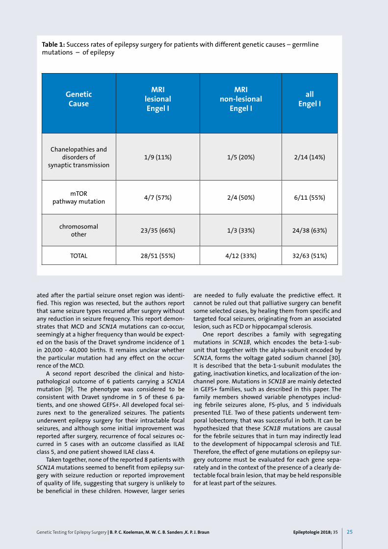
25Epileptologie 2018; 35Genetic Testing for Epilepsy Surgery | B. P. C. Koeleman, M. W. C. B. Sanders ,K. P. J. Braun
ated after the partial seizure onset region was identi-fied. This region was resected, but the authors report that same seizure types recurred after surgery without any reduction in seizure frequency. This report demon-strates that MCD and SCN1A mutations can co-occur, seemingly at a higher frequency than would be expect-ed on the basis of the Dravet syndrome incidence of 1 in 20,000 - 40,000 births. It remains unclear whether the particular mutation had any effect on the occur-rence of the MCD.
A second report described the clinical and histo-pathological outcome of 6 patients carrying a SCN1A mutation [9]. The phenotype was considered to be consistent with Dravet syndrome in 5 of these 6 pa-tients, and one showed GEFS+. All developed focal sei-zures next to the generalized seizures. The patients underwent epilepsy surgery for their intractable focal seizures, and although some initial improvement was reported after surgery, recurrence of focal seizures oc-curred in 5 cases with an outcome classified as ILAE class 5, and one patient showed ILAE class 4.
Taken together, none of the reported 8 patients with SCN1A mutations seemed to benefit from epilepsy sur-gery with seizure reduction or reported improvement of quality of life, suggesting that surgery is unlikely to be beneficial in these children. However, larger series
are needed to fully evaluate the predictive effect. It cannot be ruled out that palliative surgery can benefit some selected cases, by healing them from specific and targeted focal seizures, originating from an associated lesion, such as FCD or hippocampal sclerosis.
One report describes a family with segregating mutations in SCN1B, which encodes the beta-1-sub-unit that together with the alpha-subunit encoded by SCN1A, forms the voltage gated sodium channel [30]. It is described that the beta-1-subunit modulates the gating, inactivation kinetics, and localization of the ion-channel pore. Mutations in SCN1B are mainly detected in GEFS+ families, such as described in this paper. The family members showed variable phenotypes includ-ing febrile seizures alone, FS-plus, and 5 individuals presented TLE. Two of these patients underwent tem-poral lobectomy, that was successful in both. It can be hypothesized that these SCN1B mutations are causal for the febrile seizures that in turn may indirectly lead to the development of hippocampal sclerosis and TLE. Therefore, the effect of gene mutations on epilepsy sur-gery outcome must be evaluated for each gene sepa-rately and in the context of the presence of a clearly de-tectable focal brain lesion, that may be held responsible for at least part of the seizures.
Table 1: Success rates of epilepsy surgery for patients with different genetic causes – germline mutations – of epilepsy
GeneticCause
MRIlesionalEngel I
MRInon-lesional
Engel I
allEngel I
Chanelopathies and disorders of
synaptic transmission1/9 (11%) 1/5 (20%) 2/14 (14%)
mTORpathway mutation 4/7 (57%) 2/4 (50%) 6/11 (55%)
chromosomalother 23/35 (66%) 1/3 (33%) 24/38 (63%)
TOTAL 28/51 (55%) 4/12 (33%) 32/63 (51%)
26 Epileptologie 2018; 35 Genetic Testing for Epilepsy Surgery | B. P. C. Koeleman, M. W. C. B. Sanders ,K. P. J. Braun
In contrast to SCN1A, patients carrying mutations in genes associated with mTOR pathways appear to have better probability to become seizure free, which is in line with their association with focal epilepsy and FCD in particular. A detailed review of patients with DEPDC5 mutation showed that for all these patients, the surgi-cal approach was guided by a visible MRI lesion and/or by a circumscribed epileptogenic zone during invasive recordings (stereo-EEG). However, extensive presurgical imaging was performed and some patients were sub-jected to multiple interventions and had a wide resec-tion including eloquent cortex, with post-surgical defi-cits. The main question to evaluate in such patients is whether subtle dysplastic lesions went undetected on imaging. Furthermore, in the patients with poor surgi-cal outcome, a more complex epileptogenic network could not be excluded. A recent paper described a de-tailed analysis of a single patient with a DEPDC5 muta-tion and sleep-related hypermotor epilepsy (SHE) who underwent a SEEG, but was rejected for surgery due to the lack of a clearly localised epileptogenic zone [31]. The authors also concluded that more tailored imaging procedures aimed at identifying a wider epileptogenic network may be essential to discriminate between DEPDC5 patients who will benefit for surgery, irrespec-tive of the presence of a MR visible circumvent lesion such as a FCD.
Our literature review identified 21 surgical cases with a mutation in NF1, the gene linked to neurofi-bromatosis type 1. The disease is associated with neu-rofibromatosis that may lead to epileptogenic lesions such as hippocampal sclerosis or low-grade tumours. Nevertheless, not all patients with epilepsy presented a single delineated epileptogenic zone, which is reflected by the reported seizure freedom rate of 57% (12/21). Only one MRI-negative case with NF1 was reported, who turned out to become seizure free after surgery.
A recent publication added PCDH19 to the list of monogenic disorders that can be associated with focal seizures caused by a structural lesion [32]. The paper re-ports on five children with refractory epilepsy that was associated to PCDH19 variants that underwent pre-surgical evaluation. PCDH19 variants were confirmed to be de novo in three-, and FCD was reported in four out of the five children that were all girls. Interestingly, two patients underwent epilepsy surgery that resulted in a clear improvement of seizure control. On the other hand, one patient showed improvement at age 11 years without surgery, illustrating the previous observations that show seizure reduction over time and seizure free-dom in some patients. Mutations in PCDH19 are associ-ated with early infantile epilepsy encephalopathy type 9 [33, 34]. In some cases, the phenotype resembles that of Dravet syndrome. Similarly, it is associated with a wide range of disease severity. PCDH19 is located on the X-chromosome and heterozygous females are mostly affected, whereas males tend to show no symptoms unless they are mosaic for the mutation. This X-linked
clinical expression pattern has been contributed to a phenomenon called “cellular interference” [35]. Seizure types observed in affected females include generalized tonic, clonic or tonic-clonic, and/or focal seizures, and most females have mental retardation, developmental problems, and psychiatric comorbidities [36, 37]. These observations, and the report of the PCDH19 patients that underwent surgery, further underline the need to carefully investigate the benefit of epilepsy surgery for each epilepsy gene separately.
A growing body of evidence show that somatic mu-tations of several genes involved in the PI3K-AKT-mTOR pathway can also underlie MCD [38]. For example, low-level mosaic mutations of mTOR have been reported in the brain tissue of patients presenting with FCD type 2a [39]. Furthermore, another study showed mosaic AKT3 mutations in brain tissue of patients presenting with focal brain malformations such as hemimegalenceph-aly and polymicrogyria, whereas germline or consti-tutional mutations presented in patients with diffuse bilateral cortical malformations, megalencephaly and heterotopia [40]. An important study of 118 children with bilateral perisylvian polymicrogyria (BPP) also showed a mixture of germline and mosaic mutations with some variability in phenotype [41]. The estimated degree of mosaicism varied widely between 5 - 73% of cells analysed. Furthermore, important for our question on the utility of genetic screening before epilepsy sur-gery, the authors showed that mosaic mutations can be easily missed when testing blood-derived DNA, but can be detected in saliva-derived DNA.
These observations have implications for genetic testing, suggesting that patients presenting with these MCD types must be tested using a deep sequencing technology that is able to detect low percentage of mosaic mutations, preferably using DNA derived from saliva. The importance for epilepsy surgery evaluation and prediction of post-surgery outcome needs further study, especially as somatic mutations are also report-ed in FCD.
Perspective for genetic testing in epilepsy surgery evaluation
Evidently, there is an urgent need for larger series of patients carrying mutations in the same epilepsy genes who are evaluated for, or have undergone epilepsy sur-gery. This data, ideally collected prospectively would allow clear quantification of the impact of a genetic diagnosis on presurgical selection and on its predictive capacity regarding outcome. As outlined above, several questions should be evaluated. First, the available data already suggest that genetic mutations are suitable biomarkers for selection or rejection of putative surgi-cal candidates. This is illustrated by the data for the pa-tients carrying pathogenic SCN1A mutations, who most likely do not benefit from surgery.
27Epileptologie 2018; 35
The second question is whether genetic testing can improve presurgical evaluation in MRI-negative pa-tients. The data for the mTOR pathway genes seem to suggest that this would be possible, but clearly need more in-depth analysis of the clinical and imaging data.
Third, the broad phenotypes associated with DEP-DC5 mutations point to the question whether the pres-ence of such mutations should indicate tailored, more detailed imaging prior to invasive recordings to evalu-ate the presence of a difficult to detect FCD, or, possi-bly, more widespread epileptogenic networks, even in the presence of a visible FCD. Such strategies should be evaluated for improved rate of post-surgery seizure freedom also in the mTOR pathway MRI-positive group, in which surgical outcome was only marginally better than in the mTOR pathway MRI-negative group.
Furthermore, the presence of mutations in genes encoding ion-channels or genes associated with synap-tic function that are implicated in autosomal dominant forms of focal epilepsy – such as ADNFLE, or ADLTLE (autosomal dominant lateral temporal lobe epilepsy) – in MRI-negative patients, may exclude them from further evaluation. This would argue for early genetic testing prior to any invasive procedure. Finally, it is clear that recommendations should be based on both ob-servational data and knowledge on disease aetiology. For example, in the case of SCN1B the occurrence of TLE – which may be operable – is probably not directly related to the mutation, but is likely secondary to the early occurrence of febrile seizures. Surgery in this case is directed to cure seizures originating from the abnor-mal tissue in the temporal lobe, and not to the febrile seizures.
The existence of international multi-centre col-laborations, such as U-task, provides the ability to col-lect sufficient number of cases needed for meaningful analysis. The increase in genetic diagnostic screening of focal epilepsies, preferably with WES, will not only pro-vide the opportunity for novel gene discovery, but will also clarify the promise of precision medicine for these patients.
References
1. Orsini A, Zara F, Striano P. Recent advances in epilepsy genetics. Neurosci
Lett 2017; pii: S0304-3940(17)30402-0
2. Weckhuysen S, Mandelstam S, Suls A et al. KCNQ2 encephalopathy:
emerging phenotype of a neonatal epileptic encephalopathy. Ann Neu-
rol 2012; 71: 15-25
3. Millichap JJ, Park KL, Tsuchida T et al. KCNQ2 encephalopathy: Features,
mutational hot spots, and ezogabine treatment of 11 patients. Neurol
Genet 2016; 2: e96
4. Orhan G, Bock M, Schepers D et al. Dominant-negative effects of KCNQ2
mutations are associated with epileptic encephalopathy. Ann Neurol
2014; 75: 382-394
5. Claes L, Ceulemans B, Audenaert D et al. De novo SCN1A mutations are
a major cause of severe myoclonic epilepsy of infancy. Hum Mutat 2003;
21: 615-621
Genetic Testing for Epilepsy Surgery | B. P. C. Koeleman, M. W. C. B. Sanders ,K. P. J. Braun
6. Escayg A, MacDonald BT, Meisler MH et al. Mutations of SCN1A,
encoding a neuronal sodium channel, in two families with GEFS+2. Nat
Genet 2000; 24: 343-345
7. Lossin C, Rhodes TH, Desai RR et al. Epilepsy-associated dysfunction in
the voltage-gated neuronal sodium channel SCN1A. J Neurosci 2003; 23:
11289-11295
8. Barba C, Parrini E, Coras R et al. Co-occurring malformations of cortical
development and SCN1A gene mutations. Epilepsia 2014; 55: 1009-
1019
9. Skjei KL, Church EW, Harding BN et al. Clinical and histopathological out-
comes in patients with SCN1A mutations undergoing surgery for epilep-
sy. J Neurosurg Pediatr 2015; 16: 1-7
10. Kivity S, Oliver KL, Afawi Z et al. SCN1A clinical spectrum includes the
self-limited focal epilepsies of childhood. Epilepsy Res 2017; 131: 9-14
11. Hoffman-Zacharska D, Szczepanik E, Terczynska I et al. From focal epilep-
sy to Dravet syndrome – Heterogeneity of the phenotype due to SCN1A
mutations of the p.Arg1596 amino acid residue in the Nav1.1 subunit.
Neurol Neurochir Pol 2015; 49: 258-266
12. Kasperaviciūte D, Catarino CB, Heinzen EL et al. Common genetic varia-
tion and susceptibility to partial epilepsies: a genome-wide association
study. Brain 2010; 133: 2136-2147
13. Kasperaviciute D, Catarino CB, Matarin M et al. Epilepsy: hippocampal
sclerosis and febrile seizures linked by common genetic variation around
SCN1A. Brain 2013; 136: 3140-3150
14. Feenstra B, Pasternak B, Geller F et al. Common variants associated with
general and MMR vaccine-related febrile seizures. Nat Genet 2014; 46:
1274-1282
15. EPICURE Consortium EMINet Consortium, M. Steffens, C. Leu et al. Ge-
nome-wide association analysis of genetic generalized epilepsies impli-
cates susceptibility loci at 1q43, 2p16. 1, 2q22. 3 and 17q21. 32 .Hum
Mol Genet 2012; 21: 5359-5372
16. International League Against Epilepsy Consortium on Complex Epilep-
sies, Genetic determinants of common epilepsies: a meta-analysis of
genome-wide association studies. Lancet Neurol 2014; 13: 893-903
17. Epi4K consortium; Epilepsy Phenome/Genome Project. Ultra-rare ge-
netic variation in common epilepsies: a case-control sequencing study.
Lancet Neurol 2017; 16: 135-143
18. Weckhuysen S, Marsan E, Lambrecq V et al. Involvement of GATOR com-
plex genes in familial focal epilepsies and focal cortical dysplasia. Epilep-
sia 2016; 57: 994-1003
19. Møller RS, Weckhuysen S, Chipaux M et al. Germline and somatic muta-
tions in the MTOR gene in focal cortical dysplasia and epilepsy. Neurol
Genet 2016; 2: e118
20. Ribierre T, Baulac S. mTOR pathway in familial focal epilepsies. Oncotar-
get 2017; 8: 5674-5675
21. Ishida S, Picard F, Rudolf G et al. Mutations of DEPDC5 cause autosomal
dominant focal epilepsies. Nat Genet 2013; 45: 552-555
22. Dibbens LM, de Vries B, Donatello S et al. Mutations in DEPDC5 cause
familial focal epilepsy with variable foci. Nat Genet 2013; 45: 546-551
23. Baulac S, Weckhuysen S. DEPDC5-Related Epilepsy. In: Adam MP, Ardin-
ger HH, Pagon RA et al. (eds): Source GeneReviews® [Internet]. Seattle
(WA): University of Washington, Seattle, 1993-2017: 2016
24. Guerrini R, Duchowny M, Jayakar P et al. Diagnostic methods and treat-
ment options for focal cortical dysplasia. Epilepsia 2015; 56: 1669-1686
25. Veersema TJ, van Eijsden P, Gosselaar PH et al. 7 tesla T2*-weighted MRI
as a tool to improve detection of focal cortical dysplasia. Epileptic Disord
2016; 18: 315-323

28 Epileptologie 2018; 35 Genetic Testing for Epilepsy Surgery | B. P. C. Koeleman, M. W. C. B. Sanders ,K. P. J. Braun
26. Wang ZI, Jones SE, Jaisani Z et al. Voxel-based morphometric magnetic
resonance imaging (MRI) postprocessing in MRI-negative epilepsies. Ann
Neurol 2015; 77: 1060-1075
27. Ryvlin P, Rheims S. Predicting epilepsy surgery outcome. Curr Opin Neurol
2016; 29: 182-188
28. Blumcke I, Spreafico R, Haaker G et al. Histopathological findings in brain
tissue obtained during epilepsy surgery. N Engl J Med 2017; 377: 1648-
1656
29. Stevelink R, Sanders MWCB, Jansen FE et al. Epilepsy surgery in patients
with genetic refractory epilepsy: A systematic review. Epileptic Disord
2018; in press
30. Scheffer IE, Harkin LA, Grinton BE et al. Temporal lobe epilepsy and GEFS+
phenotypes associated with SCN1B mutations. Brain 2007; 130: 100-
109
31. Ferri L, Bisulli F, Mai R et al. A stereo EEG study in a patient with sleep-
related hypermotor epilepsy due to DEPDC5 mutation. Seizure 2017; 53:
51-54
32. Kurian M, Korff CM, Ranza E et al. Focal cortical malformations in
children with early infantile epilepsy and PCDH19 mutations: case re-
port. Dev Med Child Neurol. 2017; Oct 24. doi: 10.1111
33. Dibbens LM, Tarpey PS, Hynes K et al. X-linked protocadherin 19 muta-
tions cause female-limited epilepsy and cognitive impairment. Nat Ge-
net 2008; 40: 776-781
34. Depienne C, Bouteiller D, Keren B et al. Sporadic infantile epileptic ence-
phalopathy caused by mutations in PCDH19 resembles Dravet syndrome
but mainly affects females. PLoS Genet 2009; 5: e1000381
35. Depienne C, LeGuern E. PCDH19-related infantile epileptic encephalo-
pathy: an unusual X-linked inheritance disorder. Hum Mutat 2012; 33:
627-634
36. Marini C, Mei D, Parmeggiani L et al. Protocadherin 19 mutations in girls
with infantile-onset epilepsy. Neurology 2010; 75: 646-653
37. Marini C, Darra F, Specchio N et al. Focal seizures with affective
symptoms are a major feature of PCDH19 gene-related epilepsy. Epilep-
sia 2012; 53: 2111-2119
38. van Harssel JJ, Weckhuysen S, van Kempen MJ, et al. Clinical and genetic
aspects of PCDH19-related epilepsy syndromes and the possible role of
PCDH19 mutations in males with autism spectrum disorders. Neuroge-
netics 2013; 14: 23-34
39. Lee JH, Huynh M, Silhavy JL et al. De novo somatic mutations in compo-
nents of the PI3K-AKT3-mTOR pathway cause hemimegalencephaly. Nat
Genet 2012; 44: 941-945
40. Mirzaa GM, Campbell CD, Solovieff N et al. Association of mTOR muta-
tions with developmental brain disorders, including megalencephaly,
focal cortical dysplasia, and pigmentary mosaicism. JAMA Neurol 2016;
73: 836-845
41. Alcantara D, Timms AE, Gripp K et al. Mutations of AKT3 are associated
with a wide spectrum of developmental disorders including extreme
megalencephaly. Brain 2017; 140: 2610-2622
42. Mirzaa GM, Conti V, Timms AE et al. Characterisation of mutations of the
phosphoinositide-3-kinase regulatory subunit, PIK3R2, in perisylvian po-
lymicrogyria: a next-generation sequencing study. Lancet Neurol 2015;
14: 1182-1195
Address for correspondence:B. P. C. KoelemanUniversity Medical Center UtrechtP.O. Box 85090Heidelberglaan 100 3508 AB UTRECHTNiederlandeTel. 0031 88 756 [email protected]

29Epileptologie 2018; 35Somatic Mosaicism in Epilepsy with Focal Cortical Dysplasia | S. Baldassari, S. Baulac
Summary
Brain somatic variants are increasingly recognized as important causes of neurodevelopmental diseases, particularly in the pathogenesis of malformations of cortical development (MCDs), such as focal cortical dys-plasia (FCD) and hemimegalencephaly. Therefore, the capability to detect such variants is critical to make ge-netic diagnosis in MCDs cases. Thanks to recent genom-ic technical improvements, multiple studies have impli-cated mTOR pathway brain somatic variants in various MCDs. The present review will show the main current approaches adopted to detect brain somatic variants, the role of mTOR signaling cascade in the pathogenesis of epilepsy with FCD and the therapeutic choices avail-able at this time.
Epileptologie 2018; 35: 29 – 36
Key words: Somatic mosaicism, focal cortical dysplasia, mTOR pathway
Mosaïcisme somatique dans l’épilepsie associée à une dysplasie corticale focale
Les mutations somatiques cérébrales sont de plus en plus reconnues comme des causes importantes de maladies neurodéveloppementales, en particulier dans la pathogenèse des malformations du développement cortical (MCD), telles que la dysplasie corticale focale (FCD) et l’hémimégalencéphalie. Par conséquent, la dé-tection de telles mutations est essentielle pour le diag-nostic génétique de ces pathologies. Grâce à de récentes techniques génomiques, plusieurs études ont impliqué des variants somatiques dans des gènes de la voie mTOR dans diverses MCD. La revue présente les principales ap-proches actuelles adoptées pour détecter les variants so-matiques du cerveau, le rôle de la cascade de signalisa-tion mTOR dans la pathogenèse de l’épilepsie avec FCD et les choix thérapeutiques disponibles à ce jour.
Sara Baldassari and Stéphanie BaulacInstitut du Cerveau et de la Moelle épinière, ICM, Inserm U 1127, CNRS UMR 7225, Sorbonne Université, F-75013 Paris, France AP-HP, Hôpital Pitié-Salpêtrière, Department of Genetics, F-75013, Paris, France
Mots clés : Mosaïcisme somatique, dysplasie corticale focale, voie mTOR
Somatischer Mosaizismus bei Epilepsie mit fokaler kor-tikaler Dysplasie
Hirnsomatische Varianten werden zunehmend als wichtige Ursachen neurologischer Entwicklungsstörun-gen anerkannt, insbesondere hinsichtlich der Pathoge-nese von Fehlbildungen der Kortexentwicklung (MCDs), z. B. der fokalen kortikalen Dysplasie (FCD) und der Hemimegalenzephalie. Aus diesem Grund ist die Mög-lichkeit, derartige Varianten nachzuweisen, entschei-dend für die Erstellung einer genetischen Diagnose bei MCD-Fällen. Dank neuerer technischer Fortschritte in der Genomik haben mehrere Studien eine Beteiligung hirnsomatischer Varianten des mTOR-Signalwegs an unterschiedlichen MCDs festgestellt. Die vorliegende Übersichtsarbeit fasst die wichtigsten aktuellen An-sätze zum Nachweis hirnsomatischer Varianten, die Rolle der mTOR-Signalkaskade bei der Pathogenese der FCD-Epilepsie und die derzeit verfügbaren Therapieop-tionen zusammen.
Schlüsselwörter: Somatischer Mosaizismus, fokale kor-tikale Dysplasie, mTOR-Signalweg
Introduction
A postzygotic variant occurring during development can originate distinct populations of cells within an individual: this condition is referred to as mosaicism. If the variant occurs in the germline, it is indicated as “germline mosaicism”, and the new variant can be transmitted to the progeny. Otherwise, we talk about “somatic mosaicism” if the variant arises in a cell that will develop in the soma, and therefore will not be transmitted to the descendants. Somatic variants may occur in both dividing and non-dividing cells and can be
Somatic Mosaicism in Epilepsy with Focal Cortical Dysplasia

30 Epileptologie 2018; 35 Somatic Mosaicism in Epilepsy with Focal Cortical Dysplasia | S. Baldassari, S. Baulac
due to an error during DNA replication (only in dividing cells) or to environmental factors (the most remarkable are UV light and carcinogens) [1].
The frequency of such variants in a cell population depends on different factors: the timing during devel-opment when they occurred; whether they affect pat-terns or rates of cellular proliferation and if a selective pressure is applied to the cells carrying the variant.
This review will summarize the current method-ologies and techniques to detect somatic variants, and their role in brain development, with a focus on focal cortical dysplasia (FCD) and epilepsy.
Mosaicism discovery strategies
Somatic variants can be characterized by alterations of the DNA sequence (single nucleotide variants and small insertions/deletions) or by genomic structural variations (mobile element insertions, copy number variants, loss of heterozygosity, inversions, transloca-tions, chromosomal aneuploidies/multiploidies), both in nuclear and mitochondrial DNA [2]. Most of the ap-plied techniques can detect only a subset of these vari-ants.
Another important factor influencing the detection of a somatic variant is its frequency in the examined tissue. Rare variants (i.e. with a low alternative allele frequency, < 10%) are harder to detect, regardless of the strategy and experimental technique used. Bulk tissue analysis and single-cell analysis are two major strategies applied in the discovery of somatic variants. Bulk tissue analysis is applied when the genomic DNA is extracted directly from a primary tissue, where cells carrying the variant can be present together with cells without the variant (wild-type). The tissue can be sub-jected to the sorting of certain cell fractions (e.g. spe-cific neuronal cells by fluorescence activated cell sort-ing [FACS] of NeuN-positive cells), to analyze a more homogenous sample and to increase the percentage of cells carrying the variant. In the case of single-cell anal-ysis, the genomic DNA of an individual cell is extracted, amplified and sequenced in a single experiment.
Experimental techniques usually applied to bulk tissue analyses during the variant discovery phase are whole exome sequencing (WES), whole genome se-quencing (WGS), SNP (single nucleotide polymorphism) arrays, array-CGH (comparative genomic hybridization) and targeted high-coverage sequencing. Standard PCR (polymerase chain reaction), Sanger sequencing and digital droplet PCR are mostly applied as validation methods.
The bulk tissue approach is preferably chosen than the single-cell analysis because (i) it allows to obtain a greater amount of DNA, (ii) the preparation and han-dling of the sample is easier, (iii) it is less time consum-ing and less expensive and (iv) the technical valida-tion of the identified variant in the original sample is
equivalent to a biological duplicate. However, its main disadvantage consists in the fact that it can only detect variants at a relatively high allele frequency rate (~10%), unless a targeted high-depth sequencing is applied: in this case the detection has been proven to be as low as 0.1% [3]. The ability to detect these low frequency vari-ants is important because evidence exists that these can lead to strong phenotypic effects. In the single-cell analysis, the genome of a single nucleus is sequenced in a unique experiment, and multiple cells are usually analyzed in parallel to obtain statistically significant findings. The main advantage of using the single-cell strategy is that it allows to discover variants present in the analyzed cell, regardless of its frequency in the tis-sue. However, for very rare variants, the analysis of a great number of individual cells would be necessary and the biological validation in the primary bulk tissue or in additional selected cells can be difficult. Therefore, the techniques applied to these studies must be robust. The first step in single-cell sequencing approaches consists in the whole genomic DNA amplification (WGA), dur-ing which cytosine deamination may occur resulting in common artifacts (artificial CG->TA transitions) [4]. Therefore, an error introduced during this phase will be propagated in the following sequencing steps, leading to a false positive call. An implementation of the cur-rently used experimental procedures has been recently published, increasing the capability to detect somatic variants reducing false positive calls [5]. This will provide a greater insight into the pathogenic role of somatic variants in human disease. Lastly, the genomic sequence obtained by a single-cell analysis has always to be com-pared to the genome of a reference tissue to exclude germline variants: in single-cell sequencing, mosaic var-iants would appear at an alternative allele frequency of 50%, the same as germline heterozygous variants.
The technical and biological validation of an identi-fied somatic variant is a crucial step. Depending on the discovery approach used, there are various recommend-ed validation strategies, based on methods which dif-fer for sensitivity, throughput and cost. If a bulk tissue analysis has been performed, the confirmation of the somatic call in the original sample is usually achieved using a technique more sensitive than the one used for the discovery phase, with a consequent biological and technical validation. For this reason, a targeted high-deep sequencing or digital droplet PCR are commonly adopted. If the mosaic variant has been identified through a single-cell sequencing approach, the biologi-cal confirmation, although very important, may not be achieved in the original bulk tissue, because the variant can be present at a frequency too low to be detected by any technique. Consequently, the technical validation is fundamental in these cases, though a variant artificially introduced during the DNA amplification phase of the single-cell sequencing approach will be validated any-way, leading to a false call. A comprehensive description of the cited techniques can be found in [6].
31Epileptologie 2018; 35Somatic Mosaicism in Epilepsy with Focal Cortical Dysplasia | S. Baldassari, S. Baulac
Specific bioinformatic tools are required for the anal-ysis of next generation sequencing (NGS) data with the aim to detect somatic variants. The Broad Institute has suggested a workflow for the preprocessing of NGS data, consisting in the alignment of the raw files to the refer-ence human genome, using Burrows-Wheeler Aligner (BWA) [7] and the Genome Analysis Toolkit (GATK) [8]. Subsequently, in the so-called variant discovery phase, different algorithms have been developed, e.g. Virmid [9] and MuTect [10], to generate a list of variants, that will be then annotated with different programs, e.g. Sn-pEff [11] or Variant Effect Predictor [12]. Optimized pipe-lines for the detection of mosaic SNVs in WES data have been recently presented [13, 14].
Brain somatic mosaicism and epilepsy with FCD
Mosaicism in the brain
The mutational burden in proliferating somatic cells is estimated to be very high: hypothetically, at each cell division a genetic variation can occur, with possible ef-fects on cellular functions [15]. The role of somatic vari-ants in the pathogenesis of most cancers is well known, but several studies have also demonstrated that somat-ic variants can lead to non-neoplastic diseases as well (e.g. Proteus syndrome, McCune-Albright syndrome and Sturge-Weber syndrome, which are skin disorders caused by somatic mutations in AKT1, GNAS1 and GNAQ genes respectively), and a small number of mu-tated cells can be sufficient to cause important struc-tural/functional effects [15 - 19].
Most neurons do not face cell divisions during adult life; however, the cellular proliferation rate in the brain during the first half of the gestation period in animals is higher than in any other organ at any developmental phase. At the fourth week of gestation a limited num-ber of neuronal progenitors are found in the develop-ing brain, but at 24 gestation weeks 10^10 neurons will develop from these progenitors, with a 10^5 cell division rate per minute (higher than that of any can-cerous or other somatic cell) [19]. The brain is therefore the organ with the highest risk of accumulating so-matic variations during development, which could be linked to various neurodevelopmental disorders [19]. The development of the cerebral cortex involves differ-ent complex processes, including the proliferation of progenitor cells, migration and neuronal organization. Each of these steps may be affected by the occurrence of somatic variants in the DNA of subgroups of cells, causing phenotypes with different degrees of severity, from functional alteration of few neurons to malforma-tions of cortical development (MCDs, including cortical layer disruption or enlarged brain) [20, 21].
Focal Cortical Dysplasia (FCD): definition and clas-sification
MCDs are an important cause of pediatric and adult refractory epilepsy associated with developmental delay, in which seizures arise as a consequence of de-fective positioning of normal cortical neurons or due to abnormal cortical neurons leading to altered corti-cal circuitry [21 - 23]. A subgroup of MCDs include fo-cal cortical dysplasia (FCD) and hemimegalencephaly (HME), cortical malformations limited to a portion or one entire brain hemisphere which can be identified by neuroimaging techniques (e.g. magnetic resonance imaging, MRI, and positron emission tomography, PET). The incidence and prevalence of FCD in the population is unknown; however, it is thought to account for most refractory epilepsy cases in childhood, and the propor-tion of FCD in surgical series is 9% [24]. Moreover, many cases of the so called non-lesional refractory focal epi-lepsy, undergoing surgical resection of the epilepto-genic focus, result from small FCDs undetectable with standard MRI techniques, but which are confirmed by the histopathological analysis of the resected tissue.
Since its first description by Taylor and colleagues in 1971, several efforts for FCD classification have been made [25]. The last scheme was released by the Inter-national League Against Epilepsy (ILAE) in 2011, provid-ing evidence for differences in morphology and protein expression among the different types and subtypes of FCD (Figure 1) [26].
It is to be noted that in the context of the same le-sion, multiple FCD subtypes can coexist, as well as dif-ferent severity grades are recognized among different tissue samples of the same FCD type, suggesting a common molecular mechanism that can have a graded effect with a spatial spectrum [27]. Nevertheless, the present classification is based on histopathological findings, without any correlation with genetic etiolo-gies, which have been recently identified (see below). These findings may lead to another revision of the cur-rent classification [28].
FCDs share certain pathological phenotypes with HME: disorganized/absent cortical lamination, loss of radial neuronal orientation, and abnormal neuronal dif-ferentiation and maturation [13]. However, while HME leads to gross cortex malformation with the enlarge-ment of an entire brain hemisphere, FCD is not always visible on MRI imaging, but may often be confirmed by histopathological examination of resected brain tis-sues from patients subjected to surgical removal of the epileptogenic focus. This finding together with the fact that most FCD and HME occur sporadically, suggests that somatic variants in genes involved in main brain developmental processes, as neuronal cell growth and migration, may be the leading cause.

32 Epileptologie 2018; 35 Somatic Mosaicism in Epilepsy with Focal Cortical Dysplasia | S. Baldassari, S. Baulac
FCD and mTOR signaling
The first suspicions about the pathogenic mecha-nisms leading to the development of FCD came in 2004, only 30 years after its first report, when Babys et al. and Miyata et al. described the hyperactivation of the mech-anistic target of rapamycin (mTOR) pathway in human FCD and cortical tuber samples [29, 30]. Subsequently, this was also demonstrated in HME brain specimens [31, 32]. mTOR is a serine/threonine kinase expressed ubiquitously in mammalian tissues. Its signaling path-way has important roles in different cellular functions: protein synthesis and transcription regulation, cell growth and proliferation, metabolism, cell motility and death [33]. In the brain, mTOR signaling has been impli-cated in synaptic plasticity and learning, neurogenesis and dendritic/axonal morphology [34, 35]. Therefore, the involvement of mTOR signaling alteration was in line with the pathological findings of FCD and HME, in particular cytomegaly. However, the hyperphosphoryla-tion of mTOR targets was only confirmed in a subset of cells in the brain lesions, suggesting that the molecular cause of this cellular phenotype would have been pre-sent in the same cells and not in the entire lesion. This led to hypothesize that a somatic variant could be the cause of FCD or HME. Thanks to technical sequencing advancements, brain somatic variants in mTOR path-way genes PIK3CA, AKT3 and MTOR itself have been firstly identified in HME patients [36] and more recent-
ly MTOR brain variants have been detected in about 15 - 46% of FCD patients [37, 38]. In these cases, the mo-saic variant allele frequency rate can be as low as about 1%, further underlining that low level somatic vari-ants can cause neurodevelopmental disorders (as also shown by in vivo mouse model of FCD) [37]. After these first reports, many others have involved mTOR path-way genes in the pathogenesis of FCD, further confirm-ing that FCD belongs to the so-called “mTORopathies”. Moreover, variants in MTOR are associated with a spec-trum of brain malformations phenotypes that seem to be correlated with the levels of mosaicism [39, 40]. Not only somatic but also germline variants, and both gain of function as well as loss of function variants have been described. To date, FCD-associated gain of func-tion variants in mTOR signaling have been reported in MTOR gene itself and PIK3CA, while variants with a loss of function effect (both null and missense variants) have been identified in inhibitors of the same signal-ing cascade (TSC1, TSC2, DEPDC5, NPRL2 and NPRL3), all leading to mTORC1 activation [41 - 43, 27, 44, 37, 45, 38, 46 - 49, 39] (Figure 2). The proteins encoded by DEPDC5, NPRL2 and NPRL3 genes constitute the GATOR1 complex, a negative regulator of mTORC1 com-plex, belonging to the amino acid sensing branch of the signaling. Several reports have underlined the role of loss of function GATOR1 variants in the pathogenesis
Figure 1. FCD classification, adapted from Blümcke et al. (2011) [26].

33Epileptologie 2018; 35Somatic Mosaicism in Epilepsy with Focal Cortical Dysplasia | S. Baldassari, S. Baulac
of focal epilepsy with MCDs, despite not all patients display MRI abnormalities. This has suggested that a second somatic hit (leading to the complete removal of inhibition of mTOR signaling) could be necessary for the MCD to occur: intriguingly, a second somatic nonsense variant was identified in the resected brain tissue of a patient with FCD carrying a germline nonsense variant of DEPDC5 [41]. This mechanism could also explain the
differences seen among the brain lesions found in pa-tients with FCD: the identification of the developmen-tal time points at which the somatic variant occurs and of the progenitor cells involved may help to clarify how the same variant can lead to different histopathology features.
Figure 2. Schematic showing mTOR pathway main actors currently involved in the pathogenesis of epilepsy with FCD. Somatic and germline refer to the type of variant reported so far.
34 Epileptologie 2018; 35 Somatic Mosaicism in Epilepsy with Focal Cortical Dysplasia | S. Baldassari, S. Baulac
Epilepsy with FCD: Therapeutic strategies
A large spectrum of epileptic conditions character-izes the clinical manifestations of FCDs and depends on the age of seizures onset and on the extent and locali-zation of the dysplasia. Despite the progresses made in the diagnosis of FCD-associated epilepsies, most cases are drug-resistant [50]. The standard treatment in cases of refractory epilepsy is the surgical resection of the lesion [22], with a fluctuating seizure freedom rate after the surgery. In fact, long term seizure outcome is influenced by diverse factors, such as the identification of the lesion on MRI and its complete removal, the lo-calization and the extension of the lesion, histological findings, and the age at which the epilepsy surgery was conducted [51]. Recent publications have also high-lighted that the seizure outcome in FCD patients is also related to the type of FCD diagnosed: in a long-term analysis of 211 FCD patients, Fauser and colleagues (2015) highlighted that FCD types I, II and IIIa have sim-ilar postoperative outcomes, with Engel class I at last follow-up (> 5 years) reported in 56% of FCD type I, 61% of FCD type II and 64% of FCD type III cases, and that a complete withdrawal of anticonvulsant drugs was sig-nificantly higher in FCD II patients [52]. However, in a more recent work, a significant difference among FCD IIa and IIb subtypes was reported, with a better out-come in FCD IIb (88% Engel Ia after 5 years) compared to FCD IIa (57% Engel Ia after 5 years) [51]. Interestingly, FCD type I resulted to be the one with the lower surgery success rate, with seizure freedom achieved in 21% of the patients (at 5 years from the surgery, Engel Ia) [51]. This finding is consistent with the fact that FCD type I lesions are often difficult to see on MRI [53], which is one of the factors mostly impacting the surgery out-come, possibly due to incomplete resection of the ab-normal tissue.
Due to the high percentage of refractory seizures in FCD patients, alternative therapeutic approaches (e.g. ketogenic diet and vagus nerve stimulation) could also be considered in combination with surgical resec-tion of the lesion, or alone in those cases not suitable for surgery [54]. However, the identification of variants in mTOR pathway genes in surgically resected FCD tis-sues shows that mTOR inhibitors, such as rapamycin analogs or ATP-competitive mTOR inhibitors, could be considered as possible alternative antiepileptic drugs, as already done for TSC patients [55]. The development of novel molecules targeting specifically other mTOR pathway components such as GATOR1 complex could lead to a more specific anti-epileptogenic effect, in con-trast to the systemic effect of rapamycin, with possible fewer side effects for the patients [55].
Our present inability to adequately treat many pa-tients with refractory epilepsy caused by FCD is a sig-nificant clinical problem.
Conclusions
The present review has highlighted the established role of brain somatic variants in neurodevelopmental diseases, and FCD in particular. Advancements in DNA sequencing techniques have allowed to sequence DNA extracted from FCD tissues, leading to the discovery of brain mosaic variants in mTOR pathway genes, at allele frequencies as low as 1%. Moreover, as FCD is character-ized by a mosaic pattern of abnormal cells, the single-cell sequencing approach seems to be very promising for FCD genetic diagnosis. The molecular mechanisms leading to the development of these malformations of cortical development are not yet well understood; fur-ther efforts will be needed to elucidate how an altered mTOR signaling drives the development of FCD and other MCDs, ultimately leading to epilepsy. The epilep-tic phenotypes associated with these malformations can be severe and are often drug-resistant. The current strategies to face these conditions include the combi-nation of multiple antiepileptic drugs and surgical re-section of the epileptogenic zone, but seizure outcome depends on multiple factors and the anticipation of a long-term outcome is still difficult to achieve.
References
1. Lupski JR. Genome mosaicism – One human, multiple genomes.
Science 2013; 341: 358-359
2. Youssoufian H, Pyeritz RE. Human genetics and disease: Mechanisms
and consequences of somatic mosaicism in humans. Nat Rev Genet
2002; 3: 748
3. Newman AM, Bratman SV, To J et al. An ultrasensitive method for quan-
titating circulating tumor DNA with broad patient coverage. Nat Med
2014; 20: 548
4. Fryxell KJ, Zuckerkandl E. Cytosine deamination plays a primary role in
the evolution of mammalian isochores. Mol Biol Evol 2000; 17: 1371-
1383
5. Dong X, Zhang L, Milholland B et al. Accurate identification of single nu-
cleotide variants in whole genome amplified single cells. Nat Methods
2017; 14: 491
6. Abyzov A, Urban AE, Vaccarino FM. Principles and approaches for disco-
very and validation of somatic mosaicism in the human brain. In: Ge-
nomic Mosaicism in Neurons and Other Cell Types 3-24. New York, NY:
Humana Press, 2017. doi:10.1007/978-1-4939-7280-7_1
7. Li H, Durbin R. Fast and accurate long-read alignment with Burrows-
Wheeler transform. Bioinformatics 2010; 26: 589-595
8. McKenna A, Hanna M, Banks E et al. The Genome Analysis Toolkit: A
MapReduce framework for analyzing next-generation DNA sequencing
data. Genome Res 2010; 20: 1297-1303
9. Kim S, Jeong K, Bhutani K et al. Virmid: accurate detection of somatic
mutations with sample impurity inference. Genome Biol 2013; 14: R90
10. Cibulskis K, Lawrence MS, Carter SL et al. Sensitive detection of somatic
point mutations in impure and heterogeneous cancer samples. Nat Bio-
technol 2013; 31: 213-219
35Epileptologie 2018; 35Somatic Mosaicism in Epilepsy with Focal Cortical Dysplasia | S. Baldassari, S. Baulac
11. Cingolani P, Platts A, Wang le L et al. A program for annotating and
predicting the effects of single nucleotide polymorphisms, SnpEff. Fly
(Austin) 2012; 6: 80-92
12. McLaren W, Gill L, Hunt SE et al. The Ensemble Variant Effect Predictor.
Genome Biol 2016; Jun
13. Lim JS, Lee JH. Genomic analysis and in vivo functional validation of
brain somatic mutations leading to focal cortical malformations. In:
Genomic Mosaicism in Neurons and Other Cell Types 299-327. New
York, NY: Humana Press, 2017. doi:10.1007/978-1-4939-7280-7_15
14. do Valle ÍF, Giampieri E, Simonetti G et al. Optimized pipeline of MuTect
and GATK tools to improve the detection of somatic single nucleotide
polymorphisms in whole-exome sequencing data. BMC Bioinformatics
2016; 17: 341
15. Poduri A, Evrony GD, Cai X, Walsh CA. Somatic mutation, genomic vari-
ation, and neurological disease. Science 2013; 341: 1237758-1237758
16. Lindhurst MJ, Sapp JC, Teer JK et al. A mosaic activating mutation in
AKT1 associated with the Proteus Syndrome. N Engl J Med 2011; 365:
611-619
17. Aldred MA, Trembath RC. Activating and inactivating mutations in the
human GNAS1 gene. Hum Mutat 2000; 16: 183-189
18. Shirley MD, Tang H, Gallione CJ et al. Sturge-Weber Syndrome and port-
wine stains caused by somatic mutation in GNAQ. N Engl J Med 2013;
368: 1971-1979
19. Insel TR. Brain somatic mutations: the dark matter of psychiatric gene-
tics? Mol Psychiatry 2014; 19: 156-158
20. Guerrini R. Genetic malformations of the cerebral cortex and epilepsy.
Epilepsia 2005; 46: 32-37
21. Pang T, Atefy R, Sheen V. Malformations of cortical development. The
Neurologist 2008; 14: 181-191
22. Iffland PH, Crino PB. Focal cortical dysplasia: Gene mutations, cell signa-
ling, and therapeutic implications. Annu Rev Pathol Mech Dis 2017; 12:
547-571
23. Leventer RJ, Guerrini R, Dobyns WB. Malformations of cortical develop-
ment and epilepsy. Dialogues Clin Neurosci 2008; 10: 47-62
24. Blümcke I, Spreafico R, Haaker G et al. Histopathological findings in
brain tissue obtained during epilepsy surgery. N Engl J Med 2017; 377:
1648-1656
25. Taylor DC, Falconer MA, Bruton CJ, Corsellis JAN. Focal dysplasia of the
cerebral cortex in epilepsy. J Neurol Neurosurg Psychiatry 1971; 34: 369-
387
26. Blümcke I, Thom M, Aronica E et al. The clinicopathologic spectrum of
focal cortical dysplasias: a consensus classification proposed by an ad
hoc Task Force of the ILAE Diagnostic Methods Commission. Epilepsia
2011; 52: 158-174
27. D’Gama AM, Woodworth MB, Hossain AA et al. Somatic mutations ac-
tivating the mTOR pathway in dorsal telencephalic progenitors cause a
continuum of cortical dysplasias. Cell Rep 2017; 21: 3754-3766
28. Najm I, Sarnat HB, Blümcke I. The international consensus classification
of focal cortical dysplasia – a critical update 2018. Neuropathol Appl
Neurobiol 2018. doi:10.1111/nan.12462 Epub ahead of print
29. Baybis M, Yu J, Lee A et al. mTOR cascade activation distinguishes tubers
from focal cortical dysplasia. Ann Neurol 2004; 56: 478-487
30. Miyata H, Chiang ACY, Vinters HV. Insulin signaling pathways in cortical
dysplasia and TSC-tubers: tissue microarray analysis. Ann Neurol 2004;
56: 510-519
31. Ljungberg MC, Bhattacharjee MB, Lu Y et al. Activation of mammalian
target of rapamycin in cytomegalic neurons of human cortical dyspla-
sia. Ann Neurol 2006; 60: 420-429
32. Aronica E, Boer K, Baybis M et al. Co-expression of cyclin D1 and phos-
phorylated ribosomal S6 proteins in hemimegalencephaly. Acta Neuro-
pathol (Berl) 2007; 114: 287-293
33. Sabatini DM. Twenty-five years of mTOR: Uncovering the link from nu-
trients to growth. Proc Natl Acad Sci U. S. A. 2017; 114: 11818-11825
34. Lasarge CL, Danzer SC. Mechanisms regulating neuronal excitability
and seizure development following mTOR pathway hyperactivation.
Front Mol Neurosci 2014; 7: 18
35. Takei N, Nawa H. mTOR signaling and its roles in normal and abnormal
brain development. Front Mol Neurosci 2014; 7: 28
36. Lee JH, Huynh M, Silhavy JL et al. De novo somatic mutations in com-
ponents of the PI3K-AKT3-mTOR pathway cause hemimegalencephaly.
Nat Genet 2012; 44: 941-945
37. Lim JS, Kim WI, Kang HC et al. Brain somatic mutations in MTOR cause
focal cortical dysplasia type II leading to intractable epilepsy. Nat Med
2015. doi:10.1038/nm.3824
38. Nakashima M, Saitsu H, Takei N et al. Somatic mutations in the
MTOR gene cause focal cortical dysplasia Type IIb. Ann Neurol 2015.
doi:10.1002/ana.24444
39. Marsan E, Baulac S. mTOR pathway, focal cortical dysplasia and epilep-
sy. Neuropathol Appl Neurobiol 2018. doi:10.1111/nan.12463 Epub
ahead of print
40. Mirzaa GM, Campbell CD, Solovieff N et al. Association of MTOR mu-
tations with developmental brain disorders, including megalencephaly,
focal cortical dysplasia, and pigmentary mosaicism. JAMA Neurol 2016;
73: 836-845
41. Baulac S, Ishida S, Marsan E et al. Familial focal epilepsy with focal corti-
cal dysplasia due to DEPDC5 mutations. Ann Neurol 2015; 77: 675-683
42. Carvill GL, Crompton DE, Regan BM et al. Epileptic spasms are a feature
of DEPDC5 mTORopathy. Neurol Genet 2015; 1: e17
43. D‘Gama AM, Geng Y, Couto JA et al. Mammalian target of rapamycin
pathway mutations cause hemimegalencephaly and focal cortical dys-
plasia. Ann Neurol. 2015 Apr;77(4):720-5. doi:10.1002/ana.24357.
44. Jansen LA, Mirzaa GM, Ishak GE et al. PI3K/AKT pathway mutations
cause a spectrum of brain malformations from megalencephaly to fo-
cal cortical dysplasia. Brain 2015; 138: 1613-1628 doi:10.1093/brain/
awv045
45. Lim JS, Gopalappa R, Kim SH et al. Somatic mutations in TSC1 and TSC2
cause focal cortical dysplasia. Am J Hum Genet 2017; 100: 454-472
46. Scerri T, Riseley JR, Gillies G et al. Familial cortical dysplasia type IIA
caused by a germline mutation in DEPDC5. Ann Clin Transl Neurol 2015;
2: 575-580
47. Møller RS, Weckhuysen S, Chipaux M et al. Germline and somatic muta-
tions in the MTOR gene in focal cortical dysplasia and epilepsy. Neurol
Genet 2016; 2: e118
48. Sim JC, Scerri T, Fanjul-Fernández M et al. Familial cortical dysplasia
caused by mutation in the mammalian target of rapamycin regulator
NPRL3. Ann Neurol 2016; 79; 132-137
49. Weckhuysen S, Marsan E, Lambrecq V et al. Involvement of GATOR com-
plex genes in familial focal epilepsies and focal cortical dysplasia. Epi-
lepsia 2016; 57: 994-1003
50. Guerrini R, Duchowny M, Jayakar P et al. Diagnostic methods and treat-
ment options for focal cortical dysplasia. Epilepsia 2015; 56: 1669-1686
51. Choi SA, Kim SY, Kim H et al. Surgical outcome and predictive factors of
epilepsy surgery in pediatric isolated focal cortical dysplasia. Epilepsy
Res 2017; 139: 54-59
52. Fauser S, Essang C, Altenmüller DM et al. Long-term seizure outcome in
211 patients with focal cortical dysplasia. Epilepsia 2015; 56: 66-76
53. Kabat J, Król P. Focal cortical dysplasia – review. Pol J Radiol Pol Med Soc
Radiol 2012; 77: 35-43
54. Wang T-T, Zhou D. Non-invasive treatment options for focal cortical dys-
plasia. Exp Ther Med 2016; 11: 1537-1541
55. Myers KA, Scheffer IE. DEPDC5 as a potential therapeutic target for epi-
lepsy. Expert Opin Ther Targets 2017; 21: 591-600
Address for correspondence:Dr. Stéphanie BaulacInstitut du Cerveau et de la Moelle épinière (ICM)Hôpital de la Pitié-Salpêtrière 47 bd de l’hôpital Paris F-75013FranceTel 0033 1 5727 [email protected]
36 Epileptologie 2018; 35 Somatic Mosaicism in Epilepsy with Focal Cortical Dysplasia | S. Baldassari, S. Baulac

Schweizerische Epilepsie-LigaLigue Suisse contre l’Epilepsie Lega Svizzera contro l’EpilessiaSwiss League Against Epilepsy
Foto: www.photocase.de, Urheberrecht: Rawpixel
ZUTR
EFFE
ND
ES B
ITTE
AN
KREU
ZEN
D
F I
Anz
ahl
Send
en S
ie m
ir bi
tte:
ccc
. . . .
. . . .
. . . .
. Er
ste
Hilf
e be
i epi
lept
isch
en A
nfäl
len*
ccc
. . . .
. . . .
. . . .
. W
as s
ind
epile
ptis
che
Anfä
lle u
nd E
pile
psie
n?*
ccc
. . . .
. . . .
. . . .
. Fü
hrer
sche
in u
nd E
pile
psie
*ccc
. . . .
. . . .
. . . .
. W
icht
ige
Mer
kmal
e vo
n An
fälle
nccc
. .
. . . .
. . . .
. . .
Häu
figst
e U
rsac
hen
von
Epile
psie
nccc
. .
. . . .
. . . .
. . .
Med
ikam
entö
se B
ehan
dlun
gccc
. .
. . . .
. . . .
. . .
Reis
en u
nd E
pile
psie
cc
. . . .
. . . .
. . . .
. Sp
ort u
nd E
pile
psie
cc
. .
. . . .
. . . .
. . .
Arbe
it un
d Ep
ileps
iecc
. .
. . . .
. . . .
. . .
Epile
psie
im A
lter*
ccc
. .
. . . .
. . . .
. . .
Häu
fige
Anfa
llsfo
rmen
bei
Kin
dern
und
Juge
ndlic
hen
ccc
. .
. . . .
. . . .
. . .
Kind
erw
unsc
h un
d Ep
ileps
ie
ccc
. .
. . . .
. . . .
. . .
Frau
und
Epi
leps
ieccc
. .
. . . .
. . . .
. . .
Man
n un
d Ep
ileps
iecc
. .
. . . .
. . . .
. . .
Zusa
mm
enar
beit
mit
dem
Arz
tcc
. . . .
. . . .
. . . .
. Va
gusn
ervs
timul
atio
ncc
. .
. . . .
. . . .
. . .
Keto
gene
Diä
ten
cc
. .
. . . .
. . . .
. . .
Nic
htep
ilept
isch
e An
fälle
* Fly
er a
uch
auf P
ortu
gies
isch
und
Alb
anis
ch v
erfü
gbar
.
. .
. . . .
. . . .
. . .
Fach
zeits
chrif
t «Ep
ilept
olog
ie»
ccc
. .
. . . .
. . . .
. . .
«Epi
leps
ie N
ews»
ccc
. .
. . . .
. . . .
. . .
SOS-
Kart
eccc
. .
. . . .
. . . .
. . .
Ratg
eber
für L
egat
e
. .
. . . .
. . . .
. . .
Prog
ram
mhe
ft V
eran
stal
tung
en d
er E
pile
psie
-Lig
a
. . . .
. . . .
. . . .
. Ei
nzah
lung
ssch
ein(
e) z
ur U
nter
stüt
zung
der
Epi
leps
ie-L
iga
Ich
(wir)
möc
hte(
n):
c
Ein
zelm
itglie
d de
r Epi
leps
ie-L
iga
wer
den
(50
Fran
ken
jähr
lich)
. c
K
olle
ktiv
mitg
lied
der E
pile
psie
-Lig
a w
erde
n (1
00 F
rank
en jä
hrlic
h).
c
Gön
ner/
in d
er E
pile
psie
-Lig
a w
erde
n (m
in. 1
00 F
rank
en jä
hrlic
h).
Webcast Epilepsiebehandlung
Mit welchen neuen Medikamenten können wir in den nächsten Jahren rechnen? Wann lohnt sich eine Operation, wann eine ketogene Diät? Wer unsere Veranstaltung vom November 2017 in Basel verpasst hat, kann drei Vorträge auch im Nachhinein sehen und hören, ja sogar gezielt nach Begriffen suchen.
Fachveranstaltung: www.epi.ch/baselPublikumsveranstaltung: www.epi.ch/webcasts
Foto: © 535616296 l S-F l shutterstock.com
© 7
7310
934
l JiS
ign
l Fot
olia
.com

gt Epileptologie 2018; 35 ... | ...
Ligue Suisse contre l’Epilepsie Schweizerische Epilepsie-LigaLega Svizzera contro l’EpilessiaSwiss League Against Epilepsy
Absender | in
Vorname | Name
Beruf | Funktion
Strasse | Nummer
PLZ | Ort
Telefon
Bitte frankieren
Schweizerische Epilepsie-Liga
Seefeldstrasse 84 8008 Zürich Schweiz
Save the dates: Two ILAE congresses in Switzerland We have started preparations to host the 2019 “Jahrestagung der Deutschen und Österrei-chischen Gesellschaften für Epileptologie und der Schweizerischen Epilepsie-Liga” (Annu-al Meeting of the Austrian and German Societies for Epileptology and the Swiss Epilepsy League), also known as the “Dreiländertagung”, in Basel. Please block out 8 – 11 May 2019 on your calendar.
In addition, we are pleased to announce that the 14th European Congress on Epileptology will take place in Geneva on 4 – 8 July 2020. This biennial meeting is a collaboration of the International League Against Epilepsy (ILAE) and the International Bureau for Epilepsy (IBE). We expect around 2,000 participants from all across Europe.
And for all those who have not registered yet: this year’s annual conference takes place in Aarau on 30 – 31 May 2018: www.sgkn-congress.ch
© 7
7310
934
l JiS
ign
l Fot
olia
.com
37Epileptologie 2018; 35
Epilepsie-Liga-Mitteilungen
Forschungsförderungspreis
Förderung der wissenschaftlichen Forschung im Bereich der Epilepsie (vorwiegend Starthilfen) durch die Schweizerische Epilepsie-Liga
Die Epilepsie-Liga unterstützt wissenschaftliche Projekte im Bereich der Epileptologie im Gesamtbetrag von
CHF 25’000.—
pro Jahr. Insbesondere soll die Erforschung von Ur-sachen und Behandlungen der Epilepsie gefördert wer-den.
Stipendien für Aus- oder Weiterbildung oder Aus-landaufenthalte werden nicht ausgerichtet. Hingegen können Reise- und Aufenthaltskosten (ohne Salär) für Kurzaufenthalte (maximal einige Wochen) finanziert werden, sofern sie dem Erlernen von Methoden dienen, welche im Rahmen eines unterstützten Projektes in der Schweiz eingesetzt werden.
Falls der Antragsteller/die Antragstellerin bereits anderswo Anträge für Unterstützung gestellt hat, ist offen zu legen, bei wem und mit welchem Ergebnis. Termin für die Einreichung von Gesuchen: 31. Dezember 2018
Gesuche sind in elektronischer Form einzureichen an [email protected]
Siehe Richtlinien www.epi.ch/forschungsfoerderung
Schweizerische Epilepsie-LigaSeefeldstrasse 848008 ZürichTel. 043 488 67 77 | Fax 043 488 67 [email protected]
Bitte vormerken:
Die nächste Mitgliederversammlung findetam 30. Mai 2018 um 18.30 Uhr in Aarau statt.
Vorschau Epileptologie 2 | 2018
Die Ausgabe 2/2018 ist das 60. Heft der 2003 mit neuem Design gestalteten Fachzeitschrift „Epilepto-logie“ der Schweizerischen Epilepsie-Liga (SEL). Dies wollen wir zum Anlass nehmen, das umfangreiche Angebot für Menschen mit Epilepsie vorzustellen. Schon aus Platzgründen müssen wir uns dabei auf die Kliniken beschränken, dabei aber sowohl mit ihrem stationären als auch ambulanten Leistungsspektrum. Jede Klinik hat die Gelegenheit sich vorzustellen.
Promotionspreis
Die Schweizerische Epilepsie-Liga vergibt alle 3 Jah-re einen Preis in Höhe von
CHF 1’000.—
für die beste Dissertation an einer Schweizer Hochschu-le auf dem Gebiet der Epilep tologie.
Bewerbungen sind aus allen Fachbereichen und Berufsgruppen möglich und erwünscht, sowohl aus Grundlagen- als auch klinischen Fächern. Eine Altersbe-schränkung erfolgt nicht.
Preisrichterkollegium ist die Forschungskommissi-on der Epilepsie-Liga, die bei Bedarf zusätzlich externe Gutachter hinzuziehen kann. Es trifft seine Entschei-dung in geheimer Wahl.
Falls der Antragsteller/die Antragstellerin bereits anderswo Anträge für Unterstützung gestellt hat, ist offen zu legen, bei wem und mit welchem Ergebnis.
Die Preisverleihung erfolgt jeweils im da-rauf folgenden Jahr anlässlich der Jahrestagung oder Mitgliederversammlung der Epilepsie-Liga. Bewerbungen sind elektronisch oder in fünf Exemp- laren bis zum 31.12.2018
an die Geschäftsstelle der Epilepsie-Liga (Seefeldstras-se 84, 8008 Zürich, [email protected]) einzureichen und müssen beinhalten: • die abgeschlossene und beim Dekanat eingereichte Dissertation • und die Stellungnahme des Doktorvaters (dabei kann es sich auch um das entsprechende Gutachten für die Dissertation handeln).

Epileptologie 2018; 35
Am 29. September 2017 ist unser korrespondierendes Mitglied Dietrich Peter Blumer im Alter von 88 Jahren in Memphis, Tennessee, verstorben. Er wurde am 20. Juli 1929 in Luzern geboren. Studium, Promotion und Beginn der psychiatrischen Facharztweiterbildung er-folgten in Zürich. Von 1948 bis 1956 absolvierte er sei-nen Dienst in der Schweizer Armee. 1957 wechselte er in die USA, wo er 1962 eingebürgert wurde.
In den USA schloss er seine Weiterbildung an der Universität von Washington in Saint Louis, Missouri, und am Sheppard & Enoch Pratt Hospital in Towson, Maryland, bis 1961 ab. Von 1962 bis 1972 war er zu-nächst Instructor und dann Associate Professor für Psy-chiatrie am Johns Hopkins Hospital in Baltimore, Mary-land, wo er u.a. mit dem Neurochirurgen A. Earl Wal-ker kooperierte. Nach dessen Emeritierung wechselte er von 1972-77 als Associate Professor an die Harvard Medical School in Boston, Massachusetts, wo er u.a. mit den Neuropsychologen und Neurologen Norman Geschwind und Frank Benson zusammenarbeitete. Von 1977 bis 1987 war er Chairman für Psychiatrie sowie Abteilungsleiter für Neuropsychiatrie und Forschung am Henry Ford Hospital in Detroit, Michigan, sowie Kli-nischer Professor für Psychiatrie an der Universität von Michigan in Ann Arbor, Michigan. Während dieser Zeit war er u.a. betreuender Psychiater der „Detroit Tigers“, dem lokalen Major-League-Baseball-Team.
1987 wechselte er schliesslich nach Memphis, Tennessee, an das dortige Epilepsie-Zentrum (Epi-Care Center), wo er eine Professur für Psychiatrie innehatte und Abteilungsleiter für Neuropsychiatrie am Health Science Center der Universität von Tennessee war, zu-letzt war er dort Professor Emeritus.
Blumer prägte u.a. erstmals 1995 in Fortführung von Beschreibungen von Emil Kraepelin und Eugen Bleuler die Bezeichnung „interiktale dysphorische Störung“ für periodisch auftretende interiktale Verstimmungs-zustände bei Epilepsie [1 - 3]. Für diese von ihm detail-liert beschriebene pleomorphe affektive Störung bei Epilepsie, die neben einer labil-depressiven Stimmung, Anergie, Schmerz, und Insomnie auch durch affektive Symptome (Angst, Furcht) sowie die als „spezifisch“ postulierten Symptome einer paroxysmalen Reizbar-keit und zeitweise euphorischen Stimmung charakteri-siert ist, wurde auch die Bezeichnung Blumer-Syndrom vorgeschlagen [4].
Nachruf Dietrich Blumer (1929 – 2017)n
1. Blumer D, Montouris G, Hermann B. Psychiatric morbidity in seizure
patients on a neurodiagnostic monitoring unit. J Neuropsychiatry Clin
Neurosci 1995; 7: 445-456
2. Blumer D. Dysphoric disorders and paroxysmal affects: recognition and
treatment of epilepsy-related psychiatric disorders. Harvard Rev Psychi-
atry 2000; 8: 8-17
3. Blumer D, Montouris G, Davies K. The interictal dysphoric disorder:
recognition, pathogenesis, and treatment of the major psychiatric dis-
order of epilepsy. Epilepsy Behav 2004; 5: 826-840
4. Schmitz B. Depression and mania in patients with epilepsy. Epilepsia
(fourth series) 2005; 46: 45-49
Günter Krämer, Neurozentrum Bellevue, Zürich
Quelle: www.memorialparkfuneralandcemetery.com
38

39Epileptologie 2018; 35
Prix de la meilleure thèse
La Ligue Suisse contre l‘Epilepsie (Ligue contre l‘Epi-lepsie) décerne tous les 3 ans un prix d‘un montant de
CHF 1‘000.—
pour la meilleure thèse de doctorat à une université suisse dans le domaine de l‘épileptologie.
Tous les domaines spécialisés et tous les groupes professionnels couvrant les disciplines fondamentales ou cliniques sont invités à soumettre leur candidature. Aucune limite d‘âge n‘a été fixée.
Le jury décernant le prix se compose de la Commis-sion de la recherche de la Ligue contre l‘Epilepsie. Il peut être complété au besoin par des experts externes. La décision est prise par vote secret.
Si le requérant a déjà fait une demande de soutien ailleurs, il faut nous en informer en spécifiant où et avec quel résultat.
Le prix est toujours décerné l‘année suivante dans le cadre de l‘assemblée annuelle ou générale de la Ligue contre l‘Epilepsie.
Les dossiers de candidature doivent parvenir au Secrétariat général de la Ligue contre l’Epilepsie (Seefeldstrasse 84, 8008 Zurich, [email protected]), sous forme électronique ou en cinq exemplaires, jusqu’au
31 décembre 2018
et comporter les pièces suivantes :
• la dissertation achevée et remise au décanat,• une prise de position du directeur de thèse (il peut
par exemple s‘agir de l‘expertise concernant la thèse).
Prix d‘encouragement de la recherche
Promotion de la recherche scientifique dans le do-maine de l‘épilepsie (surtout sous forme d‘aide initiale) par la Ligue Suisse contre l‘Epilepsie (Ligue contre l‘Epi-lepsie)
La Ligue contre l‘Epilepsie soutient les projets scien-tifiques dans le domaine de l‘épileptologie par un mon-tant total de
CHF 25‘000.—
par an, la priorité étant accordée aux projets cher-chant à élucider les causes et à mettre au point des trai-tements de l‘épilepsie.
Aucune bourse ne sera octroyée pour la formation de base ou continue ou pour des séjours à l‘étranger. En revanche, la prise en charge de frais de voyage et de séjour (sans salaire) est possible pour les séjours de courte durée (quelques semaines au maximum) lorsque ces séjours servent à apprendre des méthodes appliquées dans le cadre d‘un projet bénéficiant de soutien en Suisse.
Si le requérant a déjà fait une demande de soutien ailleurs, il faut nous en informer en spécifiant où et avec quel résultat.
Délai de remise des demandes :
31 décembre 2018
Les demandes sont à adresser par voie électronique à [email protected].
Voir instructions : www.epi.ch/soutien_recherche
Ligue Suisse contre l‘EpilepsieSeefeldstrasse 848008 ZurichTél. 043 488 67 77Fax 043 488 67 [email protected]
Informations de la Ligue contre l’Epilepsie
A noter s.v.p. (en langue allemande)
La prochaine assemblée générale aura lieuà Aarau le 30 mai 2018 à 18h30.

Epileptologie 2018; 3540
Notre membre correspondant Dietrich Peter Blumer est décédé à l’âge de 88 ans le 29 septembre 2017 à Mem-phis, Tennessee. Né le 20 juillet 1929 à Lucerne, il suit ses études, obtient son doctorat et se spécialise en psy-chiatrie à Zurich. Il officie de 1948 à 1956 dans l’armée suisse, avant de rejoindre les Etats-Unis en 1957, où il est naturalisé en 1962.
Aux Etats-Unis, il termine sa formation à l’université de Washington à Saint-Louis, Missouri, et au Sheppard & Enoch Pratt Hospital à Towson, Maryland, jusqu’en 1961. De 1962 à 1972, il occupe tout d’abord la fonc-tion d’Instructor puis d’Associate Professor en psychia-trie au Johns Hopkins Hospital à Baltimore, Maryland, où il collabore notamment avec le neurochirurgien A. Earl Walker. Après le départ à la retraite de ce dernier en 1972, il devient jusqu’en 1977 Associate Professor à la Harvard Medical School à Boston, Massachusetts, où il travaille entre autres avec les neuropsychologues et neurologues Norman Geschwind et Frank Benson. De 1977 à 1987, il est Chairman en psychiatrie et direc-teur du service de neuropsychiatrie et de recherche du Henry Ford Hospital à Detroit, Michigan, ainsi que Pro-fesseur clinicien en psychiatrie à l’université du Michi-gan à Ann Arbor, Michigan. Pendant cette période, il est notamment le psychiatre qui suit l’équipe locale de la Ligue majeure de baseball, les « Detroit Tigers ».
En 1987, il s’installe finalement à Memphis, Ten-nessee, pour rejoindre le centre de l’épilepsie local (Epi-Care Center), où il occupe une chaire de psychiatrie et la fonction de directeur du service de neuropsychiatrie au Health Science Center de l’université du Tennessee, où il est en dernier lieu professeur émérite.
On doit notamment à Dietrich Blumer la désigna-tion « trouble dysphorique interictal » qu’il a utilisée pour la première fois en 1995, à la suite des descrip-tions d’Emil Kraepelin et Eugen Bleuler, pour qualifier les troubles interictaux de l’humeur survenant pério-diquement chez les patients épileptiques [1 - 3]. La désignation de syndrome de Blumer a également été proposée pour ce trouble affectif pléomorphe de l’épi-lepsie qu’il avait décrit en détail et qui, outre l’humeur dépressive instable, l’anergie, la douleur et l’insomnie auxquelles il est associé, se caractérise également par des symptômes affectifs (anxiété, peur) ainsi que par les symptômes supposés « spécifiques » d’une irrita-bilité paroxystique et d’une humeur euphorique inter-mittente [4].
Eloge funèbre de Dietrich Blumer (1929 – 2017)n
1. Blumer D, Montouris G, Hermann B. Psychiatric morbidity in seizure
patients on a neurodiagnostic monitoring unit. J Neuropsychiatry Clin
Neurosci 1995; 7: 445-456
2. Blumer D. Dysphoric disorders and paroxysmal affects: recognition and
treatment of epilepsy-related psychiatric disorders. Harvard Rev Psychi-
atry 2000; 8: 8-17
3. Blumer D, Montouris G, Davies K. The interictal dysphoric disorder:
recognition, pathogenesis, and treatment of the major psychiatric
disorder of epilepsy. Epilepsy Behav 2004; 5: 826-840
4. Schmitz B. Depression and mania in patients with epilepsy. Epilepsia
(fourth series) 2005; 46: 45-49
Günter Krämer, Neurozentrum Bellevue, Zürich
Quelle: www.memorialparkfuneralandcemetery.com

Epileptologie 2018; 35 41
Awarded for the first time in 1963 to encourage epilep-sy research in Germany, to date the MICHAEL PRIZE is one of the most highly regarded international awards for the best contributions to clinical and experimen-tal research which promote further developments in epileptology. It is awarded biennially and is specifically addressed to younger researchers not older than 45 years of age. The prize money is 20.000 Euro.
The MICHAEL PRIZE 2019 will be awarded for research in one of the following fields:
• Clinical neurophysiology • Neuropsychology, Psychology and Psychiatry• Epilepsy Genetics
The applicants may submit up to three scientific papers in English language, already published or submitted for publication; at least one of the papers, already pub-lished or not, must be from the period 2017 / 2018. The papers ( publications or manuscripts ) must be submit-ted to STIFTUNG MICHAEL before December 31, 2018 together with a curriculum vitae and with an indica-tion which of the three eligible fields the applicant‘s research is referring to.
The MICHAEL PRIZE 2019
For entry form and upload, please consult: www.michael-foundation.de/michaelpreis
The applications submitted will be rated by an inde-pendent jury consisting of:
Heidrun Potschka, Munich Germany; Yushi Inoue, Shizuoka /Japan;Jean Gotman, Montreal / Canada
The final decision will be taken by the Board of Trus-tees of the Michael Foundation.
The MICHAEL PRIZE 2019 is kindly sponsored by UCB International
For information: Stiftung Michael Alsstr. 12 D – 53227 Bonn / Germany E-Mail: [email protected] Website: www.stiftung-michael.de or www.michael-foundation.de
© F
otol
ia.co
m |
psde
sign
1

Epileptologie 2018; 3542
Kongresskalender
2018
18.4.2018 I Genève, 17 hColloque pour médecins: L’épilepsie pour les généralistes et les neurologuesInformation : Ligue Suisse contre l’Epilepsie, Seefeldstrasse 84, 8008 Zürich,Tel. 0041 / 43 / 4886777,Fax 0041 / 43 / 4886778,e-mail: [email protected], www.epi.ch 21.-27.4.2018 I Los Angeles, CA, USA70th American Academy Of Neurology (AAN) Annual Meeting 2018Information : American Academy of Neurology, 201 Chicago Avenue, Minneapolis, MN 55415, Tel: 001 / 612 / 9286000, Fax: 001 / 612 / 4542746, e-mail: [email protected], https://www.aan.com/conferences/annual-meeting/
1.-6.5.2018 | Washington, D.C., USA31st International Congress of Clinical Neurophysio-logy (ICCN) of the International Federation of Clinical Neurophysiology (IFCN)Information: International Congress of Clinical Neurophysiology, 555 E. Wells St., Suite 1100, Milwaukee, WI 53202-3823, USA, Tel. 001 / 414 / 9189803, Fax 001 / 414 / 2763349, e-mail: [email protected], http://iccn2018.acns.org/
30.-31.5.2018 I Aarau, Kongresshaus37. Jahrestagung der Schweiz. Gesellschaft für klinische Neurophysiologie zusammen mit der Schweizerischen Epilepsie-LigaInformation: Epilepsie-Liga, Seefeldstrasse 84, 8008 Zürich,Tel. 0041 / 43 / 4886777,Fax 0041 / 43 / 4886778,e-mail: [email protected], www.epi.ch
13.-16.6.2018 | Fürth, Deutschland54. Jahrestagung der Deutschen Gesellschaft für Epileptologie e.V.Information: Conventus Congressmanagement & Marketing GmbH, Juliane Schönau, Carl-Pulfrich-Strasse 1, 07745 Jena, Deutschland, Tel. 0049 / 3641 / 31 / 16347, Fax 0049 / 3641 / 31 / 16243, e-mail: [email protected], www.conventus.de, www.epilepsie-tagung.de
16.-19.6.2018 I Lissabon, Portugal4th Congress of the European Academy of NeurologyInformation: European Academy of Neurology,Breite Gasse 4/7, 1070 Wien, Österreich, Tel. 0043 / 1 / 889 / 0503, Fax 0043 / 1 / 889 / 05 03 13, e-mail: [email protected], www.ean.org/lisbon2018/
28.6.-1.7.2018 | Bali, Indonesien12th Asian & Oceanian Epilepsy CongressInformation: ILAE/IBE Congress Secretariat, 7 Priory Office Park, Stillorgan Road, Blackrock, Co. Dublin, A94 FN26, Irland, Tel. 00353 / 1 / 2056720, Fax 00353 / 1 / 2056156, e-mail: [email protected], www.epilepsybali2018.org/
7.-11.7.2018 | Berlin, Deutschland11th Forum of Neuroscience (FENS)Information: Kenes International Organizers of Congresses S.A., rue François-Versonnex 7, 1207 Genf,Tel. 0041 / 22 / 908 0488, Fax 0041 / 22 / 9089140, www.forum2018.fens.org/
16.8.2018 I Basel, 9.30 - 18.00 hBasler Epilepsie-Tag: Epilepsy mimics – ist es wirklich Epilepsie?Information: [email protected]

Epileptologie 2018; 35 43
26.-30.8.2018 | Wien, Österreich13th European Congress on EpileptologyInformation: ILAE/IBE Congress Secretariat, 7 Priory Office Park, Stillorgan Road, Blackrock, Co. Dublin, A94 FN26, Ireland, Tel. 00353 / 1 / 2056720, Fax 00353 / 1 / 2056156, email: [email protected], www.epilepsyvienna2018.org
13.9.2018 I Bürgerspital Solothurn, 14.15 hFachveranstaltung: Besser mit Epilepsie lebenInformation: Epilepsie-Liga, Seefeldstrasse 84, 8008 Zürich,Tel. 0041 / 43 / 4886777,Fax 0041 / 43 / 4886778,e-mail: [email protected], www.epi.ch
29.9.-2.10.2018 I San José, Costa Rica10th Latin American Congress on Epilepsyhttp://www.epilepsycongress.org/10th-latin-american-congress-on-epilepsy-2018/
4.10.2018 I Lugano, 16 hEpilessia e psicheInformation: Lega contro l’Epilessia, Seefeldstrasse 84, 8008 Zürich,Tel. 0041 / 43 / 4886777,Fax 0041 / 43 / 4886778,e-mail: [email protected], www.epi.ch
30.11.-4.12.2018 I New Orleans, Louisiana, USA72nd Annual Meeting of the American Epilepsy Society (AES)e-mail: [email protected], www.aesnet.org/annual_meeting
2019
4.-11.5.2019 I Philadelphia, PA, USA71st American Academy of Neurology (AAN) Annual Meeting 2019Information: American Academy of Neurology, 201 Chicago Avenue, Minneapolis, MN 55415, Tel: 001 / 612 / 9286000, Fax: 001 / 612 / 4542746, e-mail: [email protected], https://www.aan.com/conferences/annual-meeting/
8.-11.5.2019 I Basel11. Dreiländertagung der Schweizerischen Epilepsie-Liga und der Deutschen und Österreichischen Gesellschaften für EpileptologieInformation: Epilepsie-LigaSeefeldstrasse 84, 8008 Zürich,Tel. 0041 / 43 / 4886777,Fax 0041 / 43 / 488 6778,e-mail: [email protected],www.epi.ch
22.-26.6.2019 I Bangkok, Thailand33. Internationaler Epilepsie-Kongress (IEC)www.epilepsybangkok2019.org
2020
25.4.-2.5.2020 I Toronto, Ontario, Canada72nd American Academy of Neurology (AAN) Annual Meeting 2020Information: American Academy of Neurology, 201 Chicago Avenue, Minneapolis, MN 55415, Tel: 001 / 612 / 9286000, Fax: 001 / 612 / 4542746, e-mail: [email protected], https://www.aan.com/conferences/annual-meeting/
Epileptologie 2018; 3544
Impressum
Herausgeber | Administration | SchlussredaktionSchweizerische Epilepsie-LigaMargret Becker, lic. phil. ISeefeldstrasse 84 CH-8008 ZürichTel. 0041 43 477 01 39Fax 0041 43 488 67 [email protected]
Konzeption | Gestaltung | Reinzeichnungscreenblue Büro für Design | Birgit DeppingGazellenkamp 99, D-22529 [email protected], www.screenblue.de
Titelbildwww.istockphoto.com, Urheber: cosmin4000
Belichtung | Druck Bruns Druckwelt GmbH & Co. KGD-32423 Minden, www.bruns-druckwelt.de
Auflage1.100 Exemplare
VersandEingliederungs- und Dauerwerkstättedes Schweiz. Epilepsie-ZentrumsBleulerstrasse 72, 8008 Zürich


März | 2018 ISSN 1660-3656
Epileptologie
35. Jahrgang
Schweizerische Epilepsie-LigaLigue Suisse contre l’Epilepsie Lega Svizzera contro l’EpilessiaSwiss League Against Epilepsy
GENETIK
Conséquences pratiques du diagnostic génétique des épilepsies mendéliennes
Genetics in Epilepsy “plus”: Focus on the Role of CGH Array
Seizures and Epilepsies due to Channelopathies and Neurotransmitter Receptor Dysfunction: A Parallel Between Genetic and Immune Aspects
Genetic Testing for Epilepsy Surgery
Somatic Mosaicism in Epilepsy with Focal Cortical Dysplasia
35. Jahrgang März | 2018
Epileptologie
Gemeinsame Jahrestagung der SGKN und der Epilepsie-Liga
Mittwoch | WednesdayDonnerstag | Thursday
30. und 31. Mai 2018Kultur & Kongresshaus Aarau
Informationen & Registration | www.sgkn-congress.ch
shut
ters
tock
.com
/ And
rii V
odol
azhs
kyi