kyphoscoliosis - · pdf fileneuromuscular re-education ... each patient that has...
TRANSCRIPT
KyphoscoliosisMatt Orchard, Tori Orlowski, Sara Patterson, Jenna
Plummer
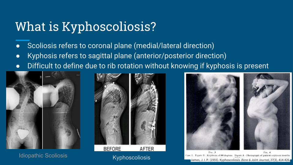
What is Kyphoscoliosis?● Scoliosis refers to coronal plane (medial/lateral direction)● Kyphosis refers to sagittal plane (anterior/posterior direction)● Difficult to define due to rib rotation without knowing if kyphosis is present
James, J. I. P. (1955). Kyphoscoliosis. Bone & Joint Journal, 37(3), 414-426.Idiopathic Scoliosis Kyphoscoliosis
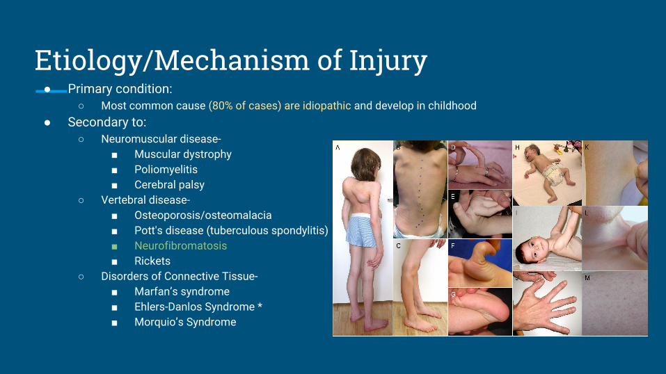
Etiology/Mechanism of Injury● Primary condition:
○ Most common cause (80% of cases) are idiopathic and develop in childhood● Secondary to:
○ Neuromuscular disease- ■ Muscular dystrophy■ Poliomyelitis■ Cerebral palsy
○ Vertebral disease- ■ Osteoporosis/osteomalacia■ Pott's disease (tuberculous spondylitis)■ Neurofibromatosis■ Rickets
○ Disorders of Connective Tissue-■ Marfan’s syndrome■ Ehlers-Danlos Syndrome *■ Morquio’s Syndrome
Patient Presentation● Signs/Symptoms
○ Morphological deformity■ Asymmetrical shoulder and/or pelvis, decreased respiratory function
○ Back pain○ Decreased trunk/UE mobility
● Risk Factors○ Early age onset, ~3 years of age○ Conditions listed on previous slide
● Complications○ Decreased pulmonary function- rib cage deformity
■ Hypercapnic, respiratory muscle fatigue■ Eventual changes in lung tissue■ Cardiorespiratory failure is typical cause of death in severe cases
○ Abnormal wear and tear on spinal structures○ Nerve damage/impingement (from curve or surgery)○ Emotional repercussions of decreased function and/or physical deformity
When can PT help?
● To manage symptoms/impairments:○ Adolescents before bones have ossified (Risser’s sign)
○ Adults until symptoms can no longer be controlled with conservative treatment.
● Pre-operatively- “conservative treatment may improve the pain and stabilize the condition however it will never correct the actual deformity.”
○ *In conjunction with primary condition when KS is secondary, as symptoms may be compounded.
● Post-operatively- achieve/maintain functional movement, manage pain, neuromuscular re-education with spine fusion.
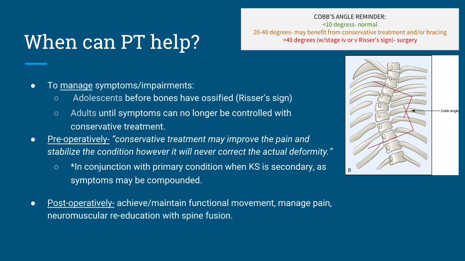
COBB’S ANGLE REMINDER:<10 degress- normal
20-40 degrees- may benefit from conservative treatment and/or bracing>40 degrees (w/stage iv or v Risser’s sign)- surgery
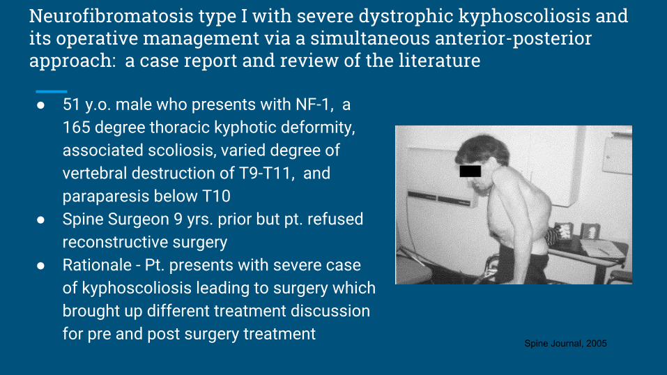
Neurofibromatosis type I with severe dystrophic kyphoscoliosis and its operative management via a simultaneous anterior-posterior approach: a case report and review of the literature
● 51 y.o. male who presents with NF-1, a 165 degree thoracic kyphotic deformity, associated scoliosis, varied degree of vertebral destruction of T9-T11, and paraparesis below T10
● Spine Surgeon 9 yrs. prior but pt. refused reconstructive surgery
● Rationale - Pt. presents with severe case of kyphoscoliosis leading to surgery which brought up different treatment discussion for pre and post surgery treatment
Spine Journal, 2005
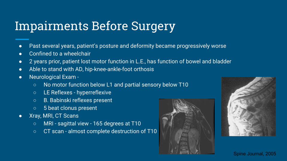
Impairments Before Surgery● Past several years, patient’s posture and deformity became progressively worse● Confined to a wheelchair● 2 years prior, patient lost motor function in L.E., has function of bowel and bladder● Able to stand with AD, hip-knee-ankle-foot orthosis● Neurological Exam -
○ No motor function below L1 and partial sensory below T10○ LE Reflexes - hyperreflexive○ B. Babinski reflexes present○ 5 beat clonus present
● Xray, MRI, CT Scans○ MRI - sagittal view - 165 degrees at T10○ CT scan - almost complete destruction of T10
Spine Journal, 2005
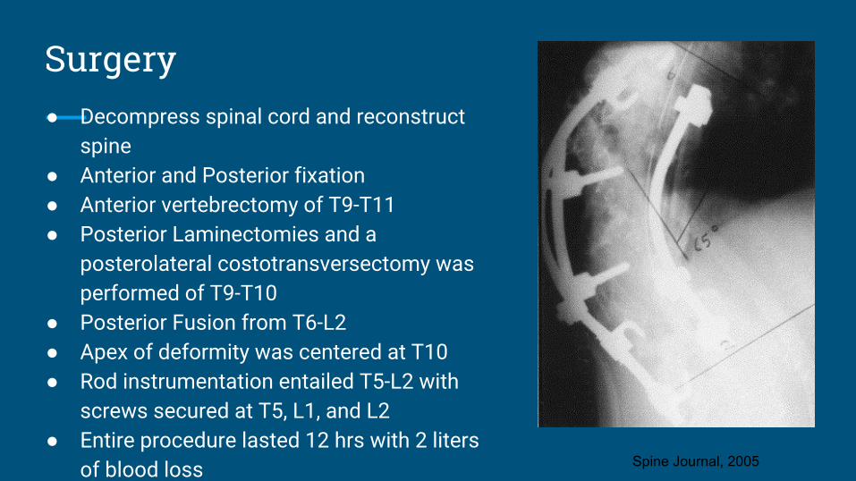
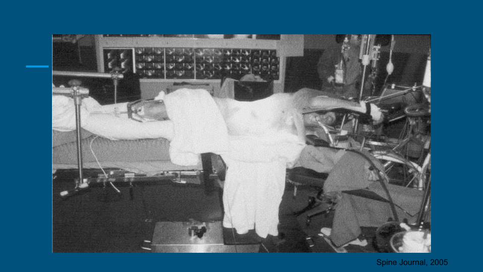
Surgery● Decompress spinal cord and reconstruct
spine● Anterior and Posterior fixation ● Anterior vertebrectomy of T9-T11● Posterior Laminectomies and a
posterolateral costotransversectomy was performed of T9-T10
● Posterior Fusion from T6-L2● Apex of deformity was centered at T10● Rod instrumentation entailed T5-L2 with
screws secured at T5, L1, and L2● Entire procedure lasted 12 hrs with 2 liters
of blood loss Spine Journal, 2005
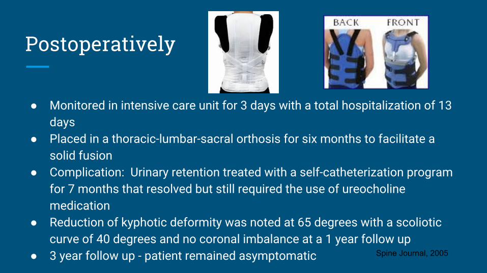
Postoperatively
● Monitored in intensive care unit for 3 days with a total hospitalization of 13 days
● Placed in a thoracic-lumbar-sacral orthosis for six months to facilitate a solid fusion
● Complication: Urinary retention treated with a self-catheterization program for 7 months that resolved but still required the use of ureocholine medication
● Reduction of kyphotic deformity was noted at 65 degrees with a scoliotic curve of 40 degrees and no coronal imbalance at a 1 year follow up
● 3 year follow up - patient remained asymptomatic Spine Journal, 2005
Spine Journal, 2005
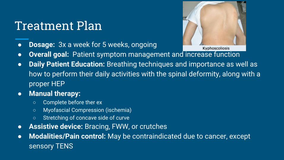
Treatment Plan● Dosage: 3x a week for 5 weeks, ongoing● Overall goal: Patient symptom management and increase function● Daily Patient Education: Breathing techniques and importance as well as
how to perform their daily activities with the spinal deformity, along with a proper HEP
● Manual therapy:○ Complete before ther ex○ Myofascial Compression (ischemia)○ Stretching of concave side of curve
● Assistive device: Bracing, FWW, or crutches● Modalities/Pain control: May be contraindicated due to cancer, except
sensory TENS
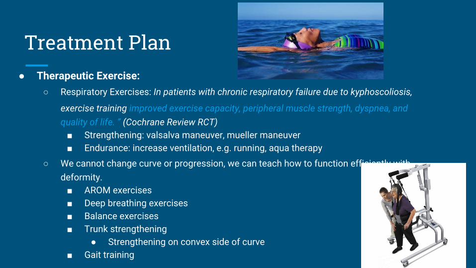
Treatment Plan● Therapeutic Exercise:
○ Respiratory Exercises: In patients with chronic respiratory failure due to kyphoscoliosis,
exercise training improved exercise capacity, peripheral muscle strength, dyspnea, and quality of life. ” (Cochrane Review RCT)
■ Strengthening: valsalva maneuver, mueller maneuver■ Endurance: increase ventilation, e.g. running, aqua therapy
○ We cannot change curve or progression, we can teach how to function efficiently with deformity.
■ AROM exercises■ Deep breathing exercises■ Balance exercises■ Trunk strengthening
● Strengthening on convex side of curve■ Gait training
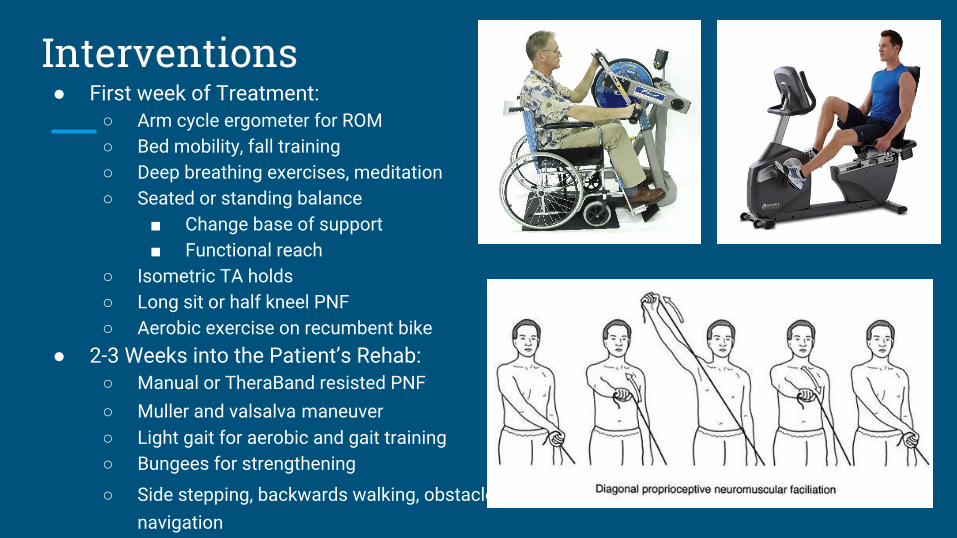
Interventions● First week of Treatment:
○ Arm cycle ergometer for ROM○ Bed mobility, fall training○ Deep breathing exercises, meditation○ Seated or standing balance
■ Change base of support■ Functional reach
○ Isometric TA holds○ Long sit or half kneel PNF○ Aerobic exercise on recumbent bike
● 2-3 Weeks into the Patient’s Rehab:○ Manual or TheraBand resisted PNF○ Muller and valsalva maneuver ○ Light gait for aerobic and gait training○ Bungees for strengthening
○ Side stepping, backwards walking, obstacle navigation
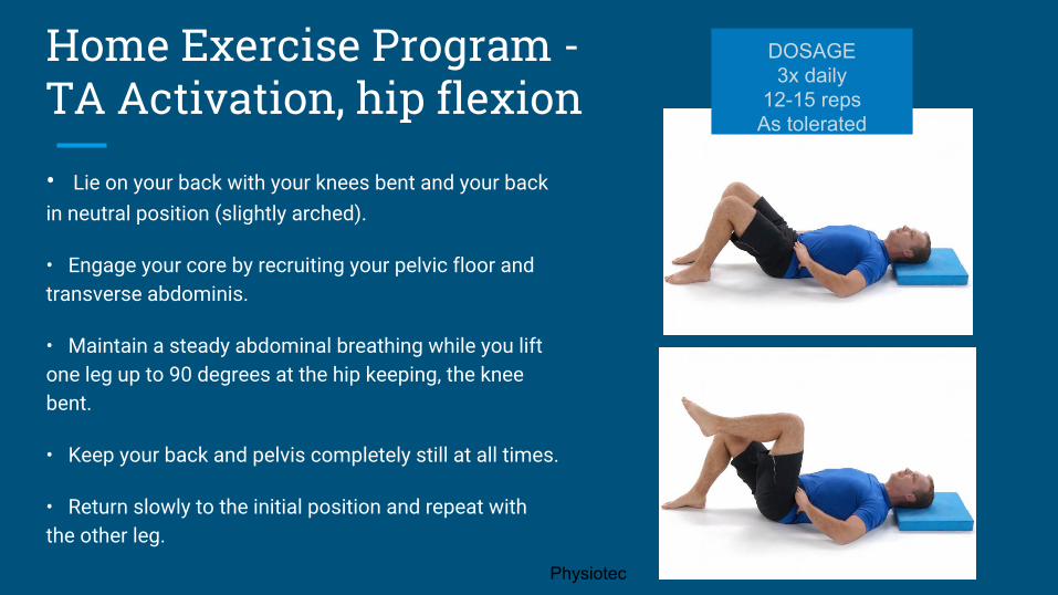
Home Exercise Program - TA Activation, hip flexion
• Lie on your back with your knees bent and your back in neutral position (slightly arched).
• Engage your core by recruiting your pelvic floor and transverse abdominis.
• Maintain a steady abdominal breathing while you lift one leg up to 90 degrees at the hip keeping, the knee bent.
• Keep your back and pelvis completely still at all times.
• Return slowly to the initial position and repeat with the other leg.
DOSAGE3x daily
12-15 repsAs tolerated
Physiotec
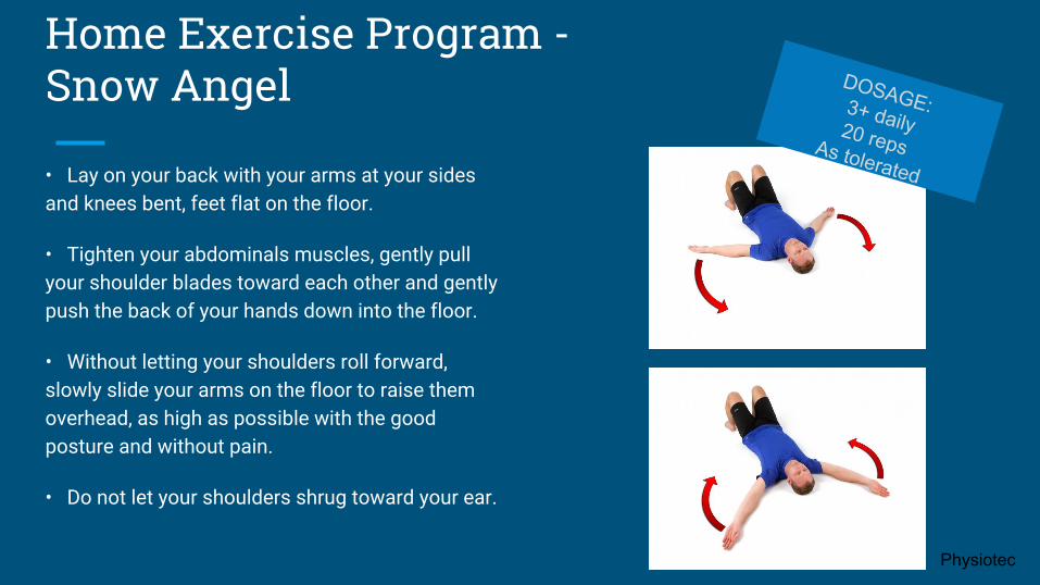
Home Exercise Program - Snow Angel
• Lay on your back with your arms at your sides and knees bent, feet flat on the floor.
• Tighten your abdominals muscles, gently pull your shoulder blades toward each other and gently push the back of your hands down into the floor.
• Without letting your shoulders roll forward, slowly slide your arms on the floor to raise them overhead, as high as possible with the good posture and without pain.
• Do not let your shoulders shrug toward your ear.
DOSAGE:3+ daily20 repsAs tolerated
Physiotec
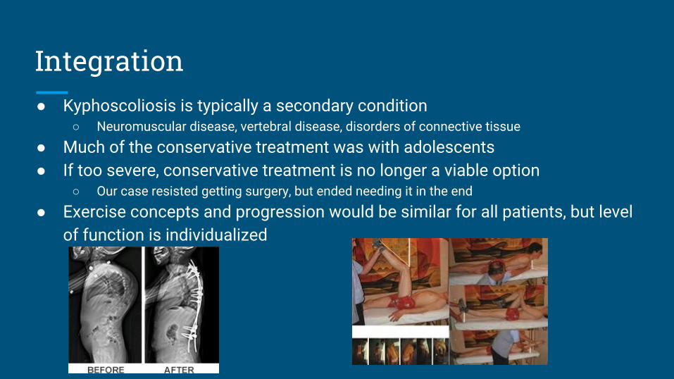
Integration● Kyphoscoliosis is typically a secondary condition
○ Neuromuscular disease, vertebral disease, disorders of connective tissue
● Much of the conservative treatment was with adolescents● If too severe, conservative treatment is no longer a viable option
○ Our case resisted getting surgery, but ended needing it in the end
● Exercise concepts and progression would be similar for all patients, but level of function is individualized
Take home message
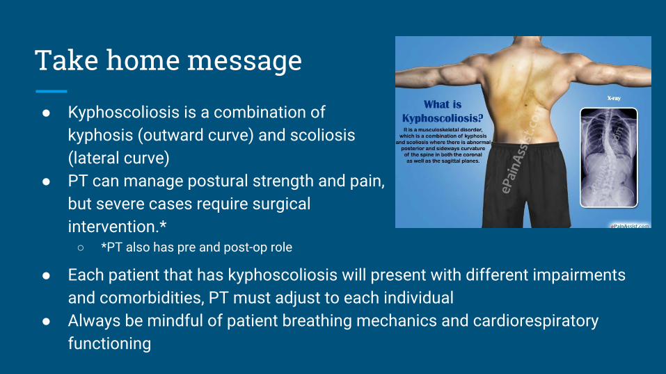
● Kyphoscoliosis is a combination of kyphosis (outward curve) and scoliosis (lateral curve)
● PT can manage postural strength and pain, but severe cases require surgical intervention.*
○ *PT also has pre and post-op role
● Each patient that has kyphoscoliosis will present with different impairments and comorbidities, PT must adjust to each individual
● Always be mindful of patient breathing mechanics and cardiorespiratory functioning
References1. James, J. I. P. (1955). Kyphoscoliosis. Bone & Joint Journal, 37(3), 414-426.2. Singh, Kern, Dino Samartzis, and Howard S. An. "Neurofibromatosis type I with severe dystrophic kyphoscoliosis
and its operative management via a simultaneous anterior-posterior approach: a case report and review of the literature." The Spine Journal 5.4 (2005): 461-466.
3. "CAS – Central Authentication Service". Uptodate.com.libproxy.nau.edu. N.p., 2016. Web. 12 July 2016.4. Reid, W. D., & Dechman, G. (1995). Considerations when testing and training the respiratory muscles. Physical
therapy, 75(11), 971-982.5. Pardy, R. L., & Rochester, D. F. (1992, January). Respiratory muscle training. In Seminars in respiratory medicine
(Vol. 13, No. 01, pp. 53-62). Copyright© 1992 by Thieme Medical Publishers, Inc..6. “Exercise training in patients with chronic respiratory failure due to kyphoscoliosis : a randomized controlled trial.”
Cejudo P , López-Márquez I , López-Campos JL , Márquez E , de laVega F , Barrot E and Ortega F,Respiratory care, 2014, 59(3), 375, Cochrane Central Register of Controlled Trials, Publication Year: 2014
7. Zaky, L. A., & Rashad, G. M. (2013). Efficacy of Ischemic Compression Followed by Exercises Therapy Versus Rehabilitation Program in Treatment of Postural Scoliosis. Bulletin of Faculty of Physical Therapy, 18(1).