lab exercise #7 epithelial tissues. let’s review…
TRANSCRIPT
Lab Exercise #7
Epithelial Tissues
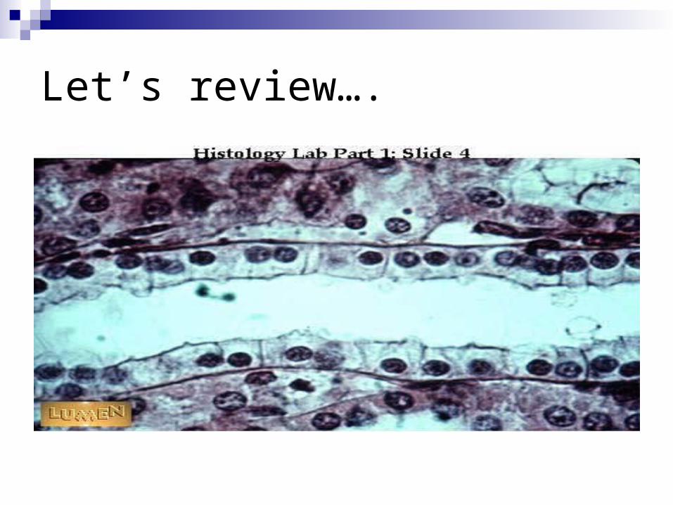
Let’s review….
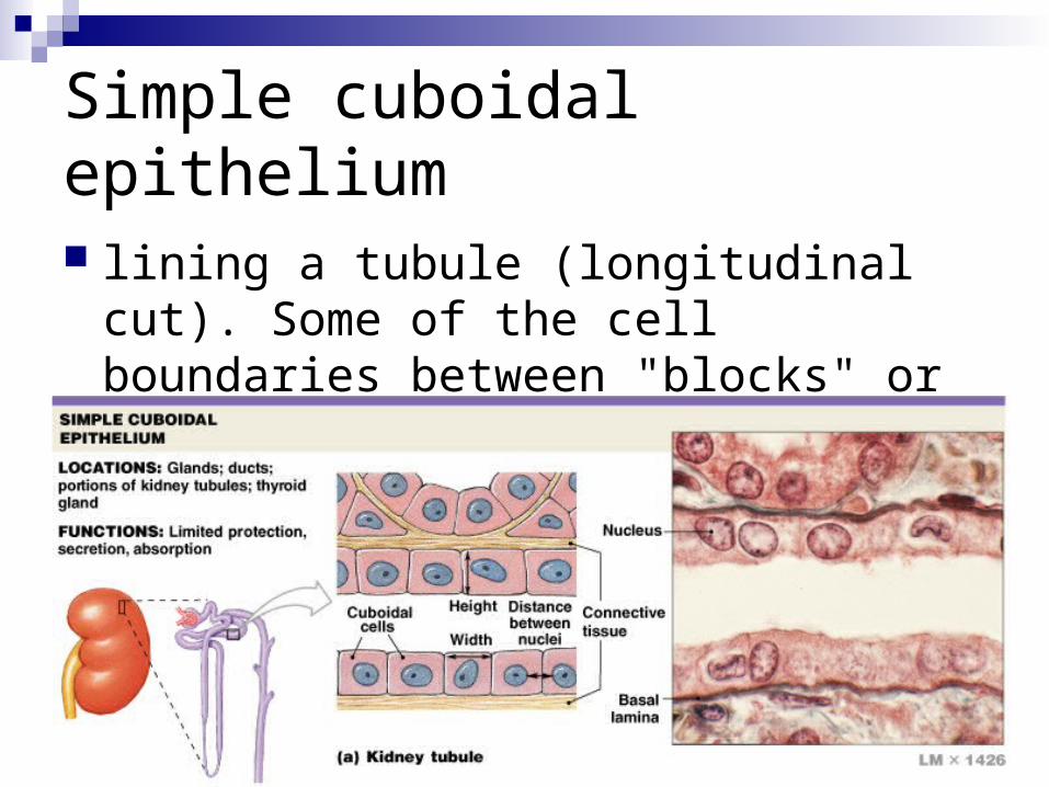
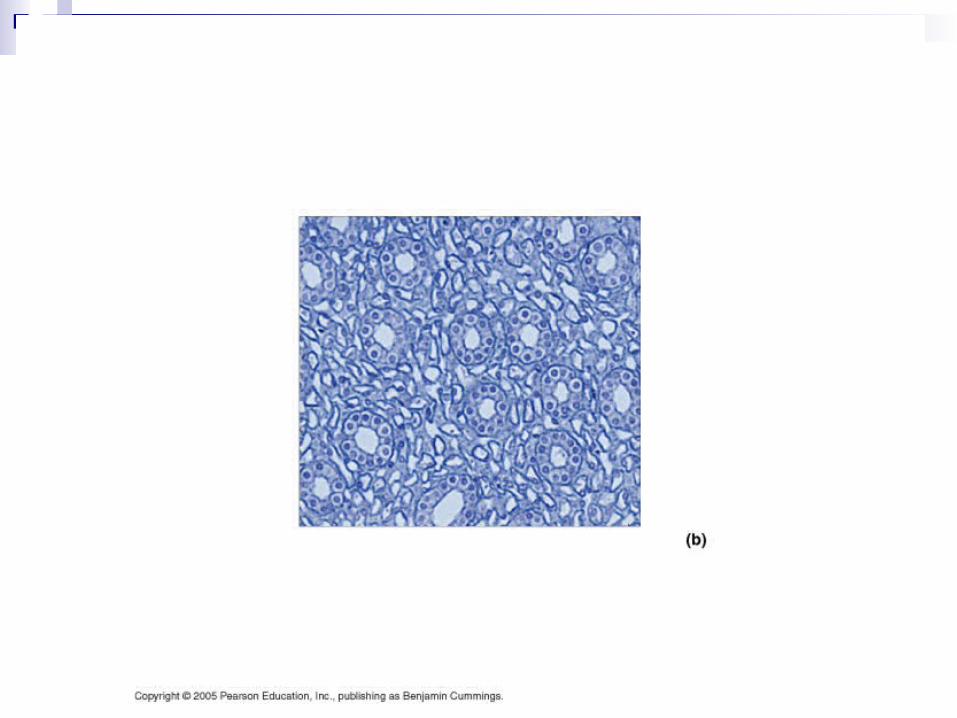
Simple cuboidal epithelium
lining a tubule (longitudinal cut). Some of the cell boundaries between "blocks" or "cubes" here are quite distinct.
Simple Cuboidal
Covers surface of ovary, lines kidney tubules and small ducts of glands (thyroid and pancreas)
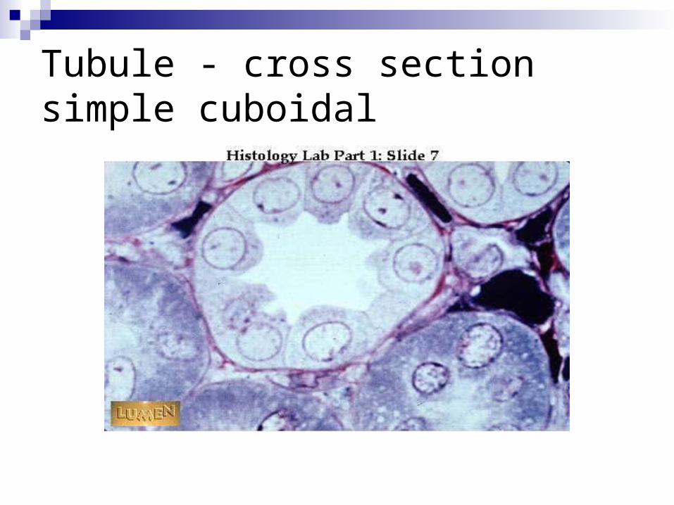
Tubule - cross sectionsimple cuboidal
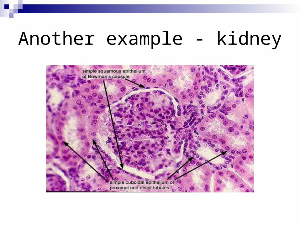
Another example - kidney
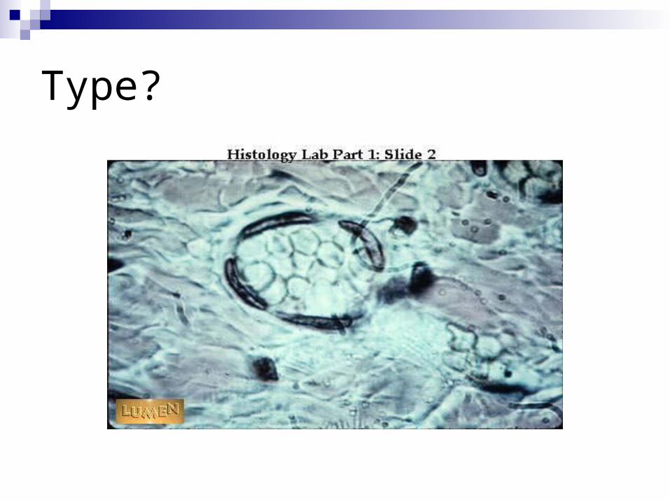
Type?
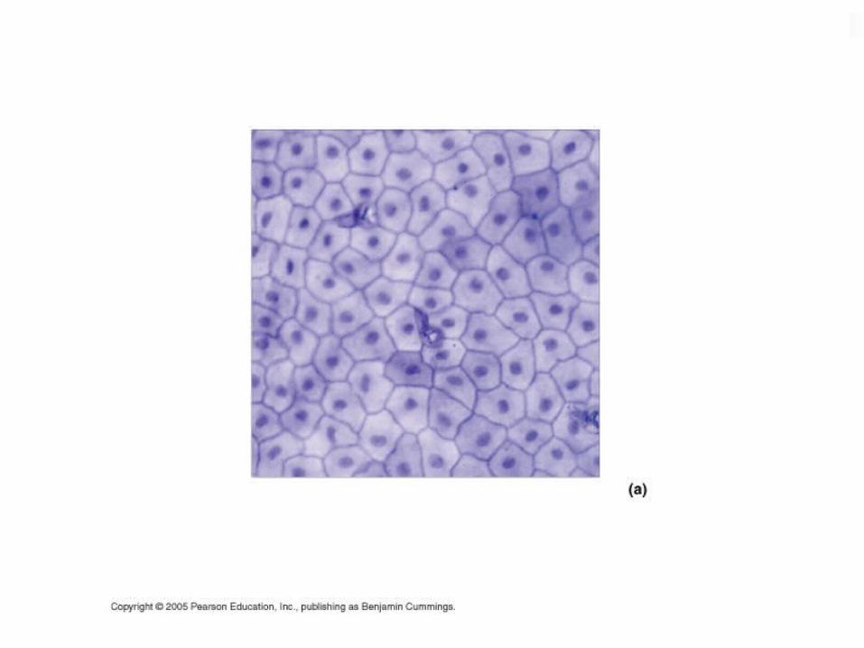
Simple squamous epithelium
High power view of endothelial cells lining a small blood vessel cut in cross-section. (You see just the nuclei - the cytoplasm between them is extremely flat.) Endothelium = the simple squamous epithelium lining blood vessels.
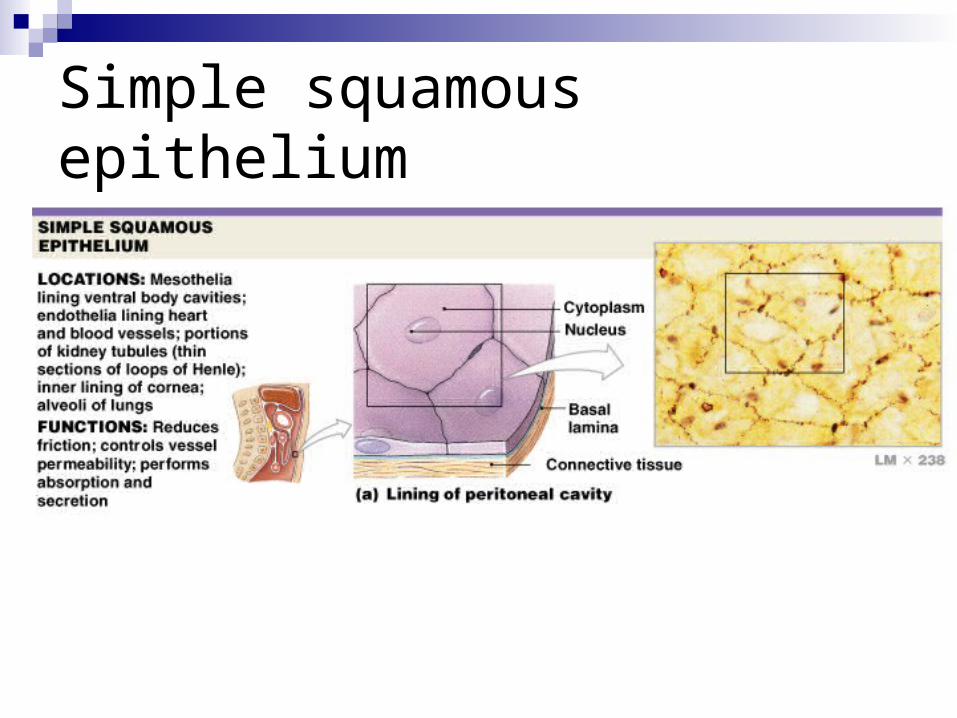
Simple squamous epithelium
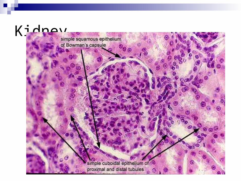
Kidney
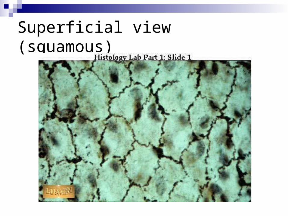
Superficial view (squamous)
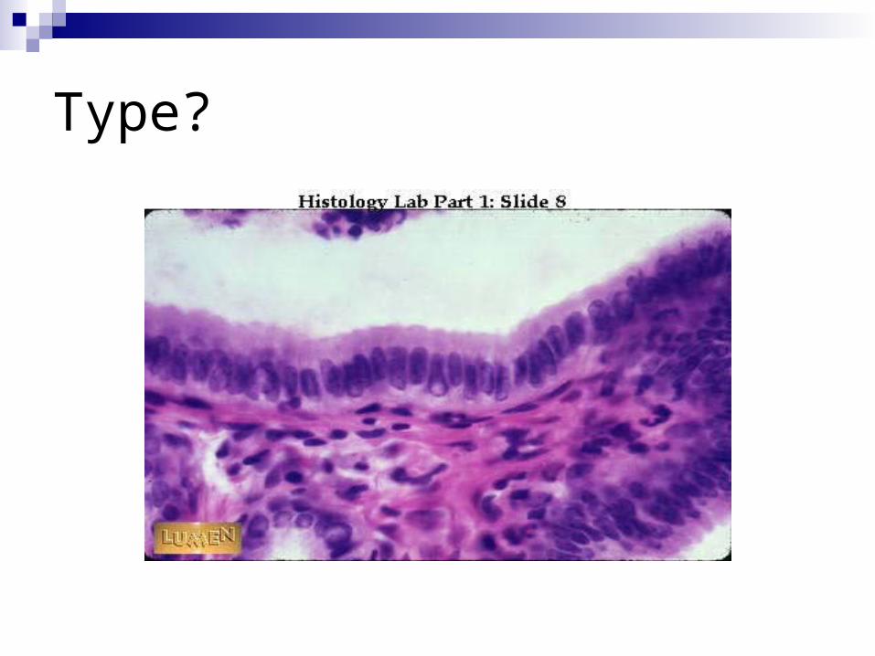
Type?
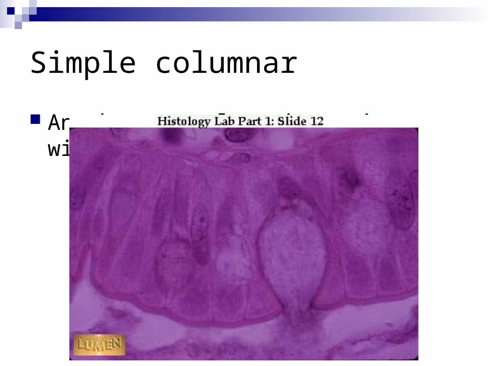
Simple columnar
Another example – intestine, with goblet
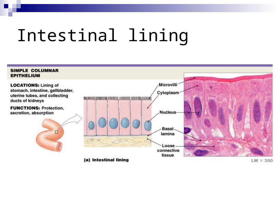
Intestinal lining
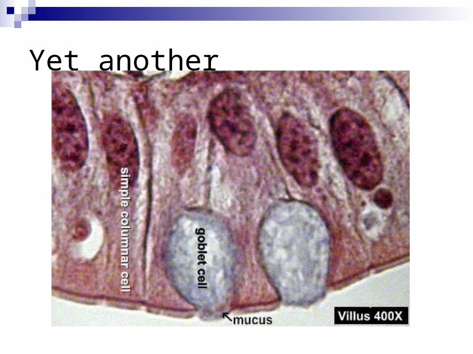
Yet another
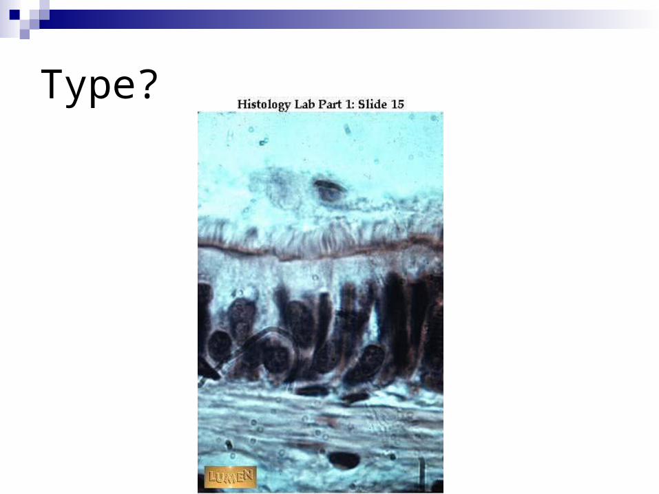
Type?
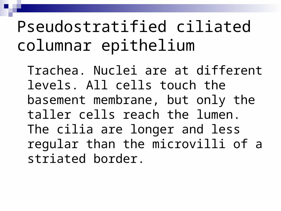
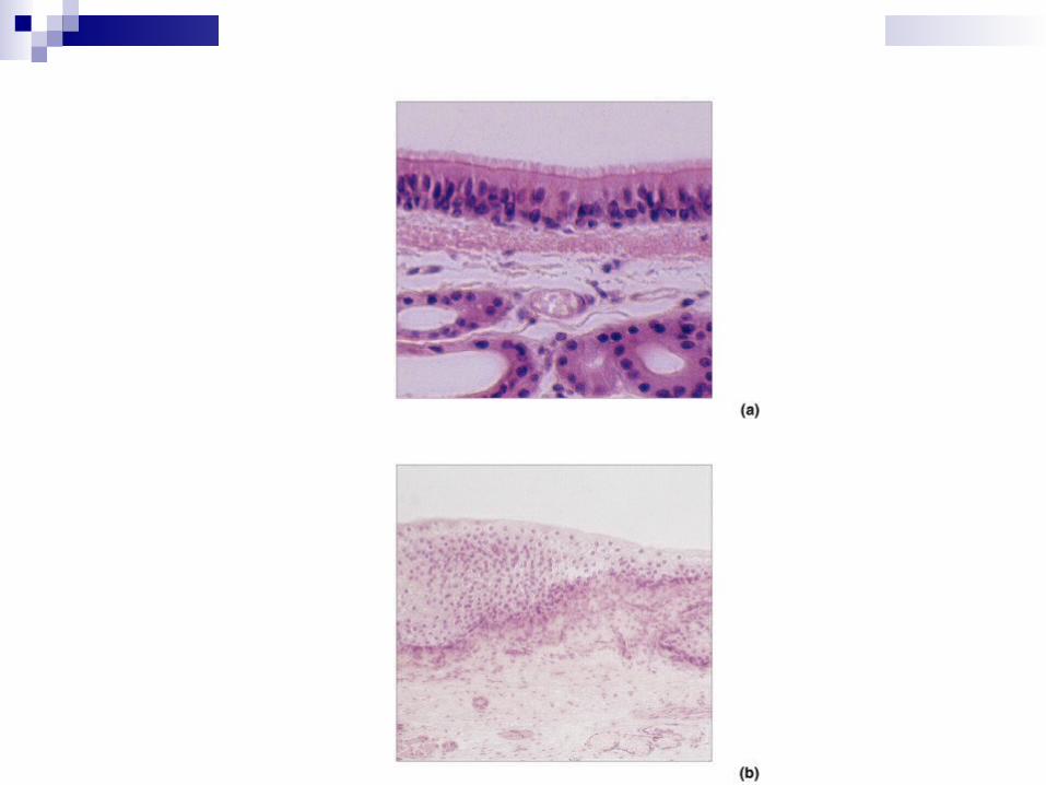
Pseudostratified ciliated columnar epithelium
Trachea. Nuclei are at different levels. All cells touch the basement membrane, but only the taller cells reach the lumen. The cilia are longer and less regular than the microvilli of a striated border.

Pseudostratified Columnar Epithelium Look like multiple layers Trachea
Figure 4–5b
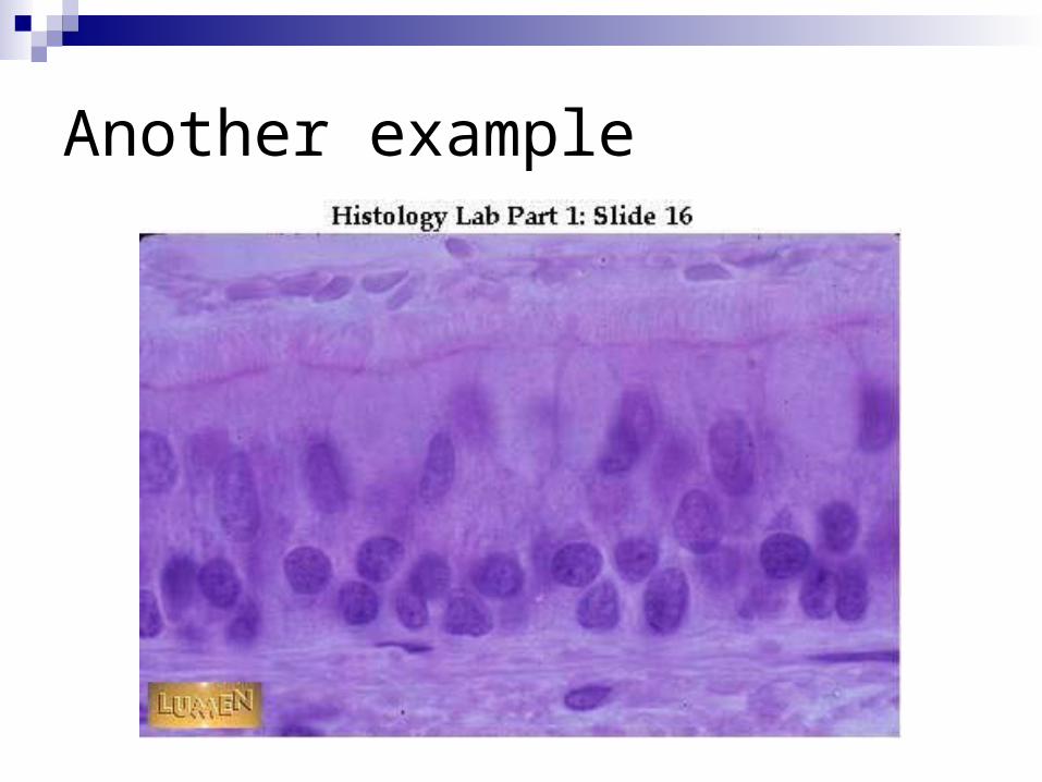
Another example
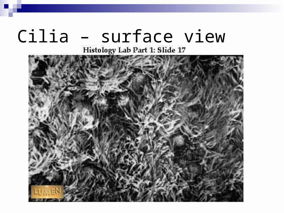
Cilia – surface view
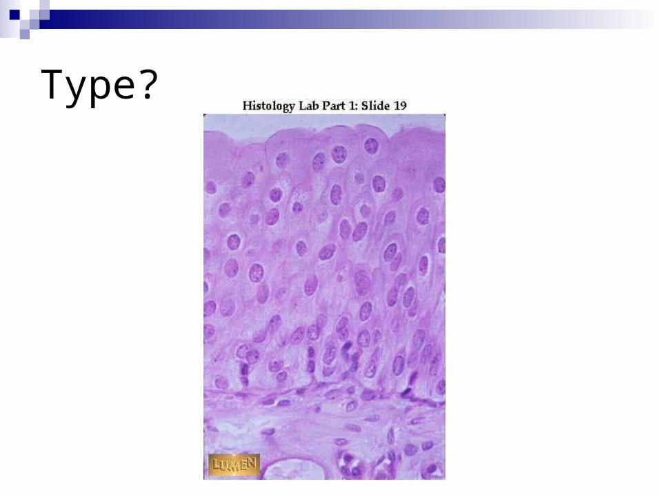
Type?
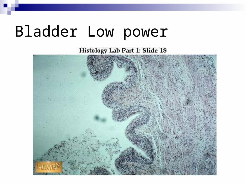
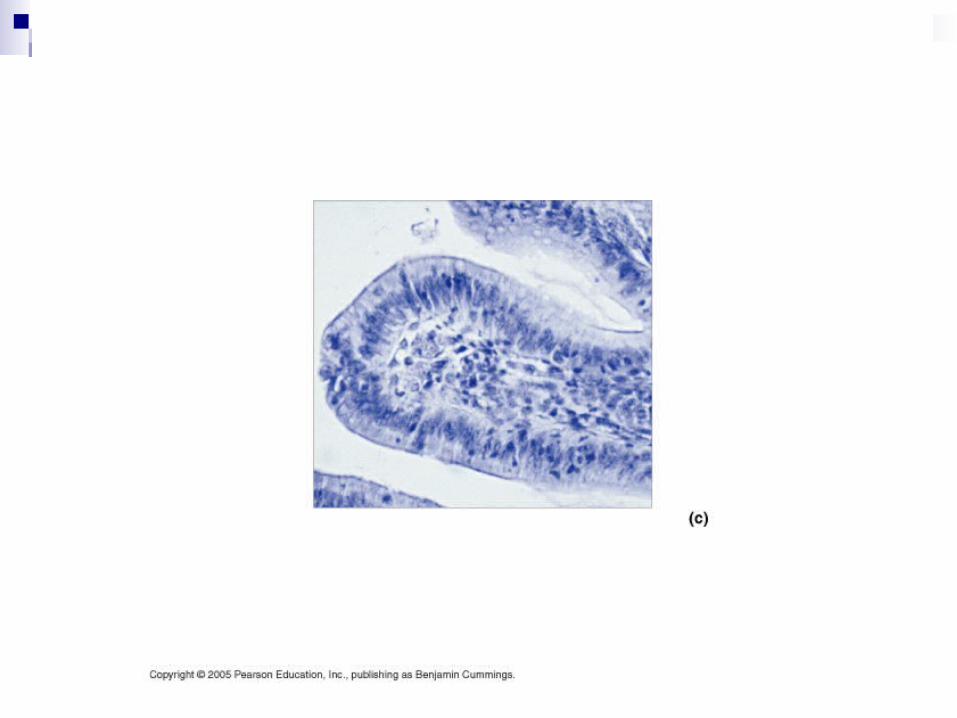
Bladder Low power
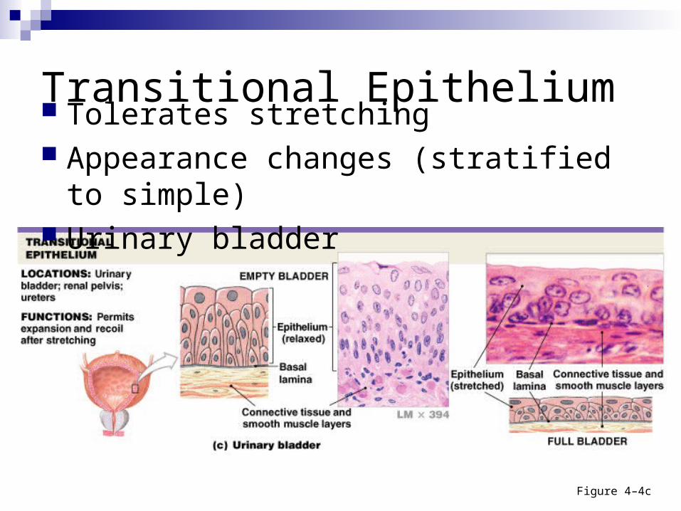
Transitional Epithelium Tolerates stretching Appearance changes (stratified to simple) Urinary bladder
Figure 4–4c
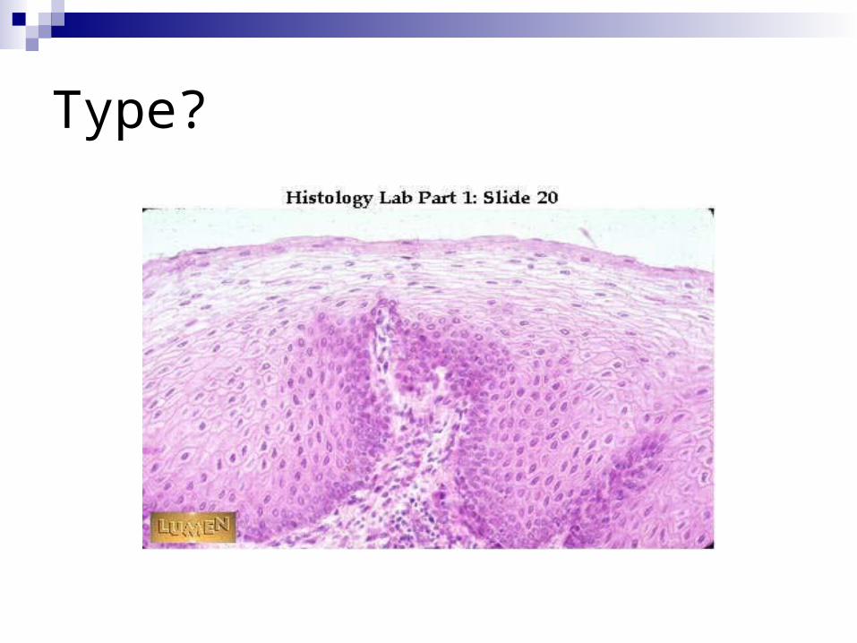
Type?
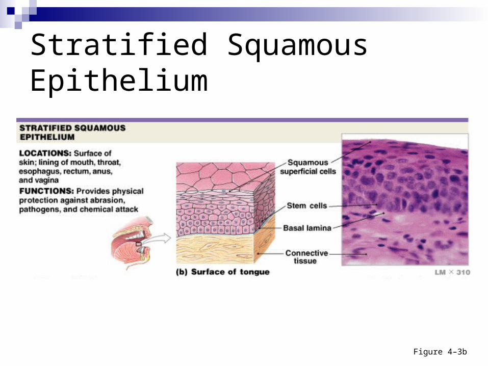
Figure 4–3b
Stratified Squamous Epithelium
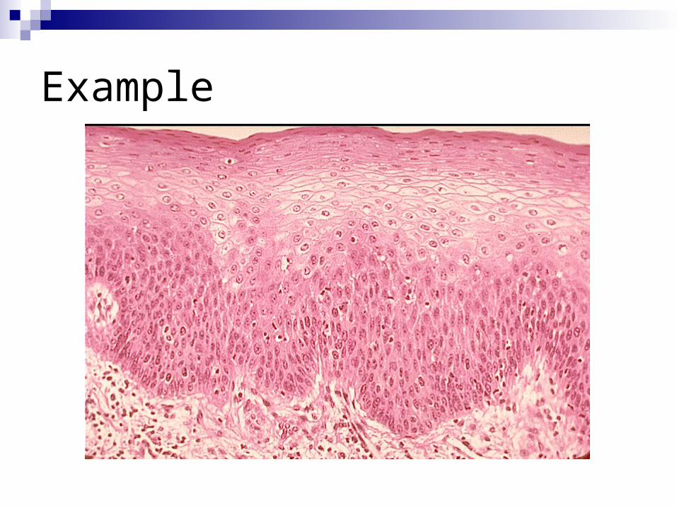
Example
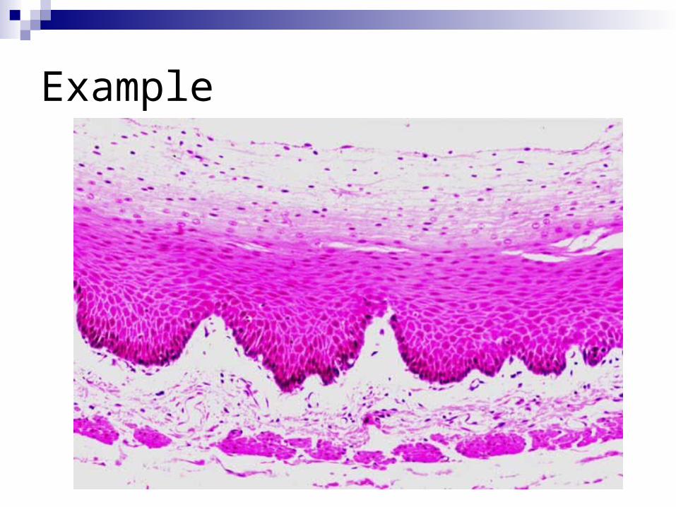
Example
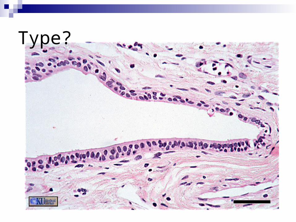
Type?
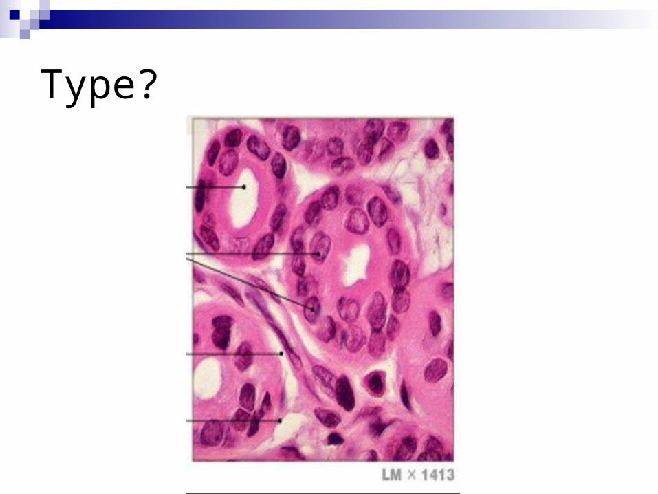
Type?
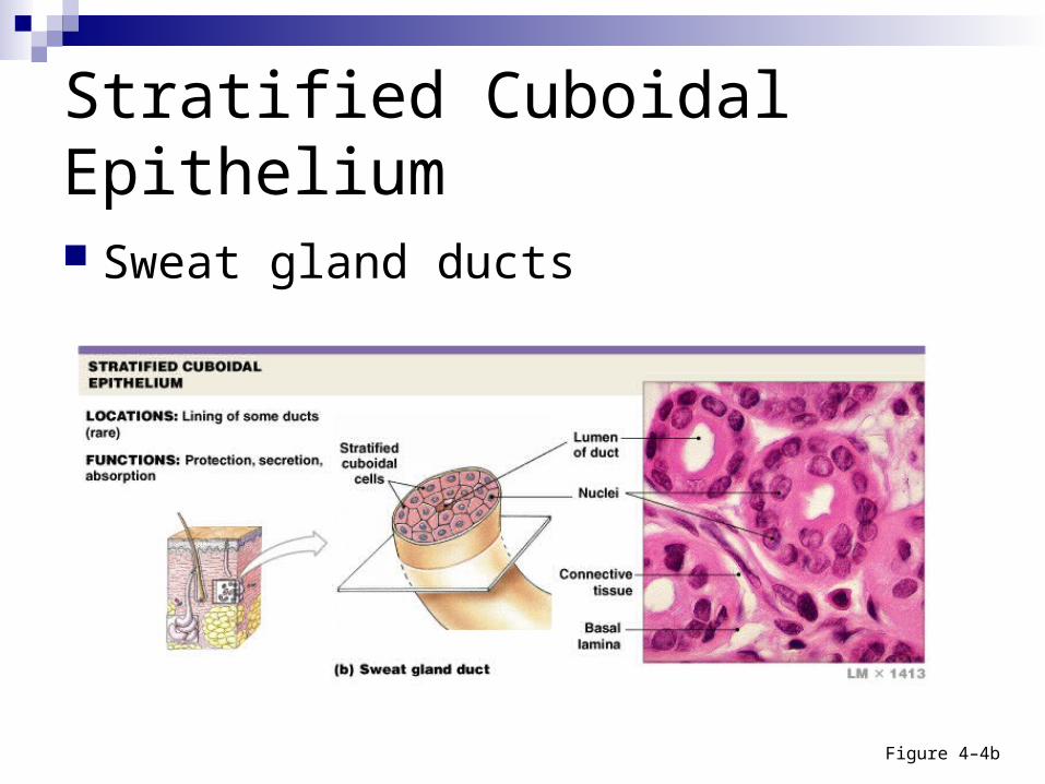
Stratified Cuboidal Epithelium
Sweat gland ducts
Figure 4–4b
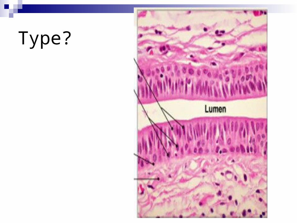
Type?
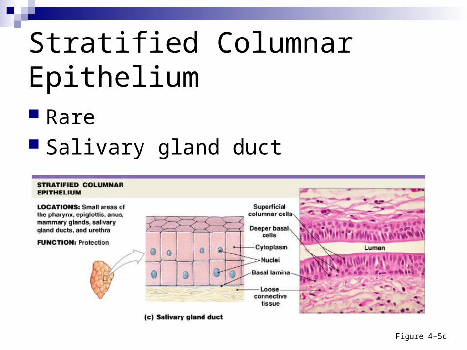
Stratified Columnar Epithelium
Rare Salivary gland duct
Figure 4–5c
Note
It is the shape of the most superficial layer that determines what type of stratified epithelium it is.
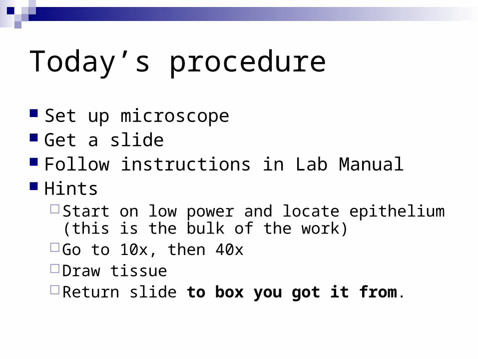
Today’s procedure
Set up microscope Get a slide Follow instructions in Lab Manual Hints
Start on low power and locate epithelium (this is the bulk of the work)
Go to 10x, then 40xDraw tissueReturn slide to box you got it from.
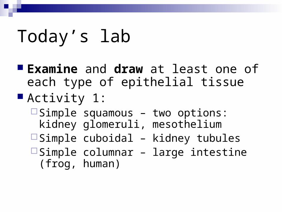
Today’s lab
Examine and draw at least one of each type of epithelial tissue
Activity 1:Simple squamous – two options: kidney
glomeruli, mesotheliumSimple cuboidal – kidney tubulesSimple columnar – large intestine (frog,
human)
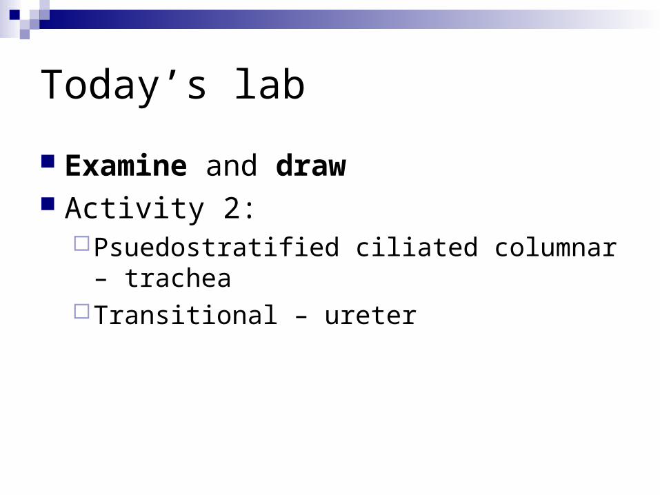
Today’s lab
Examine and draw Activity 2:
Psuedostratified ciliated columnar – trachea Transitional – ureter
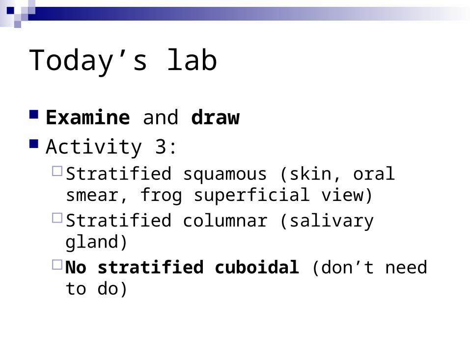
Today’s lab
Examine and draw Activity 3:
Stratified squamous (skin, oral smear, frog superficial view)
Stratified columnar (salivary gland)No stratified cuboidal (don’t need to do)
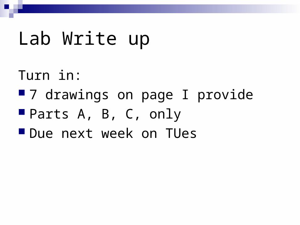
Lab Write up
Turn in: 7 drawings on page I provide Parts A, B, C, only Due next week on TUes